Mechanical Properties of Biomaterials Based onCalcium Phosphates and Bioinert Oxides forApplications in Biomedicine
Siwar Sakka, Jamel Bouaziz and Foued Ben Ayed
Additional information is available at the end of the chapter
http://dx.doi.org/10.5772/53088
1. Introduction
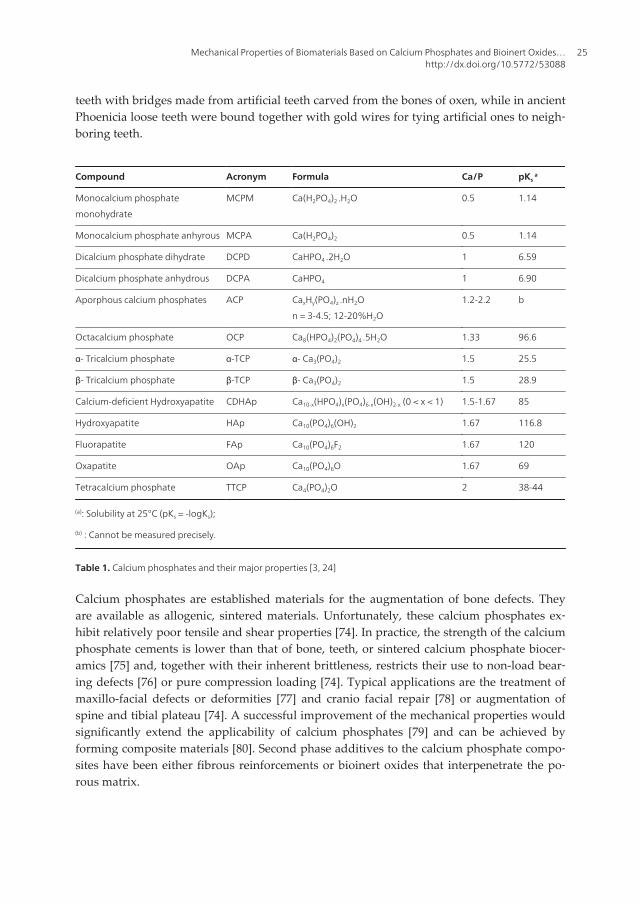
Calcium phosphates (CaP) have been sought as biomaterials for reconstruction of bonedefect in maxillofacial, dental and orthopaedic applications [1-31]. Calcium phosphateshave been used clinically to repair bone defects for many years. Calcium phosphatessuch as hydroxyapatite (Ca10(PO4)6(OH)2, HAp), fluorapatite (Ca10(PO4)6F2, FAp), tricalci‐um phosphate (Ca3(PO4)2, TCP), TCP-HAp composites and TCP-FAp composites are usedfor medical and dental applications [3, 10-29]. In general, this concept is determined byadvantageous balances of more stable (frequent by hydroxyapatite or fluorapatite) andmore resorbable (typically tricalcium phosphate) phases of calcium phosphates, while theoptimum ratios depend on the particular applications. The complete list of known calci‐um phosphates, including their major properties (such, the chemical formula, solubilitydata) is given in Table 1. The detailed information about calcium phosphates, their syn‐thesis, structure, chemistry, other properties and biomedical applications have been com‐prehensively reviewed recently in reference [24].
Calcium phosphate-based biomaterials and bioceramics are now used in a number of differ‐ent applications throughout the body, covering all areas of the skeleton. Applications in‐clude dental implants, percutaneous devices and use in periodontal treatment, treatment ofbone defects, fracture treatment, total joint replacement (bone augmentation), orthopedics,cranio-maxillofacial reconstruction, otolaryngology and spinal surgery [32-35]. Dependingupon whether a bioresorbable or a bioactive material is desired, different calcium ortho‐phosphates might be used.
In the past, many implantations failed because of infection or a lack of knowledge about thetoxicity of the selected materials. In this frame, the use of calcium phosphates is logical dueto their similarity to the mineral phase of bone and teeth [36-40]. However, according toavailable literature, the first attempt to use calcium phosphates as an artificial material to re‐pair surgically-created defects in rabbits was performed in 1920 [41]. More than fifty yearslater, the first dental application of a calcium phosphate (erroneously described as TCP) insurgically-created periodontal defects [42] and the use of dense HAp cylinders for immedi‐ate tooth root replacement were reported [43]. Since Levitt et al. described a method of pre‐paring an apatite bioceramics from FAp and suggested its possible use in medicalapplications in 1969[44]. According to the available databases, the first paper with the term‘‘bioceramics’’ in the abstract was published in 1971 [45], while those with that term in thetitle were published in 1972 [46-47]. However, application of ceramic materials as prostheseshad been known before [48-49]. Further historical details might be found in literature [50].Commercialization of the dental and surgical applications of Hap-based bioceramics occur‐red in the 1980’s, largely through the pioneering efforts by Jarcho [51], de Groot [52] and Ao‐ki [53]. Due to that, HAp has become a bioceramic of reference in the field of calciumphosphates for biomedical applications. Preparation and biomedical applications of apatitesderived from sea corals (coralline HAp) [54–56] and bovine bone were reported at the sametime [57]. Since 1990, several other calcium phosphate cements have been developed [58-62],injectable cements have been formulated [63], and growth factors have been delivered viathese cements [64]. The tetracalcium phosphate [TTCP: Ca4(PO4)2O] and dicalcium phos‐phate anhydrous [DCPA: CaHPO4] system was approved in 1996 by the Food and Drug Ad‐ministration (FDA) for repairing craniofacial defects in humans, thus becoming the firstTTCP–DCPA system for clinical use [65]. However, due to its brittleness and weakness, theuse of TTCP–DCPA system was limited to the reconstruction of non-stress-bearing bone[66-67]. To expand the use of TTCP–DCPA system to a wide range of load-bearing maxillo‐facial and orthopedic repairs, recent studies have developed natural biopolymers that areelastomeric, biocompatible and resorbable [68]. Calcium phosphates in a number of formsand compositions are currently either in use or under consideration in many areas of den‐tistry and orthopedics. For example, bulk materials, available in dense and porous forms,are used for alveolar ridge augmentation, immediate tooth replacement and maxillofacial re‐construction [35, 69]. Other examples include orbital implants (Bio-Eye) [70-71], incrementof the hearing ossicles, spine fusion and repair of bone defects [72-73]. In order to permitgrowth of new bone onto bone defects, a suitable bioresorbable material should fill the de‐fects. Otherwise, in-growth of fibrous tissue might prevent bone formation within the de‐fects [69-73]. Today, a large number of different calcium phosphate bioceramics for thetreatment of various defects are available on the market.
The performance of living tissues is the result of millions of years of evolution, while theperformance of acceptable artificial substitutions those man has designed to repair damagedhard tissues are only a few decades old. Archaeological findings exhibited in museumsshowed that materials used to replace missing human bones and teeth have included animalor human (from corpses) bones and teeth, shells, corals, ivory (elephant tusk), wood, as wellas some metals (gold or silver). For instance, the Etruscans learned to substitute missing
Advances in Biomaterials Science and Biomedical Applications24
teeth with bridges made from artificial teeth carved from the bones of oxen, while in ancientPhoenicia loose teeth were bound together with gold wires for tying artificial ones to neigh‐boring teeth.
Table 1. Calcium phosphates and their major properties [3, 24]
Calcium phosphates are established materials for the augmentation of bone defects. Theyare available as allogenic, sintered materials. Unfortunately, these calcium phosphates ex‐hibit relatively poor tensile and shear properties [74]. In practice, the strength of the calciumphosphate cements is lower than that of bone, teeth, or sintered calcium phosphate biocer‐amics [75] and, together with their inherent brittleness, restricts their use to non-load bear‐ing defects [76] or pure compression loading [74]. Typical applications are the treatment ofmaxillo-facial defects or deformities [77] and cranio facial repair [78] or augmentation ofspine and tibial plateau [74]. A successful improvement of the mechanical properties wouldsignificantly extend the applicability of calcium phosphates [79] and can be achieved byforming composite materials [80]. Second phase additives to the calcium phosphate compo‐sites have been either fibrous reinforcements or bioinert oxides that interpenetrate the po‐rous matrix.
Mechanical Properties of Biomaterials Based on Calcium Phosphates and Bioinert Oxides…http://dx.doi.org/10.5772/53088
25
Hydroxyapatite and other calcium phosphates bioceramics are important for hard tissue re‐pair because of their similarity to the minerals in natural bone, and their excellent biocom‐patibility and bioactivity [81-86]. When implanted in an osseous site, bone bioactivematerials such as HAp and other CaP implants and coatings provide an ideal environmentfor cellular reaction and colonization by osteoblasts. This leads to a tissue response termedosteoconduction in which bone grows on and bonds to the implant, promoting a functionalinterface [81, 84, 87]. Extensive efforts have significantly improved the properties and per‐formance of HAp and other CaP based implants [88-92]. Calcium phosphate cements can bemolded or injected to form a scaffold in situ, which can be resorbed and replaced by newbone [93, 65-67]. Chemically, the vast majority of calcium phosphate bioceramics is based onHAp, β-TCP, α-TCP and/or biphasic calcium phosphate (BCP), which is an intimate mixtureof either β-TCP - HAp [94-100] or α-TCP - HAp [101-111]. The preparation technique ofthese calcium phosphates has been extensively reviewed in literature [1, 4, 37, 102-104].When compared to both β- and α-TCP, HAp is a more stable phase under physiological con‐ditions, as it has a lower solubility (Table 1) [37, 109-110]. Therefore, the BCP concept is de‐termined by the optimum balance of a more stable phase of HAp and a more soluble TCP.Due to a higher biodegradability of the β - or α -TCP component, the reactivity of BCP in‐creases with the TCP-HAp the increase in ratio. Thus, in vivo bioresorbability of BCP can becontrolled through the phase composition [95]. As implants made of calcined HAp arefound in bone defects for many years after implantation, bioceramics made of more solublecalcium phosphates is preferable for the biomedical purposes [94-110]. HAp has been clini‐cally used to repair bone defects for many years [3]. However, Hap has poor mechanicalproperties [3]. Their use at high load bearing conditions has been restricted due to their brit‐tleness, poor fatigue resistance and strength.
The main reason behind the use of β-TCP as bone substitute materials is their chemicalsimilarity to the mineral component of mammalian bone and teeth [1-3]. The applicationof tricalcium phosphate as a bone substitute has received considerable attention, becauseit is remarkably biocompatible with living bodies when replacing hard tissues and be‐cause it has biodegradable properties [1-29]. Consequently, β-TCP has been used as bonegraft substitutes in many surgical fields such as orthopedic and dental surgeries [3, 11-12,16-17]. This use leads to an ultimate physicochemical bond between the implants andbone-termed osteointegration. Even so, the major limitation to the use of β-TCP as load-bearing biomaterial is their mechanical properties which make it brittle, with poor fati‐gue resistance [3, 10, 21-29]. Moreover, the mechanical properties of tricalcium phosphateare generally inadequate for many load-carrying applications (3 MPa – 5 MPa) [3, 10,20-29]. Its poor mechanical behaviour is even more evident when used to make highlyporous ceramics and scaffolds. Hence, metal oxides ceramics, such as alumina (Al2O3), ti‐tania (TiO2) and some oxides (e.g. ZrO2, SiO2) have been widely studied due to their bioi‐nertness, excellent tribological properties, high wear resistance, fracture toughness andstrength as well as relatively low friction [19, 21-22, 29-31]. However, bioinert ceramic ox‐ides having high strength are used to enhance the densification and the mechanical prop‐erties of β-TCP. In this chapter, we will try to improve the strength of β-TCP byintroducing a bioinert oxide like alumina. This is because there are few articles reporting
Advances in Biomaterials Science and Biomedical Applications26
the toughening effects of an inert oxide (like alumina (Al2O3)) on the mechanical proper‐ties of β-TCP [22, 27, 29]. Alumina has a high strength and is bio-inert with human tis‐sues [19, 22, 27, 29]. In order to improve the biocompatibility of alumina and thestrength of tricalcium phosphate effectively, and in order to search for an approach toproduce high performances of alumina-tricalcium phosphate composites, β-TCP is intro‐duced with different percentages in the alumina matrix. The aim of our study is to elabo‐rate and characterize the TCP-Al2O3 composites for biomedical applications.
This chapter proposes to study the sintering of the alumina-tricalcium phosphate compo‐sites at various temperatures (1400°C, 1450°C, 1500°C, 1550°C and 1600°C) and with differ‐ent percentages of β-TCP (10 wt%, 20 wt%, 40 wt% and 50 wt%). The characterization ofbiomaterials will be realized by using dilatometry analysis, differential thermal analysis(DTA), X-ray diffraction (XRD), magic angle spinning nuclear magnetic resonance (MASNMR), scanning electron microscopy analysis (SEM) and by using the mechanical proper‐ties, such as rupture strength (σr) of these biomaterials.
2. Materials and methods
The synthesized tricalcium phosphate and alumina (Riedel-de Haёn) were mixed in order toprepare biomaterial composites. The β-TCP powder was synthesized by solid-state reactionfrom calcium carbonate (CaCO3) and calcium phosphate dibasic anhydrous (CaHPO4) [27].Stoichiometric amounts of high purity powders such as CaHPO4 (Fluka, purity ≥ 99%) andCaCO3 (Fluka, purity ≥ 98.5%), were sintered at 1000°C for one hour to obtain the β-TCP ac‐cording to the following reaction:
( ) ( ) ( ) ( ) ( ) ( )4 3 3 4 2 222 CaHPO s CaCO s Ca PO s H O g CO g+ ® + + (1)
The β-TCP and the alumina powders were mixed in an agate mortar. The powder mixtureswere milled in ethanol for 24 hours. After milling, the mixtures were dried in a rotary vac‐uum evaporator and passed through a 70-mesh screen. After drying the powder mixtures at80°C for 24 hours, they were molded in a cylinder having a diameter of 20 mm and a thick‐ness of 6 mm, and pressed under 150 MPa. The green compacts were sintered at varioustemperatures for different lengths of time in a vertical furnace (Pyrox 2408). The heating rateis 10°C min-1. The size of the particles of the powder was measured by means of a Micromer‐itics Sedigraph 5000. The specific surface area (SSA) was measured using the BET methodand using N2 as an adsorption gas (ASAP 2010) [112]. The primary particle size (DBET) wascalculated by assuming the primary particles to be spherical:
6 / BETD Sr= (2)
where ρ is the theoretical density and S is the surface specific area.
Mechanical Properties of Biomaterials Based on Calcium Phosphates and Bioinert Oxides…http://dx.doi.org/10.5772/53088
27
The microstructure of the sintered compacts was investigated using the scanning electronmicroscope (Philips XL 30) on the fractured surfaces of the samples. The grains’ mean sizewas measured directly using SEM micrographs. The powder was analyzed by using Xraydiffraction (XRD). The Xray patterns were recorded using the Seifert XRD 3000 TT diffrac‐tometer. The Xray radiance was produced by using CuKα radiation (λ = 1.54056 Å). Thecrystalline phases were identified with the powder diffraction files (PDF) of the Internation‐al Center for Diffraction Data (ICDD). Linear shrinkage was determined using dilatometry(Setaram TMA 92 dilatometer). The heating and cooling rates were 10°C min-1 and 20°Cmin-1, respectively. Differential thermal analysis (DTA) was carried out using about 30 mg ofpowder (DTATG, Setaram Model). The heating rate was 10°C min-1. The 31P and 27Al magicangle spinning nuclear magnetic resonance (31P MAS NMR) spectra were run on a Brucker300WB spectrometer. The 31P and 27Al observational frequency were 121.49 MHz and 78.2MHz, respectively. The 31P MAS-NMR chemical shifts were referenced in parts per million(ppm) referenced to 85 wt% H3PO4. The 27Al MAS-NMR chemical shifts were referenced to astatic signal obtained from an aqueous aluminum chloride solution.
The Brazilian test was used to measure the rupture strength of biomaterials [113-114]. Therupture strength (σr) values were measured using the Brazilian test according to the equa‐tion:
2r
PD t
s ×=P × ×
(3)
where P is the maximum applied load, D the diameter, t the thickness of the disc and σr therupture strength (or mechanical strength).
3. Results and discussion
3.1. Characterization of different powders
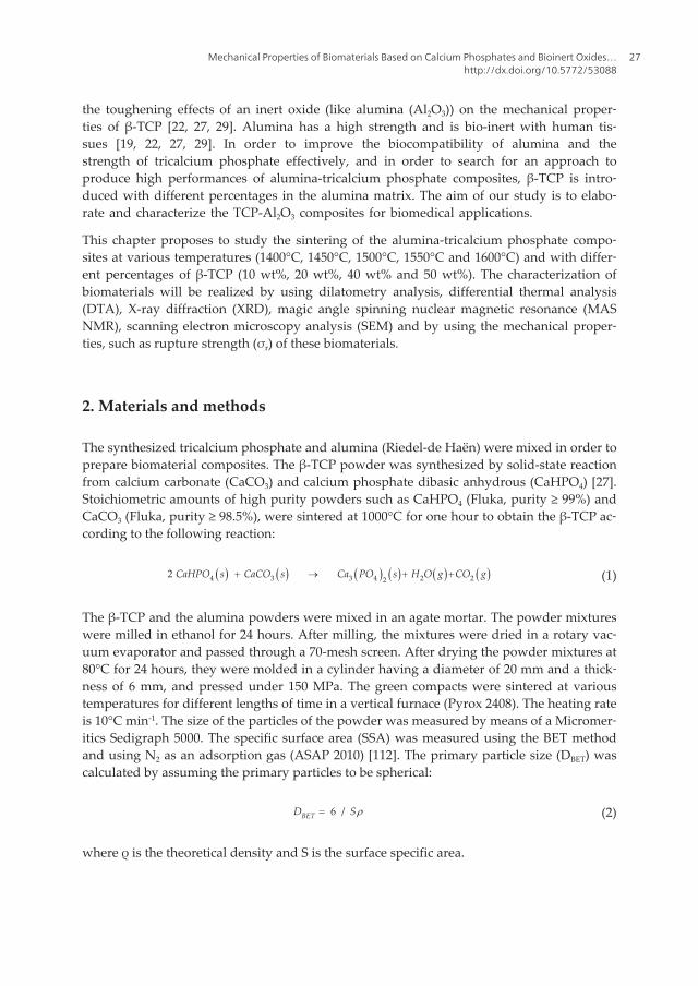
The X-ray diffraction analysis of β-TCP powder and α-alumina powder are presented in Fig‐ure 1. As it can be noticed from this figure, the X-ray diffraction pattern of tricalcium phos‐phate powder reveals only peaks of β-TCP (ICDD data file no. 70-2065) without any otherphase (Figure 1a). Consequently, the XRD pattern obtained from the alumina powder illus‐trates α phase peaks relative to ICDD data file no. 43-1484 (Figure 1b).
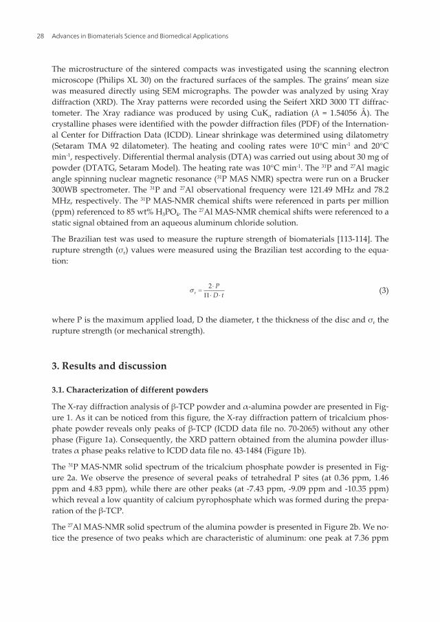
The 31P MAS-NMR solid spectrum of the tricalcium phosphate powder is presented in Fig‐ure 2a. We observe the presence of several peaks of tetrahedral P sites (at 0.36 ppm, 1.46ppm and 4.83 ppm), while there are other peaks (at -7.43 ppm, -9.09 ppm and -10.35 ppm)which reveal a low quantity of calcium pyrophosphate which was formed during the prepa‐ration of the β-TCP.
The 27Al MAS-NMR solid spectrum of the alumina powder is presented in Figure 2b. We no‐tice the presence of two peaks which are characteristic of aluminum: one peak at 7.36 ppm
Advances in Biomaterials Science and Biomedical Applications28
corresponding to octahedral Al sites (AlVI) and the other at 37.36 ppm which corresponds topentahedral Al sites (AlV). The results obtained for 31P MAS-NMR and 27Al MAS-NMR aresimilar to those previously reported by different authors [14, 22, 25-28, 31].
Figure 1. The XRD patterns of: (a) β-TCP powder and (b) α-Al2O3 powder
Figure 2. The 31P MAS-NMR spectra of: (a) β-TCP and the 27Al MAS-NMR spectra of: (b) α-Al2O3
Mechanical Properties of Biomaterials Based on Calcium Phosphates and Bioinert Oxides…http://dx.doi.org/10.5772/53088
29
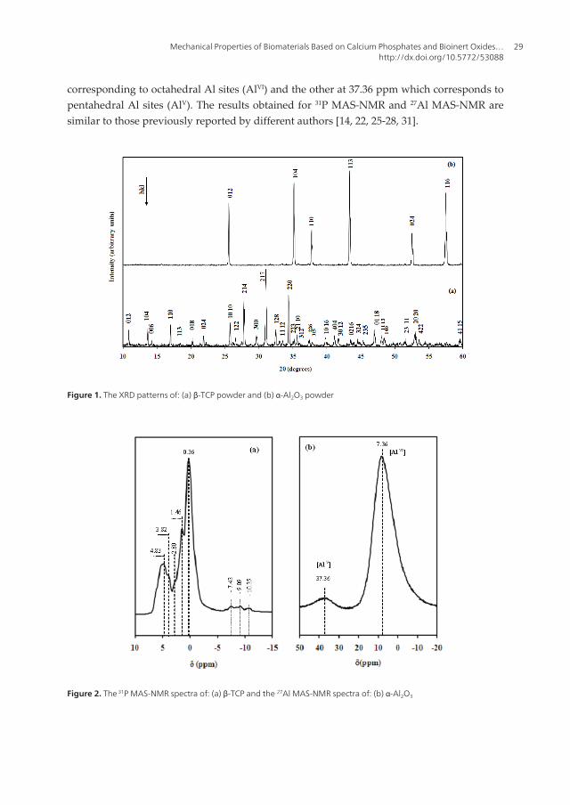
The experimental characteristics of the different powders used in this study are illustrated inTable 2. Table 2 summarizes the SSA, the DTA measurements, the sintering temperatureand the theoretical density of the different powders. The powder particles are assumed to bespherical; the size of the particles can be calculated using Eq. (2). The results from the aver‐age grain size obtained by the SSA (DBET) and from the average grain size obtained by gran‐ulometric repartition (D50) are presented in Table 2. Compared with those of the β-TCPpowder, the grains of the alumina powder have a dense morphology. These (DBET) valuesobtained by the SSA do not correspond to those obtained from the granulometric repartition(Table 2). The discrepancy may be due to the presence of agglomerates which are formedduring the preparation of the β-TCP powder at 1000°C.
Compounds SSA (m2/g)
± 1.0
DBET (µm)
± 0.2
D50 (µm)a
± 0.2
DTA measurements
(endothermic peak)
T(°C)b dc
TCP 0.70 2.79 6 1100°C-1260°C (β → α)
1470°C (α → α’)
1000 - 1300 3.070 (β)
2.860 (α)
Alumina 2.87 0.53 3 - 1400 - 1600 3.98 (α)
a : mean diameter,
b : sintering temperature domain,
c : theoretical density
Table 2. Characteristics of the powders used in the study
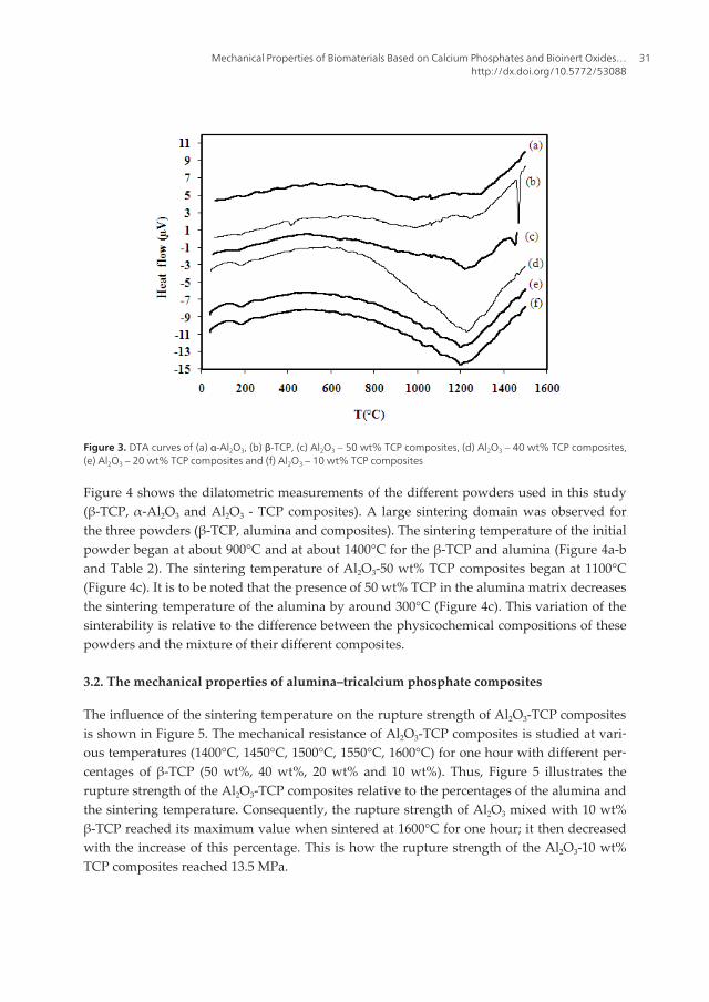
Differential thermal analysis studies of the different powders used in this study detected apotential phase change during the sintering process. The DTA thermogram of β-TCP, α-Al2O3 and different Al2O3 - TCP composites are presented in Figure 3. The DTA curve of alu‐mina reported no process relative to the sintering temperature (Figure 3a). Figure 3b showsthe DTA curve of β-TCP. The DTA thermogram of β-TCP shows two endothermic peaks,relative to the allotropic transformations of tricalcium phosphate (Figure 3b). The peak be‐tween 1100°C – 1260°C is related to the first allotropic transformation of TCP (β to α), whilethe last peak at 1470°C is related to the second allotropic transformation of TCP (α to α’). Asa matter of fact, this result is similar to the result previously reported by Destainville et al.and Ben Ayed et al. [9, 14]. Figure 3c shows the DTA curve of Al2O3-50 wt% TCP compo‐sites. This DTA curve is practically similar to the one shown in Figure 3b. Indeed, the DTAthermogram of the composites also shows two endothermic peaks. Figure 3 (d), (e) and (f)illustrate the DTA curves of Al2O3–40 wt% TCP composites, Al2O3–20 wt% TCP compositesand Al2O3–10 wt% TCP composites, respectively. The DTA thermograms of each compositesshow only one endothermic peak between 1100°C and 1260°C, which are relative to the allo‐tropic transformation of TCP (β to α). In these curves, we notice that the endothermic peakrelative to a second allotropic transformation of TCP (α to α’) has practically disappearedwhen the percentage of the alumina increases in the Al2O3 - TCP composites (Figure 3(d), (e)and (f)).
Advances in Biomaterials Science and Biomedical Applications30
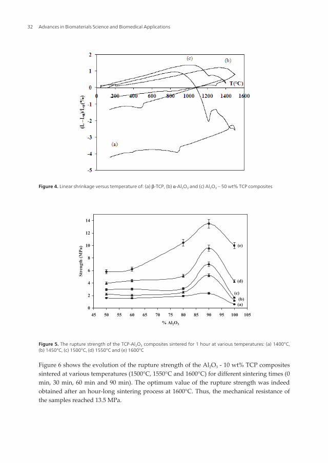
Figure 4 shows the dilatometric measurements of the different powders used in this study(β-TCP, α-Al2O3 and Al2O3 - TCP composites). A large sintering domain was observed forthe three powders (β-TCP, alumina and composites). The sintering temperature of the initialpowder began at about 900°C and at about 1400°C for the β-TCP and alumina (Figure 4a-band Table 2). The sintering temperature of Al2O3-50 wt% TCP composites began at 1100°C(Figure 4c). It is to be noted that the presence of 50 wt% TCP in the alumina matrix decreasesthe sintering temperature of the alumina by around 300°C (Figure 4c). This variation of thesinterability is relative to the difference between the physicochemical compositions of thesepowders and the mixture of their different composites.
3.2. The mechanical properties of alumina–tricalcium phosphate composites
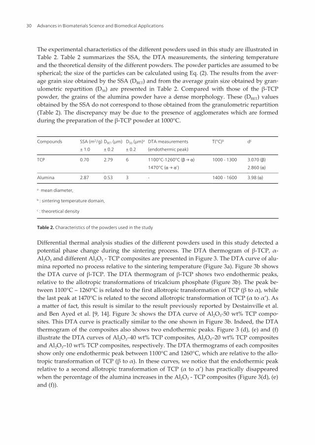
The influence of the sintering temperature on the rupture strength of Al2O3-TCP compositesis shown in Figure 5. The mechanical resistance of Al2O3-TCP composites is studied at vari‐ous temperatures (1400°C, 1450°C, 1500°C, 1550°C, 1600°C) for one hour with different per‐centages of β-TCP (50 wt%, 40 wt%, 20 wt% and 10 wt%). Thus, Figure 5 illustrates therupture strength of the Al2O3-TCP composites relative to the percentages of the alumina andthe sintering temperature. Consequently, the rupture strength of Al2O3 mixed with 10 wt%β-TCP reached its maximum value when sintered at 1600°C for one hour; it then decreasedwith the increase of this percentage. This is how the rupture strength of the Al2O3-10 wt%TCP composites reached 13.5 MPa.
Mechanical Properties of Biomaterials Based on Calcium Phosphates and Bioinert Oxides…http://dx.doi.org/10.5772/53088
31
Figure 4. Linear shrinkage versus temperature of: (a) β-TCP, (b) α-Al2O3 and (c) Al2O3 – 50 wt% TCP composites
0
2
4
6
8
10
12
14
45 50 55 60 65 70 75 80 85 90 95 100 105
% Al2O3
Stre
ngth
(MPa
) (e)
(d)
(c)(b)(a)
Figure 5. The rupture strength of the TCP-Al2O3 composites sintered for 1 hour at various temperatures: (a) 1400°C,(b) 1450°C, (c) 1500°C, (d) 1550°C and (e) 1600°C
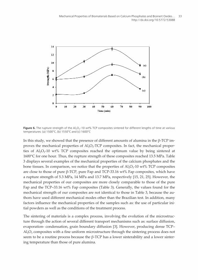
Figure 6 shows the evolution of the rupture strength of the Al2O3 - 10 wt% TCP compositessintered at various temperatures (1500°C, 1550°C and 1600°C) for different sintering times (0min, 30 min, 60 min and 90 min). The optimum value of the rupture strength was indeedobtained after an hour-long sintering process at 1600°C. Thus, the mechanical resistance ofthe samples reached 13.5 MPa.
Advances in Biomaterials Science and Biomedical Applications32
Figure 6. The rupture strength of the Al2O3–10 wt% TCP composites sintered for different lengths of time at varioustemperatures: (a) 1500°C, (b) 1550°C and (c) 1600°C
In this study, we showed that the presence of different amounts of alumina in the β-TCP im‐proves the mechanical properties of Al2O3-TCP composites. In fact, the mechanical proper‐ties of Al2O3-10 wt% TCP composites reached the optimum value by being sintered at1600°C for one hour. Thus, the rupture strength of these composites reached 13.5 MPa. Table3 displays several examples of the mechanical properties of the calcium phosphates and thebone tissues. In comparison, we notice that the properties of Al2O3-10 wt% TCP compositesare close to those of pure β-TCP, pure Fap and TCP-33.16 wt% Fap composites, which havea rupture strength of 5.3 MPa, 14 MPa and 13.7 MPa, respectively [15, 21, 25]. However, themechanical properties of our composites are more closely comparable to those of the pureFap and the TCP–33.16 wt% Fap composites (Table 3). Generally, the values found for themechanical strength of our composites are not identical to those in Table 3, because the au‐thors have used different mechanical modes other than the Brazilian test. In addition, manyfactors influence the mechanical properties of the samples such as: the use of particular ini‐tial powders as well as the conditions of the treatment process.
The sintering of materials is a complex process, involving the evolution of the microstruc‐ture through the action of several different transport mechanisms such as: surface diffusion,evaporation- condensation, grain boundary diffusion [3]. However, producing dense TCP–Al2O3 composites with a fine uniform microstructure through the sintering process does notseem to be a routine process because the β-TCP has a lower sinterability and a lower sinter‐ing temperature than those of pure alumina.
Mechanical Properties of Biomaterials Based on Calcium Phosphates and Bioinert Oxides…http://dx.doi.org/10.5772/53088
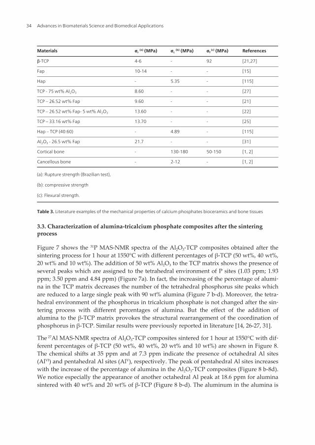
Table 3. Literature examples of the mechanical properties of calcium phosphates bioceramics and bone tissues
3.3. Characterization of alumina-tricalcium phosphate composites after the sinteringprocess
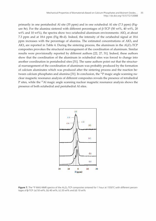
Figure 7 shows the 31P MAS-NMR spectra of the Al2O3-TCP composites obtained after thesintering process for 1 hour at 1550°C with different percentages of β-TCP (50 wt%, 40 wt%,20 wt% and 10 wt%). The addition of 50 wt% Al2O3 to the TCP matrix shows the presence ofseveral peaks which are assigned to the tetrahedral environment of P sites (1.03 ppm; 1.93ppm; 3.50 ppm and 4.84 ppm) (Figure 7a). In fact, the increasing of the percentage of alumi‐na in the TCP matrix decreases the number of the tetrahedral phosphorus site peaks whichare reduced to a large single peak with 90 wt% alumina (Figure 7 b-d). Moreover, the tetra‐hedral environment of the phosphorus in tricalcium phosphate is not changed after the sin‐tering process with different percentages of alumina. But the effect of the addition ofalumina to the β-TCP matrix provokes the structural rearrangement of the coordination ofphosphorus in β-TCP. Similar results were previously reported in literature [14, 26-27, 31].
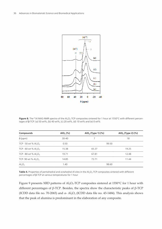
The 27Al MAS-NMR spectra of Al2O3-TCP composites sintered for 1 hour at 1550°C with dif‐ferent percentages of β-TCP (50 wt%, 40 wt%, 20 wt% and 10 wt%) are shown in Figure 8.The chemical shifts at 35 ppm and at 7.3 ppm indicate the presence of octahedral Al sites(AlVI) and pentahedral Al sites (AlV), respectively. The peak of pentahedral Al sites increaseswith the increase of the percentage of alumina in the Al2O3-TCP composites (Figure 8 b-8d).We notice especially the appearance of another octahedral Al peak at 18.6 ppm for aluminasintered with 40 wt% and 20 wt% of β-TCP (Figure 8 b-d). The aluminum in the alumina is
Advances in Biomaterials Science and Biomedical Applications34
primarily in one pentahedral Al site (35 ppm) and in one octahedral Al site (7.3 ppm) (Fig‐ure 8e). For the alumina sintered with different percentages of β-TCP (50 wt%, 40 wt%, 20wt% and 10 wt%), the spectra show two octahedral aluminum environments: AlO6 at about7.3 ppm and at 18.6 ppm (Fig 8b-d). Indeed, the intensity of the octahedral signal at 18.6ppm increases with the percentage of alumina. The estimated concentrations of AlO5 andAlO6 are reported in Table 4. During the sintering process, the aluminum in the Al2O3-TCPcomposites provokes the structural rearrangement of the coordination of aluminum. Similarresults were provisionally reported by different authors [22, 27, 31]. Indeed, these authorsshow that the coordination of the aluminum in octahedral sites was forced to change intoanother coordination in pentahedral sites [31]. The same authors point out that the structur‐al rearrangement of the coordination of aluminum was probably produced by the formationof calcium aluminates which was produced after the sintering process and the reaction be‐tween calcium phosphates and alumina [31]. In conclusion, the 31P magic angle scanning nu‐clear magnetic resonance analysis of different composites reveals the presence of tetrahedralP sites, while the 27Al magic angle scanning nuclear magnetic resonance analysis shows thepresence of both octahedral and pentahedral Al sites.
Figure 7. The 31P MAS-NMR spectra of the Al2O3-TCP composites sintered for 1 hour at 1550°C with different percen‐tages of β-TCP: (a) 50 wt%, (b) 40 wt%, (c) 20 wt% and (d) 10 wt%
Mechanical Properties of Biomaterials Based on Calcium Phosphates and Bioinert Oxides…http://dx.doi.org/10.5772/53088
35
Figure 8. The 27Al MAS-NMR spectra of the Al2O3-TCP composites sintered for 1 hour at 1550°C with different percen‐tages of β-TCP: (a) 50 wt%, (b) 40 wt%, (c) 20 wt%, (d) 10 wt% and (e) 0 wt%
Table 4. Properties of pentahedral and octahedral Al sites in the Al2O3-TCP composites sintered with differentpercentages of β-TCP at various temperatures for 1 hour
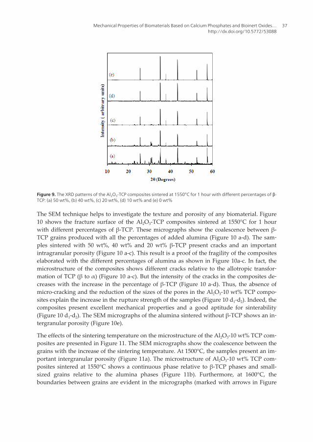
Figure 9 presents XRD patterns of Al2O3-TCP composites sintered at 1550°C for 1 hour withdifferent percentages of β-TCP. Besides, the spectra show the characteristic peaks of β-TCP(ICDD data file no. 70-2065) and α- Al2O3 (ICDD data file no. 43-1484). This analysis showsthat the peak of alumina is predominant in the elaboration of any composite.
Advances in Biomaterials Science and Biomedical Applications36
Figure 9. The XRD patterns of the Al2O3-TCP composites sintered at 1550°C for 1 hour with different percentages of β-TCP: (a) 50 wt%, (b) 40 wt%, (c) 20 wt%, (d) 10 wt% and (e) 0 wt%
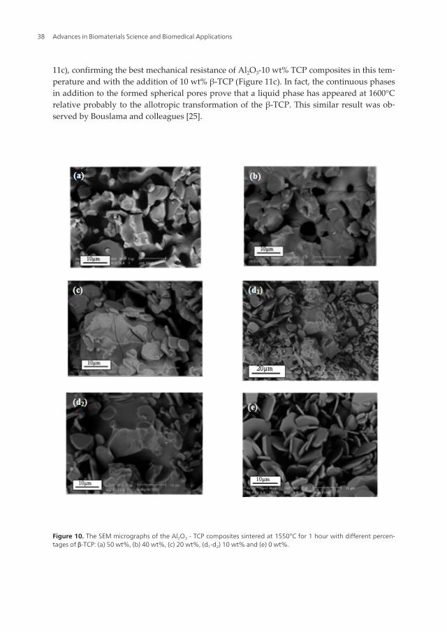
The SEM technique helps to investigate the texture and porosity of any biomaterial. Figure10 shows the fracture surface of the Al2O3-TCP composites sintered at 1550°C for 1 hourwith different percentages of β-TCP. These micrographs show the coalescence between β-TCP grains produced with all the percentages of added alumina (Figure 10 a-d). The sam‐ples sintered with 50 wt%, 40 wt% and 20 wt% β-TCP present cracks and an importantintragranular porosity (Figure 10 a-c). This result is a proof of the fragility of the compositeselaborated with the different percentages of alumina as shown in Figure 10a-c. In fact, themicrostructure of the composites shows different cracks relative to the allotropic transfor‐mation of TCP (β to α) (Figure 10 a-c). But the intensity of the cracks in the composites de‐creases with the increase in the percentage of β-TCP (Figure 10 a-d). Thus, the absence ofmicro-cracking and the reduction of the sizes of the pores in the Al2O3-10 wt% TCP compo‐sites explain the increase in the rupture strength of the samples (Figure 10 d1-d2). Indeed, thecomposites present excellent mechanical properties and a good aptitude for sinterability(Figure 10 d1-d2). The SEM micrographs of the alumina sintered without β-TCP shows an in‐tergranular porosity (Figure 10e).
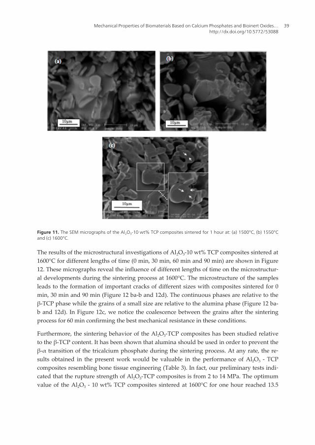
The effects of the sintering temperature on the microstructure of the Al2O3-10 wt% TCP com‐posites are presented in Figure 11. The SEM micrographs show the coalescence between thegrains with the increase of the sintering temperature. At 1500°C, the samples present an im‐portant intergranular porosity (Figure 11a). The microstructure of Al2O3-10 wt% TCP com‐posites sintered at 1550°C shows a continuous phase relative to β-TCP phases and small-sized grains relative to the alumina phases (Figure 11b). Furthermore, at 1600°C, theboundaries between grains are evident in the micrographs (marked with arrows in Figure
Mechanical Properties of Biomaterials Based on Calcium Phosphates and Bioinert Oxides…http://dx.doi.org/10.5772/53088
37
11c), confirming the best mechanical resistance of Al2O3-10 wt% TCP composites in this tem‐perature and with the addition of 10 wt% β-TCP (Figure 11c). In fact, the continuous phasesin addition to the formed spherical pores prove that a liquid phase has appeared at 1600°Crelative probably to the allotropic transformation of the β-TCP. This similar result was ob‐served by Bouslama and colleagues [25].
Figure 10. The SEM micrographs of the Al2O3 - TCP composites sintered at 1550°C for 1 hour with different percen‐tages of β-TCP: (a) 50 wt%, (b) 40 wt%, (c) 20 wt%, (d1-d2) 10 wt% and (e) 0 wt%.
Advances in Biomaterials Science and Biomedical Applications38
Figure 11. The SEM micrographs of the Al2O3-10 wt% TCP composites sintered for 1 hour at: (a) 1500°C, (b) 1550°Cand (c) 1600°C.
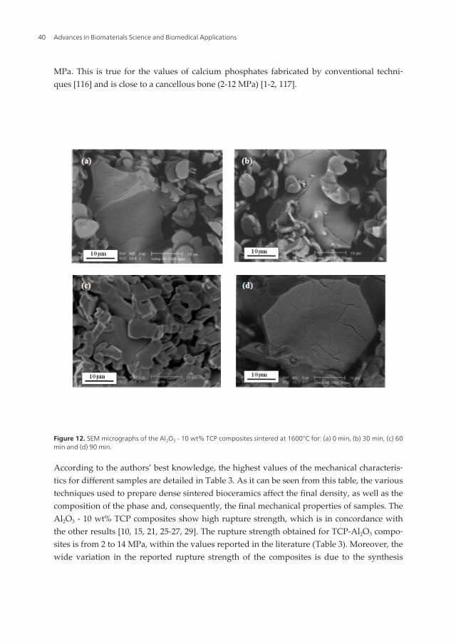
The results of the microstructural investigations of Al2O3-10 wt% TCP composites sintered at1600°C for different lengths of time (0 min, 30 min, 60 min and 90 min) are shown in Figure12. These micrographs reveal the influence of different lengths of time on the microstructur‐al developments during the sintering process at 1600°C. The microstructure of the samplesleads to the formation of important cracks of different sizes with composites sintered for 0min, 30 min and 90 min (Figure 12 ba-b and 12d). The continuous phases are relative to theβ-TCP phase while the grains of a small size are relative to the alumina phase (Figure 12 ba-b and 12d). In Figure 12c, we notice the coalescence between the grains after the sinteringprocess for 60 min confirming the best mechanical resistance in these conditions.
Furthermore, the sintering behavior of the Al2O3-TCP composites has been studied relativeto the β-TCP content. It has been shown that alumina should be used in order to prevent theβ-α transition of the tricalcium phosphate during the sintering process. At any rate, the re‐sults obtained in the present work would be valuable in the performance of Al2O3 - TCPcomposites resembling bone tissue engineering (Table 3). In fact, our preliminary tests indi‐cated that the rupture strength of Al2O3-TCP composites is from 2 to 14 MPa. The optimumvalue of the Al2O3 - 10 wt% TCP composites sintered at 1600°C for one hour reached 13.5
Mechanical Properties of Biomaterials Based on Calcium Phosphates and Bioinert Oxides…http://dx.doi.org/10.5772/53088
39
MPa. This is true for the values of calcium phosphates fabricated by conventional techni‐ques [116] and is close to a cancellous bone (2-12 MPa) [1-2, 117].
Figure 12. SEM micrographs of the Al2O3 - 10 wt% TCP composites sintered at 1600°C for: (a) 0 min, (b) 30 min, (c) 60min and (d) 90 min.
According to the authors’ best knowledge, the highest values of the mechanical characteris‐tics for different samples are detailed in Table 3. As it can be seen from this table, the varioustechniques used to prepare dense sintered bioceramics affect the final density, as well as thecomposition of the phase and, consequently, the final mechanical properties of samples. TheAl2O3 - 10 wt% TCP composites show high rupture strength, which is in concordance withthe other results [10, 15, 21, 25-27, 29]. The rupture strength obtained for TCP-Al2O3 compo‐sites is from 2 to 14 MPa, within the values reported in the literature (Table 3). Moreover, thewide variation in the reported rupture strength of the composites is due to the synthesis
Advances in Biomaterials Science and Biomedical Applications40
route of the β-TCP powder, the size of its particle as well as to its density; it is also due tothe application of different processing parameters.
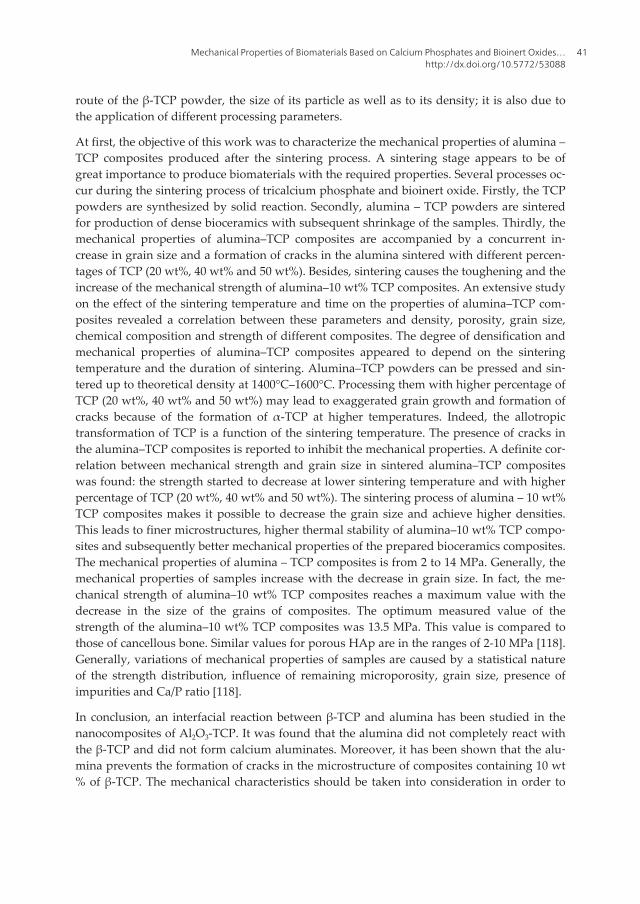
At first, the objective of this work was to characterize the mechanical properties of alumina –TCP composites produced after the sintering process. A sintering stage appears to be ofgreat importance to produce biomaterials with the required properties. Several processes oc‐cur during the sintering process of tricalcium phosphate and bioinert oxide. Firstly, the TCPpowders are synthesized by solid reaction. Secondly, alumina – TCP powders are sinteredfor production of dense bioceramics with subsequent shrinkage of the samples. Thirdly, themechanical properties of alumina–TCP composites are accompanied by a concurrent in‐crease in grain size and a formation of cracks in the alumina sintered with different percen‐tages of TCP (20 wt%, 40 wt% and 50 wt%). Besides, sintering causes the toughening and theincrease of the mechanical strength of alumina–10 wt% TCP composites. An extensive studyon the effect of the sintering temperature and time on the properties of alumina–TCP com‐posites revealed a correlation between these parameters and density, porosity, grain size,chemical composition and strength of different composites. The degree of densification andmechanical properties of alumina–TCP composites appeared to depend on the sinteringtemperature and the duration of sintering. Alumina–TCP powders can be pressed and sin‐tered up to theoretical density at 1400°C–1600°C. Processing them with higher percentage ofTCP (20 wt%, 40 wt% and 50 wt%) may lead to exaggerated grain growth and formation ofcracks because of the formation of α-TCP at higher temperatures. Indeed, the allotropictransformation of TCP is a function of the sintering temperature. The presence of cracks inthe alumina–TCP composites is reported to inhibit the mechanical properties. A definite cor‐relation between mechanical strength and grain size in sintered alumina–TCP compositeswas found: the strength started to decrease at lower sintering temperature and with higherpercentage of TCP (20 wt%, 40 wt% and 50 wt%). The sintering process of alumina – 10 wt%TCP composites makes it possible to decrease the grain size and achieve higher densities.This leads to finer microstructures, higher thermal stability of alumina–10 wt% TCP compo‐sites and subsequently better mechanical properties of the prepared bioceramics composites.The mechanical properties of alumina – TCP composites is from 2 to 14 MPa. Generally, themechanical properties of samples increase with the decrease in grain size. In fact, the me‐chanical strength of alumina–10 wt% TCP composites reaches a maximum value with thedecrease in the size of the grains of composites. The optimum measured value of thestrength of the alumina–10 wt% TCP composites was 13.5 MPa. This value is compared tothose of cancellous bone. Similar values for porous HAp are in the ranges of 2-10 MPa [118].Generally, variations of mechanical properties of samples are caused by a statistical natureof the strength distribution, influence of remaining microporosity, grain size, presence ofimpurities and Ca/P ratio [118].
In conclusion, an interfacial reaction between β-TCP and alumina has been studied in thenanocomposites of Al2O3-TCP. It was found that the alumina did not completely react withthe β-TCP and did not form calcium aluminates. Moreover, it has been shown that the alu‐mina prevents the formation of cracks in the microstructure of composites containing 10 wt% of β-TCP. The mechanical characteristics should be taken into consideration in order to
Mechanical Properties of Biomaterials Based on Calcium Phosphates and Bioinert Oxides…http://dx.doi.org/10.5772/53088
41
better assess the relationship between the processing conditions, the microstructural designas well as the mechanical response.
4. Conclusions
The biomaterials of alumina-tricalcium phosphate composites have been characterized byusing MAS NMR, XRD and SEM analysis after the sintering process. The effect of β-TCP ad‐ditive on the alumina matrix was observed in different thermal analyses: dilatometry analy‐sis and DTA analysis. The mechanical properties have been investigated by the Braziliantest. This investigation has allowed us to define the sintering temperature and the percent‐age of added alumina for which β-TCP should have an optimal densification and better me‐chanical properties. This study has also allowed us to summarize the effect of the sinteringtemperature and the length of sintering time on the mechanical properties of the Al2O3-TCPcomposites. The produced Al2O3-TCP composites with different percentages of β-TCP (50wt%; 40 wt%; 20 wt% and 10 wt%) exhibited much better mechanical properties than the re‐ported values of β-TCP without alumina. The Al2O3-TCP composites showed a higher rup‐ture strength at 1600°C, which certainly increased with the alumina content and reached theoptimum value with 90 wt%. However, no cracks were observed in the microstructure of thecomposites which contained this percentage of alumina. This is due to the allotropic trans‐formation of the tricalcium phosphate. The partial or reversal transformation of tricalciumphosphate (β to α or α to α’) during the cooling period could induce a residual stress withinthe dense bioceramics, marking it much more brittle. Accordingly, the optimum perform‐ance of alumina-tricalcium phosphate composites achieved 13.5 MPa. Furthermore, the bestmechanical properties of the composites were obtained after the sintering process at 1600°Cfor 1 hour. With different weight rations of tricalcium phosphate: alumina (50:50, 40:60 and20:80), the performance of the composites was hindered by the formation of both cracks andintragranular porosity.
Acknowledgements
The authors thank Mr Ahmed BAHLOUL for his assistance in this work.
Laboratory of Industrial Chemistry, National School of Engineering, Sfax University, Sfax,Tunisia
Advances in Biomaterials Science and Biomedical Applications42
References
[1] Hench L L. Bioceramics: From Concept to clinic. J. Am. Ceram. Soc. 1991; 74 (7) 1487.
[2] Hench L L. An Introduction to Bioceramics. J. Wilson (ed.). Vol. 1. World Scientific,Singapore; 1993.
[3] Elliott J C. Structure and Chemistry of the Apatite and Other Calcium Orthophos‐phates. Amsterdam : Elsevier Science B.V.; 1994.
[4] Landi E, Tampieri A, Celotti G, Sprio S. Densification behaviour and mechanisms ofsynthetic hydroxyapatites. J. Eur. Ceram. Soc. 2000; 20, 2377.
[5] Ben Ayed F, Bouaziz J, Bouzouita K. Pressureless sintering of fluorapatite under oxy‐gen atmosphere. J. Eur. Ceram. Soc. 2000; 20 (8) 1069.
[6] Ben Ayed F, Bouaziz J, Khattech I, Bouzouita K. Produit de solubilité apparent de lafluorapatite frittée. Ann. Chim. Sci. Mater. 2001; 26 (6) 75.
[7] Ben Ayed F, Bouaziz J, Bouzouita K. Calcination and sintering of fluorapatite underargon atmosphere. J. Alloys Compd. 2001; 322 (1-2) 238.
[8] Varma H K, Sureshbabu S. Oriented growth of surface grains in sintered b tricalciumphosphate bioceramics. Materials letters 2001; 49, 83.
[9] Destainville A, Champion E, Bernache – Assolant D. Synthesis. Characterization andthermal behavior of apatitic tricalcium phosphate. Mater. Chem. Phys. 2003; 80, 269.
[10] Wang C X, Zhou X, Wang M. Influence of sintering temperatures on hardness andYoung's modulus of tricalcium phosphate bioceramic by nanoindentation technique.Materials Characterization 2004; 52, 301.
[11] Hoell S, Suttmoeller J, Stoll V, Fuchs S, Gosheger G. The high tibial osteotomy, openversus closed wedge, a comparison of methods in 108 patients. Arch.Trauma Surg.2005; 125, 638–43.
[12] Gaasbeek R D, Toonen H G, Van Heerwaarden R J, Buma P. Mechanism of bone in‐corporation of β-TCP bone substitute in open wedge tibial osteotomy in patients. Bi‐omaterials 2005; 26, 6713–6719.
[13] Jensen S S, Broggini N, Hjorting-Hansen E, Schenk R, Buser D. Bone healing andgraft resorption of autograft, anorganic bovine bone and beta-tricalcium phosphate.A histologic and histomorphometric study in the mandibles of minipigs.. Clin. Oral.Implants Res. 2006; 17, 237–243.
[14] Ben Ayed F, Chaari K, Bouaziz J, Bouzouita K. Frittage du phosphate tricalcique. C.R. Physique, 2006; 7 (7) 825.
[15] Ben Ayed F, Bouaziz J, Bouzouita K. Résistance mécanique de la fluorapatite. Ann.Chim. Sci. Mater. 2006; 31 (4) 393.
Mechanical Properties of Biomaterials Based on Calcium Phosphates and Bioinert Oxides…http://dx.doi.org/10.5772/53088
43
[16] Gutierres M, Dias A G, Lopes M A, Hussain N S, Cabral A T, Almeid L. Openingwedge high tibial osteotomy using 3D biomodelling Bone like macroporous struc‐tures: case report. J. Mater. Sci. Mater. Med. 2007; 7 (18) 2377–2382.
[17] DeSilva G L, Fritzler A, DeSilva S P. Antibiotic-impregnated cement spacer for bonedefects of the forearm and hand. Tech. Hand Up Extrem Surg. 2007; 11, 163–7
[18] Ben Ayed F, Bouaziz J. Élaboration et caractérisation d’un biomatériau à base dephosphates de calcium. C. R. Physique, 2007; 8 (1) 101-108.
[19] Ben Ayed F, Bouaziz J. Sintering of tricalcium phosphate–fluorapatite composites byaddition of alumina. Ceramics Int. 2008; 34 (8) 1885-1892.
[20] Ben Ayed F, Bouaziz J. Sintering of tricalcium phosphate–fluorapatite compositeswith zirconia. J. Eur. Ceram. Soc., 2008; 28 (10) 1995-2002.
[21] Bouslama N, Ben Ayed F, Bouaziz J. Sintering and mechanical properties of tricalci‐um phosphate–fluorapatite composites. Ceramics Int. 2009; 35, 1909-1917.
[22] Bouslama N, Ben Ayed F, Bouaziz J. Mechanical properties of tricalcium phosphate-fluorapatite-alumina composites. Physics Procedia 2009; 2, 1441-1448.
[23] Chaari K, Ben Ayed F, Bouaziz J, Bouzouita K. Elaboration and characterization offluorapatite ceramic with controlled porosity. Materials Chemistry and Physics 2009;113, 219-226.
[24] Guha A K, Singh S, Kumarresan R, Nayar S, Sinha A. Mesenchymal cell response tonanosized biphasic calcium phosphate composites. Coll. Surf. B biointerface 2009; 73,146-51.
[25] Bouslama N, Ben Ayed F, Bouaziz J. Effect of fluorapatite additive on densificationand mechanical properties of tricalcium phosphate. J. Mechanical Behaviour of Bio‐medical Materials 2010; 3, 2-13.
[26] Ben Ayed F. Biomaterials - Physics and Chemistry, Chapter 18: Elaboration and char‐acterisation of calcium phosphate biomaterial for biomedical application, ISBN978-953-307-418-4. Croatia: In Tech; 2011. p 357 – 374.
[27] Sakka S, Ben Ayed F, Bouaziz J. Mechanical properties of tricalcium phosphate–alu‐mina composites. IOP Conf. Series : Materials Science and Engineering, 2012. 28,012028
[28] Sellami I, Ben Ayed F, Bouaziz J. Effect of fluorapatite additive on the mechanicalproperties of tricalcium phosphate-zirconia composites. IOP Conf. Series : MaterialsScience and Engineering, 2012. 28, 012029.
[29] Ben Ayed F. Current microscopy contributions to advances in science and technolo‐gy, Chapter: The effect of the sintering process on the microstructure and the me‐chanical properties of biomaterials. published by Formatex Research Center Spain; tobe published in 2012.
Advances in Biomaterials Science and Biomedical Applications44
[30] Levin I, Brandon D. Metastable Alumina Polymorphs: Crystal Structures and Transi‐tion. Sequences. J. Am. Ceram. Soc. 1998, 81.
[31] Guidera A, Chaari K, Bouaziz J. Elaboration and Characterization of Alumina-Fluo‐rapatite Composites. J. Biomat. Nano. 2011; 2, 103-113.
[32] Doremus RH. Bioceramics. J Mater Sci 1992; 27, 285–97.
[33] Vallet-Regı´ M. Ceramics for medical applications. J Chem Soc Dalton Trans 2001,97–108.
[34] Rahaman MN, Yao A, Bal BS, Garino JP, Ries MD. Ceramics for prosthetic hip andknee joint replacement. J Am Ceram Soc 2007; 90, 1965–88.
[35] Best SM, Porter AE, Thian ES, Huang J. Bioceramics: past, present and for the future.J Eur Ceram Soc 2008; 28, 1319–27.
[36] Lowenstam HA, Weiner S. On biomineralization. Oxford University Press, 1989; pp324.
[37] LeGeros RZ. Calcium phosphates in oral biology and medicine. Basel: Karger; 1991,201.
[38] Weiner S, Wagner HD. Material bone: structure-mechanical function relations. AnnRev Mater Sci 1998; 28, 271–98.
[40] Weiner S, Dove PM. An overview of biomineralization processes and the problem ofthe vital effect. In: Dove PM, de Yoreo JJ, Weiner S, editors. Biomineralization, series:reviews in mineralogy and geochemistry, vol. 54. Washington, D.C., USA: Minera‐logical Society of America; 2003. p 1–29.
[41] Albee FH. Studies in bone growth – triple calcium phosphate as stimulus to osteo‐genesis. Ann Surg 1920; 71, 32–9.
[43] Denissen HW, de Groot K. Immediate dental root implants from synthetic dense cal‐cium hydroxylapatite. J Prosthet Dent 1979; 42, 551–6.
[44] Levitt GE, Crayton PH, Monroe EA, Condrate RA. Forming methods for apatiteprosthesis. J Biomed Mater Res 1969; 3, 683–5.
[45] Blakeslee KC, Condrate Sr RA. Vibrational spectra of hydrothermally prepared hy‐droxyapatites. J Am Ceram Soc 1971; 54, 559–63.
[46] Garrington GE, Lightbody PM. Bioceramics and dentistry. J Biomed Mater Res 1972;6, 333–43.
Mechanical Properties of Biomaterials Based on Calcium Phosphates and Bioinert Oxides…http://dx.doi.org/10.5772/53088
45
[47] Cini L, Sandrolini S, Paltrinieri M, Pizzoferrato A, Trentani C. Materiali bioceramiciin funzione sostitutiva. Nota preventiva. (Bioceramic materials for replacement pur‐poses. Preliminary note.). La Chirurgia Degli Organi Di Movimento 1972; 60, 423–30.
[49] Hench LL, Splinter RJ, Allen WC, Greenlee TK. Bonding mechanisms at the interfaceof ceramic prosthetic materials. J Biomed Mater Res 1971; 2, 117–41.
[50] Hulbert SF, Hench LL, Forces D, Bowman L. History of bioceramics. In: Vincenzini P,editor. Ceramics in surgery. Amsterdam, The Netherlands: Elsevier, 1983; p 3–29.
[51] Jarcho M. Calcium phosphate ceramics as hard tissue prosthetics. Clin Orthop RelatRes 1981; 157, 259–78.
[52] de Groot K. Bioceramics consisting of calcium phosphate salts. Biomaterials 1980; 1(1) 47–50.
[53] Aoki H, Kato KM, Ogiso M, Tabata T. Studies on the application of apatite to dentalmaterials. J Dent Eng 1977; 18, 86–9.
[54] Roy DM, Linnehan SK. Hydroxyapatite formed from coral skeletal carbonate by hy‐drothermal exchange. Nature 1974; 247, 220–2.
[55] Holmes RE. Bone regeneration within a coralline hydroxyapatite implant. Plast Re‐constr Surg 1979; 63, 626–33.
[56] Elsinger EC, Leal L. Coralline hydroxyapatite bone graft substitutes. J Foot AnkleSurg 1996; 35, 396–9.
[58] Durucan C, Brown PW. Low temperature formation of calcium-deficient hydroxya‐patite-PLA/PLGA composites. J Biomed Mater Res 2000; 51A, 717–25.
[59] Ginebra MP, Rilliard A, Ferna´ndez E, Elvira C, Roma´n JS, Planell JA. Mechanicaland rheological improvement of a calcium phosphate cement by the addition of apolymeric drug. J Biomed Mater Res 2001; 57, 113–8.
[60] Yokoyama A, Yamamoto S, Kawasaki T, Kohgo T, Nakasu M. Development of calci‐um phosphate cement using chitosan and citric acid for bone substitute materials. Bi‐omaterials 2002; 23, 1091–101.
[61] Barralet JE, Gaunt T, Wright AJ, Gibson IR, Knowles JC. Effect of porosity reductionby compaction on compressive strength and microstructure of calcium phosphate ce‐ment. J Biomed Mater Res 2002; 63B, 1–9.
Advances in Biomaterials Science and Biomedical Applications46
[62] Bohner M, Gbureck U, Barralet JE. Technological issues for the development of moreefficient calcium phosphate bone cements: a critical assessment. Biomaterials 2005;26, 6423–9.
[63] Bohner M, Baroud G. Injectability of calcium phosphate pastes. Biomaterials 2005; 26,1553–63.
[64] Link DP, van den Dolder J, van den Beucken JJ, Wolke JG, Mikos AG, Jansen JA.Bone response and mechanical strength of rabbit femoral defects filled with injecta‐ble CaP cements containing TGF-b1 loaded gelatin microspheres. Biomaterials 2008;29, 675–82.
[65] Friedman CD, Costantino PD, Takagi S, Chow LC. Bone source hydroxyapatite ce‐ment: a novel biomaterial for craniofacial skeletal tissue engineering and reconstruc‐tion. J Biomed Mater Res 1998; 43B, 428–32.
[68] Muzzarelli RAA, Biagini G, Bellardini M, Simonelli L, Castaldini C, Fraatto G. Osteo‐conduction exerted by methylpyrolidinone chitosan in dental surgery. Biomaterials1993; 14, 39–43.
[69] Hing KA, Best SM, Bonfield W. Characterization of porous hydroxyapatite. J MaterSci Mater Med 1999; 10, 135–45.
[70] Jordan DR, Gilberg S, Bawazeer A. Coralline hydroxyapatite orbital implant (Bio-Eye): experience with 158 patients. Ophthal Plast Reconstr Surg 2004; 20, 69–74.
[71] Yoon JS, Lew H, Kim SJ, Lee SY. Exposure rate of hydroxyapatite orbital implants a15-year experience of 802 cases. Ophthalmology 2008; 115, 566–72.
[72] Schnettler R, Stahl JP, Alt V, Pavlidis T, Dingeldein E, Wenisch S. Calcium phos‐phate-based bone substitutes. Eur J Trauma 2004; 4, 219–29.
[73] Zyman ZZ, Glushko V, Dedukh N, Malyshkina S, Ashukina N. Porous calcium phos‐phate ceramic granules and their behaviour in differently loaded areas of skeleton. JMater Sci Mater Med 2008; 19, 2197–205.
[74] Larsson S, Hannink G. Injectable boneegraft substitutes: current products, their char‐acteristics and indications, and new developments. Injury 2011; 42, 30e4.
[75] Dorozhkin SV. Calcium orthophosphate cements for biomedical application. J MaterSci 2008; 43, 3028e57.
[76] Dorozhkin SV. Calcium orthophosphate-based biocomposites and hybrid biomateri‐als. J Mater Sci 2009; 44, 2343e87.
Mechanical Properties of Biomaterials Based on Calcium Phosphates and Bioinert Oxides…http://dx.doi.org/10.5772/53088
47
[77] Bohner M, Gbureck U, Barralet JE. Technological issues for the development of moreefficient calcium phosphate bone cements: a critical assessment. Biomaterials 2005;26, 6423e9.
[78] Von Gonten AS, Kelly JR, Antonucci JM. Load-bearing behavior of a simulated cra‐niofacial structure fabricated from a hydroxyapatite cement and bioresorbable fibermesh. J Mater Sci Mater Med 2000; 11, 95e100.
[79] Canal C, Ginebra MP. Fibre-reinforced calcium phosphate cements: a review. J MechBehav Biomed Mater 2011; 4, 1658e71.
[80] Callister Jr WD, Rethwisch DG. Materials science and engineering: an introduction.Hoboken: John Wiley Sons Inc; 2009.
[81] LeGeros RZ. Biodegradation and bioresorption of calcium phosphate ceramics. ClinMater 1993;14, 65–88.
[82] Suchanek W, Yoshimura M. Processing and properties of hydroxyapatitebased bio‐materials for use as hard tissue replacement implants. J Mater Res 1998; 13, 94–117.
[83] Hing KA, Best SM, Bonfield W. Characterization of porous hydroxyapatite. J MaterSci Mater Med 1999; 10, 135–45.
[84] Ducheyne P, Qiu Q. Bioactive ceramics: the effect of surface reactivity on bone for‐mation and bone cell function. Biomaterials 1999; 20, 2287–303.
[85] Pilliar RM, Filiaggi MJ, Wells JD, Grynpas MD, Kandel RA. Porous calcium poly‐phosphate scaffolds for bone substitute applications – in vitro characterization. Bio‐materials 2001; 22, 963–72.
[86] Chu TMG, Orton DG, Hollister SJ, Feinberg SE, Halloran JW. Mechanical and in vivoperformance of hydroxyapatite implants with controlled architectures. Biomaterials2002; 23, 1283–93.
[88] Tamai N, Myoui A, Tomita T, Nakase T, Tanaka J, Ochi T, et al. Novel hydroxyapa‐tite ceramics with an interconnective porous structure exhibit superior osteoconduc‐tion in vivo. J Biomed Mater Res 2002; 59, 110–7.
[89] Simon JL, Roy TD, Parsons JR, Rekow ED, Thompson VP, Kemnitzer J, et al. Engi‐neered cellular response to scaffold architecture in a rabbit trephine defect. J BiomedMater Res 2003; 66A, 275–82.
[90] Deville S, Saiz E, Nalla RK, Tomsia AP. Freezing as a path to build complex compo‐sites. Science 2006; 311, 515–8.
[91] Miranda P, Pajares A, Saiz E, Tomsia AP, Guiberteau F. Fracture modes under uniax‐ial compression in hydroxyapatite scaffolds fabricated by robocasting. J Biomed Ma‐ter Res 2007; 83A, 646–55.
Advances in Biomaterials Science and Biomedical Applications48
[92] Miranda P, Pajares A, Saiz E, Tomsia AP, Guiberteau F. Mechanical properties of cal‐cium phosphate scaffolds fabricated by robocasting. J Biomed Mater Res 2008; 85A,218–27.
[93] Brown WE, Chow LC. A new calcium phosphate water setting cement. In: BrownPW, editor. Cements research progress. Westerville, OH: Am Ceram Soc; 1986. p352–79.
[94] Daculsi G, Weiss P, Bouler JM, Gauthier O, Millot F, Aguado E. Biphasic calciumphosphate/hydrosoluble polymer composites: a new concept for bone and dentalsubstitution biomaterials. Bone 1999; 25 (Suppl. 2), 59S–61S.
[95] Alam I, Asahina I, Ohmamiuda K, Enomoto S. Comparative study of biphasic calci‐um phosphate ceramics impregnated with rhBMP-2 as bone substitutes. J BiomedMater Res 2001; 54, 129–38.
[96] Daculsi G, Laboux O, Malard O, Weiss P. Current state of the art of biphasic calciumphosphate bioceramics. J Mater Sci Mater Med 2003; 14, 195–200.
[97] Daculsi G. Biphasic calcium phosphate granules concept for injectable and moulda‐ble bone substitute. Adv Sci Technol 2006; 49, 9–13.
[98] LeGeros RZ, Lin S, Rohanizadeh R, Mijares D, LeGeros JP. Biphasic calcium phos‐phate bioceramics: preparation, properties and applications. J Mater Sci Mater Med2003;14, 201–9.
[99] Lecomte A, Gautier H, Bouler JM, Gouyette A, Pegon Y, Daculsi G, et al. Biphasic cal‐cium phosphate: a comparative study of interconnected porosity in two ceramics. JBiomed Mater Res B (Appl Biomater) 2008; 84B, 1–6.
[100] Tancret F, Bouler JM, Chamousset J, Minois LM. Modelling the mechanical proper‐ties of microporous and macroporous biphasic calcium phosphate bioceramics. J EurCeram Soc 2006; 26, 3647–56.
[101] Langstaff SD, Sayer M, Smith TJN, Pugh SM. Resorbable bioceramics based on stabi‐lized calcium phosphates. Part II: evaluation of biological response. Biomaterials2001; 22, 135–50.
[102] Sayer M, Stratilatov AD, Reid JW, Calderin L, Stott MJ, Yin X, et al. Structure andcomposition of silicon-stabilized tricalcium phosphate. Biomaterials 2003; 24, 369–82.
[103] Yin X, Stott MJ, Rubio A. a- and b-tricalcium phosphate: a density functional study.Phys Rev B 2003; 68, 205205.
[104] Yin X, Stott MJ. Theoretical insights into bone grafting Si-stabilized a-tricalciumphosphate. J Chem Phys 2005; 122, 024709.
[105] Reid JW, Pietak AM, Sayer M, Dunfield D, Smith TJN. Phase formation and evolu‐tion in the silicon substituted tricalcium phosphate/apatite system. Biomaterials 2005;26, 2887–97.
Mechanical Properties of Biomaterials Based on Calcium Phosphates and Bioinert Oxides…http://dx.doi.org/10.5772/53088
49
[106] Yin X, Stott MJ. Surface and adsorption properties of a-tricalcium phosphate. J ChemPhys 2006;124, 124701.
[107] Ruan JM, Zou JP, Zhou JN, Hu JZ. Porous hydroxyapatite – tricalcium phosphate bi‐oceramics. Powder Metall 2006; 49, 66–9.
[108] Reid JW, Tuck L, Sayer M, Fargo K, Hendry JA. Synthesis and characterization of sin‐gle-phase silicon substituted a-tricalcium phosphate. Biomaterials 2006; 27, 2916–25.
[109] da Silva RV, Bertran CA, Kawachi EY, Camilli JA. Repair of cranial bone defects withcalcium phosphate ceramic implant or autogenous bone graft. J Craniofac Surg 2007;18, 281–6.
[110] O’Neill WC. The fallacy of the calcium – phosphorus product. Kidney Int 2007; 72,792–6.
[111] Sanchez-Sa´ lcedo S, Arcos D, Vallet-Regı´ M. Upgrading calcium phosphate scaf‐folds for tissue engineering applications. Key Eng Mater 2008; 377, 19–42.
[112] Brunauer S, Emmet P H, Teller. Adsorption of Gases in Multimolecular Layers. J.Amer. Chem. Soc. J. 1938; 60, 310.
[113] ISRM. Suggested methods for determining tensile strength of rock materials, Int. J.Rock Mech. Min. Sci. Geomech. Abstr. 1978; 15, 99.
[114] ASTM C496, Standard test method for splitting tensile strength of cylindrical con‐crete specimens Annual Book of ASTM, Standards, vol. 0.042, ASTM, Philadelphia,1984; p 336.
[115] Balcik C, Tokdemir T, Senkoylo A, Koc N, Timucin M, Akin S, Korkusuz P, KorkusuzF. Early weight bearing of porous HA/TCP (60/40) ceramics in vivo: a longitudinalstudy in a segmental bone defect model of rabbit. Acta Biomaterialia 2007; 3, 985-996.
[116] Deville S, Saiz E, Nalla RK, Tomsia A P. Freezing as a path to build complex compo‐sites. Science. 2006; 311 (5760) 515-518.
[117] Murugan R, Ramakrishna S. Development of nanocomposites for bone grafting.Compos Sci Technol. 2005; 65 (15-16) 2385-2406.
[118] Suchanek WL, Yoshimura M. Processing and properties of hydroxyapatite based bio‐materials for use as hard tissue replacement implants. J Mater Res 1998; 13, 94–117.
Advances in Biomaterials Science and Biomedical Applications50