Homologous recombination is a universal process, conserved frombacteriophage to human, which is important for the repair of double-strand DNA breaks. Recombination in mitochondrial DNA(mtDNA) was documented more than 4 decades ago, but the under-lying molecular mechanism has remained elusive. Recent studieshave revealed the presence of a Rad52-type recombination system ofbacteriophage origin in mitochondria, which operates by a single-strand annealing mechanism independent of the canonical RecA/Rad51-type recombinases. Increasing evidence supports the notionthat, like in bacteriophages, mtDNA inheritance is a coordinated in-terplay between recombination, repair, and replication. These find-ings could have profound implications for understanding the mech-anism of mtDNA inheritance and the generation of mtDNAdeletions in aging cells.
INTRODUCTION
Mitochondria produce the bulk of cellular energy in eukary-otic cells. The maintenance of mitochondrial function is
crucial for cell survival, especially in highly energy-demanding
tissues. Because mitochondrial DNA (mtDNA) encodes compo-nents of the respiratory complexes I, III, IV, and V in the oxidativephosphorylation pathway, the integrity of mtDNA is critical forefficient energy production (1). The faithful inheritance ofmtDNA demands an incredible amount of cellular resources. Alarge cohort of proteins is required for mtDNA organization, rep-lication, repair, transmission, and other transactions. These pro-teins are all encoded by the nuclear genome and are imported intomitochondria after their synthesis by the cytosolic ribosome.
mtDNA is organized as protein-DNA complexes, known asmitochondrial nucleoids (mt-nucleoids) (2–5). Each mitochon-drion harbors 2 or 3 nucleoids which are associated with the mi-tochondrial inner membrane (6–9). mtDNA has a much highermutation rate than the nuclear genome (10, 11). The constant
attack by reactive oxygen species produced in close proximity ofthe inner membrane and the inherent error-prone feature of themtDNA replisome are held to be largely responsible for the highmutation rate in mtDNA (12). Progressive accumulation ofmtDNA mutations has been proposed to contribute to aging (13,14). This idea is supported by studies showing that transgenicanimals with increased mtDNA mutations exhibit premature-ag-ing phenotypes (15, 16).
Much less is known about how damaged DNA is repaired inmitochondria than about how it is repaired in the nucleus. So far,base excision and mismatch repair activities have been docu-mented in the organelle (17–22). These are specific mechanismsfor fixing the base lesions and DNA mismatches, respectively.However, the most dramatic damage to the mitochondrial ge-nome is probably the deletion of mtDNA, which occurs frequentlyin aged and oxyradical-rich tissues such as brain, heart, and skel-etal muscle (23–26). Although the overall abundance of themtDNA deletions is low in many aged tissues, there is experimen-tal evidence indicating that large-scale deletions in some cell typescan attain a level sufficiently high to directly affect oxidative phos-phorylation. Single-cell studies have shown that deletions can oc-cur in �32 to 80% of mtDNA molecules in substantia nigra neu-rons of brains from patients with Parkinson’s disease (26–28).Extensive deletions are also seen in these neuronal cells from nor-mally aged people. Such high levels of mtDNA deletions directlycause a cytochrome c oxidase (or complex IV) defect and respira-tory deficiency in these cells.
How do the mtDNA deletions arise in aged cells? It has previ-ously been proposed that they may be generated by replicativeslippage over repeated sequences that are scattered over the mito-chondrial genome (29). A newly emerging model instead positsthat mtDNA deletions may be produced during the repair of dou-ble-strand breaks (DSBs) by recombination-based processes (30).The latter hypothesis is supported by abundant evidence. How-ever, the actual mechanism of DNA recombination in mitochon-dria is poorly understood.
Homologous recombination (HR) is one of the most impor-tant DSB repair pathways in organisms ranging from bacterio-phage to human (31). This mechanism is also critical for the repairof interstrand cross-links and the reinitiation of DNA replicationfrom collapsed replication forks (32). Is this almost universalDNA repair mechanism also present in mitochondria for therepair and maintenance of the mitochondrial genome? In thisarticle, I first provide a brief summary of experimental data accu-mulated in the last 4 decades that document homologous recom-bination in mitochondria of fungal and lower-animal cells. Muchdiscussion is focused on evidence that supports or rejects the ex-istence of mtDNA recombination in mitochondria of higher ani-mals. I then review recent progress in the molecular dissection ofthe mitochondrial HR machineries and the possible implicationsof recombinational errors for the generation of mtDNA deletionsin aged cells.
mtDNA RECOMBINATION IN YEAST AND ANIMALS
A prerequisite for homologous recombination to occur is theavailability of a wild-type DNA template in the vicinity of thebroken DNA molecule, which is required for informational re-trieval. In the nucleus, this requirement is met when cells enterinto the S and G2 phases with the sister chromatids fully replicated.The organization of mtDNA provides a natural genetic setting in
favor of homologous recombination. Electron and superresolu-tion fluorescence microscopies have revealed that each mitochon-drion harbors numerous mt-nucleoids and each nucleoid con-tains multiple copies of mtDNA (4, 33–35). mt-nucleoids couldtherefore serve as an organizational scaffold for intranucleoidalrecombination, provided that molecular machinery for recombi-nation is in place.
mtDNA Recombination in Yeast
Genetic evidence. The transmission of mtDNA is generally bipa-rental in the model organism Saccharomyces cerevisiae. When thediploid zygotic cells are formed from two haploid strains, the pa-rental mtDNAs are rapidly mixed and randomly transmitted intothe progeny. This property has provided a powerful tool to docu-ment some early evidence for mtDNA recombination (36, 37). Bycrossing haploid strains carrying deleted (��) forms of the mito-chondrial genome with different buoyant densities or restrictionmaps, the progeny were found to have mtDNA with characteris-tics of both parents, which is indicative of recombination. Directgenetic evidence for mtDNA recombination was demonstratedwhen antibiotic-resistant genetic markers on mtDNA becameavailable (38, 39). Analyses of zygotic colonies derived fromcrosses between chloramphenicol and erythromycin resistancemarkers located in different regions of mtDNA demonstrated thestable transmission of both parental and recombinant genotypes.In general, the frequency of recombinants between two unlinkedmarkers in yeast mtDNA can reach as high as 20 to 25% (37). Thisindicates that extensive pairing and recombination events occurbetween the parental mtDNAs during zygote formation.
mtDNA recombination is not limited as an intermolecularevent (37, 40). Subgenomic recombinations also take place atshort and direct sequence repeats, a pattern consistent with theinvolvement of a recombination-excision mechanism. Theseevents underlie the frequent generation of respiration-deficientpetite mutants in S. cerevisiae, which stably transmits the exten-sively deleted �� genomes (41–43). Similar repeat-mediated re-combination events were seen in Neurospora crassa, which aremanifested by the generation of plasmid-like supercoiled sub-genomic circles in mitochondria (44). These subgenomic circlescoexist with the wild-type genome, as large mtDNA deletions arenot compatible with cell viability in the petite-negative Neuro-spora species. For a long time, a lack of efficient recombinationmachinery was thought to partly account for the inability of thepetite-negative yeasts to generate �� genomes. Studies inKluyveromyces lactis have shown that these cells are capable ofgenerating �� genomes like S. cerevisiae, provided that specificnuclear mutations that suppress cell lethality caused by the loss ofmtDNA-encoded functions are present (45–48). Thus, recombi-nation is rather an inherent property of the mitochondrial geneticsystem in yeast and other fungal species. The ability of yeastmtDNA to recombine was strongly supported by the observationthat exogenously added DNA can be integrated into the mito-chondrial genome after delivery by microprojectile bombardment(49), a procedure that is now commonly used for mitochondrialtransformation (50).
Molecular evidence. One of the landmarks in the study ofmtDNA recombination was the revelation of Holliday junctionsin mtDNA and the presence of junction-processing enzymes inmitochondria. The hypersuppressive �� genomes, which containrepeated ori/rep sequences, have a highly penetrative transmission
Homologous Recombination in Mitochondrial DNA
September 2013 Volume 77 Number 3 mmbr.asm.org 477
in crosses against the wild-type (��) cells. The Fangman groupidentified the nuclear mgt1/cce1 mutation that affects the biasedtransmission of hypersuppressive �� mtDNA (51, 52). TheMTG1/CCE1 gene encodes a Holliday junction resolvase specifi-cally localized in mitochondria (53, 54). Disruption of MTG1/CCE1 causes extensive physical linkage between mtDNA mole-cules and the formation of giant mt-nucleoids. This in turnreduces the number of heritable units and therefore the transmis-sion and hypersuppressiveness of the �� genomes. Remarkably, inthe mtg1/cce1 mutant, DNA fragments indicative of the X-shapedcross-links can be directly detected on an agarose gel after releas-ing from the highly repeated �� genomes by digestion with restric-tion enzymes. The presence of branched mtDNA structures rep-resenting recombination intermediates can also be visualized inthe wild-type mitochondrial genome using two-dimensional aga-rose gel electrophoresis (51, 55). The detection of these recombi-nation intermediates provided unequivocal molecular evidencefor the presence of active recombination in yeast mtDNA. Similarrecombination intermediates have been recently reported in Dro-sophila melanogaster mitochondria (56).
mtDNA Recombination in Mammals
The no-recombination rule. In animals whose males naturallyinherit mtDNAs from both parents, such as those in the bivalvefamilies, the occurrence of frequent recombinations has been un-ambiguously documented (57, 58). However, it has long beendebated whether or not homologous recombination takes place inhigher animals (59, 60). In these animals, mtDNA is maternallyinherited, which presents a tremendous burden for detecting re-combination. Based on extensive phylogeographical studies onnatural populations, there seems to be little direct support forrecombination between polymorphic mtDNA molecules (61–65).This has led to the general belief that animal mtDNA does notrecombine. This assumption is ostensibly logical, as the maternalinheritance of mtDNA may spare the need for recombination,whose main role is expected to be to mix genetic information fromdifferent lineages and to gain a selection advantage during evolu-tion. However, the question has persisted as to whether rare re-combination events in these species occur but are masked by fac-tors that impede their detection. A critical challenge is thatmaternally inherited mtDNA is generally homoplasmic and re-combination between homologous molecules gives rise to recom-binants which logistically cannot be distinguished from the paren-tal molecules. Maternal inheritance also gives little chance forpolymorphic mtDNAs to mix and possibly recombine in the nat-ural populations. Furthermore, somatic cells, which are routinelyused for detecting recombination, contain up to several thousandcopies of mtDNA. The frequency of recombination between po-tential heterologous molecules that result from newly arising mu-tations or from rare leakage of heterologous paternal mtDNAs(see below) may be too low to be detected by standard technolo-gies (for a review, see reference 60).
Evidence supporting recombination in mammalian mtDNA.Despite the fact that the no-recombination rule for animalmtDNA holds up fairly well, increasing exceptions violating thisrule have emerged in the last decade. There are generally two waysto describe mtDNA recombination events. Past recombinationsare detected by comparing the pattern of sequence variationsamong individuals in a population, whereas real-time recombina-tions report the novel sequence patterns in the progeny that can be
directly compared with those in their parents. Numerous poly-morphism analyses for past recombination have provided evi-dence supporting widespread mitochondrial recombinationacross the animal kingdom (66–68), although some skepticismhas been expressed about the power of this type of analysis (63, 65,69). Data strongly supporting past recombination events havecome from the analyses of mtDNA in lizard and salmon popula-tions. These recombination events result from the interbreedingof divergent populations, which may have increased paternalmtDNA leakage, thereby generating a heteroplasmic state for in-termolecular recombination (70, 71). Paternal mtDNA leakage-induced mtDNA recombination has also been reported in nema-todes (72). In contrast, in natural populations of Drosophilamelanogaster, paternal mtDNA leakage can attain a level of 6%, yetrecombinant haplotypes remains undetectable, suggesting thatcombination either is too rare to be detected or is counterselected(73).
Paternal mtDNA leakage also facilitated the capture of real-time recombination events in humans. Kraytsberg et al. definedunequivocal human mtDNA recombination products in an indi-vidual with an unusual transmission leakage of the paternalmtDNA. The paternal mtDNA leakage likely resulted from theincomplete elimination of paternal mitochondria in early em-bryogenesis (74, 75). In this remarkable circumstance, mtDNArecombination appears to be rather common in the skeletal mus-cle (76). It was proposed that the recombination products mayarise from a stalled replication fork (75). This generates a recombi-nogenic 3= end that invades a neighboring mtDNA and initiatesrecombination by means of a putative recombinase activity. Basedon these data, there seems to be no natural barrier that impedesmtDNA recombination even in humans, provided that sufficientheterologous mtDNA molecules are allowed to mix in vivo.
The analysis of deleted human mtDNAs that cause mitochon-drial diseases has revealed the frequent presence of direct repeatsthat flank the edges of the deletion points, which suggests theinvolvement of recombination events (77). In the 1960s and1970s, several groups used electron microscopy to study the struc-tural organization of mtDNA in leucocytes. They found that alarge fraction of mitochondrial DNA from cases of acute leukemiaand leukemic-phase poorly differentiated lymphocytic lymphomais present in dimeric or multiple interlocked circular forms (78–80). The leukemic patients were not subjected to chemotherapy.This provided the first evidence for the multimerization propertyof the human mitochondrial genome. Subsequent studies by oth-ers revealed dimeric and trimeric mtDNAs in cultured human celllines (81, 82). The generation and subsequent monomerization ofthese molecular species were proposed to result from homologousrecombination.
Cell line and transgenic animal models provided further sup-port for the existence of recombination in human and mousemtDNAs. Recombinant genotypes containing mitochondrialgenes from different cells can be detected when the parental cellsare forced to fuse (83). In a different experimental strategy, Bac-man et al. expressed a mitochondrially targeted restriction endo-nuclease to introduce multiple DSBs in cultured cells. Followingthe induction of mtDNA breakage, intramolecular recombina-tions were frequently detected (84). Intermolecular recombina-tion was also identified, but at a much lower frequency. Fan et al.recently traced multiple mutations in the mtDNA of mouse L celllines. The data suggested that a recombination-based reassort-
Chen
478 mmbr.asm.org Microbiology and Molecular Biology Reviews
ment of mutant alleles can generate recombinant mtDNA haplo-types with a proliferative advantage after a long-term mainte-nance of heteroplasmy (85). In a mouse model in which therestriction endonuclease is specifically expressed in neuronal cells(236), the breakage of mtDNA is followed by the formation ofmtDNA deletions. Analysis of these deletion products revealedthat the recombinations take place at sites with or without thepresence of direct repeated sequences. This suggests that in addi-tion to homologous recombination, other mechanisms such ashomeologous recombination or even nonhomologous end join-ing may also be involved.
In the last few years, recombination intermediates in humanmtDNA were directly visualized. By using two-dimensional aga-rose gel electrophoresis, an early report showed that molecularspecies suggestive of Holliday junctions exist in the human heartmtDNA (86). This observation has now been further corrobo-rated by transmission electron microscopy (87). In contrast to theconventional paradigm, heart mtDNA was found to have a com-plex organization with abundant dimeric and oligomeric mole-cules, branched structures, and prominent four- and three-wayjunctions. These molecular species are detected only in humanheart and human and mouse brains and not in other tissues ex-amined (87). This finding suggests an active homologous recom-bination in the oxyradical-rich heart and brain mitochondria. Thevarious mtDNA conformers remarkably resemble those observedin yeast (88–90) and in leukemic leukocytes as previously reportedby Firkin and Clark-Walker (78).
The dramatic observation of recombination intermediates inadult human heart and brain mtDNA reaffirms the notion thatrecombination does occur in higher animals but is highly tissuespecific. In most cell types the recombination events may be neg-ligible or be too scarce to be molecularly detected. In this regard, itis important to note that recombination is required primarily forDNA repair. If double-strand DNA breaks occur as relatively rareevents, recombination would have to be kept low. Limiting un-necessary recombination is important for preventing recombina-tion errors that are detrimental for genome stability (see below).
GENES AFFECTING mtDNA RECOMBINATION
A critical challenge in the study of mtDNA recombination is toidentify the molecular components promoting the recombina-tional reaction. A canonical recombination pathway in the eu-karyotic nucleus and in bacteria (Fig. 1) involves many proteins.The most critical ones include exonucleases that process the dou-ble-stranded DNA (dsDNA) ends, single-stranded DNA bindingproteins (SSBs) required for the stabilization of the resulting 3=single-stranded DNA (ssDNA) tails, a Rad52-type recombinationmediator for recombinase recruitment, and the Rad51/RecA-typerecombinases that directly promote dsDNA remodeling and ho-mologous pairing. Following strand invasion, DNA synthesis, andligation, the Holliday junctions are formed, which are finally re-solved by specific endonucleases (31, 91, 92). Among these pro-teins, only SSB has been clearly identified in mitochondria. Themitochondrial SSB has a prokaryotic origin and exists in a homo-tetramer form in both yeast and human (93–96). Yeast mitochon-dria also harbor the well-defined Holliday junction resolvaseMgt1/Cce1 (see above). Several other proteins that have beenidentified to affect mtDNA recombination are listed in Table 1.
DNA Helicases
Foury and Kolodynski devised a genetic screen in yeast for nuclearmutations that affect mtDNA recombination (97). In this system,a tandemly repeated �� genome carrying the oligomycin resis-
FIG 1 Simplified schematic of the early steps in a canonical homologousrecombination pathway in the eukaryotic nucleus and in bacteria. Upon DNAdamage, the free ends of a DSB are first processed by an exonuclease. Theexposed 3= ssDNA tails are coated by single-strand DNA binding proteins(SSBs) to prevent the formation of secondary structures. The recombinationmediator, Rad52 (or RecO in bacteria), then displaces SSB and recruits theRad51/RecA-type recombinase to form presynaptic helical nucleoprotein fil-aments. These filaments then initiate homology search and catalyze ATP-de-pendent strand invasion within duplex DNA templates. The invading strandprovides a free 3= end for priming DNA replication that allows the restorationof genetic information missing from the dsDNA breaks. The Rad52 protein hasa second function in this pathway, which is to capture the second end throughits single-strand annealing activity. After DNA synthesis and ligation, doubleHolliday junctions are formed. The Holliday junctions can be resolved bydifferent molecular strategies with or without DNA strand crossover. Withoutthe capturing of the second end by Rad52, the invading strand may be disso-ciated from the D-loop and reannealed to the ssDNA on the second end, aprocess known as synthesis-dependent strand annealing (not shown).
Homologous Recombination in Mitochondrial DNA
September 2013 Volume 77 Number 3 mmbr.asm.org 479
tance marker can be efficiently integrated into a �� DNA by re-combination. This gives rise to drug-resistant colonies on platescontaining a nonfermentable carbon source. It was found thatmutations in a nuclear locus named PIF1 (petite integration fre-quency 1) specifically affect the integration of the �� mtDNA intothe �� genome. The mtDNA in the pif1 mutants is hypersensitiveto UV light, suggesting a defect in mtDNA repair. The PIF1 geneencodes a 5= ¡ 3= DNA helicase that unwinds DNA duplexes (98,99). DNA helicases are known to participate in various stages ofhomologous recombination, which include double-strand DNAend resection, nucleoprotein filament stabilization, regulation ofcrossover formation, and the branch migration in Hollidayjunctions (92, 100, 101). Foury and coworkers discovered twoclasses of �� mtDNAs which are dependent or independent of Pif1for recombining with �� mtDNAs (102). Pif1-dependent ��
mtDNAs normally have high recombination frequencies in �� ��� crosses, and recombination is drastically reduced in pif1 mu-tants. These �� mtDNAs contain a dyad symmetry capable offorming cruciform structures. These secondary structures, appar-
ently promoted or stabilized by Pif1, may stimulate recombina-tion. As Pif1 preferentially unwinds forked DNA-DNA substrates,it is possible that the processing of the cruciform structures by Pif1generates single-stranded regions that favor strand invasion/annealing and recombination. In contrast, Pif1-independent ��
mtDNAs are characterized by the presence of inverted repeats.The repeated units may form large palindromic structures thatpromote recombination independent of Pif1.
Yeast pif1 mutants are respiratory deficient at the nonpermis-sive temperature. This phenotype is suppressed by overexpressingthe mitochondrial single-strand binding protein Rim1 (93). Mi-tochondrial SSB is known to stimulate DNA helicase activity (96).Indeed, Rim1 stimulates Pif1 activity by 4- to 5-fold, and these twoproteins physically interact with each other (103). In the absenceof a functional Pif1 helicase, the suppression of the respiration-deficient phenotype by Rim1 also suggests that the latter maystimulate the activity of a second DNA helicase that functionallyoverlaps with Pif1. In fact, yeast mitochondria express a secondDNA helicase activity encoded by the HMI1 gene (104). A defectin the Hmi1 function leads to mtDNA fragmentation, suggestingthat it has a role in mtDNA repair (105). HMI1 is required for themaintenance of the �� but not the highly repeated �� genomes(105–107), like other recombination genes, including MHR1 andMGM101 (see below). Hmi1 functionally overlaps with the Pif1helicase with regard to the maintenance of some �� genomes(106). One report showed that the helicase activity of Hmi1 is notessential for its mtDNA maintenance function (108). The natureof this cryptic function remains unsettled. Biochemical analysishas shown that Hmi1 unwinds dsDNA with 3=-ssDNA overhangs.It efficiently unwinds forked DNA such as the flaps resembling thechain displacement structures generated by the recombinationprocess (104). This further supports the idea that Hmi1 plays arole in mtDNA recombination.
Multiple DNA helicases have been detected in human mito-chondria, including Twinkle, Dna2, and Pif1 (109). The Twinklehelicase is a key component of the mtDNA replisome whose mu-tation causes multiple deletions in mtDNA (110). Twinkle is spe-cifically targeted into the mt-nucleoids. Interestingly, overexpres-sion of the mouse Twinkle helicase increases Holliday junctions inheart mtDNA (87), suggesting that it may also play a role in re-combination. Dna2 is a nuclease/helicase present in mitochondriato process flap intermediates during DNA replication and baseexcision repair (19, 111). Human cells also express a variant of thePif1 helicase in mitochondria known as Pif1�, in addition to anuclear version derived from a splicing variant (112). This proteinis poorly characterized, and it is unclear whether it plays a role inrecombination like its counterpart in yeast mitochondria.
Abf2 and TFAM
By using the microprojectile bombardment approach, Sia and co-workers developed an elegant genetic assay for measuring the fre-quency of repeat-mediated mtDNA recombination in yeast (113).In this scheme, expression of the mitochondrial COX2 gene,which encodes the subunit 2 of cytochrome c oxidase, is inter-rupted by the insertion of ARG8m (114). ARG8m is flanked bydirectly repeated sequences. Recombination between the repeatsresults in the deletion of ARG8m and the restoration of COX2expression, which can be readily scored by the formation of respi-ration-competent colonies. Using this system, they found thatmutations in the nuclear ABF2 gene increased repeat-mediated
TABLE 1 Proteins known to affect DNA recombination in yeast,mammalian, and plant mitochondria
recombination in mtDNA by 5-fold (115). Abf2 is a high-mobil-ity-group like protein involved in mtDNA packaging and the pro-tection of mtDNA against oxidative damage (116–119). The lossof Abf2 may enhance accessibility of mtDNA for homology searchand therefore homologous recombination. Abf2 inactivation mayalso increase double-strand breaks and therefore recombinationfrequency, due to increased oxidative damage to mtDNA (120).An early study showed that Abf2 stabilizes recombination inter-mediates and that overexpression of Abf2 increases the Hollidayjunctions (55). A decrease of mtDNA recombination intermedi-ates was confirmed in Candida albicans disrupted in the GCF1gene, encoding an Abf2 homolog (121). The binding by Abf2 maysuppress the processing of recombination intermediates. Like-wise, the mouse homolog of Abf2, TFAM, also stabilizes the Hol-liday junctions in heart mtDNA (87). TFAM is known to bindfour-way DNA junctions (122).
Exonucleases
Using the recombination reporter system developed by the Siagroup, it was also found that loss of the MRX complex decreasesrepeat-mediated recombination by 4.5-fold (123). The MRXcomplex is composed of Mre11, Rad50, and Xrs2. It defines anexonuclease activity required for resecting DSB ends and to gen-erate the recombinogenic 3= single-stranded DNA in the nucleus(100). This complex also seems to affect DNA repair in mitochon-dria. Loss of the MRX complex does not affect mtDNA stabilityunder physiological conditions. It remains unknown whether thisprotein complex is required for mtDNA repair under DNA-dam-aging conditions. Alternatively, it is possible that a second exonu-clease activity that functionally overlaps with MRX is present inmitochondria. Yeast mitochondria harbor a robust endo/exonu-clease known as Nuc1 (124), a homolog of the mammalianEndoG. Nuc1 possesses a 5=-exonuclease activity on dsDNA sub-strates (125). Disruption of NUC1 reduces recombination be-tween linked mtDNA markers and decreases the frequency of geneconversion in the mitochondrial genome (126). However, loss ofthe Nuc1 function, like that of the MRX complex, has little effecton mitochondrial function, suggesting that its role in mtDNAmaintenance is rather limited.
Much attention has recently been focused on the study of theDin7 protein. Din7 shares sequence homology with several exo-nucleases, including the Schizosaccharomyces pombe ExoI protein.Despite the facts that Din7 is specifically targeted into mitochon-dria and that its expression is induced by DNA-damaging agents(127, 128), disruption of the DIN7 gene has little effect on mtDNAstability. It has been shown that overexpression of Din7 destabi-lizes poly(GT) in mtDNA and enhances gross rearrangements inmtDNA, presumably due to enhanced recombination (129). TheSia group has shown that loss of the Din7 function does not sig-nificantly affect repeat-mediated mtDNA deletions, but it syner-gizes with the G776D allele of MSH1 (see below) to increase thisparticular recombination-based process (130). This observationseems to support a role of Din7 in suppressing rather than pro-moting this type of recombination. No recombinant Din7 hasbeen successfully prepared so far. Ling and coworkers have re-cently demonstrated that mitochondrial extracts overexpressingDin7 have increased 5=-exodeoxyribonuclease activity in an invitro assay using dsDNA as a substrate (131). It was further shownthat loss of Din7 function leads to the overaccumulation ofdsDNA breaks in mtDNA and to a defect in increasing mtDNA
copy number in response to oxidative stress. Increased expressionof Din7 appears to enhance homologous recombination in theEndo.SceI-induced polarized recombination assay. These datasupport a potential role of Din7 in generating 3=-ssDNA tails forinitiating recombinational repair of dsDNA breaks. Again, giventhat the key recombination proteins such as Mhr1 and Mgm101(see below) are essential for mtDNA maintenance, it remains un-explained why disruption of DIN7 has little effect on mtDNAstability if its primary role is to promote recombination. Cur-rently, it may be premature to conclude that Din7 is the majorexonuclease that promotes the resection of dsDNA ends prior torecombination.
A 5= DNA exonuclease activity, defined by the EXO5 gene, hasbeen recently identified in yeast mitochondria (132, 133). EXO5 isessential for the maintenance of �� but not the ori-containinghypersuppressive �� genomes. This property is reminiscent ofother mutations affecting mtDNA recombination proteins such asmhr1 and mgm101 (see below). Exo5 is distantly related to thebacterial RecB, a subunit of the RecBCD enzyme involved in ds-DNA end processing and HR. However, Exo5 seems to specificallydegrade ssDNA and does not process double-stranded DNA ends.One possible scenario for a potential role in recombination is thata 3=-5= helicase (e.g., Hmi1) is first recruited to generate a single-stranded 5=-ended substrate, which is subsequently degraded byExo5. Such a helicase/exonuclease partnership in dsDNA end pro-cessing in mitochondria is yet to be proved. The human MGME1protein was recently identified to have an ssDNA-specific 5=-3=exonuclease activity like the yeast Exo5 (134). MGME1 is pro-posed to play a role in processing DNA flap substrates duringmtDNA replication and repair. Mutations in MGME1 cause mul-tisystemic mitochondrial disease manifested by mtDNA depletionand multiple deletions.
Msh1
Msh1 is homologous to the bacterial mismatch repair proteinMutS. It specifically localizes to mitochondria and is essential formtDNA maintenance in yeast (135, 136). There is so far no clearevidence for the involvement of Msh1 in mismatch repair in mi-tochondria, although overexpression of MSH1 appears to have anantimutator phenotype (137). Moderate overexpression of MSH1seems to stimulate homologous recombination (138). The mech-anism for this effect remains unknown. Specific mutations inMsh1 predicted to affect mismatch repair activity appear to retainmitochondrial respiration, suggesting that this protein may havean additional function (139). The exact nature of this novel func-tion is not well understood. In bacteria, MutS plays a role in re-combination editing, an activity for heteroduplex rejection andthe inhibition of recombination between mismatched substrates(140). This may also be true for its mitochondrial counterpart.Disruption of the MSH1 gene in yeast increases repeat-mediatedrecombination by 50- to 170-fold (130). The MutS homolog inplant mitochondria has been clearly demonstrated to contributeto recombination surveillance, and its disruption results in en-hanced mitochondrial genome recombination at numerous re-peated sequences (141). In lower animals such as corals, MutS isencoded by mtDNA (142). This highlights the importance ofmaintaining such an activity in mitochondria during evolution. Inhuman mitochondria, the presence of Msh5, which is normallytargeted to the nucleus for mismatch repair in the form of theMsh4-Msh5 heterodimeric complex, has been recently reported
Homologous Recombination in Mitochondrial DNA
September 2013 Volume 77 Number 3 mmbr.asm.org 481
(143). Interestingly, another recent study has demonstrated thathuman mitochondria have a robust mismatch repair activitywhich is independent of the classic mismatch repair enzymes (22).Instead, the mitochondrial mismatch binding and repair activitiesmay involve the Y-box binding protein 1 (YB-1). This is in linewith the assumption that the MutS homologs in mitochondriamay carry out functions other than the conventional mismatchrepair. A comprehensive functional characterization of these pro-teins is required in the future, given the critical role of these pro-teins in the maintenance of mtDNA stability.
Mhr1
Mhr1 and gene conversion in mtDNA. Extensive studies of thecore molecular machinery for mtDNA recombination have comefrom the Ling and Shibata group. These investigators screened fornuclear mutations in yeast that sensitize mtDNA to UV irradia-tion (144). A recessive mutation termed mhr1 (for mitochondrialhomologous recombination) was identified. The mhr1-1 allele re-duces the highly polarized homing of the � intron from 98.2% inthe wild-type cells to 37.8% in crosses involving two mhr1-1 par-ents. The homing of the � intron occurs by a site-specific geneconversion mechanism, following a DSB introduced by the in-tron-encoded Endo.SceI endonuclease. The mhr1-1 mutation hasonly a slight effect on intergenic recombination between two an-tibiotic resistance markers, which is dependent on crossing over. Itappears that MHR1 has a specific role in homologous gene con-version and that homologous crossing over instigated by otherdouble-strand breaks may be dependent on an additional recom-bination pathway. This explanation is consistent with the obser-vation that mtDNA loss in the mgt1/cce1 mutant is accelerated inthe mhr1-1 background (145). Mgt1/Cce1 is a Holliday junctionresolvase, and its inactivation results in the overaccumulation ofHolliday junctions, mtDNA clustering, and, ultimately, reducedtransmission efficiency (see above). If Mhr1 is involved in theclassic homologous crossover, it would be expected that a partialdefect in Mhr1 should reduce cross-links between mtDNA mole-cules, which in turn improves transmission and increases the sta-bility of mtDNA in the mgt1/cce1 background. This turned out tonot be the case.
Biochemical properties. MHR1 encodes a DNA binding pro-tein of 27 kDa in the mitochondrial matrix that lacks sequencesimilarity to any known recombinases (144, 145). The Mhr1 pro-tein binds double- and single-stranded DNAs with comparableaffinities (146). Ling and Shibata found that Mhr1 promotes ho-mologous pairing between ssDNA and a homologous dsDNA du-plex in an in vitro D-loop formation assay (145). This activity isindependent of ATP. This led to the assumption that Mhr1 is anATP-independent DNA recombinase that mediates the strand in-vasion mode of homologous recombination like the conventionalRecA/Rad51-type recombinases (Fig. 1). In addition to its ATP-independent characteristic, several features of Mhr1 distinguish itfrom the classic strand invasion-type recombination proteins.Unlike the currently known DNA recombinases, which all formoligomers and distinct helical nucleoprotein filaments on ssDNAsubstrates, Mhr1 is monomeric in solution (146). There is no in-dication that it forms helical filaments similar to those for RecA/Rad51. In vitro assays have shown that like the RecA/Rad51 re-combinases, Mhr1 promotes the homologous pairing betweensingle-stranded DNA and a negatively supercoiled dsDNA tem-plate (145). In addition, Mhr1 seems to preferably use relaxed
rather than supercoiled dsDNA as a substrate for homologouspairing (147). The dsDNA was proposed to wrap Mhr1 in a right-handed manner, which results in DNA untwisting to compensatefor the negative supercoils generated by the wrapping step. Inter-estingly, Mhr1 shares limited sequence similarity with Rad54(148), a dsDNA-dependent helicase that regulates homologousrecombination in the nucleus (92, 101). Whether or not Mhr1 isrelated to Rad54 and affects recombination by fulfilling some ofthe Rad54-related functions is yet to be investigated. An Mhr1-related function that is unequivocally defined so far is the rolling-circle replication of mtDNA (see below), which is likely initiatedafter the invasion of mtDNA circles by a single-stranded 3= tail.This is in accordance with a role of Mhr1 in recombination.
RecA/Rad51 Homologs
Extensive efforts have been directed toward the identification ofevolutionarily conserved proteins in mitochondria that catalyzestrand invasion, a central reaction in the classic recombinationmode. In an early study, the presence of an ATP-dependent RecA-type activity in human mitochondria was postulated (149). Thiswas based on the observation that rat liver mitochondrial extractsstimulate the recombination between double- and single-stranded DNA molecules in vitro. The DNA substrates containtwo defective alleles in the kanamycin resistance (kan) gene. Re-combination restores the function of kan and gives rise to kana-mycin-resistant colonies after transformation into Escherichia coli.The recombination efficiency was therefore measured by scoringthe frequency of kanamycin-resistant colonies. One possible ca-veat for such an assay is that the mitochondrial extracts may becontaminated by the nuclear and cytosolic Rad51-type recombi-nases. More recently, the Knight group used a stringent fraction-ation protocol to demonstrate that a significant fraction of Rad51and several related proteins such as Rad51C and Xrcc3 is associ-ated with mitochondria in carcinoma cell lines (150). The levels ofthese proteins in the mitochondrial fractions are increased in re-sponse to oxidative stress. Furthermore, it was shown that cellsdepleted of these Rad51-type proteins are unable to maintainmtDNA copy number after DNA damage. These results wouldargue that mitochondria may have a homologous recombinationactivity similar to that of the nucleus. However, the presence ofrecombinational repair by the conventional strand invasion modewould also entail that other components in the pathway that arecompatible with Rad51, including Rad52 and RPA, should also bepresent in mitochondria. No experimental data are currentlyavailable to support this. Thus, whether these proteins are actuallyinvolved in homologous recombination in human mitochondrianeeds to be further substantiated. Interestingly, a subsequentstudy by the same group has shown that the recruitment of Rad51to mitochondria is increased in cells recovering from mtDNA de-pletion (151). This finding supports the idea that Rad51 is re-quired for sustaining mtDNA synthesis under conditions of rep-licative stress. It may participate in the reinitiation of mtDNAreplication from stalled or collapsed replication forks. Besides im-mortalized cell lines, the presence of Rad51 in mitochondria ofnormal tissues is yet to be proved.
Several RecA-like proteins, including RecA2 and RecA3, havebeen unequivocally identified in plant mitochondria. These pro-teins are speculated to participate in mtDNA repair especially un-der DNA-damaging conditions (152–154). Heterologous expres-sion of RECA2 and RECA3 partially complements E. coli mutants
Chen
482 mmbr.asm.org Microbiology and Molecular Biology Reviews
defective in DNA repair. Interestingly, loss of the RecA3 functionin Arabidopsis thaliana mitochondria increases rather than sup-presses mtDNA rearrangements and recombination between in-termediate-size repeats (154, 155). Suppression of repeat-medi-ated genome rearrangements by a RecA ortholog has also beenshown in the mitochondria of Physcomitrella patens (156). TheRecA orthologs in plant mitochondria seem to have acquirednovel properties that, together with Msh1, reject the invadingstrand from the D loop and prevent crossing over at short repeatsin mtDNA. This specific function directs the recombination eventinto a gene conversion or synthesis-dependent strand annealingmode (155). Thus, determination of whether or not the organellarRecA orthologs promote conventional strand invasion-type re-combination in plant mitochondria also awaits further experi-mental tests.
Mgm101
The MGM101 (for mitochondrial genome maintenance) gene wasidentified in 1993 (157), but the biochemical function of theMgm101 protein has remained largely unknown. Recent studieshave revealed that Mgm101 is a Rad52-related protein of bacte-riophage origin. This finding suggested the presence of a single-strand annealing (SSA)-based recombination system for the re-pair of mtDNA.
Mgm101 as a Rad52-related protein essential for mtDNAmaintenance. Chen et al. initially discovered the MGM101 gene ina forward genetic screen for temperature-sensitive mutations af-fecting mtDNA maintenance in S. cerevisiae (157). Cells express-ing the mgm101-1ts allele lose mtDNA at the nonpermissive tem-perature. The MGM101 gene was cloned by complementation ofthe mgm101-1ts allele for respiratory growth at 37°C and wasfound to encode a protein of 269 amino acids. Mgm101 is posi-tively charged and has a predicted isoelectric point of 10.08, sug-gesting that it may bind to DNA. Disruption of MGM101 in thepetite-negative Kluyveromyces lactis yeast is lethal because of theloss of mtDNA (158). The Nunnari group has shown thatMgm101 is a component of mt-nucleoids (159). More impor-tantly, mtDNA in the mgm101-2 mutant is hypersensitive to � rayirradiation and oxidative damage, suggesting that it plays a role inmtDNA repair. Although MGM101 is essential for the mainte-nance of �� and most ori-devoid �� genomes, the inheritance ofhypersuppressive petite genomes that contain highly repeated orisequences is not affected by the disruption of MGM101 (107). Theori sequences contain GC-rich clusters with inverted repeats ca-pable of forming cruciform structures. These sequences areknown to be hyperrecombinogenic (41). These phenotypesunderscored a possible role of Mgm101 in recombination whichcan be bypassed by the presence of the highly recombinogenicmtDNA sequences. A relatively mild mutant allele of Mgm101,mgm101N150A, has been identified. Although this allele has only amoderate effect on mtDNA stability, it reduces repeat-mediateddeletions in mtDNA by 8.1-fold (146). This finding further sug-gests that Mgm101 may be involved in mtDNA recombination.
The mature form of Mgm101 contains 247 amino acids afterproteolytic processing in the mitochondrial matrix. The biochem-ical function of Mgm101 has remained unknown for more than adecade since the first characterization of the MGM101 gene. Zuoet al. found that the central domain of Mgm101, which is highlyconserved among Mgm101 homologs from fungal species andlower animals (Fig. 2), shares low (17%) but recognizable se-
quence similarity with the N-terminal single-strand annealing(SSA) domain of the yeast Rad52 (Rad52-N-ter) (160). TheMgm101 core is predicted to have the �3-�4-�5-3 fold which ischaracteristic of the SSA domain in Rad52 (161, 162) (Fig. 3A).Recent studies by the Chen group have shown that several aminoacids in the Mgm101 core (e.g., N150, F153, and F235), which areconserved in Rad52, are essential for mtDNA maintenance in vivo(146). These observations lend important support for possiblefunctional similarities between Mgm101 and Rad52.
Biochemical, functional, and structural properties. Anotherserious challenge in the study of Mgm101 is the insolubility of theprotein when expressed in Escherichia coli, which has greatly hin-dered its biochemical characterization. This issue has recentlybeen overcome by expression of Mgm101 in a maltose bindingprotein (MBP)-fused form, which allows the production of largequantities of the protein for biochemical studies (146, 163). Thisachievement has greatly contributed to the rapid progress in theunderstanding of Mgm101 function. Indeed, it was found thatMgm101 shares biochemical, structural, and functional proper-ties with Rad52-N-ter (146). Mgm101 preferentially binds to ss-DNA over dsDNA, with Kd (dissociation constant) values of 192nM and 1.068 M, respectively. Functionally, Mgm101 catalyzesthe annealing of ssDNAs, even when the ssDNA substrates areprecomplexed with the mitochondrial single-stranded DNAbinding protein Rim1. This is consistent with the model that freessDNA is first bound by Rim1 to prevent the formation of second-ary structure. Mgm101 then displaces Rim1 and forms nucleopro-tein filaments competent for strand annealing.
Mgm101 also shares structural similarities with Rad52-relatedproteins. It forms oligomeric rings of �14-fold symmetry with alarge central channel (Fig. 3B). The Mgm101 rings have a diameterof �200 Å, as revealed by negative-stain transmission electronmicroscopy (146). More importantly, all the Rad52-related pro-teins so far known form homo-oligomeric rings of 10- to 14-foldsymmetry (161, 162, 164–166). This finding further supports acommon evolutionary origin between Mgm101 and Rad52-re-lated proteins, despite rather limited similarities in their primarysequences. A solution structure of Mgm101 has recently beensolved by small-angle X-ray scattering analysis, which confirmsthe ring-shaped higher-order structural organization (167).
Structural dynamics and functional regulation. Althoughfreshly prepared Mgm101 forms rings uniformly, the higher-or-der structure of Mgm101 is rather dynamic (146). The proteinforms highly compressed helical filaments with a pitch of only�50 in vitro after storage at 4°C. The mechanism of filamentationis currently unknown. One possibility is that it is stimulated byslow oxidation during storage. It also is not clear whether filamen-tation has some functional implications in vivo or is merely a non-physiological property in vitro. The wild-type filaments have anssDNA binding activity comparable to that of the rings. It is im-portant to note that when bound to ssDNA, no Mgm101 ringsremain visible. Instead, Mgm101 forms highly condensed nucleo-protein complexes on the ssDNA substrate. This finding supportsthe idea that the Mgm101 rings or filaments are subject to confor-mational remodeling when exposed to ssDNA.
The higher-order structure of Mgm101 may be subject to reg-ulation as a function of the redox state in mitochondria. Muta-tions in a vicinal pair of cysteines (C216 and C217) (Fig. 2 and 3A),which either disassemble the ring structure or induce the forma-tion of extended polymers/filaments, drastically destabilize
Homologous Recombination in Mitochondrial DNA
September 2013 Volume 77 Number 3 mmbr.asm.org 483
mtDNA in vivo (167). These cysteine residues are proposed to bepart of a molecular device that senses the redox or other types ofphysiological stresses (Fig. 4). Modification of the cysteines mayreconfigure the Mgm101 rings and activate its mtDNA repair ac-tivity. It was shown that the C216A C217A double mutant proteinexists in a “broken-washer” instead of a closed-ring conformation(167). This specific conformation stimulates the formation of he-lical filaments in vitro, in parallel with the loss of mtDNA mainte-nance function in vivo. On the other hand, replacement of C216and C217 by aspartic acid leads to protein aggregation. Aggregateformation may result from excessive disassembly of the ring struc-ture, which produces the otherwise highly unstable protomers ofMgm101. These findings further strengthen evidence for a poten-tial role of these two cysteines in regulating the higher-order struc-tural organization and function of Mgm101 through a redox-based mechanism. The current model maintains that the rings/filaments may be the storage form that concentrates Mgm101 inthe mt-nucleoids (Fig. 4). In response to stress, the ring structureis remodeled to activate ssDNA binding activity. The dsDNAbreaks can therefore be rapidly repaired without de novo synthesis
of Mgm101 in the cytosol and subsequent import into mitochon-dria. In respiring cells overexpressing an Mgm101-green fluores-cent protein (GFP) fusion, up to �40 to 50 bright foci can beobserved (Fig. 5). This is close to the average number of mt-nucle-oids per cell. These distinct structures remarkably resemble theRad52 foci formed in the nuclear DNA repair center under DNA-damaging conditions (168). It remains unknown how exactly thepotential regulatory cysteines are modified under stress condi-tions to activate the mtDNA repair function of Mgm101.
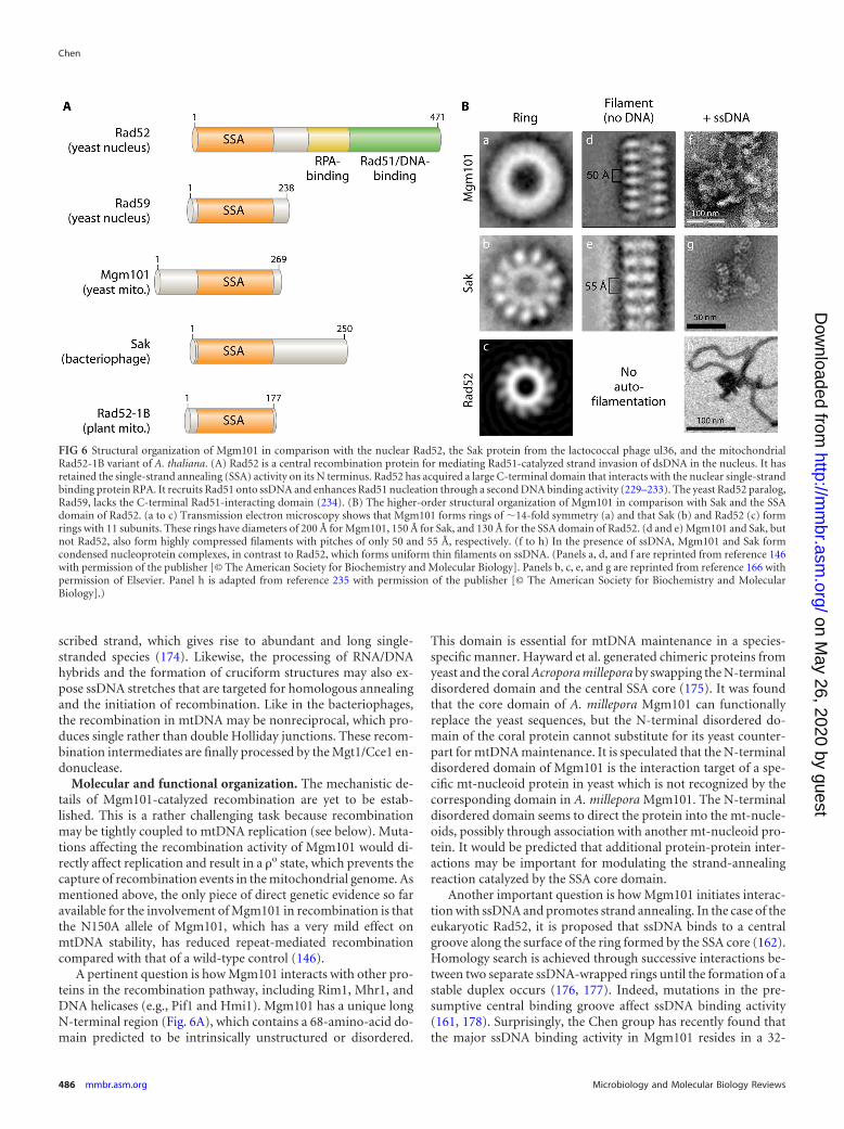
Evolutionary origin and functional implications. The Rad52-related proteins are widespread in the temperate bacteriophages(165, 169, 170), as represented by Red� and Erf from the bacte-riophages � and P22, RecT from the prophage rac, and Sak fromthe lactococcal phage ul36. These proteins are probably betterdescribed as single-strand annealing proteins (SSAPs) based ontheir unique biochemical activity (169). Unlike the eukaryoticRad52, which contains a large C-terminal domain with multipleactivities, including the recruitment of the Rad51 core recombi-nase, Mgm101 and its bacteriophage counterparts lack such a do-main (Fig. 6A). Accordingly, the SSAPs in bacteriophages catalyze
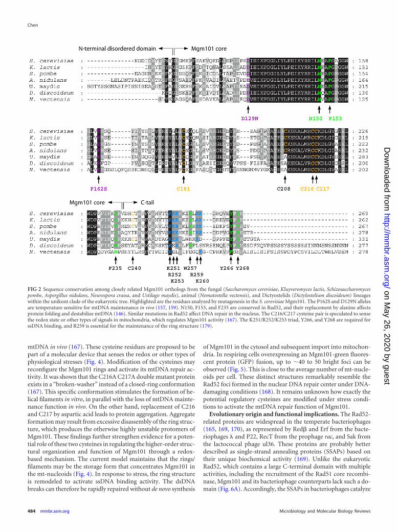
FIG 2 Sequence conservation among closely related Mgm101 orthologs from the fungal (Saccharomyces cerevisiae, Kluyveromyces lactis, Schizosaccharomycespombe, Aspergillus nidulans, Neurospora crassa, and Ustilago maydis), animal (Nematostella vectensis), and Dictyostelids (Dictylostelium discoideum) lineageswithin the unikont clade of the eukaryotic tree. Highlighted are the residues analyzed by mutagenesis in the S. cerevisiae Mgm101. The P162S and D129N allelesare temperature sensitive for mtDNA maintenance in vivo (157, 159). N150, F153, and F235 are conserved in Rad52, and their replacement by alanine affectsprotein folding and destabilize mtDNA (146). Similar mutations in Rad52 affect DNA repair in the nucleus. The C216/C217 cysteine pair is speculated to sensethe redox state or other types of signals in mitochondria, which regulates Mgm101 activity (167). The K251/R252/K253 triad, Y266, and Y268 are required forssDNA binding, and R259 is essential for the maintenance of the ring structure (179).
Chen
484 mmbr.asm.org Microbiology and Molecular Biology Reviews
recombination by the single-strand annealing mechanism inde-pendent of the bacterial RecA recombinase (171). The higher-order structure of Mgm101 is closer to that of the bacteriophageenzymes than to that of the eukaryotic Rad52, in that they allself-assemble into macromolecular filaments and form condensednucleoprotein complexes in the presence of ssDNA (Fig. 6B).Thus, Mgm101 may have evolved from an ancestral protein ofbacteriophage origin.
In light of the structural and biochemical similarities between
Mgm101 and SSAPs in the bacteriophages, it is proposed that themitochondrial recombination apparatus may operate in the sameway as its bacteriophage counterpart. The Red� protein in phage �initiates strand annealing by preferentially targeting on the laggingstrand of a replication fork (172, 173). By analogy, Mgm101 maywell function in the same manner (Fig. 7, middle panel). Instead ofinvading dsDNA duplexes like the ATP-dependent Rad51/RecAtype-recombinases, the Mgm101-ssDNA nucleoprotein com-plexes may be directly annealed to those homologous single-stranded donor sequences, thereby mediating error-free recombi-national repair of mtDNA. In addition to the lagging strand, othersingled-stranded sources may also serve as templates. For in-stance, mitochondrial transcription can displace the nontran-
FIG 4 A model for activation of Mgm101 for mtDNA repair. The Mgm101rings are proposed to be the storage form of the protein in mt-nucleoids. Inresponse to oxidative stress or other signals, the C216/C217 cysteine pair ismodified. This may induce structural remodeling in the rings, which stimu-lates ssDNA binding and the repair of dsDNA breaks.
FIG 5 Confocal microscopy showing that overexpressed Mgm101-GFP formsbright structures resembling the Rad52 foci in the nuclear DNA repair center.Mgm101-GFP was expressed from the constitutive ADH1 promoter. Scale bar,5 m. DIC, differential interference contrast.
Homologous Recombination in Mitochondrial DNA
September 2013 Volume 77 Number 3 mmbr.asm.org 485
scribed strand, which gives rise to abundant and long single-stranded species (174). Likewise, the processing of RNA/DNAhybrids and the formation of cruciform structures may also ex-pose ssDNA stretches that are targeted for homologous annealingand the initiation of recombination. Like in the bacteriophages,the recombination in mtDNA may be nonreciprocal, which pro-duces single rather than double Holliday junctions. These recom-bination intermediates are finally processed by the Mgt1/Cce1 en-donuclease.
Molecular and functional organization. The mechanistic de-tails of Mgm101-catalyzed recombination are yet to be estab-lished. This is a rather challenging task because recombinationmay be tightly coupled to mtDNA replication (see below). Muta-tions affecting the recombination activity of Mgm101 would di-rectly affect replication and result in a �o state, which prevents thecapture of recombination events in the mitochondrial genome. Asmentioned above, the only piece of direct genetic evidence so faravailable for the involvement of Mgm101 in recombination is thatthe N150A allele of Mgm101, which has a very mild effect onmtDNA stability, has reduced repeat-mediated recombinationcompared with that of a wild-type control (146).
A pertinent question is how Mgm101 interacts with other pro-teins in the recombination pathway, including Rim1, Mhr1, andDNA helicases (e.g., Pif1 and Hmi1). Mgm101 has a unique longN-terminal region (Fig. 6A), which contains a 68-amino-acid do-main predicted to be intrinsically unstructured or disordered.
This domain is essential for mtDNA maintenance in a species-specific manner. Hayward et al. generated chimeric proteins fromyeast and the coral Acropora millepora by swapping the N-terminaldisordered domain and the central SSA core (175). It was foundthat the core domain of A. millepora Mgm101 can functionallyreplace the yeast sequences, but the N-terminal disordered do-main of the coral protein cannot substitute for its yeast counter-part for mtDNA maintenance. It is speculated that the N-terminaldisordered domain of Mgm101 is the interaction target of a spe-cific mt-nucleoid protein in yeast which is not recognized by thecorresponding domain in A. millepora Mgm101. The N-terminaldisordered domain seems to direct the protein into the mt-nucle-oids, possibly through association with another mt-nucleoid pro-tein. It would be predicted that additional protein-protein inter-actions may be important for modulating the strand-annealingreaction catalyzed by the SSA core domain.
Another important question is how Mgm101 initiates interac-tion with ssDNA and promotes strand annealing. In the case of theeukaryotic Rad52, it is proposed that ssDNA binds to a centralgroove along the surface of the ring formed by the SSA core (162).Homology search is achieved through successive interactions be-tween two separate ssDNA-wrapped rings until the formation of astable duplex occurs (176, 177). Indeed, mutations in the pre-sumptive central binding groove affect ssDNA binding activity(161, 178). Surprisingly, the Chen group has recently found thatthe major ssDNA binding activity in Mgm101 resides in a 32-
amino-acid carboxyl-terminal tail instead of the central SSA core(179). The C-terminal tail is not conserved as well as the core SSAdomain among the closely related Mgm101 homologs (Fig. 2), butseveral conserved amino acids have been identified to play roles inssDNA binding. The data supported the model that the positivelycharged 251-KRK-253 triad in the C-terminal tail may initiate thecontact with the phosphate backbone of ssDNA by electrostaticinteractions. The highly conserved 266-YPY-268 motif at the ex-treme end of the protein may stabilize the interactions by basestacking. These interactions could disrupt the ring structuremaintained by salt bridges involving Arg259. This may facilitatethe deployment of additional subunits in the ring for interactionswith ssDNA. As the C-terminal tail is conserved in Mgm101 ho-mologs that function in mitochondria but not in the nuclearRad52 or bacteriophage SSAPs, it is possible that it may havearisen from convergent evolution as a successful adaptation tocatalyze recombination and DNA repair in the specific geneticsetting of mitochondria.
In addition to ssDNA binding, the C-terminal tail is also re-quired for the stabilization of the Mgm101 rings in vitro and forprotein stability in vivo (179). Mutant proteins unable to maintainthe ring structure in vitro are degraded in vivo. The degradation ofunassembled Mgm101 monomers may be necessary for the for-mation of ring-based mtDNA repair centers in the mt-nucleoids.
Mgm101 homologs. Several Mgm101 homologs have beenfunctionally characterized. Like in yeast, mitochondria in Arabi-dopsis thaliana also express a short version of a Rad52-relatedprotein similar to Mgm101, known as Rad52-1B (Fig. 6A) (180).Gualberto and coworkers independently identified Rad52-1B asODB1 (organellar DNA binding protein 1) based on its DNAbinding activity. Rad52-1B/ODB1 shares similar properties withMgm101, including its preferential binding to ssDNA and theability to promote single-strand annealing (181). More interest-ingly, these investigators also found that under genotoxic stress,the recombination in mtDNA repeats is significantly reduced inodb1 mutant plants, reminiscent of the phenotype observed with
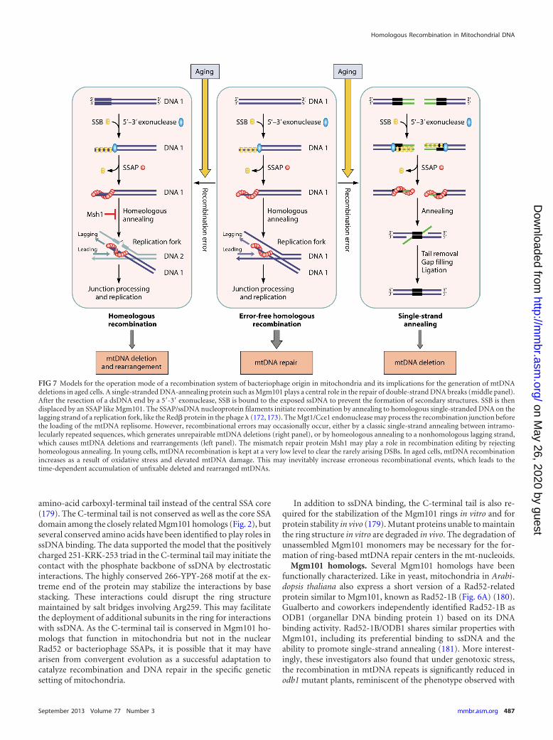
FIG 7 Models for the operation mode of a recombination system of bacteriophage origin in mitochondria and its implications for the generation of mtDNAdeletions in aged cells. A single-stranded DNA-annealing protein such as Mgm101 plays a central role in the repair of double-strand DNA breaks (middle panel).After the resection of a dsDNA end by a 5=-3= exonuclease, SSB is bound to the exposed ssDNA to prevent the formation of secondary structures. SSB is thendisplaced by an SSAP like Mgm101. The SSAP/ssDNA nucleoprotein filaments initiate recombination by annealing to homologous single-stranded DNA on thelagging strand of a replication fork, like the Red� protein in the phage � (172, 173). The Mgt1/Cce1 endonuclease may process the recombination junction beforethe loading of the mtDNA replisome. However, recombinational errors may occasionally occur, either by a classic single-strand annealing between intramo-lecularly repeated sequences, which generates unrepairable mtDNA deletions (right panel), or by homeologous annealing to a nonhomologous lagging strand,which causes mtDNA deletions and rearrangements (left panel). The mismatch repair protein Msh1 may play a role in recombination editing by rejectinghomeologous annealing. In young cells, mtDNA recombination is kept at a very low level to clear the rarely arising DSBs. In aged cells, mtDNA recombinationincreases as a result of oxidative stress and elevated mtDNA damage. This may inevitably increase erroneous recombinational events, which leads to thetime-dependent accumulation of unfixable deleted and rearranged mtDNAs.
Homologous Recombination in Mitochondrial DNA
September 2013 Volume 77 Number 3 mmbr.asm.org 487
the N150A allele of MGM101 (146). In contrast, microhomology-mediated recombination is increased in the absence of ODB1.This suggests a critical role of the protein in effective repair ofdouble-strand breaks. A defect in such a function directs the repairto the microhomology-mediated recombination mechanism.Plants lacking both ODB1 and RecA3 seem to be nonviable. Thiswould support the idea that ODB1 and RecA3 may operate inseparate but functionally redundant mtDNA repair pathways.Based on the molecular organization of Rad52-1B/ODB1, whichlacks an apparent RecA binding domain as in Rad52, it would bepredicted that it may function in a RecA-independent manner likeMgm101 and its bacteriophage counterparts.
The Sasaki group identified the Mgm101 homolog Glom2 inPhysarum polycephalum (182). Glom2 has a core domain highlyhomologous to that of Mgm101, but it also has a 218-amino-acidlong C-terminal domain containing three polyproline tracts. Theexact function of the long C-terminal domain is unknown. Itwould be interesting to know whether or not it interacts with aRecA-type protein, if such a conventional recombinase exists inPhysarum mitochondria. Downregulation of Glom2 does nothave an obvious effect on mtDNA maintenance. When combinedwith the loss of the Abf2 homolog Glom, the downregulation ofGlom2 further reduces the size of mt-nucleoids and mtDNA copynumber. A potential role of Glom2 in mtDNA recombination andrepair remains to be investigated.
OTHER FACTORS AFFECTING mtDNA RECOMBINATION
In addition to a potential difference in the basic machineries forHR, several additional factors have been proposed to account forthe wide variation of recombination frequencies in different celltypes and species. For instance, nucleoid organization, mitochon-drial dynamics, mtDNA content, and mitochondrial density mayall affect recombination. In cultured human cells, the nucleoidalorganization is highly autonomous (183, 184). Schon and co-workers fused two cell lines which were homoplasmic for dele-tions in two nonoverlapping regions affecting protein synthesis. Itwas found that the two mtDNAs can transcomplement defects inprotein synthesis but remain in independent nucleoids withoutstable intermixing. Human mitochondria in most cell lines arerather fragmented, and each nucleoid contains three or fewer cop-ies of the 16.5-kb mtDNA (33–35). The high autonomy of nucle-oids and low mtDNA content may set physical barriers for inter-and intranucleoidal recombinations, respectively. In contrast, themtDNA copy number in the myocardium is �1.9- to 3.8- and�10-fold higher than those in skeletal muscle and epithelial cells,respectively (185–187). Data on the exact copy number of mtDNAin each nucleoid and mitochondrion in these tissues are currentlyunavailable. It would be predicted that the high mtDNA content,together with a highly oxidative environment, may contribute tothe high levels of recombination in human heart mtDNA (seeabove). Likewise, the relatively high frequency of recombinationin yeast mtDNA may also be partially explained by differences ingenome size, nucleoid organization, and mitochondrial dynam-ics. The yeast mitochondrial genome is 4.5 times larger than hu-man mtDNA. Under normal growth conditions on glucose me-dium, which was used for most recombination assays, yeast cellsdevelop an elongated mitochondrial reticulum that contains mul-tiple giant nucleoids, with each harboring up to 20 copies ofmtDNA (188). These conditions may facilitate both intranucleoi-dal recombination and internucleoidal exchanges.
DSBs are the most detrimental damage to any genome. It is nowincreasingly appreciated that HR may have evolved as a molecularstrategy for the repair of DSBs. For a long time, the importance ofrecombination between homologous mtDNA molecules in themostly homoplasmic cells has been undervalued. As DSBs arise asrare events in most cell types under physiological conditions,mtDNA recombination should be expected to be low. It is nowargued that a low level of recombination may be sufficient formutational clearance in mtDNA. Evolutionarily, maintaining ho-mologous recombination at a low level is necessary to protectmtDNA against invasion of selfish elements (189, 190). A directlink between DNA recombination and repair has been supportedby the observations that the recombination proteins Mgm101 andMhr1 are both required for the maintenance of the mitochondrialgenome when yeast cells are exposed to DNA-damaging condi-tions (146, 159, 191). Consistent with this idea, mtDNA-carriedgenes from mammals accumulate more mutations than those inyeast (192). A low recombination activity of mammalian mito-chondria may account for the lower rate of mutation correctionthan in yeast, in which recombination is high.
The implications of homologous recombination could be be-yond mtDNA repair. Early studies showed that immediately fol-lowing the inactivation of MGM101, the mtDNA copy number ishalved by every cell division in yeast (107). This severe phenotypeled to the assumption that Mgm101 might be involved in mtDNAreplication, possibly by generating recombination intermediateswith free 3= ends that are used for priming replication. This modeof replication initiation may be followed by a rolling-circle mech-anism initially proposed by the Clark-Walker group (193). In fact,recombination-based replication has been proposed as an alter-native mechanism for mtDNA synthesis in several organisms (55,194, 195). The recombination-based mtDNA replication modelhas recently gained strong support from studies suggesting thatthe replicative forks in Candida albicans mtDNA are generatedfrom recombination intermediates (196). The presence of recom-bination-based replication in human heart and brain (87) and inplant mitochondria (197–199) has also been proposed. The phys-iological impact of this specific mechanism vis-à-vis the conven-tional RNA-primed replication mode is yet to be substantiated.
Ling and Shibata have provided some key evidence for theinvolvement of the Mhr1 protein in the initiation of the rolling-circle replication of yeast mtDNA (145). Rolling-circle replicationgenerates mtDNA concatemers. Mutation in MHR1 reduces con-catemer formation, which subsequently affects the partitioning ofnewly synthesized mtDNA into the buds and delays the establish-ment of mtDNA homoplasmy in the daughter cells (200, 201).Mhr1 is also required for the replicative advantage of the ori5hypersuppressive �� genome, which has apparently frequent dou-ble-strand breaks (202), consistent with its role in recombina-tional repair of mtDNA. It was initially proposed that Mhr1 mayhave a Rad52-type activity like Red� in the phage �. Red� isknown to initiate rolling-circle replication by annealing the Red�-ssDNA filaments preferentially on the lagging strand of a replica-tion fork, followed by template switch and DNA synthesis (172,173, 203). The recent finding of Mgm101 as a Rad52-type proteinraises the possibility that Mgm101 may fulfill such a role, in amanner dependent on or independent of Mhr1 (146, 167). �he
Chen
488 mmbr.asm.org Microbiology and Molecular Biology Reviews
Nunnari laboratory has shown that Mgm101 is preferentially as-sociated with actively replicating mtDNA nucleoids in vivo, andthat Mgm101 coimmunoprecipitates with the mitochondrialDNA polymerase Mip1 (204). Thus, Mgm101 may directly recruitthe mtDNA replisome following the strand-annealing reaction.Future studies are required to support a role of Mgm101 in initi-ating the strand-annealing reaction prior to the rolling-circle rep-lication. It is important to note that mhr1 and mgm101 mutantsshare similar phenotypes: both genes are essential for the stabilityof �� but not the highly recombinogenic �� genomes (107, 144).The loss of Mhr1 has a less severe effect on the maintenance of theN1 �� genome than the disruption of MGM101 (107). These twoproteins likely function in the same pathway in initiating recom-bination-based mtDNA replication. Mhr1 does not stimulate thesingle-strand annealing activity of Mgm101, and Mgm101 has lit-tle effect on the D-loop formation activity of Mhr1 in vitro (ourunpublished observation). How these two proteins cooperate ininitiating recombination-based mtDNA repair and replication is apressing issue in the field.
HOMOLOGOUS VERSUS HOMEOLOGOUS RECOMBINATION:IMPLICATIONS FOR mtDNA DELETION/REARRANGEMENT
No biological system is perfect. A controlled recombination activ-ity benefits mtDNA repair, but hyperrecombination could be det-rimental for mitochondrial genome stability. Extensive mtDNAdeletions in dopaminergic neurons from aged individuals andthose suffering from Parkinson’s disease have been reported (26,27). Krishnan et al. proposed that these deletions arise by a mech-anism resembling the classic SSA (30) (Fig. 7, right panel). Aftersymmetrical resection of dsDNA ends by a 5=-3= exonuclease, re-peated sequences exposed on the other strands misanneal. Theremoval of the unpaired tails by exonucleases followed by gapfilling and ligation generates intramolecular deletions in mtDNA.The Rad52-related SSAPs are well known for their activity in pro-moting single-strand annealing (205–207), which generates dele-tions of genomic sequences between directly repeated regions.
In light of its functional similarity to SSAPs, Mgm101 may beinvolved in single-strand annealing and mtDNA deletion. How-ever, this operation mode would require an extensive resection ofdsDNA ends in order to expose the homologous sequences. Thiscould become a limiting factor when the homologous sequencesare separated by large intervening regions. Alternatively, it is pos-sible that homeologous recombination, which occurs betweenmismatched sequences, may play a major role in generatingmtDNA deletions/rearrangements (Fig. 7, left panel). It has beenwell recognized that the SSAPs catalyze recombination betweenrelated but diverged DNA sequences. For example, a sequencedivergence of 4% between the target genes has little effect on therecombination catalyzed by the Red� protein in phage � (208).This is in sharp contrast to recombination in the E. coli chromo-some catalyzed by RecA, in which a 4% divergence reduces recom-bination by 270-fold (209). Red� can tolerate a sequence diver-gence in the recombination target genes of as high as 22%,although the recombination efficiency is reduced by �100-fold.Interestingly, detailed analysis of human mtDNA suggested thatdeletions most likely happen between imperfectly matched DNAsequences that are capable of forming long and stable duplexes(210). Homeologous recombination is the simplest way to pro-mote these events.
To what extent can Mgm101 tolerate sequence divergence in
the recombination targets and how homeologous recombinationis controlled remain to be determined. The annealing of mis-matched single-stranded DNA by the bacteriophage Red� is sup-pressed by the host mismatch repair system (211). It may be ex-pected that its counterpart in mitochondria, Msh1, could also playa role in the suppression of homeologous recombination. Thismay explain the dramatic hyperrecombinogenic phenotype asso-ciated with mutations in MSH1 and the rapid disintegration ofmtDNA in an msh1 null mutant (130). In plant mitochondria, lossof Msh1 function leads to DNA exchange between sequences shar-ing only 85% identity (212). The barrier for preventing homeolo-gous recombination in mtDNA seems to be naturally low in someanimal species. In sea mussels, recombination between maternaland paternal mtDNA regions in the obligatorily heteroplasmicmales that are diverged by 20% can be readily detected (213).
mtDNA RECOMBINATION IN AGING
Recombination errors, introduced by either homeologous recom-bination or SSA, could have important implications for mtDNAintegrity in aging cells. Aging seems to increase mtDNA recombi-nation, concomitant with increased mtDNA deletions/rearrange-ments. This has been shown by experiments carried out in Po-dospora anserina (214). Mitochondria in this filamentousascomycete are inherited in a strictly uniparental manner (215,216). However, heteroplasmons may be formed in the contactzone of heterokaryon incompatible strains, in which leakage ofpaternal mitochondria into the maternal mycelium occurs aftertransient hyphal fusion. This property provided a unique oppor-tunity for examining the frequency of mtDNA recombination inthe heteroplasmons derived from cells at different time points intheir life spans. In reciprocal confrontational crosses betweenstrains harboring different mtDNA markers, van Diepeningen etal. demonstrated that the levels of recombinant mtDNA remainlow in crosses involving young maternal mycelium. Juvenilestrains have little to no recombination in mtDNA. The recombi-nation rate only increases marginally over the first 3/4 of the lifespan. Remarkably, in crosses generated from the maternal myce-lium in the last 1/4 of its life span, the percentage of offspring withrecombined mtDNA increases to nearly 50% (214). These obser-vations strongly suggest that mtDNA becomes hyperrecombino-genic in aged cells.
The senescence of Podospora is associated with the accumula-tion of various classes of mtDNA derivatives which can be bothcircularly and tandemly arranged, with or without coding func-tion. Some of these mtDNA species are thought to contribute tocellular senescence (217). These “senescent” mtDNA moleculesresult from intramolecular recombination between short directrepeats (218). This raises the possibility that the hyperrecombino-genic state in aged cells may become a significant source ofmtDNA instability. In young cells, because both DNA damage andrecombination activity are low, mtDNA deletions also remainlow. In contrast, the high mtDNA recombination activity in agedcells may result in the increased incidence of recombinational er-rors, which overwhelms the cell’s capacity to clear damaged mito-chondria by processes such as mitophagy. The mechanism ofmtDNA recombination in Podospora and exactly how this activityis elevated in aged cells are unknown. It is possible that the recom-binase is directly activated by reactive oxygen species. A closelyrelated Mgm101 homolog is present in Podospora anserine (ourunpublished observation), and its role in mtDNA recombination
Homologous Recombination in Mitochondrial DNA
September 2013 Volume 77 Number 3 mmbr.asm.org 489
is yet to be established. Genetic runaway may occur. Mitochon-drial dysfunction in aged cells increases the production of reactiveoxygen species (ROS), which activates the Mgm101-based recom-bination mechanism. Hyperactive recombination increases theoverall recombination errors and mtDNA deletions, which fur-ther damages mitochondria and increases ROS production(Fig. 8).
Age-dependent increases in mtDNA recombination in otherorganisms have yet to be explored. Replicatively aged yeast cellshave destabilized mtDNA (219, 220); however, it is unknownwhether or not mtDNA disintegration is caused by hyperrecom-bination. Interestingly, four-way junctions in human heartmtDNA are not detected in newborns. They are acquired duringearly childhood concomitant with increased mtDNA copy num-ber (221). This has been interpreted as a cellular adaptive processthat responds to increased energy demand, ROS production, anddouble-strand DNA breaks (222). In humans, a common deletionin mtDNA is the 4,977-bp deletion which occurs between two13-bp direct repeats. This deletion likely results from a recombi-nation-excision mechanism and is increased in an age-dependentmanner, with a correlation coefficient of 0.82 in specific tissuessuch as the anterior wall of the left ventricle in the heart (23).Extensive large-scale mtDNA deletions have been reported inneuronal cells during aging (26). It remains unknown whetherthis results from hyperrecombination.
CONCLUSION AND PERSPECTIVES
Accumulating evidence indicates that mtDNA recombination oc-curs not only in the traditionally studied yeast and lower animalsthat have biparental inheritance but also in fungal and animalspecies with uniparental inheritance and even in specific humantissues, including the heart and brain. Although controversy per-sists, increasing evidence suggests that mtDNA recombination ismore widespread than was previously thought. In animals withhomoplasmic mtDNA, recombinant products are indistinguish-able from their progenitor molecules. Unequivocally capturingthe actual mtDNA recombination events in most cell types is stilla challenge in the field. Next-generation sequencing technology isalready showing its power in detecting rare alleles and subge-nomes of mtDNA (212, 223, 224). These technologies would be
expected to detect possible recombinations between the newlyarising rare mutations. This may also help to answer the questionregarding the universality of mtDNA recombination across differ-ent biota (225).
Despite the fact that mtDNA recombination has been reportedfor over 4 decades, the study of the underlying molecular machin-ery has just made its debut. The field of mtDNA recombination isgaining strong momentum following the recent discovery of abacteriophage-type SSAP in mitochondria as defined by Mgm101.Understanding the functional relationship between Mgm101 andother components in the mtDNA recombination pathway, partic-ularly Mhr1, will be critical for advancing this field. In the comingyears, a critical question to be addressed is whether human mito-chondria express a similar mtDNA recombination system, insteadof using a canonical recombination pathway involving a RecA/Rad51-type recombinase. Human mitochondria also harbor SSAactivity conferred by proteins ostensibly unrelated to SSAPs (e.g.,the Twinkle helicase [226]). Close orthologs of Mgm101 havebeen identified in lower animals (e.g., Nematostella vectensis) us-ing current bioinformatic tools. However, the presence of a dis-tantly related SSAP in higher animals cannot be completely ruledout. The SSAPs are often conserved at the structural level but notin their primary sequences. For instance, Mgm101 shares struc-tural and functional similarities with the SSAPs in bacteriophagessuch as Red�, Erf, and Sak, but sequence similarity between theseproteins is hardly recognizable. Nonetheless, mitochondria andbacteriophages seem to share more molecular machineries thanpreviously thought. Both the mitochondrial DNA and RNA poly-merases are phylogenetically originated from the T-odd lineage ofbacteriophages instead of alphaproteobacteria, the ancestor of mi-tochondria (227). The experimental data now also show that mi-tochondria and bacteriophages share similar molecular mecha-nisms for mtDNA recombination and repair. It is possible thatthese enzymes were acquired together from an infecting phage atthe onset of endosymbiosis. Future studies should be focused onunderstanding how the processes of recombination, repair, andreplication are interlinked and coordinated in mitochondria,which is a salient feature of DNA metabolism in bacteriophages(228).
ACKNOWLEDGMENTS
I thank George Desmond Clark-Walker (Australian National University)and the Chen lab members for comments and critical reading of the man-uscript, the anonymous reviewers for their valuable suggestions, StephanWilkens for help in constructing the molecular models of Mgm101, andXiaoming Zuo for generating the Mgm101-GFP construct.
I am supported by grant AG023731 from the National Institute onAging, National Institutes of Health.
REFERENCES1. Wallace DC. 2007. Why do we still have a maternally inherited mito-
chondrial DNA? Insights from evolutionary medicine. Annu. Rev.Biochem. 76:781– 821.
2. Chen XJ, Butow RA. 2005. The organization and inheritance of themitochondrial genome. Nat. Rev. Genet. 6:815– 825.
3. Holt IJ, He J, Mao CC, Boyd-Kirkup JD, Martinsson P, Sembongi H,Reyes A, Spelbrink JN. 2007. Mammalian mitochondrial nucleoids:organizing an independently minded genome. Mitochondrion 7:311–321.
drial DNA in nucleoids: history, recent developments, and future chal-lenges. IUBMB Life 62:19 –32.
6. Nass MM. 1969. Mitochondrial DNA. I. Intramitochondrial distribu-tion and structural relations of single- and double-length circular DNA.J. Mol. Biol. 42:521–528.
7. Albring M, Griffith J, Attardi G. 1977. Association of a proteinstructure of probable membrane derivation with HeLa cell mitochon-drial DNA near its origin of replicaton. Proc. Natl. Acad. Sci. U. S. A.74:1348 –1352.
8. Prachar J. 2010. Mouse and human mitochondrial nucleoid— detailedstructure in relation to function. Gen. Physiol. Biophys. 29:160 –174.
9. Kopek BG, Shtengel G, Xu CS, Clayton DA, Hess HF. 2012. Correla-tive 3D superresolution fluorescence and electron microscopy reveal therelationship of mitochondrial nucleoids to membranes. Proc. Natl.Acad. Sci. U. S. A. 109:6136 – 6141.
10. Brown WM, George M, Jr., Wilson AC. 1979. Rapid evolution ofanimal mitochondrial DNA. Proc. Natl. Acad. Sci. U. S. A. 76:1967–1971.
11. Howell N. 1996. Mutational analysis of the human mitochondrial ge-nome branches into the realm of bacterial genetics. Am. J. Hum. Genet.59:749 –755.
12. Larsson NG. 2010. Somatic mitochondrial DNA mutations in mamma-lian aging. Annu. Rev. Biochem. 79:683–706.
13. Harman D. 1972. The biological clock: the mitochondria? J. Am. Geriatr.Soc. 20:145–147.
14. Linnane AW, Marzuki S, Ozawa T, Tanaka M. 1989. MitochondrialDNA mutations as an important contributor to ageing and degenerativediseases. Lancet i:642– 645.
15. Kujoth GC, Hiona A, Pugh TD, Someya S, Panzer K, Wohlgemuth SE,Hofer T, Seo AY, Sullivan R, Jobling WA, Morrow JD, Van RemmenH, Sedivy JM, Yamasoba T, Tanokura M, Weindruch R, Leeuwen-burgh C, Prolla TA. 2005. Mitochondrial DNA mutations, oxidativestress, and apoptosis in mammalian aging. Science 309:481– 484.
16. Trifunovic A, Wredenberg A, Falkenberg M, Spelbrink JN, Rovio AT,Bruder CE, Bohlooly YM, Gidlof S, Oldfors A, Wibom R, Tornell J,Jacobs HT, Larsson NG. 2004. Premature ageing in mice expressingdefective mitochondrial DNA polymerase. Nature 429:417– 423.
17. Weissman L, de Souza-Pinto NC, Stevnsner T, Bohr VA. 2007. DNArepair, mitochondria, and neurodegeneration. Neuroscience 145:1318 –1329.
18. Wilson DM, III, Bohr VA. 2007. The mechanics of base excision repair,and its relationship to aging and disease. DNA Repair (Amst.) 6:544 –559.
19. Zheng L, Zhou M, Guo Z, Lu H, Qian L, Dai H, Qiu J, YakubovskayaE, Bogenhagen DF, Demple B, Shen B. 2008. Human DNA2 is amitochondrial nuclease/helicase for efficient processing of DNA replica-tion and repair intermediates. Mol. Cell 32:325–336.
20. Kazak L, Reyes A, Holt IJ. 2012. Minimizing the damage: repair path-ways keep mitochondrial DNA intact. Nat. Rev. Mol. Cell Biol. 13:659 –671.
21. Sykora P, Wilson DM, III, Bohr VA. 2012. Repair of persistent strandbreaks in the mitochondrial genome. Mech. Ageing Dev. 133:169 –175.
22. de Souza-Pinto NC, Mason PA, Hashiguchi K, Weissman L, Tian J,Guay D, Lebel M, Stevnsner TV, Rasmussen LJ, Bohr VA. 2009. NovelDNA mismatch-repair activity involving YB-1 in human mitochondria.DNA Repair (Amst.) 8:704 –719.
23. Meissner C, Bruse P, Mohamed SA, Schulz A, Warnk H, Storm T,Oehmichen M. 2008. The 4977 bp deletion of mitochondrial DNA inhuman skeletal muscle, heart and different areas of the brain: a usefulbiomarker or more? Exp. Gerontol. 43:645– 652.
24. Cortopassi GA, Arnheim N. 1990. Detection of a specific mitochondrialDNA deletion in tissues of older humans. Nucleic Acids Res. 18:6927–6933.
25. Bua E, Johnson J, Herbst A, Delong B, McKenzie D, Salamat S, AikenJM. 2006. Mitochondrial DNA-deletion mutations accumulate intracel-lularly to detrimental levels in aged human skeletal muscle fibers. Am. J.Hum. Genet. 79:469 – 480.
26. Kraytsberg Y, Kudryavtseva E, McKee AC, Geula C, Kowall NW,Khrapko K. 2006. Mitochondrial DNA deletions are abundant and causefunctional impairment in aged human substantia nigra neurons. Nat.Genet. 38:518 –520.
27. Bender A, Krishnan KJ, Morris CM, Taylor GA, Reeve AK, PerryRH, Jaros E, Hersheson JS, Betts J, Klopstock T, Taylor RW,Turnbull DM. 2006. High levels of mitochondrial DNA deletions in
substantia nigra neurons in aging and Parkinson disease. Nat. Genet.38:515–517.
28. Bender A, Schwarzkopf RM, McMillan A, Krishnan KJ, Rieder G,Neumann M, Elstner M, Turnbull DM, Klopstock T. 2008. Dopami-nergic midbrain neurons are the prime target for mitochondrial DNAdeletions. J. Neurol. 255:1231–1235.
29. Shoffner JM, Lott MT, Voljavec AS, Soueidan SA, Costigan DA,Wallace DC. 1989. Spontaneous Kearns-Sayre/chronic external oph-thalmoplegia plus syndrome associated with a mitochondrial DNA de-letion: a slip-replication model and metabolic therapy. Proc. Natl. Acad.Sci. U. S. A. 86:7952–7956.
30. Krishnan KJ, Reeve AK, Samuels DC, Chinnery PF, Blackwood JK,Taylor RW, Wanrooij S, Spelbrink JN, Lightowlers RN, Turnbull DM.2008. What causes mitochondrial DNA deletions in human cells? Nat.Genet. 40:275–279.
31. San Filippo J, Sung P, Klein H. 2008. Mechanism of eukaryotic homol-ogous recombination. Annu. Rev. Biochem. 77:229 –257.
32. Gangavarapu V, Prakash S, Prakash L. 2007. Requirement of RAD52group genes for postreplication repair of UV-damaged DNA in Saccha-romyces cerevisiae. Mol. Cell. Biol. 27:7758 –7764.
33. Iborra FJ, Kimura H, Cook PR. 2004. The functional organization ofmitochondrial genomes in human cells. BMC Biol. 2:9.
34. Brown TA, Tkachuk AN, Shtengel G, Kopek BG, Bogenhagen DF,Hess HF, Clayton DA. 2011. Superresolution fluorescence imaging ofmitochondrial nucleoids reveals their spatial range, limits, and mem-brane interaction. Mol. Cell. Biol. 31:4994 –5010.
35. Kukat C, Wurm CA, Spahr H, Falkenberg M, Larsson NG, Jakobs S.2011. Super-resolution microscopy reveals that mammalian mitochon-drial nucleoids have a uniform size and frequently contain a single copyof mtDNA. Proc. Natl. Acad. Sci. U. S. A. 108:13534 –13539.
36. Birky CW, Jr. 1978. Transmission genetics of mitochondria and chlo-roplasts. Annu. Rev. Genet. 12:471–512.
37. Dujon B, Slonimski PP, Weill L. 1974. Mitochondrial genetics IX: amodel for recombination and segregation of mitochondrial genomes inSaccharomyces cerevisiae. Genetics 78:415– 437.
38. Thomas DY, Wilkie D. 1968. Recombination of itochondrial drug re-sistance factors in Saccharomyces cerevisiae. Biochem. Biophys. Res.Commun. 30:368 –372.
39. Rank GH, Bech-Hansen NT. 1972. Somatic segretation, recombination,asymmetrical distribution and complementation tests of cytoplasmical-ly-inherited antibiotic-resistance mitochondrial markers in S. cerevisiae.Genetics 72:1–15.
40. Michaelis G, Petrochilo E, Slonimski PP. 1973. Mitochondrial genetics.3. Recombined molecules of mitochondrial DNA obtained from crossesbetween cytoplasmic petite mutants of Saccharomyces cerevisiae: physicaland genetic characterization. Mol. Gen. Genet. 123:51– 65.
41. Gaillard C, Strauss F, Bernardi G. 1980. Excision sequences in themitochondrial genome of yeast. Nature 283:218 –220.
42. de Zamaroczy M, Faugeron-Fonty G, Bernardi G. 1983. Excisionsequences in the mitochondrial genome of yeast. Gene 21:193–202.
43. Clark-Walker GD. 1989. In vivo rearrangement of mitochondrialDNA in Saccharomyces cerevisiae. Proc. Natl. Acad. Sci. U. S. A. 86:8847– 8851.
44. Gross SR, Levine PH, Metzger S, Glaser G. 1989. Recombination andreplication of plasmid-like deriviatives of a short section of the mito-chondrial genome of Neurospora crassa. Genetics 121:693–701.
45. Chen XJ, Clark-Walker GD. 1993. Mutations in MGI genes convertKluyveromyces lactis into a petite-positive yeast. Genetics 133:517–525.
46. Chen XJ, Clark-Walker GD. 1995. Specific mutations in - and �-sub-units of F1-ATPase affect mitochondrial genome integrity in the petite-negative yeast Kluyveromyces lactis. EMBO J. 14:3277–3286.
47. Chen XJ, Clark-Walker GD. 1996. The mitochondrial genome integritygene, MGI1, of Kluyveromyces lactis encodes the beta-subunit of F1-ATPase. Genetics 144:1445–1454.
48. Chen XJ, Clark-Walker GD. 2000. The petite mutation in yeasts: 50years on. Int. Rev. Cytol. 194:197–238.
49. Johnston SA, Anziano PQ, Shark K, Sanford JC, Butow RA. 1988.Mitochondrial transformation in yeast by bombardment with micropro-jectiles. Science 240:1538 –1541.
50. Bonnefoy N, Fox TD. 2007. Directed alteration of Saccharomyces cerevi-siae mitochondrial DNA by biolistic transformation and homologousrecombination. Methods Mol. Biol. 372:153–166.
Homologous Recombination in Mitochondrial DNA
September 2013 Volume 77 Number 3 mmbr.asm.org 491
51. Lockshon D, Zweifel SG, Freeman-Cook LL, Lorimer HE, Brewer BJ,Fangman WL. 1995. A role for recombination junctions in the segrega-tion of mitochondrial DNA in yeast. Cell 81:947–955.
52. Zweifel SG, Fangman WL. 1991. A nuclear mutation reversing a biasedtransmission of yeast mitochondrial DNA. Genetics 128:241–249.
53. Kleff S, Kemper B, Sternglanz R. 1992. Identification and characteriza-tion of yeast mutants and the gene for a cruciform cutting endonuclease.EMBO J. 11:699 –704.
54. Ezekiel UR, Zassenhaus HP. 1993. Localization of a cruciform cuttingendonuclease to yeast mitochondria. Mol. Gen. Genet. 240:414 – 418.
55. MacAlpine DM, Perlman PS, Butow RA. 1998. The high mobility groupprotein Abf2p influences the level of yeast mitochondrial DNA recom-bination intermediates in vivo. Proc. Natl. Acad. Sci. U. S. A. 95:6739 –6743.
56. Joers P, Jacobs HT. 2013. Analysis of replication intermediates indicatesthat Drosophila melanogaster mitochondrial DNA replicates by a strand-coupled theta mechanism. PLoS One 8:e53249. doi:10.1371/journal.pone.0053249.
57. Ladoukakis ED, Zouros E. 2001. Direct evidence for homologous re-combination in mussel (Mytilus galloprovincialis) mitochondrial DNA.Mol. Biol. Evol. 18:1168 –1175.
58. Burzynski A, Zbawicka M, Skibinski DO, Wenne R. 2003. Evidence forrecombination of mtDNA in the marine mussel Mytilus trossulus fromthe Baltic. Mol. Biol. Evol. 20:388 –392.
59. Hagelberg E. 2003. Recombination or mutation rate heterogeneity? Im-plications for Mitochondrial Eve. Trends Genet. 19:84 –90.
60. White DJ, Wolff JN, Pierson M, Gemmell NJ. 2008. Revealing thehidden complexities of mtDNA inheritance. Mol. Ecol. 17:4925–4942.
61. Berlin S, Smith NG, Ellegren H. 2004. Do avian mitochondria recom-bine? J. Mol. Evol. 58:163–167.
62. Ballard JW, Whitlock MC. 2004. The incomplete natural history ofmitochondria. Mol. Ecol. 13:729 –744.
63. Elson JL, Andrews RM, Chinnery PF, Lightowlers RN, Turnbull DM,Howell N. 2001. Analysis of European mtDNAs for recombination. Am.J. Hum. Genet. 68:145–153.
64. Innan H, Nordborg M. 2002. Recombination or mutational hot spots inhuman mtDNA? Mol. Biol. Evol. 19:1122–1127.
65. Piganeau G, Eyre-Walker A. 2004. A reanalysis of the indirect evidencefor recombination in human mitochondrial DNA. Heredity (Edinb.)92:282–288.
66. Piganeau G, Gardner M, Eyre-Walker A. 2004. A broad survey ofrecombination in animal mitochondria. Mol. Biol. Evol. 21:2319 –2325.
67. Ladoukakis ED, Eyre-Walker A. 2004. Evolutionary genetics: directevidence of recombination in human mitochondrial DNA. Heredity 93:321.
68. Tsaousis AD, Martin DP, Ladoukakis ED, Posada D, Zouros E. 2005.Widespread recombination in published animal mtDNA sequences.Mol. Biol. Evol. 22:925–933.
69. Eyre-Walker A, Awadalla P. 2001. Does human mtDNA recombine? J.Mol. Evol. 53:430 – 435.
70. Ciborowski KL, Consuegra S, Garcia de Leaniz C, Beaumont MA,Wang J, Jordan WC. 2007. Rare and fleeting: an example of inter-specific recombination in animal mitochondrial DNA. Biol. Lett.3:554 –557.
71. Ujvari B, Dowton M, Madsen T. 2007. Mitochondrial DNA recombi-nation in a free-ranging Australian lizard. Biol. Lett. 3:189 –192.
72. Hoolahan AH, Blok VC, Gibson T, Dowton M. 2012. Evidence ofanimal mtDNA recombination between divergent populations of thepotato cyst nematode Globodera pallida. Genetica 140:19 –29.
73. Nunes MD, Dolezal M, Schlotterer C. 2013. Extensive paternal mtDNAleakage in natural populations of Drosophila melanogaster. Mol. Ecol.22:2106 –2117.
74. Schwartz M, Vissing J. 2002. Paternal inheritance of mitochondrialDNA. N. Engl. J. Med. 347:576 –580.
75. Kraytsberg Y, Schwartz M, Brown TA, Ebralidse K, Kunz WS, ClaytonDA, Vissing J, Khrapko K. 2004. Recombination of human mitochon-drial DNA. Science 304:981.
76. Zsurka G, Kraytsberg Y, Kudina T, Kornblum C, Elger CE, KhrapkoK, Kunz WS. 2005. Recombination of mitochondrial DNA in skeletalmuscle of individuals with multiple mitochondrial DNA heteroplasmy.Nat. Genet. 37:873– 877.
77. Mita S, Rizzuto R, Moraes CT, Shanske S, Arnaudo E, Fabrizi GM,Koga Y, DiMauro S, Schon EA. 1990. Recombination via flanking directrepeats is a major cause of large-scale deletions of human mitochondrialDNA. Nucleic Acids Res. 18:561–567.
78. Firkin FC, Clark-Walker GD. 1979. Abnormal mitochondrial DNA inacute leukaemia and lymphoma. Br. J. Haematol. 43:201–206.
79. Clayton DA, Vinograd J. 1967. Circular dimer and catenate forms ofmitochondrial DNA in human leukaemic leucocytes. Nature 216:652–657.
80. Clayton DA, Vinograd J. 1969. Complex mitochondrial DNA in leuke-mic and normal human myeloid cells. Proc. Natl. Acad. Sci. U. S. A.62:1077–1084.
81. Tang Y, Manfredi G, Hirano M, Schon EA. 2000. Maintenance ofhuman rearranged mitochondrial DNAs in long-term cultured transmi-tochondrial cell lines. Mol. Biol. Cell 11:2349 –2358.
82. Holt IJ, Dunbar DR, Jacobs HT. 1997. Behaviour of a population ofpartially duplicated mitochondrial DNA molecules in cell culture: segre-gation, maintenance and recombination dependent upon nuclear back-ground. Hum. Mol. Genet. 6:1251–1260.
83. D’Aurelio M, Gajewski CD, Lin MT, Mauck WM, Shao LZ, Lenaz G,Moraes CT, Manfredi G. 2004. Heterologous mitochondrial DNA re-combination in human cells. Hum. Mol. Genet. 13:3171–3179.
84. Bacman SR, Williams SL, Moraes CT. 2009. Intra- and inter-molecularrecombination of mitochondrial DNA after in vivo induction of multipledouble-strand breaks. Nucleic Acids Res. 37:4218 – 4226.
85. Fan W, Lin CS, Potluri P, Procaccio V, Wallace DC. 2012. mtDNAlineage analysis of mouse L-cell lines reveals the accumulation of multi-ple mtDNA mutants and intermolecular recombination. Genes Dev. 26:384 –394.
86. Kajander OA, Karhunen PJ, Holt IJ, Jacobs HT. 2001. Prominentmitochondrial DNA recombination intermediates in human heart mus-cle. EMBO Rep. 2:1007–1012.
87. Pohjoismaki JL, Goffart S, Tyynismaa H, Willcox S, Ide T, Kang D,Suomalainen A, Karhunen PJ, Griffith JD, Holt IJ, Jacobs HT. 2009.Human heart mitochondrial DNA is organized in complex catenatednetworks containing abundant four-way junctions and replication forks.J. Biol. Chem. 284:21446 –21457.
88. Sena EP, Revet B, Moustacchi E. 1986. In vivo homologous recombi-nation intermediates of yeast mitochondrial DNA analyzed by electronmicroscopy. Mol. Gen. Genet. 202:421– 428.
89. Williamson D. 2002. The curious history of yeast mitochondrial DNA.Nat. Rev. Genet. 3:475– 481.
90. Bendich AJ. 1996. Structural analysis of mitochondrial DNA moleculesfrom fungi and plants using moving pictures and pulsed-field gel elec-trophoresis. J. Mol. Biol. 255:564 –588.
91. Kowalczykowski SC, Dixon DA, Eggleston AK, Lauder SD, RehrauerWM. 1994. Biochemistry of homologous recombination in Escherichiacoli. Microbiol. Rev. 58:401– 465.
92. Symington LS. 2002. Role of RAD52 epistasis group genes in homolo-gous recombination and double-strand break repair. Microbiol. Mol.Biol. Rev. 66:630 – 670.
93. Van Dyck E, Foury F, Stillman B, Brill SJ. 1992. A single-stranded DNAbinding protein required for mtiochondrial DNA replication in S. cerevi-siae is homologous to E. coli SSB. EMBO J. 11:3421–3430.
94. Curth U, Urbanke C, Greipel J, Gerberding H, Tiranti V, Zeviani M.1994. Single-stranded-DNA-binding proteins from human mitochon-dria and Escherichia coli have analogous physicochemical properties. Eur.J. Biochem. 221:435– 443.
95. Yang C, Curth U, Urbanke C, Kang C. 1997. Crystal structure of humanmitochondrial single-stranded DNA binding protein at 2.4 A resolution.Nat. Struct. Biol. 4:153–157.
96. Oliveira MT, Kaguni LS. 2010. Functional roles of the N- and C-termi-nal regions of the human mitochondrial single-stranded DNA-bindingprotein. PLoS One 5:e15379. doi:10.1371/journal.pone.0015379.
97. Foury F, Kolodynski J. 1983. Pif mutation blocks recombination be-tween mitochondrial rho� and rho� genomes having tandemly arrayedrepeat units in Saccharomyces cerevisiae. Proc. Natl. Acad. Sci. U. S. A.80:5345–5349.
98. Lahaye A, Leterme S, Foury F. 1993. PIF1 DNA helicase from Saccha-romyces cerevisiae— biochemical characterization of the enzyme. J. Biol.Chem. 268:26155–26161.
99. Lahaye A, Stahl H, Thines-Sempoux D, Foury F. 1991. PIF1: a DNAhelicase in yeast mitochondria. EMBO J. 10:997–1007.
Chen
492 mmbr.asm.org Microbiology and Molecular Biology Reviews
100. Mimitou EP, Symington LS. 2009. DNA end resection: many nucleasesmake light work. DNA Repair (Amst.) 8:983–995.
101. Mazin AV, Mazina OM, Bugreev DV, Rossi MJ. 2010. Rad54, themotor of homologous recombination. DNA Repair (Amst.) 9:286 –302.
102. Foury F, Dyck EV. 1985. A PIF-dependent recombinogenic signal in themitochondrial DNA of yeast. EMBO J. 4:3525–3530.
103. Ramanagoudr-Bhojappa R, Blair LP, Tackett AJ, Raney KD. 2013.Physical and functional interaction between yeast Pif1 helicase andRim1 single-stranded DNA binding protein. Nucleic Acids Res. 41:1029 –1046.
104. Kuusk S, Sedman T, Joers P, Sedman J. 2005. Hmi1p from Saccharo-myces cerevisiae mitochondria is a structure-specific DNA helicase. J.Biol. Chem. 280:24322–24329.
105. Sedman T, Joers P, Kuusk S, Sedman J. 2005. Helicase Hmi1 stimulatesthe synthesis of concatemeric mitochondrial DNA molecules in yeastSaccharomyces cerevisiae. Curr. Genet. 47:213–222.
106. Sedman T, Kuusk S, Kivi S, Sedman J. 2000. A DNA helicase requiredfor maintenance of the functional mitochondrial genome in Saccharo-myces cerevisiae. Mol. Cell. Biol. 20:1816 –1824.
107. Zuo XM, Clark-Walker GD, Chen XJ. 2002. The mitochondrial nucle-oid protein, Mgm101p, of Saccharomyces cerevisiae is involved in themaintenance of rho� and ori/rep-devoid petite genomes but is not re-quired for hypersuppressive rho� mtDNA. Genetics 160:1389 –1400.
108. Monroe DS, Jr, Leitzel AK, Klein HL, Matson SW. 2005. Biochemicaland genetic characterization of Hmi1p, a yeast DNA helicase involved inthe maintenance of mitochondrial DNA. Yeast 22:1269 –1286.
110. Spelbrink JN, Li FY, Tiranti V, Nikali K, Yuan QP, Tariq M, WanrooijS, Garrido N, Comi G, Morandi L, Santoro L, Toscano A, Fabrizi GM,Somer H, Croxen R, Beeson D, Poulton J, Suomalainen A, Jacobs HT,Zeviani M, Larsson C. 2001. Human mitochondrial DNA deletionsassociated with mutations in the gene encoding Twinkle, a phage T7 gene4-like protein localized in mitochondria. Nat. Genet. 28:223–231.
111. Copeland WC, Longley MJ. 2008. DNA2 resolves expanding flap inmitochondrial base excision repair. Mol. Cell 32:457– 458.
112. Futami K, Shimamoto A, Furuichi Y. 2007. Mitochondrial and nuclearlocalization of human Pif1 helicase. Biol. Pharm. Bull. 30:1685–1692.
113. Phadnis N, Sia RA, Sia EA. 2005. Analysis of repeat-mediated deletionsin the mitochondrial genome of Saccharomyces cerevisiae. Genetics 171:1549 –1559.
114. Steele DF, Butler CA, Fox TD. 1996. Expression of a recoded nucleargene inserted into yeast mitochondrial DNA is limited by mRNA-specifictranslational activation. Proc. Natl. Acad. Sci. U. S. A. 93:5253–5257.
115. Sia RA, Carrol S, Kalifa L, Hochmuth C, Sia EA. 2009. Loss of themitochondrial nucleoid protein, Abf2p, destabilizes repetitive DNA inthe yeast mitochondrial genome. Genetics 181:331–334.
116. Diffley JF, Stillman B. 1991. A close relative of the nuclear, chromo-somal high-mobility group protein HMG1 in yeast mitochondria. Proc.Natl. Acad. Sci. U. S. A. 88:7864 –7868.
117. Diffley JFX, Stillman B. 1992. DNA binding properties of an HMG1-related protein from yeast mitochondria. J. Biol. Chem. 267:3368 –3374.
118. Friddle RW, Klare JE, Martin SS, Corzett M, Balhorn R, Baldwin EP,Baskin RJ, Noy A. 2004. Mechanism of DNA compaction by yeastmitochondrial protein Abf2p. Biophys. J. 86:1632–1639.
119. Brewer LR, Friddle R, Noy A, Baldwin E, Martin SS, Corzett M,Balhorn R, Baskin RJ. 2003. Packaging of single DNA molecules by theyeast mitochondrial protein Abf2p. Biophys. J. 85:2519 –2524.
120. O’Rourke TW, Doudican NA, Mackereth MD, Doetsch PW, ShadelGS. 2002. Mitochondrial dysfunction due to oxidative mitochondrialDNA damage is reduced through cooperative actions of diverse proteins.Mol. Cell. Biol. 22:4086 – 4093.
121. Visacka K, Gerhold JM, Petrovicova J, Kinsky S, Joers P, Nosek J,Sedman J, Tomaska L. 2009. Novel subfamily of mitochondrial HMGbox-containing proteins: functional analysis of Gcf1p from Candida al-bicans. Microbiology 155:1226 –1240.
122. Ohno T, Umeda S, Hamasaki N, Kang D. 2000. Binding of humanmitochondrial transcription factor A, an HMG box protein, to a four-way DNA junction. Biochem. Biophys. Res. Commun. 271:492– 498.
123. Kalifa L, Quintana DF, Schiraldi LK, Phadnis N, Coles GL, Sia RA, Sia
EA. 2012. Mitochondrial genome maintenance: roles for nuclear nonho-mologous end joining proteins in Saccharomyces cerevisiae. Genetics 190:951–964.
124. Zassenhaus HP, Hofmann TJ, Uthayashanker R, Vincent RD, Zona M.1988. Construction of a yeast mutant lacking the mitochondrial nuclease.Nucleic Acids Res. 16:3283–3296.
125. Dake E, Hofmann TJ, McIntire S, Hudson A, Zassenhaus HP. 1988.Purification and properties of the major nuclease from mitochondria ofSaccharomyces cerevisiae. J. Biol. Chem. 263:7691–7702.
126. Zassenhaus HP, Denniger G. 1994. Analysis of the role of the Nuc1endo/exonuclease in yeast mitochondrial DNA recombination. Curr.Genetics 25:142–149.
127. Mieczkowski PA, Fikus MU, Ciesla Z. 1997. Characterization of a novelDNA damage-inducible gene of Saccharomyces cerevisiae, DIN7, which isa structural homolog of the RAD2 and RAD27 DNA repair genes. Mol.Gen. Genet. 253:655– 665.
128. Fikus MU, Mieczkowski PA, Koprowski P, Rytka J, Sledziewska-Gojska E, Ciesla Z. 2000. The product of the DNA damage-induciblegene of Saccharomyces cerevisiae, DIN7, specifically functions in mito-chondria. Genetics 154:73– 81.
129. Koprowski P, Fikus MU, Dzierzbicki P, Mieczkowski P, Lazowska J,Ciesla Z. 2003. Enhanced expression of the DNA damage-inducible geneDIN7 results in increased mutagenesis of mitochondrial DNA in Saccha-romyces cerevisiae. Mol. Genet. Genomics 269:632– 639.
130. Mookerjee SA, Sia EA. 2006. Overlapping contributions of Msh1p andputative recombination proteins Cce1p, Din7p, and Mhr1p in large-scalerecombination and genome sorting events in the mitochondrial genomeof Saccharomyces cerevisiae. Mutat. Res. 595:91–106.
131. Ling F, Hori A, Yoshitani A, Niu R, Yoshida M, Shibata T. 17 April2013. Din7 and Mhr1 expression levels regulate double-strand-break-induced replication and recombination of mtDNA at ori5 in yeast. Nu-cleic Acids Res. [Epub ahead of print.] doi:10.1093/nar/gkt273.
132. Burgers PM, Bauer GA, Tam L. 1988. Exonuclease V from Saccharo-myces cerevisiae. A 5=-3= deoxyribonuclease that produces dinucleotidesin a sequential fashion. J. Biol. Chem. 263:8099 – 8105.
134. Kornblum C, Nicholls TJ, Haack TB, Scholer S, Peeva V, DanhauserK, Hallmann K, Zsurka G, Rorbach J, Iuso A, Wieland T, Sciacco M,Ronchi D, Comi GP, Moggio M, Quinzii CM, Dimauro S, Calvo SE,Mootha VK, Klopstock T, Strom TM, Meitinger T, Minczuk M, KunzWS, Prokisch H. 2013. Loss-of-function mutations in MGME1 impairmtDNA replication and cause multisystemic mitochondrial disease. Nat.Genet. 45:214 –219.
135. Chi NW, Kolodner RD. 1994. Purification and characterization ofMSH1, a yeast mitochondrial protein that binds to DNA mismatches. J.Biol. Chem. 269:29984 –29992.
136. Reenan RA, Kolodner RD. 1992. Isolation and characterization of twoSaccharomyces cerevisiae genes encoding homologs of the bacterial HexAand MutS mismatch repair proteins. Genetics 132:963–973.
137. Dzierzbicki P, Koprowski P, Fikus MU, Malc E, Ciesla Z. 2004. Repairof oxidative damage in mitochondrial DNA of Saccharomyces cerevisiae:involvement of the MSH1-dependent pathway. DNA Repair (Amst.)3:403– 411.
138. Kaniak A, Dzierzbicki P, Rogowska AT, Malc E, Fikus M, Ciesla Z.2009. Msh1p counteracts oxidative lesion-induced instability of mtDNAand stimulates mitochondrial recombination in Saccharomyces cerevi-siae. DNA Repair (Amst.) 8:318 –329.
139. Mookerjee SA, Lyon HD, Sia EA. 2005. Analysis of the functionaldomains of the mismatch repair homologue Msh1p and its role in mito-chondrial genome maintenance. Curr. Genet. 47:84 –99.
140. Rayssiguier C, Thaler DS, Radman M. 1989. The barrier to recombi-nation between Escherichia coli and Salmonella typhimurium is disruptedin mismatch-repair mutants. Nature 342:396 – 401.
141. Abdelnoor RV, Yule R, Elo A, Christensen AC, Meyer-Gauen G,Mackenzie SA. 2003. Substoichiometric shifting in the plant mitochon-drial genome is influenced by a gene homologous to MutS. Proc. Natl.Acad. Sci. U. S. A. 100:5968 –5973.
Gervais S, Lespinasse F, Vandenbos F, Pradelli LA, Ricci JE, Rotig A,Michiels JF, Vande Velde C, Paquis-Flucklinger V. 2012. The humanMSH5 (MutS Homolog 5) protein localizes to mitochondria and pro-tects the mitochondrial genome from oxidative damage. Mitochondrion12:654 – 665.
144. Ling F, Makishima F, Morishima N, Shibata T. 1995. A nuclear mu-tation defective in mitochondrial recombination in yeast. EMBO J. 14:4090 – 4101.
145. Ling F, Shibata T. 2002. Recombination-dependent mtDNA partition-ing: in vivo role of Mhr1p to promote pairing of homologous DNA.EMBO J. 21:4730 – 4740.
146. Mbantenkhu M, Wang X, Nardozzi JD, Wilkens S, Hoffman E, PatelA, Cosgrove MS, Chen XJ. 2011. Mgm101 is a Rad52-related proteinrequired for mitochondrial DNA recombination. J. Biol. Chem. 286:42360 – 42370.
147. Ling F, Yoshida M, Shibata T. 2009. Heteroduplex joint formation freeof net topological change by Mhr1, a mitochondrial recombinase. J. Biol.Chem. 284:9341–9353.
148. Masuda T, Ling F, Shibata T, Mikawa T. 2010. Analysis of DNA-binding sites on Mhr1, a yeast mitochondrial ATP-independent homol-ogous pairing protein. FEBS J. 277:440 – 452.
149. Thyagarajan B, Padua RA, Campbell C. 1996. Mammalian mitochon-dria possess homologous DNA recombination activity. J. Biol. Chem.271:27536 –27543.
150. Sage JM, Gildemeister OS, Knight KL. 2010. Discovery of a novelfunction for human Rad51: maintenance of the mitochondrial genome.J. Biol. Chem. 285:18984 –18990.
151. Sage JM, Knight KL. 2013. Human Rad51 promotes mitochondrialDNA synthesis under conditions of increased replication stress. Mito-chondrion 13:350 –356.
152. Khazi FR, Edmondson AC, Nielsen BL. 2003. An Arabidopsis homo-logue of bacterial RecA that complements an E. coli recA deletion is tar-geted to plant mitochondria. Mol. Genet. Genomics 269:454 – 463.
153. Odahara M, Inouye T, Fujita T, Hasebe M, Sekine Y. 2007. Involve-ment of mitochondrial-targeted RecA in the repair of mitochondrialDNA in the moss, Physcomitrella patens. Genes Genet. Syst. 82:43–51.
154. Miller-Messmer M, Kuhn K, Bichara M, Le Ret M, Imbault P, Gual-berto JM. 2012. RecA-dependent DNA repair results in increased het-eroplasmy of the Arabidopsis mitochondrial genome. Plant Physiol. 159:211–226.
156. Odahara M, Kuroiwa H, Kuroiwa T, Sekine Y. 2009. Suppression ofrepeat-mediated gross mitochondrial genome rearrangements by RecAin the moss Physcomitrella patens. Plant Cell 21:1182–1194.
157. Chen XJ, Guan MX, Clark-Walker GD. 1993. MGM101, a nuclear geneinvolved in maintenance of the mitochondrial genome in Saccharomycescerevisiae. Nucleic Acids Res. 21:3473–3477.
158. Clark-Walker GD, Chen XJ. 1996. A vital function for mitochondrialDNA in the petite-negative yeast Kluyveromyces lactis. Mol. Gen. Genet.252:746 –750.
159. Meeusen S, Tieu Q, Wong E, Weiss E, Schieltz D, Yates JR, NunnariJ. 1999. Mgm101p is a novel component of the mitochondrial nucleoidthat binds DNA and is required for the repair of oxidatively damagedmitochondrial DNA. J. Cell Biol. 145:291–304.
160. Zuo X, Xue D, Li N, Clark-Walker GD. 2007. A functional core of themitochondrial genome maintenance protein Mgm101p in Saccharomy-ces cerevisiae determined with a temperature-conditional allele. FEMSYeast Res. 7:131–140.
161. Kagawa W, Kurumizaka H, Ishitani R, Fukai S, Nureki O, Shibata T,Yokoyama S. 2002. Crystal structure of the homologous-pairing domainfrom the human Rad52 recombinase in the undecameric form. Mol. Cell10:359 –371.
162. Singleton MR, Wentzell LM, Liu Y, West SC, Wigley DB. 2002.Structure of the single-strand annealing domain of human RAD52 pro-tein. Proc. Natl. Acad. Sci. U. S. A. 99:13492–13497.
163. Wang X, Mbantenkhu M, Wierzbicki S, Chen XJ. Preparation of theMgm101 recombination protein by MBP-based tagging strategy J. Vis.Exp., in press.
164. Poteete AR, Sauer RT, Hendrix RW. 1983. Domain structure andquaternary organization of the bacteriophage P22 Erf protein. J. Mol.Biol. 171:401– 418.
165. Passy SI, Yu X, Li Z, Radding CM, Egelman EH. 1999. Rings andfilaments of beta protein from bacteriophage lambda suggest a superfam-ily of recombination proteins. Proc. Natl. Acad. Sci. U. S. A. 96:4279 –4284.
166. Ploquin M, Bransi A, Paquet ER, Stasiak AZ, Stasiak A, Yu X, Cies-linska AM, Egelman EH, Moineau S, Masson JY. 2008. Functional andstructural basis for a bacteriophage homolog of human RAD52. Curr.Biol. 18:1142–1146.
167. Nardozzi JD, Wang X, Mbantenkhu M, Wilkens S, Chen XJ. 2012. Aproperly configured ring structure is critical for the function of the mi-tochondrial DNA recombination protein, Mgm101. J. Biol. Chem. 287:37259 –37268.
168. Lisby M, Rothstein R, Mortensen UH. 2001. Rad52 forms DNA repairand recombination centers during S phase. Proc. Natl. Acad. Sci. U. S. A.98:8276 – 8282.
169. Iyer LM, Koonin EV, Aravind L. 2002. Classification and evolutionaryhistory of the single-strand annealing proteins, RecT, Redbeta, ERF andRAD52. BMC Genomics 3:8.
170. Lopes A, Amarir-Bouhram J, Faure G, Petit MA, Guerois R. 2010.Detection of novel recombinases in bacteriophage genomes unveilsRad52, Rad51 and Gp2.5 remote homologs. Nucleic Acids Res. 38:3952–3962.
171. Muyrers JP, Zhang Y, Buchholz F, Stewart AF. 2000. RecE/RecT andRedalpha/Redbeta initiate double-stranded break repair by specifi-cally interacting with their respective partners. Genes Dev. 14:1971–1982.
172. Lim SI, Min BE, Jung GY. 2008. Lagging strand-biased initiation of redrecombination by linear double-stranded DNAs. J. Mol. Biol. 384:1098 –1105.
173. Poteete AR. 2008. Involvement of DNA replication in phage lambdaRed-mediated homologous recombination. Mol. Microbiol. 68:66 –74.
174. MacAlpine DM, Kolesar J, Okamoto K, Butow RA, Perlman PS. 2001.Replication and preferential inheritance of hypersuppressive petite mi-tochondrial DNA. EMBO J. 20:1807–1817.
175. Hayward DC, Dosztanyi Z, Clark-Walker GD. 2013. The N-terminalintrinsically disordered domain of Mgm101p is localized to the mito-chondrial nucleoid. PLoS One 8:e56465. doi:10.1371/journal.pone.0056465.
176. Rothenberg E, Grimme JM, Spies M, Ha T. 2008. Human Rad52-mediated homology search and annealing occurs by continuous interac-tions between overlapping nucleoprotein complexes. Proc. Natl. Acad.Sci. U. S. A. 105:20274 –20279.
177. Grimme JM, Honda M, Wright R, Okuno Y, Rothenberg E, Mazin AV,Ha T, Spies M. 2010. Human Rad52 binds and wraps single-strandedDNA and mediates annealing via two hRad52-ssDNA complexes. Nu-cleic Acids Res. 38:2917–2930.
178. Lloyd JA, McGrew DA, Knight KL. 2005. Identification of residuesimportant for DNA binding in the full-length human Rad52 protein. J.Mol. Biol. 345:239 –249.
179. Mbantenkhu M, Wierzbicki S, Wang X, Guo S, Wilkens S, Chen XJ.2013. A short carboxyl-terminal tail is required for ssDNA-binding,higher order structural organization and stability of the mitochon-drial single strand annealing protein Mgm101. Mol. Biol. Cell 24:1507–1518.
180. Samach A, Melamed-Bessudo C, Avivi-Ragolski N, Pietrokovski S,Levy AA. 2011. Identification of plant RAD52 homologs and character-ization of the Arabidopsis thaliana RAD52-like genes. Plant Cell 23:4266 – 4279.
181. Janicka S, Kuhn K, Le Ret M, Bonnard G, Imbault P, Augustyniak H,Gualberto JM. 2012. A RAD52-like single-stranded DNA binding pro-tein affects mitochondrial DNA repair by recombination. Plant J. 72:423– 435.
182. Itoh K, Izumi A, Mori T, Dohmae N, Yui R, Maeda-Sano K, Shirai Y,Kanaoka MM, Kuroiwa T, Higashiyama T, Sugita M, Murakami-Murofushi K, Kawano S, Sasaki N. 2011. DNA packaging proteinsGlom and Glom2 coordinately organize the mitochondrial nucleoid ofPhysarum polycephalum. Mitochondrion 11:575–586.
183. Gilkerson RW, Schon EA. 2008. Nucleoid autonomy: an underlyingmechanism of mitochondrial genetics with therapeutic potential. Com-mun. Integr. Biol. 1:34 –36.
184. Gilkerson RW, Schon EA, Hernandez E, Davidson MM. 2008. Mito-chondrial nucleoids maintain genetic autonomy but allow for functionalcomplementation. J. Cell Biol. 181:1117–1128.
Chen
494 mmbr.asm.org Microbiology and Molecular Biology Reviews
185. Barthelemy C, Ogier de Baulny H, Diaz J, Cheval MA, Frachon P,Romero N, Goutieres F, Fardeau M, Lombes A. 2001. Late-onsetmitochondrial DNA depletion: DNA copy number, multiple deletions,and compensation. Ann. Neurol. 49:607– 617.
186. Miller FJ, Rosenfeldt FL, Zhang C, Linnane AW, Nagley P. 2003.Precise determination of mitochondrial DNA copy number in humanskeletal and cardiac muscle by a PCR-based assay: lack of change of copynumber with age. Nucleic Acids Res. 31:e61. doi:10.1093/nar/gng060.
187. Nekhaeva E, Bodyak ND, Kraytsberg Y, McGrath SB, Van Orsouw NJ,Pluzhnikov A, Wei JY, Vijg J, Khrapko K. 2002. Clonally expandedmtDNA point mutations are abundant in individual cells of human tis-sues. Proc. Natl. Acad. Sci. U. S. A. 99:5521–5526.
188. Shiiba D, Fumoto SI, Miyakawa I, Sando N. 1997. Isolation of giantmitochondrial nucleoids from the yeast Saccharomyces cerevisiae. Proto-plasma 198:177–185.
189. Neiman M, Taylor DR. 2009. The causes of mutation accumulation inmitochondrial genomes. Proc. Biol. Sci. 276:1201–1209.
190. Barr CM, Neiman M, Taylor DR. 2005. Inheritance and recombinationof mitochondrial genomes in plants, fungi and animals. New Phytol.168:39 –50.
191. Ling F, Morioka H, Ohtsuka E, Shibata T. 2000. A role for MHR1, agene required for mitochondrial genetic recombination, in the repair ofdamage spontaneously introduced in yeast mtDNA. Nucleic Acids Res.28:4956 – 4963.
192. Clark-Walker GD. 1991. Contrasting mutation rates in mitochon-drial and nuclear genes of yeasts versus mammals. Curr. Genet. 20:195–198.
193. Maleszka MR, Skelly PJ, Clark-Walker GD. 1991. Rolling circle repli-caton of DNA in yeast mitochondria. EMBO J. 10:3923–3929.
194. Fangman WL, Henly JW, Brewer BJ. 1990. RPO41-independent main-tenance of �� mitochondrial DNA in Saccharomyces cerevisiae. Mol. Cell.Biol. 10:10 –15.
195. Preiser PR, Wilson RJM, Moore PW, McCready SM, HajibagheriM, Blight AN, Strath KJ, Williamson DH. 1996. Recombinationassociated with replication of malarial mitochondrial DNA. EMBO J.15:684 – 693.
196. Gerhold JM, Aun A, Sedman T, Joers P, Sedman J. 2010. Strandinvasion structures in the inverted reseat of Candida albicans mitochon-drial DNA reveal a role for homologous recombination in replication.Mol. Cell 39:851– 861.
197. Backert S. 2002. R-loop-dependent rolling-circle replication and a newmodel for DNA concatemer resolution by mitochondrial plasmid mp1.EMBO J. 21:3128 –3136.
198. Backert S, Borner T. 2000. Phage T4-like intermediates of DNA repli-cation and recombination in the mitochondria of the higher plant Che-nopodium album (L.). Curr. Genet. 37:304 –314.
199. Backert S, Dorfel P, Lurz R, Borner T. 1996. Rolling-circle replicationof mitochondrial DNA in the higher plant Chenopodium album (L.).Mol. Cell. Biol. 16:6285– 6294.
200. Ling F, Shibata T. 2004. Mhr1p-dependent concatemeric mitochondrialDNA formation for generating yeast mitochondrial homoplasmic cells.Mol. Biol. Cell 15:310 –322.
201. Shibata T, Ling F. 2007. DNA recombination protein-dependentmechanism of homoplasmy and its proposed functions. Mitochon-drion 7:17–23.
202. Ling F, Hori A, Shibata T. 2007. DNA recombination-initiation plays arole in the extremely biased inheritance of yeast rho� mitochondrialDNA that contains the replication origin ori5. Mol. Cell. Biol. 27:1133–1145.
203. Silberstein Z, Maor S, Berger I, Cohen A. 1990. Lambda Red-mediatedsynthesis of plasmid linear multimers in Escherichia coli K12. Mol. Gen.Genet. 223:496 –507.
204. Meeusen S, Nunnari J. 2003. Evidence for a two membrane-spanningautonomous mitochondrial DNA replisome. J. Cell Biol. 163:503–510.
205. Davis AP, Symington LS. 2001. The yeast recombinational repair pro-tein Rad59 interacts with Rad52 and stimulates single-strand annealing.Genetics 159:515–525.
206. Feng Q, During L, de Mayolo AA, Lettier G, Lisby M, Erdeniz N,Mortensen UH, Rothstein R. 2007. Rad52 and Rad59 exhibit bothoverlapping and distinct functions. DNA Repair (Amst.) 6:27–37.
207. Wu Y, Sugiyama T, Kowalczykowski SC. 2006. DNA annealing medi-ated by Rad52 and Rad59 proteins. J. Biol. Chem. 281:15441–15449.
208. Martinsohn JT, Radman M, Petit MA. 2008. The lambda red proteins
promote efficient recombination between diverged sequences: implica-tions for bacteriophage genome mosaicism. PLoS Genet. 4:e1000065.doi:10.1371/journal.pgen.1000065.
209. Elez M, Radman M, Matic I. 2007. The frequency and structure ofrecombinant products is determined by the cellular level of MutL. Proc.Natl. Acad. Sci. U. S. A. 104:8935– 8940.
210. Guo X, Popadin KY, Markuzon N, Orlov YL, Kraytsberg Y, KrishnanKJ, Zsurka G, Turnbull DM, Kunz WS, Khrapko K. 2010. Repeats,longevity and the sources of mtDNA deletions: evidence from ‘deletionalspectra’. Trends Genet. 26:340 –343.
211. Costantino N, Court DL. 2003. Enhanced levels of lambda Red-mediated recombinants in mismatch repair mutants. Proc. Natl. Acad.Sci. U. S. A. 100:15748 –15753.
212. Davila JI, Arrieta-Montiel MP, Wamboldt Y, Cao J, Hagmann J,Shedge V, Xu YZ, Weigel D, Mackenzie SA. 2011. Double-strand breakrepair processes drive evolution of the mitochondrial genome in Arabi-dopsis. BMC Biol. 9:64.
213. Ladoukakis ED, Theologidis I, Rodakis GC, Zouros E. 2011. Homol-ogous recombination between highly diverged mitochondrial sequences:examples from maternally and paternally transmitted genomes. Mol.Biol. Evol. 28:1847–1859.
214. van Diepeningen AD, Goedbloed DJ, Slakhorst SM, KoopmanschapAB, Maas MF, Hoekstra RF, Debets AJ. 2010. Mitochondrial recom-bination increases with age in Podospora anserina. Mech. Ageing Dev.131:315–322.
215. Belcour L, Begel O. 1977. Mitochondrial genes in Podospora anserina:recombination and linkage. Mol. Gen. Genet. 153:11–21.
216. Belcour L. 1975. Cytoplasmic mutations isolated from protoplasts ofPodospora anserina. Genet. Res. 25:155–161.
217. Osiewacz HD. 2002. Genes, mitochondria and aging in filamentousfungi. Ageing Res. Rev. 1:425– 442.
218. Jamet-Vierny C, Boulay J, Briand JF. 1997. Intramolecular cross-oversgenerate deleted mitochondrial DNA molecules in Podospora anserina.Curr. Genet. 31:162–170.
219. Veatch JR, McMurray MA, Nelson ZW, Gottschling DE. 2009. Mito-chondrial dysfunction leads to nuclear genome instability via an iron-sulfur cluster defect. Cell 137:1247–1258.
221. Pohjoismaki JL, Goffart S, Taylor RW, Turnbull DM, Suomalainen A,Jacobs HT, Karhunen PJ. 2010. Developmental and pathologicalchanges in the human cardiac muscle mitochondrial DNA organization,replication and copy number. PLoS One 5:e10426. doi:10.1371/journal.pone.0010426.
222. Pohjoismaki JL, Goffart S. 2011. Of circles, forks and humanity: topo-logical organisation and replication of mammalian mitochondrial DNA.Bioessays 33:290 –299.
223. He Y, Wu J, Dressman DC, Iacobuzio-Donahue C, Markowitz SD,Velculescu VE, Diaz LA, Jr, Kinzler KW, Vogelstein B, PapadopoulosN. 2010. Heteroplasmic mitochondrial DNA mutations in normal andtumour cells. Nature 464:610 – 614.
224. Williams SL, Huang J, Edwards YJ, Ulloa RH, Dillon LM, Prolla TA,Vance JM, Moraes CT, Zuchner S. 2010. The mtDNA mutation spec-trum of the progeroid Pol� mutator mouse includes abundant controlregion multimers. Cell Metab. 12:675– 682.
225. Rokas A, Abbot P. 2009. Harnessing genomics for evolutionary insights.Trends Ecol. Evol. 24:192–200.
226. Sen D, Nandakumar D, Tang GQ, Patel SS. 2012. Human mitochon-drial DNA helicase TWINKLE is both an unwinding and annealing he-licase. J. Biol. Chem. 287:14545–14556.
227. Shutt TE, Gray MW. 2006. Bacteriophage origins of mitochondrialreplication and transcription proteins. Trends Genet. 22:90 –95.
228. Mosig G. 1998. Recombination and recombination-dependent DNAreplication in bacteriophage T4. Annu. Rev. Genet. 32:379 – 413.
229. Mortensen UH, Bendixen C, Sunjevaric I, Rothstein R. 1996. DNAstrand annealing is promoted by the yeast Rad52 protein. Proc. Natl.Acad. Sci. U. S. A. 93:10729 –10734.
230. Sung P. 1997. Function of yeast Rad52 protein as a mediator betweenreplication protein A and the Rad51 recombinase. J. Biol. Chem. 272:28194 –28197.
231. New JH, Sugiyama T, Zaitseva E, Kowalczykowski SC. 1998. Rad52
Homologous Recombination in Mitochondrial DNA
September 2013 Volume 77 Number 3 mmbr.asm.org 495
protein stimulates DNA strand exchange by Rad51 and replication pro-tein A. Nature 391:407– 410.
232. Plate I, Hallwyl SC, Shi I, Krejci L, Muller C, Albertsen L, Sung P,Mortensen UH. 2008. Interaction with RPA is necessary for Rad52 repaircenter formation and for its mediator activity. J. Biol. Chem. 283:29077–29085.
233. Seong C, Sehorn MG, Plate I, Shi I, Song B, Chi P, Mortensen U, Sung P,Krejci L. 2008. Molecular anatomy of the recombination mediator functionof Saccharomyces cerevisiae Rad52. J. Biol. Chem. 283:12166–12174.
234. Bai Y, Symington LS. 1996. A Rad52 homolog is required for RAD51-independent mitotic recombination in Saccharomyces cerevisiae. GenesDev. 10:2025–2037.
235. Kagawa W, Kurumizaka H, Ikawa S, Yokoyama S, Shibata T. 2001.Homologous pairing promoted by the human Rad52 protein. J. Biol.Chem. 276:35201–35208.
236. Fukui H, Moraes CT. 2009. Mechanisms of formation and accumula-tion of mitochondrial DNA deletions in aging neurons. Hum. Mol.Genet. 18:1028 –1036.
Xin Jie Chen is an Associate Professor in theDepartment of Biochemistry and MolecularBiology, State University of New York UpstateMedical University. He received his B.S. degreein animal science from Zhejiang AgriculturalUniversity (now Zhejiang University), hisD.E.A. from Université Nancy I, and his Ph.Dfrom Université Paris-Sud (XI). His Ph.D workwas focused on yeast molecular genetics in thelaboratory of Dr. Hiroshi Fukuhara at InstitutCurie (Orsay, France). He conducted postdoc-toral work with Dr. George Desmond Clark-Walker at the Australian Na-tional University, studying mitochondrial DNA inheritance. He also servedas a faculty member at the Australian National University, Nanyang Tech-nological University, and University of Texas Southwestern Medical Centerat Dallas. For the last 2 decades, Dr. Chen’s research interests have beenfocused on mitochondrial genetics and aging.
Chen
496 mmbr.asm.org Microbiology and Molecular Biology Reviews