Hindawi Publishing CorporationJournal of ChemistryVolume 2013, Article ID 783058, 9 pageshttp://dx.doi.org/10.1155/2013/783058
Research ArticleMechanism of Selective Inhibition of Yohimbine andIts Derivatives in Adrenoceptor 𝛼2 Subtypes
Liu Hai-Bo,1 Peng Yong,1 Huang Lu-qi,2 Xu Jun,3 and Xiao Pei-Gen1
1 Institute of Medicinal Plant Development, Chinese Academy of Medical Sciences, Beijing 100193, China2 Institute of Chinese Materia Medica, China Academy of Chinese Medical Sciences, Beijing 100700, China3 School of Pharmaceutical Sciences, Sun Yat-sen University, Guangzhou 510006, China
Some natural alkaloids frommedicinal plants, such as yohimbine and its derivatives, have been reportedwith adrenoceptor (AR) 𝛼2subtypes inhibiting activity. In trying to address the possible mechanism of the action, a set of homology models of AR 𝛼2 was builtbased onMOE. After that, docking andmolecular dynamics methods were used to investigate the bindingmodes of yohimbine andits 2 derivatives in the active pocket of adrenoceptor 𝛼2 subtype A, B, and C. The key interactions between the 3 ligands and the 3receptors weremapped. Bindingmode analysis presents a strong identity in the key residues in each subtype. Only a few differencesplay the key role in modulating selectivity of yohimbine and its derivatives. These results can guide the design of new selective AR𝛼2 inhibitors.
1. Introduction
Adrenoceptor (AR) is a signal receptor of epinephrine andnorepinephrine and is mainly distributed in myocardial,vascular, and nervous system. Till now, 9 AR subtypes havebeen reported. They are classified into subtypes 𝛼1, 𝛼2 and 𝛽according to the pharmacological functions and sequences.𝛼1 receptors have subtype A, B, and D. 𝛼2 receptors havesubtypes A, B, and C. 𝛽 receptors have 𝛽1, 𝛽2, 𝛽3 [1, 2].
Since discovered in 1948, AR family has always been oneof the most important targets for drug discovery. Both theagonist and the antagonist on different AR subtypes can beleading compounds with pharmacological effects, particu-larly the significant cardiovascular effect. Looking for thecandidate with high selectivity among the AR subtypes is theemphasis in drug development. The 𝛼2 ARs are widely dis-tributed throughout the peripheral and CNS. Agonists actingat 𝛼2 ARs have analgesic properties following supraspinal [3],spinal [4], peripheral [5], and systemic administration [6].
Some natural products acting on the AR receptor havebeen found as the active ingredients of traditional Chinesemedicine. All the compounds are alkaloids and are the ARblockers. According to the skeleton, the compounds can be
divided into three categories: rauwolfia alkaloids, such asyohimbine; protoberberine, such as berberine; and apor-phine, such as xylopine (Table 1).
Yohimbine (CAS: 146-48-5) was first isolated from barkof a West African tree Pausinystalia yohimbe (Rubiaceae) in1945.The plant has a long history of human use as a stimulantand aphrodisiac, especially as a herbal supplement to improveerectile function. To date, Yohimbine was also found inAlchornea floribunda Mull. Arg. (Euphorbiaceae) [14], Rau-volfia vomitoria, Rauwolfia nitida, Rauwolfia serpentine, Rau-volfia yunnanensis, and others [15].
The pharmacological research on Yohimbine has beenstarted since 1949 [16]. The reported activities include antid-iuretic, antiadrenaline, mydriatic, serotonin antagonist, andothers. The most remarkable activity is selective inhibitionon ARs, high affinity for the AR 𝛼2 subtypes, and moderateaffinity for the 𝛼1 subtypes. Binding affinities among the AR𝛼2 subtypes are 𝛼2C (0.88 nM) > 𝛼2A (1.4 nM) > 𝛼2B(7.1 nM) [13, 17, 18]. Because of the high selectivity on ARsubtypes, Yohimbine attracted more attention [11, 12]. Lots ofpharmacological data have been reported, while the mecha-nism of the action is still unclear.
2 Journal of Chemistry
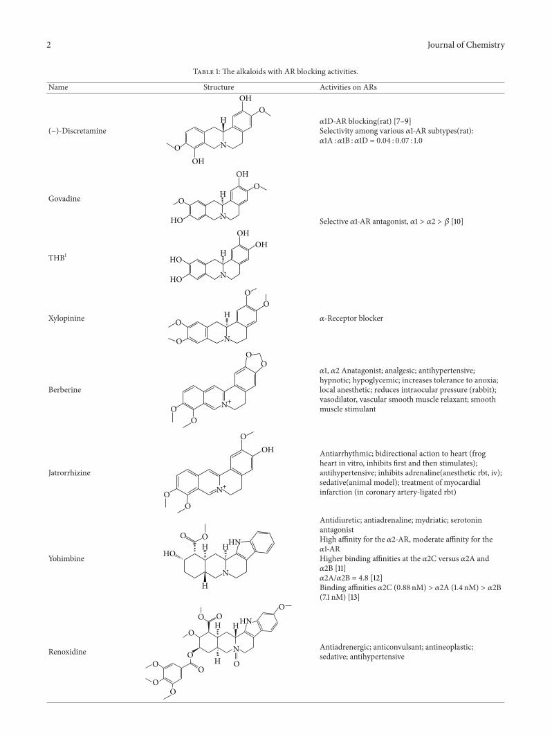
Table 1: The alkaloids with AR blocking activities.
Name Structure Activities on ARs
(−)-Discretamine
NO
H
OH
OH
O𝛼1D-AR blocking(rat) [7–9]Selectivity among various 𝛼1-AR subtypes(rat):𝛼1A :𝛼1B :𝛼1D = 0.04 : 0.07 : 1.0
OOH Antiarrhythmic; bidirectional action to heart (frog
heart in vitro, inhibits first and then stimulates);antihypertensive; inhibits adrenaline(anesthetic rbt, iv);sedative(animal model); treatment of myocardialinfarction (in coronary artery-ligated rbt)
YohimbineHHN
H
H
N
OO
HO
Antidiuretic; antiadrenaline; mydriatic; serotoninantagonistHigh affinity for the 𝛼2-AR, moderate affinity for the𝛼1-ARHigher binding affinities at the 𝛼2C versus 𝛼2A and𝛼2B [11]𝛼2A/𝛼2B = 4.8 [12]Binding affinities 𝛼2C (0.88 nM) > 𝛼2A (1.4 nM) > 𝛼2B(7.1 nM) [13]
In this paper, three-dimensional homology models werebuilt for each AR 𝛼2 subtype. By using molecule stimulationmethod, the bindingmodes of Yohimbine, Yohimbinemono-glycine ester (YME), and Yohimbine alkyl amine (YAA) wereanalyzed (Figure 1).Themechanism of selectivity and the keyresidues were investigated.
2. Data and Methods
2.1. Homo Modeling. The AR receptor belongs to heterotri-meric guanine nucleotide-binding protein coupled receptors(GPCRs). The proteins in GPCR superfamily usually havelow sequence identities but with high structure similarities.The binding sites usually locate among the seven transmem-brane (TM) helices; together with three extracellular loops.GPCR receptors play important roles in drug discovery[19]. Due to the hardness in the crystallization of GPCRs,homology modeling is a main approach for the structure-based drug design of GPCRs [20].
The secondary structure assignments are annotated withcolor-coded bars above the sequences. Helixes are in red,strands are in yellow, 1–4 turns are in blue, and 1–5 turns are ingreen. The conserved residues were marked with dark color.
To date, our understanding of GPCR structure is mainlybased on the high-resolution crystal protein structure. Com-putational modeling has also predicted that GPCRs sharea seven membrane-spanning a-helix topology as a com-mon structural property. Likewise, mutagenesis studies havetentatively identified individual transmembrane domainswith specific roles in signal transduction function that areconserved throughout the GPCR family. Since 2007, severalHomo AR 𝛽-subtypes were reported [21–24]. Among thecrystal data, 2RH1, a complex of 𝛽2 subtype with agonistcarazolol, is an ideal template for the modeling of AR family[25, 26].
The alignment errors increase rapidly when the sequenceidentity of two proteins is less than 30% [27]. For GPCRhomology, the sequence identity is usually less than 25% [20].Therefore, automated homology modeling of GPCRs is likelyto result in more errors [28].The errors in alignments cannotbe justified by energy optimizations, molecular dynamics, ordistance geometry refinements. The alignments become crit-ical for the quality of the homologymodel.Multiple sequencealignments are adopted in order to enhance the alignmentquality. We have used 8 AR subtypes in the sequencealignment. As shown in Figure 2, all the subtypes keep a high
4 Journal of Chemistry
2.5 3.5
4.5 5.5
2RH1𝛽1𝛼1A𝛼1B𝛼1D𝛼2A𝛼2B𝛼2C
1.5
6.5 7.5
Figure 2: Alignment of the 8AR subtypes.
𝛼2A
𝛼2B
𝛼2C
Figure 3: The homology models of three 𝛼2 subtypes. The left isthe template superposed by the three homology models, 2RH1: red,𝛼2A: green, 𝛼2B: blue, and 𝛼2C: purple.
identity in the TM helices. For the binding site is exactlyconstituted by the TMs, the homo model may be of highreliability to estimate the interaction between the ligand andAR.
Homomodels of AR 𝛼2A, B and C were constructed withthe homology modeling module of the molecular operating
environment (MOE) [29]. The AMBER99 force field wasapplied to the homology modeling. The carboxyl-terminaland amino-terminal modeling was disabled. The scoringmethod was GB/VI [30]. Ten models were built by MOEfor each subtype, kept in a MOE database, and ranked bythe GB/VI scores. The best model was kept for the followingenergy optimizing.
Partial charges for all atoms were calculated. Then, theenergywasminimized bymeans ofAMBER99 force field, andthe ending condition of the calculation was the root meansquare (RMS) of the two conformer energies being less than0.1 kcalmol−1 A−1. Furthermore, molecular mechanics (MD)simulations were applied, in MOE. The process consisted oftwo steps.
(1) The MD simulations were performed with all C𝛼atoms being fixed. The temperature was increasedfrom 0K to 300K within 60 ps (picoseconds); anNVT (constant volume/constant-temperature) mo-lecular dynamic simulation of 100 ps at 300K wascarried out with a time-step length of 0.002 ps.
(2) The atoms in the TM helixes were set flexible forconformation changes. The MD experiments sim-ulated conformation changes in 1 ns timescale. Thefinal model was obtained by energy minimization tillan RMS of 0.05 kcalmol−1 A−1 was reached.
2.2. Docking. Docking experiments were carried out toexplore the binding modes of three antagonists, Yohimbine,
Figure 4: The RMSD fluctuation during MD simulation.
HHHN
HN
+
OO
HO
Ile
TyrTyr196
190
394
Ser
Ser
200
204
Figure 5: The binding mode of yohimbine with AR 𝛼2A.
YME, and YAA. All the ligands were built in MOE Builder.Their three-dimensional structures were energy minimizedby MOE using MMFF94x force field. The initial poses wereassigned by Flexible Alignment module of MOE, with theagonist carazolol in 2RH1 as the template. The 3 ligands were
docked into 3 𝛼2 subtypes, respectively. Totally 9 dockingexperiments were carried out.
Docking used the algorithm MOE Dock. The defaultsettings were used. For each position, 700 iterationswere gen-erated to optimize the interactions at a location. A database
6 Journal of Chemistry
𝛼2A-Yohimbine 𝛼2A-YME 𝛼2A-YAA
𝛼2B-Yohimbine 𝛼2B-YME 𝛼2B-YAA
𝛼2C-Yohimbine 𝛼2C-YME 𝛼2C-YAA
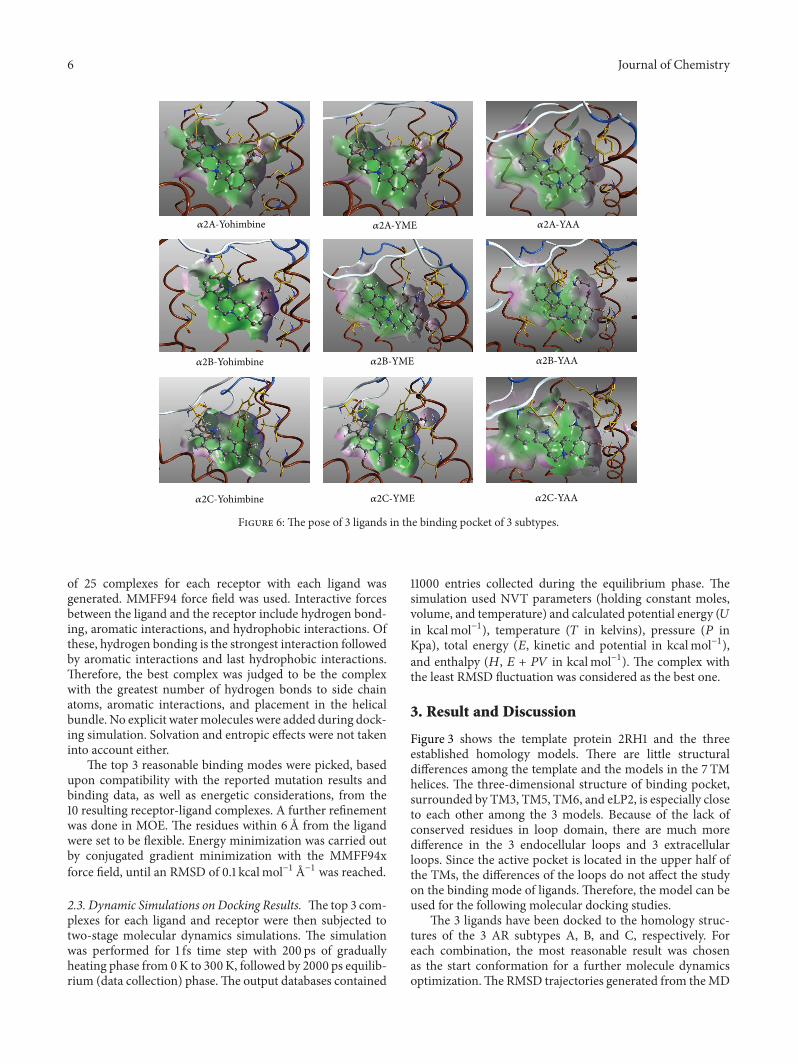
Figure 6: The pose of 3 ligands in the binding pocket of 3 subtypes.
of 25 complexes for each receptor with each ligand wasgenerated. MMFF94 force field was used. Interactive forcesbetween the ligand and the receptor include hydrogen bond-ing, aromatic interactions, and hydrophobic interactions. Ofthese, hydrogen bonding is the strongest interaction followedby aromatic interactions and last hydrophobic interactions.Therefore, the best complex was judged to be the complexwith the greatest number of hydrogen bonds to side chainatoms, aromatic interactions, and placement in the helicalbundle. No explicit watermolecules were added during dock-ing simulation. Solvation and entropic effects were not takeninto account either.
The top 3 reasonable binding modes were picked, basedupon compatibility with the reported mutation results andbinding data, as well as energetic considerations, from the10 resulting receptor-ligand complexes. A further refinementwas done in MOE. The residues within 6 A from the ligandwere set to be flexible. Energy minimization was carried outby conjugated gradient minimization with the MMFF94xforce field, until an RMSD of 0.1 kcalmol−1 A−1 was reached.
2.3. Dynamic Simulations on Docking Results. The top 3 com-plexes for each ligand and receptor were then subjected totwo-stage molecular dynamics simulations. The simulationwas performed for 1 fs time step with 200 ps of graduallyheating phase from 0K to 300K, followed by 2000 ps equilib-rium (data collection) phase.The output databases contained
11000 entries collected during the equilibrium phase. Thesimulation used NVT parameters (holding constant moles,volume, and temperature) and calculated potential energy (𝑈in kcalmol−1), temperature (𝑇 in kelvins), pressure (𝑃 inKpa), total energy (𝐸, kinetic and potential in kcalmol−1),and enthalpy (𝐻, 𝐸 + 𝑃𝑉 in kcalmol−1). The complex withthe least RMSD fluctuation was considered as the best one.
3. Result and Discussion
Figure 3 shows the template protein 2RH1 and the threeestablished homology models. There are little structuraldifferences among the template and the models in the 7 TMhelices. The three-dimensional structure of binding pocket,surrounded by TM3, TM5, TM6, and eLP2, is especially closeto each other among the 3 models. Because of the lack ofconserved residues in loop domain, there are much moredifference in the 3 endocellular loops and 3 extracellularloops. Since the active pocket is located in the upper half ofthe TMs, the differences of the loops do not affect the studyon the binding mode of ligands. Therefore, the model can beused for the following molecular docking studies.
The 3 ligands have been docked to the homology struc-tures of the 3 AR subtypes A, B, and C, respectively. Foreach combination, the most reasonable result was chosenas the start conformation for a further molecule dynamicsoptimization.The RMSD trajectories generated from theMD
Journal of Chemistry 7
Ile
Ile
Ile
Lys
LeuGln
Gln
Asp
Asp
Cys
Phe
Phe
PhePhe
Phe
Phe
PhePhe
Phe
Phe
H2N
H
H HHH
HH
H
H
H
H H
H H
H
H
HH
H
HH
H
HH
H
H
H
H
H H
HH
H
H
HHH
HN
HN HN
HN
N
HN
HN
HN
HN
HN+
HN+
+
HN
HN
HN
HN
+
HO HO
HO
HOHO
HO
HO HO
HO
OO O
O
O
OO
O
O
OOO
OO
OO
O
O
TyrTyr196
TyrTyr
Tyr196
190
180
190
190
390
391
387387
387
388
412412
423
412
192
92
96
174
394
Tyr394
Tyr394
Tyr196
Ser
Ser180Ser
214Ser
214Ser
218Ser
218Ser
214Ser
218Ser
180Ser
Ser
200
Ser179 172
Tyr172
Tyr172
Tyr402
Tyr402
Tyr210
Tyr210
Tyr210
391
Tyr391
166
166
168
168
Ser200
204Ser204
Ser204
+NH
+NH+NH
+NH
NH2
NH
NH
2
+NH
NH
NH
Leu
204Leu
204Leu
204Leu
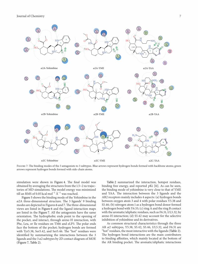
𝛼2A-Yohimbine 𝛼2A-YME 𝛼2A-YAA
𝛼2B-Yohimbine 𝛼2B-YME 𝛼2B-YAA
𝛼2C-Yohimbine 𝛼2C-YME 𝛼2C-YAA
HN
Figure 7: The binding modes of the 3 antagonists to 3 subtypes. Blue arrows represent hydrogen bonds formed with backbone atoms; greenarrows represent hydrogen bonds formed with side-chain atoms.
simulation were shown in Figure 4. The final model wasobtained by averaging the structures from the 1.5∼2 ns trajec-tories of MD simulations. The model energy was minimizedtill an RMS of 0.05 kcalmol−1 A−1 was reached.
Figure 5 shows the bindingmode of the Yohimbine in the𝛼2A three-dimensional structure. The 3 ligands’ 9 bindingmodes are depicted in Figures 6 and 7.The three-dimensionalviews are listed in Figure 6 and the ligand interaction mapsare listed in the Figure 7. All the antagonists have the sameorientation. The hydrophobic ends point to the opening ofthe pocket, and interact, through arene-H interaction, withPhe, Leu, or Ile residues on TM6 and eLP3. The polar endsface the bottom of the pocket; hydrogen bonds are formedwith Tyr5.38, Ser5.42, and Ser5.46. The “hot” residues wereidentified by summarizing the interactions between the 3ligands and the 3 𝛼2 subtypes by 2D contact diagram ofMOE(Figure 7, Table 2).
Table 2 summarized the interaction, hotspot residues,binding free energy, and reported 𝑝Ki [11]. As can be seen,the binding mode of yohimbine is very close to that of YMEand YAA. The interaction between the 3 ligands and theAR2 receptors mainly includes 4 aspects: (a) hydrogen bondsbetween oxygen atom 3 and 4 with polar residues Y5.38 andS5.46; (b) nitrogen atom 1 as a hydrogen bond donor formeda hydrogen bondwith Y6.55; (c) ring A and the ring B contactwith the aromatic/aliphatic residues, such as F6.51, I/L5.32, byarene-H interaction; (d) S5.42 may account for the selectiveinhibition of yohimbine and its derivatives.
As common structural characteristics through the threeAR 𝛼2 subtypes, Y5.38, S5.42, S5.46, I/L5.32, and F6.51 are“hot” residues, themost interactive with the ligands (Table 2).The hydrogen bond interactions are the main contributorsto binding affinities, which mainly located at the bottom ofthe AR binding pocket. The aromatic/aliphatic interactions
8 Journal of Chemistry
Table 2: Summary on the interactions between 3 ligands and the 3 𝛼2 subtypes.
ligands ARs Hydrogen bond Arene-H/arene-arene𝐸 (kJ ⋅mol−1) 𝑝Ki (𝜇M)
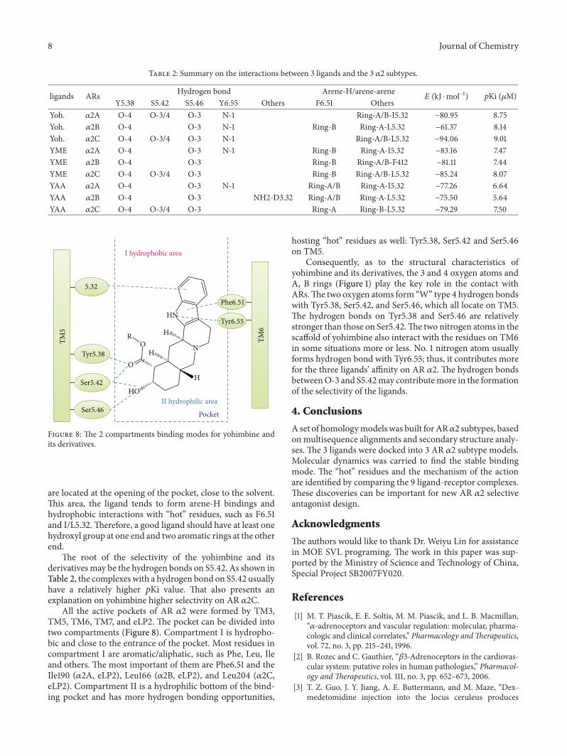
Figure 8: The 2 compartments binding modes for yohimbine andits derivatives.
are located at the opening of the pocket, close to the solvent.This area, the ligand tends to form arene-H bindings andhydrophobic interactions with “hot” residues, such as F6.51and I/L5.32.Therefore, a good ligand should have at least onehydroxyl group at one end and two aromatic rings at the otherend.
The root of the selectivity of the yohimbine and itsderivativesmay be the hydrogen bonds on S5.42. As shown inTable 2, the complexes with a hydrogen bond on S5.42 usuallyhave a relatively higher 𝑝Ki value. That also presents anexplanation on yohimbine higher selectivity on AR 𝛼2C.
All the active pockets of AR 𝛼2 were formed by TM3,TM5, TM6, TM7, and eLP2. The pocket can be divided intotwo compartments (Figure 8). Compartment I is hydropho-bic and close to the entrance of the pocket. Most residues incompartment I are aromatic/aliphatic, such as Phe, Leu, Ileand others. The most important of them are Phe6.51 and theIle190 (𝛼2A, eLP2), Leu166 (𝛼2B, eLP2), and Leu204 (𝛼2C,eLP2). Compartment II is a hydrophilic bottom of the bind-ing pocket and has more hydrogen bonding opportunities,
hosting “hot” residues as well: Tyr5.38, Ser5.42 and Ser5.46on TM5.
Consequently, as to the structural characteristics ofyohimbine and its derivatives, the 3 and 4 oxygen atoms andA, B rings (Figure 1) play the key role in the contact withARs.The two oxygen atoms form “W” type 4 hydrogen bondswith Tyr5.38, Ser5.42, and Ser5.46, which all locate on TM5.The hydrogen bonds on Tyr5.38 and Ser5.46 are relativelystronger than those on Ser5.42.The two nitrogen atoms in thescaffold of yohimbine also interact with the residues on TM6in some situations more or less. No. 1 nitrogen atom usuallyforms hydrogen bond with Tyr6.55; thus, it contributes morefor the three ligands’ affinity on AR 𝛼2. The hydrogen bondsbetweenO-3 and S5.42may contributemore in the formationof the selectivity of the ligands.
4. Conclusions
Aset of homologymodelswas built forAR𝛼2 subtypes, basedonmultisequence alignments and secondary structure analy-ses. The 3 ligands were docked into 3 AR 𝛼2 subtype models.Molecular dynamics was carried to find the stable bindingmode. The “hot” residues and the mechanism of the actionare identified by comparing the 9 ligand-receptor complexes.These discoveries can be important for new AR 𝛼2 selectiveantagonist design.
Acknowledgments
The authors would like to thank Dr. Weiyu Lin for assistancein MOE SVL programing. The work in this paper was sup-ported by the Ministry of Science and Technology of China,Special Project SB2007FY020.
References
[1] M. T. Piascik, E. E. Soltis, M. M. Piascik, and L. B. Macmillan,“𝛼-adrenoceptors and vascular regulation: molecular, pharma-cologic and clinical correlates,” Pharmacology andTherapeutics,vol. 72, no. 3, pp. 215–241, 1996.
[2] B. Rozec and C. Gauthier, “𝛽3-Adrenoceptors in the cardiovas-cular system: putative roles in human pathologies,” Pharmacol-ogy andTherapeutics, vol. 111, no. 3, pp. 652–673, 2006.
[3] T. Z. Guo, J. Y. Jiang, A. E. Buttermann, and M. Maze, “Dex-medetomidine injection into the locus ceruleus produces
Journal of Chemistry 9
antinociception,” Anesthesiology, vol. 84, no. 4, pp. 873–881,1996.
[4] S. V. R. Reddy and T. L. Yaksh, “Spinal noradrenergic terminalsystem mediates antinociception,” Brain Research, vol. 189, no.2, pp. 391–401, 1980.
[5] K. D. Davis, R. D. Treede, S. N. Raja, R. A. Meyer, and J. N.Campbell, “Topical application of clonidine relieves hyperalge-sia in patients with sympathetically maintained pain,” Pain, vol.47, no. 3, pp. 309–317, 1991.
[6] L. Paalzow, “Analgesia produced by clonidine in mice and rats,”Journal of Pharmacy and Pharmacology, vol. 26, no. 5, pp. 361–363, 1974.
[7] F. N. Ko, J. H. Guh, S.M. Yu, Y. S. Hou, Y. C.Wu, andC.M. Teng,“(-)-Discretamine, a selective 𝛼(1D)-adrenoceptor antagonist,isolated from Fissistigma glaucescens,” The British Journal ofPharmacology, vol. 112, no. 4, pp. 1174–1180, 1994.
[8] F. N. Ko, S. M. Yu, M. J. Su, Y. C. Wu, and C. M. Teng,“Pharmacological activity of (-)-discretamine, a novel vascular𝛼-adrenoceptor and 5-hydroxytrypamine receptor antagonist,isolated from Fissistigma glaucescens,” The British Journal ofPharmacology, vol. 110, no. 2, pp. 882–888, 1993.
[9] A. Carrieri, N. B. Centeno, J. Rodrigo, F. Sanz, and A. Carotti,“Theoretical evidence of a salt bridge disruption as the initiatingprocess for the 𝛼1d-adrenergic receptor activation: a moleculardynamics and docking study,” Proteins, vol. 43, no. 4, pp. 382–394, 2001.
[10] F. N. Ko, Y. L. Chang, C. M. Chen, and C. M. Teng, “(±)-go-vadine and (±)-THP, two tetrahydroprotoberberine alkaloids,as selective 𝛼1-adrenoceptor antagonists in vascular smoothmuscle cells,” Journal of Pharmacy and Pharmacology, vol. 48,no. 6, pp. 629–634, 1996.
[11] S. A. Bavadekar,G.Ma, S.M.Mustafa, B.M.Moore,D.D.Miller,and D. R. Feller, “Tethered yohimbine analogs as selectivehuman 𝛼 2Cadrenergic receptor ligands,” Journal of Pharmacol-ogy and Experimental Therapeutics, vol. 319, no. 2, pp. 739–748,2006.
[12] W. Zheng, L. Lei, S. Lalchandani, G. Sun, D. R. Feller, and D. D.Miller, “Yohimbine dimers exhibiting binding selectivities forhuman 𝛼(2a)- versus 𝛼(2b)-adrenergic receptors,” Bioorganicand Medicinal Chemistry Letters, vol. 10, no. 7, pp. 627–630,2000.
[13] S.M.Mustafa, S. A. Bavadekar, G.Ma, B.M.Moore, D. R. Feller,and D. D.Miller, “Synthesis and biological studies of yohimbinederivatives on human 𝛼2C-adrenergic receptors,” Bioorganicand Medicinal Chemistry Letters, vol. 15, no. 11, pp. 2758–2760,2005.
[14] R. Paris and R. Goutarel, “African species of Alchornea; thepresence of yohimbine in Alchornea floribunda (Euphor-biaceae).,” Annales pharmaceutiques francaises, vol. 16, no. 1, pp.15–20, 1958.
[15] J. J. Zhou, G. R. Xie, and X. J. Yan, Chemical Components ofSource Plants in Traditional Chinese Medicine, Science Press,Beijing, China, 2009.
[16] C. M. Mac, C. I. Mc, and F. H. Shaw, “The action of yohimbineon excitation and propagation in nerve,”The Australian Journalof Experimental Biology and Medical Science, vol. 27, part 1, pp.115–122, 1949.
[17] M. J. Millan, A. Newman-Tancredi, V. Audinot et al., “Agonistand antagonist actions of yohimbine as compared to fluparoxanat alpha(2)-adrenergic receptors (AR)s, serotonin (5-HT)(1A),5-HT(1B), 5-HT(1D) and dopamine D(2) and D(3) receptors.
Significance for the modulation of frontocortical monoaminer-gic transmission and depressive states,” Synapse, vol. 35, no. 2,pp. 79–95, 2000.
[18] D. D. Schwartz and T. P. Clark, “Selectivity of atipamezole,yohimbine and tolazoline for alpha-2 adrenergic receptor sub-types: implications for clinical reversal of alpha-2 adrenergicreceptor mediated sedation in sheep,” Journal of VeterinaryPharmacology andTherapeutics, vol. 21, no. 5, pp. 342–347, 1998.
[19] J. Saunders, “G-protein-coupled receptors in drug discovery,”Bioorganic & Medicinal Chemistry Letters, vol. 15, article 3653,2005.
[20] R. Heilker, M. Wolff, C. S. Tautermann, and M. Bieler, “G-protein-coupled receptor-focused drug discovery using a targetclass platform approach,”Drug Discovery Today, vol. 14, no. 5-6,pp. 231–240, 2009.
[21] T. Warne, M. J. Serrano-Vega, J. G. Baker et al., “Structure of a𝛽1-adrenergicG-protein-coupled receptor,”Nature, vol. 454, no.7203, pp. 486–491, 2008.
[22] S. G. F. Rasmussen, H. J. Choi, D. M. Rosenbaum et al., “Crystalstructure of the human𝛽2 adrenergic G-protein-coupled recep-tor,” Nature, vol. 450, no. 7168, pp. 383–387, 2007.
[23] M. A. Hanson, V. Cherezov, M. T. Griffith et al., “A specificcholesterol binding site is established by the 2.8 A structure ofthe human beta2-adrenergic receptor,” Structure, vol. 16, no. 6,pp. 897–905, 2008.
[24] M. P. Bokoch, Y. Zou, S. G. F. Rasmussen et al., “Ligand-specificregulation of the extracellular surface of a G-protein-coupledreceptor,” Nature, vol. 463, no. 7277, pp. 108–112, 2010.
[25] L. Ostopovici-Halip, R. Curpan, M. Mracec, and C. G. Bologa,“Structural determinants of the alpha2 adrenoceptor subtypeselectivity,” Journal ofMolecular Graphics andModelling, vol. 29,no. 8, pp. 1030–1038, 2011.
[26] V. Cherezov, D. M. Rosenbaum, M. A. Hanson et al., “High-resolution crystal structure of an engineered human 𝛽2-adrenergic G protein-coupled receptor,” Science, vol. 318, no.5854, pp. 1258–1265, 2007.
[27] D. Baker and A. Sali, “Protein structure prediction and struc-tural genomics,” Science, vol. 294, no. 5540, pp. 93–96, 2001.
[28] I. D. Pogozheva, M. J. Przydzial, and H. I. Mosberg, “Homologymodeling of opioid receptor-ligand complexes using experi-mental constraints,” AAPS Journal, vol. 7, no. 2, pp. E434–E448,2005.
[29] “Molecular Operating Environment in, Chemical ComputingGroup Inc,” Sherbrooke Street West, Suite 910, Montreal, H3A2R7, Canada, 2007.
[30] P. Labute, “The generalized born/volume integral implicit sol-vent model: estimation of the free energy of hydration usingLondon dispersion instead of atomic surface area,” Journal ofComputational Chemistry, vol. 29, no. 10, pp. 1693–1698, 2008.