Mechanism of Transfection of Chicken Embryo Fibroblasts byRous Sarcoma Virus DNA
GEOFFREY M. COOPER* AND SHARON OKENQUIST
Sidney Farber Cancer Institute and Department of Pathology, Harvard Medical School, Boston,Massachusetts 02115
Received for publication 15 May 1978
The mechanism of transfection by Rous sarcoma virus DNA was investigatedby assaying DNA-mediated transformation under conditions which restrictedsecondary virus infection. Chicken embryo fibroblasts which were geneticallyresistant to virus infection as a result of the absence of receptors for viruspenetration were also resistant to transformation by integrated or unintegratedRous sarcoma virus DNA. In addition, DNA of replication-defective Bryan high-titer Rous sarcoma virus was noninfectious, and transformation by DNA of a
temperature-sensitive DNA polymerase mutant was temperature sensitive. Theseresults indicated that secondary virus infection was necessary for transformationby Rous sarcoma virus DNA. Since transformation was assayed by colonyformation in soft agar, as well as by focus formation, the requirement for secondaryvirus infection was not an artifact of potential difficulty in detection of foci formedby division of single transformed cells. Therefore, it appeared that donor DNAdid not stably transform recipient cells by direct integration. Instead, the resultswere consistent with the hypothesis that transfection of chicken embryo fibro-blasts by Rous sarcoma virus DNA proceeded by transcription of donor DNA,formation of extracellular progeny virus, and secondary virus infection of sensitivecells.
Assays of the biological activity of virus DNAsby transfection have been used to study inte-grated and unintegrated DNAs of avian andmammalian retroviruses. However, the mecha-nism of transfection by retrovirus DNAs has notbeen established. Since this mechanism mightbe a significant consideration in interpretationof the biological activity of virus DNAs, weinitiated the present study to investigate themechanism of transfection of chicken embryofibroblasts by DNA of Rous sarcoma virus(RSV).Two general alternative pathways of transfec-
tion were considered: (i) donor RSV DNA mightintegrate directly into the genome of recipientcells to form a stably inherited DNAprovirus or (ii) donor RSV DNA might serve asa template for RNA synthesis, possibly withoutintegration of the donor DNA. In the secondpathway progeny viral RNA would serve as atemplate for synthesis of RSV DNA whichwould subsequently integrate into cellular DNA.Synthesis and integration of RSV DNA mightoccur either within the cell which was the initialrecipient for uptake and transcription of thedonor DNA or via formation of extracellularprogeny virus and secondary infection of othercells in the DNA-treated recipient cell culture.
To distinguish between these pathways, weinvestigated RSV DNA-mediated transforma-tion of chicken embryo fibroblasts under condi-tions which restricted secondary virus infection.Transformation of recipient cells by RSV DNAwas found to require formation of extracellularprogeny virus and secondary infection of sensi-tive cells. Therefore, it appeared that donor RSVDNA did not stably transform recipient cells bydirect integration. These results were thus con-sistent with the hypothesis that transfection ofchicken embryo fibroblasts proceeded primarilyby transcription of donor RSV DNA.
MATERIALS AND METHODSCells and viruses. Cells were grown in Temin
modified Eagle minimal essential medium containing20% tryptose phosphate broth (ET medium) and sup-plemented with calf and fetal bovine sera. Fertilechicken eggs were purchased from SPAFAS, Norwich,Conn. (C/E phenotype), from Hyline Farms, DallasCenter, Iowa (C/O and C/BDE phenotypes), and fromH and N Farms, Redmond, Wash. (C/ACE pheno-type). Chicken embryo fibroblasts were virus negative,chicken helper factor negative, and avian leukosisvirus group-specific antigen negative. Fertile Orloppturkey eggs were provided by the Wilmar Poultry Co.,Wilmar, Minn. Turkey embryo fibroblasts were T/BD,virus negative, helper factor negative, and avian leu-
Schmidt-Ruppin RSV subgroup D (SR-RSV-D), andLA335 (pol), an early temperature-sensitive mutantof Prague RSV subgroup C which encodes a thermo-labile DNA polymerase (15, 18, 29), were previouslydescribed (3). Bryan high-titer RSV (BH-RSV), whichis defective for envelope glycoprotein synthesis (env-)(22), was obtained as a Rous-associated virus-I (RAV-1) pseudotype [BH-RSV(RAV-1)] from H. M. Temin,Madison, Wis.
Preparation of DNA. Total cellular DNA wasextracted from RSV-infected chicken embryo fibro-blasts 5 to 10 days after infection by a previouslydescribed modification (4) of the procedure of Marmur(17).
Unintegrated RSV DNA was extracted 3 days afterinfection of chicken embryo fibroblasts with SR-RSV-D (approximately 0.1 focus-forming unit [FFU]/cell).Cellular DNA was 'H-labeled by culturing the cells for24 h before DNA extraction in medium which con-tained 1 jLCi of [;'Hithymidine per ml. Cells wereharvested by trypsinization, suspended at a density of5 x 10' cells/ml in 10 mM Tris-hydrochloride-10 mMEDTA (pH 7.4), and fractionated by the method ofHirt (12). Approximately 12% of the 'H-labeled DNAwas recovered in the Hirt supernatant fraction. TheHirt supernatant was digested with Pronase (250jug/ml, 30 min, 37°C) and was extracted twice withchloroform-isoamyl alcohol (24:1). DNA was precipi-tated with ethanol overnight at -20°C. The precipi-tate was collected by centrifugation, dissolved in SSC(0.15 M NaCl-0.015 M sodium citrate, pH 7.0), digestedwith RNase A (100 /Lg/ml, 30 min, 37°C), digested withPronase (250 ,g/ml, 30 min, 37°C), and extracted sixtimes with chloroform-isoamyl alcohol. NaCl wasadded to a final concentration of 0.3 M, and DNA wasprecipitated with ethanol overnight at -20°C. Theprecipitate was collected by centrifugation and wasdissolved in sterile SSC. The final yield of Hirt super-natant DNA corresponded to approximately 3% of theinitial 'H-labeled cellular DNA.Agarose gel electrophoresis. Hirt supernatant
DNA was subjected to electrophoresis in 0.7% agarosegels to separate linear and circular forms ofRSV DNA.Electrophoresis was performed as described by Sharpet al. (24), except that both the gels and the electro-phoresis buffer contained 0.1 M NaCl and 4 [ig ofethidium bromide per ml to optimize separation ofcircular and linear DNA forms (13). Covalently closedcircular and linear DNAs of the plasmid pmDm691(molecular weight, 6.2 x 10") (D. J. Finnegan, G. M.Rubin, and D. S. Hogness, in preparation) were usedas markers. pmDm691 DNA was kindly provided byG. M. Rubin. The positions of infectious RSV DNAswere determined by transfection assays ofDNA elutedfrom gel slices as described by Fritsch and Temin (7).The positions of marker DNAs in parallel gels weredetermined by UV fluorescence.
Transfection assay of RSV DNA. Cultures ofchicken embryo fibroblasts were prepared and exposedto DNA by the calcium method of Graham and Vander Eb (10) as previously described (5). Recipient cellswere incubated with DNA for 4 h, the medium waschanged to ET medium supplemented with 2% fetal
J. VIROL.
bovine serum, and the cells were incubated at 37°C.Media were changed at 3-day intervals, and foci oftransformed cells were counted 5 to 7 days after ex-posure to DNA.Colony formation in soft agar. Colony formation
by transformed cells was assayed by a modification ofthe method of Graf (8). Feeder layers of mitomycin C-treated turkey embryo fibroblasts in 60-mm tissueculture dishes were overlaid with 3 ml of medium 199supplemented with 20% tryptose phosphate broth,0.06% sodium bicarbonate, 1% dimethyl sulfoxide, 5%calf serum, 1% heat-inactivated chicken serum, and0.6% agar. DNA-treated chicken embryo fibroblastswere plated at a density of approximately 10' cells perdish in 4 ml of medium 199 supplemented as abovebut containing 0.36% agar. Cultures were incubated at37°C, and colonies of transformed cells were counted7 to 10 days after plating.
RESULTSTransformation of sensitive and resist-
ant chicken embryo fibroblasts by RSVDNA. Transfection assays of RSV DNA werepreviously quantitated by end point dilution ofthe donor DNA (4). However, by use of thecalcium method of DNA treatment (10), trans-fection by RSV DNA can be quantitated byenumeration of foci on the original DNA-treatedplates. The advantage of the calcium method inthis respect appears to be that it is significantlyless toxic to the recipient cells than the DEAE-dextran method used previously (4). Represent-ative transfection assays are presented in Fig.1A. The kinetics of transfection by RSV DNAs
30- A B77V-CC/E Cells 0
20_
10 _ SR-RSV-D0~~~~*
1 -iA.
"I-,
40
30
20
10
BC/ACE Cells SR-RSV-D
0
,
B77V-C
0 2 3 4 5DNA(g/plate)
FIG. 1. Focu.s assay ofRSV DNA. Cultures of C/Eor C/ACE chicken embryo fibroblasts were exposedto DNA extracted from B77V-C-infected cells (B77V-C DNA, 0) or from SR-RSV-D-infected cells (SR-RSV-D DNA, *). Foci were counted 6 days afterDNAtreatment. Each point is the average of duplicateplates.
were one hit, and the specific infectivities ofRSV DNAs were approximately 5 FFU/,ug (Fig.1A). The specific infectivities of RSV DNAsassayed by focus formation were therefore sim-ilar to specific infectivities previously deter-mined by end point dilution assays (approxi-mately 10 infectious units/,tg of DNA) (4).The availability of a direct focus assay for
RSV DNA enabled us to investigate transfor-mation by RSV DNA in the absence of second-ary virus infection. We therefore assayed theinfectivity ofRSV DNA on recipient cells whichwere genetically resistant to secondary virus in-fection as a result of the absence of receptorsrequired for virus penetration (6, 21). B77V-Cand SR-RSV-D DNAs had similar specific infec-tivities (5 and 4 FFU/,g of DNA, respectively)when assayed on C/E cells, which were sensitiveto infection by both subgroup C and subgroupD RSV (Fig. 1A). However, B77V-C DNA was
not infectious (<0.03 FFU/j,g of DNA) whenassayed on C/ACE cells, which were resistant toinfection by subgroup C RSV (Fig. 1B). In con-
trast, the infectivity of SR-RSV-D DNA on
C/ACE cells (Fig. 1B) was similar to its infectiv-ity on C/E cells (Fig. 1A). Therefore, it appearedthat recipient cells which were resistant to infec-tion by extracellular RSV were also resistant totransfection by RSV DNA. Since subgroup-spe-cific resistance to RSV infection appears to beexpressed at the level of virus penetration (6,21), these results indicated that focus formationby RSV DNA required formation of extracellu-lar progeny virus and secondary infection ofsensitive cells.To test the possibility that secondary virus
infection was only necessary for development ofdetectable foci under the conditions of transfec-tion assays, we used colony formation in softagar to assay transformation by RSV DNA.Since soft agar colonies are formed by divisionof single transformed cells, this assay eliminatedthe potential requirement for secondary virusinfection in focus development.
Preliminary experiments were performed todetermine the optimal time for transfer ofDNA-treated recipient cells into suspension in softagar. Recipient cells transferred into soft agarimmediately after exposure to RSV DNA didnot give rise to transformed cell colonies, butcolonies were formed by cells transferred intosoft agar 1 to 2 days after transfection. Thefailure of cells to form colonies in soft agarimmediately after transfection by RSV DNAwas consistent with the hypothesis that forma-tion of progeny virus and secondary virus infec-tion were needed for establishment of geneticallystable transformation. The number of soft agarcolonies formed by cells transferred 1 day after
transfection ranged from 10 to 50% of the num-ber of foci formed on parallel plates maintainedunder liquid medium, whereas the number ofcolonies formed by cells transferred 2 days aftertransfection equaled or exceeded the number offoci obtained. Therefore, recipient cells wereroutinely transferred into soft agar 2 days aftertransfection by RSV DNA.B77V-C and SR-RSV-D DNAs were assayed
on C/0, C/ACE, and C/BDE chicken embryofibroblasts (Table 1). Some of the recipient cellcultures were maintained under liquid mediumto assay focus formation. The rest of the recipi-ent cell cultures were trypsinized 2 days afterexposure to DNA and were plated in soft agarmedium to assay colony formation. B77V-CDNA was infectious for C/0 and C/BDE cellsbut not for C/ACE cells (resistant to infectionby B77V-C). Reciprocally, SR-RSV-D DNA wasinfectious for C/0 and C/ACE cells but not forC/BDE cells (resistant to infection by SR-RSV-D). With both DNAs, the results of soft agarcolony assays agreed with the results of focusassays. Therefore, secondary virus infection ap-peared necessary for genetically stable transfor-mation of recipient chicken cells by RSV DNA.Transfection by DNAs of LA335 (polk)
and BH-RSV (env-). The role of secondaryvirus infection was further investigated in trans-fection experiments with DNAs of replication-defective mutant RSV. The mutants used wereLA335 (pot'), which has a temperature-sensitivemutation in the gene encoding the virion RNA-directed DNA polymerase (15, 18, 29), and BH-RSV (env-), which has a deletion of the geneencoding the virion envelope glycoprotein (22).DNA of LA335-infected cells was assayed for
colony formation in soft agar at permissive
TABLE 1. Assay ofRSVDNA by colony formationin soft agar'
"Cultures of chicken embryo fibroblasts were ex-posed to 5 yg of B77V-C or SR-RSV-D DNA. Somecultures were maintained under liquid medium andfoci were counted 7 days after DNA treatment. Othercultures were transferred into soft agar 2 days afterDNA treatment and transformed cell colonies werecounted 8 days later.
"Average of quadruplicate DNA-treated recipientcultures.
(35°C) and nonpermissive (41°C) temperatures.The infectivity of LA335 (pot') DNA was ap-proximately 10-fold lower at 41 than 35°C (Table2). In contrast, the infectivity of B77V-C (pol+)DNA was threefold higher at 41 than 35°C (Ta-ble 2), possibly as a result of enhanced produc-tion of progeny virus at the higher temperature.The temperature sensitivity of transformationby LA335 DNA indicated that DNA polymeraseactivity was required for transfection. The lowertemperature sensitivity of transfection by LA335DNA (approximately 10-fold) as compared tothe temperature sensitivity of virus infection byLA335 (approximately 10 '-fold) (data notshown) may be due to continuous production ofmultiple progeny virus particles by the DNA-treated cells (see Fig. 3).To test the infectivity of DNA of BH-RSV
(env-), we assayed transfection by DNA of cellsinfected with BH-RSV(RAV-1). This DNA wasnegative when assayed for transformation eitherby focus formation (<0.05 FFU/tig of DNA) orby colony formation in soft agar (<0.03 colony-forming unit/jg of DNA). However, BH-RSV(RAV-1) DNA was infectious (approxi-mately 0.5 infectious unit/Ag of DNA) whenassayed for production of nontransforming virusby determination of sedimentable DNA polym-erase activity in culture fluids (5). Therefore,BH-RSV (RAV-1)-infected cells appeared tocontain infectious DNA of RAV-1 but not ofBH-RSV (env-). The lack of transformation byBH-RSV (enu-) DNA further indicated thatsecondary virus infection was required for RSVDNA-mediated transformation.A control experiment was performed to deter-
mine whether BH-RSV-transformed nonpro-ducer cells formed soft agar colonies under theconditions used for transfection assays. Culturesof chicken embryo fibroblasts were treated withsalmon sperm DNA and were then infected withserial dilutions of a BH-RSV(RAV-1) stockwhich titered approximately 2 x 10' FFU/ml instandard focus assays. Two days after infection,
T'ABLF 2. Tran.sfection by LA.335 (pol',) DNA"Colonies/plate"
D)NA350C 41°C
B77V-C (pol+) 31 92LA335 (pol'") 5.3 0.4
Cultures of C/O chicken embryo fibroblasts wereexposed to 5 ,ig of LA335 (pol") or B77V-C (pol+)I)NA and were incubated at either 35 or 41°C. Thecells were transferred into soft agar 2 days after DNAtreatment.
" Average of three plates treated with B77V-C(pol+) DNA or 27 plates treated with LA335 (pol")I)NA.
J. VIROL.
the cells were trypsinized and plated in soft agar.The efficiency of colony formation correspondedto approximately 5 x 10' colony-formingunits/ml of the original virus stock and was thussimilar to the titer determined by focus assay.Twelve transformed cell colonies from plateswhich had been exposed to a 10' dilution ofvirus were picked and tested for production ofinfectious progeny RSV. Nine of the 12 coloniesfailed to yield infectious progeny virus, indicat-ing that transformed nonproducer cells were ca-pable of forming soft agar colonies under trans-fection assay conditions.Transfection by unintegrated RSV DNA.
Since total cellular DNA of RSV-infected cellsdid not appear to stably transform recipient cellsby direct integration, it was of interest to inves-tigate transfection by newly synthesized unin-tegrated RSV DNA, which is the probable pre-cursor to integrated proviral DNA in virus-in-fected cells. Unintegrated DNA of SR-RSV-D-infected cells was extracted by Hirt fraction-ation (12) 3 days after infection at a multiplicityof 0.1 FFU per cell. The yield of infectious Hirtsupernatant RSV DNA corresponded to approx-imately 1 x 10⁴ FFU from 3 x 10⁸ cells.The infectious Hirt supernatant DNA was
analyzed by electrophoresis in agarose gels withthe use of linear and closed circular DNAs ofplasmid pmDm691 (molecular weight, 6.2 x 10⁶)as markers (Fig. 2). Infectious RSV DNA yieldeda single peak in the position of linear DNA ofapproximately 6 x 10⁶ daltons. No infectivity
.G30~
pm Dm691pm Dm691 ClosedLinear Circular
10 15
FractionFIG. 2. Agarose gel electrophoresis of Hirt super-
natant RSV DNA. Hirt supernatant SR-RSV-D DNAwas subjected to electrophoresis for 10 h at 30 V in a0.7%lc agarose gel which contained 0.1 M NaCI and 4pg of ethidium bromide per ml. The gel was fraction-ated into 3-mm slices, and the DNA was eluted andassayed for infectivity. Marker DNAs of plasmidpmDm691 (molecular weight, 6.2 x KY') were subjectedto electrophoresis in a parallel gel.
(<10%) was detected in the position of closedcircular DNA. Therefore, the infectious Hirtsupernatant DNA consisted primarily of unin-tegrated linear RSV DNA molecules.The results of transfection assays of uninte-
grated SR-RSV-D DNA by both focus and col-ony formation are presented in Table 3. TheHirt supernatant DNA was infectious for C/Ocells but not for C/BDE cells (resistant to infec-tion by SR-RSV-D). Therefore, transfection byunintegrated linear RSV DNA also appeared torequire formation of extracellular progeny virusand secondary virus infection.Production of progeny virus by resistant
cells exposed to RSV DNA. Since transfectionappeared to proceed by transcription of donorDNA and formation of extracellular progenyvirus, it was of interest to investigate virus pro-
duction by resistant cells which had been ex-
posed to RSV DNA.C/ACE cells (resistant to infection by
B77V-C) were exposed to B77V-C DNA, theDNA was removed, and cells which were sensi-tive to infection by subgroup C RSV (C/E or
T/BD cells) were added to determine whetherthe DNA-treated C/ACE cells could serve as
infectious centers. T/BD cells were resistant totransfection by B77V-C DNA (unpublisheddata) and were therefore used in this experimentto exclude the possibility that transfection couldoccur by uptake of residual DNA by the addedsensitive cells. The specific infectivity of B77V-C DNA was approximately 6 FFU/,ug on C/Ecells, but less than 0.1 FFU/,ug on C/ACE cells(Table 4). However, addition of C/E or T/BDcells to DNA-treated C/ACE cell cultures re-
sulted in focus formation at frequencies of 2 to3 FFU/ug of DNA (Table 4). Therefore, C/ACEcells served as infectious centers after exposureto B77V-C DNA with an efficiency comparableto the efficiency of transfection of sensitive cells.The kinetics of production of progeny virus
TABLE 3. Transfection by unintegrated SR-RSV-DDNA"
Recip- Foci/ Colonies/DNA ient plate" plate'
SR-RSV-D Hirt C/O 7 12supernatant C/BDE 0 0
B77V-C C/O 24 50C/BDE 9 48
a Recipient cultures of C/O or C/BDE chicken em-
bryo fibroblasts were exposed to SR-RSV-D Hirt su-
pernatant DNA or to 5 pg of B77V-C DNA. Cultureswere either maintained under liquid medium or trans-ferred into soft agar 2 days after DNA treatment.
'Average of quadruplicate plates.
TABLE 4. Infectious center assay of virusproduction by resistant cells exposed to RSV DNA"
Phenotype of Phenotype of cells FoiDNA-treated cells added after re- pFloatimoval of DNA pae
C/E None' 33C/ACE None" 0C/ACE C/E 17C/ACE T/BD 9
Cultures of C/E or C/ACE chicken embryo fibro-blasts were incubated for 4 h with 5 ,ug of B77V-CDNA. The DNA was removed, and C/E or T/BD cellswere added to some cultures. The cells were main-tained under liquid medium and foci were counted 7days after DNA treatment.
"Average of triplicate plates.No cells were added after DNA treatment of these
cultures.
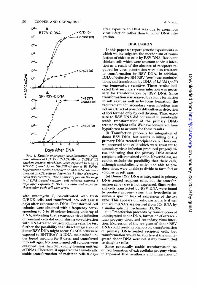
after exposure of sensitive or resistant cells toB77V-C and SR-RSV-D DNAs are presented inFig. 3. Progeny virus was first detected 2 daysafter DNA treatment of either sensitive or re-sistant chicken embryo fibroblasts. The titer ofprogeny virus produced by sensitive cells in-creased to 107 to 10' FFU of B77V-C or to 106FFU of SR-RSV-D per culture by 7 to 10 daysafter DNA treatment, at which time transfor-mation of the DNA-treated cultures approachedconfluency. DNA-treated resistant cells pro-duced progeny virus at a titer of 102 to 10; FFUper culture for at least 10 days after exposure toDNA, although no foci of transformed cells wereevident.The persistence of progeny virus production
for 10 days after transfection of resistant cellssuggested that stably transformed cells might bepresent at later times after DNA infection. Totest this possibility, C/BDE cells (resistant toinfection by SR-RSV-D) were exposed to SR-RSV-D DNA, maintained under liquid mediumfor 8 days, and then transferred into soft agar.Transformed cell colonies were obtained with afrequency corresponding to approximately 10colony-forming units/,ug ofDNA, indicating thatgenetically stable transformation had occurred.
Stable transformation of resistant cells 8 daysafter transfection could be due either to directintegration of donor RSV DNA or to exogenousvirus infection of the resistant cells during co-cultivation with cells which were producingprogeny RSV via transcription of unintegrateddonor RSV DNA. Crittenden (6) previously ob-served that C/A cells were susceptible to infec-tion with subgroup A RSV during co-cultivationwith lethally irradiated RSV-A-producing cells.To test this possibility in the present experi-ments, C/BDE cells were killed 2 days aftertransfection with SR-RSV-D DNA by treatment
Days After DNAFIG. 3. Kinetics ofprogeny virus formation. Dupli-
cate cultureaof CrE (x), CcACE (cu), or CcBDE (d)chicken embryo fibroblasts were exposed to 5peg ofB77V-C (panel A) or SR-RSV-D (panel B) DNAs.Supernatant media harCested at 24-h intervals wereassayed on CeO cells transferre iter ofprogenyvirus (FFU/culture). The number of foci on the orig-inal DNA-treated recipient cell cultures, counted 6days after exposure to DNA, are indicated in paren-theses after each cell phenotype.
with mitomycin C, co-cultivated with freshC/BDE cells, and transferred into soft agar 8days after exposure to DNA. Transformed cellcolonies were obtained with a frequency corre-sponding to 5 to 10 colony-forming units/ag ofDNA, indicating that exogenous virus infectionof resistant cells did occur during co-cultivationwith DNA-treated virus-producing cells. To testfurther the possibility that direct integration ofdonor RSV DNA might occur, C/ACE cells wereexposed to BHT(RAV-1) DNA, maintained un-der liquid medium for 8 days, and transferredinto soft agar. No transformed cell colonies wereobtained (less than 0.01 colony-forming unit/,ugof DNA). Therefore, it appeared that geneticallystable transformation of resistant cells 8 days
after exposure to DNA was due to exogenousvirus infection rather than to donor DNA inte-gration.
DISCUSSIONIn this paper we report genetic experiments in
which we investigated the mechanism of trans-fection of chicken cells by RSV DNA. Recipientchicken cells which were resistant to virus infec-tion as a result of the absence of receptors re-quired for virus penetration were also resistantto transformation by RSV DNA. In addition,DNA of defective BH-RSV (env-) was noninfec-tious, and transfection by DNA of LA335 (pols)was temperature sensitive. These results indi-cated that secondary virus infection was neces-sary for transformation by RSV DNA. Sincetransformation was assayed by colony formationin soft agar, as well as by focus formation, therequirement for secondary virus infection wasnot an artifact of possible difficulties in detectionof foci formed only by cell division. Thus, expo-sure to RSV DNA did not result in geneticallystable transformation of the primary DNA-treated recipient cells. We have considered threehypotheses to account for these results.
(i) Transfection proceeds by integration ofdonor RSV DNA, but results in killing of theprimary DNA-treated recipient cells. However,we observed that cells which were resistant tosecondary virus infection produced progeny vi-rus, indicating that the primary DNA-treatedrecipient cells remained viable. Nevertheless, wecannot exclude the possibility that these cells,although metabolically active and able to pro-duce virus, were unable to divide to form foci orcolonies in soft agar.
(ii) Donor RSV DNA is integrated in primaryDNA-treated recipient cells, but the transfor-mation gene (src) is not expressed. Since resist-ant cells transfected by RSV DNA were foundto produce progeny virus, this hypothesis as-sumes a specific lack of expression of the srcgene. This appears unlikely, particularly if envand src mRNA's are derived from 35S RNA bya similar splicing mechanism (19, 30).
(iii) Transfection proceeds by transcription ofunintegrated donor DNA, formation of extracel-lular progeny virus, and secondary virus infec-tion. Expression of the src gene of donor RSVDNA could result in phenotypic transformationof primary DNA-treated recipient cells, buttransformation would be abortive if the uninte-grated donor DNA were not stably transmittedto daughter cells.
Since genetically stable transformation re-quired formation of extracellular progeny virus,it appeared that synthesis and integration of
progeny RSV DNA did not occur in the samecell which was the primary recipient for uptakeand transcription of donor RSV DNA. Forma-tion of extracellular virions might be requiredfor completion of processes which occur duringvirion assembly or maturation. Such processescould include formation of the 70S RNA com-plex (1, 2), formation of DNA polymerase byprecursor polypeptide cleavage (20), or associa-tion of DNA polymerase with primer and tem-plate RNAs.
In contrast to the present observations, trans-formation of some mouse cell lines by DNA ofdefective murine sarcoma virus occurs at highefficiency, indicating that murine sarcoma virusDNA integrates directly into the recipientmouse cell genome (14, 16, M. Goldfarb and R.A. Weinberg, personal communication). Wehave confirmed these results with murine sar-coma virus DNA and have also demonstratedtransformation of NIH 3T3 mouse cells by SR-RSV-D DNA (N. Copeland and G. M. Cooper,unpublished data). Thus, it appears that trans-fecting retrovirus DNAs integrate efficiently inNIH 3T3 mouse cells, but not in chicken embryofibroblasts. The basis of this difference in trans-fection might be related to the recipient cellspecies, to the fact that NIH 3T3 cells are anestablished cell line, or to factors which mightbe unique to the particular cells studied. Thedifferent fates of donor DNAs in different recip-ient cells might be related to differences in thecellular contents of enzymes involved in DNAintegration.The lack of direct transformation of chicken
cells by RSV DNA also differs from the trans-forming activity of fragments of papova virus,adenovirus, and herpes simplex virus DNAs (re-viewed in 9). However, the efficiency of trans-formation by fragments of these viral DNAs isgenerally less than 1 FFU per 108 viral genomeequivalents (9). Since this is approximately i0}-fold lower than the efficiency of transfection byRSV DNA, we would not have detected a similarfrequency of direct transformation by RSV DNAin the present experiments.
Transfection by unintegrated linear RSVDNA, as well as by total cell DNA, appeared toproceed by transcription of the donor DNA.However, unintegrated linear DNA synthesizedin the cytoplasm of RSV-infected cells appearsto be the precursor of closed circular DNA (23),which is the probable substrate for integration.Therefore, the activity of unintegrated virusDNAs in transfection assays (7, 11, 26, 27) maynot be indicative of their possible roles as pre-cursors of integrated proviral DNA in virus-in-fected cells. If transfection proceeds by tran-
scription of unintegrated donor DNA, the infec-tivity of unintegrated linear RSV DNA alsoindicates that transcription of viral DNA canoccur in the absence of adjacent cellular DNAsequences. Since significant amounts of uninte-grated viral DNAs may persist for several weeksafter infection (7, 11, 28), these observationssuggest the possibility that these unintegratedDNAs, as well as integrated proviral DNA,might serve as templates for viral RNA synthe-sis.The hypothesis that transfection by RSV
DNA proceeds via transcription of donor DNAis also relevant to the interpretation of experi-ments in which transfection was used to studythe endogenous RAV-0 DNA of uninfectedchicken cells (5). In contrast to the infectivity ofRAV-0 proviral DNA of RAV-0-infected cells,the endogenous RAV-0 genome of uninfected V+chicken cells was not infectious (5). Since thetiter of RAV-0 produced by uninfected V+ cellswas 10 3- to 104-fold lower than the titer of RAV-0 produced by RAV-0-infected cells (5, 25), itwas proposed that the lack of infectivity of theendogenous RAV-0 genome was a consequenceof linkage to a cis-acting control element whichinhibited its transcription (5). The present re-sults indicate that transcription of donor DNAis a primary event in transfection and therebyclarify a possible mechanism by which a linkedcis-acting transcriptional control element couldaffect the infectivity of endogenous RAV-0DNA.
ACKNOWLEDGMENTS
We thank G. M. Rubin for supplying pmDm691 DNA, L.Silverman for technical assistance, and N. Copeland and D.M. Livingston for helpful comments on the manuscript. Weare grateful to D. R. Lowy and R. A. Weinberg for discussionsof unpublished results.
This investigation was supported by Public Health Servicegrants CA18689 and CA21082 awarded by the National CancerInstitute.
LITERATURE CITED
1. Canaani, E., K. Von der Helm, and P. Duesberg. 1973.Evidence for 30-40S RNA as precursor of the 60-70SRNA of Rous sarcoma virus. Proc. Natl. Acad. Sci.U.S.A. 70:401-405.
2. Cheung, K.-S., R. E. Smith, M. P. Stone, and W. K.Joklik. 1972. Comparison of immature (rapid harvest)and mature Rous sarcoma virus particles. Virology50:851-864.
3. Cooper, G. M., and S. B. Castellot. 1977. Assay ofnoninfectious fragments of DNA of avian leukosis virus-infected cells by marker rescue. J. Virol. 22:300-307.
4. Cooper, G. M., and H. M. Temin. 1974. Infectious Roussarcoma virus and reticuloendotheliosis virus DNAs. J.Virol. 14:1132-1141.
5. Cooper, G. M., and H. M. Temin. 1976. Lack of infectiv-ity of the endogenous avian leukosis virus-related genesin the DNA of uninfected chicken cells. J. Virol.17:422-430.
6. Crittenden, L. B. 1968. Observations on the nature of agenetic cellular resistance to avian tumor viruses. J.Natl. Cancer Inst. 41:145-153.
7. Fritsch, E., and H. M. Temin. 1977. Formation andstructure of infectious DNA of spleen necrosis virus. J.Virol. 21:119-130.
8. Graf, T. 1973. Two types of target cells for transformationwith avian myelocytomatosis virus. Virology54:398-413.
9. Graham, F. L. 1977. Biological activity of tumor virusDNA. Adv. Cancer Res. 25:1-51.
10. Graham, F. L., and A. J. Van der Eb. 1973. A newtechnique for the assay of infectivity of human adeno-virus 5 DNA. Virology 52:456-467.
11. Guntaka, R. V., 0. C. Richards, P. R. Shank, H.-J.Jung, N. Davidson, E. Fritsch, J. M. Bishop, andH. E. Varmus. 1976. Covalently closed circular DNAof avian sarcoma virus: purification from nuclei of in-fected quail tumor cells and measurement by electronmicroscopy and gel electrophoresis. J. Mol. Biol.106:337-357.
12. Hirt, B. 1967. Selective extraction of polyoma DNA frominfected mouse cell cultures. J. Mol. Biol. 26:365-369.
13. Johnson, P. H., and L. I. Grossman. 1977. Electropho-resis of DNA in agarose gels. Optimizing separations ofconformational isomers of double- and single-strandedDNAs. Biochemistry 16:4217-4224.
14. Karpas, A., and C. Milstein. 1973. Recovery of thegenome of murine sarcoma virus (MSV) after infectionof cells with nuclear DNA from MSV transformed non-virus producing cells. Eur. J. Cancer 9:295-299.
15. Linial, M., and W. S. Mason. 197.3. Characterization oftwo conditional early mutants of Rous sarcoma virus.Virology 53:258-273.
16. Lowy, D. R., E. Rands, and E. M. Scolnick. 1978.Helper-independent transformation by unintegratedHarvey sarcoma virus DNA. J. Virol. 26:291-298.
17. Marmur, J. 1961. A procedure for the isolation of deoxy-ribonucleic acid from micro-organisms. J. Mol. Biol.3:208-218.
18. Mason, W. S., R. R. Friis, M. Linial, and P. K. Vogt.1974. Determination of the defective function in twomutants of Rous sarcoma virus. Virology 61:559-574.
19. Mellon, P., and P. H. Duesberg. 1977. Subgenomic,
cellular Rous sarcoma virus RNAs contain oligonucle-otides from the 3' half and the 5' terminus of virionRNA. Nature (London) 270:631-634.
20. Oppermann, H., J. M. Bishop, H. E. Varmus, and L.Levintow. 1977. A joint product of the genes gag andpol of avian sarcoma virus: a possible precursor ofreverse transcriptase. Cell 12:993-1006.
21. Piraino, F. 1967. The mechanism of genetic resistance ofchick embryo cells to infection by Rous sarcoma virus-Bryan strain (BS-RSV). Virology 32:700-707.
22. Scheele, C. M., and H. Hanafusa. 1971. Proteins ofhelper-dependent RSV. Virology 45:401-41(0.
23. Shank, P. R., and H. E. Varmus. 1978. Virus-specificDNA in the cytoplasm of avian sarcoma virus-infectedcells is a precursor to covalently closed circular viralDNA in the nucleus. .I. Virol. 25:104-114.
24. Sharp, P. A., B. Sugden, and J. Sambrook. 1973.Detection of two restriction endonuclease activities inHaemophilus parainfluenzae using analytical agarose-ethidium bromide electrophoresis. Biochemistry12:3055-:306:3.
25. Smith, E. J., L. B. Crittenden, and T. H. Brinsfield,Jr. 1974. Status of the endogenous avian leukosis virusin resistant cells from a producing line. Virology61:594-596.
26. Smotkin, D., A. M. Gianni, S. Rozenblatt, and R. A.Weinberg. 1975. Infectious viral DNA of murine leu-kemia virus. Proc. Natl. Acad. Sci. U.S.A. 72:4910-4913.
27. Smotkin, D., F. K. Yoshimura, and R. A. Weinberg.1976. Infectious linear, unintegrated DNA of Moloneymurine leukemia virus. J. Virol. 20:621-626.
28. Varmus, H. E., and P. R. Shank. 1976. Unintegratedviral DNA is synthesized in the cytoplasm of aviansarcoma virus-transformed duck cells by viral DNApolymerase. J. Virol. 18:567-573.
29. Verma, I. M., W. S. Mason, S. D. Drost, and D.Baltimore. 1974. DNA polymerase activity from twotemperature/sensitive mutants of Rous sarcoma virusis thermolabile. Nature (London) 251:27-31.
3t0. Weiss, S. R., H. E. Varmus, and J. M. Bishop. 1977.The size and genetic composition of virus-specific RNAsin the cytoplasm of cells producing avian sarcoma-leu-kosis viruses. Cell 12:983-992.