Mechanisms of Bacterial (Serratia marcescens) Attachment to,Migration along, and Killing of Fungal Hyphae
Tal Hover,a Tal Maya,a Sapir Ron,a Hani Sandovsky,a Yana Shadkchan,a Nitzan Kijner,a Yulia Mitiagin,a Boris Fichtman,b
Amnon Harel,b Robert M. Q. Shanks,c Roberto E. Bruna,d Eleonora García-Véscovi,d Nir Osherova
Department of Clinical Microbiology and Immunology, Sackler School of Medicine, Tel-Aviv University, Tel-Aviv, Israela; Faculty of Medicine in the Galilee, Bar-IlanUniversity, Safed, Israelb; Charles T. Campbell Microbiology Laboratory, Department of Ophthalmology, University of Pittsburgh Medical Center, Pittsburgh, Pennsylvania,USAc; Instituto de Biología Molecular y Celular de Rosario (IBR), Consejo Nacional de Investigaciones Científicas y Técnicas, Universidad Nacional de Rosario, Rosario,Argentinad
We have found a remarkable capacity for the ubiquitous Gram-negative rod bacterium Serratia marcescens to migrate along andkill the mycelia of zygomycete molds. This migration was restricted to zygomycete molds and several basidiomycete species. Nomigration was seen on any molds of the phylum Ascomycota. S. marcescens migration did not require fungal viability or sur-rounding growth medium, as bacteria migrated along aerial hyphae as well. S. marcescens did not exhibit growth tropism to-ward zygomycete mycelium. Bacterial migration along hyphae proceeded only when the hyphae grew into the bacterial colony.S. marcescens cells initially migrated along the hyphae, forming attached microcolonies that grew and coalesced to generate abiofilm that covered and killed the mycelium. Flagellum-defective strains of S. marcescens were able to migrate along zygomy-cete hyphae, although they were significantly slower than the wild-type strain and were delayed in fungal killing. Bacterial at-tachment to the mycelium does not necessitate type 1 fimbrial adhesion, since mutants defective in this adhesin migrated equallywell as or faster than the wild-type strain. Killing does not depend on the secretion of S. marcescens chitinases, as mutants inwhich all three chitinase genes were deleted retained wild-type killing abilities. A better understanding of the mechanisms bywhich S. marcescens binds to, spreads on, and kills fungal hyphae might serve as an excellent model system for such interactionsin general; fungal killing could be employed in agricultural fungal biocontrol.
Prokaryote-eukaryote interactions are ubiquitous. Althoughthe relationships between bacteria and plants or animals have
gained the most attention, encounters among bacteria and fungiare likely far more common. Several studies have demonstratednumerous possible interactions between bacteria and fungi, in-cluding mutualism, commensalism, and pathogenesis (1–4). Ho-gan and Kolter (5) showed that the pathogenic Gram-negativebacterium Pseudomonas aeruginosa is able to attach to, spread on,and kill the dimorphic pathogenic fungus Candida albicans in itsfilamentous form. P. aeruginosa initially forms discrete hypha-attached bacterial colonies and then forms biofilm (6). Spreadingis reduced on the yeast form of C. albicans (7). In a phenomenoncalled “bacterial hitchhiking,” Staphylococcus aureus is able to in-vade host tissue and disseminate via adherence to the invasivehyphal elements of C. albicans (8). Furuno et al. (1) showed thatdispersal of the bacterium Pseudomonas putida across air gaps,simulating nonsaturated soil, was made possible along the myce-lium of the plant-pathogenic fungus Pythium ultimum, suggestinga mechanism for soil bacterial translocation across soil air pockets.
Preliminary research in our laboratory found that a nonpig-mented strain of Serratia marcescens, isolated from compost, wascapable of rapidly traveling over the mycelium of the fungus Rhi-zopus oryzae and other species of fungi from the phylum Zygomy-cota. To date, and to the best of our knowledge, a trophic interac-tion between the bacterium S. marcescens and zygomycete fungihas not been described.
Serratia spp. are ubiquitous inhabitants of soil, water, andplant surfaces and are commonly associated with food spoilage. S.marcescens, in particular, represents an important nosocomialpathogen capable of causing pneumonia, intravenous-catheter-
associated infections, ocular and urinary tract infections, osteo-myelitis, and endocarditis (9).
S. marcescens is unique among enteric bacteria in many aspects.It secretes extracellular DNase, gelatinase, lipase, several pro-teases, a red pigment (prodigiosin), three chitinases, and a chitinbinding protein. It is in fact believed to be one of the most efficientchitin-degrading bacteria in the environment (10).
Fungi of the order Mucorales, phylum Zygomycota, are fila-mentous aseptate molds. Most members of the Mucorales arethermotolerant saprotrophs and are common worldwide in soiland in decaying matter. Mucormycosis, an infection caused byfungi of the order Mucorales, is an uncommon yet often deadlyinfection in immunocompromised patients (11). It is noteworthythat zygomycetes are the only members of the fungal kingdomwhose cell wall is composed primarily of chitin (12), which issusceptible to enzymatic breakdown by chitinases secreted by S.marcescens (10).
To define the complex interactions between the bacterium S.
Received 21 December 2015 Accepted 12 February 2016
Accepted manuscript posted online 19 February 2016
Citation Hover T, Maya T, Ron S, Sandovsky H, Shadkchan Y, Kijner N, Mitiagin Y,Fichtman B, Harel A, Shanks RMQ, Bruna RE, García-Véscovi E, Osherov N. 2016.Mechanisms of bacterial (Serratia marcescens) attachment to, migration along,and killing of fungal hyphae. Appl Environ Microbiol 82:2585–2594.doi:10.1128/AEM.04070-15.
Editor: A. A. Brakhage, HKI and University of Jena
marcescens and fungi, we tested the hypotheses that the bacteriumhas the ability to use fungal hyphae as a means of translocation andthat this ability is most significant when the bacterium is in contactwith fungi of the phylum Zygomycota. We also attempted toidentify the molecular mechanisms underlying the ability of S.marcescens to spread along and kill fungal mycelia. It has beensuggested in the past that some of the bacterial virulence arsenalused against humans has evolved to combat microbial eukaryotesfound in their surroundings; therefore, a better understanding ofthese microscopic encounters may help to improve both bacterialinfection prophylaxis and fungicidal therapies.
MATERIALS AND METHODSOrganisms and culture conditions. All fungal strains used in this studyare listed in Table 1. Fungi were cultivated on YAG (0.5% [wt/vol] yeastextract, 1% [wt/vol] glucose, 10 mM MgSO4, 1.5% [wt/vol] agar) supple-mented with trace elements and vitamins. Spores were harvested in 0.2%(vol/vol) Tween 20, resuspended in double-distilled water, and countedwith a hemocytometer. Due to a lack of spore formation, basidiomycetestrains were cultured by seeding mycelial plugs �2 cm2 in size from a 2- to5-day-old mycelium grown on YAG at 37°C. Mycelia were harvested witha sterile surgical blade under aseptic conditions.
The bacterial strains used in this study are listed in Table 1. Unlessotherwise specified, all strains were cultivated at 37°C on modified SOC(0.5% [wt/vol] yeast extract, 2% [wt/vol] tryptone, 0.5% [wt/vol] glucose,10 mM NaCl, 2.5 mM KCl, 10 mM MgSO4, 1.5% [wt/vol] agar) in thepresence of antibiotic selection as required for each strain. Optical density(OD) was determined spectrophotometrically at 600 nm. The antibioticsused in the study were ampicillin (100 �g/ml), chloramphenicol (20 �g/ml), gentamicin (10 �g/ml), and kanamycin (100 �g/ml).
Qualitative measurement of S. marcescens spreading over fungalmycelium. Ascomycota/Zygomycota and S. marcescens 1 or RM66262were inoculated as streaks at a 45° angle on solid medium and incubated at30 or 37°C for 48 h. Inoculation was performed by streaking a spore-bacterial suspension; the first streak was allowed to dry thoroughly priorto the next to avoid dragging the spores along the path of the bacteria.Bacterial spreading along the mycelium was assessed visually.
For nonsporulating basidiomycetes, mature mycelial plugs wereseeded on an SOC plate, forming a line. After 2 to 5 days at 30°C, 10 �l ofS. marcescens 1 or S. marcescens RM66262 suspension (OD, 0.5) wereinoculated adjacent to the newly formed mycelium and incubated at 30 or37°C. Bacterial spreading along the mycelium was visually evaluated.
Quantitative measurement of S. marcescens spreading over R.oryzae 3465 mycelium. To measure the rate of bacterial spreading overfungal mycelia, fresh spores of R. oryzae 3465 were spread over an SOC plateat a concentration of 2.5 � 104 spores/cm2, allowed to germinate for 12 h at37°C, and then inoculated at the center of the plate by pipetting 5 �l of bac-terial suspension grown at 30°C in SOC to exponential phase (OD � 0.5) onthe agar surface. After incubation at 30°C, the plates were sampled by theinsertion of a 96-pin metal replicator (of which 66 pins fit into the plate) intothe agar and replication onto fresh SOC plates at 4, 8, 12, and 24 h postinoc-ulation. Replicated plates were incubated for 24 h at 37°C, when bacterialcolonies were clearly visible. The number of positive pinned colonies was usedas an estimate for the rate of bacterial spreading. Student’s t test was used todetermine the statistical significance of bacterial spreading at each time point.Significance was defined at a P value of 0.05.
S. marcescens killing of R. oryzae 3465 mycelium. Fresh spores of R.oryzae 3465 were spread over 24-well SOC agar plates (1.5 ml of SOC/well)at a concentration of 2.5 �104 spores/well and allowed to grow for 12 h at37°C. Each mycelium-covered well was then inoculated at the center bypipetting 5 �l of S. marcescens suspension grown at 30°C in SOC to expo-nential phase (OD � 0.5). Plates were incubated at 30°C for the timeperiods indicated in Fig. 7B and 8B. At each time point, the agar plug(containing bacteria and mold) was removed from the well with a sterile
spatula, placed inverted on the surface of an SOC agar plate containingantibiotics for 24 h at 37°C, and scored for radial growth of the fungus. Nodetectable fungal growth was defined as 100% killing by the bacteria.
S. marcescens migration over dead fungal mycelium. A total of 105 R.oryzae spores were grown in a petri dish containing 20 ml of liquid YAG at37°C for 24 h, forming a thick mycelium in which sporulation had notbegun. The medium was then gently suctioned from the plate, and themycelium was frozen intact at �70°C and lyophilized. For preparation ofthe Aspergillus fumigatus mycelium, 105 A. fumigatus spores were grown in100 ml of liquid YAG under shaking at 37°C for 24 h. The mycelial sus-pension was poured over Miracloth to form a thin layer, dried, frozenintact at �70°C, and lyophilized.
TABLE 1 Bacterial and fungal strains used in this study
S. marcescens strain or fungal phylum,genus, species, and/or strain Comment(s) (reference)
S. marcescens strains1 Environmental isolate, pigmentedRM66262 Wild-type clinical isolate,
BasidiomycotaAlternaria alternata ATCC 1044Cladosporium cladosporioides ATCC 9836Phanerochaete chrysosporium HUJI Rehovot, IsraelPleurotus ostreatus ATCC 58053Pleurotus pulmonarius Institute of Technical Microbiology,
Jena, GermanySchizophyllum commune HUJI Rehovot, IsraelUstilago sphaerogena ATCC 12421Gloeophyllum trabeum 512 University of Göttingen, GermanyTrametes versicolor 522 University of Göttingen, GermanyLentinus lepideus 519 University of Göttingen, GermanyGanoderma lucidum IMI109910 CABI Culture Collection
Hover et al.
2586 aem.asm.org May 2016 Volume 82 Number 9Applied and Environmental Microbiology
Mycelia were then exposed to UV radiation (wavelength, 312 nm;10,000 �W/cm2; 60 min for each side of the mycelium) on a Vilber Lour-mat ECX-F15m UV table. Complete mycelial death was confirmed byplacing the mycelia on solid SOC agar for 24 h, during which no growthwas seen. Bacterial migration was tested by cutting the dead myceliuminto triangles with a sterile surgical blade and placing the pieces 2 to 3 mmapart from a bacterial colony made of 5 �l of logarithmic-phase (OD �0.5) bacteria suspended in SOC. The mycelium and bacterial colony wereplaced on the plate simultaneously but without contact to avoid capillarysuction of bacteria along the hyphae; instead, the bacterial colony wasallowed to grow for 24 to 48 h at 30°C or 37°C until contact was naturallymade with the edge of the mycelium.
S. marcescens movement over aerial fungal hyphae. The experimentwas conducted on plastic stages raised �3 mm inside a petri dish andseparated by �3 mm. The plastic stages were bent to the desired shape,autoclaved, and aseptically fixed to a sterile dry petri dish with epoxy glue.Humidity was maintained by pouring 3 ml of a sterile 1.5% (wt/vol) agarsolution on each side of the plate. Blocks of SOC agar were cut off a freshlypoured plate and placed on top of the stages, inoculated with R. oryzaespores and an S. marcescens 1 suspension at opposite sides, and incubatedat 37°C for 48 to 96 h to allow bridging of the gap by the fungal hyphae.Bacterial movement was confirmed by microscopy and by sampling thefungal mycelium with a sterile wooden stick; these samples were pointinoculated on solid SOC agar and grown for 24 h at 37°C.
Fluorescence microscopy of S. marcescens attachment to fungal hy-phae on solid medium. R. oryzae 3465 spores were placed and allowed togerminate at 37°C overnight on SOC agar plates. A 5-�l suspension of S.marcescens RM66262 pGFP grown in liquid SOC (OD � 0.5) was placedas a droplet next to the fungal hyphae. The plates were incubated over-night at 37°C and viewed by fluorescence microscopy on an OlympusIX50 microscope equipped for fluorescence, at a magnification of �400.Images were recorded with a digital Olympus DP70 camera.
SEM analysis. S. marcescens RM66262 and R. oryzae 3465 were grownon SOC agar plates, as described in the previous section. Fixation andprocessing of samples for scanning electron microscopy (SEM) were per-formed, essentially as described previously (13). Briefly, 5- by 5-mm frag-ments were carefully excised from the zone of interaction and vapor fixedwith 8% paraformaldehyde and 4% glutaraldehyde dissolved in water for1 h in a closed chamber. Secondary vapor fixation was then performed inan aqueous solution of 2% osmium tetroxide for 1 h. The samples weresubmerged for 10 min in double-distilled water (DDW) and then dehy-drated (10 min, twice for each step) under a series of ethanol concentra-tions (7.5%, 15%, 30%, 50%, 70%, 90%, 95%, and 100%). Next, samplesunderwent critical-point drying using a K850 critical-point dryer (Quo-rum Technologies, United Kingdom). Coating was performed with 3 nmof iridium using a Q150T coater (Quorum Technologies). Samples wereimaged with a Merlin scanning electron microscope (Zeiss, Germany).
RESULTSS. marcescens migrates along zygomycete and some basidiomy-cete molds. While studying the interaction between various bac-teria and molds isolated from compost heaps, we noticed that abacterial S. marcescens isolate was able to spread rapidly over themycelia of zygomycete molds isolated from the same microenvi-ronment, whereas no spreading of S. marcescens over Aspergillusflavus mycelium or the surrounding agar surface was seen (Fig. 1).To better define this phenomenon, 26 fungal species representingthree phyla (Table 1) were tested against one red-pigmented en-vironmental strain of S. marcescens 1 and one nonpigmented pa-tient-isolated strain (RM66262) at 30 or 37°C on 1.5% SOC agar.We decided to use two temperatures, since Soo et al. (14) showedthat a wild-type clinical isolate of S. marcescens (CH-1) exhibited acharacteristic swarmer phenotype on L-broth solidified with 0.8%agar at 30°C, but no swarming was noted on identical plates incu-
bated at 37°C, even after 48 h. Results were assessed visually fol-lowing photography of the plates. Both Serratia strains migratedalong all zygomycete species tested at both 30 and 37°C, whereasno migration was seen along ascomycete fungi (Aspergillus andPenicillium). Interestingly, both S. marcescens 1 and S. marcescensRM66262 migrated along three basidiomycete species: Phanero-chaete chrysosporium, Ustilago sphaerogena, and Gloeophyllum tra-beum. Both strains migrated over the mycelial periphery of Lenti-nus lepideus and Ganoderma lucidum and did not migrate alongthe mycelia of the basidiomycetes Alternaria alternata, Cladospo-rium cladosporioides, Pleurotus ostreatus, Pleurotus pulmonarius,Schizophyllum commune, and Trametes versicolor (Table 1).
S. marcescens does not exhibit active chemotaxis toward R.oryzae. We hypothesized that under optimal conditions for bac-terial migration, S. marcescens may exhibit active chemotaxis to-ward the fungal colony. To test this, we point inoculated S. marc-escens RM66262 next to point-inoculated R. oryzae 3465 on SOCagar. Following incubation of 24 h at 37°C, no change was seen inthe shape or outline of the bacterial colony toward the fungalcolony (Fig. 2A). Interestingly, fungal growth was reduced in thevicinity of the bacterial colony. Bacterial migration commencedonly after hyphae made contact with the edge of the bacterialcolony, suggesting the necessity of physical contact (Fig. 2B). Sim-ilar results were seen with either S. marcescens 1 or RM66262 pointinoculated next to R. oryzae 3465 at 30°C (not shown).
S. marcescens migrates over dead fungal hyphae. To test theability of S. marcescens to migrate over killed fungal hyphae, S.marcescens 1 was inoculated 2 to 3 mm from the edge of UV-killedand lyophilized R. oryzae 3465 or A. fumigatus mycelium. As con-trols, we used live mycelia. As described above, live R. oryzae my-
FIG 1 S. marcescens spreads over Rhizopus oryzae mold. S. marcescens 1 (red)was cocultured at a 45° angle to various mold species (white). (A) Aspergillusflavus AflI and S. marcescens 1 after 48 h of coincubation. No spreading of S.marcescens over A. flavus mycelium is seen. (B to D) R. oryzae and S. marcescens1 after 24 h (B), 30 h (C), and 48 h (D) of incubation. Spreading of pigmentedS. marcescens over R. oryzae mycelium can be clearly seen preceding the grow-ing hyphal tips at 24 and 30 h (arrows) and covering the entire mycelium after48 h. All samples were incubated at 37°C on SOC plates.
Bacterial Migration and Killing of Fungal Hyphae
May 2016 Volume 82 Number 9 aem.asm.org 2587Applied and Environmental Microbiology
celium supported bacterial migration, whereas live A. fumigatusmycelium did not. Interestingly, S. marcescens 1 in contact with liveA. fumigatus showed decreased color (prodigiosin production), pos-sibly as a result of toxins produced by the fungus. Both dead R. oryzaeand A. fumigatus mycelia were entirely covered by bacteria after 36 hof incubation at 30°C (Fig. 3). Expansion in other directions, or be-yond the margin of the mycelium, was negligible. Similar results wereseen at 37°C (not shown). These findings suggest that (i) live A. fu-migatus actively repels invasion by S. marcescens, whereas R. oryzaedoes not, and (ii) dead A. fumigatus does not actively repel S.marcescens, allowing it, like R. oryzae, to serve as a passive scaffoldand highway for the spread of the bacteria.
S. marcescens can migrate directly across aerial hyphae with-out contacting the agar surface. To test the ability of S. marcescensto migrate exclusively along fungal hyphae, we designed a modelcontaining two separate blocks of agar, one inoculated with S.marcescens 1 and the other with R. oryzae 3465 or R. oryzae 5698,separated by an aerial gap (Fig. 4A to C). Movement between theisolated blocks is possible only via the bridge of aerial hyphaeformed between them after �48 h of incubation at 37°C in ahumid chamber. Successful bacterial “bridging” was defined by
positive growth of the bacterium on the fungal agar block or thefungal mycelia bridging the air gap after sampling with a woodentoothpick. After 96 h of incubation, bacteria were found on hy-phae bridging the air gap in 100% of the R. oryzae samples. In halfof the samples, bacteria were found on the opposing agar blockinoculated with R. oryzae alone, indicating that they had bothbridged the gap and colonized the fungus (Table 2). Microscopicexamination clearly identified red patches of bacteria migratingover the bridging aerial hyphae (Fig. 4C, bottom).
Microscopic analysis of S. marcescens movement along R.oryzae hyphae on solid medium by fluorescence and SEM. For fluo-
FIG 2 S. marcescens strain RM66262 does not show chemotaxis toward R.oryzae 5698. Bacterial and fungal strains were point inoculated adjacently. (A)No visible change in bacterial colony morphology is observed, indicating thatthere is no chemotaxis. (B) S. marcescens migration begins only after hyphaehave invaded the bacterial colony. Isolates were grown on SOC at 37°C for 24h. Identical results were seen with S. marcescens 1 and R. oryzae 3465.
FIG 3 S. marcescens 1 migrates over killed mycelium of R. oryzae 3465 and A.fumigatus but fails to migrate on live A. fumigatus mycelium. Following 36 h ofincubation at 30°C on SOC plates, red bacterial coloration is visible on theentire mycelial surface and edges of live or killed R. oryzae mycelium (top). Incontrast, S. marcescens spreads on killed A. fumigatus mycelium but fails to doso on live mycelium (bottom).
FIG 4 S. marcescens 1 crosses an aerial bridge of R. oryzae 3465 hyphae. (A andB) Scheme of bridging experiment plate, containing two separate raised SOCagar blocks (orange in panel B) inoculated with bacterial and fungal inoculumseparately and incubated in a humid chamber. To eliminate bacterial migra-tion in the water condensate, the SOC agar blocks are placed over raised plasticstages (yellow in panels B and C). Humidity is maintained by pouring 3 ml ofa 1.5% agar solution at each side of the plate (colorless in panel B). (C) The airgap is bridged by fungal hyphae after �48 h at 37°C. The box enlarged at thebottom of panel C (top view with a binocular microscope) shows that S. marc-escens 1 (red arrow) spreads along R. oryzae 3465 aerial hyphae (red arrow),bridging the gap between the agar blocks after 48 h of incubation at 37°C.
Hover et al.
2588 aem.asm.org May 2016 Volume 82 Number 9Applied and Environmental Microbiology
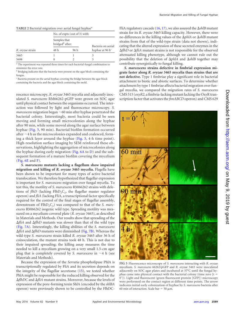
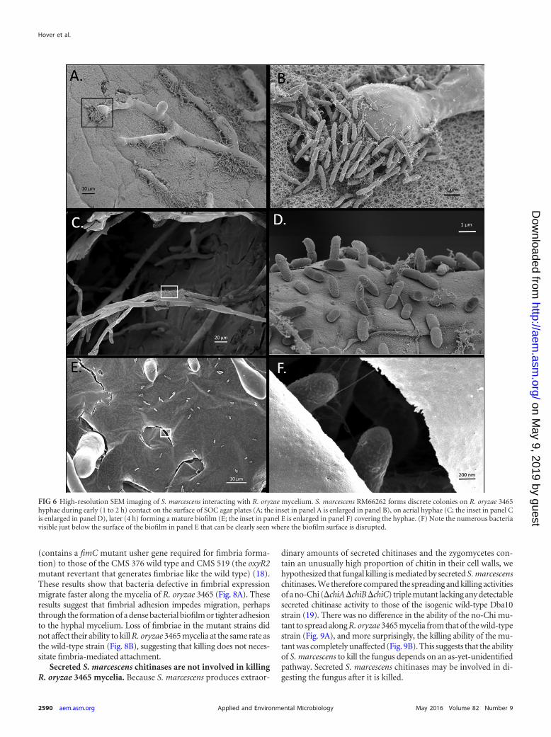
rescence microscopy, R. oryzae 3465 mycelia and adjacently inoc-ulated S. marcescens RM66262-pGFP were grown on SOC agaruntil physical contact between the organisms occurred. The inter-action was followed by light and fluorescence microscopy. S.marcescens migration began �60 min after hyphae penetrated thebacterial colony. Interestingly, most bacteria could be seenmoving and forming small microcolonies along the hyphaeafter 90 min, while some moved along the agar surface betweenhyphae (Fig. 5, 90 min). Bacterial biofilm formation occurredafter �4 h as the microcolonies expanded and coalesced, form-ing a thick layer around the hyphae (Fig. 5, 4-h time point).High-resolution surface imaging by SEM reinforced these ob-servations, highlighting the aggregation of microcolonies alongthe hyphae during early migration (Fig. 6A to D) and the sub-sequent formation of a mature biofilm covering the mycelium(Fig. 6E and F).
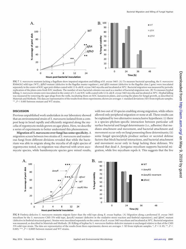
S. marcescens mutants lacking a flagellum show impairedmigration and killing of R. oryzae 3465 mycelia. Flagella havebeen shown to be important for many types of active bacterialtranslocation. We therefore hypothesized that flagellar expressionis important for S. marcescens migration over fungal mycelia. Totest this, the motility of S. marcescens RM66262 strains with dele-tions of flhD (lacking FlhD2C2, the flagellar master regulatoroperon) and fliA (lacking FliA, a transcriptional factor specificallyrequired for the control of the final stages of flagellar assembly,downstream of FlhD2C2) was compared to that of the S. marc-escens RM66262 isogenic wild type. Spreading motility was mea-sured on a mycelium-covered plate (R. oryzae 3465), as describedin Materials and Methods. Our results show that spreading of the�fliA and �flhD mutants was slower than that of the wild type(Fig. 7A). Interestingly, the killing abilities of the S. marcescens�fliA and �flhD mutants were diminished (Fig. 7B). Whereas thewild-type S. marcescens strain killed R. oryzae 3465 after 36 h ofcoinoculation, the mutant strains took 48 h. This is not due totheir impaired spreading; the killing assay measures the timeneeded to kill a mycelium growing on a very small 1.5-cm agarplug that is completely covered by S. marcescens in �4 h (seeMaterials and Methods).
Because the expression of the Serratia phospholipase PhlA istranscriptionally regulated by FliA and its secretion depends onthe integrity of the flagellar secretome (15), we tested whetherPhlA might be responsible for the reduced killing observed for the�flhDC and �fliA mutant strains. Moreover, because the levels ofexpression of the pore-forming toxin ShlA (encoded by the shlBAoperon) were previously shown to be controlled by the FlhDC-
FliA regulatory cascade (16, 17), we also assayed the �shlB mutantstrain for its R. oryzae 3465 killing capacity. However, there wereno differences in the killing values of the �phlA or �shlB mutantstrains from that of the wild-type strain (data not shown), indi-cating that the altered expression of these secreted enzymes in the�flhD or �fliA mutant strains is not responsible for the observedattenuated killing phenotype, although we cannot rule out thepossibility that the deletion of �phlA and �shlB together maycontribute synergistically to fungal killing.
S. marcescens strains defective in fimbrial expression mi-grate faster along R. oryzae 3465 mycelia than strains that arenot defective. Type 1 fimbriae play a significant role in bacterialattachment to biotic and abiotic surfaces. To determine whetherattachment by type 1 fimbriae affects bacterial migration over fun-gal mycelia, we compared the migration rates of S. marcescensCMS 515 (oxyR2, a fimbria-lacking mutant lacking the OxyR tran-scription factor that activates the fimABCD operon) and CMS 629
FIG 5 Fluorescence microscopy of S. marcescens interacting with R. oryzaemycelium. S. marcescens 66262/pGFP and R. oryzae 3465 were inoculatedadjacently on SOC agar plates and incubated at 37°C until the fungal hy-phae came into physical contact with the bacterial colony (time zero [t �0=]). Light and fluorescent (green fluorescent protein [GFP]) microscopywere performed on the contact region at different time points. The arrowindicates initial early colonization of hyphae by S. marcescens bacteria after60 min of interaction. Scale bar � 50 �m.
TABLE 2 Bacterial migration over aerial fungal hyphaea
R. oryzae strain
No. of expts (out of 3) with:
Samples thatbridgedb after:
Bacteria on aerialhyphae at 96 hc48 h 96 h
3465 1 1 35698 0 2 3a The experiment was repeated three times for each bacterial-fungal combination tominimize the error rate.b Bridging indicates that the bacteria were present on the agar block containing thefungus.c Bacteria present on the aerial hyphae covering the bridge between the agar blockcontaining the bacteria and the agar block containing the mold.
Bacterial Migration and Killing of Fungal Hyphae
May 2016 Volume 82 Number 9 aem.asm.org 2589Applied and Environmental Microbiology
(contains a fimC mutant usher gene required for fimbria forma-tion) to those of the CMS 376 wild type and CMS 519 (the oxyR2mutant revertant that generates fimbriae like the wild type) (18).These results show that bacteria defective in fimbrial expressionmigrate faster along the mycelia of R. oryzae 3465 (Fig. 8A). Theseresults suggest that fimbrial adhesion impedes migration, perhapsthrough the formation of a dense bacterial biofilm or tighter adhesionto the hyphal mycelium. Loss of fimbriae in the mutant strains didnot affect their ability to kill R. oryzae 3465 mycelia at the same rate asthe wild-type strain (Fig. 8B), suggesting that killing does not neces-sitate fimbria-mediated attachment.
Secreted S. marcescens chitinases are not involved in killingR. oryzae 3465 mycelia. Because S. marcescens produces extraor-
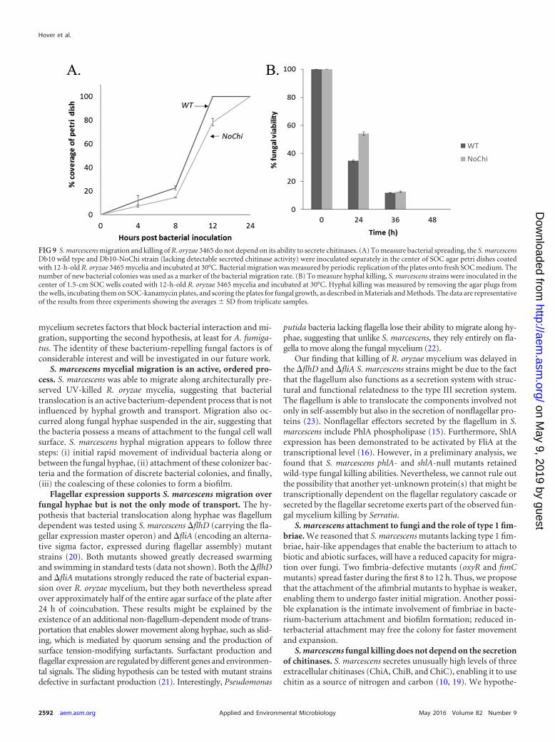
dinary amounts of secreted chitinases and the zygomycetes con-tain an unusually high proportion of chitin in their cell walls, wehypothesized that fungal killing is mediated by secreted S. marcescenschitinases. We therefore compared the spreading and killing activitiesof a no-Chi (�chiA�chiB�chiC) triple mutant lacking any detectablesecreted chitinase activity to those of the isogenic wild-type Dba10strain (19). There was no difference in the ability of the no-Chi mu-tant to spread along R. oryzae 3465 mycelia from that of the wild-typestrain (Fig. 9A), and more surprisingly, the killing ability of the mu-tant was completely unaffected (Fig. 9B). This suggests that the abilityof S. marcescens to kill the fungus depends on an as-yet-unidentifiedpathway. Secreted S. marcescens chitinases may be involved in di-gesting the fungus after it is killed.
FIG 6 High-resolution SEM imaging of S. marcescens interacting with R. oryzae mycelium. S. marcescens RM66262 forms discrete colonies on R. oryzae 3465hyphae during early (1 to 2 h) contact on the surface of SOC agar plates (A; the inset in panel A is enlarged in panel B), on aerial hyphae (C; the inset in panel Cis enlarged in panel D), later (4 h) forming a mature biofilm (E; the inset in panel E is enlarged in panel F) covering the hyphae. (F) Note the numerous bacteriavisible just below the surface of the biofilm in panel E that can be clearly seen where the biofilm surface is disrupted.
Hover et al.
2590 aem.asm.org May 2016 Volume 82 Number 9Applied and Environmental Microbiology
Previous unpublished work undertaken in our laboratory showedthat an environmental strain of S. marcescens isolated from a com-post heap in Israel rapidly and efficiently migrated along the my-celia of zygomycete molds grown on agar plates. Here, we describea series of experiments to better understand this phenomenon.
Migration of S. marcescens over fungi has some specificity. Amigration screen between two strains of S. marcescens and numer-ous fungi from different divisions revealed that while the bacte-rium was able to migrate along the mycelia of all eight species ofzygomycetes tested, no migration was observed with seven asco-mycete species, while basidiomycete species gave mixed results,
with two out of 10 species enabling strong migration, while othersallowed only peripheral migration or none at all. These results canbe explained by two alternative nonexclusive hypotheses: (i) thereis a species–phylum-specific interaction between particular cellsurface bacterial and fungal determinants (i.e., adhesins) that me-diates attachment and movement, and bacterial attachment andmovement occur only on fungi possessing these determinants; (ii)some fungal species/phyla produce surface or secreted defensefactors that block bacterial interactions, and bacterial attachmentand movement occur only in fungi lacking these defenses. Weshowed that dead A. fumigatus mycelium supports bacterial mi-gration, while live mycelium repels it. This suggests that the live
FIG 7 S. marcescens mutants lacking a flagellum show impaired migration and killing of R. oryzae 3465. (A) To measure bacterial spreading, the S. marcescensRM66262 wild type (WT), �flhD mutant (defective in the flagellar master regulator), and �fliA mutant (defective in the flagellar class 2 gene) were inoculatedseparately in the center of SOC agar petri dishes coated with 12-h-old R. oryzae 3465 mycelia and incubated at 30°C. Bacterial migration was measured by periodicreplication of the plates onto fresh SOC medium. The number of new bacterial colonies was used as a marker of bacterial migration rate. (B) To measure hyphalkilling, S. marcescens strains were inoculated in the center of 1.5-cm SOC wells coated with 12-h-old R. oryzae 3465 mycelia and incubated at 30°C. Hyphal killingwas measured by removing the agar plugs from the wells, incubating them on SOC-kanamycin plates, and scoring the plates for fungal growth, as described inMaterials and Methods. The data are representative of the results from three experiments; shown are averages standard deviations (SD) from triplicate samples.**, P � 0.005 between mutant and WT strains.
FIG 8 Fimbria-defective S. marcescens mutants migrate faster than the wild type along R. oryzae hyphae. (A) Migration along a preformed R. oryzae 3465mycelium by the S. marcescens CMS 376 wild type, �oxyR2 mutant (defective in the oxidative-stress reaction and fimbrial expression), and �fimC mutant(defective in fimbrial structural genes). Strains were inoculated separately on the center of an R. oryzae 3465 mycelium and incubated at 30°C. Bacterial migrationwas measured, as described in Materials and Methods. (B) Killing of hyphae by S. marcescens fimbria-defective mutants is unchanged relative to that of the CMS376 wild-type strain. The data are representative of the results from three experiments; shown are averages SD from triplicate samples. *, P � 0. 05; **, P �0.005; ***, P � 0.0005 between mutant and WT strains.
Bacterial Migration and Killing of Fungal Hyphae
May 2016 Volume 82 Number 9 aem.asm.org 2591Applied and Environmental Microbiology
mycelium secretes factors that block bacterial interaction and mi-gration, supporting the second hypothesis, at least for A. fumiga-tus. The identity of these bacterium-repelling fungal factors is ofconsiderable interest and will be investigated in our future work.
S. marcescens mycelial migration is an active, ordered pro-cess. S. marcescens was able to migrate along architecturally pre-served UV-killed R. oryzae mycelia, suggesting that bacterialtranslocation is an active bacterium-dependent process that is notinfluenced by hyphal growth and transport. Migration also oc-curred along fungal hyphae suspended in the air, suggesting thatthe bacteria possess a means of attachment to the fungal cell wallsurface. S. marcescens hyphal migration appears to follow threesteps: (i) initial rapid movement of individual bacteria along orbetween the fungal hyphae, (ii) attachment of these colonizer bac-teria and the formation of discrete bacterial colonies, and finally,(iii) the coalescing of these colonies to form a biofilm.
Flagellar expression supports S. marcescens migration overfungal hyphae but is not the only mode of transport. The hy-pothesis that bacterial translocation along hyphae was flagellumdependent was tested using S. marcescens �flhD (carrying the fla-gellar expression master operon) and �fliA (encoding an alterna-tive sigma factor, expressed during flagellar assembly) mutantstrains (20). Both mutants showed greatly decreased swarmingand swimming in standard tests (data not shown). Both the �flhDand �fliA mutations strongly reduced the rate of bacterial expan-sion over R. oryzae mycelium, but they both nevertheless spreadover approximately half of the entire agar surface of the plate after24 h of coincubation. These results might be explained by theexistence of an additional non-flagellum-dependent mode of trans-portation that enables slower movement along hyphae, such as slid-ing, which is mediated by quorum sensing and the production ofsurface tension-modifying surfactants. Surfactant production andflagellar expression are regulated by different genes and environmen-tal signals. The sliding hypothesis can be tested with mutant strainsdefective in surfactant production (21). Interestingly, Pseudomonas
putida bacteria lacking flagella lose their ability to migrate along hy-phae, suggesting that unlike S. marcescens, they rely entirely on fla-gella to move along the fungal mycelium (22).
Our finding that killing of R. oryzae mycelium was delayed inthe �flhD and �fliA S. marcescens strains might be due to the factthat the flagellum also functions as a secretion system with struc-tural and functional relatedness to the type III secretion system.The flagellum is able to translocate the components involved notonly in self-assembly but also in the secretion of nonflagellar pro-teins (23). Nonflagellar effectors secreted by the flagellum in S.marcescens include PhlA phospholipase (15). Furthermore, ShlAexpression has been demonstrated to be activated by FliA at thetranscriptional level (16). However, in a preliminary analysis, wefound that S. marcescens phlA- and shlA-null mutants retainedwild-type fungal killing abilities. Nevertheless, we cannot rule outthe possibility that another yet-unknown protein(s) that might betranscriptionally dependent on the flagellar regulatory cascade orsecreted by the flagellar secretome exerts part of the observed fun-gal mycelium killing by Serratia.
S. marcescens attachment to fungi and the role of type 1 fim-briae. We reasoned that S. marcescens mutants lacking type 1 fim-briae, hair-like appendages that enable the bacterium to attach tobiotic and abiotic surfaces, will have a reduced capacity for migra-tion over fungi. Two fimbria-defective mutants (oxyR and fimCmutants) spread faster during the first 8 to 12 h. Thus, we proposethat the attachment of the afimbrial mutants to hyphae is weaker,enabling them to undergo faster initial migration. Another possi-ble explanation is the intimate involvement of fimbriae in bacte-rium-bacterium attachment and biofilm formation; reduced in-terbacterial attachment may free the colony for faster movementand expansion.
S. marcescens fungal killing does not depend on the secretionof chitinases. S. marcescens secretes unusually high levels of threeextracellular chitinases (ChiA, ChiB, and ChiC), enabling it to usechitin as a source of nitrogen and carbon (10, 19). We hypothe-
FIG 9 S. marcescens migration and killing of R. oryzae 3465 do not depend on its ability to secrete chitinases. (A) To measure bacterial spreading, the S. marcescensDb10 wild type and Db10-NoChi strain (lacking detectable secreted chitinase activity) were inoculated separately in the center of SOC agar petri dishes coatedwith 12-h-old R. oryzae 3465 mycelia and incubated at 30°C. Bacterial migration was measured by periodic replication of the plates onto fresh SOC medium. Thenumber of new bacterial colonies was used as a marker of the bacterial migration rate. (B) To measure hyphal killing, S. marcescens strains were inoculated in thecenter of 1.5-cm SOC wells coated with 12-h-old R. oryzae 3465 mycelia and incubated at 30°C. Hyphal killing was measured by removing the agar plugs fromthe wells, incubating them on SOC-kanamycin plates, and scoring the plates for fungal growth, as described in Materials and Methods. The data are representativeof the results from three experiments showing the averages SD from triplicate samples.
Hover et al.
2592 aem.asm.org May 2016 Volume 82 Number 9Applied and Environmental Microbiology
sized that S. marcescens utilizes secreted chitinases to specificallytarget and kill zygomycete molds, because they contain unusuallyhigh levels of chitin (in the form of chitosan) in their cell walls(12). Surprisingly, we found that an S. marcescens triple-deletionmutant (�chiA �chiB �chiC) with no detectable secreted chitinaseactivity retained wild-type fungal killing abilities. We propose thatS. marcescens-mediated fungal killing is carried out by additionalas-yet-unknown virulence factors and that chitinases are used todegrade the cell wall during the next step of digestion and assim-ilation. P. aeruginosa killing of C. albicans hyphae is understood inmuch greater detail: it involves both contact-mediated and solublesecreted factors, such as phospholipase C and phenazine deriva-tives (5, 6, 24), suggesting similar approaches to pursue in ourfuture studies.
It is interesting to compare the S. marcescens-zygomycete in-teractions that we have described here to those found for P. aerugi-nosa-C. albicans interactions, which are the most extensively studiedsystem of bacterial-fungal interaction. In both cases and over similartime scales, the bacteria formed biofilms on the hyphal surface, lead-ing to fungal death (5). However, P. aeruginosa-C. albicans bindingoccurs in liquid medium, and unlike S. marcescens, P. aeruginosadoes not rapidly spread over fungal mycelia growing on agar surfaces(5). While flagellum-defective S. marcescens strains were delayed inspreading and killing of the fungus, similar P. aeruginosa mutantsshowed normal killing. While pilus-deficient S. marcescens strainsshowed increased spreading rates along hyphae and normal kill-ing, P. aeruginosa mutants showed normal biofilm formation butdelayed killing. These findings suggest that the molecular mechanismof S. marcescens-zygomycete interaction differs substantially fromthat of P. aeruginosa and C. albicans.
In summary, our characterization of the mechanisms allowingS. marcescens to migrate over fungal mycelia revealed a complex,multigenetic, and multifactorial system. The complexity of theinteraction, which takes place on solid agar surfaces and not insuspension in liquid medium, makes it difficult to analyze exper-imentally. It is unclear whether the fungi serve as a highway, anutrient source, or both. The chemical and physical nature ofbacterial attachment seems to involve numerous mechanisms,and the common properties of the fungi found to support or repelmigration will have to be further characterized as well.
ACKNOWLEDGMENTS
We thank Frank Sargent from the University of Dundee, United King-dom, for his generous donation of bacterial mutant strains and DanaLevinson from the faculty of Agriculture, Food, and Environment, Re-hovot, Israel, for the generous donation of fungal basidiomycete strains.
We declare no conflicts of interest.
FUNDING INFORMATIONThis work, including the efforts of Nir Osherov, was funded by BinationalScience Foundation US-Israel (2011322).
mycelia allow chemotactic dispersal of polycyclic aromatic hydrocarbon-degrading bacteria in water-unsaturated systems. Environ Microbiol 12:1391–1398.
2. Kohlmeier S, Smits TH, Ford RM, Keel C, Harms H, Wick LY. 2005.Taking the fungal highway: mobilization of pollutant-degrading bacteriaby fungi. Environ Sci Technol 39:4640 – 4646. http://dx.doi.org/10.1021/es047979z.
H. 2007. Effect of fungal hyphae on the access of bacteria to phenanthrenein soil. Environ Sci Technol 41:500 –505. http://dx.doi.org/10.1021/es061407s.
4. Peleg AY, Hogan DA, Mylonakis E. 2010. Medically important bacterial-fungal interactions. Nat Rev Microbiol 8:340 –349. http://dx.doi.org/10.1038/nrmicro2313.
5. Hogan DA, Kolter R. 2002. Pseudomonas-Candida interactions: an eco-logical role for virulence factors. Science 296:2229 –2232. http://dx.doi.org/10.1126/science.1070784.
6. Brand A, Barnes JD, Mackenzie KS, Odds FC, Gow NA. 2008. Cell wallglycans and soluble factors determine the interactions between the hyphaeof Candida albicans and Pseudomonas aeruginosa. FEMS Microbiol Lett287:48 –55. http://dx.doi.org/10.1111/j.1574-6968.2008.01301.x.
7. Gibson J, Sood A, Hogan DA. 2009. Pseudomonas aeruginosa-Candidaalbicans interactions: localization and fungal toxicity of a phenazine de-rivative. Appl Environ Microbiol 75:504 –513. http://dx.doi.org/10.1128/AEM.01037-08.
9. Mahlen SD. 2011. Serratia infections: from military experiments to cur-rent practice. Clin Microbiol Rev 24:755–791. http://dx.doi.org/10.1128/CMR.00017-11.
10. Vaaje-Kolstad G, Horn SJ, Sørlie M, Eijsink VG. 2013. The chitinolyticmachinery of Serratia marcescens—a model system for enzymatic degra-dation of recalcitrant polysaccharides. FEBS J 280:3028 –3049. http://dx.doi.org/10.1111/febs.12181.
11. Ibrahim AS, Kontoyiannis DP. 2013. Update on mucormycosis patho-genesis. Curr Opin Infect Dis 26:508 –515. http://dx.doi.org/10.1097/QCO.0000000000000008.
12. Mélida H, Sain D, Stajich JE, Bulone V. 2015. Deciphering the unique-ness of Mucoromycotina cell walls by combining biochemical and phylog-enomic approaches. Environ Microbiol 17:1649 –1662. http://dx.doi.org/10.1111/1462-2920.12601.
13. Fichtman B, Shaulov L, Harel A. 2014. Imaging metazoan nuclear porecomplexes by field emission scanning electron microscopy. Methods CellBiol 122:41–58. http://dx.doi.org/10.1016/B978-0-12-417160-2.00002-3.
14. Soo PC, Wei JR, Horng YT, Hsieh SC, Ho SW, Lai HC. 2005. Charac-terization of the dapA-nlpB genetic locus involved in regulation of swarm-ing motility, cell envelope architecture, hemolysin production, and cellattachment ability in Serratia marcescens. Infect Immun 73:6075– 6084.http://dx.doi.org/10.1128/IAI.73.9.6075-6084.2005.
15. Givskov M, Eberl L, Christiansen G, Benedik MJ, Molin S. 1995.Induction of phospholipase- and flagellar synthesis in Serratia liquefa-ciens is controlled by expression of the flagellar master operon flhD.Mol Microbiol 15:445– 454. http://dx.doi.org/10.1111/j.1365-2958.1995.tb02258.x.
16. Di Venanzio G, Stepanenko TM, Garcia Véscovi E. 2014. Serratiamarcescens ShlA pore-forming toxin is responsible for early induction ofautophagy in host cells and is transcriptionally regulated by RcsB. InfectImmun 82:3542–3554. http://dx.doi.org/10.1128/IAI.01682-14.
17. Lin CS, Horng JT, Yang CH, Tsai YH, Su LH, Wei CF, Chen CC, HsiehSC, Lu CC, Lai HC. 2010. RssAB-FlhDC-ShlBA as a major pathogenesispathway in Serratia marcescens. Infect Immun 78:4870 – 4881. http://dx.doi.org/10.1128/IAI.00661-10.
18. Shanks RM, Stella NA, Kalivoda EJ, Doe MR, O’Dee DM, Lathrop KL,Guo FL, Nau GJ. 2007. A Serratia marcescens OxyR homolog mediatessurface attachment and biofilm formation. J Bacteriol 189:7262–7272.http://dx.doi.org/10.1128/JB.00859-07.
19. Hamilton JJ, Marlow VL, Owen RA, Costa MDAA, Guo M, BuchananG, Chandra G, Trost M, Coulthurst SJ, Palmer T, Stanley-Wall NR,Sargent F. 2014. A holin and an endopeptidase are essential for chitino-lytic protein secretion in Serratia marcescens. J Cell Biol 207:615– 626.http://dx.doi.org/10.1083/jcb.201404127.
20. Fedrigo GV, Campoy EM, Di Venanzio G, Colombo MI, Garcia VéscoviE. 2011. Serratia marcescens is able to survive and proliferate in autophag-ic-like vacuoles inside non-phagocytic cells. PLoS One 6:e24054. http://dx.doi.org/10.1371/journal.pone.0024054.
21. Li H, Tanikawa T, Sato Y, Nakagawa Y, Matsuyama T. 2005. Serratiamarcescens gene required for surfactant serrawettin W1 production en-codes putative aminolipid synthetase belonging to nonribosomal peptide
Bacterial Migration and Killing of Fungal Hyphae
May 2016 Volume 82 Number 9 aem.asm.org 2593Applied and Environmental Microbiology
22. Pion M, Bshary R, Bindschedler S, Filippidou S, Wick LY, Job D, JunierP. 2013. Gains of bacterial flagellar motility in a fungal world. Appl Envi-ron Microbiol 79:6862– 6867. http://dx.doi.org/10.1128/AEM.01393-13.
23. Coburn B, Sekirov I, Finlay BB. 2007. Type III secretion systems anddisease. Clin Microbiol Rev 20:535–549. http://dx.doi.org/10.1128/CMR.00013-07.
24. Morales DK, Grahl N, Okegbe C, Dietrich LE, Jacobs NJ, Hogan DA.2013. Control of Candida albicans metabolism and biofilm formation by
25. Bruna RE, Revale S, Garcia Vescovi E, Mariscotti JF. 2015. Draftwhole-genome sequence of Serratia marcescens strain RM66262, isolatedfrom a patient with a urinary tract infection. Genome Announc 3(6):e01423-15. http://dx.doi.org/10.1128/genomeA.01423-15.
26. Castelli ME, Fedrigo GV, Clementin AL, Ielmini MV, Feldman MF,Garcia Véscovi E. 2008. Enterobacterial common antigen integrity is acheckpoint for flagellar biogenesis in Serratia marcescens. J Bacteriol 190:213–220. http://dx.doi.org/10.1128/JB.01348-07.
Hover et al.
2594 aem.asm.org May 2016 Volume 82 Number 9Applied and Environmental Microbiology