All rights reserved. However, in accordance with the Copyright Act of Canada, this work may be reproduced, without authorization, under the conditions for Fair Dealing. Therefore, limited
reproduction of this work for the purposes of private study, research, criticism, review and news
reporting is likely to be in accordance with the law, particularly if cited appropriately.
Name:
Degree:
APPROVAL
Syama Gauri Dasi Chatterton
Doctor of Philosophy
Tit le of Thesis:
Mechanisms of biological control of Fusar ium root and stem rot of greenhouse
cucumber by Gliocladium catenulatum
Examin ing Commit tee:
Chair: Dr. J. Revnolds. Professor
Dr. Z. Punja, Professor. Senior SupervisorDepartment of B io logica l Sc iences, S.F.U.
Dr. M. Moore. ProfessorDepartment of B io logica l Sc iences, S.F.U.
Dr. A. Plant. Associate Professor (Retired) and Adjunct ProfessorDepart rnent of B io logica l Sc iences, S.F.U.
Dr. R. Hamelin. ProfessorDepartment of Forestry, The University of Bri t ish ColumbiaPublic Examiner
Dr. T. Pauli tz. Research Plant PathologistDepartrnent of Plant Pathology. Washington State UniversityExternal Examiner
I 1 January 2010Date Approved
Last revision: Spring 09
Declaration of Partial Copyright Licence
The author, whose copyright is declared on the title page of this work, has granted to Simon Fraser University the right to lend this thesis, project or extended essay to users of the Simon Fraser University Library, and to make partial or single copies only for such users or in response to a request from the library of any other university, or other educational institution, on its own behalf or for one of its users.
The author has further granted permission to Simon Fraser University to keep or make a digital copy for use in its circulating collection (currently available to the public at the “Institutional Repository” link of the SFU Library website <www.lib.sfu.ca> at: <http://ir.lib.sfu.ca/handle/1892/112>) and, without changing the content, to translate the thesis/project or extended essays, if technically possible, to any medium or format for the purpose of preservation of the digital work.
The author has further agreed that permission for multiple copying of this work for scholarly purposes may be granted by either the author or the Dean of Graduate Studies.
It is understood that copying or publication of this work for financial gain shall not be allowed without the author’s written permission.
Permission for public performance, or limited permission for private scholarly use, of any multimedia materials forming part of this work, may have been granted by the author. This information may be found on the separately catalogued multimedia material and in the signed Partial Copyright Licence.
While licensing SFU to permit the above uses, the author retains copyright in the thesis, project or extended essays, including the right to change the work for subsequent purposes, including editing and publishing the work in whole or in part, and licensing other parties, as the author may desire.
The original Partial Copyright Licence attesting to these terms, and signed by this author, may be found in the original bound copy of this work, retained in the Simon Fraser University Archive.
Simon Fraser University Library Burnaby, BC, Canada
iii
ABSTRACT
Gliocladium catenulatum strain J1446 (formulated as Prestop WP,
Verdera Oy) is a biological control agent of Fusarium root and stem rot caused
by Fusarium oxysporum f. sp. radicis-cucumerinum on greenhouse cucumber
plants. The mechanisms involved in biocontrol efficacy are currently unknown.
Following transformation of G. catenulatum with the -glucuronidase (uidA) gene,
blue-stained mycelia could be seen growing on the surface and within epidermal
and cortical cells of roots, stems and shoots 3 weeks after treatment. Application
of G. catenulatum preceding inoculation with Fusarium significantly reduced
pathogen populations on roots compared to plants inoculated with Fusarium
alone, while densities of the biocontrol agent increased in the presence of the
pathogen. Factors influencing root population levels included nutrient solution
pH, temperature and growing media type, while cucumber cultivar, root wounding
and addition of nutrients did not appear to significantly affect colonization. In
culture, G. catenulatum produced chitinase and ß-1,3-glucanase enzymes on
chitin or laminarin as a sole carbon source, respectively, and caused localized
degradation of Fusarium hyphae. Cucumber root extracts from G. catenulatum-
colonized plants had significantly higher levels of glucanase at 7 days post-
application compared to untreated controls. Reverse-transcription polymerase
chain reaction using primers designed to amplify a -1,3-glucanase gene
confirmed G. catenulatum glucanase expression on roots. In a split-root system,
iv
G. catenulatum applied to one-half of the roots prior to inoculation with Fusarium
on the other half did not significantly reduce disease compared to plants treated
with Fusarium only. There was no detectable increase in chitinase, peroxidase
or polyphenol oxidase enzyme activity in roots and leaves following treatment
with G. catenulatum. Competitive colonization of the rhizosphere by G.
catenulatum, which is facilitated by its mycoparasitic ability, are the primary
mechanisms by which pathogen development and disease incidence is reduced.
1.1 Pathogens in the greenhouse environment ........................................ 1
1.2 Biological control of pathogens in greenhouses ................................. 3
1.2.1 Biological control of root pathogens using fungal biocontrol agents ............................................................................................. 4
1.3 Biocontrol by Gliocladium catenulatum .............................................. 6
1.3.1 Taxonomy of the genus Gliocladium ............................................... 6 1.3.2 Diseases suppressed by Gliocladium catenulatum ......................... 7
1.4 Mechanisms of disease suppression by biological control agents ................................................................................................ 9
1.5 Root colonization .............................................................................. 26 1.6 Environmental factors determining the success of biocontrol
agents .............................................................................................. 29 1.7 Research objectives ......................................................................... 32
2: Colonization of cucumber plants by the biocontrol fungus Gliocladium catenulatum ................................................................................. 34
2.1 Introduction ...................................................................................... 34 2.2 Materials and Methods ..................................................................... 36
2.2.1 Fungal strains and culture conditions ............................................ 36 2.2.2 Biological control activity of G. catenulatum .................................. 37 2.2.3 Survival of G. catenulatum ............................................................ 38
2.2.4 Scanning electron microscopic studies ......................................... 39
ix
2.2.5 Extent of internal colonization ....................................................... 40 2.2.6 GUS-transformation ...................................................................... 40 2.2.7 Growth of GUS-transformed G. catenulatum on cucumber
tissues ........................................................................................... 42 2.2.8 Estimation of fungal biomass ........................................................ 43 2.2.9 Survival of F. oxysporum on cucumber roots in the presence
of G. catenulatum .......................................................................... 44
2.3 Results ............................................................................................. 45 2.3.1 Biological control activity and survival of G. catenulatum. ............. 45
2.3.2 Scanning electron microscopic observations. ............................... 46
2.3.3 Extent of internal tissue colonization. ............................................ 49 2.3.4 GUS transformation ...................................................................... 49 2.3.5 Growth of GUS transformed G. catenulatum on cucumber
2.3.6 Estimation of fungal biomass ........................................................ 54 2.3.7 Survival of F. oxysporum on cucumber roots in the presence
of G. catenulatum .......................................................................... 54
3: Chitinase and -1,3-glucanase enzyme production by Gliocladium catenulatum against the fungal plant pathogens Fusarium and Pythium ............................................................................................................. 66
3.2 Materials and Methods ..................................................................... 68 3.2.1 Production of antifungal metabolic compounds G.
catenulatum that inhibit growth of F. oxysporum. .......................... 68
3.2.2 Chitinase and glucanase production in culture ............................. 69 3.2.3 Scanning electron microscopy (SEM) ........................................... 71 3.2.4 Effect of crude enzyme extracts on Pythium and Fusarium
mycelial growth and conidial germination ..................................... 72 3.2.5 Release of glucose or NAGA from Fusarium or Pythium cell
wall fragments ............................................................................... 73 3.2.6 Measurement of chitinase and glucanase enzymes in
3.3 Results ............................................................................................. 77 3.3.1 Production of antifungal metabolic compounds G.
catenulatum that inhibit growth of F. oxysporum. .......................... 77
3.3.2 Chitinase and glucanase production in culture ............................. 80 3.3.3 Scanning electron microscopy ...................................................... 83 3.3.4 Effect of crude enzyme extracts on Pythium and Fusarium
growth and conidial germination ................................................... 86 3.3.5 Release of glucose or NAGA from Fusarium or Pythium cell
4.3 Results ........................................................................................... 108 4.3.1 Factors affecting population levels of G. catenulatum on
5: Evaluation of Gliocladium catenulatum to induce systemic resistance in cucumber ................................................................................. 126
5.2 Materials and Methods ................................................................... 128
5.2.1 Fungal strains and culture conditions .......................................... 128 5.2.2 Disease development of Fusarium root and stem rot and
biocontrol by G. catenulatum in hydroponic culture .................... 130
5.2.3 Split-root assays ......................................................................... 131 5.2.4 Measurement of defense-related enzymes in cucumber
plants .......................................................................................... 132 5.2.5 Disease development of P. syringae pv. lachrymans (Psl) in
5.3.1 Disease development of Fusarium root and stem rot and biocontrol by G. catenulatum in hydroponic culture .................... 134
5.3.2 Split-root assays. ........................................................................ 135 5.3.3 Measurement of defense-related enzymes in cucumber
plants. ......................................................................................... 137 5.3.4 Disease development of P. syringae pv. lachrymans in
Figure 2-1. Disease severity index (DSI) of cucumber plants and population densities of G. catenulatum on cucumber roots, tissues and growing medium. .............................................................. 47
Figure 2-2. Scanning electron micrographs of cucumber roots 7 days after inoculation with G. catenulatum (A-F), or F. oxysporum (G-I), or G. catenulatum followed 3 days later by F. oxysporum (J-L). .............. 48
Figure 2-3 Colonization of root, stem, cotyledon and leaf pieces by G. catenulatum after 0, 10, 30 or 60 s of surface sterilization, 3 weeks following application of Prestop WP to the seed. ...................... 50
Figure 2-4 Colonization pattern of cucumber plants by G. catenulatum strain J1446 expressing the GUS gene. .............................................. 53
Figure 2-5 Relationship between fungal biomass or protein content and GUS activity in a transformant of G. catenulatum. ............................... 55
Figure 2-6 Fungal biomass of G. catenulatum expressing the GUS gene in different tissues of cucumber plants following application to seed or rockwool block. ....................................................................... 56
Figure 2-7 Population size of Forc in the roots (top) or crown area (bottom) of cucumber plants treated with G. catenulatum or Prestop WP ......................................................................................... 57
Figure 3-1 Dual culture plates of G. catenulatum (Gc) and F. oxysporum (Fo) or P. aphanidermatum.................................................................. 78
Figure 3-2 Colony diameter (top) and conidia production (bottom) of F. oxysporum that was grown alone or after 7 days exposure to cultures of G. catenulatum growing for 7 days or 14 days on a Petri dish attached by parafilm. ........................................................... 79
Figure 3-3 Time course of (A) chitinase and (B) -1,3-glucanase production by Gliocladium catenulatum on MSM (no carbon source) and on medium containing chitin or laminarin as a carbon source, respectively. ................................................................ 81
Figure 3-4 Effect of carbon source on chitinase and -1,3-glucanase production after 7 days of growth of Gliocladium catenulatum. ........... 82
xii
Figure 3-5 .Effect of initial medium pH on (A) chitinase and (B) -1,3-glucanase production by Gliocladium catenulatum grown for 7 days on chitin or laminarin as the carbon source, respectively. ........... 84
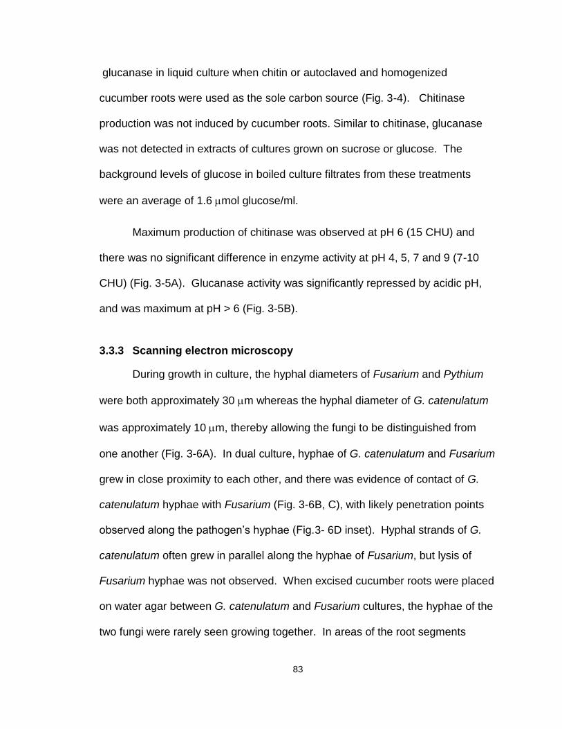
Figure 3-6 Scanning electron micrographs of the interactions between Gliocladium catenulatum (c) and Fusarium oxysporum (f) in dual culture on water agar (A-D), or between G. catenulatum and Pythium (p) on excised cucumber roots (r) on water agar (E,F). ......... 85
Figure 3-7 Effect of Gliocladium catenulatum culture filtrates on (A) growth of Fusarium oxysporum or Pythium aphanidermatum mycelia or (B) germination of Fusarium conidia. .................................. 87
Figure 3-8 Effect of Gliocladium catenulatum culture filtrates on the release of (A) NAGA from Fusarium cell wall fragments or (B) glucose from Fusarium or (C) Pythium cell wall fragments. ................. 88
Figure 3-9 Glucanase activity in roots of 10-day-old cucumber seedlings at 2, 3 and 7 days following inoculation with Gliocladium catenulatum (Gc), treatment with salicylic acid (SA) or water (Con). .................................................................................................. 90
Figure 3-10 SDS-PAGE of -1,3-glucanase isoforms from G. catenulatum. ........................................................................................ 91
Figure 3-11 Detection of -1,3-glucanase expression by Gliocladium catenulatum on colonized cucumber roots using reverse transcription – polymerase chain reaction (RT-PCR). ......................... 93
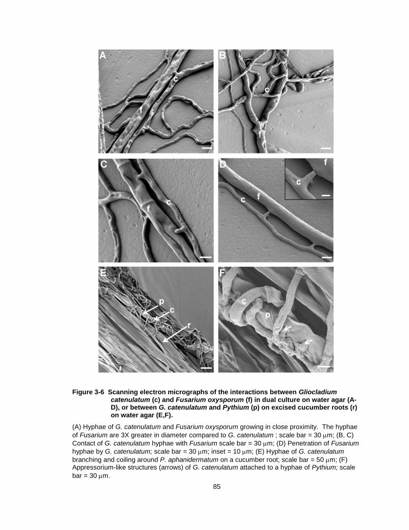
Figure 3-12 Northern blot analysis of expression of a -1,3-glucanase gene from Gliocladium catenulatum. ................................................... 94
Figure 4-1 Population levels of G. catenulatum on cucumber roots determined by colony plate counts (A) or GUS expression (B) as influenced by pH of the nutrient solution. ........................................... 110
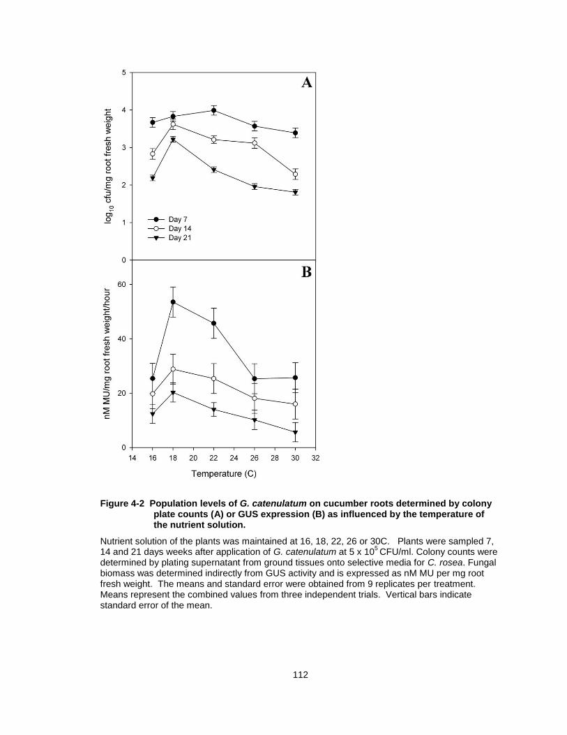
Figure 4-2 Population levels of G. catenulatum on cucumber roots determined by colony plate counts (A) or GUS expression (B) as influenced by the temperature of the nutrient solution. ...................... 112
Figure 4-3 Population levels of G. catenulatum on cucumber roots grown in different media types determined by colony plate counts (A) or GUS expression (B). .......................................................................... 113
Figure 4-4 Population levels of G. catenulatum on the roots of 5 cucumber cultivars (Averyl, Bodega, Ladner, Marcel or Sienna) determined by colony plate counts (A) or GUS expression (B).......... 115
Figure 4-5 Population levels of G. catenulatum on cucumber roots as affected by addition of glucose or asparagine to the nutrient solution determined by colony plate counts (A) or GUS expression (B). .................................................................................. 117
xiii
Figure 4-6 Population levels of G. catenulatum on unwounded (control) cucumber roots or wounded roots determined by colony plate counts (A) or GUS expression (B). .................................................... 118
Figure 5-1 Disease severity index (A) and root fresh weight (B) of cucumber plants inoculated with Fusarium only or inoculated with Fusarium three days after application of G. catenulatum (1 x 10⁶ conidia/ml NS) to the roots (Gc + Fus). .................................... 136
Figure 5-2 Disease severity index (A) and root fresh weight (B) of cucumber plants grown in hydroponic solution and either untreated (control) or treated with Gliocladium on one half of the roots (Gc), treated with Forc on one half of the roots only (Forc) or treated with Gliocladium on one half of the roots 3 days before treatment with Forc on the other half of the roots (Forc+Gc). ............ 138
Figure 5-3 Enzyme activities in roots or leaves of 10-day-old cucumber seedlings at 2, 3 and 7 days following application of INA (2 mg/L), G. catenulatum (1x 10⁶ cfu/ml) or SA (2 mM). ........................ 141
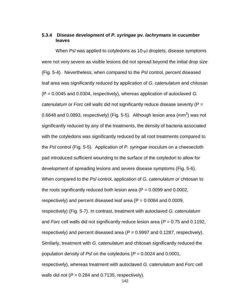
Figure 5-4 Disease symptoms of Pseudomonas syringae pv. lachrymans when applied as droplets on cotyledons of cucumber plants, 5 days after pathogen inoculation. ........................................................ 143
Figure 5-5 Disease severity of Pseudomonas syringae pv. lachrymans when applied as droplets on cotyledons of cucumber plants measured by lesion area, mm2 (A), % diseased leaf area (B), and population levels of Psl on the surface of cotyledons (C). .......... 144
Figure 5-6 Disease symptoms of Pseudomonas syringae pv. lachrymans when applied using a cheesecloth rub on cotyledons of cucumber plants, 5 days after pathogen inoculation. ......................... 145
Figure 5-7 Disease severity of Pseudomonas syringae pv. lachrymans when applied using a cheesecloth rub on cotyledons of cucumber plants measured by lesion area, mm2 (A), % diseased leaf area (B), and population levels of Psl on the surface of cotyledons (C). .................................................................................. 146
xiv
LIST OF TABLES
Table 2-1 Slope values (b) from linear regression analysis of the population levels of Forc in the roots or crown area of cucumber plants treated with G. catenulatum (107 conidia/ml) versus log10-transformed initial inoculum of Forc. .................................................... 59
Table 2-2 Population levels of G. catenulatum in the roots or crown area of cucumber plants treated with G. catenulatum applied to the rockwool blocks (107 conidia/ml) prior to inoculation with Forc at an initial inoculum level of either 104, 105, or 106 conidia/ml. ............... 60
xv
LIST OF ACRONYMS AND ABBREVIATIONS USED
APDA Acidified potato dextrose agar
ANOVA Analysis of variance.
AS Acetosyringone
BCA Biocontrol agent
CFU Colony forming unit
cm centimetre
CHU Chitinase units
DPA Days post-application
DPI Days post-inoculation
DSI Disease severity index
oC Degree Celsius
Forc Fusarium oxysporum f. sp. radicis-cucumerinum
gpd glyceraldehyde-3-phosphate dehydrogenase
GFP Green fluorescent protein
GU Glucanase units
GUS -glucuronidase
IM Induction media
INA 2,6-dichloroisonicotinic acid
Log10 Logarithm to base 10
xvi
M Molarity
min Minutes
MSM Minimal synthetic (or salts) media
MU Methylumbelliferone
MUG Methylumbelliferyl β-D-glucuronide hydrate
NAGA N-acetyl glucosamine
NS Nutrient solution
PCA Paraquat chloramphenicol agar
PDA Potato dextrose agar
PDAtt Potato dextrose agar + Triton + tetracycline
PDB Potato dextrose broth
Psl Pseudomonas syringae pv lachrymans
RFW Root fresh weight
s seconds
SA Salicylic acid
SDW Sterile distilled water
VOC Volatile organic compound
1
1: INTRODUCTION
1.1 Pathogens in the greenhouse environment
One of the motivating factors in developing greenhouse cropping systems
was the theoretical potential to eliminate or minimize plant diseases (Stanghellini
and Rasmussen, 1994). Certainly, the diversity of pathogens is lower in the
greenhouse, but most pathogens cannot be effectively excluded from the
greenhouse environment (Paulitz and Belanger, 2001). Hence, several fungi
have emerged as major root and foliar pathogens that can cause devastating
crop losses when introduced into the enclosed system (Stanghellini and
Rasmussen, 1994). Soilless substrates such as peat or rockwool lack the
microbial diversity of natural soil. Thus, soilborne pathogens such as Pythium
aphanidermatum, Rhizoctonia solani and Fusarium oxysporum can quickly grow
and spread (Paulitz and Belanger, 2001). The warm temperatures and high
humidity maintained in the greenhouse that ensure optimal plant growth also
provide ideal conditions for infection by foliar pathogens such as Botrytis cinerea
and powdery mildews. High density planting of greenhouse crops and the
recirculation of nutrient solution can facilitate rapid spread of pathogens.
Management practices, such as pruning and continuous harvesting, also provide
sources of infection through wounds.
In British Columbia, two common root-infecting pathogens of greenhouse
cucumbers are P. aphanidermatum, causing damping-off and root rot and
2
Fusarium oxysporum f. sp. radicis-cucumerinum (Forc), causing Fusarium root
and stem rot (Punja and Parker, 2000). F. oxysporum is an ascomycete fungus
in the Order Hypocreales (Michielse and Rep, 2009). P. aphanidermatum is an
oomycete, which are phylogenetically more closely related to algae than fungi,
although its classification under a new Kingdom Stramenopiles or Chromista
within the Domain Eukarya is under debate (Adl et al., 2005). Distinct
characteristics of oomycetes are cell walls that lack chitin, and are instead
composed primarily of -1,3-glucan, and the production of motile flagellated
aquatic zoospores (Viterbo et al., 2002b; Stanghellini and Rasmussen, 1994).
Symptoms of P. aphanidermatum infection include extensive browning and
rotting of the root system and crown area (Wulff et al., 1998; Punja and Yip,
2003). Symptoms of F. oxysporum infection include a brown discoloration at the
crown and vertical stem lesions accompanied by the presence of white mycelium
and masses of orange conidia (Punja and Parker, 2000; Vakalounakis, 1996). In
advanced stages of both diseases, plants begin to wilt and eventually die. While
there are no cucumber cultivars resistant to P. aphanidermatum (Punja and Yip,
2003), cultivars of cucumber differ in their tolerance to F. oxysporum, with some
cultivars showing high levels of resistance (Rose and Punja, 2004). Although
seedlings appear to be most susceptible to infection by both F. oxysporum and
P. aphanidermatum, disease symptoms often are most apparent in older plants
(Punja and Parker, 2000; Favrin et al., 1988). Therefore, the pathogens may be
present in the plant without causing disease until the plants become stressed by
events such as fruiting, or by environmental factors such as high temperatures or
3
imbalances in the nutrient solution (Cherif et al., 1997; Favrin et al., 1988; Punja
and Parker, 2000). Both pathogens are likely introduced into the greenhouse on
infected plant material, contaminated growing substrates or irrigation water, with
subsequent spread occurring throughout the greenhouse by water dispersal or
contaminated equipment (Paulitz and Belanger, 2001; Stanghellini and
Rasmussen, 1994).
1.2 Biological control of pathogens in greenhouses
Greenhouses and protected structures provide a unique niche for the
implementation of biological control programs. Currently, over half of the
commercially available biocontrol products are registered for use in greenhouses
and nurseries, making the use of biocontrol agents in greenhouses more
prevalent than in field crops (van Lenteren, 2000). Many of these biocontrol
agents (BCAs) have been developed specifically against the major greenhouse
pathogens, such as Pythium, Rhizoctonia and Fusarium (Paulitz and Belanger,
2001). Differences between field and greenhouse environments may help to
explain the success of biocontrol agents in the greenhouse. Environmental
parameters, such as temperature and relative humidity, can be adjusted to
provide a consistency of performance of biocontrol agents that is not often
observed under natural field conditions (Paulitz and Belanger, 2001).
Greenhouses are relatively isolated units and therefore a limited number of pest
species occur within them (van Lenteren, 2000). Introduced biological control
agents are likely to establish and proliferate due to the low biological diversity in
soilless substrates observed in the early stages of greenhouse production
4
(Fravel, 2005). To aid in dispersal throughout the cropping system, biocontrol
agents, if formulated correctly, can be easily added to the nutrient solution, thus
facilitating uniform distribution (Paulitz, 1997).
Growers working in greenhouses often prefer biological control over
chemical control agents for a number of reasons. Unlike their behaviour in soil,
fungicides applied to the roots often cause toxicity to the crop plant under soilless
conditions due to the lack of adsorption and dispersal (van Lenteren, 2000). In
addition, there are relatively few fungicides registered for use in Canadian
greenhouses. The use of biocontrol agents does not disrupt scheduled
operations such as the prolonged re-entry times required after pesticide use
(Harman, 2000). Consumer demands for pesticide-free foods also stimulate the
use of biocontrol agents (van Lenteren, 2000). There is considerable
precedence for the use of biological control against insect pests in greenhouses
and this has become a well-established practice by growers. However, in spite
of the great optimism and extensive research efforts, progress in achieving
commercial, large-scale use of biological disease control has been slow and has
yet to achieve the same successes realized for insect control (Spadaro and
Gullino, 2005).
1.2.1 Biological control of root pathogens using fungal biocontrol agents
1.2.1.1 Trichoderma spp.
By far, the most studied biocontrol agents are Trichoderma spp.
accounting for 90% of fungal strains that have been evaluated for use as
biocontrol agents of plant diseases (Benitez et al., 2004). The most common
5
BCAs from the Trichoderma genus are strains of T. virens, T. viride, T.
asperellum and T. harzianum (Punja and Utkhede, 2003). The success of
Trichoderma strains as BCAs is due to their high reproductive capacity, their
ability to survive under unfavourable environmental conditions, efficient utilization
of nutrients, capability to colonize the plant rhizosphere, and strong
aggressiveness against plant pathogenic fungi (Benitez et al., 2004). T.
harzianum strain T-22 is marketed by Bioworks, Geneva, NY as a granular
promoter. Agrobacterium tumefaciens strain AGL-1 (provided by Dr. S. Marek,
Oklahoma State University) was transformed with the binary vector (An et al.,
1988). Positive transformants were verified using PCR, stored in -80oC glycerol
stocks and were used to transform G. catenulatum according to the following
procedure (Dr. S. Marek, personal communication). Cells from glycerol stock
were plated onto Agrobacterium minimal medium containing kanamycin
(100g/ml) and carbenicillin (100g/ml) and incubated at 28oC for 2-3 days.
Single colonies were used to inoculate liquid minimal medium and incubated at
28oC with shaking at 250 rpm for 2 days. Cultures were then diluted in induction
medium (IM) containing 40 mM MES, 0.5% glycerol (w/v) and 200 M
acetosyringone (AS) (Mozo and Hooykaas, 1991) to an optical density of 0.2
absorbance units at 600 nm and then incubated overnight at 28oC with shaking
at 250 rpm. The induced suspension was then diluted again to an optical density
of 0.2 absorbance units at 600 nm in IM for use in transformations. Conidia of G.
catenulatum were diluted in IM to a concentration of 2x10⁵, 2x10⁴ or 2x10³
conidia/ml and 500 l of each conidia suspension was mixed with 500 l of the
induced Agrobacterium cells. Aliquots (200l) were plated onto Hybond
membranes (Amersham) placed on IM plates containing 200 M AS and
incubated at room temperature for 72 h. The membranes were transferred to
YPS plates containing 300 g/ml timentin to inhibit Agrobacterium growth and
200 g/ml hygromycin to select the Gliocladium transformants and incubated at
room temperature for 7-10 days. Colonies growing on the selective medium
were tested for GUS activity by placing mycelia from each colony in the well of a
42
microtiter plate containing 200 l of 10 mM sodium phosphate buffer (pH 7.0) and
4 l of X-Gluc substrate (12 mg/ml 5-bromo-4-chloro-3-indolyl-b-D-glucuronic
acid substrate, Sigma). For each positive isolate, single-conidial isolations were
carried out to ensure mitotic stability. The transformed isolate displaying the
highest level of GUS expression as determined by staining intensity was chosen
for further studies. To test for genetic stability, this isolate was cultivated under
non-selective conditions for 2 months, after which time colonies were tested for
hygromycin B resistance and GUS activity. Integration of the uidA gene was
verified by Southern blot hybridization following EcoRI digestion of the genomic
DNA (Wally et al., 2008).
2.2.7 Growth of GUS-transformed G. catenulatum on cucumber tissues
Cucumber plants were grown aseptically in Magenta boxes or in rockwool
blocks as described previously. Seeds were treated with GUS-transformed G.
catenulatum prior to planting by soaking in 107 conidia/ml for 10 minutes, then
air-drying for 30 minutes and immediately planted, or G. catenulatum was applied
to the nutrient solution in Magenta boxes when plants were 10 days old or to
rockwool blocks at seeding as described above. Tissues were harvested at
intervals between 2 and 10 days and histochemical staining of germinating
seeds, root, stem, apical meristem and leaf pieces was performed according to
Jefferson et al. (1986) with modifications according to Wally et al. (2008). Tissue
segments that displayed characteristic blue staining were mounted in resin,
sectioned (4 m thickness) and viewed under 40 X magnification using bright-
field microscopy. To verify the results of GUS staining, root, stem and leaf
43
pieces were simultaneously plated onto PDA+hygromycin to confirm presence of
the transformed fungus.
2.2.8 Estimation of fungal biomass
Fungal biomass of GUS-transformed G. catenulatum on cucumber tissues
was estimated from GUS activity using a modification of the fluorometric method
(Jefferson et al., 1986). A standard curve correlating fungal biomass to 4-
methylumbelliferyl (MU) concentration was first developed by measuring GUS
activity in protein extracts from fungal mycelia as described by Bao et al. (2000).
Briefly, five-day-old mycelia, grown in PDB, was harvested by vacuum filtration
through Whatman No. 1 filter paper, and washed twice with SDW. 50 mg
aliquots were ground in 1 ml ice-cold GUS buffer in 2-ml polypropylene tubes
containing one chrome steel bead (6.35 mm) and glass beads (0.5 mm). The
tissue was homogenized for 1 min using a MiniBeadBeater-8 (BioSpec Products,
Bartlesville, OK) and the resulting homogenate was then serially diluted. The dry
weight of tissue in each serial dilution was determined by transferring 200 l
aliquots to weigh boats, which were then dried for 24 h and reweighed.
Homogenates were centrifuged twice at 10 000 x g for 15 min at 4oC, and the
supernatants were measured for protein concentration using the Bradford assay
and were also used for GUS activity assays. 20 l of extract was mixed with 200
L 1mM MUG in GUS buffer and incubated for 1 h at 37oC. The reaction was
terminated by adding 780 l stop buffer (0.2 M Na2CO3) and the MU
concentration was measured using a fluorometer (Hoefer DyNA Quant 200,
Amersham Biosciences), with an excitation wavelength of 365 nm and emission
44
wavelength of 455 nm. GUS activity was calculated as the amount (nmoles) of
MU produced per hour per milligram of mycelium.
2.2.9 Survival of F. oxysporum on cucumber roots in the presence of G. catenulatum
To determine the effect of G. catenulatum on survival of F. oxysporum on
cucumber roots, cucumber plants were grown in rockwool blocks as described
previously. Rockwool blocks were treated with Prestop WP as described above
or 50 ml of 1x10⁷ conidia/ml of transformed G. catenulatum (GcA1) was applied
to the rockwool blocks at the time of seeding. Three-week-old plants were then
inoculated with a 50 ml suspension of either 104, 105 or 106 conidia/ml of Forc.
Treatments consisted of Forc alone, Prestop WP alone, the transformant alone,
Forc+Prestop WP and Forc+transformant. There were 10 replicate plants per
treatment, and the experiment was conducted twice. Two weeks post-
inoculation, samples of roots and crown tissues were collected. Root samples
were divided into 3 portions: main taproot within the rockwool blocks, lateral roots
that were growing outside the rockwool blocks, and root tips excised from the
lateral roots. All root and crown tissues were rinsed thrice before grinding for 30s
in 1 ml SDW using one glass bead (6.35 mm) in a Mini Beadbeater. The
suspensions were diluted and plated onto Fusarium-selective medium (Komada,
1975) and onto PDAtt. Suspensions from treatments with GUS-transformed G.
catenulatum were plated onto PDA+hygromycin. Root samples from these
plants were also stained with X-gluc to visualize colonization.
45
2.2.10 Statistical analyses
For all experiments, analysis of variance was performed using the Proc
GLM method in Statistical Analysis System, version 8.0 (SAS Institute, 1999).
Means of treatments were compared to the appropriate control treatment using
Dunnett’s test or contrast partitioning. Density estimates of G. catenulatum
associated with roots were log transformed (y+1) prior to analysis. Density
estimates of Forc associated with roots or crown relative to Forc inoculum levels
were subjected to regression analysis, and slopes of curves from the various
treatments were compared using contrast partitioning. Normality of all
dependent variables was tested by means of the univariate procedure, and
homogeneity of experimental errors was examined by plotting residuals versus
predicted values using the mixed model and plot procedure. Outlier detection
was performed using studentized residuals and Cook’s distance measure. Data
for repetitions of the experiments were pooled for analysis when F-tests indicated
that variances of the data did not differ significantly. The Type 1 error rate (α)
was set at 0.05 for all statistical tests.
2.3 Results
2.3.1 Biological control activity and survival of G. catenulatum.
Cucumber seeds treated with Prestop WP followed by treatment with Forc
30 days later resulted in a significantly lower DSI compared to cucumber plants
treated with Forc alone in growth room trials (Fig. 2-1A). When applied at
seeding, G. catenulatum persisted on cucumber roots for at least 50 days at
levels above 1x10⁵ CFU/g root fresh weight (RFW) (Fig. 2-1B). By 60 days post-
46
application (DPA), population levels were slightly below 1x10⁵ CFU/g RFW. The
biocontrol agent was also recovered from the crown region of cucumber plants at
densities of 5x10⁴ CFU/ g stem tissue at 60 DPA (Fig. 2-1C). Rockwool blocks
also had population levels at 60 DPA of 1x10⁵ CFU/ cm3 of rockwool. Lower
levels of G. catenulatum were recovered from the nutrient solution inside the
plastic bags.
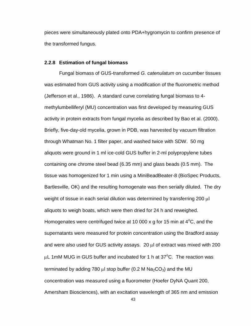
2.3.2 Scanning electron microscopic observations.
Cucumber roots were extensively colonized by G. catenulatum hyphae
within 7 days after application and formed a dense network over the root surface.
Hyphae were found intertwined near root hairs and were closely associated with
the junction of epidermal cells (Fig. 2-2A-C). Sporulation was observed, with
production of characteristic verticilliate conidiophores and conidia on the root
surface (Fig. 2-2D-F). A dense network of hyphae was also visible on the
surface of cucumber roots treated with Forc alone 7 days after inoculation (Fig.
2-2G-I) but no penetration or collapse of epidermal cells was observed. On roots
treated with G. catenulatum followed by Forc, hyphae of G. catenulatum and
Forc were distinguishable on the basis of their size (Fig. 2-2K). Colonization by
G. catenulatum was clearly visible and conidiophores and conidia were present
on the root surface (Fig. 2-2J); there was less evidence of the presence of Forc
hyphae. There was no visible hyphal interaction between the two fungi even
when they were observed growing in close proximity to one another (Fig. 2-2J-L).
47
Figure 2-1. Disease severity index (DSI) of cucumber plants and population densities of G. catenulatum on cucumber roots, tissues and growing medium.
(A) DSI of cucumber plants treated with Prestop WP (containing G. catenulatum) 30 days before inoculation with F. oxysporum f. sp. radicis-cucumerinum. (B) Population densities, expressed as log(10) colony forming units/ g root fresh weight, of G. catenulatum associated with cucumber roots and (C) cucumber crown region and growing medium over a 60 day period. (C) Crown = log(10) CFU/ g tissue; substrate = log(10) CFU/cm
3; nutrient solution = log(10) CFU/ml
48
Figure 2-2. Scanning electron micrographs of cucumber roots 7 days after inoculation with G. catenulatum (A-F), or F. oxysporum (G-I), or G. catenulatum followed 3 days later by F. oxysporum (J-L).
(A) Colonization of root hair zone; scale bar = 100 m; (B, C) Extensive colonization of the root
surface; scale bar = 100 m; (D, E, F) Sporulation of G. catenulatum on the cucumber root
surface; scale bar (D,E) = 100 m, scale bar (F) = 30 m; (G, H, I) Extensive colonization of the
root surface by F. oxysporum; scale bar (G) = 500m, scale bar (H,I) = 100 m; (J, K) Hyphal contact between G. catenulatum (g) and F. oxysporum (f) on the root surface; scale bar = 100
m; (L) Hyphae and conidia of G. catenulatum (g) and F. oxysporum (f) in close proximity to each
other; scale bar = 100 m.
49
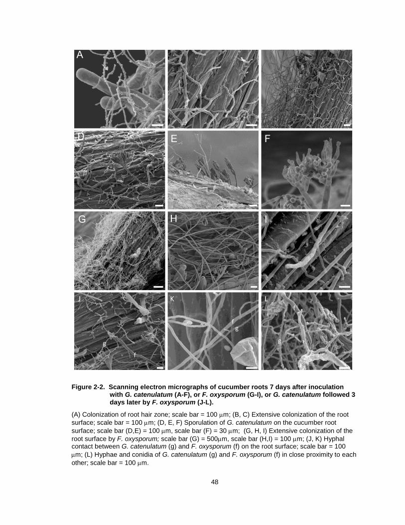
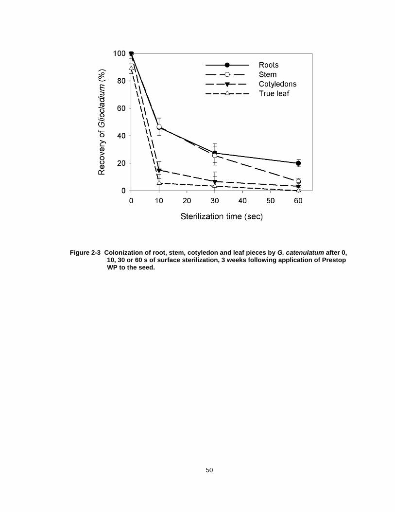
2.3.3 Extent of internal tissue colonization.
When root, stem and leaf tissues were not surface-sterilized, G.
catenulatum was recovered at frequencies of almost 100%. By comparison, the
biocontrol agent was not recovered from leaf tissues after surface-sterilization for
10, 30 or 60 s (Fig. 2-3). It was recovered from 50 % of root and stem pieces
after sterilization for 10 s, but recovery from both tissues was reduced to 25%
after 30 s of sterilization. Recovery was 20% from root pieces after 60 s of
sterilization and 0% from stem pieces (Fig. 2-3). There was no recovery of any
other microbes at any of the sterilization times from the tissues plated.
2.3.4 GUS transformation
Approximately 25 colonies per plate grew on the selection medium when
Agrobacterium was incubated with G. catenulatum at a concentration of 103
conidia/ml; at higher conidial densities, the putative transformant colonies were
too numerous and were not discernible. All 25 putative transformants were
screened for GUS activity by histochemical staining on microtiter plates and for
hygromycin resistance. Approximately 20% of colonies displayed spontaneous
resistance to hygromycin and were not transformed. In total, 12 colonies
displayed the GUS phenotype, yielding a transformation frequency of 3%.
Southern hybridization analysis showed that the transformants contained a single
copy of the uidA gene while the wild-type strain did not contain sequences similar
to this gene (data not shown). Transformants were grown for three successive
transfers on non-selective agar medium. Suspensions of conidia were then
plated onto PDA and randomly selected colonies were tested for GUS
50
Figure 2-3 Colonization of root, stem, cotyledon and leaf pieces by G. catenulatum after 0, 10, 30 or 60 s of surface sterilization, 3 weeks following application of Prestop WP to the seed.
51
expression. All the single-spore colonies were positive for hygromycin
resistance and GUS activity, indicating that the genes were stably maintained.
The colony morphology of transformant GcA1 and its ability to colonize cucumber
roots were not significantly different from the parent strainGcJ1446. The average
recovery from cucumber roots was 5x10⁶ CFU/ g RFW 5 weeks post-inoculation,
which is similar to the wild-type strain. When stem and root pieces were plated
onto PDA+ hygromycin, 100% of tissue pieces were colonized by the
transformed strain. Randomly selected colonies arising from the tissues all
stained positively for GUS, indicating the transformant was stable.
2.3.5 Growth of GUS transformed G. catenulatum on cucumber tissues
Following seed treatment with conidia of GUS-transformed G.
catenulatum, colonization of the seed coat was visible within 24 hr and the
endosperm and emerging radicle were also colonized (Fig. 2-4A). On seeds in
Magenta boxes, colonization of the developing cotyledons and the main root
occurred along the margin and predominantly near the root tips, respectively,
within 3 days after seed germination (Fig. 2-4B). After 14 days, roots were
primarily colonized at the root tip and at the junctions of lateral root emergence
(Fig. 2-4C, D), although discontinuous colonization along the surface of the
mature root zone was also observed. The crown area, shoot meristem and
emerging true leaves were also colonized by G. catenulatum (Fig. 2-4E-G). G.
catenulatum was found associated externally with trichomes on the stem and
appeared to form a network of hyphae over the epidermis (Fig. 2-4E). When the
tissues were sectioned, hyphae could be seen growing internally in epidermal
52
53
Figure 2-4 Colonization pattern of cucumber plants by G. catenulatum strain J1446 expressing the GUS gene.
(A) Seed coat and endosperm and (B) developing cotyledons colonized by G. catenulatum 24 h and 72 h after seed application, respectively. (C, D) Colonization of roots, (E, F) stem, (G) meristem and true leaves (F) by G. catenulatum 14 days after seed treatment in Magenta boxes. Colonization of roots was visible at junctions of lateral roots and at root tips (D), while hyphae were found associated with (E) trichomes on the stem surface. (H-M) Light micrographs showing internal colonization by G. catenulatum. (H) Colonization of the epidermal and cortical cells in roots of plants grown in Magenta boxes and (I) 3-week-old plants grown in rockwool blocks. (J) Hyphae on the surface of the stem with ingress into trichomes and cortical cells and (K) xylem vascular elements of plants grown in Magenta boxes. (L) Hyphae in cortical cells of stems of plants grown in rockwool blocks. (M) Hyphae in the epidermal layer of young true leaves of plants grown in Magenta boxes.
54
and cortical cells of roots (Fig 2-4H, I). On stems, blue-stained hyphae were
observed on the surface of trichomes (Fig. 2-4J), and internal colonization of the
epidermal, cortical and vascular regions was evident (Fig. 2-4J-L). Colonization
of the epidermal layer of young true leaves was observed on plants grown both in
Magenta boxes and rockwool blocks (Fig. 2-4M).
2.3.6 Estimation of fungal biomass
There was a linear relationship between GUS activity and mycelial dry
weight and between GUS activity and protein content (Fig. 2-5). Using GUS
activity, as little as 1 ng of mycelia could be detected (Fig. 2-5). Application of G.
catenulatum to the rockwool blocks resulted in significantly higher root
colonization compared to the lower stem and other plant tissues (Fig. 2-6). Seed
treatment resulted in colonization of the true leaves, lower stem and roots at low
levels that were not significantly different from each other (Fig. 2-6).
2.3.7 Survival of F. oxysporum on cucumber roots in the presence of G. catenulatum
There was no significant difference in the levels of Forc or G. catenulatum
on the three different root portions; therefore, data were combined to give overall
CFU levels per mg of root. In addition, there was no significant difference in the
CFU levels of G. catenulatum applied as Prestop WP or GcA1, so the data were
combined for these treatments. Application of G. catenulatum to the rockwool
blocks before inoculation with Forc resulted in a significant decrease in Forc
levels on the roots and crown when compared to plants inoculated with Forc
only, regardless of the initial Forc inoculum concentration (Fig. 2-7). At a Forc
55
Figure 2-5 Relationship between fungal biomass or protein content and GUS activity in a transformant of G. catenulatum.
GUS activity was measured in dilutions of mycelial extracts from 3-day-old cultures. The lines were generated by simple regression analysis.
56
Figure 2-6 Fungal biomass of G. catenulatum expressing the GUS gene in different tissues of cucumber plants following application to seed or rockwool block.
Fungal biomass was determined indirectly from GUS activity. Means represent the combined values from two independent trials (n=10). Vertical bars indicate standard error of the mean.
57
Figure 2-7 Population size of Forc in the roots (top) or crown area (bottom) of cucumber plants treated with G. catenulatum or Prestop WP
Prestop was applied at the recommended rate or conidia of GcA1 (107 cfu/ml) was applied to the
rockwool blocks prior to inoculation with Forc at an initial inoculum level of either 104, 10
5, or 10
6
conidia/ml. Plants were sampled 2 weeks after inoculation with Forc. Population levels were determined by plating supernatant from ground tissues onto selective media for Forc. The means and standard error were obtained from 10 replicates per treatment.
58
concentration of 104 conidia/ml, treatment with G. catenulatum reduced
pathogen levels on the roots to undetectable levels. Linear regression
coefficients for slopes were significantly lower in the roots and crown of plants
treated with the biocontrol agent followed by Forc inoculum compared to plants
treated with Forc only (Table 2-1), indicating that Forc survival was reduced in
the presence of G. catenulatum.
Population levels of G. catenulatum on the roots were significantly higher
at 4000, 6700 or 5200 CFU/ mg fresh weight (P= 0.0469, 0.0001, and 0.0072,
respectively, Table 2-2) when Forc was added at inoculum levels of 104, 105 or
106 conidia/ml compared to G. catenulatum only treatments (1200 CFU/ mg). On
the crown, population densities of G. catenulatum on plants receiving Forc at all
inoculum levels were not significantly different from plants treated with G.
catenulatum only (913 CFU/ mg) (P=0.4695) (Table 2-2).
2.4 Discussion
The results from this study showed that G. catenulatum provided protection
to cucumber against Fusarium root and stem rot for a period of up to 60 days
following a single application to the rockwool growing medium at seeding. In
previous studies, pathogen challenge occurred within 24 h to 3 days after G.
catenulatum was applied (McQuilken et al., 2001; Punja and Yip, 2003; Rose et
al., 2003). We observed that disease suppression occurred even when pathogen
challenge occurred 30 days following application of the biocontrol agent. Root
colonization plating data revealed that the density of G. catenulatum was about
5x10⁵ CFU/g
59
Table 2-1 Slope values (b) from linear regression analysis of the population levels of Forc in the roots or crown area of cucumber plants treated with G. catenulatum (10
7
conidia/ml) versus log10-transformed initial inoculum of Forc.
Treatmenta Rootsbc Crown
Forc only 389.7 (25.9) 15.4 (2.1)
Forc + Prestop WP 49.8 (28.7)* 1.2 (2.1)*
Forc + GcA1 75.9 (25.9)* 1.6 (2.1)*
aPlants were sampled 2 weeks after inoculation with Forc. Forc was applied to
rockwool blocks at inoculum levels of 104, 105 or 106 conidia/ml, 3 weeks
following application of G. catenulatum (107 conidia/ml) at seeding. The means
and standard errors were obtained from 10 replicates per treatment. Population
levels were determined by plating tissues onto selective media for Forc.
bValues in parentheses indicate standard error of the mean
cValues within a column followed by an asterisk are significantly different from the
pathogen control (contrast partitioning, < 0.05). Graphical depiction of
regression lines are shown in Figure 2-7.
60
Table 2-2 Population levels of G. catenulatum in the roots or crown area of cucumber plants treated with G. catenulatum applied to the rockwool blocks (10
7
conidia/ml) prior to inoculation with Forc at an initial inoculum level of either 10
4, 10
5, or 10
6 conidia/ml.
Forc levels Gc CFU / mg fresh weighta
Rootsbc Crown
0 1199 (867) 913 (141)
104 4141 (862)* 1019 (199)
105 6707 (862)* 1056 (199)
106 5172 (944)* 750 (115)
aPlants were sampled 5 weeks and 2 weeks after inoculation with G. catenulatum
and Forc, respectively. The means and standard errors were obtained from 10
replicates per treatment. Population levels were determined by plating tissues
onto selective media for G. catenulatum.
bValues in parentheses indicate standard error of the mean
cValues in a column followed by an asterisk differed significantly from the G.
catenulatum only control (contrast partitioning, < 0.05)
61
root fresh weight at the time of pathogen challenge, and populations remained
near this level over the duration of the experiment. These high densities of the
biocontrol agent likely play an important role in biocontrol activity since the
proportion of the root surface colonized and the efficacy of biocontrol is
dependent on a high ratio of the biocontrol strain to the pathogen (Larkin and
Fravel, 1999; Whipps, 2001). Plating of root segments also indicated that almost
100% of root sections were colonized by G. catenulatum even after 60 days.
Scanning electron microscopic observations confirmed the ability of G.
catenulatum to sporulate abundantly on the root surface, and colonization was
evident in the root hair zones and at the origins of lateral roots. As well, G.
catenulatum sporulated extensively on the surface of the rockwool, with some
spread occurring to leaves and stems in growth room trials. Since the rockwool
medium was initially sterile with no competing microorganisms, this may have
allowed G. catenulatum to establish high densities. The competitive saprophytic
ability of G. catenulatum in nonsterile soil is not known.
In this study, G. catenulatum was transformed with the GUS marker gene
using Agrobacterium to facilitate further study of its colonization behaviour and
distribution on greenhouse cucumber plants. The use of Agrobacterium-
mediated transformation of fungi is increasing due to the reported high
transformation efficiencies, ease and reliability of the procedure (de Groot et al.,
1998; Duarte et al., 2007). However, G. catenulatum was resistant to
hygromycin at 200 mg/L, a characteristic that has also been observed in closely
related fungi (Lubeck et al., 2002). This made selection of transformants very
62
difficult as spontaneous resistance to hygromycin occurred at a fairly high rate
(20%), even on nutrient-poor media. Nonetheless, 12 positive transformants
were identified and found to be mitotically stable after single spore transfers.
Transformation did not change the growth or antagonistic properties of the
transformant compared to the wildtype.
The ability of G. catenulatum to colonize cucumber plants internally was
initially suggested by its recovery from root and stem pieces even after 60 s of
sterilization. Recovery of a microbe after such a rigorous sterilization procedure
has been suggested to be indirect evidence of its ability to be an endophyte
(Schulz and Boyle, 2005). Direct evidence was obtained by the visualization of
blue-stained hyphae in sections of the epidermis and cortical regions of roots and
stems using the GUS-marked strain. Hyphae were also observed in the xylem
vessels of cucumber stems in plants grown in nutrient solution in enclosed
Magenta boxes. Endophytic growth of fungi within roots can be inter- or
intracellular and become systemic (Schulz and Boyle, 2005). Histochemical
observations showed that G. catenulatum also colonized the epidermal layer of
true leaves. Colonization of above-ground tissues by fungal endophytes is
usually intracellular and results in localized growth (Schulz and Boyle, 2005).
Our observations suggest that G. catenulatum meets the criteria of an
opportunist, since it has the capability to grow both endophytically and
saprophytically (Schulz and Boyle, 2005). Many endophytes can induce plant
defence reactions that limit their growth inside the plant (Peters et al., 1998). For
example, T. harzianum was shown to colonize the epidermis of cucumber roots
63
and induce systemic resistance during the early stages of colonization (Yedidia
et al., 2000). Whether or not G. catenulatum can also induce defense responses
in cucumber plants remains to be determined.
We observed G. catenulatum to actively colonize the seed coat and
developing roots following seed treatment. This rhizosphere competence can
protect the germinating seed and emerging radicle against damping-off
pathogens, such as Pythium spp. and Rhizoctonia solani (McQuilken et al., 2001;
Punja and Yip, 2003). Furthermore, seed treatment or application to rockwool
blocks resulted in colonization of above-ground parts, including the apical shoot
meristem. G. catenulatum has been shown to reduce the incidence of
anthracnose caused by C. acutatum when applied to blueberry flowers,
suggesting that colonization must have occurred to preclude pathogen invasion
(Verma et al., 2006). Strains of G. catenulatum which colonize roots or leaves
have also been shown to be effective biocontrol agents against seed-borne
diseases of cereals (Lubeck et al., 2002) and against Botrytis cinerea on
strawberry and raspberry leaves (Peng and Sutton, 1991; Sutton et al., 1997).
Population levels of Forc on roots and crown tissue were significantly lower
in the presence of G. catenulatum compared to plants inoculated with Forc alone,
suggesting that this fungus can exclude Forc from colonizing the roots. Using
scanning electron microscopy, mycelia of Forc were rarely observed at sites
colonized by the biocontrol agent and infrequently found on the roots compared
to roots from treatments with Forc alone. Infection by F. oxysporum f. sp. radicis-
lycopersici on tomato was shown to occur near root hairs and through cellular
64
junctions along the main root (Lagopodi, 2002; Bolwerk et al., 2005). G.
catenulatum was often observed associated with root hairs and formed a dense
network over the cucumber root epidermis, with hyphae closely associated with
junctions of epidermal cells. Therefore, colonization of these niches by G.
catenulatum prior to Forc infection likely reduced the infection sites available for
Forc. Interestingly, levels of G. catenulatum on roots were higher in the
presence of Forc compared to plants inoculated with G. catenulatum alone.
Nutrients released from infection sites could have favoured growth of the
biocontrol agent. Other studies have observed a similar phenomenon: T.
harzianum levels were higher on roots that were more diseased and had higher
populations of P. ultimum (Green et al., 2001). Similarly, densities of non-
pathogenic F. oxysporum were higher in roots infected with pathogenic F.
oxysporum f. sp. radicis-lycopersici (Bao and Lazarovits, 2001). A higher density
of the biocontrol agent relative to the pathogen is usually required for biocontrol
in which competition for nutrients or infection sites is the primary mode of action
(Larkin and Fravel, 1999; Bolwerk et al., 2005). Therefore, in all of the biocontrol
and population density experiments, G. catenulatum was introduced at densities
much higher than the pathogen, usually at 1 x 10⁶ cfu/ g roots. Since G.
catenulatum was also applied preceding the pathogen, it is probable that roots
were colonized at the time of pathogen introduction, thus effectively excluding
Forc infection. This strategy is an important factor to ensure the success of G.
catenulatum in biocontrol approaches in commercial greenhouse settings. Since
Fusarium stem and root rot develops when primary infections occur early in the
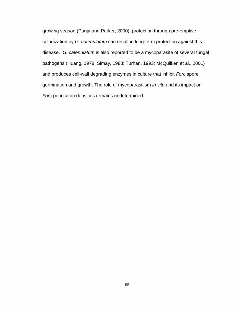
65
growing season (Punja and Parker, 2000), protection through pre-emptive
colonization by G. catenulatum can result in long-term protection against this
disease. G. catenulatum is also reported to be a mycoparasite of several fungal
Random primers were used to label the 750 bp Glu1 mRNA fragment using [α-
32P] dCTP and Prime-A-gene labelling kit (Promega, Madison, WI, USA)
following manufacturers protocols, and used as a radioactive RNA probe.
Hybridization procedures were carried out as described by Wally et al. (2008).
3.2.9 Statistical analyses
For all data collected, variance analysis was performed using the proc glm
method using Statistical Analysis System, version 8.0 (SAS Institute, 1999).
Means of treatments were compared using Tukey’s HSD test. Normality of all
dependent variables was tested by means of the univariate procedure, and
homogeneity of experimental errors was examined by plotting residuals versus
predicted values using the mixed model and plot procedure. Outlier detection
was performed using studentized residuals and Cook’s distance measure. Data
77
for repetitions of the experiments were pooled for analysis when F-tests indicated
that variances of the data did not differ significantly. The Type 1 error rate (α)
was set at 0.05 for all statistical tests.
3.3 Results
3.3.1 Production of antifungal metabolic compounds G. catenulatum that inhibit growth of F. oxysporum.
G. catenulatum did not inhibit the growth of Forc, when the two fungi were
co-inoculated on PDA (Fig. 3-1A). In contrast, on WA, hyphae of G. catenulatum
overgrew and sporulated on Fusarium hyphae (Fig 3-1B). On PDA, G.
catenulatum overgrew colonies of P. aphanidermatum (Fig 3-1C). In the
experiments to detect volatile antibiotic production by G. catenulatum (Fig 3-1D,
E), radial growth of Forc was not significantly inhibited by the presence of G.
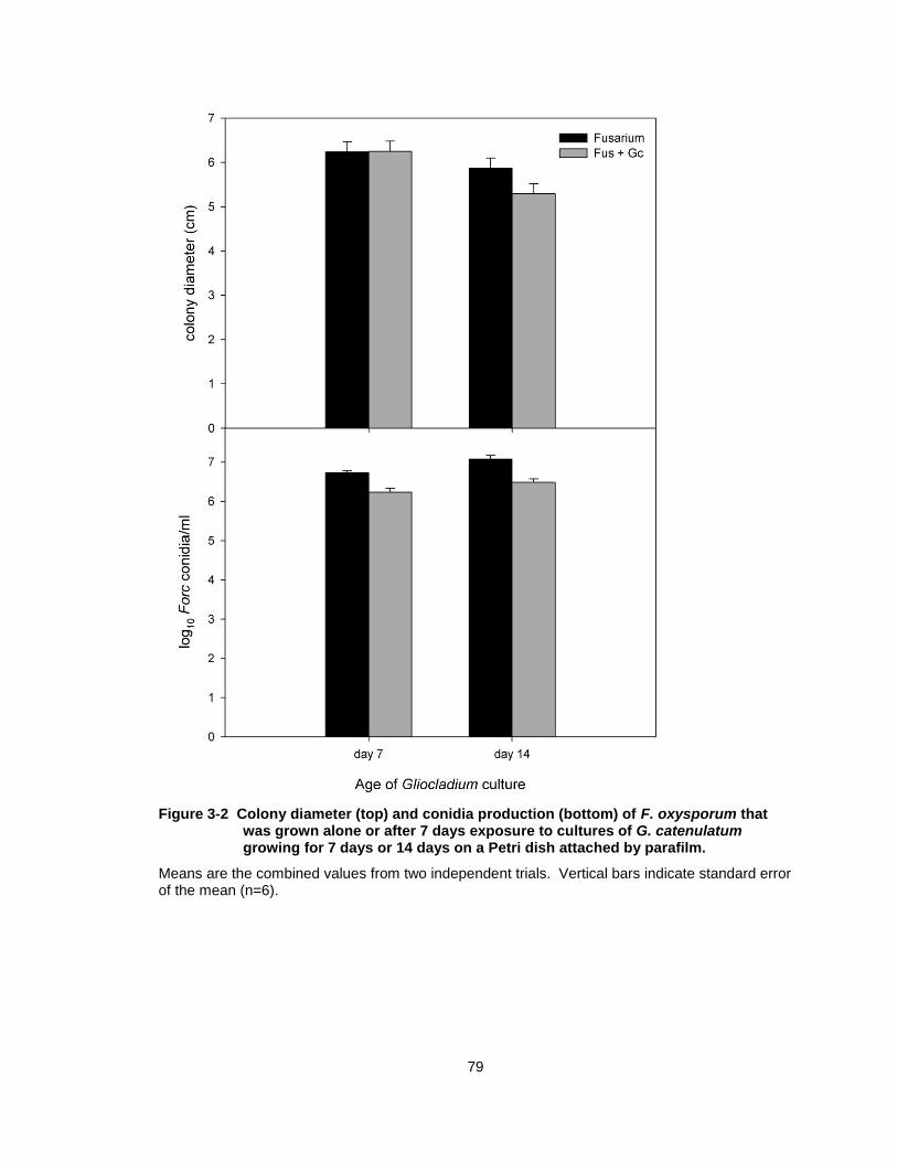
catenulatum at any of the treatment combinations (Fig 3-2). Sporulation of Forc
was significantly decreased in all treatments in the presence of G. catenulatum,
but only by an average 5-fold decrease from approximately 1 x 10⁷ conidia per ml
in control treatments to 2 x 10⁶ conidia per ml in Forc + G. catenulatum
treatments (Fig 3-2).
78
Figure 3-1 Dual culture plates of G. catenulatum (Gc) and F. oxysporum (Fo) or P. aphanidermatum.
Dual culture plates of G. catenulatum (Gc) and F. oxysporum (Fo) on (A) PDA and (B) water agar, and (C) G. catenulatum and P. aphanidermatum (Pa) on PDA, 7 days after co-inoculation. Samples for SEM processing were taken from the interaction zones (circles). (D) Method to test for production of volatile organic compounds (VOCs) by G. catenulatum. (E) Control culture of F. oxysporum (Fo), and culture of F. oxysporum (Fo) exposed to a 14-day-old culture of G. catenulatum (Gc) for 7 days.
79
Figure 3-2 Colony diameter (top) and conidia production (bottom) of F. oxysporum that was grown alone or after 7 days exposure to cultures of G. catenulatum growing for 7 days or 14 days on a Petri dish attached by parafilm.
Means are the combined values from two independent trials. Vertical bars indicate standard error of the mean (n=6).
80
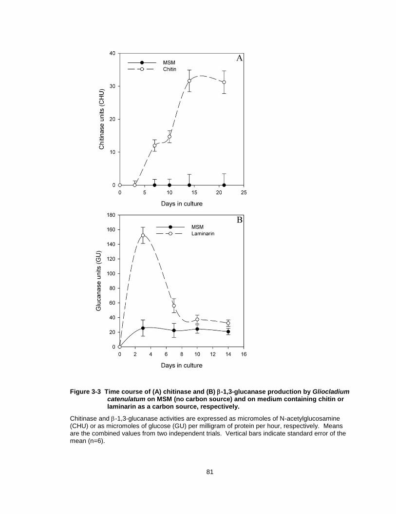
3.3.2 Chitinase and glucanase production in culture
G. catenulatum produced extracellular chitinase and -1,3-glucanase on
chitin or laminarin as the sole carbon source, respectively. In chitin-amended
medium, production of chitinase increased steadily and peaked at 14 days (30
CHU) after which time the levels remained constant (Fig. 3-3A). When grown on
MSM only, chitinase activity was not detected. Production of -1,3-glucanase
peaked at 3 days when grown on laminarin and was highest at 150 glucanase
units (GU) than at any other time period (Fig. 3-3B). Glucanase levels were
lower after 7 days in culture and remained steady at around 40 GU for the
duration of the experiment. When grown on MSM only, G. catenulatum produced
a constant low level of glucanase over the time period assayed.
An incubation period of 7 days was chosen to test the effect of different
carbon sources on production of chitinase and -1,3-glucanase by G
catenulatum. Chitinase production was significantly higher in medium containing
Fusarium cell walls (28 CHU), with activity almost 3.5-fold higher than on chitin-
containing medium (8 CHU) (P < 0.0001) (Fig. 3-4). Chitinase activity was not
detected in extracts from cultures grown on laminarin, glucose, sucrose, and N-
acetylglucosamine (data not shown). The production of -1,3-glucanase by G.
catenulatum was not significantly different on laminarin (32 GU) compared to
when Fusarium or Pythium cell walls were used as the sole carbon source (24
and 31 GU, respectively). G. catenulatum also produced extracellular -1,3-
81
Figure 3-3 Time course of (A) chitinase and (B) -1,3-glucanase production by Gliocladium catenulatum on MSM (no carbon source) and on medium containing chitin or laminarin as a carbon source, respectively.
Chitinase and -1,3-glucanase activities are expressed as micromoles of N-acetylglucosamine (CHU) or as micromoles of glucose (GU) per milligram of protein per hour, respectively. Means are the combined values from two independent trials. Vertical bars indicate standard error of the mean (n=6).
82
Figure 3-4 Effect of carbon source on chitinase and -1,3-glucanase production after 7 days of growth of Gliocladium catenulatum.
Chitinase and -1,3-glucanase activities are expressed as micromoles of N-acetylglucosamine (CHU) or as micromoles of glucose (GU) per milligram of protein per hour, respectively. Values are the combined means from two independent trials. Carbon sources were as follows (1mg/ml): minimal synthetic medium with no carbon source (MSM), chitin, laminarin (Lam) Fusarium cell wall (FCW), Pythium cell walls (PCW), and homogenized cucumber roots (cuc). Values for columns in each group followed by the same letter did not differ significantly (Tukey’s HSD, P < 0.05). Vertical bars indicate standard error of the mean. Means represent the combined values from two independent trials (n=6).
83
glucanase in liquid culture when chitin or autoclaved and homogenized
cucumber roots were used as the sole carbon source (Fig. 3-4). Chitinase
production was not induced by cucumber roots. Similar to chitinase, glucanase
was not detected in extracts of cultures grown on sucrose or glucose. The
background levels of glucose in boiled culture filtrates from these treatments
were an average of 1.6 mol glucose/ml.
Maximum production of chitinase was observed at pH 6 (15 CHU) and
there was no significant difference in enzyme activity at pH 4, 5, 7 and 9 (7-10
CHU) (Fig. 3-5A). Glucanase activity was significantly repressed by acidic pH,
and was maximum at pH > 6 (Fig. 3-5B).
3.3.3 Scanning electron microscopy
During growth in culture, the hyphal diameters of Fusarium and Pythium
were both approximately 30 m whereas the hyphal diameter of G. catenulatum
was approximately 10 m, thereby allowing the fungi to be distinguished from
one another (Fig. 3-6A). In dual culture, hyphae of G. catenulatum and Fusarium
grew in close proximity to each other, and there was evidence of contact of G.
catenulatum hyphae with Fusarium (Fig. 3-6B, C), with likely penetration points
observed along the pathogen’s hyphae (Fig.3- 6D inset). Hyphal strands of G.
catenulatum often grew in parallel along the hyphae of Fusarium, but lysis of
Fusarium hyphae was not observed. When excised cucumber roots were placed
on water agar between G. catenulatum and Fusarium cultures, the hyphae of the
two fungi were rarely seen growing together. In areas of the root segments
84
Figure 3-5 .Effect of initial medium pH on (A) chitinase and (B) -1,3-glucanase production by Gliocladium catenulatum grown for 7 days on chitin or laminarin as the carbon source, respectively.
Chitinase and -1,3-glucanase activities are expressed as micromoles of N-acetylglucosamine (CHU) or as micromoles of glucose (GU) per milligram of protein per hour, respectively. Values followed by the same letter did not differ significantly (Tukey’s HSD, P < 0.05). Vertical bars indicate standard error of the mean. Means are the combined values from two independent trials. (n=6).
85
Figure 3-6 Scanning electron micrographs of the interactions between Gliocladium catenulatum (c) and Fusarium oxysporum (f) in dual culture on water agar (A-D), or between G. catenulatum and Pythium (p) on excised cucumber roots (r) on water agar (E,F).
(A) Hyphae of G. catenulatum and Fusarium oxysporum growing in close proximity. The hyphae
of Fusarium are 3X greater in diameter compared to G. catenulatum ; scale bar = 30 m; (B, C)
Contact of G. catenulatum hyphae with Fusarium scale bar = 30 m; (D) Penetration of Fusarium
hyphae by G. catenulatum; scale bar = 30 m; inset = 10 m; (E) Hyphae of G. catenulatum
branching and coiling around P. aphanidermatum on a cucumber root; scale bar = 50 m; (F) Appressorium-like structures (arrows) of G. catenulatum attached to a hyphae of Pythium; scale
bar = 30 m.
86
colonized by both fungi, there was evidence of contact of Fusarium hyphae by G.
catenulatum similar to that observed in dual cultures, but penetration was not
observed (data not shown). In contrast, on excised cucumber roots, G.
catenulatum hyphae were observed to coil around the hyphae of Pythium,
producing short branches that surrounded the host hyphae (Fig. 3-6E).
Spherical, appressorium-like structures were produced when attachment of the
host hyphae to the pathogen’s hyphae was observed (Fig. 3-6F).
3.3.4 Effect of crude enzyme extracts on Pythium and Fusarium growth and conidial germination
Colony growth of Fusarium was significantly reduced (P < 0.05) on PDA
containing crude extracts of filtrates from G. catenulatum with glucanase or
chitinase activity when compared to boiled extracts or PDA alone (Figure 3-7A).
Chitinase and glucanase levels in these filtrates ranged from 15 to 30 CHU or 40
to 50 GU, respectively (data not shown). Germination of Fusarium conidia was
also significantly reduced (by up to 50 %) in the presence of these extracts
(Figure 3-7B). Colony growth of Pythium was significantly reduced (P < 0.05) on
PDA containing crude extracts of filtrates with glucanase, but not chitinase,
activity compared to boiled extracts or PDA alone (Figure 3-7A).
3.3.5 Release of glucose or NAGA from Fusarium or Pythium cell walls
When Fusarium or Pythium cell walls were incubated with culture filtrates of
G. catenulatum containing chitinase or-1,3-glucanase activity, NAGA or
glucose was released, respectively (Fig. 3-8). All filtrates containing glucanase
activity were capable of degrading cell walls of Fusarium and Pythium to release
87
Figure 3-7 Effect of Gliocladium catenulatum culture filtrates on (A) growth of Fusarium oxysporum or Pythium aphanidermatum mycelia or (B) germination of Fusarium conidia.
Filtrates were obtained from extracts when G. catenulatum was grown on: no carbon source (MSM), laminarin medium (Lam) or chitin medium (Chit). Boiled filtrates were used as controls. Means represent the combined values from two independent trials. Values for columns in each group followed by the same letter did not differ significantly (Tukey’s HSD, P < 0.05) Vertical bars indicate standard error of the mean (n=6).
88
Figure 3-8 Effect of Gliocladium catenulatum culture filtrates on the release of (A) NAGA from Fusarium cell wall fragments or (B) glucose from Fusarium or (C) Pythium cell wall fragments.
Filtrates were obtained from extracts when G. catenulatum was grown on: no carbon source (MSM), chitin medium (Ch), laminarin medium (Lam), or Fusarium or Pythium cell walls. Boiled filtrates or phosphate buffer was used as a control for background levels of glucose or NAGA. Means represent the combined values from two independent trials. Values in a column followed by the same letter did not differ significantly (Tukey’s HSD, P < 0.05). Vertical bars indicate standard error of the mean (n=6).
89
glucose. Filtrates containing chitinase activity released NAGA from Fusarium cell
walls only. Filtrates obtained from chitin or Fusarium cell wall-containing media
possessed chitinase and glucanase activity that released both NAGA and
glucose from Fusarium cell wall fragments (Fig. 3-8A, B). Filtrates obtained from
laminarin-containing medium possessed glucanase activity that was capable of
releasing glucose, but not significant amounts of NAGA, from Fusarium or.
Pythium cell wall fragments (Fig. 3-8A, B). All activity levels were significantly
higher than those in boiled filtrates and phosphate buffer
3.3.6 Measurement of chitinase and glucanase in cucumber plants
Chitinase and glucanase activities were measured in the leaves and roots
2, 3 and 7 days after treatment with G. catenulatum. There was no detectable
increase in chitinase activity in the roots or leaves after treatment with the
biocontrol agent at most of the sampling times (see Chapter 5). However, there
was a significant increase in glucanase activity in the roots (Fig. 3-9), but not
leaves (see Chapter 5) 7 days after treatment with G. catenulatum
3.3.7 -1,3-glucanase isoforms
When G. catenulatum was grown on laminarin, three bands corresponding
to -1,3-glucanase activity were resolved on SDS-PAGE gels (Fig. 3-10), two of
which were approximately 20 kDa and one was approximately 45kDa in size.
Only one band, corresponding to 45 kDa, was detected in culture filtrates when
G. catenulatum was grown on MSM. Two 20 kDa bands were visible in extracts
originating from roots that had been inoculated with G. catenulatum 7 days prior
90
Figure 3-9 Glucanase activity in roots of 10-day-old cucumber seedlings at 2, 3 and 7 days following inoculation with Gliocladium catenulatum (Gc), treatment with salicylic acid (SA) or water (Con).
Enzyme activity is expressed as micromoles of glucose (GU) per milligram of protein per hour. Values for columns in each sampling time followed by the same letter did not differ significantly (Tukey’s HSD, P < 0.05). Means represent the combined values from three independent trials. Vertical bars indicate standard error of the mean (n=9).
91
Figure 3-10 SDS-PAGE of -1,3-glucanase isoforms from G. catenulatum.
G. catenulatum was grown on media containing no carbon source (MSM) (lane 1) or laminarin (lane 2), or in extracts from cucumber roots treated with G. catenulatum for 7 days (lane 3),
control roots (lane 4) and plants treated with INA (2g/ml) (lane 5). Samples (60 l) of culture
filtrates or plant extracts were loaded on a gel containing -1,3-glucan. Bands with glucanase activity (size = 45 or 20 kDA) appeared as dark zones under UV illumination after staining with aniline blue.
92
to extraction. No bands were visible in root extracts from control plants or those
treated with INA.
3.3.8 Glucanase gene expression
3.3.8.1 PCR
PCR with first-strand cDNA synthesized from total RNA extracted from
cucumber plants colonized by G. catenulatum using the primer pair designed to
amplify a 750 bp fragment did not consistently produce amplicons that were
visible on an agarose gel (Fig. 3-11A). Therefore, to detect glucanase
expression, the PCR products were used in a subsequent nested PCR protocol
using a primer pair designed to amplify an internal fragment of a -1,3-glucanase
gene from G. catenulatum (GenBank accession DQ975304). PCR from cDNA
synthesized from total RNA from cucumber plants colonized by G. catenulatum
produced an amplicon of approximately 250 bp, which was absent in control
plants not treated with G. catenulatum (Fig. 3-11B). The identity of the 750 and
250 bp amplicons, as determined by sequencing, showed 100% similarity with
the sequence of the glucanase gene fragment from G. catenulatum.
3.3.8.1 Northern analysis
Glucanase mRNA was detected when G. catenulatum was grown on
w/v), or in low glucose levels (0.1% w/v) representing starvation conditions (Fig.
3-12). Lower intensity of probe hybridization was observed when the initial
93
Figure 3-11 Detection of -1,3-glucanase expression by Gliocladium catenulatum on colonized cucumber roots using reverse transcription – polymerase chain reaction (RT-PCR).
(A) Glu1F and Glu1R primer set and (B) nested PCR with PCR products from above using Glu1npF and Glu1npR primer set. Lane 1, water control; Lanes 3, 4, uninoculated cucumber roots; Lanes 2, 5, 6, cucumber roots colonized by G. catenulatum; Lane 7, G. catenulatum mycelium grown on laminarin; Lane M, DNA ladder. Amplicons of (A) 750 bp or (B) 220 bp are indicated by an arrow.
94
Figure 3-12 Northern blot analysis of expression of a -1,3-glucanase gene from Gliocladium catenulatum.
The fungus was grown as a shake culture in MSM supplemented with 0.2% cell wall fragments of Fusarium (Fo) or Pythium (Pa), homogenized cucumber roots (R), 2% glucose (G2) or 0.1% glucose (G0.1). Approximately five micrograms of total RNA, extracted after 3 days or 7 days of growth, was electrophoresed on a formaldehyde gel, blotted, and hybridized to a radiolabelled
probe designed from a 750 bp fragment of a -1,3-glucanase gene isolated from G. catenulatum (top panel). The bottom panel shows ethidium bromide-stained rRNA.
95
glucose concentration was increased to 2%, both 3 and 7 days after incubation
(Fig. 3-12). Background glucose levels in boiled culture filtrates from 2% glucose
treatments were 42 and 4.8 mol glucose/ml at day 3 and 7, respectively. In
contrast, glucose level was 0.6 mol/ml in the 0.1% glucose treatment at day 3,
and by day 7, glucose was not detected in the culture filtrates. Growth of G.
catenulatum on Fusarium or Pythium cell walls enhanced glucanase expression
compared to cucumber roots and low glucose treatments. Detection of
glucanase mRNA was higher after 7 days than 3 days of incubation.
3.4 Discussion
Secretion of extracellular enzymes capable of lysing cell walls of
pathogenic fungi is important in the mycoparasitic process and these enzymes
are well characterized in many biocontrol agents, especially T. harzianum
(Viterbo et al., 2002b; Whipps, 2001). Purified endo--1,3-glucanase and
endochitinases from T. harzianum inhibited spore germination and had lytic
activity against the cell walls of a number of plant pathogenic fungi, including
Botrytis cinerea, Rhizoctonia solani, Sclerotium rolfsii and F. oxysporum f. sp.
melonis (Lorito et al., 1994a; Viterbo et al., 2001), providing evidence for their
involvement in mycoparasitism. Hydrolytic enzyme activity has not been
previously characterized in G. catenulatum. We observed that enzymes capable
of degrading chitin and -1,3-glucan, both major cell wall components in
Fusarium (Schoffelmeer et al., 1999), were produced in culture by G.
catenulatum. Both enzymes inhibited the growth and germination of conidia of
Fusarium, while only glucanase inhibited the growth of Pythium. The lack of
96
effect of chitinase on Pythium was not unexpected because the cell walls of
plant-pathogenic oomycetes, such as P. aphanidermatum, are composed mainly
of ß-1,3- and ß-1,6-glucans and cellulose instead of chitin (Viterbo et al., 2002b).
Interestingly, even though glucanase activity was detected in culture filtrates of
G. catenulatum grown on chitin, these filtrates did not inhibit growth of P.
aphanidermatum in culture. Glucanase activity in filtrates from chitin media was
approximately 1.5-fold lower than that from laminarin media. Therefore, it is
likely that the glucanase levels from chitin-amended media were too low to inhibit
Pythium growth, since growth inhibition by cell-wall degrading enzymes is often
concentration dependent. For example, growth inhibition of B. cinerea by
chitinases and glucanases produced by T. harzianum followed a dose-dependent
response (Lorito et al., 1994a).
Activities of both chitinase and glucanase from G. catenulatum were
repressed by glucose and were induced by fungal cell wall extracts, or by
polymers such as laminarin and chitin. Northern analysis of glucanase mRNA
generally revealed the same trends as enzyme activity data, demonstrating that
expression of glucanase mRNA is induced by fungal cell walls. However, in
enzyme activity assays, glucanase was not detected in low percent glucose-
amended media, even though glucanase mRNA was detected in these glucose
concentrations (0.1% w/v). To ensure that adequate levels of growth would be
obtained for RNA extractions, the initial inoculum of G. catenulatum in the MS
medium (1 x 10⁶ conidia/ml) was much higher in the experiments designed for
Northern analysis. Therefore, it may be that carbon starvation was attained
97
much more rapidly in these cultures than those grown for enzyme activity assays,
where a single mycelial plug was used as the initial inoculum source. The
enhanced expression of genes encoding hydrolytic enzymes under conditions of
physiological stress or carbon starvation has been reported for mycoparasitic
fungi such as T. harzianum and Coniothyrium minitans (Donzelli et al., 2005;
Giczey et al., 2001). Interactions between a mycoparasite and its host can also
induce expression of chitinase and glucanase genes due to the action of small
diffusible molecules derived from host cell walls (Lu et al., 2004; Viterbo et al.,
2002a; Cohen-Kupiec et al., 1999).
The initial pH of the growth medium affected the activity of both glucanase
and chitinase. In T. harzianum, pH has also been found to be an important
factor regulating the production of hydrolytic enzymes (Donzelli et al., 2005). The
pH of homogenized cucumber root extracts grown in rockwool cubes and
watered with nutrient solution ranged from 5.8 to 6.3. Since the optimal pH range
for glucanase and chitinase production in G. catenulatum (pH 6.0-7.0) coincides
with the pH of cucumber roots, enzyme production in situ is likely to occur. The
high initial peak of glucanase activity after 3 days in culture, along with its
detection on cucumber roots in vivo, suggest that this enzyme may be important
in the biocontrol activity of G. catenulatum against root pathogens. In contrast,
fungal chitinase activity was not enhanced on colonized cucumber roots, and
activity was not induced by cucumber roots, suggesting chitinase may be less
important than glucanase.
98
Although attachment of G. catenulatum to Fusarium hyphae was observed
in dual culture, this mycoparasitic behaviour was rarely detected on the root
surface. It may be that direct interactions between an antagonist and a pathogen
in the rhizosphere were rare, or just not observed (Whipps, 2001). In contrast,
on excised cucumber roots, coiling and formation of appressorium-like structures
by G. catenulatum on Pythium were frequently seen. This mycoparasitic
behaviour by G. catenulatum was not observed on colonized cucumber roots in
the absence of the pathogen (described in the previous chapter), indicating that
hyphal branching and appressoria-like structures were induced by the presence
of fungal host hyphae. In T. atroviride, hyphal branching and formation of
specialized structures such as appressoria and papillae on colonized cucumber
roots appeared to be an active response to the presence of a fungal host (Lu et
al., 2004).
Despite the absence of direct penetration of Fusarium hyphae by G.
catenulatum on cucumber roots, it is conceivable that glucanase levels could
inhibit growth of Fusarium in situ when the two fungi are in close proximity to
each other. This mechanism, termed hyphal interference, can occur through the
action of diffusible metabolites, despite a physical separation between the
interacting organisms (Thrane et al., 1997). Under conditions of carbon
starvation and reduced growth, many fungi can actively secrete high levels of
hydrolytic enzymes (Tweddell et al., 1994; Ramot et al., 2000; Viterbo et al.,
2002a). For example, T. harzianum retained its ability to produce glucanase in
99
the presence of easily fermented carbon components such as those found in
plant exudates (Thrane et al., 2000).
There were no detectable changes in enzyme activity in cucumber roots or
leaves 2-3 days following treatment with G. catenulatum or salicylic acid.
Salicylic acid and INA treatments were included to compare the effects of G.
catenulatum to known plant chemical defense inducers, since SA and INA have
been reported to induce resistance to fungal pathogens by enhancing expression
of defense-related proteins (Walters et al., 2005; Jayaraj et al., 2008). In our
experimental system, however, neither SA nor G. catenulatum appeared to
induce the production of plant -1,3-glucanase or chitinase. However, a
significant elevation in -1,3-glucanase or chitinase levels may not be revealed
with the application of a biocontrol agent alone, but may also require pathogen
challenge. For example, inoculation with T. asperellum resulted in a significant
increase in mRNA levels of both chitinase and glucanase genes in cucumber
leaves, but only when challenge-inoculated with a pathogen (Shoresh et al.,
2005b). Therefore, whether or not G. catenulatum can induce defense
responses in cucumber plants remains to be determined. In our study,
glucanase activity was significantly higher in roots 7 days after application of G.
catenulatum when compared to the control and SA-treated plants, and the
glucanase isoform patterns indicated that the glucanase activity was of fungal
origin. This was further supported by detection of fungal glucanase mRNA
expression on colonized cucumber roots using reverse-transcription PCR. An
amplicon corresponding to a 250 bp fragment of a G. catenulatum -1,3-
100
glucanase was detected in nested PCR samples from colonized cucumber roots
which was absent in roots not treated with G. catenulatum. This provides strong
evidence for the in situ expression of an important fungal enzyme known to be
involved in biological control activity.
Rhizosphere competence is also strongly related to biocontrol efficacy in
mycoparasitic isolates of Trichoderma spp. (Thrane et al., 1997; Whipps, 2001).
Therefore, production of antifungal -1,3-glucanases by G. catenulatum in the
rhizosphere could create an environment that is inhibitory to growth and
colonization by Fusarium or Pythium , prior to the introduction of pathogen
inoculum (pre-emptive colonization). This is supported by the significant
biocontrol efficacy of G. catenulatum when applied 24 h to 3 days prior to
pathogen inoculation (McQuilken et al., 2001; Punja and Yip, 2003; Rose et al.,
2003) and by findings, described in Chapter 2, that the density of Fusarium
propagules on cucumber root and crown tissues was significantly reduced in the
presence of G. catenulatum. Preliminary investigations into the role of volatile
and nonvolatile antibiotics by G. catenulatum indicated that these were absent in
culture. The extensive root colonization ability of this fungus, coupled with its
ability to rapidly produce glucanase in situ as reported here, are likely major
contributors to its efficacy as a biocontrol agent against Fusarium and Pythium.
101
4: FACTORS INFLUENCING COLONIZATION OF CUCUMBER ROOTS BY GLIOCLADIUM CATENULATUM
Published: Chatterton, S., and Z. K. Punja. 2009. Factors influencing
colonization of cucumber roots by Clonostachys rosea f. catenulata, a biological
control agent of Fusarium root and stem rot. Biocontrol Science & Technology,
20: 37-55.
4.1 Introduction
The fungus Gliocladium catenulatum Gilman & Abbott strain J1446 [syn.
Clonostachys rosea f. catenulata (Gilman & Abbott) Schroers; teleomorph
Bionectria ochroleuca (Schw.) Schroers & Samuels (Schroers, 2001; Schroers et
al., 1999)] is a commercially formulated biocontrol agent (Prestop WP, Verdera
Oy, Finland) with broad-spectrum activity against plant pathogens (Lahdenpera
and Korteniemi, 2005; McQuilken et al., 2001). A comparative study on
biological control strategies to reduce Fusarium root and stem rot of cucumbers
demonstrated that this biocontrol agent was effective in reducing seedling
mortality and was the best of three commercially available fungal biocontrol
agents in reducing disease severity in growth room trials (Rose et al., 2003).
Under semicommercial growing conditions, G. catenulatum also reduced root
and stem rot when applied prior to pathogen inoculation (Rose et al., 2003).
When applied to rockwool blocks at seeding, G. catenulatum persisted on
cucumber roots for at least 50 days at levels above 1 x 10⁵ CFU/g root fresh
102
weight (Chapter 2). Hyphae of G. catenulatum were found to extensively
colonize cucumber roots, forming a dense network over the root surface within 7
days after application, and hyphae were often observed growing internally in root
epidermal cells. Application of G. catenulatum to rockwool blocks before
inoculation with Fusarium oxysporum f. sp. radicis-cucumerinum (Forc) resulted
in a significant decrease in pathogen levels on the roots and crown when
compared to plants inoculated with Forc only. The biocontrol agent also secretes
cell-wall degrading enzymes that can degrade hyphae of Forc and -1,3-
glucanase activity was detected on colonized cucumber roots (Chapter 3).
Therefore, the root colonization ability of G. catenulatum coupled with
mycoparasitism appear to be important for its efficacy.
A major challenge in biocontrol programs to manage root diseases is
ensuring that stable populations of the introduced agents are maintained in the
rhizosphere so that beneficial effects are sustained throughout the growing
season (Leandro et al., 2007). The survival of biocontrol agents and their
efficacy against pathogens are known to be influenced by environmental
conditions. Inconsistent environmental conditions in the field have often been
cited as a reason for failure or variable performance of biocontrol agents (Fravel,
2005). Therefore, identification of factors that can affect the efficacy of an
antagonist should make biocontrol a more predictable and successful strategy.
Since most biocontrol agents have an optimum environmental range for best
efficacy, these conditions should be defined from laboratory studies (Paulitz,
2000). Environmental conditions including temperature, moisture, and soil
103
physical and chemical characteristics can affect the physiology of the host plant
and can also alter the interactions among the plant, pathogen and biocontrol
agent (Larkin and Fravel, 2002). Understanding the parameters that favour
establishment of G. catenulatum on cucumber roots may help identify strategies
for use under greenhouse conditions that will maximize survival of the biocontrol
agent, potentially resulting in more consistent disease reduction. The objectives
of this study, therefore, were to determine the influence of root zone temperature,
nutrient solution pH, cucumber cultivar, root wounding and growing media on root
colonization and population density of G. catenulatum on cucumber plants.
4.2 Materials and Methods
4.2.1 Fungal strains and plant growing conditions
A GUS-modified, hygromycin-resistant strain of G. catenulatum (Chapter
2) was maintained on PDA amended with hygromycin (50 mg/liter). To produce
inoculum, the fungus was cultured onto half-strength PDA amended with
tetracycline (20 mg/liter) and Triton X-100 (2 ml/liter) (PDAtt, McQuilken et al.
2001) and grown under laboratory conditions (21-24oC) for 10-14 days with
ambient fluorescent lights. Conidia were obtained by flooding colonies with
sterile distilled water, followed by scraping the surface with a glass rod. The
resulting suspension was filtered through a double layer of cheesecloth to
remove mycelia. The total number of spores was determined using a
haemocytometer and adjusted to the desired concentration.
Surface-sterilized cucumber seeds cv. Averyl (Rijk Zwaan, De Lier, the
Netherlands) were placed on filter paper strips (3 mm thick) suspended on a wire
Zwaan), Marcel (Nunhems) and Sienna (Nunhems) were grown under
hydroponic conditions as previously described. Roots were excised and
population levels determined as described previously.
107
4.2.2.5 Addition of nutrients to the hydroponic solution
To determine whether addition of nutrients to the nutrient solution affected
colonization by G. catenulatum, plants were grown in hydroponic solution as
described above. Glucose (0.01% w/v), asparagine (0.005% w/v) or glucose and
asparagine (0.01% and 0.005% w/v, respectively) were added to the nutrient
solution just prior to application of G. catenulatum to the roots. The nutrient
solution containing the additives was replaced with regular nutrient solution after
3 days. Plants grown in nutrient solution without any additives served as the
control.
4.2.2.6 Wounding
To determine the effect of wounding on colonization by G. catenulatum,
roots were either not wounded (control), wounded by cutting off the distal 2 cm of
the roots at the time of application (cut), wounded by piercing the main root 10
times with a sterilized fine needle (an insect mounting pin) at the time of
application (stab), or wounded by piercing the main root 10 times with a fine
needle daily after application of the biocontrol agent (daily).
4.2.3 Statistical analysis
For all experiments conducted using the hydroponic system, there were 3
replicate containers per treatment, and 3 plants were harvested on each
sampling day. Each experiment was conducted three times, except for the
growing media experiment, which was conducted two times. For the growing
media experiment, there were 10 replicate pots, and 5 plants were sampled on
108
each harvesting day. Data from repeated experiments were combined for
analysis when F-tests indicated that variances of the data did not differ
significantly. Density estimates of G. catenulatum associated with cucumber
roots were log transformed (y+1) prior to analysis. Analysis of variance was
performed using the proc mixed statement to determine significance of main
treatment effects and the interaction of treatment with other experimental effects
(day, variety) using the Statistical Analysis System, version 9.1 (SAS Institute,
2008). Data sets from each sampling day were analyzed separately when
analysis of variance indicated that day was a significant source of variation. For
all experiments, significant differences between treatment means were separated
using Fisher`s Protected LSD. The Type 1 error rate (α) was set at 0.05 for all
statistical tests.
4.3 Results
4.3.1 Factors affecting population levels of G. catenulatum on cucumber roots
4.3.1.1 Nutrient solution pH
The pH of the nutrient solution had a significant effect on the population
levels of G. catenulatum on cucumber roots at all sampling days. Seven days
after application of G. catenulatum, population levels were highest on roots that
were grown at pH 5, 6, or 7 with average populations of 3.7 - 3.8 log10 CFU per
mg root fresh weight at all 3 pH’s (Figure 4-1). Populations on roots grown at pH
4 or 8 were significantly lower, at 3.5 and 3.1 log10 CFU per mg root fresh weight
(P = 0.018 and 0.0001, respectively). GUS activity was highest in roots grown at
109
pH 5 (31.2 nM MU/mg root fresh weight/hr), but there was no significant
difference in GUS activity from pH 4, 6 and 7 treatments, which ranged in value
from 25 to 27 nM MU per mg root fresh weight (Figure 4-1). GUS activity was
lowest on roots grown at pH 8, with a value of 20 nM MU. Fourteen days after
application, population levels of G. catenulatum were highest on roots from the
pH 5 treatment (3.4 log10 CFU) but these levels were not significantly different
from the pH 6 treatment (3.2 log10 CFU, P = 0.0862). The lowest level was
observed on roots from the pH 8 treatment (2.5 log10 CFU) which was
significantly lower compared to the other pH treatments (P < 0.0001). GUS
activity levels from pH 5, 6 and 7 treatments were not significantly different from
each other (24.2, 24.4 and 26.4 nM MU/mg root fresh weight, respectively), while
activity was significantly lower at pH 8 (14.4, P = 0.0001). By day 21, population
levels on roots had decreased in all treatments, with the highest level occurring
at pH 5 (2.9 log10 CFU) and the lowest level at pH 8 (1.9 log10 CFU). GUS activity
levels from pH 4, 5, 6 and 7 treatments were not significantly different from each
other, while a significantly lower value was observed in the pH 8 treatment (P <
0.0003). There was no GUS activity detected in control roots not receiving G.
catenulatum treatment.
4.3.1.2 Temperature of the nutrient solution
The temperature of the root zone had a significant effect on colonization
by G. catenulatum on all sampling days. Seven days after application of the
biocontrol agent, population levels were highest at 22 oC (4.0 log10 CFU/mg root
110
Figure 4-1 Population levels of G. catenulatum on cucumber roots determined by colony plate counts (A) or GUS expression (B) as influenced by pH of the nutrient solution.
Plants were sampled 7, 14 and 21 days weeks after application of G. catenulatum at 5 x 10⁵
CFU/ml. Colony counts were determined by plating supernatant from ground tissues onto selective media for G. catenulatum. Fungal biomass was determined indirectly from GUS activity and is expressed as nM MU per mg root fresh weight. The means and standard error were obtained from 9 replicates per treatment. Means represent the combined values from three independent trials. Vertical bars indicate standard error of the mean.
111
fresh weight). However, this level was not significantly different from that
observed at 16 oC (3.7 log10 CFU, P = 0.0743) and 18 oC (3.8 log10 CFU, P =
0.3486), but was significantly higher than levels at 26 oC (3.6 log10 CFU, P =
0.0284) and 30 oC (3.3 log10 CFU, P = 0.0032) (Figure 4-2). GUS activity was
highest at 18 oC (53.5 nM MU), but this value was not significantly different than
at 22 oC (45.69 nM MU, P = 0.2650). GUS activity in roots that were grown at 16,
26 and 30 oC were not significantly different from each other, with mean levels
close to 25 nM MU per mg root fresh weight in all three treatments. By day 14,
population levels were highest in roots grown at 18 oC (3.6 log10 CFU) and this
number was significantly higher than levels observed on roots from 16 oC (2.8
log10 CFU, P = 0.0043), 22 oC (3.2 log10 CFU, P = 0.0471), 26 oC (3.1 log10 CFU,
P = 0.0383) and 30 oC (2.2 log10 CFU, P = 0.0002). Similar trends were observed
by day 21, with the highest levels of colonization occurring on roots that were
grown at 18 oC (3.2 log10 CFU). These levels were significantly higher than those
observed in all other treatments (P < 0.0001). However, there was no significant
difference in GUS activity levels between any of the treatments (P = 0.2851) at
day 14 or 21.
4.3.1.3 Growing media
Colonization of cucumber roots by G. catenulatum was significantly
affected by the medium in which the cucumbers were grown. Plants grown in
nutrient solution in hydroponic containers supported the highest population levels
of G. catenulatum at both 14 and 21 days after application of the biocontrol agent
(Figure 4-3). Colony forming units of G. catenulatum on these roots on day 14
112
Figure 4-2 Population levels of G. catenulatum on cucumber roots determined by colony plate counts (A) or GUS expression (B) as influenced by the temperature of the nutrient solution.
Nutrient solution of the plants was maintained at 16, 18, 22, 26 or 30C. Plants were sampled 7, 14 and 21 days weeks after application of G. catenulatum at 5 x 10
5 CFU/ml. Colony counts were
determined by plating supernatant from ground tissues onto selective media for C. rosea. Fungal biomass was determined indirectly from GUS activity and is expressed as nM MU per mg root fresh weight. The means and standard error were obtained from 9 replicates per treatment. Means represent the combined values from three independent trials. Vertical bars indicate standard error of the mean.
113
Figure 4-3 Population levels of G. catenulatum on cucumber roots grown in different media types determined by colony plate counts (A) or GUS expression (B).
Cucumber plants were grown in nutrient solution (Hydro), rockwool blocks (rock), sawdust (saw), soilless potting mix (peat) or field soil (soil). Plants were sampled 14 and 21 days weeks after application of G. catenulatum. Colony counts were determined by plating supernatant from ground tissues onto selective media for G. catenulatum. Fungal biomass was determined indirectly from GUS activity and is expressed as nM MU per mg root fresh weight. The means and standard error were obtained from 10 replicates per treatment. Means represent the combined values from two independent trials. Vertical bars indicate standard error of the mean.
114
were significantly higher than levels on roots grown in rockwool or sawdust (P =
0.0222 and 0.0001, respectively), which in turn were significantly higher than
populations found in peat and soil (P < 0.0001). By day 21, however, colony
forming units on roots grown in nutrient solution or in rockwool were not
significantly different from each other (P = 0.1222), whereas these levels were
significantly higher than those found in all of the other growing media. GUS
activity was also highest on roots grown in nutrient solution at 14 days (58.9 nM
MU) and was significantly higher than activities from all of the other media types
(Figure 4-3). By 21 days after application, GUS activities on roots grown in
nutrient solution, rockwool or sawdust were not significantly different from each
other. Lowest colonization levels, as determined by both colony counts and GUS
activity, were found on cucumber roots grown in the potting mix and field soil at
both sampling times.
4.3.1.4 Cucumber cultivar
Analysis of variance indicated that cultivar type was not a significant
source of variation contributing to colony forming unit values at day 7, 14, or 21
(P = 0.1323, 0.3523, or 0.0626, respectively) or to GUS activity values at day 7,
14 or 21 (P = 0.2162, 0.6881, or 0.0602) (Figure 4-4).
4.3.1.5 Addition of nutrients to the hydroponic solution
Analysis of variance indicated that the addition of glucose, asparagine or
glucose + asparagine to the nutrient solution was not a significant source of
variation contributing to colony forming unit values at day 7, 14, or 21 (P =
115
Figure 4-4 Population levels of G. catenulatum on the roots of 5 cucumber cultivars (Averyl, Bodega, Ladner, Marcel or Sienna) determined by colony plate counts (A) or GUS expression (B).
Plants were sampled 7, 14 and 21 days weeks after application of G. catenulatum at 5 x 10⁵
CFU/ml. Colony counts were determined by plating supernatant from ground tissues onto selective media for G. catenulatum. Fungal biomass was determined indirectly from GUS activity and is expressed as nM MU per mg root fresh weight. The means and standard error were obtained from 9 replicates per treatment. Means represent the combined values from three independent trials. Vertical bars indicate standard error of the mean.
116
0.3870, 0.0940, or 0.0960, respectively) or to GUS activity values at day 7, 14 or
21 (P = 0.8703, 0.1153, or 0.2061) (Figure 4-5).
4.3.1.6 Wounding
Wounding of the roots had a significant effect on the population levels of
G. catenulatum, but only at 14 and 21 days after application of the biocontrol
agent. Seven days after application, there was no significant difference between
the treatments and colonization levels in all treatments ranged from 3.6 to 3.9
log10 CFU/mg root fresh weight (P = 0.1160) and GUS activity levels ranged from
26.6 to 38.2 nM MU/mg root fresh weight (P = 0.2649) (Figure 4-6). By day 14,
population levels on the roots that were wounded daily with a fine needle were
significantly lower than those on unwounded roots (P = 0.0002) or on roots that
were wounded at the time of inoculation only (P = 0.0001). Roots that were
wounded by cutting had population levels that were significantly higher than all
other treatments (P = 0.0001). GUS activity levels were significantly higher on
roots that were wounded by cutting (27.7 nM MU) compared to all other
treatments, which showed similar GUS activity levels ranging from 16.7 to 19.7
nM MU. By day 21, population levels on roots that were wounded daily fell to 0.
6 log10 CFU/ mg root fresh weight, which was significantly lower than all other
treatments (P < 0.0001). Population levels on the roots that were wounded by
cutting were higher than on non-wounded roots (P = 0.0052) and on roots that
were wounded once by stabbing (P = 0.0052). Only roots that were wounded
daily with a needle displayed GUS activities that were significantly lower than all
other treatments.
117
Figure 4-5 Population levels of G. catenulatum on cucumber roots as affected by addition of glucose or asparagine to the nutrient solution determined by colony plate counts (A) or GUS expression (B).
Nutrient solution of the plants was either left untreated or glucose (0.01%w/v), asparagine (0.001% w/v) or glucose + asparagine were added to the nutrient solution at the time of G. catenulatum application. Plants were sampled 7, 14 and 21 days weeks after application of G. catenulatum at 5 x 10
5 CFU/ml. Colony counts were determined by plating supernatant from
ground tissues onto selective media for G. catenulatum. Fungal biomass was determined indirectly from GUS activity and is expressed as nM MU per mg root fresh weight. The means and standard error were obtained from 9 replicates per treatment. Means represent the combined values from three independent trials. Vertical bars indicate standard error of the mean.
118
Figure 4-6 Population levels of G. catenulatum on unwounded (control) cucumber roots or wounded roots determined by colony plate counts (A) or GUS expression (B).
Roots were wounded by stabbing with a fine tip needle daily (daily) or once at the time of biocontrol application (stab) or by cutting the distal 2 cm of roots at the time of application (cut). Plants were sampled 7, 14 and 21 days weeks after application of G. catenulatum at 5 x 10
5
CFU/ml. Colony counts were determined by plating supernatant from ground tissues onto selective media for G. catenulatum. Fungal biomass was determined indirectly from GUS activity and is expressed as nM MU per mg root fresh weight. The means and standard error were obtained from 9 replicates per treatment. Means represent the combined values from three independent trials. Vertical bars indicate standard error of the mean.
119
4.4 Discussion
Inconsistencies in achieving biocontrol of root pathogens due to varying
environmental conditions have often been cited as a limiting factor in using
biocontrol agents for disease management strategies (Fravel, 2005). Currently,
over half of the commercially available biocontrol products are registered for use
in greenhouses, making the use of biocontrol agents in this environment more
prevalent than in field crops (van Lenteren, 2000). In greenhouses,
environmental conditions are relatively uniform, and can be adjusted to provide
optimal conditions for growth of a biocontrol agent, provided that these conditions
coincide with conditions required for optimal plant health (Paulitz and Belanger,
2001). The success of an introduced biocontrol agent in suppressing disease
can be influenced by the conduciveness of the environment to the disease and to
the establishment and proliferation of the antagonist in the root zone (Landa,
2004). In this study, a number of abiotic factors were evaluated for their effects
on colonization of cucumber roots by the biocontrol fungus G. catenulatum since
there have been no previous studies to determine this.
There were some discrepancies in data values between population levels
expressed as CFU/mg root fresh weight or nM MU/mg root fresh weight.
However, the general data trends and the conclusions that could be drawn from
the results did not differ greatly between the two enumeration methods. The
largest difference in results between CFU and GUS activity was for the
temperature experiments, and especially at 7 days after application. The
biomass of G. catenulatum on the roots was much higher at 18 and 22oC
120
compared to the other temperatures, as measured by GUS activity levels,
whereas this difference was not observed in the results for CFU/ mg root fresh
weight. The dilution-plating technique does not differentiate among different
propagules (hyphal fragments, conidia and chlamydospores), all of which may
generate colonies when plated on agar, and thus is not a true estimate of fungal
biomass (Park et al., 1992; Bae and Knudsen, 2000). When expression of GUS
is under the control of a constitutive promoter, the production of the enzyme
correlates with the general physiological condition of the organism, and thus
qualifies as an expression of the metabolic activity of the fungus in planta (Green
and Jensen, 1995; Bao et al., 2000). Therefore, in this case, GUS activity
measurements were likely correlated to the fungal biomass of G. catenulatum on
the roots, whereas CFU/mg would more closely describe sporulation occurring
on the roots, thus accounting for any inconsistency between the two results.
Population levels of G. catenulatum associated with cucumber roots were
significantly affected by pH of the nutrient solution, temperature, and growing
media, while root wounding, cucumber cultivar and nutrient additives did not
have a significant effect. The ability to thrive over a wide range of external pH
values is an important component of the complex set of variables that a
biocontrol agent encounters during its interaction with the host plant (Benitez et
al., 2004). Biocontrol agents that can survive over a range of pH conditions by
adapting their own metabolism, especially those functions related to biocontrol
activity, could consequently reduce the activity of phytopathogens which are
unable to withstand changes in pH (Benitez et al., 2004). In this study, G.
121
catenulatum survived over the range of pH levels tested, but population levels
were highest on roots that were grown at pH 5 - 7. The target pH of the nutrient
solution supplied to cucumber plants is between 5.5 and 6.0 (OMAFRA, 2003).
The pH optimum levels of G. catenulatum fall within the common range for other
biocontrol fungi such as T. atroviride and T. harzianum (Longa et al., 2008;
Ahmad and Baker, 1987). Ambient pH regulates gene expression involved in the
mycoparasitic response and antagonistic abilities have been shown to function at
an optimal pH in filamentous fungi such as T. harzianum strain CECT 2413
(Moreno-Mateos et al., 2007) and T. atroviride strain P1 (Donzelli et al., 2005).
Production of chitinase and -1,3-glucanase by G. catenulatum was influenced
by pH, with highest activities of both enzymes occurring at pH 6 (Chapter 3).
G. catenulatum was able to grow and sporulate on healthy cucumber
roots, and wounding of cucumber roots at the time of G. catenulatum application
did not significantly affect population levels compared to healthy roots. This
single wounding event likely induced transient changes in root exudates, and
their effect on population levels would not be detected at the first sampling time
which occurred 7 days after application of the antagonist. However, when
cucumber roots were wounded daily, population levels of G. catenulatum
significantly declined over time, and staining with X-Gluc revealed that G.
catenulatum was not present at the wound sites (data not shown). Wounding of
plants can induce responses that strongly resemble those induced by pathogen
attack, resulting in elevated levels of defense-related proteins and phenolic
compounds that function in preventing fungal growth (Baron and Zambryski,
122
1995). Therefore, it is likely that daily wounding resulted in reduced G.
catenulatum colonization due to an enhanced tissue defense response. In
contrast, hyphae of T. harzianum (T3) were found especially in association with
small wounds on cucumber roots, and the presence of organic substrates such
as seed coats, decaying roots and wounds enhanced root colonization (Thrane
et al., 1995; Green et al., 2001).
Temperature is a key factor influencing colonization by biocontrol agents
and expression of biocontrol mechanisms (Landa, 2004). The optimum
temperature range for colonization of cucumber roots by G. catenulatum was 18-
22 oC, although at 18 oC, populations were maintained at higher levels on the
roots over time. In culture, G. catenulatum grew best at 25 oC, but growth also
occurred at temperatures ranging from 15 to 30oC. The target root temperature
for cucumber plants in commercial greenhouses is 18-22oC (OMAFRA, 2003).
Pre-emptive colonization of the cucumber root zone by G. catenulatum prevented
infection by Forc and significantly reduced pathogen populations on the roots
when plants were grown at 22oC (Chapter 2). Severity of Fusarium root and
stem rot on cucumber seedlings is highest at 17-24oC (Vakalounakis, 1996;
Punja and Parker, 2000). This would indicate that even under disease-
conducive temperatures, G. catenulatum is effective at suppressing Fusarium
infections, and these two fungi have similar temperature requirements for root
establishment.
The type of growing medium in which cucumbers were grown appeared to
have the greatest influence on colonization of roots by G. catenulatum. Plants
123
grown under conditions where roots were suspended in a nutrient solution
supported the highest levels of root colonization. When roots were grown in a
rockwool or sawdust substrate, populations of G. catenulatum were similar to
those observed in nutrient solution alone. In the greenhouse, biological control
agents introduced to the root zone in soilless substrates are expected to
establish and proliferate due to the low microbial diversity, especially during the
early stages of greenhouse production (Fravel, 2005; Menzies et al., 2005). For
example, tomatoes grown in a hydroponic system supported more abundant
growth of non-pathogenic F. oxysporum Fo47 on the root surface, allowing the
fungus to colonize the root elongation zone and apex. This colonization pattern
was rarely observed in soil environments (Nahalkova et al. 2008; Olivain et al.
2006). A greater density of fungi was found associated with cucumber roots
grown in soil, followed by rockwool and sawdust, with the least number occurring
in nutrient solution (Menzies et al., 2005). Higher levels of microbial soil biomass
induced a shift from hyphal growth to sporulation in T. harzianum, resulting in
reduced biocontrol efficacy (Bae and Knudsen, 2005). These types of effects
may be due to soil fungistasis, which is largely dependent on the soil microbial
community composition (de Boer et al., 2003). This could explain the very low
population levels of G. catenulatum associated with cucumber roots in field soil.
In two types of peat-based potting mixes, Rhizoctonia damping off of poinsettia
was not controlled effectively by T. hamatum (T-382), due to a rapid decline in
the population levels of the biocontrol agent (Krause et al., 2001). Peat-based
potting mixes often have a lower microbial carrying capacity (Hoitink et al., 2006),
124
and this could explain the low colonization levels by G. catenulatum in the potting
mix used in this study. Most biocontrol agents have a threshold level required for
biocontrol activity (Paulitz, 2000). Biocontrol activity by Trichoderma strains has
been reported at populations of 105–107 CFU/g of growing medium, and when
Trichoderma strains are present at lower levels, biocontrol can be ineffective
(Leandro et al., 2007). A similar population level of G. catenulatum was
observed on roots of cucumber plants grown in nutrient solution, rockwool and
sawdust throughout the duration of the experiment, but levels in potting mix and
field soil were below 105 CFU/g root fresh weight 21 days after application.
There was no influence of cucumber cultivar on the ability of G.
catenulatum to colonize cucumber roots. The cultivars used in this study
represent host genotypes with different levels of resistance to Fusarium root and
stem rot (Rose and Punja, 2004), suggesting host genotype did not influence the
antagonist-plant association. Of the cultivars tested, Sienna is highly
susceptible, Ladner and Bodega display moderate susceptibility and Averyl and
Marcel are resistant to infection by Forc (Rose and Punja, 2004). In studies on
biocontrol of tomato wilt caused by F. oxysporum f.sp. lycopersici, similar disease
control was achieved by non-pathogenic isolates of F. oxysporum regardless of
the tomato cultivar used (Larkin and Fravel, 2002). However, on chickpea,
different cultivars significantly affected the extent and consistency of suppression
of Fusarium wilt of chickpea using non-pathogenic F. oxysporum (Hervas et al.,
1998).
125
Addition of exogenous nutrient sources can be used to enhance
population levels of an introduced biocontrol agent to the maximum carrying
capacity of the growth medium (Weaver and Kenerley, 2005; Schlatter et al.,
2009). However, application of glucose or asparagine to the nutrient solution did
not significantly increase the population density of G. catenulatum in the present
study. This could indicate that the maximum carrying capacity of the roots in the
nutrient solution had already been reached. In addition, addition of glucose and
asparagine to the nutrient solution caused a significant increase in the number of
bacteria (data not shown), which may have reduced proliferation of G.
catenulatum, through competition.
126
5: EVALUATION OF GLIOCLADIUM CATENULATUM TO INDUCE SYSTEMIC RESISTANCE IN CUCUMBER
5.1 Introduction
The fungus Gliocladium catenulatum Gilman & Abbott strain J1446 [syn.
Clonostachys rosea f. catenulata (Gilman & Abbott) Schroers; teleomorph
Bionectria ochroleuca (Schw.) Schroers & Samuels (Schroers, 2001; Schroers et
al. 1999)], is a commercially formulated biocontrol agent (Prestop WP, Verdera
Oy, Finland) with broad-spectrum activity against plant pathogens (Lahdenpera
and Korteniemi, 2005; McQuilken et al., 2001). Recent studies on the use of
biological control strategies to reduce root diseases of cucumbers have shown
that Prestop WP and Prestop Mix (Verdera Oy, Finland) were effective in
reducing disease severity of Fusarium root and stem rot and Pythium rot in
growth room trials (Rose et al., 2003; Punja and Yip, 2003). The biocontrol agent
has also shown efficacy in reducing damping-off on ginseng seedlings caused by
a complex of soilborne pathogens (Rahman and Punja, 2007), reducing
anthracnose development caused by Colletotrichum acutatum Simmonds in
blueberries (Verma et al., 2006), and suppressed sporulation of Botrytis spp. on
dead onion leaves (Kohl et al., 1995). These studies indicate that G.
catenulatum has activity against both rhizosphere- and phyllosphere-infecting
fungi of plants.
127
In general, methods by which a microbial antagonist can achieve disease
suppression include competition for nutrients or space in the rhizosphere or on
the roots, mycoparasitism, antibiosis, and induced systemic resistance (ISR)
(Whipps, 2001). Biocontrol often results from a concurrent or synergistic action
of several of these mechanisms, and is dependent on factors such as
environmental conditions and pathogen inoculum level (Green et al., 2001).
Induced systemic resistance, as a mode of disease suppression, has been
implicated in the biocontrol efficacy of several Trichoderma spp. and non-
pathogenic isolates of F. oxysporum. Non-pathogenic F. oxysporum strain Fo47
protected tomatoes against Fusarium wilt in a split-root assay in which the
biocontrol agent and the pathogen were physically separated from each other
(Larkin and Fravel, 1999). Application of T. asperellum T-203 to roots of
cucumber significantly reduced lesions caused by Pseudomonas syringae pv.
lachrymans (Psl) on foliage (Yedidia et al., 2003; Shoresh et al., 2005a) and
enhanced enzyme levels of chitinase, glucanase and peroxidase in the absence
of pathogen inoculation (Yedidia et al., 1999; Yedidia et al., 2000). Treatment of
tobacco roots with G. roseum resulted in increased activity of glucanases and
chitinases in leaf extracts, and leaves of these plants showed less severe
symptoms of powdery mildew compared with the control (Lahoz et al., 2004).
There are generally four characteristics of a biocontrol agent that support
ISR as a likely mechanism of biocontrol: i) nonspecificity of control of diseases
caused by unrelated pathogens; ii) requirement of host colonization prior to
challenge by the pathogen; iii) dose-independence in application and iv) the
128
ability to reduce disease when spatially separated from the pathogen (Olson and
Benson, 2007). Results reported previously in this thesis (Chapter 2) show that
G. catenulatum exhibits dose-independence in its ability to reduce pathogen
propagules of Forc, and that host colonization is an essential aspect of biocontrol
by G. catenulatum. However, the ability of G. catenulatum to reduce Fusarium
root and stem root when spatially separated from the pathogen or to protect
against foliar pathogens when applied to the roots has not yet been assessed.
Pseudomonas syringae pv lachrymans, the causal agent of angular leaf spot of
cucumber, has been utilized for demonstration of nonspecificity and spatial
separation in short-term cucumber experiments (Yedidia et al., 2003; Shoresh et
al., 2005b; Shoresh, 2006; Viterbo et al., 2007). Control of a foliar pathogen by
G. catenulatum would support induced systemic resistance as a mechanism of
biological control. Therefore, the objectives of this study were to determine the
ability of G. catenulatum to a) reduce Fusarium root and stem rot in a split-root
system, b) induce defense enzymes in cucumber plants in the absence of a
pathogen, and c) reduce foliar infection by P. syringae pv lachrymans.
5.2 Materials and Methods
5.2.1 Fungal strains and culture conditions
An isolate of Fusarium oxysporum f.sp. radicis-cucumerinum (Forc) was
obtained from cucumber plants displaying symptoms of Fusarium root and stem
rot in a commercial greenhouse. The isolate was maintained on potato dextrose
agar (Difco) amended with 2 ml/liter of lactic acid (APDA). To ensure the isolate
retained its virulence, reisolations were made at regular intervals from artificially
129
inoculated plants by surface-sterilizing diseased tissues in 20% bleach (Javex)
for 30 s, followed by 70% ethanol for 60 s, and then rinsing in sterile distilled
water. The tissues were plated onto Komada’s medium (Komada, 1975),
followed by transfer to APDA after 10-14 days of growth. For inoculum
production, conidia were obtained by flooding 14-day-old cultures with sterile
distilled water, followed by scraping the surface with a glass rod. The resulting
suspension was filtered through a double layer of cheesecloth to remove
mycelia. The total number of both macro- and micro-conidia was determined
using a haemacytometer and adjusted to the desired concentration.
Strain J1446 of G. catenulatum was recovered from a commercial
formulation (Prestop Mix, Verdera Oy, Finland) by plating samples onto PDA
amended with tetracycline (20 mg/liter) and Triton X-100 (2 ml/liter) (McQuilken
et al., 2001). The fungus was then cultured onto PDA and grown under
laboratory conditions (21-24oC) under ambient fluorescent lights.
P. syringae pv. lachrymans (Psl, obtained from Dr. D. Guttmann,
University of Toronto) was grown in tryptic soy broth (TSB) overnight at 25oC.
Bacterial cells were centrifuged at 5,000 rpm. The pellet was resuspended in
sterile saline-phosphate buffer, and the concentration of the suspension was
determined spectrophotometrically. The bacterial density of the suspension was
adjusted to OD600=0.3 in 0.5 X PBS + 0.01% Tween 20 for plant inoculation.
130
5.2.2 Disease development of Fusarium root and stem rot and biocontrol by G. catenulatum in hydroponic culture
Cucumber seeds were planted into cavities made in rockwool blocks (3.5
x 3.5 cm, Westgro, Delta, BC) and maintained in a growth room as described
previously (Chapter 4). After 10 days, seedlings were placed into individual
hydroponic units which consisted of 800 ml plastic containers (Fisher Scientific)
filled with nutrient solution (NS), as described in previously (Chapters 2 and 4).
Seven days after transplant, conidia of G. catenulatum were applied to the
nutrient solution at a final concentration of 1 x 10⁶ cfu/ mL NS. Three days later,
plants were inoculated with F. oxysporum by adding a conidial suspension to the
NS at a final concentration of 5 x 10², 5 x 10³ or 5 x 10⁴ cfu/mL NS. Treatment
combinations consisted of i) Gc + Forc at each of the 3 inoculum doses, ii) Forc
only at each of the 3 inoculum doses, and iii) a control treatment which received
Gc only. Percent plant mortality was assessed at 15 and 30 days, while shoot
height and root fresh weight was determined at 30 days. The mortality and
height data were used to calculate a disease severity index as follows: DSI = (no.
of dead plants at 15 days/15) + (no. of dead plants at 30 days/30) + [1-(height of
surviving plants/height of controls)]. Experiments were conducted in a growth
room under ambient temperature (21-24oC) with a 16 h photoperiod provided by
sodium vapour lights (light intensity of 100 moles/m2/s). There were three
replicate plants per treatment and the experiment was repeated twice.
131
5.2.3 Split-root assays
Cucumber seeds were planted into cavities made in rockwool blocks (3.5
x 3.5 cm, Westgro, Delta, BC) and maintained in a growth room as described in
Chapter 2. After 3 weeks, the lower portion of the stem was split in half from the
hypocotyl down to the main root system using a sterile scalpel. Each half of the
root system was placed into an individual hydroponic unit which consisted of a
400 ml plastic container (Fisher Scientific) filled with NS. Seven days later, the
following treatments were applied: i) conidia of G. catenulatum were added at a
final concentration of 106 per ml NS to one-half of the roots while the other half
received no treatment; ii) G. catenulatum was added to one-half of the roots,
followed 3 days later by Forc on the other half at a final concentration of 5 x 10⁴
conidia per ml NS; iii) Forc was applied to one-half of the roots only; iv) control.
To ensure that there was no contamination between Gliocladium and Fusarium
on the two sides of the root system, root pieces from each side were plated, at
various time intervals, onto Komada’s medium and onto PDAtt for detecting the
presence of G. catenulatum. The number of dead plants in all of the treatments
was recorded 14 and 28 days after treatment. At the end of the experiment, the
root fresh weight of each half of the root system was measured, along with plant
height and shoot fresh weight. The mortality and height data were used to
calculate a disease severity index as follows: DSI = (no. of dead plants at 14
days/14)+(no. of dead plants at 28 days/28)+[1-(height of surviving plants/height
of controls)]. The experiment was conducted six times.
132
5.2.4 Measurement of defense-related enzymes in cucumber plants
Cucumber seedlings were grown aseptically inside Magenta boxes with
NS as described previously (Chapters 2 and 3). After 10 days, the following
treatments were applied: i) conidia of G. catenulatum were added to the NS; ii)
2,6-dichloroisonicotinic acid (INA) (2 g/ml) was added to the NS; iii) salicylic
acid (5 mM) was applied to the foliage; iv) control plants. At 2, 3 and 7 days after
treatment, the roots and shoots were excised, weighed, and ground separately in
a mortar and pestle under liquid nitrogen. The material was suspended in cold
phosphate buffer (10 mM, pH 6.0) and ground again with silica sand in
microcentrifuge tubes using a polypropylene pestle (Sigma). The resulting
suspension was centrifuged twice at 10,000 x g at 4oC and the supernatant was
stored at -20oC and used in enzyme assays. The protein concentration was
determined by the bicinchoninic acid method (Smith et al., 1985), using bovine
serum albumin (Sigma) as the standard. There were 3 replicate plant samples
for each treatment. The experiment was conducted three times.
Chitinase and -1,3-glucanase activity in root and shoot samples was
assayed as described in Chapter 3. Peroxidase activity was determined
according to the method described by Chen et al. (2000). The reaction mixture
contained 100 l of root or shoot extract, 792 l of phosphate buffer (5 mM, pH
6.5) and 7.5 l pyrogallol (60 mM). The reaction was started by adding 100 l of
0.6M H202 and the increase in absorbance at 420 nm was measured at 30 s
intervals for 3 min. Enzyme activity was expressed as the change in
absorbance/min/mg protein. Polyphenol oxidase activity was determined
133
according to the procedure of Mayer et al. (1966). Briefly, 200 l of the enzyme
extract from roots or shoots was mixed with 1.5 mL of 0.1 M sodium phosphate
buffer (ph 6.5). To start the reaction, 200 l of 0.01 M catechol was added to the
mixture and activity was expressed as the change in absorbance at 495
nm/min/mg protein.
5.2.5 Disease development of P. syringae pv. lachrymans (Psl) in cucumber leaves
Cucumber seeds cv. Sienna were planted in small rockwool blocks (3.5 x
3.5 cm, Westgro, Delta, BC), groups of 2 blocks each were placed in individual
Petri dish lids, and watered daily with NS. When seedlings were 10 days-old, 3
ml of each of following treatments were applied to the roots: i) SDW as a control
treatment, ii) a spore suspension of G. catenulatum (5 x 10⁶ cfu/ml), iii) an
autoclaved spore suspension of G. catenulatum, iv) an autoclaved suspension of
Forc cell walls (1mg/10 ml), prepared as described previously (Chapter 3), and v)
0.02% (w/v) chitosan . Three days later, plants were inoculated with Psl in one of
two ways i) 3 X 10 l drops of Psl suspension was applied to each cotyledon or ii)
500 l of Psl suspension was applied to a sterile cheesecloth pad (3 cm X 3 cm,
4 layers), which was then rubbed on each cotyledon twice. Cotyledons were also
mock inoculated with 0.5 X PBS + 0.01% Tween 20. For each of the two
inoculation methods, there were six treatments in total: i) control, ii) Psl only, iii)
Gc + Psl, iv) autoclaved Gc + Psl, v) Forc cell walls + Psl, and vi) chitosan + Psl.
Plants were kept in a humidex chamber for the duration of the experiment. Five
days after Psl inoculations , the cotyledons were harvested, photographed and
134
weighed. Cotyledons were then immediately washed 2 times in SDW, and
suspended in 10 ml sterile PBS. To determine colony-forming units of Psl,
leaves were ground using a mortar and pestle, and dilutions plated onto King’s B
media. The number of lesions, lesion size and percent diseased leaf area was
calculated from photographs using Quantity One 1-D Analysis Software (BioRad,
Hercules, California). There were 3 replicate plants per treatment, and the
experiment was repeated twice.
5.2.6 Statistical analysis
Data from individual trials were combined for analysis when F-tests
indicated that variances of the data did not differ significantly. Density estimates
of Psl associated with cucumber were log transformed (y+1) prior to analysis.
Analysis of variance was performed using the proc glm statement to determine
significance of treatment effects using the Statistical Analysis System, version
9.1 (SAS Institute, 2008). For all experiments, except those with Psl, significant
differences between treatment means were separated using Tukey’s HSD test.
For the Psl experiments, treatment means were compared to the Psl only control
using Dunnet’s Method. The Type 1 error rate (α) was set at 0.05 for all
statistical tests.
5.3 Results
5.3.1 Disease development of Fusarium root and stem rot and biocontrol by G. catenulatum in hydroponic culture
Development of Fusarium root and stem rot in hydroponic culture was
dependent on the initial inoculum dose of Forc. Cucumber plants only displayed
135
mortality at an initial Forc dosage of 1 x 10⁵ cfu/ml NS (~ 60%), and the resulting
DSI was close to 6 (Fig. 5-1). The percent mortality in treatments that received 1
x 103 or 1x10⁴ cfu/ml suspension of Forc was very low (< 20%), and the
corresponding DSI levels were close to 1. Application of G. catenulatum conidia
to the NS before inoculation with Forc resulted in low percent mortality (< 20%) in
all treatments, regardless of the Forc dose, with corresponding DSI levels
ranging from 0.5 to 1.0. Although inoculation with Forc did not always result in
mortality of cucumber plants, the roots in all treatments were brown and
decayed, with a significant reduction in root mass compared to the control (5.8
g). There was no significant difference in the fresh weight of roots in any of the
Forc-treated plants, regardless of the initial inoculum dosage. Furthermore,
treatment with G. catenulatum prior to Forc inoculation did not reduce the
severity of decayed roots as measured by root fresh weight.
5.3.2 Split-root assays.
The split-root assay provided a physical separation of the antagonist and
pathogen on either side and permitted direct observation of development of
disease symptoms and root growth. However, only three of the six replicate
experiments could be combined for analysis, owing to the variability in disease
development of Fusarium root and stem rot. Treatment with G. catenulatum on
one-half of the roots did not significantly reduce the DSI on plants treated with
Forc on the other half of the roots (DSI = 0.73), compared to treatments with Forc
only on one half (DSI = 1.46) (Fig. 5-2). However, the disease levels in the
hydroponic system were low (mortality < 15%), and there was significant
136
Figure 5-1 Disease severity index (A) and root fresh weight (B) of cucumber plants inoculated with Fusarium only or inoculated with Fusarium three days after application of G. catenulatum (1 x 10
6 conidia/ml NS) to the roots (Gc + Fus).
Fusarium was applied at an initial inoculum dose of 5 x 10², 5 x 10
3 or 5 x 10
4
conidia/ml NS. Root weight was measured 28 days after treatment with Forc. Means represent the combined values from two independent trials. Vertical bars indicate standard error of the mean.
137
variation in disease levels between repeated trials. A better assessment of
disease severity in the split-root system was obtained from root weight, which
was proportional to the level of root decay observed (Fig. 5-2). Fresh weight of
cucumber roots treated with Forc on one half of the roots and G. catenulatum on
the other half was not significantly different from that of plants treated with Forc
only on one half of the roots, and was significantly lower than the root weight of
the uninoculated control (Fig. 5-2). All roots that were inoculated with Forc were
extensively colonized and were decayed. The split-root system was effective in
spatially separating the two fungi as G. catenulatum colonies were not detected
growing on roots treated with Forc. Conversely, Fusarium colonies were not
present on roots treated with G. catenulatum (data not shown).
5.3.3 Measurement of defense-related enzymes in cucumber plants.
Chitinase, peroxidase, polyphenol oxidase and glucanase activities were
measured in the leaves and roots 2, 3 and 7 days after treatment with G.
catenulatum. Although the experiment was repeated three times, data could not
be combined for analysis due to the variability in enzyme levels among trials.
Therefore, representative results from the best trial are shown. Polyphenol
oxidase (PPO) was not detected in the roots of any of the treatments, at any of
the sampling days (Fig. 5-3). In leaves, however, PPO activity was measurable
in all the treatment at all sampling days. There was no difference in PPO activity
on day 2 between any of the treatments. At day 3, leaves from plants treated
with G. catenulatum to the roots showed significantly higher levels of PPO than
any other treatment, but this was not observed by day 7. In salicylic acid treated-
138
Figure 5-2 Disease severity index (A) and root fresh weight (B) of cucumber plants grown in hydroponic solution and either untreated (control) or treated with Gliocladium on one half of the roots (Gc), treated with Forc on one half of the roots only (Forc) or treated with Gliocladium on one half of the roots 3 days before treatment with Forc on the other half of the roots (Forc+Gc).
Root weight was measured 28 days after treatment with Forc. Means represent the combined values from three independent trials. Vertical bars indicate standard error of the mean
139
plants, leaves showed significantly higher levels of PPO at day 7 only
There was no significant difference in peroxidase units in the roots between
any of the treatments on 2 or 3 days after application (Fig. 5-3). By day 7,
peroxidase levels in the roots of INA, SA and G. catenulatum-treated plants were
significantly higher compared to control plants. In leaves, peroxidase units were
significantly higher in SA-treated plants at 2, 3 and 7 days compared to the
corresponding sampling days in both control and G. catenulatum-treated plants.
There was a significant increase in peroxidase units in leaves of INA-treated
plants, but only at day 7.
Measurable units of chitinase were only detected in the roots of control and
treated plants at day 7, and there was no significant difference in these levels
between any of the treatment (Fig. 5-3). In the leaves, there was no detectable
increase in the levels of chitinase after treatment with INA, SA or G. catenulatum
at any of the sampling times.
Glucanase activity in the roots was not significantly different between any of
the treatments at day 2 (Fig. 5-3). However, application of INA, SA and G.
catenulatum caused an increase in glucanase activity in the roots at day 3
compared to the control, but differences from the control were not significant.
There was a marked increase in glucanase activity in the roots 7 days after
treatment with G. catenulatum, at levels that were significantly higher than in any
of the other treatments. Glucanase activity was not detected in the leaves from
any of the treatments at day 2. Glucanase activity in the leaves was not
significantly different between any of the treatments at day 3 or day 7.
140
141
Figure 5-3 Enzyme activities in roots or leaves of 10-day-old cucumber seedlings at 2, 3 and 7 days following application of INA (2 mg/L), G. catenulatum (1x 10
6
cfu/ml) or SA (2 mM).
Enzyme activities are expressed as follows: Polyphenol oxidase (PPO) and peroxidase units =
absorbance/min/mg protein; chitinase units = moles NAGA/mg protein/h; and glucanase
unitsmoles glucose/mg protein/h respectively. Values represent the means from one representative trial, with three replicate samples per treatment. Vertical bars indicate standard error of the mean.
142
5.3.4 Disease development of P. syringae pv. lachrymans in cucumber leaves
When Psl was applied to cotyledons as 10-l droplets, disease symptoms
were not very severe as visible lesions did not spread beyond the initial drop size
(Fig. 5-4). Nevertheless, when compared to the Psl control, percent diseased
leaf area was significantly reduced by application of G. catenulatum and chitosan
(P = 0.0045 and 0.0304, respectively), whereas application of autoclaved G.
catenulatum or Forc cell walls did not significantly reduce disease severity (P =
0.6648 and 0.0893, respectively) (Fig. 5-5). Although lesion area (mm2) was not
significantly reduced by any of the treatments, the density of bacteria associated
with the cotyledons was significantly reduced by all root treatments compared to
the Psl control (Fig. 5-5). Application of P. syringae inoculum on a cheesecloth
pad introduced sufficient wounding to the surface of the cotyledon to allow for
development of spreading lesions and severe disease symptoms (Fig. 5-6).
When compared to the Psl control, application of G. catenulatum or chitosan to
the roots significantly reduced both lesion area (P = 0.0099 and 0.0002,
respectively) and percent diseased leaf area (P = 0.0084 and 0.0009,
respectively) (Fig. 5-7). In contrast, treatment with autoclaved G. catenulatum
and Forc cell walls did not significantly reduce lesion area (P = 0.75 and 0.1192,
respectively) and percent diseased area (P = 0.9997 and 0.1287, respectively).
Similarly, treatment with G. catenulatum and chitosan significantly reduced the
population density of Psl on the cotyledons (P = 0.0024 and 0.0001,
respectively), whereas treatment with autoclaved G. catenulatum and Forc cell
walls did not (P = 0.284 and 0.7135, respectively).
143
Figure 5-4 Disease symptoms of Pseudomonas syringae pv. lachrymans when applied as droplets on cotyledons of cucumber plants, 5 days after pathogen inoculation.
The following treatments were applied to the roots: SDW (control) (A), SDW (Psl only control) (B), G. catenulatum (C), Forc cell walls (D), chitosan (E), or autoclaved G. catenulatum (F). 24 h after
root treatments, Psl was applied to the cotyledons in 10 l droplets, except for the control.
144
Figure 5-5 Disease severity of Pseudomonas syringae pv. lachrymans when applied as droplets on cotyledons of cucumber plants measured by lesion area, mm
2 (A),
% diseased leaf area (B), and population levels of Psl on the surface of cotyledons (C).
The following treatments were applied to the roots: SDW (control) (con), SDW (Psl only control) (Ps), autoclaved G. catenulatum (auto), Forc cell walls (CW), chitosan (Chit), or G. catenulatum
(Gc). Psl was applied to the cotyledons 24 h after root treatments in 10 l droplets in all treatments, except the control. Means represent the combined values from two independent trials. Vertical bars indicate standard error of the mean. Bars with an asterisk were significantly
different from the Psl control (Dunnett’s Control test, < 0.05)
145
Figure 5-6 Disease symptoms of Pseudomonas syringae pv. lachrymans when applied using a cheesecloth rub on cotyledons of cucumber plants, 5 days after pathogen inoculation.
A cheesecloth pad soaked in inoculum of Psl was rubbed on the surface of the cotyledons 24 h after root treatment. The following treatments were applied to the roots: SDW (control) (A), SDW (Psl only control) (B), G. catenulatum (C), Forc cell walls (D), chitosan (E), or autoclaved G. catenulatum (F).
146
Figure 5-7 Disease severity of Pseudomonas syringae pv. lachrymans when applied using a cheesecloth rub on cotyledons of cucumber plants measured by lesion area, mm
2 (A), % diseased leaf area (B), and population levels of Psl on the
surface of cotyledons (C).
The following treatments were applied to the roots: SDW (con), SDW (Psl only control) (Ps), autoclaved G. catenulatum (auto), Forc cell walls (CW), chitosan (Chit), or G. catenulatum (Gc). 24 h after root treatment, a cheesecloth pad soaked in a Psl suspension was rubbed on the leaf surface in all treatments, except the control. Means represent the combined values from two independent trials. Vertical bars indicate standard error of the mean. Bars with an asterisk were
significantly different from the Psl only control (Dunnett’s Control test, < 0.05).
147
5.4 Discussion
An F. oxysporum inoculum dosage of 5 X 10⁴ was chosen for the split root
assays based on the results obtained from disease development in hydroponics.
At this inoculum level, Forc was capable of infecting the plant, resulting in
symptom development in the roots, but did not always cause mortality. The
lower inoculum dosage more accurately reflects inoculum levels that are likely to
be present in a commercial greenhouse. However, unlike assays performed with
whole root systems, in the split-root system, infection by Forc on one-half of the
roots rarely resulted in visible lesions on the crown and stem area even though
inoculated roots displayed symptoms of Fusarium infection. Thus, it was difficult
to assess the effect of the biocontrol agent on disease suppression as measured
by a disease severity index. However, in both the whole root and split-root
systems, pre-treatment with G. catenulatum did not reduce root browning or root
biomass loss caused by F. oxysporum. Interestingly, in the whole root assays,
G. catenulatum was successful in reducing mortality caused by Forc, despite root
infection, likely by preventing the spread of pathogen hyphae to the crown and
stem regions. F. oxysporum f. sp. lycopersici was better and faster at colonizing
tomato roots growing in hydroponic culture than in soil (Nahalkova et al., 2008),
indicating that the outcome of biocontrol studies can be influenced by the culture
conditions. Therefore, although the hydroponic system was invaluable for
visualization and quantification of root health, it did not provide the ideal
conditions for measuring disease suppression using a split-root assay.
148
Another method of evaluating the ability of a biocontrol agent to induce
resistance is to determine whether application of the fungus to the plant will
induce changes in defense-related enzymes relative to the control. A number of
biocontrol fungi have been shown to initiate increased levels of defense-related
enzymes, such as peroxidases, -1,3-glucanase, chitinase, cellulase, polyphenol
oxidase and phenylalanine ammmonia lyase, after their application (Yedidia et
al., 1999; Yedidia et al., 2000; Xue et al., 1998; Duijff et al., 1998; Fuchs et al.,
1997). Therefore, cucumber roots treated with G. catenulatum were assayed for
enhanced levels of defense enzymes relative to control plants. Although
detectable changes in enzyme activity in roots or leaves following treatment with
G. catenulatum were observed for some enzymes at different time points, there
was no consistent trend evident from the data that allowed conclusions to be
drawn regarding induced systemic resistance by G. catenulatum. Only
glucanase levels were significantly higher in roots compared to SA, INA and
control-treated plants at 7 days post-application of G. catenulatum. But as
discussed in Chapter 3, this enhanced glucanase activity was due to fungal, not
plant, activity. These results are in contrast to findings published for other
biocontrol fungi. However, results presented in the literature on induction of
defense related enzymes are sometimes contradictory (Alabouvette et al., 2007).
Nonpathogenic strain Fo47 was ineffective in inducing systemic resistance in
tomato, yet its application enhanced levels of PR proteins (Duijff et al., 1998). In
the absence of pathogen challenge, glucanase, chitinase and peroxidase levels
in cucumber plants increased at 48 and 72 h after application of T. harzianum
149
(T-203) compared to untreated controls (Yedidia et al., 1999; Yedidia et al.,
2000). However, there was no change in chitinase, glucanase and peroxidase
gene expression levels, measured using quantitative PCR, after application of T.
asperellum (T-203) only (Shoresh et al., 2005a). An increase in transcript levels,
after application of T-203, relative to non-inoculated plants was not detected until
the plants were challenge inoculated with Psl on the leaves. Similarly, non-
pathogenic binucleate Rhizoctonia spp. caused substantial increases in activities
of peroxidases, glucanases and chitinases in bean seedlings in the absence or
presence of plant pathogens (Xue et al., 1998). However, gene expression
studies showed that levels of glucanase, phenylalanine ammonia lyase, and
chalcone synthase were significantly lower in plants treated with non-pathogenic
Rhizoctonia spp. in either the absence or presence of pathogenic R. solani (Wen
et al., 2005). Therefore, the technique that is used to measure host-mediated
changes resulting from biocontrol application, the timing of the plant sampling
after application, and the presence or absence of pathogen challenge can greatly
affect the outcome and conclusions of the experiment.
The induced resistance effect also appears to be strongly dependent on
the strain of the antagonist, the species/cultivar of the plant, the type of growing
media and the pathogen (Hoitink et al., 2006; Woo and Lorito, 2007). It is
possible that although G. catenulatum does not appear to induce systemic
resistance in cucumber against F. oxysporum, this mechanism can be effective
against other pathogens. Therefore, the ability of G. catenulatum, when applied
to the roots, to reduce disease severity of the foliar pathogen P. syringae pv.
150
lachrymans was also tested. When Psl was applied as 10 l droplets to
cucumber cotyledons, spreading lesions did not develop much past the border of
the inoculating drop, and measurements of disease severity were low.
Inoculating the pathogen on a cheesecloth pad resulted in much higher disease
levels, most likely due to wounding of the leaf surface, allowing entry and
colonization of the bacteria. However, in both systems, application of G.
catenulatum to the roots resulted in a 4-fold reduction in the percentage of
diseased leaf area. A similar reduction in the pathogen population on the leaf
surface was also observed. Application of autoclaved G. catenulatum mycelia or
Forc cell walls to the roots did not appear to have any effect on disease
reduction, indicating that only live G. catenulatum exerted a protective effect.
Application of chitosan to the roots resulted in the same disease suppression
levels provided by G. catenulatum. Chitosan is a deacetylated derivative of chitin
that can elicit a resistance response in plant tissues against pathogens by
induction of structural barriers and defense-related enzymes (chitinase and -
1,3-glucanases) (Bautista-Baos et al., 2006; Amborabe et al., 2008; Benhamou
and Theriault, 1992; ElGhaouth et al., 1994). Similar results for suppression of
Psl were found for T. asperellum T-203, where application of T-203 to the roots
of cucumber plants resulted in significant reduction of necrotic lesion area and
bacteria populations on cucumber leaves (Shoresh et al., 2005b; Yedidia et al.,
2003). This reduction in disease severity was correlated with an increase in the
plant mRNA of defense-related genes such as PAL and hydroxyperoxide lyase
(HPL), along with an accumulation of phenolic products (Yedidia et al., 2003).
151
Preliminary evidence from this study indicates that G. catenulatum does
have the capability of inducing a defense response in cucumber, as indicated by
its ability to reduce disease symptoms caused by a foliar bacterial pathogen.
However, application of G. catenulatum to the roots, in the absence of pathogen
challenge, does not appear to stimulate the production of defense enzymes in
the leaves. In Chapter 2, it was shown that G. catenulatum can colonize cells in
the epidermal layer of the roots, and that under axenic hydroponic growing
conditions, can penetrate even further into the root zones. Similar patterns of
colonization have been found for Trichoderma spp. (Yedidia et al., 1999; Yedidia
et al., 2000), and invasion of the outer root cells by Trichoderma strains likely
results in systemic induced resistance (Harman et al., 2004). Therefore, to
ensure that the biocontrol fungus is contained within the first few cell layers of the
roots, the same type of defense reactions should be exhibited against
colonization by G. catenulatum. Using the techniques in this study, we were
unable to find evidence of such a defense response. In order to conclude that G.
catenulatum can induce a biochemical response in the host plant, future studies
should focus on quantifying mRNA levels of defense-related genes in treated
plants with and without pathogen challenge at specific time points. Compared to
quantification of enzyme activities, measurement of mRNA levels using qPCR
appears to be a more robust and consistent method for determining changes that
occur in the plant after application of a biocontrol agent (Wen et al., 2005;
Shoresh et al., 2005a).
152
6: GENERAL DISCUSSION AND CONCLUSIONS
The mechanisms of action of the biocontrol agent Gliocladium
catenulatum in suppressing disease severity of Fusarium root and stem rot of
cucumbers were explored in detail. Methods of disease suppression by a
biocontrol agent includes four recognized principles: a) competition for nutrients
or space in the rhizosphere, b) mycoparasitism, c) antibiosis, and d) induced
systemic resistance. Data gathered in this thesis supports evidence for
competition in conjunction with mycoparasitism. The success of an introduced
biocontrol agent in suppressing disease can be influenced by its root colonization
ability and the population size on the roots at the time of pathogen infection.
Therefore, the root colonizing ability of G. catenulatum was evaluated in detail.
Carbon is most likely the primary growth-limiting factor in agricultural
systems; therefore, effective use of carbon resources may be of greatest
importance in governing interactions between fungal species in the rhizosphere
(Sivan and Chet, 1989; Couteaudier and Alabouvette, 1990a). The rhizosphere
competence of G. catenulatum demonstrated in this thesis indicates that this
strain is able to successfully outcompete other rhizosphere microflora for the
limited carbon resources available on a plant root. This implied rhizosphere
competence is challenged by the observation that population density of G.
catenulatum on roots was highest in hydroponic culture, a growing medium that
is initially low in indigenous rhizosphere. In field soil and soil-less potting mix,
153
which have their own populations of indigenous microflora, colonization of
cucumber by G. catenulatum was low. Similarly, when exogenous nutrients were
introduced to the nutrient solution, allowing bacterial species to flourish, the
population density of G. catenulatum was reduced. These findings could indicate
that the competitive ability of G. catenulatum in microbial-rich environments may
be fairly poor. However, McQuilken et al. (2001) found that G. catenulatum
survived in pathogen-containing peat-based growing media at levels over 106
CFU/cm3 for up to 28 days after application. Similarly, G. catenulatum survived
in rockwool blocks containing F. oxysporum for up to 60 days at levels close to
105 CFU/ g root fresh weight. Therefore, it is likely that the presence of pathogen
propagules, especially when artificially inoculated at high dosages, can provide
an alternate carbon source for this mycoparasitic fungi, thus enhancing
population densities. Furthermore, under greenhouse conditions, where
cucumbers are planted into new sterile rockwool blocks, the initial microbial
population present in the rhizosphere is low. Therefore, for adequate
establishment of propagules of G. catenulatum, the biocontrol product should be
applied early in the growing season.
Although G. catenulatum has long been considered a mycoparasite of
plant pathogenic fungi, this thesis provided evidence for the first time for the
production of cell-wall degrading enzymes, a characteristic that is required for
mycoparasitism. Results from this study showed that -1,3-glucanase was
produced on the roots in vivo by G. catenulatum at detectable levels. An
interesting area for further research would be to evaluate the synergistic effect
154
between mycoparasitism, root colonization and competition. An increase in -
1,3-glucanase activity was not observed on the roots until 7 days after application
of G. catenulatum. This could indicate that either the population density of G.
catenulatum was not sufficiently high enough on the roots until this time, or that
population levels of G. catenulatum were sufficiently high enough to have utilized
all the carbon exudates from the roots, and thus a switch to nutrition through
mycoparasitism would be warranted. Cell-wall degrading enzymes are often
regulated through catabolite repression, and the cellular processes required for a
mycoparasitic mode of nutrition are not activated until glucose levels are low
(Viterbo et al., 2002a; Martin et al., 2007). This form of regulation was also
observed for G. catenulatum. Thus, it is evident that the mycoparasitic behaviour
of G. catenulatum could also function to enhance its competitive ability to utilize
the scarce carbon resources present in the rhizosphere environment. By
producing cell-wall degrading enzymes on the roots, an inhibitory environment is
created that would succeed in limiting growth of other rhizosphere microflora.
Furthermore, these cell-wall degrading enzymes are also induced by the cell
walls of other fungi, thus providing a positive feedback loop for enhanced
production of these enzymes. Therefore, the population dynamics of G.
catenulatum in relation to enzyme production on the plant root, combined with
the effect of pathogen presence, warrants further investigation, to conclusively
prove that these three activities acting in concert with each other are responsible
for the biocontrol effect of this fungus. The efficacy of introduced BCAs in
suppressing disease can also be influenced by the environmental conditions and
155
the host plant susceptibility (Larkin et al., 2002). Temperature, pH, wounding
and plant cultivar are critical factors for disease development of Fusarium
diseases. Severity of Fusarium root and stem rot on cucumber seedlings is
highest at 17-24oC (Punja and Parker, 2000; Vakalounakis, 1996). External pH
is a determining factor in the virulence of many pathogenic F. oxysporum strains,
as pathogenicity factors are often produced within a very narrow range of pHs
(Caracuel et al., 2003; Penalva and Arst, 2002). Cucumber cultivars display a
range of different resistance levels to Fusarium root and stem rot, with some
cultivars showing complete resistance to infection by Forc (Rose and Punja,
2004). F. oxysporum infects roots through wound sites. Therefore, the effect of
these environmental and host factors on the association of G. catenulatum with
the roots was studied in depth. Results showed that environmental factors such
as temperature and pH appeared to have a larger influence on colonization by G.
catenulatum than did host factors such as cultivar and wounding. A further step
in this research would be to determine how varying these environmental factors
affects disease suppression provided by G. catenulatum against Forc. However,
considering that the majority of the biocontrol studies were performed under the
optimal conditions for Forc development (17-24oC, pH of 6), this would suggest
that biocontrol is effective even under the most conducive conditions for
Fusarium root and stem rot development.
Another factor often cited as the cause for inconsistent performance of
biological control agents is the variability in root colonization by the introduced
BCA (Weller, 1988; Whipps, 2001; Fravel, 2005). Colonization of the roots by G.
156
catenulatum was fairly consistent among experimental repetitions and different
experiments, with levels remaining above 1 x 10⁵ cfu/g root fresh weight in
rockwool and hydroponic trials. Furthermore, it was found that G. catenulatum
appears to persist on cucumber roots for at least up to 60 days. Of equal
importance to biocontrol efficacy is the pattern of distribution of the biocontrol
agent on the roots relative to pathogen distribution. To visualize the distribution
patterns of G. catenulatum on the roots, a GUS-marked strain was developed.
This strain showed that colonization by G. catenulatum often occurred at the root
hair zone, forming a dense network over the cucumber root epidermis. However,
colonization of the entire root zone was discontinuous, with some root areas not
supporting any growth of G. catenulatum. Nonetheless, the presence of G.
catenulatum was sufficient to significantly reduce pathogen propagules levels
detected on the roots. As discussed earlier, efficient root colonization ability
driven by competition for the scarce nutrient resources in the rhizosphere,
coupled with the mycoparasitic ability of G. catenulatum, may act synergistically
to produce an inhibitory environment against fungal root-infecting pathogens.
The ability to induce systemic resistance in the host has been shown be
an important characteristic of fungal biocontrol agents (Harman et al., 2004).
However, it was difficult to determine whether part of the biocontrol efficacy of G.
catenulatum against Forc resulted from induced resistance in the host. This is
likely due to the complexity of mechanisms that are involved in host resistance to
F. oxysporum pathogens. Major resistance (R) genes have been found against
specific F. oxysporum races in several crops, including cultivars of tomato,
157
melon, cucumber, and avirulence genes have been identified in F. oxysporum
f.sp. lycopersici (Michielse and Rep, 2009). Alternately, some cultivars display
resistance to infection by F. oxysporum through polygenic resistance
mechanisms. Using Arabidopsis thaliana as a model host plant for determining
resistance mechanisms to F. oxysporum infection, it appears that the salicylic
acid, jasmonate and ethylene pathways are all involved in limiting disease
development. However, some studies suggest that the jasmonate pathway is not
involved in resistance to F. oxysporum, and that PR proteins do not contribute to
resistance (Diener and Ausubel, 2005). Given the complexity of the nature of a
plant’s response to infection by F. oxysporum, it is not surprising that unravelling
the potential for induced resistance against this pathogen by a biocontrol agent is
equally complex. This could be one potential reason why we failed to prove that
induced systemic resistance occurred against F. oxysporum. Nevertheless,
induced resistance by G. catenulatum did reduce disease development by the
foliar-infecting pathogen, P. syringae pv. lachrymans. Furthermore, endophytic
colonization of the epidermis and outer cortex region of the cucumber roots
suggests that a plant response to G. catenulatum invasion should occur. To
provide incontrovertible evidence for an induced systemic resistance response
that is effective in preventing F. oxysporum infection, it may first be necessary to
determine which resistance pathway is elicited by G. catenulatum colonization.
In addition, the choice of cultivar used for ISR experiments might affect the
outcome of the experiment. Therefore, further research into the ability of G.
catenulatum to induce resistance in cucumbers against root-infecting pathogens
158
is justified, but caution must be used when selecting the appropriate cultivar and
resistance indicators for the experiment. The mechanism of resistance
governing the interaction between cucumber cultivars and Forc is poorly
characterized, and needs to be better understood first to fully evaluate ISR by G.
catenulatum against this pathogen.
The development of a biocontrol strategy to control both Pythium root rot
and Fusarium root and stem rot of greenhouse cucumbers based on application
of the formulated product, Prestop®, which contains G. catenulatum as the active
ingredient is needed. This product can be effective as a broad-spectrum anti-
fungal agent, as the fungus appears to be active against many different fungal
pathogens (Lahdenperä and Korteniemi, 2005; McQuilken et al., 2001). A
drawback of biocontrol programs to manage diseases is often the narrow target
range of the antagonist. Furthermore, the finding that G. catenulatum was also
present on the leaves after application to the roots, indicates that this fungus may
have usefulness against foliar-infecting pathogens. Ultimately, the goal of this
study was to determine the primary mechanisms of action responsible for the
efficacy of this biocontrol agent, with the aim to improve understanding of its
implementation in a biocontrol program. Results indicate that the biocontrol
agent will be most effective when applied early in the growing season, will be
more advantageous in greenhouse crops grown in a soilless system, and that
populations will persist for some time in the roots. Growers that employ this
biocontrol agent may also have the added benefit of some foliar protection
against pathogens, but that aspect warrants further investigation.
159
7: REFERENCES
Adams, D. J. (2004). Fungal cell wall chitinases and glucanases. Microbiology-Sgm, 150, 2029-2035.
Adl, S. M., Simpson, A. G. B., Farmer, M. A., Andersen, R. A., Anderson, O. R., Barta, J. R., Bowser, S. S., Brugerolle, G., Fensome, R. A., Fredericq, S., James, T. Y., Karpov, S., Kugrens, P., Krug, J. et al. (2005). The new higher level classification of eukaryotes with emphasis on the taxonomy of protists. Journal of Eukaryotic Microbiology, 52, 399-451.
Ahmad, J. S. and Baker, R. (1985). Induction of rhizosphere competence in Trichoderma harzianum. Phytopathology, 75, 1302.
Ahmad, J. S. and Baker, R. (1987). Rhizosphere competence of Trichoderma harzianum. Phytopathology, 77, 182-189.
Alabouvette, C., Olivain, C., L'Haridon, F., Aime, S., and Steinberg, C. (2007). Using strains of Fusarium oxysporum to control Fusarium wilts: dream or reality? In M.Vurro & J. Gressel (Eds.), Novel biotechnologies for biocontrol agent enhancement and management (pp. 157-177). Springer.
Amborabe, B. E., Bonmort, J., Fleurat-Lessard, P., and Roblin, G. (2008). Early events induced by chitosan on plant cells. Journal of Experimental Botany, 59, 2317-2324.
An, G., Ebert, P. R., Mitra, A., and Ha, S. B. (1988). Binary vectors. In S.B.Gelvin, R. Chilperoort, & D. P. Verma (Eds.), Plant Molecular Biology Manual (A3 ed., pp. 1-19). The Netherlands.: Kluwer Academic Pub.
Avni, A., Bailey, B. A., Mattoo, A. K., and Anderson, J. D. (1994). Induction of ethylene biosynthesis in Nicotiana tabacum by a Trichoderma viride xylanase is correlated to the accumulation of 1-aminocyclopropane-1-carboxylic acid (Acc) Synthase and Acc oxidase transcripts. Plant Physiology, 106, 1049-1055.
Bae, Y. S. and Knudsen, G. R. (2000). Cotransformation of Trichoderma harzianum with beta-glucuronidase and green fluorescent protein genes provides a useful tool for monitoring fungal growth and activity in natural soils. Applied and Environmental Microbiology, 66, 810-815.
160
Bae, Y. S. and Knudsen, G. R. (2005). Soil microbial biomass influence on growth and biocontrol efficacy of Trichoderma harzianum. Biological Control, 32, 236-242.
Baek, J. M., Howell, C. R., and Kenerley, C. M. (1999). The role of an extracellular chitinase from Trichoderma virens Gv29-8 in the biocontrol of Rhizoctonia solani. Current Genetics, 35, 41-50.
Bao, J. R. and Lazarovits, G. (2001). Differential colonization of tomato roots by nonpathogenic and pathogenic Fusarium oxysporum strains may influence Fusarium wilt control. Phytopathology, 91, 449-456.
Bao, J. R., Velema, J., Dobinson, K. F., and Lazarovits, G. (2000). Using GUS expression in a nonpathogenic Fusarium oxysporum strain to measure fungal biomass. Canadian Journal of Plant Pathology, 22, 70-78.
Baron, C. and Zambryski, P. C. (1995). The plant response in pathogenesis, symbiosis, and wounding: Variations on a common theme? Annual Review of Genetics, 29, 107-129.
Bautista-Baos, S., Hernandez-Lauzardo, A. N., Velazquez-del Valle, M. G., Hernandez-Lopez, M., Ait Barka, E., Bosquez-Molina, E., and Wilson, C. L. (2006). Chitosan as a potential natural compound to control pre and postharvest diseases of horticultural commodities. Crop Protection, 25, 108-118.
Belien, T., Van Campenhout, S., Robben, J., and Volckaert, G. (2006). Microbial endoxylanases: effective weapons to breach the plant cell-wall barrier or, rather, triggers of plant defense systems? Molecular Plant-Microbe Interactions, 19, 1072-1081.
Benhamou, N., Garand, C., and Goulet, A. (2002). Ability of nonpathogenic Fusarium oxysporum strain Fo47 to induce resistance against Pythium ultimum infection in cucumber. Applied and Environmental Microbiology, 68, 4044-4060.
Benhamou, N. and Theriault, G. (1992). Treatment with chitosan enhances resistance of tomato plants to the crown and root-rot pathogen Fusarium oxysporum f. sp. radicis-lycopersici. Physiological and Molecular Plant Pathology, 41, 33-52.
Benitez, T., Rincon, A. M., Limon, M. C., and Codon, A. C. (2004). Biocontrol mechanisms of Trichoderma strains. International Microbiology, 7, 249-260.
Bolwerk, A., Lagopodi, A. L., Lugtenberg, B. J. J., and Bloemberg, G. V. (2005). Visualization of interactions between a pathogenic and a beneficial
161
Fusarium strain during biocontrol of tomato foot and root rot. Molecular Plant-Microbe Interactions, 18, 710-721.
Brotman, Y., Briff, E., Viterbo, A., and Chet, I. (2008). Role of swollenin, an expansin-like protein from Trichoderma, in plant root colonization. Plant Physiology, 147, 779-789.
Caracuel, Z., Roncero, M. I. G., Espeso, E. A., Gonzalez-Verdejo, C. I., Garcia-Maceira, F. I., and Di Pietro, A. (2003). The pH signalling transcription factor PacC controls virulence in the plant pathogen Fusarium oxysporum. Molecular Microbiology, 48, 765-779.
Carsolio, C., Benhamou, N., Haran, S., Cortes, C., Gutierrez, A., Chet, I., and Herrera-Estrella, A. (1999). Role of the Trichoderma harzianum endochitinase gene, ech42, in mycoparasitism. Applied and Environmental Microbiology, 65, 929-935.
Chalfie, M., Tu, Y., Euskirchen, G., Ward, W. W., and Prasher, D. C. (1994). Green fluorescent protein as a marker for gene expression. Science, 263, 802-805.
Chen, C. Q., Belanger, R. R., Benhamou, N., and Paulitz, T. C. (2000). Defense enzymes induced in cucumber roots by treatment with plant growth-promoting rhizobacteria (PGPR) and Pythium aphanidermatum. Physiological and Molecular Plant Pathology, 56, 13-23.
Cherif, M., Tirilly, Y., and Belanger, R. R. (1997). Effect of oxygen concentration on plant growth, lipid peroxidation, and receptivity of tomato roots to Pythium F under hydroponic conditions. European Journal of Plant Pathology, 103, 255-264.
Chomczynski, P. and Sacchi, N. (1987). Single-step method of RNA isolation by acid guanidinium thiocyanate phenol chloroform extraction. Analytical Biochemistry, 162, 156-159.
Cohen-Kupiec, R., Broglie, K. E., Friesem, D., Broglie, R. M., and Chet, I. (1999). Molecular characterization of a novel beta-1,3-exoglucanase related to mycoparasitism of Trichoderma harzianum. Gene, 226, 147-154.
Cohen-Kupiec, R. and Chet, I. (1998). The molecular biology of chitin digestion. Current Opinion in Biotechnology, 9, 270-277.
Conrath, U., Beckers, G. J. M., Flors, V., Garcia-Agustin, P., Jakab, G., Mauch, F., Newman, M. A., Pieterse, C. M. J., Poinssot, B., Pozo, M. J., Pugin, A., Schaffrath, U., Ton, J., Wendehenne, D. et al. (2006). Priming: Getting ready for battle. Molecular Plant-Microbe Interactions, 19, 1062-1071.
162
Cortes, C., Gutierrez, A., Olmedo, V., Inbar, J., Chet, I., and Herrera-Estrella, A. (1998). The expression of genes involved in parasitism by Trichoderma harzianum is triggered by a diffusible factor. Molecular and General Genetics, 260, 218-225.
Couteaudier, Y. and Alabouvette, C. (1990a). Quantitative comparison of Fusarium oxysporum competitiveness in relation to carbon utilization. FEMS Microbiology Ecology, 74, 261-267.
Couteaudier, Y. and Alabouvette, C. (1990b). Survival and inoculum potential of conidia and chlamydospores of Fusarium oxysporum f. sp. lini in soil. Canadian Journal of Microbiology, 36, 551-556.
Dahiya, N., Tewari, R., and Hoondal, G. S. (2006). Biotechnological aspects of chitinolytic enzymes: a review. Applied Microbiology and Biotechnology, 71, 773-782.
de Boer, W., Verheggen, P., Klein Gunnewiek, P. J. A., Kowalchuk, G. A., and van Veen, J. A. (2003). Microbial community composition affects soil fungistasis. Applied and Environmental Microbiology, 69, 835-844.
de Groot, M. J. A., Bundock, P., Hooykaas, P. J. J., and Beijersbergen, A. G. M. (1998). Agrobacterium tumefaciens-mediated transformation of filamentous fungi. Nature Biotechnology, 16, 839-842.
Diener, A. C. and Ausubel, F. M. (2005). Resistance to Fusarium oxysporum 1, a dominant Arabidopsis disease-resistance gene, is not race specific. Genetics, 171, 305-321.
Djonovic, S., Pozo, M. J., Dangott, L. J., Howell, C. R., and Kenerley, C. M. (2006). Sm1, a proteinaceous elicitor secreted by the biocontrol fungus Trichoderma virens induces plant defense responses and systemic resistance. Molecular Plant-Microbe Interactions, 19, 838-853.
Djonovic, S., Vittone, G., Mendoza-Herrera, A., and Kenerley, C. M. (2007a). Enhanced biocontrol activity of Trichoderma virens transformants constitutively coexpressing beta-1,3- and beta-1,6-glucanase genes. Molecular Plant Pathology, 8, 469-480.
Djonovic, S., Vargas, W. A., Kolomiets, M. V., Horndeski, M., Wiest, A., and Kenerley, C. M. (2007b). A proteinaceous elicitor Sm1 from the beneficial fungus Trichoderma virens is required for induced systemic resistance in maize. Plant Physiology, 145, 875-889.
Donzelli, B. G. G., Siebert, K. J., and Harman, G. E. (2005). Response surface modeling of factors influencing the production of chitinolytic and beta-1,3-glucanolytic enzymes in Trichoderma atroviride strain P1. Enzyme and Microbial Technology, 37, 82-92.
163
Duarte, R. T. D., Staats, C. C., Fungaro, M. H. P., Schrank, A., Vainsten, M. H., Furlaneto-Maia, L., Nakamura, C. V., de Souza, W., and Furlaneto, M. C. (2007). Development of a simple and rapid Agrobacterium tumefaciens-mediated transformation system for the entomopathogenic fungus Metarhizium anisopliae var. acridum. Letters in Applied Microbiology, 44, 248-254.
Dugan, F., Lupien, S., Hernandez-Bello, M., Peever, T., and Chen, W. (2005). Fungi resident in chickpea debris and their suppression of growth and eeproduction of Didymella rabiei under laboratory conditions. Journal of Phytopathology, 153, 431-439.
Duijff, B. J., Pouhair, D., Olivain, C., Alabouvette, C., and Lemanceau, P. (1998). Implication of systemic induced resistance in the suppression of Fusarium wilt of tomato by Pseudomonas fluorescens WCS417r and by nonpathogenic Fusarium oxysporum Fo47. European Journal of Plant Pathology, 104, 903-910.
Duo-Chuan, L. (2006). Review of fungal chitinases. Mycopathologia, 161, 345-360.
El Ghaouth, A., Arul, J., Grenier, J., Benhamou, N., Asselin, A., and Belanger, R. (1994). Effect of chitosan on cucumber plants - Suppression of Pythium aphanidermatum and induction of defense reactions. Phytopathology, 84, 313-320.
Eparvier, A. and Alabouvette, C. (1994). Use of Elisa and Gus-transformed strains to study competition between pathogenic and nonpathogenic Fusarium oxysporum for root colonization. Biocontrol Science and Technology, 4, 35-47.
Favrin, R. J., Rahe, J. E., and Mauza, B. (1988). Pythium spp. associated with crown rot of cucumbers in British Columbia greenhouses. Plant Disease, 72, 683-687.
Fravel, D., Olivain, C., and Alabouvette, C. (2003). Fusarium oxysporum and its biocontrol. New Phytologist, 157, 493-502.
Fravel, D. R. (2005). Commercialization and implementation of biocontrol. Annual Review of Phytopathology, 43, 337-359.
Fuchs, J. G., MoenneLoccoz, Y., and Defago, G. (1997). Nonpathogenic Fusarium oxysporum strain Fo47 induces resistance to Fusarium wilt in tomato. Plant Disease, 81, 492-496.
Gan, Z., Yang, J., Tao, N., Yu, Z., and Zhang, K. Q. (2007). Cloning and expression analysis of a chitinase gene Crchi1 from the mycoparasitic
Gerrity, R. G. and Forbes, G. W. (2003). Microwave processing in diagnostic electron microscopy. Microscopy Today, 11, 38-41.
Giczey, G., Kerenyi, Z., Fulop, L., and Hornok, L. (2001). Expression of cmg1, an exo-beta-1,3-glucanase gene from Coniothyrium minitans, increases during sclerotial parasitism. Applied and Environmental Microbiology, 67, 865-871.
Green, H., Heiberg, N., Lejbolle, K., and Jensen, D. F. (2001). The use of a GUS transformant of Trichoderma harzianum, strain T3a, to study metabolic activity in the spermosphere and rhizosphere related to biocontrol of Pythium damping-off and root rot. European Journal of Plant Pathology, 107, 349-359.
Green, H. and Jensen, D. F. (1995). A tool for monitoring Trichoderma harzianum. 2. The use of a Gus transformant for ecological studies in the rhizosphere. Phytopathology, 85, 1436-1440.
Grenier, J. and Asselin, A. (1993). Detection of b-1,3-glucanase activity in gels containing alkali-soluble yeast glucan. Analytical Biochemistry, 212, 301-302.
Hanson, L. E. and Howell, C. R. (2004). Elicitors of plant defense responses from biocontrol strains of Trichoderma virens. Phytopathology, 94, 171-176.
Harman, G. E. (2000). Myths and dogmas of biocontrol - Changes in perceptions derived from research on Trichoderma harzianum T-22. Plant Disease, 84, 377-393.
Harman, G. E. (2006). Overview of mechanisms and uses of Trichoderma spp. Phytopathology, 96, 190-194.
Harman, G. E., Howell, C. R., Viterbo, A., Chet, I., and Lorito, M. (2004). Trichoderma species - Opportunistic, avirulent plant symbionts. Nature Reviews Microbiology, 2, 43-56.
Harman, G. E. and Shoresh, M. (2007). The mechanisms and applications of symbiotic opportunistic plant symbionts. In M.Vurro & J. Gressel (Eds.), Novel biotechnologies for biocontrol agent enhancement and management (pp. 131-155). Springer.
Health Canada Pest Management Regulatory Agency (2008). Proposed Registration Decision : Gliocladium catenulatum strain J1446. Catalogue Number: H113-9/2008-3E-PDF
165
Heil, M. and Bostock, R. M. (2002). Induced systemic resistance (ISR) against pathogens in the context of induced plant defences. Annals of Botany, 89, 503-512.
Hervas, A., Landa, B., Datnoff, L. E., and Jimenez-Diaz, R. M. (1998). Effects of commercial and indigenous microorganisms on Fusarium wilt development in chickpea. Biological Control, 13, 166-176.
Hoitink, H. A. J. and Boehm, M. J. (1999). Biocontrol within the context of soil microbial communities: A substrate-dependent phenomenon. Annual Review of Phytopathology, 37, 427-446.
Hoitink, H. A. J., Madden, L. V., and Dorrance, A. E. (2006). Systemic Resistance induced by Trichoderma spp.: Interactions between the host, the pathogen, the biocontrol agent, and soil organic matter quality. Phytopathology, 96, 186-189.
Howell, C. R. (2006). Understanding the mechanisms employed by Trichoderma virens to effect biological control of cotton diseases. Phytopathology, 96, 178-180.
Huang, H. C. (1978). Gliocladium catenulatum - Hyper-parasite of Sclerotinia sclerotiorum and Fusarium species. Canadian Journal of Botany, 56, 2243-2246.
Inbar, J. and Chet, I. (1997). Lectins and biocontrol. Critical Reviews in Biotechnology, 17, 1-20.
Inglis, G. D. and Kawchuk, L. M. (2002). Comparative degradation of oomycete, ascomycete, and basidiomycete cell walls by mycoparasitic and biocontrol fungi. Canadian Journal of Microbiology, 48, 60-70.
Jayaraj, J., Wan, A., Rahman, M., and Punja, Z. K. (2008). Seaweed extract reduces foliar fungal diseases on carrot. Crop Protection, 27, 1360-1366.
Jefferson, R. A., Burgess, S. M., and Hirsh, D. (1986). Beta-glucuronidase from Escherichia coli as a gene-fusion marker. Proceedings of the National Academy of Sciences of the United States of America, 83, 8447-8451.
Jensen, B., Knudsen, I. M. B., and Jensen, D. F. (2000). Biological seed treatment of cereals with fresh and long-term stored formulations of Clonostachys rosea: Biocontrol efficacy against Fusarium culmorum. European Journal of Plant Pathology, 106, 233-242.
Jensen, B., Knudsen, I. M. B., Madsen, M., and Jensen, D. F. (2004). Biopriming of infected carrot seed with an antagonist, Clonostachys rosea, selected for control of seedborne Alternaria spp. Phytopathology, 94, 551-560.
166
Jensen, D. F., Knudsen, I. M. B., Lubeck, M., Mamarabadi, M., Hockenhull, J., and Jensen, B. (2007). Development of a biocontrol agent for plant disease control with special emphasis on the near commercial fungal antagonist Clonostachys rosea strain 'IK726'. Australasian Plant Pathology, 36, 95-101.
Khan, J., Ooka, J. J., Miller, S. A., Madden, L. V., and Hoitink, H. A. J. (2004). Systemic resistance induced by Trichoderma hamatum 382 in cucumber against Phytophthora crown rot and leaf blight. Plant Disease, 88, 280-286.
Kohl, J., Vanderplas, C. H., Molhoek, W. M. L., and Fokkema, N. J. (1995). Effect of interrupted leaf wetness periods on suppression of sporulation of Botrytis allii and Botrytis cinerea by antagonists on dead onion leaves. European Journal of Plant Pathology, 101, 627-637.
Komada, H. (1975). Development of a selective medium for qualitative isolation of Fusarium oxysporum from natural soil. Reviews in Plant Protection Research, 8, 114-125.
Krause, M. S., Madden, L. V., and Hoitink, H. A. J. (2001). Effect of potting mix microbial carrying capacity on biological control of Rhizoctonia damping-off of radish and Rhizoctonia crown and root rot of Poinsettia. Phytopathology, 91, 1116-1123.
Kredics, L., Antal, Z., Manczinger, L., Szekeres, A., Kevei, F., and Nagy, E. (2003). Influence of environmental parameters on Trichoderma strains with biocontrol potential. Food Technology and Biotechnology, 41, 37-42.
Lagopodi, A. L. (2002). Novel aspects of tomato root colonization and infection by Fusarium oxysporum f. sp radicis-lycopersici revealed by confocal laser scanning microscopic analysis using the green fluorescent protein as a marker. Molecular Plant-Microbe Interactions, 15, 172-179.
Lahdenperä, M. A. L. and Korteniemi, M. (2005). Application of microbial fungicides on horticultural crops: a case study with Gliocladium catenulatum J1446. In S.Sorvari & O. Toldi (Eds.), Proceedings of the 1st International Conference on Plant Microbe Interactions: Endophytes and Biocontrol Agents. (pp. 80-90).
Lahoz, E., Contillo, R., and Porrone, F. (2004). Induction of systemic resistance to Erysiphe orontii cast in tobacco by application on roots of an isolate of Gliocladium roseum Bainier. Journal of Phytopathology, 152, 465-470.
Landa, B. B. (2004). Influence of temperature on plant-rhizobacteria interactions related to biocontrol potential for suppression of Fusarium wilt of chickpea. Plant Pathology, 53, 341-352.
167
Larkin, R. P. and Fravel, D. R. (1999). Mechanisms of action and dose-response relationships governing biological control of Fusarium wilt of tomato by nonpathogenic Fusarium spp. Phytopathology, 89, 1152-1161.
Larkin, R. P. and Fravel, D. R. (2002). Effects of varying environmental conditions on biological control of Fusarium wilt of tomato by nonpathogenic Fusarium spp. Phytopathology, 92, 1160-1166.
Le Floch, G., Benhamou, N., Mamaca, E., Salerno, M. I., Tirilly, Y., and Rey, P. (2005). Characterisation of the early events in atypical tomato root colonisation by a biocontrol agent, Pythium oligandrum. Plant Physiology and Biochemistry, 43, 1-11.
Le Floch, G., Tambong, J., Vallance, J., Tirilly, Y., Levesque, A., and Rey, P. (2007). Rhizosphere persistence of three Pythium oligandrum strains in tomato soilless culture assessed by DNA macroarray and real-time PCR. FEMS Microbiology Ecology, 61, 317-326.
Leandro, L. F. S., Guzman, T., Ferguson, L. M., Fernandez, G. E., and Louws, F. J. (2007). Population dynamics of Trichoderma in fumigated and compost-amended soil and on strawberry roots. Applied Soil Ecology, 35, 237-246.
Lemanceau, P., Bakker, P. A. H. M., Jandekogel, W., Alabouvette, C., and Schippers, B. (1993). Antagonistic effect of nonpathogenic Fusarium oxysporum Fo47 and Pseudobactin 358 upon pathogenic Fusarium oxysporum f. sp. dianthi. Applied and Environmental Microbiology, 59, 74-82.
Li, G. Q., Huang, H. C., Kokko, E. G., and Acharya, S. N. (2002). Ultrastructural study of mycoparasitism of Gliocladium roseum on Botrytis cinerea. Botanical Bulletin of Academia Sinica, 43, 211-218.
Lo, C. T., Nelson, E. B., Hayes, C. K., and Harman, G. E. (1998). Ecological studies of transformed Trichoderma harzianum strain 1295-22 in the rhizosphere and on the phylloplane of creeping bentgrass. Phytopathology, 88, 129-136.
Longa, C. M. O., Pertot, I., and Tosi, S. (2008). Ecophysiological requirements and survival of a Trichoderma atroviride isolate with biocontrol potential. Journal of Basic Microbiology, 48, 269-277.
Lorito, M., Hayes, C. K., Dipietro, A., Woo, S. L., and Harman, G. E. (1994a). Purification, characterization, and synergistic activity of a glucan 1,3-beta-glucosidase and an N-acetyl-beta-glucosaminidase from Trichoderma harzianum. Phytopathology, 84, 398-405.
Lorito, M., Peterbauer, T. C., Hayes, C. K., and Harman, G. E. (1994b). Synergistic interaction between fungal cell-wall degrading enzymes and
168
different antifungal compounds enhances inhibition of spore germination. Microbiology-UK, 140, 623-629.
Lorito, M., Woo, S. L., DAmbrosio, M., Harman, G. E., Hayes, C. K., Kubicek, C. P., and Scala, F. (1996). Synergistic interaction between cell wall degrading enzymes and membrane affecting compounds. Molecular Plant-Microbe Interactions, 9, 206-213.
Lu, Z. X., Tombolini, R., Woo, S., Zeilinger, S., Lorito, M., and Jansson, J. K. (2004). In vivo study of Trichoderma-pathogen-plant interactions, using constitutive and inducible green fluorescent protein reporter systems. Applied and Environmental Microbiology, 70, 3073-3081.
Lubeck, M., Knudsen, I. M. B., Jensen, B., Thrane, U., Janvier, C., and Jensen, D. F. (2002). GUS and GFP transformation of the biocontrol strain Clonostachys rosea IK726 and the use of these marker genes in ecological studies. Mycological Research, 106, 815-826.
Mamarabadi, M., Jensen, B., Jensen, D. F., and Lubeck, M. (2008a). Real-time RT-PCR expression analysis of chitinase and endoglucanase genes in the three-way interaction between the biocontrol strain Clonostachys rosea IK726, Botrytis cinerea and strawberry. FEMS Microbiology Letters, 285, 101-110.
Mamarabadi, M., Jensen, B., and Lubeck, M. (2008b). Three endochitinase-encoding genes identified in the biocontrol fungus Clonostachys rosea are differentially expressed. Current Genetics, 54, 57-70.
Mamarabadi, M., Jensen, D. F., and Lubeck, M. (2009). An N-acetyl-beta-D-glucosaminidase gene, cr-nag1, from the biocontrol agent Clonostachys rosea is up-regulated in antagonistic interactions with Fusarium culmorum. Mycological Research, 113, 33-43.
Markovich, N. A. and Kononova, G. L. (2003). Lytic enzymes of Trichoderma and their role in plant defense from fungal diseases: A review. Applied Biochemistry and Microbiology, 39, 341-351.
Martin, K., McDougall, B. M., McIlroy, S., Jayus, Chen, J. Z., and Seviour, R. J. (2007). Biochemistry and molecular biology of exocellular fungal beta-(1,3)- and beta-(1,6)-glucanases. FEMS Microbiology Reviews, 31, 168-192.
Mayer, A. M., Harel, E., and Benshaul, R. (1966). Assay of catechol oxidase - A critical comparison of methods. Phytochemistry, 5, 783-&.
McQuilken, M. P., Gemmell, J., and Lahdenpera, M. L. (2001). Gliocladium catenulatum as a potential biological control agent of damping-off in bedding plants. Journal of Phytopathology, 149, 171-178.
169
Menzies, J. G., Ehret, D. L., Koch, C., Hall, J. W., Seifert, K. A., Bissett, J., and Barr, D. J. S. (2005). Fungi associated with roots of cucumber grown in different greenhouse root substrates. Canadian Journal of Botany, 83, 80-92.
Michielse, C. B. and Rep, M. (2009). Pathogen profile update: Fusarium oxysporum. Molecular Plant Pathology, 10, 311-324.
Migheli, Q., Gonzalez-Candelas, L., Dealessi, L., Camponogara, A., and Ramon-Vidal, D. (1998). Transformants of Trichoderma longibrachiatum overexpressing the beta-1,4-endoglucanase gene egl1 show enhanced biocontrol of Pythium ultimum on cucumber. Phytopathology, 88, 673-677.
Moreno-Mateos, M. A., Delgado-Jarana, J., Codon, A. C., and Benitez, T. (2007). pH and Pac1 control development and antifungal activity in Trichoderma harzianum. Fungal Genetics and Biology, 44, 1355-1367.
Mozo, T. and Hooykaas, P. J. J. (1991). Electroporation of megaplasmids into Agrobacterium. Plant Molecular Biology, 16, 917-918.
Nahalkova, J., Fatehi, J., Olivain, C., and Alabouvette, C. (2008). Tomato root colonization by fluorescent-tagged pathogenic and protective strains of Fusarium oxysporum in hydroponic culture differs from root colonization in soil. FEMS Microbiology Letters, 286, 152-157.
Olivain, C., Humbert, C., Nahalkova, J., Fatehi, J., L'Haridon, F., and Alabouvette, C. (2006). Colonization of tomato root by pathogenic and nonpathogenic Fusarium oxysporum strains inoculated together and separately into the soil. Applied and Environmental Microbiology, 72, 1523-1531.
Olson, H. A. and Benson, D. M. (2007). Induced systemic resistance and the role of binucleate Rhizoctonia and Trichoderma hamatum 382 in biocontrol of Botrytis blight in geranium. Biological Control, 42, 233-241.
Park, Y. H., Kenerley, C. M., and Stack, J. P. (1992). Inoculum dynamics of Gliocladium virens associated with roots of cotton seedlings. Microbial Ecology, 23, 169-179.
Paulitz, T. C. (1997). Biological control of root pathogens in soilless and hydroponic systems. HortScience, 32, 193-196.
Paulitz, T. C. (2000). Population dynamics of biocontrol agents and pathogens in soils and rhizospheres. European Journal of Plant Pathology, 106, 401-413.
170
Paulitz, T. C. and Belanger, R. R. (2001). Biological control in greenhouse systems. Annual Review of Phytopathology, 39, 103-133.
Penalva, M. A. and Arst, H. N. (2002). Regulation of gene expression by ambient pH in filamentous fungi and yeasts. Microbiology and Molecular Biology Reviews, 66, 426-446.
Peng, G. and Sutton, J. C. (1991). Evaluation of microorganisms for biocontrol of Botrytis cinerea in strawberry. Canadian Journal of Plant Pathology, 13, 247-257.
Peters, S., Dammeyer, B., and Schulz, B. (1998). Endophyte-host interactions. I. Plant defense reactions to endophytic and pathogenic fungi. Symbiosis, 25, 193-211.
Pitson, S. M., Seviour, R. J., and McDougall, B. M. (1993). Noncellulolytic fungal beta-glucanases: Their physiology and regulation. Enzyme and Microbial Technology, 15, 178-192.
Punja, Z. K. and Parker, M. (2000). Development of Fusarium root and stem rot, a new disease on greenhouse cucumber in British Columbia, caused by Fusarium oxysporum f. sp. radicis-cucumerinum. Canadian Journal of Plant Pathology, 22, 349-363.
Punja, Z. K. and Utkhede, R. S. (2003). Using fungi and yeasts to manage vegetable crop diseases. Trends in Biotechnology, 21, 400-407.
Punja, Z. K. and Yip, R. (2003). Biological control of damping-off and root rot caused by Pythium aphanidermatum on greenhouse cucumbers. Canadian Journal of Plant Pathology, 25, 411-417.
Punt, P. J. and van den Hondel, C. A. M. J. J. (1992). Transformation of filamentous fungi based on hygromycin-B and phleomycin resistance markers. Methods in Enzymology, 216, 447-457.
Rahman, M. and Punja, Z. K. (2007). Biological control of damping-off on American ginseng (Panax quinquefolius) by Clonostachys rosea f. catenulata (= Gliocladium catenulatum). Canadian Journal of Plant Pathology, 29, 203-207.
Ramot, O., Cohen-Kupiec, R., and Chet, I. (2000). Regulation of beta-1,3-glucanase by carbon starvation in the mycoparasite Trichoderma harzianum. Mycological Research, 104, 415-420.
Reissig, J. L., Strominger, J. L., and Leloir, L. F. (1955). A modified colorimetric method for the estimation of N-acetylamino sugars. Journal of Biological Chemistry, 217, 959-966.
171
Roberts, I. N., Oliver, R. P., Punt, P. J., and van den Hondel, C. A. M. J. J. (1989). Expression of the Escherichia coli beta-glucuronidase gene in industrial and phytopathogenic filamentous fungi. Current Genetics, 15, 177-180.
Roberts, W. K. and Selitrennikoff, C. P. (1988). Plant and bacterial chitinases differ in antifungal activity. Journal of General Microbiology, 134, 169-176.
Rose, S., Parker, M., and Punja, Z. K. (2003). Efficacy of biological and chemical treatments for control of fusarium root and stem rot on greenhouse cucumber. Plant Disease, 87, 1462-1470.
Rose, S. and Punja, Z. K. (2004). Greenhouse cucumber cultivars differ in susceptibility to fusarium root and stem rot. Horttechnology, 14, 240-242.
Schlatter, D., Fubuh, A., Xiao, K., Hernandez, D., Hobbie, S., and Kinkel, L. (2009). Resource amendments influence density and competitive phenotypes of Streptomyces in soil. Microbial Ecology, 57, 413-420.
Schoffelmeer, E. A. M., Klis, F. M., Sietsma, J. H., and Cornelissen, B. J. C. (1999). The cell wall of Fusarium oxysporum. Fungal Genetics and Biology, 27, 275-282.
Schroers, H. J. (2000). Generic delimitation of Bionectria (Bionectriaceae, Hypocreales) based on holomorph characters and rDNA sequences. Studies in Mycology, 63-82.
Schroers, H. J. (2001). A monograph of Bionectria (Ascomycota, Hypocreales, Bionectriaceae) and its Clonostachys anamorphs. Studies in Mycology, 1-211.
Schroers, H. J., Samuels, G. J., Seifert, K. A., and Gams, W. (1999). Classification of the mycoparasite Gliocladium roseum in Clonostachys as C. rosea, its relationship to Bionectria ochroleuca, and notes on other Gliocladium-like fungi. Mycologia, 91, 365-385.
Schulz, B. and Boyle, C. (2005). The endophytic continuum. Mycological Research, 109, 661-686.
Shoresh, M. (2006). Characterization of a mitogen-activated protein kinase gene from cucumber required for Trichoderma-conferred plant resistance. Plant Physiology, 142, 1169-1179.
Shoresh, M., Yedidia, I., and Chet, I. (2005a). Involvement of jasmonic acid/ethylene signaling pathway in the systemic resistance induced in cucumber by Trichoderma asperellum T203. Phytopathology, 95, 76-84.
172
Shoresh, M., Yedidia, I., and Chet, I. (2005b). Involvement of jasmonic acid/ethylene signaling pathway in the systemic resistance induced in cucumber by Trichoderma asperellum T203. Phytopathology, 95, 76-84.
Simay, E. I. (1988). In vivo occurrence of hyperparasitism of Botrytis cinerea Pers by Gliocladium catenulatum Gilman Abbott. Acta Phytopathologica Et Entomologica Hungarica, 23, 133-135.
Simon, A. and Sivasithamparam, K. (1989). Pathogen-suppression - A case-study in biological suppression of Gaeumannomyces graminis var. tritici in soil. Soil Biology & Biochemistry, 21, 331-337.
Sivan, A. and Chet, I. (1989). The possible role of competition between Trichoderma harzianum and Fusarium oxysporum on rhizosphere colonization. Phytopathology, 79, 198-203.
Smith, P. K., Krohn, R. I., Hermanson, G. T., Mallia, A. K., Gartner, F. H., Provenzano, M. D., Fujimoto, E. K., Goeke, N. M., Olson, B. J., and Klenk, D. C. (1985). Measurement of protein using bicinchoninic acid. Analytical Biochemistry, 150, 76-85.
Spadaro, D. and Gullino, M. L. (2005). Improving the efficacy of biocontrol agents against soilborne pathogens. Crop Protection, 24, 601-613.
Stanghellini, M. E. and Rasmussen, S. L. (1994). Hydroponics - A solution for zoosporic pathogens. Plant Disease, 78, 1129-1138.
Steyaert, J. M., Ridgway, H. J., Elad, Y., and Stewart, A. (2003). Genetic basis of mycoparasitism: a mechanism of biological control by species of Trichoderma. New Zealand Journal of Crop and Horticultural Science, 31, 281-291.
Stinson, M., Ezra, D., Hess, W. M., Sears, J., and Strobel, G. (2003). An endophytic Gliocladium sp. of Eucryphia cordifolia producing selective volatile antimicrobial compounds. Plant Science, 165, 913-922.
Strobel, G. (2006). Muscodor albus and its biological promise. Journal of Industrial Microbiology & Biotechnology, 33, 514-522.
Sutton, J. C., Li, D. W., Peng, G., Yu, H., Zhang, P. G., and ValdebenitoSanhueza, R. M. (1997). Gliocladium roseum - A versatile adversary of Botrytis cinerea in crops. Plant Disease, 81, 316-328.
Teperi, E., Keskinen, M., Ketoja, E., and Tahvonen, R. (1998). Screening for fungal antagonists of seedborne Fusarium culmorum on wheat using in vivo tests. European Journal of Plant Pathology, 104, 243-251.
173
Thrane, C., Jensen, D. F., and Tronsmo, A. (2000). Substrate colonization, strain competition, enzyme production in vitro, and biocontrol of Pythium ultimum by Trichoderma spp. isolates P1 and T3. European Journal of Plant Pathology, 106, 215-225.
Thrane, C., Lubeck, M., Green, H., Degefu, Y., Allerup, S., Thrane, U., and Jensen, D. F. (1995). A tool for monitoring Trichoderma harzianum.1. Transformation with the GUS gene by protoplast technology. Phytopathology, 85, 1428-1435.
Thrane, C., Tronsmo, A., and Jensen, D. F. (1997). Endo-1,3-beta-glucanase and cellulase from Trichoderma harzianum: Purification and partial characterization, induction of and biological activity against plant pathogenic Pythium spp. European Journal of Plant Pathology, 103, 331-344.
Turhan, G. (1993). Mycoparasitism of Alternaria alternata by an additional 8 fungi indicating the existence of further unknown candidates for biological control. Journal of Phytopathology, 138, 283-292.
Tweddell, R. J., Jabaji-Hare, S. H., and Charest, P. M. (1994). Production of chitinases and beta-1,3-glucanases by Stachybotrys elegans, a mycoparasite of Rhizoctonia solani. Applied and Environmental Microbiology, 60, 489-495.
Utkhede, R. S. and Koch, C. A. (2004). Evaluation of biological and chemical treatments for control of gummy stem blight on cucumber plants grown hydroponically in greenhouses. Biocontrol, 49, 109-117.
Utkhede, R. S. and Mathur, S. (2002). Biological control of stem canker of greenhouse tomatoes caused by Botrytis cinerea. Canadian Journal of Microbiology, 48, 550-554.
Vakalounakis, D. J. (1996). Root and stem rot of cucumber caused by Fusarium oxysporum f. sp. radicis-cucumerinum f. sp. nov. Plant Disease, 80, 313-316.
van Lenteren, J. C. (2000). A greenhouse without pesticides: fact or fantasy? Crop Protection, 19, 375-384.
Van Loon, L. C. (1997). Induced resistance in plants and the role of pathogenesis-related proteins. European Journal of Plant Pathology, 103, 753-765.
VandenWymelenberg, A. J., Cullen, D., Spear, R. N., Schoenike, B., and Andrews, J. H. (1997). Expression of green fluorescent protein in Aureobasidium pullulans and quantification of the fungus on leaf surfaces. Biotechniques, 23, 686-690.
174
Verma, N., MacDonald, L., and Punja, Z. K. (2006). Inoculum prevalence, host infection and biological control of Colletotrichum acutatum: causal agent of blueberry anthracnose in British Columbia. Plant Pathology, 55, 442-450.
Vinale, F., Sivasithamparam, K., Ghisalberti, E. L., Marra, R., Woo, S. L., and Lorito, M. (2008). Trichoderma-plant-pathogen interactions. Soil Biology & Biochemistry, 40, 1-10.
Viterbo, A. and Chet, I. (2006). TasHyd1, a new hydrophobin gene from the biocontrol agent Trichoderma asperellum, is involved in plant root colonization. Molecular Plant Pathology, 7, 249-258.
Viterbo, A., Haran, S., Friesem, D., Ramot, O., and Chet, I. (2001). Antifungal activity of a novel endochitinase gene (chit36) from Trichoderma harzianum Rifai TM. FEMS Microbiology Letters, 200, 169-174.
Viterbo, A., Harel, M., and Chet, I. (2004). Isolation of two aspartyl proteases from Trichoderma asperellum expressed during colonization of cucumber roots. FEMS Microbiology Letters, 238, 151-158.
Viterbo, A., Montero, M., Ramot, O., Friesem, D., Monte, E., Llobell, A., and Chet, I. (2002a). Expression regulation of the endochitinase chit36 from Thichoderma asperellum (T. harzianum T-203). Current Genetics, 42, 114-122.
Viterbo, A., Ramot, O., Chernin, L., and Chet, I. (2002b). Significance of lytic enzymes from Trichoderma spp. in the biocontrol of fungal plant pathogens. Antonie Van Leeuwenhoek International Journal of General and Molecular Microbiology, 81, 549-556.
Viterbo, A., Wiest, A., Brotman, Y., Chet, I., and Kenerley, C. (2007). The 18mer peptaibols from Trichoderma virens elicit plant defence responses. Molecular Plant Pathology, 8, 737-746.
Wally, O., Jayaraj, J., and Punja, Z. K. (2008). Comparative expression of beta-glucuronidase with five different promoters in transgenic carrot (Daucus carota L.) root and leaf tissues. Plant Cell Reports, 27, 279-287.
Walters, D., Walsh, D., Newton, A., and Lyon, G. (2005). Induced resistance for plant disease control: Maximizing the efficacy of resistance elicitors. Phytopathology, 95, 1368-1373.
Weaver, M. and Kenerley, C. (2005). Density independent population dynamics by Trichoderma virens in soil and defined substrates. Biocontrol Science and Technology, 15, 847-857.
Weller, D. M. (1988). Biological control of soilborne plant-pathogens in the rhizosphere with bacteria. Annual Review of Phytopathology, 26, 379-407.
175
Wen, K., Seguin, P., St-Arnaud, M., and Jabaji-Hare, S. (2005). Real-time quantitative RT-PCR of defense-associated gene transcripts of Rhizoctonia solani-infected bean seedlings in response to inoculation with a nonpathogenic binucleate Rhizoctonia isolate. Phytopathology, 95, 345-353.
Whipps, J. M. (2001). Microbial interactions and biocontrol in the rhizosphere. Journal of Experimental Botany, 52, 487-511.
Woo, S. L., Donzelli, B., Scala, F., Mach, R., Harman, G. E., Kubicek, C. P., Del Sorbo, G., and Lorito, M. (1999). Disruption of the ech42 (endochitinase-encoding) gene affects biocontrol activity in Trichoderma harzianum P1. Molecular Plant-Microbe Interactions, 12, 419-429.
Woo, S. L. and Lorito, M. (2007). Exploiting the interactions between fungal antagonists, pathogens and the plant for biocontrol. In M.Vurro & J. Gressel (Eds.), Novel biotechnologies for biocontrol agent enhancement and management (pp. 107-130). Springer, Dordrecht, The Netherlands.
Wulff, E. G., Pham, A. T. H., Cherif, M., Rey, P., Tirilly, Y., and Hockenhull, J. (1998). Inoculation of cucumber roots with zoospores of mycoparasitic and plant pathogenic Pythium species: Differential zoospore accumulation, colonization ability and plant growth response. European Journal of Plant Pathology, 104, 69-76.
Xue, L., Charest, P. M., and Jabaji-Hare, S. H. (1998). Systemic induction of peroxidases, 1,3-beta-glucanases, chitinases, and resistance in bean plants by binucleate Rhizoctonia species. Phytopathology, 88, 359-365.
Yates, I. E., Hiett, K. L., Kapczynski, D. R., Smart, W., Glenn, A. E., Hinton, D. M., Bacon, C. W., Meinersmann, R., Liu, S., and Jaworski, A. J. (1999). GUS transformation of the maize fungal endophyte Fusarium moniliforme. Mycological Research, 103, 129-136.
Yedidia, I., Benhamou, N., and Chet, I. (1999). Induction of defense responses in cucumber plants (Cucumis sativus L.) by the biocontrol agent Trichoderma harzianum. Applied and Environmental Microbiology, 65, 1061-1070.
Yedidia, I., Benhamou, N., Kapulnik, Y., and Chet, I. (2000). Induction and accumulation of PR proteins activity during early stages of root colonization by the mycoparasite Trichoderma harzianum strain T-203. Plant Physiology and Biochemistry, 38, 863-873.
Yedidia, I., Shoresh, M., Kerem, Z., Benhamou, N., Kapulnik, Y., and Chet, I. (2003). Concomitant induction of systemic resistance to Pseudomonas syringae pv. lachrymans in cucumber by Trichoderma asperellum (T-203) and accumulation of phytoalexins. Applied and Environmental Microbiology, 69, 7343-7353.