Mechanisms of mesenchymal stem/stromalcell functionJeffrey L. Spees1,3*, Ryang Hwa Lee2 and Carl A. Gregory2*
Abstract
The past decade has seen an explosion of research directed toward better understanding of the mechanisms ofmesenchymal stem/stromal cell (MSC) function during rescue and repair of injured organs and tissues. In additionto delineating cell–cell signaling and molecular controls for MSC differentiation, the field has made particularprogress in defining several other mechanisms through which administered MSCs can promote tissue rescue/repair.These include: 1) paracrine activity that involves secretion of proteins/peptides and hormones; 2) transfer ofmitochondria by way of tunneling nanotubes or microvesicles; and 3) transfer of exosomes or microvesiclescontaining RNA and other molecules. Improved understanding of MSC function holds great promise for theapplication of cell therapy and also for the development of powerful cell-derived therapeutics for regenerativemedicine. Focusing on these three mechanisms, we discuss MSC-mediated effects on immune cell responses, cellsurvival, and fibrosis and review recent progress with MSC-based or MSC-derived therapeutics.
BackgroundMesenchymal stem cells, also referred to as multipotentstromal cells or mesenchymal stromal cells (MSCs), havebeen the subject of intense scientific investigation sincetheir initial discovery by Alexander Friedenstein in thelate 1960s [1–5]. In their early studies, Friedenstein andcolleagues demonstrated that MSCs, likely originatingfrom the mesoderm, had the capacity to differentiateinto a variety of mesenchymal tissue lineages such as os-teoblasts, chondrocytes, and adipocytes. These observa-tions sparked a substantial degree of interest in thepotential application of MSCs for the repair of serious
connective tissue trauma and disease [6–10]. It was ori-ginally hypothesized that, upon administration, MSCswould migrate to sites of injury, engraft, and differentiateinto functional cells, resulting in regeneration of dam-aged or diseased connective tissues (Fig. 1a). Surpris-ingly, results from hundreds of animal studies and manyhuman trials conducted over the past few decades havechallenged this classic paradigm. In short, while MSCswere found to exhibit a remarkable degree of efficacy ina variety of disease models, it became increasingly appar-ent that the cells did not engraft in significant numbersor for durations sufficient to explain the results in termsof tissue replacement [11–15]. More surprisingly, MSCswere reported to engraft and differentiate into functionalcells of tissues that did not originate from mesoderm[16, 17], questioning the long-established dogma thatdifferentiation of adult stem cells is typically restricted
* Correspondence: [email protected]; [email protected] of Vermont, Burlington, VT, USA2Institute for Regenerative Medicine, Texas A & M University College of Medicine,206 Olsen Blvd., Room 228, MS1114, College Station, TX 77845, USAFull list of author information is available at the end of the article
to tissues derived from their germ layer of origin [18–20].Later studies confirmed that the majority of results de-scribing cross-germ line differentiation of MSCs could beascribed to limitations in methodology or cell fusionevents (Fig. 1b) [21–23]. Still largely unsolved, the mysteryof efficacy without long-term engraftment, especially innon-mesodermal tissues, remains a source of considerabledebate [24, 25]. In retrospect, a partial explanation for thebenefits of MSC administration traces back to some of thevery first observations made with bone marrow stromal
cells. In the 1970s, Dexter and colleagues were the first todemonstrate that adherent stromal cells from bone mar-row (later identified as MSCs) could sustain the growth,viability, and multipotent status of hematopoietic stemcells in long-term co-cultures that lacked growth factorsupplementation [26–29]. Of particular interest was thatthe cultures achieved homeostasis with the self-renewal ofprogenitor cells balanced against the development of com-mitted hematopoietic cells. These initial studies suggestedthat MSCs had the capacity to sustain the growth and
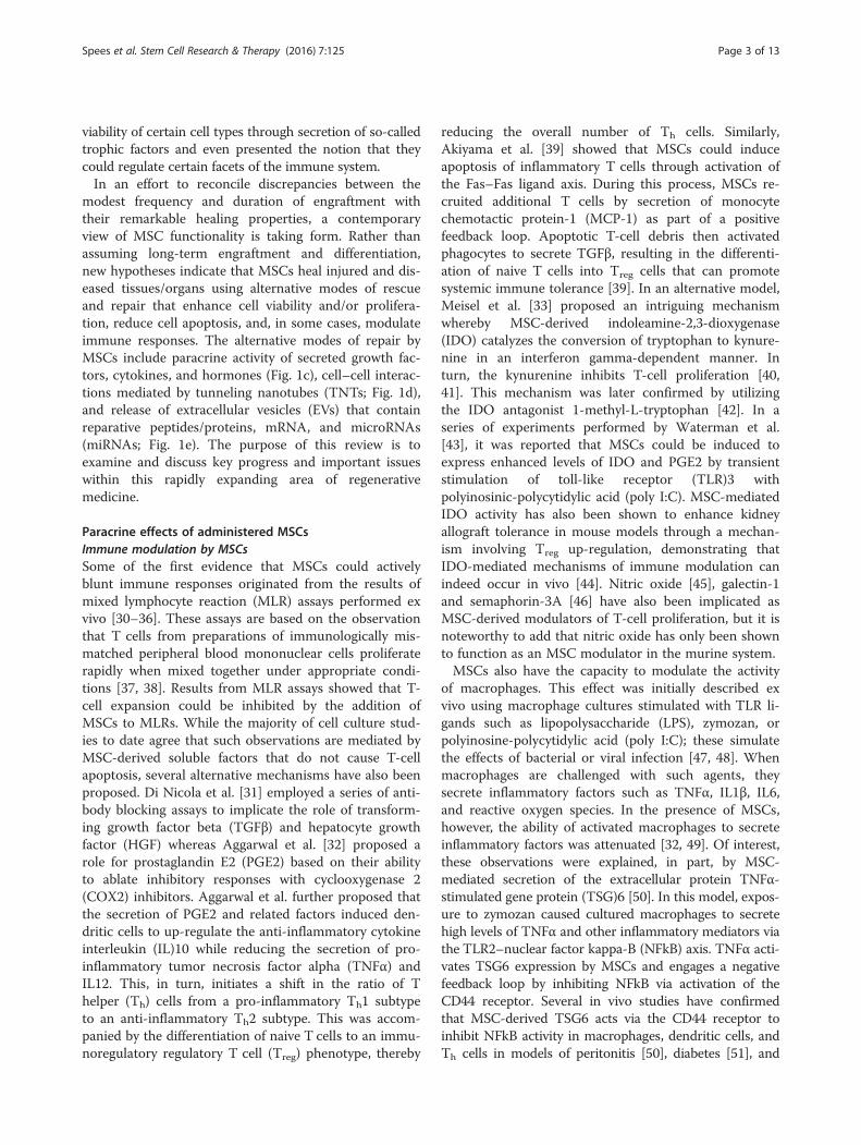
a. Differentiation of MSCs to replace cells.
c. Paracrine activity of MSCs that promotes tissue rescue/repair.
d. MSC-mediated transfer of organelles and/or molecules by TNTs.
e. Transfer of molecules from MSC-derived exosomes or microvesicles.
MSCs
MSC
VEGF-A
HGF
ANG1
SDF-1
PDGF-B
IL-11PGE2
Target tissue
MSC
Mitochondria
Ca2+, Mg2+, RNAs
Proteins/peptides Target cell
Exosomes (30-100 nm)
Microvesicles (50-1000 nm)
MSC
IGF-1TSG-6
b. MSC/cell fusion.
Target cell
MSC
Fig. 1 MSCs rescue and/or repair injured cells and tissues by diverse mechanisms. a Differentiation into replacement cell types. b Rescue of damagedor dying cells through cell fusion. c Secretion of paracrine factors such as growth factors, cytokines, and hormones. VEGF vascular endothelial growthfactor, PDGF platelet-derived growth factor, ANG1 angiopoietin-1, IL-11 interleukin-11, PGE2 prostaglandin E2, TSG-6 TNF-stimulated gene-6, SDF-1stromal-derived factor-1, HGF hepatocyte growth factor, IGF-1 insulin-like growth factor-1. d Transfer of organelles (e.g., mitochondria) and/or moleculesthrough tunneling nanotubes (TNTs). Ca2+ calcium, Mg2+ magnesium. e MSC-mediated transfer of proteins/peptides, RNA, hormones, and/or chemicalsby extracellular vesicles such as exosomes or microvesicles. Exosomes are generated through the endocytic pathway and released through exocytosis.By contrast, microvesicles are produced by cell surface budding and released directly from the plasma membrane. Note that the figure is not drawn toscale. Also, use of mechanisms a–e is not equivalent. For example, for MSCs administered intravenously, use of mechanism c is likely more relevantthan are mechanisms (a) or (b)
Spees et al. Stem Cell Research & Therapy (2016) 7:125 Page 2 of 13
viability of certain cell types through secretion of so-calledtrophic factors and even presented the notion that theycould regulate certain facets of the immune system.In an effort to reconcile discrepancies between the
modest frequency and duration of engraftment withtheir remarkable healing properties, a contemporaryview of MSC functionality is taking form. Rather thanassuming long-term engraftment and differentiation,new hypotheses indicate that MSCs heal injured and dis-eased tissues/organs using alternative modes of rescueand repair that enhance cell viability and/or prolifera-tion, reduce cell apoptosis, and, in some cases, modulateimmune responses. The alternative modes of repair byMSCs include paracrine activity of secreted growth fac-tors, cytokines, and hormones (Fig. 1c), cell–cell interac-tions mediated by tunneling nanotubes (TNTs; Fig. 1d),and release of extracellular vesicles (EVs) that containreparative peptides/proteins, mRNA, and microRNAs(miRNAs; Fig. 1e). The purpose of this review is toexamine and discuss key progress and important issueswithin this rapidly expanding area of regenerativemedicine.
Paracrine effects of administered MSCsImmune modulation by MSCsSome of the first evidence that MSCs could activelyblunt immune responses originated from the results ofmixed lymphocyte reaction (MLR) assays performed exvivo [30–36]. These assays are based on the observationthat T cells from preparations of immunologically mis-matched peripheral blood mononuclear cells proliferaterapidly when mixed together under appropriate condi-tions [37, 38]. Results from MLR assays showed that T-cell expansion could be inhibited by the addition ofMSCs to MLRs. While the majority of cell culture stud-ies to date agree that such observations are mediated byMSC-derived soluble factors that do not cause T-cellapoptosis, several alternative mechanisms have also beenproposed. Di Nicola et al. [31] employed a series of anti-body blocking assays to implicate the role of transform-ing growth factor beta (TGFβ) and hepatocyte growthfactor (HGF) whereas Aggarwal et al. [32] proposed arole for prostaglandin E2 (PGE2) based on their abilityto ablate inhibitory responses with cyclooxygenase 2(COX2) inhibitors. Aggarwal et al. further proposed thatthe secretion of PGE2 and related factors induced den-dritic cells to up-regulate the anti-inflammatory cytokineinterleukin (IL)10 while reducing the secretion of pro-inflammatory tumor necrosis factor alpha (TNFα) andIL12. This, in turn, initiates a shift in the ratio of Thelper (Th) cells from a pro-inflammatory Th1 subtypeto an anti-inflammatory Th2 subtype. This was accom-panied by the differentiation of naive T cells to an immu-noregulatory regulatory T cell (Treg) phenotype, thereby
reducing the overall number of Th cells. Similarly,Akiyama et al. [39] showed that MSCs could induceapoptosis of inflammatory T cells through activation ofthe Fas–Fas ligand axis. During this process, MSCs re-cruited additional T cells by secretion of monocytechemotactic protein-1 (MCP-1) as part of a positivefeedback loop. Apoptotic T-cell debris then activatedphagocytes to secrete TGFβ, resulting in the differenti-ation of naive T cells into Treg cells that can promotesystemic immune tolerance [39]. In an alternative model,Meisel et al. [33] proposed an intriguing mechanismwhereby MSC-derived indoleamine-2,3-dioxygenase(IDO) catalyzes the conversion of tryptophan to kynure-nine in an interferon gamma-dependent manner. Inturn, the kynurenine inhibits T-cell proliferation [40,41]. This mechanism was later confirmed by utilizingthe IDO antagonist 1-methyl-L-tryptophan [42]. In aseries of experiments performed by Waterman et al.[43], it was reported that MSCs could be induced toexpress enhanced levels of IDO and PGE2 by transientstimulation of toll-like receptor (TLR)3 withpolyinosinic-polycytidylic acid (poly I:C). MSC-mediatedIDO activity has also been shown to enhance kidneyallograft tolerance in mouse models through a mechan-ism involving Treg up-regulation, demonstrating thatIDO-mediated mechanisms of immune modulation canindeed occur in vivo [44]. Nitric oxide [45], galectin-1and semaphorin-3A [46] have also been implicated asMSC-derived modulators of T-cell proliferation, but it isnoteworthy to add that nitric oxide has only been shownto function as an MSC modulator in the murine system.MSCs also have the capacity to modulate the activity
of macrophages. This effect was initially described exvivo using macrophage cultures stimulated with TLR li-gands such as lipopolysaccharide (LPS), zymozan, orpolyinosine-polycytidylic acid (poly I:C); these simulatethe effects of bacterial or viral infection [47, 48]. Whenmacrophages are challenged with such agents, theysecrete inflammatory factors such as TNFα, IL1β, IL6,and reactive oxygen species. In the presence of MSCs,however, the ability of activated macrophages to secreteinflammatory factors was attenuated [32, 49]. Of interest,these observations were explained, in part, by MSC-mediated secretion of the extracellular protein TNFα-stimulated gene protein (TSG)6 [50]. In this model, expos-ure to zymozan caused cultured macrophages to secretehigh levels of TNFα and other inflammatory mediators viathe TLR2–nuclear factor kappa-B (NFkB) axis. TNFα acti-vates TSG6 expression by MSCs and engages a negativefeedback loop by inhibiting NFkB via activation of theCD44 receptor. Several in vivo studies have confirmedthat MSC-derived TSG6 acts via the CD44 receptor toinhibit NFkB activity in macrophages, dendritic cells, andTh cells in models of peritonitis [50], diabetes [51], and
Spees et al. Stem Cell Research & Therapy (2016) 7:125 Page 3 of 13
corneal transplant rejection [52]. In addition to the ac-tion(s) of TSG6, MSC-derived PGE2 has also been dem-onstrated to have potent effects on macrophages in vivo.In a murine model of sepsis, Nemeth et al. [53] demon-strated that, upon activation by LPS or TNFα, MSCs se-creted PGE2. This caused the release of anti-inflammatoryIL10 by macrophages and improved cell survival. Indeed,the role of PGE2 in MSC-mediated macrophage modula-tion is a common theme in many culture models [54, 55].In an alternative mechanism proposed by Chen et al. [56],placental human MSCs inhibited the interaction of TLR4with a key effector molecule, MyD88 [48], resulting in in-hibition of secretory factors by macrophages. This processwas inhibited by addition of a COX2 inhibitor, suggestingthat the process was PGE2-dependent.MSCs were reported to modulate the proliferation, dif-
ferentiation, and immunoglobulin secretion of B cellswithout induction of apoptosis [57]. Transwell assaysseparating the two cell types but allowing for exchangeof secreted factors showed that such MSC-mediated ef-fects derived, in part, from the paracrine activity of sol-uble factors secreted by MSCs. These experimentalresults have since been replicated using purified B cellsand unpurified preparations of peripheral blood mono-nuclear cells [58–60]; however, the paracrine mechanismwas recently challenged by a co-culture study that sug-gested physical interaction between T cells and MSCswas necessary for MSCs to inhibit the activities of B cells[61]. Using a mouse model of allergy, Nemeth et al. [62]reported that MSC-derived TGFβ was critical in sup-pressing B-cell mediated allergic responses in vivo. Theyspeculated that MSCs may recruit Treg cells that down-regulate allergy-specific cytokine and immunoglobulinproduction as well as lung eosinophil infiltration. Consist-ent with their immune-modulatory properties, efficacywith MSC treatment has been demonstrated in a varietyof inflammatory models of disease, including arthritis [63],Crohn’s disease [64], multiple sclerosis [65, 66], myocar-dial infarction [14], diabetes [51, 67], graft versus host dis-ease [34, 68, 69], and corneal rejection [52].
Promotion of cell survival by MSCsIn addition to the paracrine effects of MSCs on immunecells, they also secrete a diverse repertoire of factors thatsupport cell survival, including growth factors, cytokines,and extracellular matrix (ECM). Together, the compo-nents of the MSC secretome have the theoretical cap-acity to rescue injured cells, reduce tissue damage, andaccelerate repair. This is exemplified by their naturalroles as reticular cells that support the hematopoieticstem cell niche [26–28, 70, 71] and as vascular pericytesthat support endothelial cells [72, 73]. The observationthat MSCs can be isolated from a wide variety of tissues,such as bone marrow, adipose, ligament, skin, placenta,
dental pulp, synovium, placenta, umbilical cord, andother fetal tissues [72, 74], lends support to the con-cept that they function endogenously as stromal sup-port cells.The pro-survival effect(s) of the MSC secretome on
other cell types was first recognized through studies oflong-term bone marrow cultures [26–29, 75] and embry-onic cells [76]. Collectively, these cell culture studiesprovide for an attractive, paracrine-based explanation forthe ability of MSCs to promote healing across a broadrange of developmentally unrelated tissues and for myr-iad diseases and injury types. Detailed analysis of theMSC transcriptome and proteome has confirmed thatthey secrete a vast repertoire of paracrine pro-survivalfactors commonly referred to as trophic factors or medi-ators [77–82]. Of interest, the MSC-secreted factorscomprise a diverse group of soluble peptides and pro-teins with complementary set(s) of biological activitiesthat can accelerate progenitor cell self-renewal, stimulateangiogenesis, and minimize apoptosis and/or inflamma-tion. Despite several decades of research and progress,the specific paracrine mechanisms by which adminis-tered MSCs improve cell survival and self-renewal underparticular contexts of tissue rescue/repair remain largelyundefined [75, 77].In line with the traditional model of paracrine biology
whereby cells secrete factors that regulate adjacent cells,it was initially thought that engrafted MSCs readily mi-grated into injured tissue and then remained to orches-trate repair. For many models of tissue injury, however,what was originally perceived as “MSC migration”turned out to be far less directed (e.g., non-specific, tran-sient trapping of MSCs within the microvasculature andcapillary network). Of particular interest, depending ontheir relative size (i.e., diameter), the majority of intra-venously administered MSCs will typically lodge in thelung microvasculature upon the first pass through thecirculation, regardless of the presence or absence oflung-specific injury. Notably, after intravenous MSC in-fusion, paracrine factors released into the blood by cir-culating MSCs or from trapped MSCs may indirectlyinfluence survival signaling and the fate of distal cellspreviously compromised by injury or disease. Thus, foreffect, paracrine factors produced by MSCs appear notto depend on long-term MSC engraftment, nor do theyrequire the unlikely differentiation of mesodermal pro-genitors into tissues of ectodermal or endodermallineages.Some of the best evidence supporting an indirect role
for MSCs in the repair of tissues/organs originates fromstudies of heart with infarction. In a rat model of myo-cardial infarction, MSCs modified with the gene encod-ing protein kinase B (a.k.a. Akt) engrafted into themyocardium, reduced pathological remodeling, and
Spees et al. Stem Cell Research & Therapy (2016) 7:125 Page 4 of 13
improved cardiac function [83]. The observed efficacywas later attributed to a paracrine effect mediated by se-creted frizzled related protein (sFRP), a Wnt signalinginhibitor that reduces cardiomyocyte apoptosis [84–86].Since these studies, a number of additional mechanismsfor the paracrine action of MSC-derived factors on car-diac repair have been proposed, including secretion ofangiogenic factors [87–89], stromal cell derived factor-1(SDF-1) [90], and Jagged/Notch signaling [89, 91]. Ofinterest, MSC-mediated improvements in cardiac func-tion could be achieved without long-term engraftmentof MSCs [11]. Using a different approach, MSC-conditioned medium was employed to prime cardiacstem/progenitor cells prior to cardiac grafting in a ratmodel of myocardial infarction. The conditionedmedium (CM) improved cardiac stem cell engraftmentthrough mechanisms involving connective tissue growthfactor and insulin signaling [92].The role of MSCs in the protection of other damaged
tissues has also been demonstrated. For example, intra-peritoneally and intravenously administered MSCs frommurine bone marrow and adipose tissue had a protectiveeffect in a cisplatin-induced acute kidney injury (AKI)model [93], as evidenced by a reduction in the apoptosisof tubule cells and improved renal function. This effectappeared to be mediated by secreted factors since the re-sults could be repeated by intraperitoneal administrationof CM generated from the MSCs (MSC-CM). In con-trast, Xing et al. [94] reported that murine MSC-CMcontaining HGF, vascular endothelial growth factor(VEGF)-A and insulin-like growth factor (IGF)-1 failedto protect the kidneys of mice against ischemia-reperfusion injury, whereas live MSCs had a significantprotective effect. This is one of several examples in thefield where apparently minor differences in the cellsource, the culture conditions, duration of medium con-ditioning, and dosage can profoundly affect outcome.Such complexities have made elucidation of the mecha-nism(s) responsible for the protective effect of MSCs onkidney tissue challenging, but some progress has beenmade. For example, Zarjou et al. [95] demonstrated thatthe stress-responsive enzyme heme-oxygenase-1 (HO-1)played a role by utilizing MSC from bone marrow ofHO-1-/- mice. In this study, HO-1+/+ MSC-CM rescuedpathology associated with cisplatin-induced AKI, whileHO-1-/- MSC-CM was ineffective. The authors attrib-uted the difference in effect to enhanced levels of SDF-1,VEGF-A, and HGF in the HO-1+/+ MSCs. Indeed, im-munological and transcriptional blocking experimentsboth confirm a protective role for VEGF-A [96–98] andIGF-1 [99] in mice with AKI and for VEGF-A in ratswith cerebral ischemia (stroke) [100].The utility of MSCs and their secreted products to
protect cells and to foster tissue repair has been
demonstrated in numerous efficacy-based studies acrossa broad range of tissue injury and disease models. Whilea comprehensive summary of the associated literature isbeyond the scope of this review, some key examples ofMSC-derived benefits include facilitation of wound heal-ing [101], improved treatment of diabetes [102], en-hancement of bone repair [103, 104], and effect(s) oncancer [105].
Effects of MSCs on fibrosisFibrosis is generally defined as a an accelerated accumu-lation of ECM factors (predominantly collagen type I)that prevents the regeneration of tissue. It can occur invirtually any tissue as a result of trauma, inflammation,immunological rejection, chemical toxicity, or oxidativestress. Current clinical strategies generally have pooroutcomes in terms of efficacy and adverse effects [106].Given the immunomodulatory and trophic properties ofMSCs, they have become attractive candidates for thetreatment of fibrosis and preclinical studies suggest theyhave a promising level of efficacy in a variety of models.While the anti-fibrotic effects of MSCs are likely to over-lap with their anti-inflammatory and angiogenic proper-ties, the specific mechanisms remain poorly understood.Nevertheless, a comprehensive review by Usuner et al.[107] suggests that their modes of action seem to fallunder four categories: i) immune modulation, ii) inhib-ition of TGFβ-mediated differentiation of various cellstypes into ECM-secreting myofibroblasts by epithelial tomesenchymal transition, iii) inhibition of oxidative stress,and iv) matrix remodeling. For example, Ortiz et al. dem-onstrated that systemic murine MSC administration at-tenuated fibrosis in a bleomycin-induced lung injurymodel [108]. This was achieved through MSC-mediatedsecretion of IL1 receptor antagonist, which reduced infil-tration of lymphocytes and neutrophils and their produc-tion of inflammatory and fibrotic mediators such as IL1and TNFα. Using the same model, it was recently reportedthat MSCs had the capacity to inhibit fibrosis through theaction of the secreted protein stanniocalcin-1 (STC-1)[109]. The authors demonstrated that STC-1 acted inmultiple ways by reducing the secretion of collagen by fi-broblasts, by reducing TGFβ output by endothelial cellsand also through alleviating oxidative stress by uncouplingmitochondrial respiration via the induction of uncouplingprotein 2. Using a model of chronic kidney injury, Huuskeset al. [110] demonstrated that MSCs improved kidneymorphology and functionality when co-administered withthe putatively anti-fibrotic hormone recombinant humanrelaxin (serelaxin). In this system, MSCs and serelaxinacted synergistically to reduce TGFβ-induced myofibro-blast differentiation and collagen deposition while increas-ing the level of matrix metalloproteinase 2 (MMP2), acollagen-degrading enzyme.
Spees et al. Stem Cell Research & Therapy (2016) 7:125 Page 5 of 13
Transfer of mitochondria by TNTs and microvesiclesDiscovery of TNTsRustom et al. [111] first reported TNTs as a communi-cating intercellular transport network formed in culturesof transformed cells (human 293 cells and rat PC12cells) as well as primary cells from rat kidney. Endocyticorganelles (lysosomes) and vesicles were shown to movethrough thin, 50–200 nm diameter filaments thatstretched between cells. Incubation of cells in the inhibi-tor latrunculin B demonstrated a requirement for poly-merized F-actin in TNT formation. Onfelt et al. [112]reported TNTs in human immune cells (e.g., naturalkiller cells, macrophages, and B cells) and later demon-strated that TNTs between macrophages had differentproperties and potentially differing functions; they ob-served thin filaments containing F-actin and also athicker subset (0.7 microns) that contained both F-actinand microtubules. The thicker TNT subset was shownto transport mitochondria and lysosomal vesicles [113].Other studies demonstrated that some TNTs wereactinomyosin-dependent [114, 115]. For example, theGerdes group showed that kidney cells treated with S-(-)-blebbistatin, a myosin II-specific inhibitor, increasedthe number of TNTs formed and also organelle transfer,whereas a general myosin inhibitor increased TNT num-ber but significantly reduced organelle transfer [114].
Discovery of mitochondrial transfer by cultured MSCsThe first evidence that transfer of mitochondria mightbenefit injured target cells came from studies of humanMSCs co-cultured with a unique lung epithelial cell linethat lacked functional mitochondria (A549rho cells)[116]. Utilizing a complementation screen to detectmitochondrial transfer and resulting cell growth, theProckop group reported that human MSCs could restoreaerobic respiration to A549rho cells by transfer of mito-chondria or mitochondrial DNA (mtDNA). Mitochon-drial transfer from MSCs to rescued A549rho cells wasdemonstrated by tracking genetic tags (i.e., mtDNA andnuclear DNA) and by time-lapse photomicroscopy ofMSCs transduced with lentiviral vectors to targetDsRed2 to mitochondria [116]. MSCs are now under-stood to transfer mitochondria to several different celltypes, including epithelial cells, endothelial cells, andcardiac myocytes [117]. Such transfers are particularlyevident when the potential target cells are injured orunder stress. For example, MSCs were recently shown toprevent apoptosis in endothelial cells by transferringmitochondria during hypoxic/ischemic stress [118].
TNT formation and mitochondrial transfer in vivoThe first evidence that TNTs could form in vivo camefrom studies of the eye. Using wild-type, eGFP chimericmice, and Cx3cr1(GFP) transgenic mice and confocal
microscopy tracking, Chinnery et al. [119] documentedmembrane nanotubes that formed between bone marrow-derived MHC class II(+) cells in whole-mounted cornealtissue. Notably, they observed an increase in TNT fre-quency during corneal injury or inflammation. In a follow-up study with live imaging of myeloid cells in inflamedcorneal explants from Cx3cr1(GFP) and CD11c(eYFP)transgenic mice, Seyed-Razavi et al. [120] showed de novoformation of nanotubes at a rate of 15.5 μm/min. Theseresults demonstrated that TNTs could form in the absenceof actual cell–cell contact and, furthermore, that theycould then be directed from one cell toward another. Add-itional evidence for in vivo mitochondria or mtDNA trans-fer between cells came from studies of a remarkablecanine transmissible venereal tumor that had persisted inferal dog populations for about 10,000 years. Rebbeck etal. [121] showed that the transmitted tumor cell line hadobtained mitochondria (mtDNA) from multiple caninehosts over time. They suggested that fitness/persistence ofcanine transmissible venereal tumor benefited from the ac-quisition of host-derived mtDNA and through shedding ofmutant and/or damaged mtDNA that could negatively im-pact mitochondrial biogenesis. Importantly, multiple re-search groups have shown that intercellular transfer oforganelles and mtDNA is not limited only to the animalkingdom. Intercellular organelle trafficking and horizontalgene transfer in plants has been reported for both plastids[122] and mitochondria [123].
Proteins shown to control transfer of mitochondria by MSCsafter tissue injurySeveral recent studies have provided compelling evi-dence that administered MSCs can transfer mitochon-dria in vivo and, furthermore, that mitochondria transferfrom MSCs can rescue injured pulmonary cells andameliorate lung injury. Islam et al. [124] demonstratedthat airway instillation of human MSCs could reduceLPS-mediated lung injury, in part, through transfer ofmitochondria. Using live optical imaging, they docu-mented transfer of vesicles containing labeled mitochon-dria from MSCs to alveolar epithelial cells that increasedalveolar ATP levels and cell survival. Unlike wild-typeMSCs, MSCs genetically modified for connexin 43 thatwere incapable of forming gap junctions and MSCs withdysfunctional mitochondria did not reduce acute lunginjury [124].Recent data from a cigarette smoke-induced model of
lung injury suggest that donor source and age may affectrepair by mitochondria transfer by MSC. Li et al. [125]found that transplantation of MSCs derived from in-duced pluripotent stem cells may provide enhanced re-pair after transplantation by virtue of increased TNTformation and mitochondria transfer relative to adult-derived MSCs.
Spees et al. Stem Cell Research & Therapy (2016) 7:125 Page 6 of 13
Using loss- and gain-of-function approaches, Ahmadet al. [126] elegantly demonstrated that Miro-1, an outermitochondrial membrane Rho-like GTPase, regulatedthe amount of mitochondrial transfer from MSCs to cul-tured lung epithelial cells. Enhanced expression of Miro-1 was shown to increase transfer of mitochondria fromMSCs and treatment of mice with MSCs overexpressingMiro-1 reduced Rotenone lung injury and airway hyper-responsiveness and negative remodeling in severalmodels of asthma [126].
Regulators of mitochondria transport identified in other celltypes that may orchestrate mitochondrial transfer by MSCsIn addition to Miro-1, other proteins known to regulateintracellular mitochondrial dynamics (e.g., fusion, fission,tethering, and trafficking) [127, 128] may also promoteor inhibit intercellular mitochondria transfer. Miro-1and Miro-2 belong to a group of dynamin-related pro-teins that regulate mitochondrial division and fusion.They interact with TRAK1 and TRAK2 (identified asMilton in Drosophila), adaptor proteins that recruitkinesin motor proteins to mitochondria. The resultingadaptor–motor protein complex shuttles mitochondriaalong microtubules and was demonstrated to be criticalfor neuronal transport of mitochondria to axons, den-drites, and synapses [129–131]. Mitofusin 1 and 2 mayalso regulate mitochondria transfer as they are known tointeract with Miro-1 and Miro-2 as well as TREK1/TREK2 in the adaptor–motor protein complex [132].Perhaps not surprising, motor proteins are likely to berequired for generation of some forms of TNTs. Myo-X(Myo10) is a myosin motor protein that localizes to theends of cellular filapodia. It is unique in that it does notrequire substrate attachment to induce filapodia exten-sion [133]. Co-culture studies in neuronal cells demon-strated that Myo10 was required for TNT formationfrom filapodia and overexpression of Myo10 resulted inincreased TNT formation and vesicle transfer betweencells [134].Although the damage/injury signals that initiate mito-
chondrial transfer have yet to be identified, it is plausiblethat differences in intracellular Ca+2 or energy stores(e.g., glucose, ATP) may play a role in directing one cellto transfer mitochondria to another. For example, intra-cellular movement of mitochondria is highly sensitive tocytosolic Ca+2 levels. Wang and Schwartz [135] elegantlydemonstrated that Ca+2 promotes Miro to interact withthe motor domain of kinesin, thus blocking kinesin fromthe microtubule. Accordingly, mitochondria transferfrom cell to cell may be affected by differences in intra-cellular Ca+2 concentration and/or localization. Consist-ent with this concept, TNTs have been shown to transferCa2+ and even electrical signals to neighboring cellsthrough TNT-associated gap junctions [136, 137]. In
addition, the level of available nutrients can alter move-ment of mitochondria. In neurons, Pekkurnaz et al.[138] reported that extracellular glucose and the enzymeO-GlcNAc transferase (OGT) affect mitochondrial mo-tility by altering GlcNAcylation of Milton, an OGT sub-strate. As OGT activity is dependent on glucose,increased glucose was shown to decrease mitochondrialmotility.Of special interest, several reports indicate regulatory
overlap or some form of integration between TNT for-mation and endosomal trafficking, as both interact withcomponents of the exocyst complex that regulates ves-icular transport from the Golgi apparatus to the plasmamembrane [139, 140]. For example, Hase et al. [141] re-ported that M-sec, part of the exocyst complex, inter-acted with the small GTPase RalA and was required forTNT formation in a macrophage cell line. Furthermore,they showed that M-sec expression could induce cellprotrusions de novo, some of which formed TNTs withadjacent cells. Subsequently, Schiller et al. [142] foundthat the transmembrane MHC class III protein leukocytespecific transcript 1 (LST1) was also required for TNTformation. At the cell membrane, LST1 was shown tointeract with M-Sec, myosin, and myoferlin and also torecruit RalA, promoting its interaction with the exocystcomplex [142]. Notably, some mechanisms (e.g., pro-teins) controlling TNT formation and/or mitochondrialtransfer may be specific to specialized cell types such asneurons. However, in light of the conserved nature ofintracellular adaptor/kinesin motor protein complexes,mitochondrial dynamics, and endosomal trafficking, it isprobable that many mechanisms that control TNT for-mation and/or mitochondrial transfer are similar be-tween many cell types, including MSCs.
Modifying mitochondrial transfer and/or mitochondria forclinical applicationFor future clinical application, harnessing mitochondrialtransfer in a controlled and predictable manner willlikely require further mechanistic insight. Importantly,recent advances in targeting of DNA to mitochondriamay provide new tools to track or even perhaps to gen-etically alter mitochondria by modifying mtDNA as op-posed to nuclear genes for proteins targeted tomitochondria (e.g., genes for mitochondrial membraneproteins). For example, Yu et al. [143] restored ATP syn-thesis in cells carrying mutant mtDNA for humanNADH ubiquinone oxidoreductase subunit 4 (ND4) byinfecting cells with an adeno-associated virus capsid(VP2) fused to a mitochondrial targeting sequence andthe wild-type ND4 mitochondrial gene sequence. Fol-lowing recent successful testing in non-human primatesand human eyes ex vivo, the innovative method maysoon be applied in clinical trials for treatment of Leber
Spees et al. Stem Cell Research & Therapy (2016) 7:125 Page 7 of 13
hereditary optic neuropathy, a disease caused by a muta-tion in the ND4 mitochondrial gene [144].Despite the potential benefits of mitochondrial transfer
or other TNT-mediated effects, it is worth noting thatcell–cell communication by way of TNTs may also havesome negative consequences. In contrast to their potentialtherapeutic benefits, TNTs also have potential to act asdisease vectors for transmission of HIV/AIDS [145], bac-teria [113], Prions [146], and oncogenic miRNAs [147].
Transfer of RNAs and other molecules by EVsThe general term “extracellular vesicle” (EV) refers tomembrane-bound vesicles released from most, if not all,somatic cell types (reviewed in [140, 148, 149]). To-gether, the EVs include exosomes, 30–100-nm plasmamembrane-coated vesicles of endocytic origin; microve-sicles, 50–1000-nm vesicles of non-endocytic origin; andapoptotic bodies, 1–5-μm vesicles released during mem-brane blebbing of apoptotic cells [150].Cellullar exosomes are released when multivesicular
bodies traffic to and fuse with the plama membrane in aregulated manner. Exosomes were first identified andisolated from cultures of normal and transformed cellsduring the 1980s [151–153]. Valadi et al. [154] made akey contribution when they demonstrated that bothmRNA and miRNA could be exchanged between cellsby virtue of exosomal transfer. Studying xenogenic co-cultures, they observed expression of various mouse pro-teins in human mast cells after exosomal transfer frommurine cells, indicating successful translation of exoso-mally delivered mRNA into protein. As with exosomesisolated from diverse cell types, MSC-derived exosomesare reported to contain lipid raft domains [155] and tetra-spanins known to alter the fusion state of cell membranes(e.g., CD9, CD81), Alix, a calcium-binding protein withroles in both endosomal trafficking and cell death, andTSG101, a tumor suppressor protein [156, 157]. Com-pared with exosomes, which are relatively homogenousupon release, microvesicles are heterogenous in both sizeand composition. Furthermore, regulatory mechanismsfor microvesicular shedding from the membrane surfaceremain poorly understood.Exosomes purified from MSCs have garnered tremen-
dous interest in the field of regenerative medicine basedon their ability to reduce apoptosis/necrosis in rodentsafter ischemic injury to the heart [158, 159], brain [160,161], lung [162], liver [163], or kidney [164]. In addition,exosomal transfer from MSCs is reported to reduce in-flammation and to increase cell proliferation during tis-sue repair [162, 165, 166]. Tomasoni et al. [167] showedthat MSCs transferred exosomes with mRNA for IGF1Rand IGF1 to cisplatin-damaged proximal tubular cells;this resulted in their expression of IGF1R, thereby in-creasing sensitization to IGF-1. The exosomal transfer
improved renal cell survival and increased proliferationduring repair after injury. In multiple drug-inducedmodels of liver injury, treatment with MSC exosomes atthe time of injury increased the number of proliferatingcell nuclear antigen-positive proliferation cells while re-ducing the number of hepatocytes undergoing apoptoticcell death [168]. Treatment of a murine carbontetrachloride-based injury model with exosomes fromhuman umbilical cord-derived MSCs was shown to re-duce liver fibrosis [169]. Following stroke in rats, treat-ment with MSC-derived exosomes was shown topromote angiogenesis, neurogenesis, neurite outgrowth,and recovery by virtue of transfer of miR-133b [170,171]. In addition to RNAs, exosomes and microvesiclescan deliver peptide/protein-based paracrine effectorssuch as growth factors, cytokines, and hormones. For ex-ample, transfer of Wnt4 by exosomes from human um-bilical cord-derived MSCs improved repair of skinwounds in rats by altering cell proliferation [172].Currently, many investigators and clinicians are inter-
ested in the potential of MSC-derived EV therapeutics forrepair of injured and diseased tissue and to treat cancer[173, 174]. Most studies with exosome-based treatment ofinjured tissues/organs report positive outcomes, However,whether or not MSC-mediated transfer of exosomes,microvesicles, and/or their constituents promote or inhibitthe activities of transformed cells in a way that wouldpositively or negatively impact cancer remains context-dependent and controversial. For example, bone marrowMSCs were shown to reduce the growth of cultured breastcancer cells by transferring miR-127, -197, -222, and -223through gap junctions and exosomes; these miRNAs areknown to target CXCL12 (a.k.a. SDF-1) [175]. Lee et al.[176] suggested that exosomes from MSCs might suppressangiogenesis based on their containing miR-16, a miRNAthat targets VEGF and was shown to reduce its expressionin a breast cancer cell line. By contrast, Zhu et al. [177]reported that exosomes from human MSCs actually pro-moted tumor growth in vivo by inducing VEGF expres-sion in tumor cells. Boelens et al. [178] reported cross-talkbetween stromal cells and breast cancer cells wherebystromal exosomes induced paracrine antiviral signals andstimulated juxtacrine Notch3 signaling that increased thenumber of therapy-resistant tumor-initiating cells. As withother paracrine effects of cell-based therapy or treatmentsbased on administration of signaling agonists (e.g., growthfactors), it is clear that care must be taken to avoid poten-tial off-target treatment effects of administered EVs toavoid cancer cell propagation and/or metastasis.Towards standardization of exosome-based therapy
using MSCs or any cell type, identification of the mostreliable and consistent vesicle isolation methods will becritical so that different laboratories can effectively com-pare their results. At present, several different methods
Spees et al. Stem Cell Research & Therapy (2016) 7:125 Page 8 of 13
of isolation are widely used, including centrifugation,filtration, immunoaffinity isolation with beads, andmicrofluidics. Notably, exosomes isolated from thesame source by different methods may differ in amountand/or content [179–181].Research aimed at improved understanding of mecha-
nisms controlling cargo loading of exosomes will also beimportant. For protein-based cargo, Shen et al. [182]have reported some progress using expressed plasmamembrane anchors. For miRNA-based cargo, Villarroya-Beltri et al. [183] recently identified specific miRNA se-quence motifs that direct their loading into exosomes. Fur-thermore, they determined that sumoylated heterogenousnuclear ribonucleoprotein (hnRNPA2B1) was required forsorting of miRNAs into exosomes based on the specificmotifs. Detailed characterization of MSC exosome contentunder various conditions and from all tissues will likelyaid in a more predictable product in terms of therapy.For example, MSCs isolated from various tissues differin terms of exosome content [184, 185] and MSCs frombone marrow with multiple myeloma were reported todiffer in miRNA content relative to MSCs from controlbone marrow [183].
ConclusionsIn light of promising results in animal models and pa-tients, therapeutic use of MSCs and MSC-based prod-ucts for treatment of tissue injury and disease is likely toundergo continued evaluation. As next steps, focusingefforts toward achieving standardized methods of MSCisolation, characterization, and administration has greatpotential to provide powerful new treatments with MSCsor MSC-derived products. In regard to the predominantmechanisms of MSC function, clarification of the rela-tive role(s) that each mechanism plays during the rescueand repair of damaged tissues/organs following MSC ad-ministration may serve to improve treatment safety, effi-cacy, and predictability of outcome for patients.
AcknowledgmentsThis work was funded in part by research grants from the North AmericanSpine Society (C.A.G.), The Scott & White Research Grants Program (C.A.G.),The Texas Engineering Experiment Station Strategic Initiative (C.A.G.), TheCenter for the Advancement of Science in Space (C.A.G.), NSF CollaborativeResearch CBET-1264832 (C.A.G.), NIH NIAMS R01 AR066033 (C.A.G.), NIHNINDS/NIGMS R01 NS073815 (J.L.S.), NIH NHLBI R01 HL132264 (J.L.S.), andSPARKVT (J.L.S.).
Authors’ contributionsAll authors wrote, read, and approved this manuscript.
Competing interestsThe authors declare that they have no competing interests.
DisclosuresJeffrey L. Spees is co-founder of Samba BioLogics, Inc. and Carl A. Gregory isa member of the Scientific Advisory Board of Theocorp Holding Co.
Author details1University of Vermont, Burlington, VT, USA. 2Institute for RegenerativeMedicine, Texas A & M University College of Medicine, 206 Olsen Blvd., Room228, MS1114, College Station, TX 77845, USA. 3Department of Medicine, StemCell Core, University of Vermont, 208 South Park Drive, Ste 2, Colchester, VT05446, USA.
Kuralesova AI, Latzinik NW, Gerasimow UW. Origin of bone marrow stromalmechanocytes in radiochimeras and heterotopic transplants. Exp Hematol.1978;6(5):440–4.
2. Friedenstein AJ. Precursor cells of mechanocytes. Int Rev Cytol. 1976;47:327–59.3. Friedenstein AJ, Gorskaja JF, Kulagina NN. Fibroblast precursors in normal
and irradiated mouse hematopoietic organs. Exp Hematol. 1976;4(5):267–74.4. Friedenstein AJ, Chailakhjan RK, Lalykina KS. The development of fibroblast
colonies in monolayer cultures of guinea-pig bone marrow and spleen cells.Cell Tissue Kinet. 1970;3(4):393–403.
5. Friedenstein AJ, Piatetzky II S, Petrakova KV. Osteogenesis in transplants ofbone marrow cells. J Embryol Exp Morphol. 1966;16(3):381–90.
6. Pittenger M, Vanguri P, Simonetti D, Young R. Adult mesenchymal stemcells: potential for muscle and tendon regeneration and use in genetherapy. J Musculoskelet Neuronal Interact. 2002;2(4):309–20.
7. Prockop DJ. Marrow stromal cells as stem cells for nonhematopoietictissues. Science. 1997;276(5309):71–4.
8. Caplan AI, Bruder SP. Mesenchymal stem cells: building blocks for molecularmedicine in the 21st century. Trends Mol Med. 2001;7(6):259–64.
9. Horwitz EM, Gordon PL, Koo WK, Marx JC, Neel MD, McNall RY, Muul L,Hofmann T. Isolated allogeneic bone marrow-derived mesenchymal cellsengraft and stimulate growth in children with osteogenesis imperfecta:Implications for cell therapy of bone. Proc Natl Acad Sci U S A.2002;99(13):8932–7.
11. Iso Y, Spees JL, Serrano C, Bakondi B, Pochampally R, Song YH, Sobel BE,Delafontaine P, Prockop DJ. Multipotent human stromal cells improvecardiac function after myocardial infarction in mice without long-termengraftment. Biochem Biophys Res Commun. 2007;354(3):700–6.
12. Hofstetter CP, Schwarz EJ, Hess D, Widenfalk J, El Manira A, Prockop DJ,Olson L. Marrow stromal cells form guiding strands in the injured spinalcord and promote recovery. Proc Natl Acad Sci U S A. 2002;99(4):2199–204.
13. Lee RH, Seo MJ, Pulin AA, Gregory CA, Ylostalo J, Prockop DJ. The CD34-likeprotein PODXL and alpha6-integrin (CD49f) identify early progenitor MSCswith increased clonogenicity and migration to infarcted heart in mice.Blood. 2009;113(4):816–26.
14. Lee RH, Pulin AA, Seo MJ, Kota DJ, Ylostalo J, Larson BL, Semprun-Prieto L,Delafontaine P, Prockop DJ. Intravenous hMSCs improve myocardialinfarction in mice because cells embolized in lung are activated to secretethe anti-inflammatory protein TSG-6. Cell Stem Cell. 2009;5(1):54–63.
15. Dai W, Hale SL, Martin BJ, Kuang JQ, Dow JS, Wold LE, Kloner RA. Allogeneicmesenchymal stem cell transplantation in postinfarcted rat myocardium:short- and long-term effects. Circulation. 2005;112(2):214–23.
16. Herzog EL, Chai L, Krause DS. Plasticity of marrow-derived stem cells. Blood.2003;102(10):3483–93.
17. Krause DS, Theise ND, Collector MI, Henegariu O, Hwang S, Gardner R,Neutzel S, Sharkis SJ. Multi-organ, multi-lineage engraftment by a singlebone marrow-derived stem cell. Cell. 2001;105(3):369–77.
18. Theise ND. New principles of cell plasticity. C R Biol. 2002;325(10):1039–43.19. Fuchs E, Segre JA. Stem cells: a new lease on life. Cell. 2000;100(1):143–55.20. Wagers AJ, Weissman IL. Plasticity of adult stem cells. Cell.
2004;116(5):639–48.21. Theise ND. On experimental design and discourse in plasticity research.
Stem Cell Rev. 2005;1(1):9–13.22. Gruh I, Martin U. Transdifferentiation of stem cells: a critical view. Adv
Biochem Eng Biotechnol. 2009;114:73–106.23. Prockop DJ, Oh JY. Medical therapies with adult stem/progenitor cells
(MSCs): a backward journey from dramatic results in vivo to the cellular andmolecular explanations. J Cell Biochem. 2012;113(5):1460–9.
Spees et al. Stem Cell Research & Therapy (2016) 7:125 Page 9 of 13
24. Prockop DJ. Repair of tissues by adult stem/progenitor cells (MSCs):controversies, myths, and changing paradigms. Mol Ther. 2009;17(6):939–46.
25. Bianco P, Cao X, Frenette PS, Mao JJ, Robey PG, Simmons PJ, Wang CY. Themeaning, the sense and the significance: translating the science ofmesenchymal stem cells into medicine. Nat Med. 2013;19(1):35–42.
26. Dexter TM, Spooncer E. Growth and differentiation in the hemopoieticsystem. Annu Rev Cell Biol. 1987;3:423–41.
27. Dexter TM, Wright EG, Krizsa F, Lajtha LG. Regulation of haemopoietic stemcell proliferation in long term bone marrow cultures. Biomedicine.1977;27(9-10):344–9.
28. Dexter TM, Spooncer E, Toksoz D, Lajtha LG. The role of cells and theirproducts in the regulation of in vitro stem cell proliferation and granulocytedevelopment. J Supramol Struct. 1980;13(4):513–24.
29. Quesenberry PJ, Mcniece IK, McGrath HE, Temeles DS, Baber GB,Deacon DH. Stromal regulation of hematopoiesis. Ann N Y Acad Sci.1989;554:116–24.
30. Le Blanc K, Tammik L, Sundberg B, Haynesworth SE, Ringdén O.Mesenchymal stem cells inhibit and stimulate mixed lymphocyte culturesand mitogenic responses independently of the major histocompatibilitycomplex. Scand J Immunol. 2003;57(1):11–20.
31. Di Nicola M, Carlo-Stella C, Magni M, Milanesi M, Longoni PD, Matteucci P,Grisanti S, Gianni AM. Human bone marrow stromal cells suppress T-lymphocyteproliferation induced by cellular or nonspecific mitogenic stimuli. Blood.2002;99(10):3838–43.
36. Zappia E, Casazza S, Pedemonte E, Benvenuto F, Bonanni I, Gerdoni E, GiuntiD, Ceravolo A, Cazzanti F, Frassoni F, et al. Mesenchymal stem cellsameliorate experimental autoimmune encephalomyelitis inducing T-cellanergy. Blood. 2005;106(5):1755–61.
37. Lyons AB, Parish CR. Determination of lymphocyte division by flowcytometry. J Immunol Methods. 1994;171(1):131–7.
38. Suni MA, Maino VC, Maecker HT. Ex vivo analysis of T-cell function. CurrOpin Immunol. 2005;17(4):434–40.
39. Akiyama K, Chen C, Wang D, Xu X, Qu C, Yamaza T, Cai T, Chen W, Sun L,Shi S. Mesenchymal-stem-cell-induced immunoregulation involvesFAS-ligand-/FAS-mediated T cell apoptosis. Cell Stem Cell.2012;10(5):544–55.
40. Hwu P, Du MX, Lapointe R, Do M, Taylor MW, Young HA. Indoleamine2,3-dioxygenase production by human dendritic cells results in theinhibition of T cell proliferation. J Immunol. 2000;164(7):3596–9.
41. Mellor AL, Munn DH. Tryptophan catabolism and T-cell tolerance:immunosuppression by starvation? Immunol Today. 1999;20(10):469–73.
42. Ryan JM, Barry F, Murphy JM, Mahon BP. Interferon-gamma does not break,but promotes the immunosuppressive capacity of adult humanmesenchymal stem cells. Clin Exp Immunol. 2007;149(2):353–63.
43. Waterman RS, Tomchuck SL, Henkle SL, Betancourt AM. A newmesenchymal stem cell (MSC) paradigm: polarization into a pro-inflammatory MSC1 or an Immunosuppressive MSC2 phenotype. PLoS One.2010;5(4):e10088.
44. Ge W, Jiang J, Arp J, Liu W, Garcia B, Wang H. Regulatory T-cell generationand kidney allograft tolerance induced by mesenchymal stem cellsassociated with indoleamine 2,3-dioxygenase expression. Transplantation.2010;90(12):1312–20.
45. Sato K, Ozaki K, Oh I, Meguro A, Hatanaka K, Nagai T, Muroi K, Ozawa K.Nitric oxide plays a critical role in suppression of T-cell proliferation bymesenchymal stem cells. Blood. 2007;109(1):228–34.
46. Lepelletier Y, Lecourt S, Renand A, Arnulf B, Vanneaux V, Fermand JP,Menasché P, Domet T, Marolleau JP, Hermine O, et al. Galectin-1 andsemaphorin-3A are two soluble factors conferring T-cell
immunosuppression to bone marrow mesenchymal stem cell. Stem CellsDev. 2010;19(7):1075–9.
48. Watters TM, Kenny EF, O’Neill LA. Structure, function and regulation of theToll/IL-1 receptor adaptor proteins. Immunol Cell Biol. 2007;85(6):411–9.
49. Tsyb AF, Petrov VN, Konoplyannikov AG, Saypina EV, Lepechina LA, KalsinaSS, Semenkova IV, Agaeva EV. In vitro inhibitory effect of mesenchymalstem cells on zymosan-induced production of reactive oxygen species. BullExp Biol Med. 2008;146(1):158–64.
50. Choi H, Lee RH, Bazhanov N, Oh JY, Prockop DJ. Anti-inflammatory proteinTSG-6 secreted by activated MSCs attenuates zymosan-induced mouseperitonitis by decreasing TLR2/NF-kB signaling in resident macrophages.Blood. 2011;118(2):330–8.
51. Kota DJ, Wiggins LL, Yoon N, Lee RH. TSG-6 produced by hMSCs delays theonset of autoimmune diabetes by suppressing Th1 development andenhancing tolerogenicity. Diabetes. 2013;62(6):2048–58.
52. Oh JY, Lee RH, Yu JM, Ko JH, Lee HJ, Ko AY, Roddy GW, Prockop DJ.Intravenous mesenchymal stem cells prevented rejection of allogeneiccorneal transplants by aborting the early inflammatory response. Mol Ther.2012;20(11):2143–52.
53. Németh K, Leelahavanichkul A, Yuen PS, Mayer B, Parmelee A, Doi K, RobeyPG, Leelahavanichkul K, Koller BH, Brown JM, et al. Bone marrow stromalcells attenuate sepsis via prostaglandin E(2)-dependent reprogramming ofhost macrophages to increase their interleukin-10 production. Nat Med.2009;15(1):42–9.
54. Maggini J, Mirkin G, Bognanni I, Holmberg J, Piazzón IM, Nepomnaschy I,Costa H, Cañones C, Raiden S, Vermeulen M, et al. Mouse bone marrow-derivedmesenchymal stromal cells turn activated macrophages into a regulatory-likeprofile. PLoS One. 2010;5(2):e9252.
55. Bartosh TJ, Ylöstalo JH, Mohammadipoor A, Bazhanov N, Coble K, ClaypoolK, Lee RH, Choi H, Prockop DJ. Aggregation of human mesenchymalstromal cells (MSCs) into 3D spheroids enhances their antiinflammatoryproperties. Proc Natl Acad Sci U S A. 2010;107(31):13724–9.
56. Chen CP, Tsai PS, Huang CJ. Antiinflammation effect of human placentalmultipotent mesenchymal stromal cells is mediated by prostaglandin E2 viaa myeloid differentiation primary response gene 88-dependent pathway.Anesthesiology. 2012;117(3):568–79.
57. Corcione A, Benvenuto F, Ferretti E, Giunti D, Cappiello V, Cazzanti F, RissoM, Gualandi F, Mancardi GL, Pistoia V, et al. Human mesenchymal stem cellsmodulate B-cell functions. Blood. 2006;107(1):367–72.
58. Asari S, Itakura S, Ferreri K, Liu CP, Kuroda Y, Kandeel F, Mullen Y.Mesenchymal stem cells suppress B-cell terminal differentiation. ExpHematol. 2009;37(5):604–15.
59. Bochev I, Elmadjian G, Kyurkchiev D, Tzvetanov L, Altankova I, Tivchev P,Kyurkchiev S. Mesenchymal stem cells from human bone marrow oradipose tissue differently modulate mitogen-stimulated B-cellimmunoglobulin production in vitro. Cell Biol Int. 2008;32(4):384–93.
60. Franquesa M, Hoogduijn MJ, Bestard O, Grinyó JM. Immunomodulatoryeffect of mesenchymal stem cells on B cells. Front Immunol. 2012;3:212.
61. Rosado MM, Bernardo ME, Scarsella M, Conforti A, Giorda E, Biagini S,Cascioli S, Rossi F, Guzzo I, Vivarelli M, et al. Inhibition of B-cell proliferationand antibody production by mesenchymal stromal cells is mediated by Tcells. Stem Cells Dev. 2015;24(1):93–103.
62. Nemeth K, Keane-Myers A, Brown JM, Metcalfe DD, Gorham JD, Bundoc VG,Hodges MG, Jelinek I, Madala S, Karpati S, et al. Bone marrow stromalcells use TGF-beta to suppress allergic responses in a mouse model ofragweed-induced asthma. Proc Natl Acad Sci U S A. 2010;107(12):5652–7.
63. Liu Y, Wu J, Zhu Y, Han J. Therapeutic application of mesenchymal stemcells in bone and joint diseases. Clin Exp Med. 2014;14(1):13–24.
67. Bassi Ê, Moraes-Vieira PM, Moreira-Sá CS, Almeida DC, Vieira LM, Cunha CS,Hiyane MI, Basso AS, Pacheco-Silva A, Câmara NO. Immune regulatoryproperties of allogeneic adipose-derived mesenchymal stem cells in the
Spees et al. Stem Cell Research & Therapy (2016) 7:125 Page 10 of 13
treatment of experimental autoimmune diabetes. Diabetes.2012;61(10):2534–45.
68. Baron F, Storb R. Mesenchymal stromal cells: a new tool against graft-versus-hostdisease? Biol Blood Marrow Transplant. 2012;18(6):822–40.
69. Ren G, Zhang L, Zhao X, Xu G, Zhang Y, Roberts AI, Zhao RC, Shi Y.Mesenchymal stem cell-mediated immunosuppression occurs viaconcerted action of chemokines and nitric oxide. Cell Stem Cell.2008;2(2):141–50.
70. Sugiyama T, Nagasawa T. Bone marrow niches for hematopoietic stem cellsand immune cells. Inflamm Allergy Drug Targets. 2012;11(3):201–6.
71. Sacchetti B, Funari A, Michienzi S, Di Cesare S, Piersanti S, Saggio I, TagliaficoE, Ferrari S, Robey PG, Riminucci M, et al. Self-renewing osteoprogenitors inbone marrow sinusoids can organize a hematopoietic microenvironment.Cell. 2007;131(2):324–36.
72. Murray IR, West CC, Hardy WR, James AW, Park TS, Nguyen A,Tawonsawatruk T, Lazzari L, Soo C, Péault B. Natural history of mesenchymalstem cells, from vessel walls to culture vessels. Cell Mol Life Sci.2014;71(8):1353–74.
73. Crisan M, Yap S, Casteilla L, Chen CW, Corselli M, Park TS, Andriolo G, Sun B,Zheng B, Zhang L, et al. A perivascular origin for mesenchymal stem cells inmultiple human organs. Cell Stem Cell. 2008;3(3):301–13.
74. Gregory CA. Mesenchymal stem cells: from culture to clinic. In: Levicar N,Habib N, Gordon M, Dimarakis I, editors. Stem cell repair and regeneration,vol. 3. London: Imperial College Press; 2008. p. 21–44.
75. Haynesworth SE, Baber MA, Caplan AI. Cytokine expression by humanmarrow-derived mesenchymal progenitor cells in vitro: effects ofdexamethasone and IL-1 alpha. J Cell Physiol. 1996;166(3):585–92.
76. Cheng L, Hammond H, Ye Z, Zhan X, Dravid G. Human adult marrow cellssupport prolonged expansion of human embryonic stem cells in culture.Stem Cells. 2003;21(2):131–42.
77. Caplan AI, Dennis JE. Mesenchymal stem cells as trophic mediators. J CellBiochem. 2006;98(5):1076–84.
78. Silva WA, Covas DT, Panepucci RA, Proto-Siqueira R, Siufi JL, Zanette DL,Santos AR, Zago MA. The profile of gene expression of human marrowmesenchymal stem cells. Stem Cells. 2003;21(6):661–9.
79. Schinköthe T, Bloch W, Schmidt A. In vitro secreting profile of humanmesenchymal stem cells. Stem Cells Dev. 2008;17(1):199–206.
80. Phinney DG, Hill K, Michelson C, DuTreil M, Hughes C, Humphries S,Wilkinson R, Baddoo M, Bayly E. Biological activities encoded by the murinemesenchymal stem cell transcriptome provide a basis for theirdevelopmental potential and broad therapeutic efficacy. Stem Cells.2006;24(1):186–98.
81. Phinney DG. A SAGE view of mesenchymal stem cells. Int J Stem Cells.2009;2(1):1–10.
82. Park HW, Shin JS, Kim CW. Proteome of mesenchymal stem cells.Proteomics. 2007;7(16):2881–94.
83. Mangi AA, Noiseux N, Kong D, He H, Rezvani M, Ingwall JS, Dzau VJ.Mesenchymal stem cells modified with Akt prevent remodeling and restoreperformance of infarcted hearts. Nat Med. 2003;9(9):1195–201.
84. Mirotsou M, Zhang Z, Deb A, Zhang L, Gnecchi M, Noiseux N, Mu H,Pachori A, Dzau V. Secreted frizzled related protein 2 (Sfrp2) is the keyAkt-mesenchymal stem cell-released paracrine factor mediating myocardialsurvival and repair. Proc Natl Acad Sci U S A. 2007;104(5):1643–8.
85. Gnecchi M, He H, Liang OD, Melo LG, Morello F, Mu H, Noiseux N, Zhang L,Pratt RE, Ingwall JS, et al. Paracrine action accounts for marked protection ofischemic heart by Akt-modified mesenchymal stem cells. Nat Med.2005;11(4):367–8.
86. Noiseux N, Gnecchi M, Lopez-Ilasaca M, Zhang L, Solomon SD, Deb A, DzauVJ, Pratt RE. Mesenchymal stem cells overexpressing Akt dramatically repairinfarcted myocardium and improve cardiac function despite infrequentcellular fusion or differentiation. Mol Ther. 2006;14(6):840–50.
87. Tang YL, Zhao Q, Qin X, Shen L, Cheng L, Ge J, Phillips MI. Paracrine actionenhances the effects of autologous mesenchymal stem cell transplantationon vascular regeneration in rat model of myocardial infarction. Ann ThoracSurg. 2005;80(1):229–36. discussion 236–227.
88. Miyahara Y, Nagaya N, Kataoka M, Yanagawa B, Tanaka K, Hao H, Ishino K,Ishida H, Shimizu T, Kangawa K, et al. Monolayered mesenchymal stem cellsrepair scarred myocardium after myocardial infarction. Nat Med.2006;12(4):459–65.
89. Sassoli C, Pini A, Chellini F, Mazzanti B, Nistri S, Nosi D, Saccardi R, QuercioliF, Zecchi-Orlandini S, Formigli L. Bone marrow mesenchymal stromal cells
stimulate skeletal myoblast proliferation through the paracrine release ofVEGF. PLoS One. 2012;7(7):e37512.
90. Zhang M, Mal N, Kiedrowski M, Chacko M, Askari AT, Popovic ZB, Koc ON,Penn MS. SDF-1 expression by mesenchymal stem cells results introphic support of cardiac myocytes after myocardial infarction. FASEBJ. 2007;21(12):3197–207.
91. Sassoli C, Pini A, Mazzanti B, Quercioli F, Nistri S, Saccardi R, Zecchi-OrlandiniS, Bani D, Formigli L. Mesenchymal stromal cells affect cardiomyocytegrowth through juxtacrine Notch-1/Jagged-1 signaling and paracrinemechanisms: clues for cardiac regeneration. J Mol Cell Cardiol.2011;51(3):399–408.
92. Iso Y, Rao KS, Poole CN, Zaman AK, Curril I, Sobel BE, Kajstura J, Anversa P,Spees JL. Priming with ligands secreted by human stromal progenitor cellspromotes grafts of cardiac stem/progenitor cells after myocardial infarction.Stem Cells. 2014;32(3):674–83.
93. Bi B, Schmitt R, Israilova M, Nishio H, Cantley LG. Stromal cells protectagainst acute tubular injury via an endocrine effect. Clin J Am Soc Nephrol.2007;18(9):2486–96.
94. Xing L, Cui R, Peng L, Ma J, Chen X, Xie RJ, Li B. Mesenchymal stem cells,not conditioned medium, contribute to kidney repair after ischemia-reperfusion injury. Stem Cell Res Ther. 2014;5(4):101.
95. Zarjou A, Kim J, Traylor AM, Sanders PW, Balla J, Agarwal A, Curtis LM.Paracrine effects of mesenchymal stem cells in cisplatin-induced renal injuryrequire heme oxygenase-1. Am J Physiol Renal Physiol. 2011;300(1):F254–62.
96. Togel F, Cohen A, Zhang P, Yang Y, Hu Z, Westenfelder C. Autologous andallogeneic marrow stromal cells are safe and effective for the treatment ofacute kidney injury. Stem Cells Dev. 2009;18(3):475–85.
97. Togel F, Zhang P, Hu Z, Westenfelder C. VEGF is a mediator of therenoprotective effects of multipotent marrow stromal cells in acute kidneyinjury. J Cell Mol Med. 2009;13(8B):2109–14.
98. Jang HR, Park JH, Kwon GY, Lee JE, Huh W, Jin HJ, Choi SJ, Oh W, Oh HY,Kim YG. Effect of preemptive treatment with human umbilical cordblood-derived mesenchymal stem cells on the development of renalischemia-reperfusion injury in mice. Am J Physiol Renal Physiol.2014;307(10):F1149–61.
99. Imberti B, Morigi M, Tomasoni S, Rota C, Corna D, Longaretti L, Rottoli D,Valsecchi F, Benigni A, Wang J, et al. Insulin-like growth factor-1 sustainsstem cell mediated renal repair. J Am Soc Nephrol. 2007;18(11):2921–8.
100. Deng YB, Ye WB, Hu ZZ, Yan Y, Wang Y, Takon BF, Zhou GQ, Zhou YF.Intravenously administered BMSCs reduce neuronal apoptosis and promoteneuronal proliferation through the release of VEGF after stroke in rats.Neurol Res. 2010;32(2):148–56.
101. Chen L, Tredget EE, Wu PY, Wu Y. Paracrine factors of mesenchymal stemcells recruit macrophages and endothelial lineage cells and enhancewound healing. PLoS One. 2008;3(4):e1886.
102. Lee RH, Seo MJ, Reger RL, Spees JL, Pulin AA, Olson SD, Prockop DJ.Multipotent stromal cells from human marrow home to and promote repairof pancreatic islets and renal glomeruli in diabetic NOD/scid mice. Proc NatlAcad Sci U S A. 2006;103(46):17438–43.
103. Zeitouni S, Krause U, Clough BH, Halderman H, Falster A, Blalock DT,Chaput CD, Sampson HW, Gregory CA. Human mesenchymal stemcell-derived matrices for enhanced osteoregeneration. Sci Transl Med.2012;4(132):132ra155.
104. Clough BH, McCarley MR, Krause U, Zeitouni S, Froese JJ, McNeill EP, ChaputCD, Sampson HW, Gregory CA. Bone regeneration with osteogenicallyenhanced mesenchymal stem cells and their extracellular matrix protein. JBone Miner Res. 2014;30(1):83–94.
105. Lee RH, Yoon N, Reneau JC, Prockop DJ. Preactivation of human MSCs withTNF-alpha enhances tumor-suppressive activity. Cell Stem Cell.2012;11(6):825–35.
106. Wynn TA. Cellular and molecular mechanisms of fibrosis. J Pathol.2008;214(2):199–210.
107. Usunier B, Benderitter M, Tamarat R, Chapel A. Management offibrosis: the mesenchymal stromal cells breakthrough. Stem Cells Int.2014;2014:340257.
108. Ortiz LA, Gambelli F, McBride C, Gaupp D, Baddoo M, Kaminski N, PhinneyDG. Mesenchymal stem cell engraftment in lung is enhanced in responseto bleomycin exposure and ameliorates its fibrotic effects. Proc Nat Acad SciU S A. 2003;100(14):8407–11.
109. Ono M, Ohkouchi S, Kanehira M, Tode N, Kobayashi M, Ebina M, Nukiwa T,Irokawa T, Ogawa H, Akaike T, et al. Mesenchymal stem cells correct
Spees et al. Stem Cell Research & Therapy (2016) 7:125 Page 11 of 13
inappropriate epithelial-mesenchyme relation in pulmonary fibrosis usingstanniocalcin-1. Mol Ther. 2014;23(3):549–60.
110. Huuskes BM, Wise AF, Cox AJ, Lim EX, Payne NL, Kelly DJ, Samuel CS,Ricardo SD. Combination therapy of mesenchymal stem cells and serelaxineffectively attenuates renal fibrosis in obstructive nephropathy. FASEB J. 2014;29(2):540–53.
111. Rustom A, Saffrich R, Markovic I, Walther P, Gerdes HH. Nanotubular highwaysfor intercellular organelle transport. Science. 2004;303(5660):1007–10.
113. Onfelt B, Nedvetzki S, Benninger RK, Purbhoo MA, Sowinski S, Hume AN,Seabra MC, Neil MA, French PM, Davis DM. Structurally distinct membranenanotubes between human macrophages support long-distance vesiculartraffic or surfing of bacteria. J Immunol. 2006;177:8476–83.
114. Gurke S, Barroso JF, Hodneland E, Bukoreshtliev NV, Schlicker O, Gerdes HH.Tunneling nanotube (TNT)-like structures facilitate a constitutive,actomyosin-dependent exchange of endocytic organelles between normalrat kidney cells. Exp Cell Res. 2008;314:3669–83.
115. Bukoreshtliev NV, Wang X, Hodneland E, Gurke S, Barroso JF, Gerdes HH.Selective block of tunneling nanotube (TNT) formation inhibits intercellularorganelle transfer between PC12 cells. FEBS Lett. 2009;583:1481–8.
116. Spees JL, Olson SD, Whitney MJ, Prockop DJ. Mitochondrial transferbetween cells can rescue aerobic respiration. Proc Natl Acad Sci U S A.2006;103:1283–8.
118. Liu K, Ji K, Guo L, Wu W, Lu H, Shan P, Yan C. Mesenchymal stem cellsrescue injured endothelial cells in an in vitro ischemia-reperfusion model viatunneling nanotube like structure-mediated mitochondrial transfer.Microvasc Res. 2014;92:10–8.
119. Chinnery HR, Pearlman E, McMenamin PG. Membrane nanotubes in vivo: afeature of MHC class II+ cells in the mouse cornea. J Immunol. 2008;180:5779–83.
120. Seyed-Razavi Y, Hickey MJ, Kuffová L, McMenamin PG, Chinnery HR.Membrane nanotubes in myeloid cells in the adult mouse cornearepresent a novel mode of immune cell interaction. Immunol Cell Biol.2013;91:89–95.
121. Rebbeck CA, Leroi AM, Burt A. Mitochondrial capture by a transmissiblecancer. Science. 2011;331:303.
122. Thyssen G, Svab Z, Maliga P. Cell-to-cell movement of plastids in plants.Proc Natl Acad Sci U S A. 2012;109:2439–43.
123. Rice DW, Alverson AJ, Richardson AO, Young GJ, Sanchez-Puerta MV,Munzinger J, Barry K, Boore JL, Zhang Y, dePamphilis CW, et al. Horizontaltransfer of entire genomes via mitochondrial fusion in the angiospermAmborella. Science. 2013;342(6165):1468–73.
124. Islam MN, Das SR, Emin MT, Wei M, Sun L, Westphalen K, Rowlands DJ,Quadri SK, Bhattacharya S, Bhattacharya J. Mitochondrial transfer frombone-marrow-derived stromal cells to pulmonary alveoli protects againstacute lung injury. Nat Med. 2012;18:759–65.
125. Li X, Zhang Y, Yeung SC, Liang Y, Liang X, Ding Y, Ip MS, Tse HF, Mak JC,Lian Q. Mitochondrial transfer of induced pluripotent stem cell-derivedmesenchymal stem cells to airway epithelial cells attenuates cigarettesmoke-induced damage. Am J Respir Cell Mol Biol. 2014;51:455–65.
126. Ahmad T, Mukherjee S, Pattnaik B, Kumar M, Singh S, Kumar M, Rehman R,Tiwari BK, Jha KA, Barhanpurkar AP, et al. Miro1 regulates intercellularmitochondrial transport & enhances mesenchymal stem cell rescue efficacy.EMBO J. 2014;33:994–1010.
127. Hoppins S, Lackner L, Nunnari J. The machines that divide and fusemitochondria. Annu Rev Biochem. 2007;76:751–80.
128. Lackner LL. Shaping the dynamic mitochondrial network. BMC Biol.2014;12:35.
129. Stowers RS, Megeath LJ, Górska-Andrzejak J, Meinertzhagen IA, Schwarz TL.Axonal transport of mitochondria to synapses depends on milton, a novelDrosophila protein. Neuron. 2002;36:1063–77.
130. Glater EE, Megeath LJ, Stowers RS, Schwarz TL. Axonal transport ofmitochondria requires milton to recruit kinesin heavy chain and is lightchain independent. J Cell Biol. 2006;173:545–57.
131. van Spronsen M, Mikhaylova M, Lipka J, Schlager MA, van den Heuvel DJ,Kuijpers M, Wulf PS, Keijzer N, Demmers J, Kapitein LC, et al. TRAK/Milton
motor-adaptor proteins steer mitochondrial trafficking to axons anddendrites. Neuron. 2013;77:485–502.
132. Misko A, Jiang S, Wegorzewska I, Milbrandt J, Baloh RH. Mitofusin 2 isnecessary for transport of axonal mitochondria and interacts with the Miro/Milton complex. J Neurosci. 2010;30(12):4232–40.
133. Bohil AB, Robertson BW, Cheney RE. Myosin-X is a molecular motor thatfunctions in filopodia formation. Proc Natl Acad Sci U S A. 2006;103:12411–6.
134. Gousset K, Marzo L, Commere PH, Zurzolo C. Myo10 is a key regulator ofTNT formation in neuronal cells. J Cell Sci. 2013;126:4424–35.
135. Wang X, Schwarz TL. The mechanism of Ca2+-dependent regulation ofkinesin-mediated mitochondrial motility. Cell. 2009;136:163–74.
136. Smith IF, Shuai J, Parker I. Active generation and propagation of Ca2+signals within tunneling membrane nanotubes. Biophys J. 2011;100:L37–9.
137. Wang X, Veruki ML, Bukoreshtliev NV, Hartveit E, Gerdes HH. Animal cellsconnected by nanotubes can be electrically coupled through interposedgap-junction channels. Proc Natl Acad Sci U S A. 2010;107(40):17194–9.
138. Pekkurnaz G, Trinidad JC, Wang X, Kong D, Schwarz TL. Glucose regulatesmitochondrial motility via Milton modification by O-GlcNAc transferase. Cell.2014;158:54–68.
139. Kimura S, Hase K, Ohno H. The molecular basis of induction and formationof tunneling nanotubes. Cell Tissue Res. 2013;352(1):67–76.
141. Hase K, Kimura S, Takatsu H, Ohmae M, Kawano S, Kitamura H, Ito M,Watarai H, Hazelett CC, Yeaman C, Ohno H. M-Sec promotes membranenanotube formation by interacting with Ral and the exocyst complex. NatCell Biol. 2009;11:1427–32.
142. Schiller C, Diakopoulos KN, Rohwedder I, Kremmer E, von Toerne C, UeffingM, Weidle UH, Ohno H, Weiss EH. LST1 promotes the assembly of amolecular machinery responsible for tunneling nanotube formation. J CellSci. 2013;126:767–77.
143. Yu H, Koilkonda RD, Chou TH, Porciatti V, Ozdemir SS, Chiodo V, Boye SL, BoyeSE, Hauswirth WW, Lewin AS, Guy J. Gene delivery to mitochondria bytargeting modified adenoassociated virus suppresses Leber’s hereditary opticneuropathy in a mouse model. Proc Natl Acad Sci U S A. 2012;109:E1238–47.
144. Koilkonda RD, Yu H, Chou TH, Feuer WJ, Ruggeri M, Porciatti V, Tse D,Hauswirth WW, Chiodo V, Boye SL, et al. Safety and effects of the vector forthe Leber hereditary optic neuropathy gene therapy clinical trial. JAMAOphthalmol. 2014;132:409–20.
145. Sowinski S, Jolly C, Berninghausen O, Purbhoo MA, Chauveau A, Köhler K,Oddos S, Eissmann P, Brodsky FM, Hopkins C, et al. Membrane nanotubesphysically connect T cells over long distances presenting a novel route forHIV-1 transmission. Nat Cell Biol. 2008;10:211–9.
146. Gousset K, Schiff E, Langevin C, Marijanovic Z, Caputo A, Browman DT,Chenouard N, de Chaumont F, Martino A, Enninga J, et al. Prions hijacktunnelling nanotubes for intercellular spread. Nat Cell Biol.2009;11:328–36.
147. Thayanithy V, Dickson EL, Steer C, Subramanian S, Lou E. Tumor-stromalcross talk: direct cell-to-cell transfer of oncogenic microRNAs via tunnelingnanotubes. Transl Res. 2014;164(5):359–65.
148. Lee Y, El Andaloussi S, Wood MJ. Exosomes and microvesicles: extracellularvesicles for genetic information transfer and gene therapy. Hum Mol Genet.2012;21(R1):R125–34.
150. Lai RC, Tan SS, Yeo RW, Choo AB, Reiner AT, Su Y, Shen Y, Fu Z, Alexander L,Sze SK, Lim SK. MSC secretes at least 3 EV types each with a uniquepermutation of membrane lipid, protein and RNA. J Extracell Vesicles.2016;5:29828. doi:10.3402/jev.v5.29828. eCollection 2016.
151. Trams EG, Lauter CJ, Salem Jr N, Heine U. Exfoliation of membraneecto-enzymes in the form of micro-vesicles. Biochim Biophys Acta.1981;645(1):63–70.
152. Pan BT, Johnstone RM. Fate of the transferrin receptor during maturation ofsheep reticulocytes in vitro: selective externalization of the receptor. Cell.1983;33(3):967–78.
153. Johnstone RM, Adam M, Hammond JR, Orr L, Turbide C. Vesicle formationduring reticulocyte maturation. Association of plasma membrane activitieswith released vesicles (exosomes). J Biol Chem. 1987;262(19):9412–20.
154. Valadi H, Ekström K, Bossios A, Sjöstrand M, Lee JJ, Lötvall JO. Exosome-mediatedtransfer of mRNAs and microRNAs is a novel mechanism of geneticexchange between cells. Nat Cell Biol. 2007;9(6):654–9.
Spees et al. Stem Cell Research & Therapy (2016) 7:125 Page 12 of 13
155. Tan SS, Yin Y, Lee T, Lai RC, Yeo RW, Zhang B, Choo A, Lim SK. TherapeuticMSC exosomes are derived from lipid raft microdomains in the plasmamembrane. J Extracell Vesicles. 2013;2. doi:10.3402/jev.v2i0.22614.eCollection 2013.
156. Mathivanan S, Simpson RJ. ExoCarta: A compendium of exosomal proteinsand RNA. Proteomics. 2009;9(21):4997–5000.
157. Kim HS, Choi DY, Yun SJ, Choi SM, Kang JW, Jung JW, Hwang D, Kim KP,Kim DW. Proteomic analysis of microvesicles derived from humanmesenchymal stem cells. J Proteome Res. 2012;11(2):839–49.
158. Lai RC, Arslan F, Lee MM, Sze NS, Choo A, Chen TS, Salto-Tellez M, TimmersL, Lee CN, El Oakley RM, et al. Exosome secreted by MSC reducesmyocardial ischemia/reperfusion injury. Stem Cell Res. 2010;4(3):214–22.
159. Arslan F, Lai RC, Smeets MB, Akeroyd L, Choo A, Aguor EN, Timmers L, vanRijen HV, Doevendans PA, Pasterkamp G, et al. Mesenchymal stem cell-derived exosomes increase ATP levels, decrease oxidative stress and activatePI3K/Akt pathway to enhance myocardial viability and prevent adverseremodeling after myocardial ischemia/reperfusion injury. Stem Cell Res.2013;10(3):301–12.
160. Xin H, Li Y, Chopp M. Exosomes/miRNAs as mediating cell-based therapy ofstroke. Front Cell Neurosci. 2014;8:377.
161. Xin H, Li Y, Cui Y, Yang JJ, Zhang ZG, Chopp M. Systemic administration ofexosomes released from mesenchymal stromal cells promote functionalrecovery and neurovascular plasticity after stroke in rats. J Cereb Blood FlowMetab. 2013;33(11):1711–5.
162. Lee C, Mitsialis SA, Aslam M, Vitali SH, Vergadi E, Konstantinou G, Sdrimas K,Fernandez-Gonzalez A, Kourembanas S. Exosomes mediate thecytoprotective action of mesenchymal stromal cells on hypoxia-inducedpulmonary hypertension. Circulation. 2012;126(22):2601–11.
163. Kanazawa H, Fujimoto Y, Teratani T, Iwasaki J, Kasahara N, Negishi K,Tsuruyama T, Uemoto S, Kobayashi E. Bone marrow-derived mesenchymalstem cells ameliorate hepatic ischemia reperfusion injury in a rat model.PLoS One. 2011;6(4):e19195.
164. Gatti S, Bruno S, Deregibus MC, Sordi A, Cantaluppi V, Tetta C, Camussi G.Microvesicles derived from human adult mesenchymal stem cells protectagainst ischaemia-reperfusion-induced acute and chronic kidney injury.Nephrol Dial Transplant. 2011;26(5):1474–83.
165. Bruno S, Grange C, Deregibus MC, Calogero RA, Saviozzi S, Collino F,Morando L, Busca A, Falda M, Bussolati B, et al. Mesenchymal stemcell-derived microvesicles protect against acute tubular injury. J Am SocNephrol. 2009;20(5):1053–67.
166. Zhang B, Yin Y, Lai RC, Tan SS, Choo AB, Lim SK. Mesenchymal stem cellssecrete immunologically active exosomes. Stem Cells Dev.2014;23(11):1233–44.
167. Tomasoni S, Longaretti L, Rota C, Morigi M, Conti S, Gotti E, Capelli C,Introna M, Remuzzi G, Benigni A. Transfer of growth factor receptor mRNAvia exosomes unravels the regenerative effect of mesenchymal stem cells.Stem Cells Dev. 2013;22(5):772–80.
168. Tan CY, Lai RC, Wong W, Dan YY, Lim SK, Ho HK. Mesenchymal stemcell-derived exosomes promote hepatic regeneration in drug-induced liverinjury models. Stem Cell Res Ther. 2014;5(3):76.
169. Li T, Yan Y, Wang B, Qian H, Zhang X, Shen L, Wang M, Zhou Y, Zhu W, LiW, Xu W. Exosomes derived from human umbilical cord mesenchymal stemcells alleviate liver fibrosis. Stem Cells Dev. 2013;22(6):845–54.
170. Xin H, Li Y, Buller B, Katakowski M, Zhang Y, Wang X, Shang X, Zhang ZG,Chopp M. Exosome-mediated transfer of miR-133b from multipotentmesenchymal stromal cells to neural cells contributes to neurite outgrowth.Stem Cells. 2012;30(7):1556–64.
171. Xin H, Li Y, Liu Z, Wang X, Shang X, Cui Y, Zhang ZG, Chopp M. MiR-133bpromotes neural plasticity and functional recovery after treatment of strokewith multipotent mesenchymal stromal cells in rats via transfer ofexosome-enriched extracellular particles. Stem Cells. 2013;31(12):2737–46.
172. Zhang B, Wang M, Gong A, Zhang X, Wu X, Zhu Y, Shi H, Wu L, Zhu W,Qian H, Xu W. HucMSC-exosome mediated Wnt4 signaling is requiredfor cutaneous wound healing. Stem Cells. 2014. doi:10.1002/stem.1771[Epub ahead of print].
173. Baglio SR, Pegtel DM, Baldini N. Mesenchymal stem cell secreted vesiclesprovide novel opportunities in (stem) cell-free therapy. Front Physiol. 2012;3:359.
174. Akyurekli C, Le Y, Richardson RB, Fergusson D, Tay J, Allan DS. A systematicreview of preclinical studies on the therapeutic potential of mesenchymalstromal cell-derived microvesicles. Stem Cell Rev. 2015;11(1):150–60. doi:10.1007/s12015-014-9545-9.
175. Lim PK, Bliss SA, Patel SA, Taborga M, Dave MA, Gregory LA, Greco SJ, BryanM, Patel PS, Rameshwar P. Gap junction-mediated import of microRNA frombone marrow stromal cells can elicit cell cycle quiescence in breast cancercells. Cancer Res. 2011;71(5):1550–60.
176. Lee JK, Park SR, Jung BK, Jeon YK, Lee YS, Kim MK, Kim YG, Jang JY, Kim CW.Exosomes derived from mesenchymal stem cells suppress angiogenesis bydown-regulating VEGF expression in breast cancer cells. PLoS One.2013;8(12):e84256.
177. Zhu W, Huang L, Li Y, Zhang X, Gu J, Yan Y, Xu X, Wang M, Qian H, Xu W.Exosomes derived from human bone marrow mesenchymal stem cellspromote tumor growth in vivo. Cancer Lett. 2012;315(1):28–37.
178. Boelens MC, Wu TJ, Nabet BY, Xu B, Qiu Y, Yoon T, Azzam DJ, Twyman-SaintVictor C, Wiemann BZ, Ishwaran H, et al. Exosome transfer from stromalto breast cancer cells regulates therapy resistance pathways. Cell.2014;159(3):499–513.
179. Tauro BJ, Greening DW, Mathias RA, Ji H, Mathivanan S, Scott AM, SimpsonRJ. Comparison of ultracentrifugation, density gradient separation, andimmunoaffinity capture methods for isolating human colon cancer cell lineLIM1863-derived exosomes. Methods. 2012;56(2):293–304.
180. Kalra H, Simpson RJ, Ji H, Aikawa E, Altevogt P, Askenase P, Bond VC, BorràsFE, Breakefield X, Budnik V, et al. Vesiclepedia: a compendium forextracellular vesicles with continuous community annotation. PLoS Biol.2012;10(12):e1001450.
181. Taylor DD, Shah S. Methods of isolating extracellular vesicles impact down-streamanalyses of their cargoes. Methods. 2015. doi:10.1016/j.ymeth.2015.02.019[Epub ahead of print].
182. Shen B, Wu N, Yang JM, Gould SJ. Protein targeting to exosomes/microvesiclesby plasma membrane anchors. J Biol Chem. 2011;286(16):14383–95.
183. Villarroya-Beltri C, Gutiérrez-Vázquez C, Sánchez-Cabo F, Pérez-Hernández D,Vázquez J, Martin-Cofreces N, Martinez-Herrera DJ, Pascual-Montano A,Mittelbrunn M, Sánchez-Madrid F. Sumoylated hnRNPA2B1 controls thesorting of miRNAs into exosomes through binding to specific motifs. NatCommun. 2013;4:2980.
184. Roccaro AM, Sacco A, Maiso P, Azab AK, Tai YT, Reagan M, Azab F, FloresLM, Campigotto F, Weller E, et al. BM mesenchymal stromal cell-derivedexosomes facilitate multiple myeloma progression. J Clin Invest.2013;123(4):1542–55.
185. Collino F, Deregibus MC, Bruno S, Sterpone L, Aghemo G, Viltono L, Tetta C,Camussi G. Microvesicles derived from adult human bone marrow andtissue specific mesenchymal stem cells shuttle selected pattern of miRNAs.PLoS One. 2010;5(7):e11803.
Spees et al. Stem Cell Research & Therapy (2016) 7:125 Page 13 of 13