j ourna l homepage: www.e lsev ie r.com/ locate /vph
Mechanisms of nitric oxide synthase uncoupling in endotoxin-induced acutelung injury: Role of asymmetric dimethylarginine
Shruti Sharma a, Anita Smith a, Sanjiv Kumar a, Saurabh Aggarwal a, Imran Rehmani a, Connie Snead a,Cynthia Harmon b, Jeffery Fineman b,c, David Fulton a, John D. Catravas a, Stephen M. Black a,⁎a Vascular Biology Center, Medical College of Georgia, Augusta, GA 30912, USAb Department of Pediatrics, University of California, San Francisco CA 94143-0106, USAc Cardiovascular Research Institute, University of California, San Francisco CA 94143-0106, USA
Article history:Received 10 September 2009Received in revised form 2 November 2009Accepted 29 November 2009
Keywords:NitrationSuperoxideArginine metabolism
Acute lung injury (ALI) is associated with severe alterations in lung structure and function and ischaracterized by hypoxemia, pulmonary edema, low lung compliance and widespread capillary leakage.Asymmetric dimethylarginine (ADMA), a known cardiovascular risk factor, has been linked to endothelialdysfunction and the pathogenesis of a number of cardiovascular diseases. However, the role of ADMA in thepathogenesis of ALI is less clear. ADMA is metabolized via hydrolytic degradation to L-citrulline anddimethylamine by the enzyme, dimethylarginine dimethylaminohydrolase (DDAH). Recent studies suggestthat lipopolysaccharide (LPS) markedly increases the level of ADMA and decreases DDAH activity inendothelial cells. Thus, the purpose of this study was to determine if alterations in the ADMA/DDAH pathwaycontribute to the development of ALI initiated by LPS-exposure in mice. Our data demonstrate that LPSexposure significantly increases ADMA levels and this correlates with a decrease in DDAH activity but notprotein levels of either DDAH I or DDAH II isoforms. Further, we found that the increase in ADMA levels causean early decrease in nitric oxide (NOx) and a significant increase in both NO synthase (NOS)-derivedsuperoxide and total nitrated lung proteins. Finally, we found that decreasing peroxynitrite levels with eitheruric acid or Manganese (III) tetrakis (1-methyl-4-pyridyl) porphyrin (MnTymPyp) significantly attenuatedthe lung leak associated with LPS-exposure in mice suggesting a key role for protein nitration in theprogression of ALI. In conclusion, this is the first study that suggests a role of the ADMA/DDAH pathwayduring the development of ALI in mice and that ADMAmay be a novel therapeutic biomarker to ascertain therisk for development of ALI.
Acute lung injury (ALI) and Acute respiratory distress syndrome(ARDS) are acute inflammatory states which are characterized by anonset of dyspnea, severe hypoxemia, neutrophil pulmonary seques-tration, and pulmonary edema secondary to disruption of pulmonarycapillary integrity thus leading to significant morbidity and mortality(Martinez et al., 2009). In ALI/ARDS, the integrity of the separationbetween the alveolus and the pulmonary circulation is compromisedeither by endothelial and/or epithelial injury. This damage leads toincreased vascular permeability, alveolar flooding, and surfactantabnormalities (due to damage of type II pneumocytes). ALI can occurin response to a number of insults that either directly or indirectly
induce lung injury. The most common indirect pulmonary insultleading to ALI is the release of lipopolysaccharide (LPS; endotoxin)from the outer cell wall of most gram-negative bacteria producingsepsis (Erickson et al., 2009). Despite great advances in understandingthe pathophysiology of ALI/ARDS, the available therapies have not ledto a significant reduction in mortality or an increased quality of life insurvivors. Thus, a greater understanding of the mechanisms by whichthe pathways leading to ALI are disrupted could lead to thedevelopment of more effective therapies.
ADMA is an endogenously produced competitive inhibitor of NOsynthases (Vallance et al., 1992) and has been shown to be acardiovascular risk factor for numerous diseases. ADMA is constantlyproduced in the course of normal protein turnover in many tissues,including vascular endothelial cells, and is derived from the hydrolysisof methylated proteins (Kakimoto and Akazawa, 1970). ADMA ismetabolized via hydrolytic degradation to citrulline and dimethylamineby the enzyme dimethylarginine dimethylaminohydrolase (DDAH)(Kimoto et al., 1995). Elevated ADMA levels have been shown toattenuate endothelium-dependent vasodilation in humans (Boger,
183S. Sharma et al. / Vascular Pharmacology 52 (2010) 182–190
2003a; Boger and Bode-Boger, 2000). In addition, inhibition of DDAHresults in vasoconstriction of vascular segments that can be reversed byL-arginine (MacAllister et al., 1996). Earlier studies have shown thatADMA can disrupt NO signaling and induce endothelial dysfunction(Boger, 2003a,b, 2004; Boger and Bode-Boger, 2000; Tran et al., 2003).There is also increasing evidence that ADMA causes NOS uncoupling inendothelial cells leading to increased superoxide generation (Sud et al.,2008; Antoniades et al., 2009). Superoxide free radicals can react withNO to form peroxynitrite (ONOO–), which is a potent reactive nitrogenspecies (RNS) that causes the irreversible nitration of tyrosine residueswithin proteins that can in turn lead to cellular damage and cytotoxicity.Nitrotyrosine (3-NT) is a major product formed by peroxynitritemediated nitration of proteins (Szabo, 2003). Our previous studieshave shown that ADMA uncouples eNOS leading to an increase insuperoxide production resulting in increased peroxynitrite generationand nitrotyrosine protein levels in endothelial cells (Sud et al., 2008).
In a recent study, LPS was found to increase the levels of ADMA anddecrease DDAH activity in human endothelial cells. LPS also increasedintracellular reactive oxygen species production in these cells (Xin et al.,2007). Another study has shown that ADMA levels were elevated inpatients with septic shock (O'Dwyer et al., 2006). Peroxynitrite has beenshown to play a role in the pathogenesis of endotoxin-inducedhomodynamic instability and organ dysfunction (Zingarelli et al., 1997).Previous studies in animalmodels of ALI have shown the elevated levels of3-NT levels in the pulmonary tissue and BALfluid (Laffey et al., 2004; Chenet al., 2003; Tsuji et al., 2000; Shang et al., 2008) while increases in 3-NTlevels inALIhavepreviouslybeenshowntobe iNOS-dependent (Tsuji et al.,2000; Chenet al., 2003; Razavi et al., 2005).However, at present therehavebeen no studies that evaluate the early effects on ADMA levels and NOSsignaling in themurinemodel of ALI induced by LPS. Thus, in this studyweutilized the LPS-induced mouse model of ALI to investigate whetheralterations in the ADMA/DDAH pathwaymay contribute to the pathogen-esis of this disease. Furthermore, we explored the role played by increasedADMA levels in increasing nitrosative stress and subsequent nitration ofproteins as a possible mechanism leading to LPS induced ALI. Finally, weevaluatedwhether the scavengingofONOO– couldexert aprotectiveeffecton the lung leak associated with ALI.
2. Materials and methods
2.1. In vivo experiments
2.1.1. LPS treatmentAdult male C57BL/6NHsdmice (7–8 weeks; Harlan Indianapolis, IN)
were used in all experiments. All animal care and experimentalprocedureswere approvedby theCommitteeonAnimalUse inResearchand Education of the Medical College of Georgia (Augusta, GA). Stocksolutions of lipopolysaccharide (LPS), purified from Escherichia coli(serotype 0111:B4) was prepared in 0.9% saline. Mice received vehicle(10% DMSO in saline) or LPS (6.75×10⁴ EU/gm body wt) intraper-itoneally. Mice were then euthanized at 0, 2, 4, and 12 h after LPSinjection and the lungs were flushed with 1 ml of ice-cold EDTA-PBSexcised, snap-frozen in liquid nitrogen, and stored at−80 °C until used.
(MnTymPyp, A.G. Scientific, Inc. San Diego, CA), was prepared indistilled water, 0 h control mice received an intraperitoneal injection(IP) of water. Uric acid was dissolved in 25% glycerol and 75%, of 0.9%saline, 0 h control mice received an intraperitoneal injection (IP) of25% glycerol and 75% of 0.9% saline. In the experiments to determinelung leak (Evans Blue), MnTymPyp (5 mg/kg body weight), uric acid(5 mg/kg body weight) or corresponding vehicle was injected I.P.30 min prior to LPS injections. Subsequent doses of uric acid wereinjected 3 and 6 h post LPS injection (Hooper et al., 1998). After 12 hof LPS exposure, animals were anesthetized and Evans Blue surgery
was performed. To determine total nitration levels, MnTymPyp, uricacid, or vehicle was injected I.P. 30 min prior to LPS injections. Asubsequent dose of uric acid was injected 3 h post LPS injection.Animals were then euthanized, blood was collected by ventricularpuncture and the lungs were flushed with ice-cold phospho-bufferedsaline and EDTA. The lungs for total nitration were then excised, snapfrozen in liquid nitrogen and stored at −80 °C until used.
2.2. Lung tissue homogenates
Lung protein extracts were prepared by homogenizing mouselung tissues in Triton lysis buffer (50 mM Tris–HCL, pH 7.6, 0.5%TritonX-100, 20% glycerol) containing a protease inhibitor cocktail (Sigma).Extracts were then clarified by centrifugation (15,000 g×10 min at4 °C). Supernatant fractions were then assayed for protein concen-tration using the Bradford reagent (Bio-Rad, Richmond, CA).
2.3. Western blot analyses
Western blot analysis was performed as previously described(Sharma et al., 2008, 2007; Sud et al., 2008)). Briefly, protein extracts(25–50 μg) were separated on 4–20% denaturing polyacrylamide gelsand transferred to Immunoblot-PVDF membranes (Biorad Lab,Hercules, CA). The membranes were blocked with 5% nonfat drymilk in TBS containing 0.1% Tween. After blocking, the membraneswere incubated overnight at 4 °C with eNOS (1:1000, BD Transduc-tion), nNOS (1:1000, BD Transduction), iNOS (1:1000, Upstate),DDAH I (1:500, Biosynthesis Inc., Louisville, TX) and DDAH II(Biosynthesis Inc., Louisville, TX), 3-nitrotyrosine (3-NT) antibody(1:1000, Calbiochem, San Diego, CA), mouse β-actin (1:10,000,Sigma), washed with TBS containing 0.1% Tween, and then incubatedwith a goat anti-mouse IgG-horseradish peroxidase. After washing,the protein bands were visualized with chemiluminescence (WestFemto kit, Pierce) using a Kodak Digital Science Image Station. Allprotein bands were densitometrically analyzed using Kodak Imagingsoftware. To normalize for protein loading, blots were re-probed withβ-actin, the housekeeping protein.
2.4. Measurement of ADMA levels
ADMA levels were analyzed by high-performance liquid chroma-tography (HPLC) as we have previously published (Sud et al., 2008).The crude fraction of cell lysate was isolated using a solid phaseextraction column and subsequently, ADMA was separated usingpre-column derivatization with ortho-phthaldialdehyde (OPA)reagent (4.5 mg/mL in borate buffer, pH 8.5, containing 3.3 μl/mLβ-mercaptoethanol) prior to injection. HPLC was performed using aShimadzu UFLC system with a Nucleosil phenyl reverse phasecolumn (4.6×250 mm; Supelco, Bellefonte, PA), equipped with anRF-10AXL fluorescence detector (Shimadzu USA ManufacturingCorporation). ADMA levels were quantified by fluorescence detec-tion at 450 nm (emission) and 340 nm (excitation). Mobile phase Awas composed of 95% potassium phosphate (50 mM, pH 6.6), 5%methanol and mobile phase B was composed of 100% methanol.ADMAwas separated using a pre-gradient wash of 25% mobile phaseB (flow rate 0.8 mL/min), followed by a linear increase in mobilephase B concentration from 20% to 25% over 7 min followed by aconstant flow at 25% for 10 min and another linear increase from 25%to 27% mobile phase B over 5 min followed by constant flow at 27%mobile phase B for another 7 min. Retention time for ADMA wasapproximately 28 min. ADMA concentrations were calculated usingstandards and an internal homoarginine standard. The detectionlimit of the assay was 0.1 μmol/L.
TheEndothelium
&ALI
184 S. Sharma et al. / Vascular Pharmacology 52 (2010) 182–190
2.5. Measurement of DDAH activity
DDAH activity in LPS treated mouse lungs was assessed directly bymeasuring the amount of ADMA metabolized by this enzyme aspreviously described (Lin et al., 2002). DDAH activity is defined as theamount of ADMA degraded per mg protein.
2.6. Measurement of BH4 levels
BH4 levels were determined using the differential iodine oxidationmethod as we have previously described (Kumar et al., 2009;Wainwright et al., 2005). Lung tissue was homogenized in anextraction buffer (50 mM pH 7.4 Tris–HCl, 1 mM EDTA, 1 mM DTT)and divided into equal volumes between two centrifuge tubescontaining either 1 M NaOH or 1 M H3PO4. A solution of 1% I2 in 2%KI was added to each tube and samples were then incubated in thedark at RT for 90 min. 1 M H3PO4 was then added to the tubescontaining NaOH. Excess I2 was removed from the samples by adding2% ascorbic acid and samples were centrifuged at 15,000×g for 10 minto remove the precipitated protein. Eac supernatant was thenanalyzed for BH4 content by HPLC using a Spherisorb ODS-1 column(Waters, Franklin MA). BH4 levels were calculated by subtracting thearea of the biopterin peak resulting from the oxidation of BH2 in thebase solution from the peak resulting from the oxidation of both BH2
and BH4 in the acidic solution. Levels were normalized for proteinconcentration by Bradford assay.
2.7. Assessment of lung capillary leakage
Mice were anesthetized 12 h after LPS administration, withketamine (80 mg/kg) and xylazine–HCl (8 mg/kg). Evans blue dye(EB) dissolved in saline was injected (100 mg/kg) through the leftjugular vein, using a 30-gauge needle inserted to PE-10 tubing. After30 min, blood was withdrawn via cardiac puncture and stored at 4 °C.The lungs were flushed with 1 ml of EDTA–PBS (pH 7.4, 4 °C), excised,snap-frozen in liquid nitrogen, and stored at −80 °C. Frozen lungswere homogenized in ice-cold PBS (1 ml/100 mg tissue), incubatedwith 2 volumes of formamide (60 °C, 18 h), and centrifuged (5,000×gfor 30 min), and supernatant absorbance at 620 nm (A620) and740 nm (A740) was recorded. Tissue EB content was calculated bycorrecting the A620 optical density for the presence of hemepigments: A620 (corrected)=A620−(1.426×A740+0.030) and bythen comparing this value with a standard curve of EB in formamide–PBS. Total EB leak was expressed as lungEB content divided by serumEB
content.
2.8. Superoxide quantitation in lung tissue
Superoxide levels in mouse lung tissue taken from 0, 2, 4, and 12 hpost LPS treatments, were estimated by electronic paramagneticresonance (EPR) assay using the spin-trap compound 1-hydroxy-3-methoxycarbonyl-2,2,5,5-tetramethylpyrrolidine HCl (CMH) as wehave previously described (Lakshminrusimha et al., 2007). Briefly,0.1 g of tissue was sectioned from fresh-frozen lung tissue andimmediately immersed, while still frozen, in 200 µl of EPR Buffer (PBSsupplemented with 5 μM diethydithiocarbamate [DETC, Sigma-Aldrich], and 25 μM desferrioxamine [Def MOS, Sigma-Aldrich]). Allsamples were then incubated for 30 min on ice then homogenized for30 s with a VWR PowerMAX AHS 200 tissue homogenizer. Followingincubation, samples were analyzed for protein content using Bradfordanalysis. Sample volumes were then adjusted with EPR buffer and25 mg/ml CMH-hydrochloride in order to achieve equal proteincontent and a final CMH concentration of 5 mg/ml. Samples werefurther incubated for 60 min on ice and centrifuged at 14,000×g for15 min at room temperature. 35 μl of supernatant from each samplewas loaded into a 50 μl capillary tube and analyzed with a MiniScope
MS200 ESR (Magnettech, Berlin, Germany) at a microwave power of40 mW, modulation amplitude of 3000 mG, and modulation frequen-cy of 100 kHz, with a magnetic strength of 333.95–3339.94 mT.Resulting EPR spectra were analyzed using ANALYSIS v.2.02 software(Magnettech), whereby the EPR maximum and minimum spectralamplitudes for the CM superoxide spin-trap product waveform werequantified. Experimental groups were normalized to fold vs. untreat-ed control samples, then compared for differences in O2U¯ concentra-tion using statistical analysis. The specificity for superoxide and thelevel of NOS-derived superoxide were determined by incubatingduplicate samples with either PEG-SOD (100 U) or the NOS inhibitorNG-monomethyl L-arginine (L-NMMA; 100 µM) respectively.
2.9. Measurement of NOx levels
In order to quantify bioavailable NO, NO and its metabolites weredetermined in mouse lung tissue. In solution, NO reacts withmolecular oxygen to form nitrite, and with oxyhemoglobin andsuperoxide anion to form nitrate. Nitrite and nitrate are reduced usingvanadium (III) and hydrochloric acid at 90 °C. NO is purged fromsolution resulting in a peak of NO for subsequent detection bychemiluminescence (NOA 280, Sievers Instruments Inc. Boulder CO),as we have previously described (Black et al., 1999; McMullan et al.,2000). The sensitivity is 1×10⁻¹² mol, with a concentration range of1×10−9 to 1×10⁻³ mol of nitrate.
2.10. Human lung microvascular endothelial cell isolation and culture
Isolation and culture of human lung microvascular endothelial cell(HLMVEC)was performedby amodification of themethod ofHewett andMurray (Hewett and Murray, 1993) and Burg et al (Burg et al., 2002).Normal human lung tissue was obtained from lobectomy specimensresecteddue to lungdisease. Briefly, isolation ofHPMECwasperformed asfollows: subpleural lung tissuewas cut into small fragmentswith scissors.After removal of debris and erythrocytes by filtering through a 40 µmnylon net, the tissue was treated with dispase (1 U/ml at 4 °C for 18 h).After filtration through a 100 µm nylon net, the tissue was treated in avolumeof 15 ml M199, 15% FBS, 1 mgdispase/ml at 37 °C for 1 h followedby a further flitration through a 100 µmnylon net. The cell clumpswithinthe filtrate were repeatedly resuspended in M199 and filtered through a40 µm net, followed by centrifugation for 10 min and resuspension inM199 containing 20% serum. Undigested tissue was washed from the100 µmnet, collected anddigested again in1 mgdispase/ml as above. Thepositive selection of HLMVEC was achieved by interacting the cellsuspensionwithmagnetic beads (Tosyl activated Dynabeads: Invitrogen)coated with Ulex europaeus I according to the method of Jackson et al(Jackson et al., 1990). After purification, cells were cultured inM199, 20%FBS, 100U Heparin/ml, 150 µg ECGF/ml, 1 µg hydrocortisone/ml, 292mgL-glutamine/l, and 110 mg sodium pyruvate/l. EC identity was confirmedby uptake of 1,1_-dioctadecyl-1,3,3_,3_-tetramethyl-indocarbocyanine-acetylated low-density lipoprotein (Dil-Ac-LDL) and used betweenpassages 1-3.
2.11. Measurement of transendothelial cell electrical impedance
Transendothelial impedance was measured using an electric cellimpedance sensing (ECIS) apparatus (Applied Biophysics, Troy, NY).Equal number of HLMVEC were seeded on L-cysteine coated goldelectrode arrays (8W10E) and allowed to grow to confluence thenserum starved for 4 h. Transendothelial impedance was monitored for30 min to establish baseline. The cells were treated or not with ADMA(5 μM) in the presence or absence of vascular endothelial growthfactor (VEGF,100 ng) and the effect on endothelial permeabilitymeasured over 2 h.
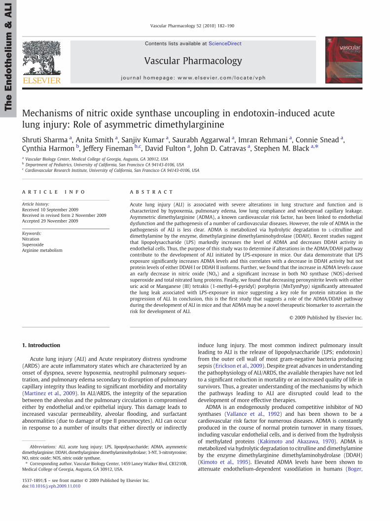
Fig. 2. Lung tissueNOx and tetrahydrobiopterin levels after LPS exposure. TissueNOx levelsare significantly decreased 2 h after LPS exposure. However, NOx levels were significantlyincreased 4- and 12-h after LPS exposure (A). Lung BH4 levels were measured by HPLC.Therewas no change in BH4 levels 2 h after LPS exposure, but BH4 levelswere significantlyincreased 4- and 12-h post LPS treatment (B). Values aremean±SE,N=5 for each group.⁎Pb0.05 compared to no LPS, †Pb0.05 compared to previous time point.
TheEndothelium
&ALI
185S. Sharma et al. / Vascular Pharmacology 52 (2010) 182–190
2.12. Statistical analysis
Statistical analysis was performed using GraphPad Prism version4.01 forWindows (GraphPadSoftware, SanDiego, CA). Themean±SEMwere calculated for all samples and significance was determined eitherby the unpaired t-test (for 2 groups) and ANOVA (for≥3 groups)followed by Newman-Keuls multiple comparisons test. A value ofPb0.05 was considered significant.
3. Results
3.1. Superoxide levels in LPS treated mouse lungs
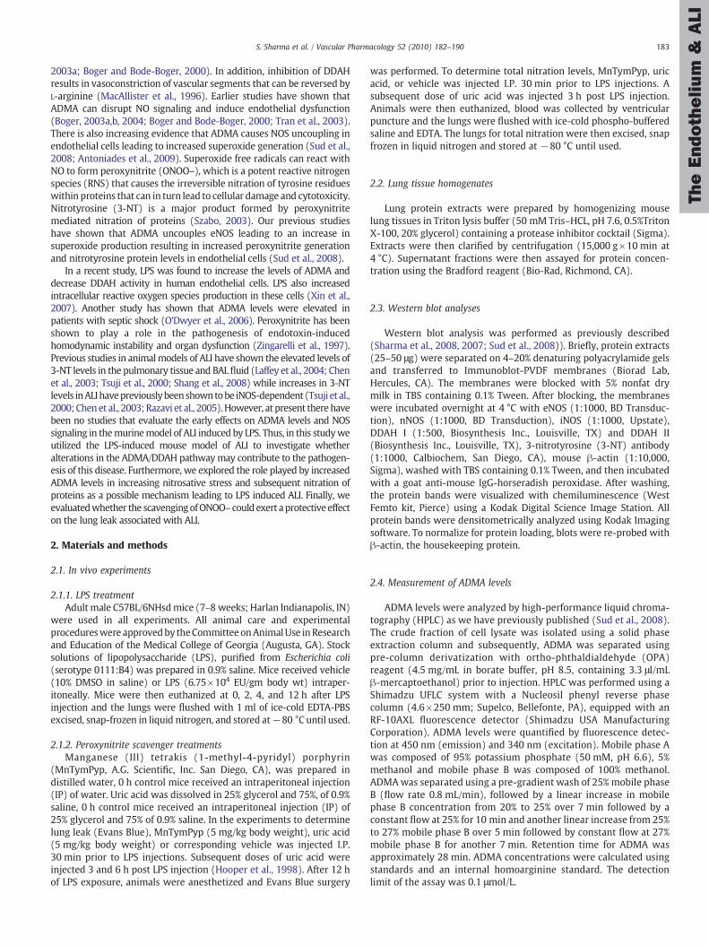
Relative superoxide levels were determined by EPR in lung tissuesharvested from 0- (Control), 2-, 4-, and 12-h after LP exposure (Fig. 1).Our data indicate that lung superoxide levels were significantlyincreased early after LPS-exposure: 2 h (∼2-fold) and 4 h (∼1.5 fold)and that this was a transient event as there was no change insuperoxide levels 12 h post-LPS (Fig. 1). To determine the contribu-tion of uncoupled eNOS in the increased superoxide generation,duplicate samples were incubated with the NOS inhibitor, NG-monomethyl L-arginine (L-NMMA; 100 μM, 30 min) on ice prior tothe addition of CMH. The LPS-mediated increase in superoxidegeneration was blocked in the presence of L-NMMA suggesting thatNOS uncoupling is a significant contributor to superoxide generationafter LPS exposure (Fig. 1). Specificity of the EPR assay for superoxidewas confirmed by a significant reduction in the waveform amplitudewith the addition of superoxide scavenger, polyethylene glycol-conjugated superoxide dismutase (PEG-SOD) to the samples (Fig. 1).
3.2. NOx and BH4 levels after LPS exposure
NOx levels were determined in mouse lung tissue 0-, 2-, 4-, and12-h after LPS treatment. Correlating with the increase in NOSuncoupling, we found there was a significant decrease in NOx levels2 h (−40%) after LPS exposure whereas we found an increase in NOx
levels 4 h (+60%) and 12 h (+160%) after LPS administration (Fig. 2 A).In addition, wemeasured lung BH4 levels after LPS exposure. BH4 levelswere unaltered 2 h after LPS exposure but were significantly elevated at4 h (∼2 fold) and 12 h (∼3 fold) post LPS treatment (Fig. 2 B).
3.3. NOS expression in control and ALI mouse lungs
To attempt to correlate the changes in NOx levels with NOS isoformprotein levels, whole lung homogenates were assessed for changes in
Fig. 1. Superoxide levels in the mouse lung after LPS exposure. Relative superoxidelevels were determined by electron paramagnetic resonance (EPR) in LPS-treatedmouse lungs. There is a significant increase in superoxide radical generation 2- and 4 hafter LPS exposure that is blocked by the addition of the NOS inhibitor, NG-monomethylL-arginine (L-NMMA; 100 µM) or polyethylene glycol-superoxide dismutase (PEG-SOD; 100 U/ml). There is no change in superoxide generation 12 h after LPS exposure.Values are means±SE, N=6. ⁎Pb0.05 vs. no LPS, †Pb0.05 vs. LPS alone.
the expression of NOS isoforms (eNOS, nNOS and iNOS) 2- and 4-h afterLPS exposure. Western blot analyses showed that there was nodifference in eNOS (Fig. 3 A) or nNOS protein levels (Fig. 3 B). However,iNOS protein levels although unchanged 2 h post-LPS treatment weresignificantly increased (∼6-fold) 4 h after LPS treatment (Fig. 3C)suggesting the increase in NOx levels 4 h post-LPS is likely due toincreases in iNOS protein levels.
3.4. Elevated ADMA levels and decreased DDAH activity in LPS-treatedmouse lungs
In an attempt to evaluate the mechanism responsible for theincrease in NOS uncoupling early after LPS exposure, we nextdetermined if LPS altered the levels of ADMA in the mouse lung. Wefound significantly increased ADMA levels in LPS treated mouse lungsat 2 h (12.13±0.84 vs. 7.53±0.57 nmol/gww; Fig. 4 A), 4 h (13.40±2.10 vs. 7.53±0.57 nmol/gww; Fig. 4 A) and 12 h (19.10±1.90 vs.7.53±0.57 nmol/gww; Fig. 4 A) after LPS exposure. To determinewhether the increased ADMA levels in the LPS treated mouse lungswere a result of decreased DDAH I and/or DDAH II protein expression,we measured protein levels of the two isoforms by Western blotanalyses. DDAH I and DDAH II anti-serum detected ∼37-kDA and∼33-kDA bands, respectively in lung tissue homogenates. Nosignificant differences were detected between protein levels of eitherDDAH I (Fig. 4 B) or DDAH II (Fig. 4 C) in mouse lung tissue post-LPS.However, we found that DDAH enzyme activity was significantlydecreased (∼2-fold) in mouse lung tissue homogenates both 2- and4-h after LPS exposure (Fig. 4 D).
Fig. 3. NOS isoform protein levels in the mouse lung after LPS exposure. Protein levelsfor eNOS, nNOS, and iNOS were determined in lung tissues 2- and 4-h after LPSexposure byWestern blot analysis using specific antisera raised against eNOS, nNOS, oriNOS respectively and re-probed with β-actin to normalize for loading. RepresentativeWestern blots are shown for eNOS (panel A), nNOS (panel B), and iNOS (panel C). Thereare no changes in eNOS or nNOS protein levels but iNOS protein levels are significantlyincreased 4 h after LPS exposure. Values are mean±SE, N=5 for each group ⁎Pb0.05compared to no LPS.
TheEndothelium
&ALI
186 S. Sharma et al. / Vascular Pharmacology 52 (2010) 182–190
3.5. Elevated nitrotyrosine levels after LPS exposure
Superoxides react with NO to form peroxynitrite that can modifyproteinsby interactingwith andnitrating tyrosine residues to form3-NT.To determine the presence of tyrosine-nitrated proteins in the mouselungs after LPS exposure, the levels of 3-NT were assessed by Westernblotting to detect nitrated proteins. The 3-NT levels were quantified byobtaining the densitometric units of all nitrated proteins (Fig. 5 A). Ourdata indicate that LPS exposure significantly increases 3-NT levels 4 hafter LPS-treatment in the mouse lung (Fig. 5 B).
3.6. Peroxynitrite scavengers cause a reduction in nitrated proteins afterLPS exposure.
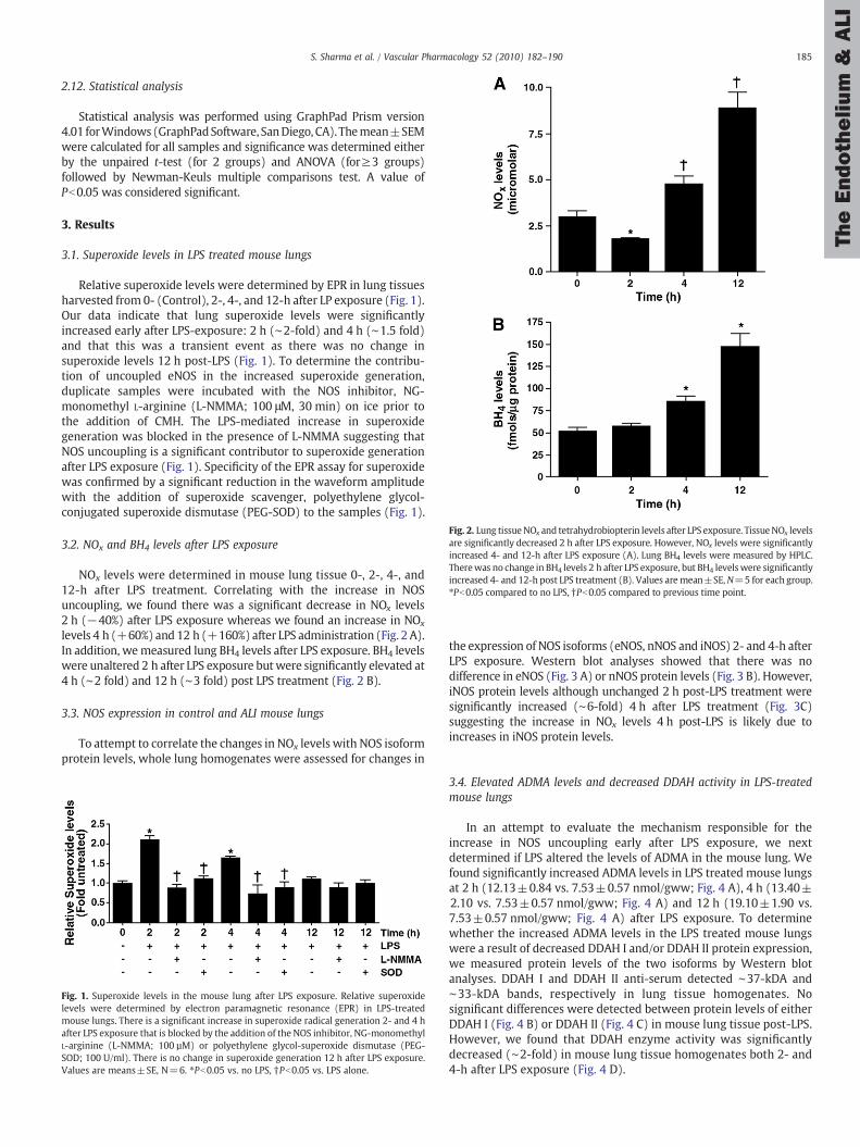
To determine the effect of peroxynitrite scavenging on LPS-mediated 3-NT levels, we used two potent peroxynitrite scavengers,MnTymPyp and uric acid. The animalswere treatedwith peroxynitritescavengers prior to LPS exposure and 3-NT levels were determined 4 hpost-LPS. Both, MnTymPyp and uric acid significantly attenuated theLPS-induced increase in 3-NT levels (Fig. 6 A).
3.7. Peroxynitrite scavengers attenuate the LPS mediated increase in lungpermeability.
The increase in lung permeability in response to LPS wasdetermined by measuring the Evan Blue dye leak, 12 h after LPSchallenge. Our data indicate that therewas a significant increase in thelung leak in the LPS treated mice (∼1.7 fold) while this increase inlung leak was significantly reduced in the animals pre-treated withthe peroxynitrite scavengers (MnTymPyp and uric acid) (Fig. 6 B).
3.8. ADMA potentiates VEGF-induced decrease in endothelial barrierfunction
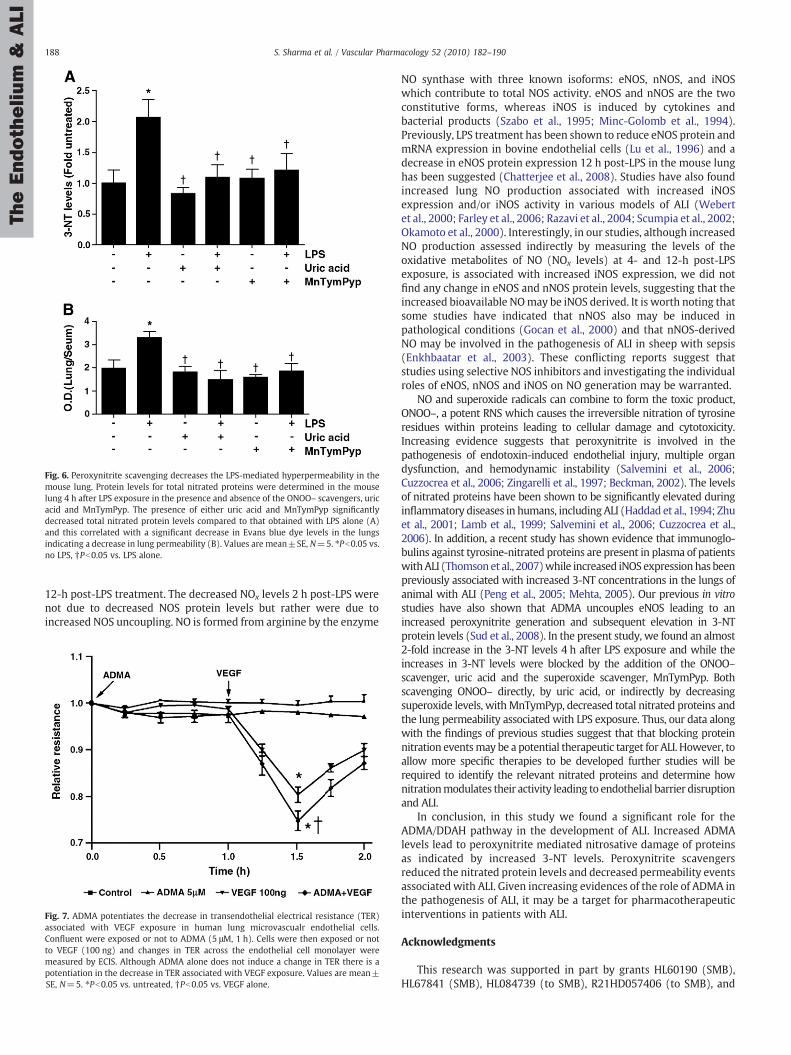
To determine if increases in ADMA alone are sufficient to induceendothelial cell barrier disruption, HLMVEC were exposed or not toADMA (5 µM) in the presence or absence of VEGF (100 ng) and theeffect on endothelial barrier function estimated bymeasuring changesin transendothelial resistance (TER) using an ECIS apparatus. Our dataindicate that ADMA alone is not sufficient to induce barrier disruptionbut it does potentiate the VEGF-mediated reduction in TER (Fig. 7).
4. Discussion
This study provides insight into a novel mechanism bywhich ADMAmediatednitrosative damagemay cause a loss of lung function in an LPS-induced mouse model of ALI. We found: (1) decreased DDAH activity,which was correlated with increased ADMA levels; (2) increased NOSuncoupling at early stages and increased iNOS expression at later stagesof the pathogenesis; (3) peroxynitrite mediated increase in 3-NT levelsand subsequent increase in lung permeability; (4) reduced 3-NT levelsand decreased lung leak in mice receiving peroxynitrite scavengersbefore endotoxin exposure. Interestingly our data also indicate thatalone increases in ADMA levels do not cause endothelial barrierdisruption in vitro using HLMVEC. However, ADMA does potentiateVEGF-induced barrier disruption suggesting ADMA may be necessarybut not sufficient to induce ALI. Thus, ADMA may act in concert withother proteins to produce endothelial barrier dysfunction. Indeed arecent study has shown that the ADMA/DDAH pathway regulatespulmonary endothelial barrier function through the modulation of Rac1signaling (Wojciak-Stothard et al., 2009). Further studies will berequired to elucidate the mechanism and key targets involved inADMA mediated EC barrier disruption.
We, and others, have previously shown that increased levels of theendogenous NOS inhibitor, ADMA can cause uncoupling of NOS andincreased production of both reactive oxygen species (ROS) andreactive nitrogen species (RNS) (Sud et al., 2008; Boger et al., 2000),which results in oxidative and nitrosative stress in the cell. There isgrowing evidence that increased ADMA levels are involved in thepathogenesis of a number of cardiovascular diseases (Miyazaki et al.,1999; Takiuchi et al., 2004; Boger, 2003c; Bae et al., 2005). Further,ADMA has been shown to cause increased peroxynitrite generation,and increased nitration events leading to pathological conditions (Sudet al., 2008). However, the role of ADMA in ALI has not been clarified.In this study, we demonstrate that lung tissue ADMA levels weresignificantly increased within 2 h of LPS exposure suggesting that thisis an early event in the pathogenesis of the disease. ADMA is degraded
Fig. 4. LPS exposure leads to elevated ADMA and decreased DDAH activity in the mouse lung. ADMA levels and DDAH activity were analyzed by high-performance liquidchromatography (HPLC) in the mice lungs after LPS exposure. There was a progressive increase in ADMA levels after 2-, 4- and 12-h LPS exposure (A). Although there were nochanges in either DDAH I (B) or DDAH II (C) protein levels there was a significant decrease in DDAH activity 2- and 4-h after LPS exposure (panel D). Values are mean±SE, N=5 foreach group, ⁎Pb0.05 compared to no LPS.
TheEndothelium
&ALI
187S. Sharma et al. / Vascular Pharmacology 52 (2010) 182–190
through active metabolism by the enzyme, DDAH. DDAH has twoisoforms and it has been previously shown that mRNA (Tran et al.,2000) and protein (Arrigoni et al., 2003) for both DDAH isoforms areexpressed in the lung. We observed a decrease in DDAH activity in ALIlung tissue, however our Western blot analyses did not detectalterations in either DDAH I or DDAH II protein levels. This suggeststhat the decrease in DDAH activity is not due to altered protein levelsbut is perhaps due to a post-translational modification. However,further studies will be necessary to elucidate the mechanism by which
Fig. 5. Nitrated protein levels are increased in the mouse lung after LPS exposure.Protein levels for total nitrated proteins were determined in the mouse lung 4 h afterLPS exposure. Homogenates (25 µg) were separated on a 4–20% denaturingpolyacrylamide gel, electrophoretically transferred to PVDF-nitrocellulose membranes,and analyzed using specific antiserum raised against 3-NT residues. A representativeimage for theWestern blot analysis for 3-NT protein levels is shown (A). The boxed areashows the region of total nitrated proteins that was used to quantify 3-NT levels.Densitometric analysis indicates that LPS exposure increases total nitrated proteins inthe mouse lung (B). Values are mean±SE, N=5. ⁎Pb0.05 vs. no LPS.
LPS regulates DDAH activity. Consistent with our results other studieshave shown that reduced DDAH activity, but not expression, isresponsible for the plasma ADMA elevation in hypercholesterolemiaand hyperhomocysteinemia (Boger et al., 1998; Lin et al., 2002;Stuhlinger et al., 2001). It has also been shown that the treatment ofmice with a DDAH inhibitor can cause higher plasma and blood vesselconcentrations of ADMA (Leiper et al., 2007). Our results are alsoconsistent with a recent study in which LPS significantly increased thelevels of ADMA, decreased DDAH activity, and increased intracellularROS production in human endothelial cells (Xin et al., 2007).While priorstudieshave reportedelevatedADMA levels inpatientswith septic shock(O'Dwyer et al., 2006) and found that serum ADMAwas associated withincreased vascular superoxide generation and eNOS uncoupling inhuman atherosclerosis (Antoniades et al., 2009). Our data also indicatethat the NOS uncoupling is a transient phenomenon after LPS exposureand as all three NOS isoforms are present our data cannot determine ifone isoform predominates. NOS uncoupling is a complex process thatcan be induced by a variety of conditions including increases in ADMA(Sud et al., 2008), decreases in the substrate L-arginine (Settergren et al.,2009), and decreases in the NOS cofactor, BH4 (Bevers et al., 2006; Caiet al., 2005).We found that there was no change in lung BH4 levels after2 h LPS exposure but there was a substantial increase after 4- and 12-h.This suggests that the increase in BH4 at the later timepointsmaybe ableto overcome the uncoupling effect of the increases in ADMA. Althoughthis is speculative our BH4 data are in agreement with a prior study thatreported increases in BH4 levels in the rat in both the plasma and tissues3 h after LPS exposure (Hattori et al., 1996).
Tissue NOx levels, a stable metabolite of NO, were significantlydecreased 2 h after LPS exposure but were markedly elevated 4- and
Fig. 6. Peroxynitrite scavenging decreases the LPS-mediated hyperpermeability in themouse lung. Protein levels for total nitrated proteins were determined in the mouselung 4 h after LPS exposure in the presence and absence of the ONOO– scavengers, uricacid and MnTymPyp. The presence of either uric acid and MnTymPyp significantlydecreased total nitrated protein levels compared to that obtained with LPS alone (A)and this correlated with a significant decrease in Evans blue dye levels in the lungsindicating a decrease in lung permeability (B). Values are mean±SE, N=5. ⁎Pb0.05 vs.no LPS, †Pb0.05 vs. LPS alone.
TheEndothelium
&ALI
188 S. Sharma et al. / Vascular Pharmacology 52 (2010) 182–190
12-h post-LPS treatment. The decreased NOx levels 2 h post-LPS werenot due to decreased NOS protein levels but rather were due toincreased NOS uncoupling. NO is formed from arginine by the enzyme
Fig. 7. ADMA potentiates the decrease in transendothelial electrical resistance (TER)associated with VEGF exposure in human lung microvascualr endothelial cells.Confluent were exposed or not to ADMA (5 μM, 1 h). Cells were then exposed or notto VEGF (100 ng) and changes in TER across the endothelial cell monolayer weremeasured by ECIS. Although ADMA alone does not induce a change in TER there is apotentiation in the decrease in TER associated with VEGF exposure. Values are mean±SE, N=5. ⁎Pb0.05 vs. untreated, †Pb0.05 vs. VEGF alone.
NO synthase with three known isoforms: eNOS, nNOS, and iNOSwhich contribute to total NOS activity. eNOS and nNOS are the twoconstitutive forms, whereas iNOS is induced by cytokines andbacterial products (Szabo et al., 1995; Minc-Golomb et al., 1994).Previously, LPS treatment has been shown to reduce eNOS protein andmRNA expression in bovine endothelial cells (Lu et al., 1996) and adecrease in eNOS protein expression 12 h post-LPS in the mouse lunghas been suggested (Chatterjee et al., 2008). Studies have also foundincreased lung NO production associated with increased iNOSexpression and/or iNOS activity in various models of ALI (Webertet al., 2000; Farley et al., 2006; Razavi et al., 2004; Scumpia et al., 2002;Okamoto et al., 2000). Interestingly, in our studies, although increasedNO production assessed indirectly by measuring the levels of theoxidative metabolites of NO (NOx levels) at 4- and 12-h post-LPSexposure, is associated with increased iNOS expression, we did notfind any change in eNOS and nNOS protein levels, suggesting that theincreased bioavailable NOmay be iNOS derived. It is worth noting thatsome studies have indicated that nNOS also may be induced inpathological conditions (Gocan et al., 2000) and that nNOS-derivedNO may be involved in the pathogenesis of ALI in sheep with sepsis(Enkhbaatar et al., 2003). These conflicting reports suggest thatstudies using selective NOS inhibitors and investigating the individualroles of eNOS, nNOS and iNOS on NO generation may be warranted.
NO and superoxide radicals can combine to form the toxic product,ONOO–, a potent RNS which causes the irreversible nitration of tyrosineresidues within proteins leading to cellular damage and cytotoxicity.Increasing evidence suggests that peroxynitrite is involved in thepathogenesis of endotoxin-induced endothelial injury, multiple organdysfunction, and hemodynamic instability (Salvemini et al., 2006;Cuzzocrea et al., 2006; Zingarelli et al., 1997; Beckman, 2002). The levelsof nitrated proteins have been shown to be significantly elevated duringinflammatory diseases in humans, including ALI (Haddad et al., 1994; Zhuet al., 2001; Lamb et al., 1999; Salvemini et al., 2006; Cuzzocrea et al.,2006). In addition, a recent study has shown evidence that immunoglo-bulins against tyrosine-nitrated proteins are present in plasma of patientswithALI (Thomsonet al., 2007)while increased iNOSexpressionhas beenpreviously associated with increased 3-NT concentrations in the lungs ofanimal with ALI (Peng et al., 2005; Mehta, 2005). Our previous in vitrostudies have also shown that ADMA uncouples eNOS leading to anincreased peroxynitrite generation and subsequent elevation in 3-NTprotein levels (Sud et al., 2008). In the present study, we found an almost2-fold increase in the 3-NT levels 4 h after LPS exposure and while theincreases in 3-NT levels were blocked by the addition of the ONOO–scavenger, uric acid and the superoxide scavenger, MnTymPyp. Bothscavenging ONOO– directly, by uric acid, or indirectly by decreasingsuperoxide levels, withMnTymPyp, decreased total nitrated proteins andthe lung permeability associated with LPS exposure. Thus, our data alongwith the findings of previous studies suggest that that blocking proteinnitration eventsmay be a potential therapeutic target for ALI. However, toallow more specific therapies to be developed further studies will berequired to identify the relevant nitrated proteins and determine hownitrationmodulates their activity leading to endothelial barrier disruptionand ALI.
In conclusion, in this study we found a significant role for theADMA/DDAH pathway in the development of ALI. Increased ADMAlevels lead to peroxynitrite mediated nitrosative damage of proteinsas indicated by increased 3-NT levels. Peroxynitrite scavengersreduced the nitrated protein levels and decreased permeability eventsassociated with ALI. Given increasing evidences of the role of ADMA inthe pathogenesis of ALI, it may be a target for pharmacotherapeuticinterventions in patients with ALI.
Acknowledgments
This research was supported in part by grants HL60190 (SMB),HL67841 (SMB), HL084739 (to SMB), R21HD057406 (to SMB), and
TheEndothelium
&ALI
189S. Sharma et al. / Vascular Pharmacology 52 (2010) 182–190
HL61284 (to JRF) all from the National Institutes of Health, 0550133Zfrom the American Heart Association, Pacific Mountain Affiliates(SMB), 09BGIA2310050 from the Southeast Affiliates (to SS), and by aTransatlantic Network Development Grant from the Fondation Leducq(to SMB & JRF). This work was also supported by a ProgrammaticDevelopment award (to SMB, JC, and DF) and Seed Awards (to SS andSJ) from the Cardiovascular Discovery Institute of the Medical Collegeof Georgia. Anita Smith was supported in part by NIH training Grant5T32HL06699.
References
Antoniades, C., Shirodaria, C., Leeson, P., Antonopoulos, A., Warrick, N., Van-Assche, T.,Cunnington, C., Tousoulis, D., Pillai, R., Ratnatunga, C., Stefanadis, C., Channon,K.M., 2009.Association of plasma asymmetrical dimethylarginine (ADMA) with elevated vascularsuperoxide production and endothelial nitric oxide synthaseuncoupling: implicationsfor endothelial function in human atherosclerosis. Eur. Heart J. 30, 1142–1150.
Arrigoni, F.I., Vallance, P., Haworth, S.G., Leiper, J.M., 2003. Metabolism of asymmetricdimethylarginines is regulated in the lung developmentally and with pulmonaryhypertension induced by hypobaric hypoxia. Circulation 107, 1195–1201.
Bae, S.W., Stuhlinger, M.C., Yoo, H.S., Yu, K.H., Park, H.K., Choi, B.Y., Lee, Y.S., Pachinger, O.,Choi, Y.H., Lee, S.H., Park, J.E., 2005. Plasma asymmetric dimethylarginine concentra-tions innewly diagnosedpatientswith acutemyocardial infarction or unstable anginapectoris during two weeks of medical treatment. Am. J. Cardiol. 95, 729–733.
Beckman, J.S., 2002. Protein tyrosine nitration and peroxynitrite. Faseb. J. 16, 1144.Bevers, L.M., Braam, B., Post, J.A., van Zonneveld, A.J., Rabelink, T.J., Koomans, H.A.,
Verhaar, M.C., Joles, J.A., 2006. Tetrahydrobiopterin, but not L-arginine, decreasesNO synthase uncoupling in cells expressing high levels of endothelial NO synthase.Hypertension 47, 87–94.
Black, S.M., Heidersbach, R.S., McMullan, D.M., Bekker, J.M., Johengen, M.J., Fineman, J.R.,1999. Inhaled nitric oxide inhibits NOS activity in lambs: potential mechanism forrebound pulmonary hypertension. Am. J. Physiol. 277, H1849–H1856.
Boger, R.H., 2003a. Association of asymmetric dimethylarginine and endothelialdysfunction. Clin. Chem. Lab. Med. 41, 1467–1472.
Boger, R.H., 2003b. The emerging role of asymmetric dimethylarginine as a novelcardiovascular risk factor. Cardiovasc. Res. 59, 824–833.
Boger, R.H., 2003c. When the endothelium cannot say 'NO’ anymore. ADMA, anendogenous inhibitor of NO synthase, promotes cardiovascular disease. Eur. Heart J.24, 1901–1902.
Boger, R.H., 2004. Asymmetric dimethylarginine, an endogenous inhibitor of nitricoxide synthase, explains the “L-arginine paradox” and acts as a novel cardiovascularrisk factor. J. Nutr. 134, 2842S–2847S discussion 2853S.
Burg, J., Krump-Konvalinkova, V., Bittinger, F., Kirkpatrick, C.J., 2002. GM-CSFexpression by human lung microvascular endothelial cells: in vitro and in vivofindings. Am. J. Physiol. Lung Cell. Mol. Physiol. 283, L460–L467.
Cai, S., Khoo, J., Channon, K.M., 2005. Augmented BH4 by gene transfer restores nitric oxidesynthase function in hyperglycemic human endothelial cells. Cardiovasc. Res. 65,823–831.
Chatterjee, A., Snead, C., Yetik-Anacak, G., Antonova, G., Zeng, J., Catravas, J.D., 2008.Heat shock protein 90 inhibitors attenuate LPS-induced endothelial hyperperme-ability. Am. J. Physiol. Lung Cell. Mol. Physiol. 294, L755–L763.
Chen, L.W., Wang, J.S., Chen, H.L., Chen, J.S., Hsu, C.M., 2003. Peroxynitrite is an importantmediator in thermal injury-induced lung damage. Crit. Care Med. 31, 2170–2177.
Cuzzocrea, S., Mazzon, E., Di Paola, R., Esposito, E., Macarthur, H., Matuschak, G.M.,Salvemini, D., 2006. A role for nitric oxide-mediated peroxynitrite formation in amodel of endotoxin-induced shock. J. Pharmacol. Exp. Ther. 319, 73–81.
Enkhbaatar, P.,Murakami, K., Shimoda,K.,Mizutani, A.,McGuire, R., Schmalstieg, F., Cox, R.,Hawkins, H., Jodoin, J., Lee, S., Traber, L., Herndon, D., Traber, D., 2003. Inhibition ofneuronal nitric oxide synthase by 7-nitroindazole attenuates acute lung injury in anovine model. Am. J. Physiol. Regul. Integr. Comp. Physiol. 285, R366–R372.
Farley, K.S., Wang, L.F., Razavi, H.M., Law, C., Rohan, M., McCormack, D.G., Mehta, S.,2006. Effects of macrophage inducible nitric oxide synthase in murine septic lunginjury. Am. J. Physiol. Lung Cell. Mol. Physiol. 290, L1164–L1172.
Gocan, N.C., Scott, J.A., Tyml, K., 2000. Nitric oxide produced via neuronal NOS mayimpair vasodilatation in septic rat skeletal muscle. Am. J. Physiol. Heart Circ.Physiol. 278, H1480–H1489.
Haddad, I.Y., Pataki, G., Hu, P., Galliani, C., Beckman, J.S., Matalon, S., 1994. Quantitationof nitrotyrosine levels in lung sections of patients and animals with acute lunginjury. J. Clin. Invest. 94, 2407–2413.
Hattori, Y., Nakanishi, N., Kasai, K., Murakami, Y., Shimoda, S., 1996. Tetrahydrobiop-terin and GTP cyclohydrolase I in a rat model of endotoxic shock: relation to nitricoxide synthesis. Exp. Physiol. 81, 665–671.
Hooper, D.C., Spitsin, S., Kean, R.B., Champion, J.M., Dickson, G.M., Chaudhry, I.,Koprowski, H., 1998. Uric acid, a natural scavenger of peroxynitrite, inexperimental allergic encephalomyelitis and multiple sclerosis. Proc. Natl. Acad.Sci. U. S. A. 95, 675–680.
Jackson, C.J., Garbett, P.K., Nissen, B., Schrieber, L., 1990. Binding of human endotheliumto Ulex europaeus I-coated dynabeads: application to the isolation of microvascularendothelium. J. Cell Sci. 96 (Pt 2), 257–262.
Kakimoto, Y., Akazawa, S., 1970. Isolation and identification of N-G, N-G- and N-G, N′-G-dimethyl-arginine, N-epsilon-mono-, di-, and trimethyllysine, and glucosylgalac-tosyl- and galactosyl-delta-hydroxylysine from human urine. J. Biol. Chem. 245,5751–5758.
Kimoto, M., Whitley, G.S., Tsuji, H., Ogawa, T., 1995. Detection of NG, NG-dimethylarginine dimethylaminohydrolase in human tissues using a monoclonalantibody. J. Biochem. (Tokyo) 117, 237–238.
Kumar, S., Sun, X., Sharma, S., Aggarwal, S., Ravi, K., Fineman, J.R., Black, S.M., 2009. GTPcyclohydrolase I expression is regulated by nitric oxide: role of cyclic AMP. Am. J.Physiol. Lung Cell Mol. Physiol. 297, L309–L317.
Lakshminrusimha, S.,Wiseman,D., Black, S.M., Russell, J.A., Gugino, S.F., Oishi, P., Steinhorn,R.H., Fineman, J.R., 2007. The role of nitric oxide synthase-derived reactive oxygenspecies in the altered relaxation of pulmonary arteries from lambs with increasedpulmonary blood flow. Am. J. Physiol. Heart Circ. Physiol. 293, H1491–H1497.
Lamb, N.J., Quinlan, G.J., Westerman, S.T., Gutteridge, J.M., Evans, T.W., 1999. Nitration ofproteins in bronchoalveolar lavage fluid from patients with acute respiratory distresssyndrome receiving inhaled nitric oxide. Am. J. Respir. Crit. CareMed. 160, 1031–1034.
Leiper, J., Nandi, M., Torondel, B., Murray-Rust, J., Malaki, M., O'Hara, B., Rossiter, S.,Anthony, S., Madhani, M., Selwood, D., Smith, C., Wojciak-Stothard, B., Rudiger, A.,Stidwill, R., McDonald, N.Q., Vallance, P., 2007. Disruption of methylargininemetabolism impairs vascular homeostasis. Nat. Med. 13, 198–203.
Lin, K.Y., Ito, A., Asagami, T., Tsao, P.S., Adimoolam, S., Kimoto, M., Tsuji, H., Reaven, G.M.,Cooke, J.P., 2002. Impaired nitric oxide synthase pathway in diabetes mellitus: roleof asymmetric dimethylarginine and dimethylarginine dimethylaminohydrolase.Circulation 106, 987–992.
Lu, J.L., Schmiege III, L.M., Kuo, L., Liao, J.C., 1996. Downregulation of endothelialconstitutive nitric oxide synthase expression by lipopolysaccharide. Biochem.Biophys. Res. Commun. 225, 1–5.
MacAllister, R.J., Parry, H., Kimoto, M., Ogawa, T., Russell, R.J., Hodson, H., Whitley, G.S.,Vallance, P., 1996. Regulation of nitric oxide synthesis by dimethylargininedimethylaminohydrolase. Br. J. Pharmacol. 119, 1533–1540.
Martinez, O., Nin, N., Esteban, A., 2009. Prone position for the treatment of acuterespiratory distress syndrome: a review of current literature. Arch. Bronconeumol.45, 291–296.
McMullan, D.M., Bekker, J.M., Parry, A.J., Johengen,M.J., Kon, A., Heidersbach, R.S., Black, S.M.,Fineman, J.R., 2000. Alterations in endogenous nitric oxide production aftercardiopulmonary bypass in lambs with normal and increased pulmonary blood flow.Circulation 102, III172–III178.
Mehta, S., 2005. The effects of nitric oxide in acute lung injury. Vascul. Pharmacol. 43,390–403.
Minc-Golomb, D., Tsarfaty, I., Schwartz, J.P., 1994. Expression of inducible nitric oxidesynthase by neurones following exposure to endotoxin and cytokine. Br. J.Pharmacol. 112, 720–722.
Miyazaki, H., Matsuoka, H., Cooke, J.P., Usui, M., Ueda, S., Okuda, S., Imaizumi, T., 1999.Endogenous nitric oxide synthase inhibitor: a novel marker of atherosclerosis.Circulation 99, 1141–1146.
O'Dwyer, M.J., Dempsey, F., Crowley, V., Kelleher, D.P., McManus, R., Ryan, T., 2006. Septicshock is correlated with asymmetrical dimethyl arginine levels, which may beinfluenced by a polymorphism in the dimethylarginine dimethylaminohydrolase IIgene: a prospective observational study. Crit. Care 10, R139.
Okamoto, I., Abe, M., Shibata, K., Shimizu, N., Sakata, N., Katsuragi, T., Tanaka, K., 2000.Evaluating the role of inducible nitric oxide synthase using a novel and selectiveinducible nitric oxide synthase inhibitor in septic lung injury produced by cecalligation and puncture. Am. J. Respir. Crit. Care Med. 162, 716–722.
Peng, X., Abdulnour, R.E., Sammani, S., Ma, S.F., Han, E.J., Hasan, E.J., Tuder, R., Garcia, J.G.,Hassoun, P.M., 2005. Inducible nitric oxide synthase contributes to ventilator-inducedlung injury. Am. J. Respir. Crit. Care Med. 172, 470–479.
Razavi, H.M., Wang le, F., Weicker, S., Rohan, M., Law, C., McCormack, D.G., Mehta, S.,2004. Pulmonary neutrophil infiltration in murine sepsis: role of inducible nitricoxide synthase. Am. J. Respir. Crit. Care Med. 170, 227–233.
Razavi, H.M., Wang, L., Weicker, S., Quinlan, G.J., Mumby, S., McCormack, D.G., Mehta, S.,2005. Pulmonary oxidant stress in murine sepsis is due to inflammatory cell nitricoxide. Crit. Care Med. 33, 1333–1339.
Salvemini, D., Doyle, T.M., Cuzzocrea, S., 2006. Superoxide, peroxynitrite and oxidative/nitrative stress in inflammation. Biochem. Soc. Trans. 34, 965–970.
Settergren, M., Bohm, F., Malmstrom, R.E., Channon, K.M., Pernow, J., 2009. L-arginineand tetrahydrobiopterin protects against ischemia/reperfusion-induced endothe-lial dysfunction in patients with type 2 diabetes mellitus and coronary arterydisease. Atherosclerosis 204, 73–78.
Shang, Y., Li, X., Prasad, P.V., Xu, S., Yao, S., Liu, D., Yuan, S., Feng, D., 2008. Erythropoietinattenuates lung injury in lipopolysaccharide treated rats. J. Surg. Res.
TheEndothelium
&ALI
190 S. Sharma et al. / Vascular Pharmacology 52 (2010) 182–190
Sharma, S., Grobe, A.C., Wiseman, D.A., Kumar, S., Englaish, M., Najwer, I., Benavidez, E.,Oishi, P., Azakie, A., Fineman, J.R., Black, S.M., 2007. Lung antioxidant enzymes areregulated by development and increased pulmonary blood flow. Am. J. Physiol.Lung Cell. Mol. Physiol. 293, L960–L971.
Sharma, S., Sud, N., Wiseman, D.A., Carter, A.L., Kumar, S., Hou, Y., Rau, T., Wilham, J.,Harmon, C., Oishi, P., Fineman, J.R., Black, S.M., 2008. Altered carnitine homeostasisis associated with decreased mitochondrial function and altered nitric oxidesignaling in lambs with pulmonary hypertension. Am. J. Physiol. Lung Cell. Mol.Physiol. 294, L46–L56.
Stuhlinger, M.C., Tsao, P.S., Her, J.H., Kimoto, M., Balint, R.F., Cooke, J.P., 2001.Homocysteine impairs the nitric oxide synthase pathway: role of asymmetricdimethylarginine. Circulation 104, 2569–2575.
Sud, N., Wells, S.M., Sharma, S., Wiseman, D.A., Wilham, J., Black, S.M., 2008.Asymmetric dimethylarginine inhibits HSP90 activity in pulmonary arterialendothelial cells: role of mitochondrial dysfunction. Am. J. Physiol. Cell Physiol.294, C1407–C1418.
Szabo, C., 2003. Multiple pathways of peroxynitrite cytotoxicity. Toxicol. Lett. 140–141,105–112.
Szabo, C., Salzman, A.L., Ischiropoulos, H., 1995. Endotoxin triggers the expression of aninducible isoform of nitric oxide synthase and the formation of peroxynitrite in therat aorta in vivo. FEBS Lett. 363, 235–238.
Takiuchi, S., Fujii, H., Kamide, K., Horio, T., Nakatani, S., Hiuge, A., Rakugi, H., Ogihara, T.,Kawano, Y., 2004. Plasma asymmetric dimethylarginine and coronary andperipheral endothelial dysfunction in hypertensive patients. Am. J. Hypertens. 17,802–808.
Thomson, L., Christie, J., Vadseth, C., Lanken, P.N., Fu, X., Hazen, S.L., Ischiropoulos, H.,2007. Identification of immunoglobulins that recognize 3-nitrotyrosine in patientswith acute lung injury after major trauma. Am. J. Respir. Cell Mol. Biol. 36,152–157.
Tran, C.T., Fox, M.F., Vallance, P., Leiper, J.M., 2000. Chromosomal localization, genestructure, and expression pattern of DDAH1: comparison with DDAH2 andimplications for evolutionary origins. Genomics 68, 101–105.
Tsuji, C., Shioya, S., Hirota, Y., Fukuyama, N., Kurita, D., Tanigaki, T., Ohta, Y., Nakazawa, H.,2000. Increased production of nitrotyrosine in lung tissue of rats with radiation-induced acute lung injury. Am. J. Physiol. Lung Cell. Mol. Physiol. 278, L719–L725.
Vallance, P., Leone, A., Calver, A., Collier, J., Moncada, S., 1992. Endogenousdimethylarginine as an inhibitor of nitric oxide synthesis. J. Cardiovasc. Pharmacol.20 (Suppl 12), S60–S62.
Wainwright, M.S., Arteaga, E., Fink, R., Ravi, K., Chace, D.H., Black, S.M., 2005. Tetrahy-drobiopterin and nitric oxide synthase dimer levels are not changed following hypoxia-ischemia in the newborn rat. Brain Res. Dev. Brain Res. 156, 183–192.
Webert, K.E., Vanderzwan, J., Duggan,M., Scott, J.A., McCormack, D.G., Lewis, J.F., Mehta, S.,2000. Effects of inhaled nitric oxide in a rat model of Pseudomonas aeruginosapneumonia. Crit. Care Med. 28, 2397–2405.
Zhu, S., Ware, L.B., Geiser, T., Matthay, M.A., Matalon, S., 2001. Increased levels of nitrateand surfactant protein a nitration in the pulmonary edema fluid of patients withacute lung injury. Am. J. Respir. Crit. Care Med. 163, 166–172.
Zingarelli, B., Day, B.J., Crapo, J.D., Salzman, A.L., Szabo, C., 1997. The potential role ofperoxynitrite in the vascular contractile and cellular energetic failure in endotoxicshock. Br. J. Pharmacol. 120, 259–267.