Previous studies demonstrate that glyphosate exposure is associated with oxidative damage and neu-rotoxicity. Therefore, the mechanism of glyphosate-induced neurotoxic effects needs to be determined.The aim of this study was to investigate whether Roundup® (a glyphosate-based herbicide) leads toneurotoxicity in hippocampus of immature rats following acute (30 min) and chronic (pregnancy andlactation) pesticide exposure. Maternal exposure to pesticide was undertaken by treating dams orallywith 1% Roundup® (0.38% glyphosate) during pregnancy and lactation (till 15-day-old). Hippocampalslices from 15 day old rats were acutely exposed to Roundup® (0.00005–0.1%) during 30 min and experi-ments were carried out to determine whether glyphosate affects 45Ca2+ influx and cell viability. Moreover,we investigated the pesticide effects on oxidative stress parameters, 14C-�-methyl-amino-isobutyric acid(14C-MeAIB) accumulation, as well as glutamate uptake, release and metabolism. Results showed thatacute exposure to Roundup® (30 min) increases 45Ca2+ influx by activating NMDA receptors and voltage-dependent Ca2+ channels, leading to oxidative stress and neural cell death. The mechanisms underlyingRoundup®-induced neurotoxicity also involve the activation of CaMKII and ERK. Moreover, acute expo-sure to Roundup® increased 3H-glutamate released into the synaptic cleft, decreased GSH content and
increased the lipoperoxidation, characterizing excitotoxicity and oxidative damage. We also observedthat both acute and chronic exposure to Roundup® decreased 3H-glutamate uptake and metabolism,while induced 45Ca2+ uptake and 14C-MeAIB accumulation in immature rat hippocampus. Taken together,these results demonstrated that Roundup® might lead to excessive extracellular glutamate levels andconsequently to glutamate excitotoxicity and oxidative stress in rat hippocampus.
The annual consumption of pesticides in Brazil has increasedlarmingly in recent years. The justification for the use of these sub-tances is based on the improvement of agricultural productivity,
Please cite this article in press as: Cattani, D., et al., Mechanisms underimmature rat hippocampus: Involvement of glutamate excitotoxicity.
hich places the country as one of the world’s largest produc-rs of food. However, the risks to the environment and health areeglected. According to the Brazilian National Trade Union for the
∗ Corresponding author at: Departamento de Bioquímica, Centro de Ciênciasiológicas, UFSC, Campus Universitário, Bairro Trindade, CEP: 88040-970, Flori-nópolis, Santa Catarina, Brazil. Tel.: +55 48 3721 4747; fax: +55 48 3721 9672.
Agricultural Defense Products (Sindicato Nacional da Indústria deProdutos para Defesa Agrícola – SINDAG), in 2008 Brazil became thelargest consumer of pesticides in the world. The increase in pesti-cide consumption leads to high rates of intoxication among farmers.Moreover, it has been suggested important associations betweenthe bulk sale of pesticides and the increased rates of several types ofcancer, endocrine disorders and a high prevalence of neurodegen-erative diseases in agricultural workers (Londres, 2011), reinforcingthe need for mechanistic investigations.
Glyphosate-based herbicides lead the pesticide world market.Moreover, glyphosate is the primary active ingredient present
lying the neurotoxicity induced by glyphosate-based herbicide in Toxicology (2014), http://dx.doi.org/10.1016/j.tox.2014.03.001
in Roundup® (Monsanto Company, St. Louis, MO). It has beensuggested that the toxicity of Roundup® is probably due to syner-gistic effects between glyphosate and other formulation products(Marc et al., 2002; El-Shenawy, 2009), such as the surfactant
olyethoxylated tallowamine (POEA) which might facilitatelyphosate penetration through plasma membranes potentiatingts toxicity (Williams et al., 2000; Tsui and Chu, 2003; Richard etl., 2005; Benachour and Séralini, 2009).
The planting of transgenic soybeans resistant to glyphosate-oundup® (Glyphosate resistant soybeans, GRS) has greatlynhanced the consumption of this herbicide in Brazilian crops.n this context, it was recently reported a teratogenic action oferbicides based on glyphosate to vertebrates (Paganelli et al.,010), warning of the need to evaluate the use of the product
n agriculture. Moreover, Benachour and Séralini (2009) recentlyemonstrated that glyphosate formulations induce apoptosis andecrosis in human umbilical, embryonic, and placental cells. Thiserbicide was also associated with induction of oxidative stressnd neuroinflamation (El-Shenawy, 2009; Astiz et al., 2012). More-ver, glyphosate was able to provoke oxidative stress in specificrain regions: substantia nigra, cerebral cortex and hippocampus.owever, most of these studies were carried out by using the com-ercial formulations, and not pure glyphosate, suggesting that
urther studies are necessary to examine whether the pesticideormulation may lead to neurotoxicity.
It has been suggested that pesticide exposure could be a riskactor for neurodegenerative disorders (Le Couteur et al., 1999;irby et al., 2001; Barlow et al., 2005; Patel et al., 2006). In thisontext, occupational pesticide exposure leads to oxidative dam-ge, increases the risk of incidence of Parkinson’s and Alzheimer’sisease, and also might accelerate age-related neurodegenerationWang et al., 2006; Peng et al., 2007; Hayden et al., 2010); how-ver, it is unclear as to which pesticides and what mechanismsf action may contribute to the neurodegenerative condition. Inhis context, it has been reported that acute and chronic expo-ure to glyphosate might cause parkinsonism, a condition similaro Parkinson’s disease (Barbosa et al., 2001; Wang et al., 2011).arbosa and colleagues (2001) reported a case of a 54-year-oldan who accidentally sprayed himself with the chemical agent
lyphosate and one month later he developed parkinsonism. Oneear later, the patient presented a slow resting tremor in the leftand and arm, accompanied by impairment of short-term mem-ry. Further, Wang et al. (2011) reported a case of parkinsonismollowing chronic exposure to glyphosate in a previously healthy4-year-old woman who worked for 3 years in a chemical factory,xclusively in the glyphosate production division. The glyphosateeurotoxicity was associated with rigidity, slowness and restingremor in all four limbs with no impairment of short-term memory.
It has been recently demonstrated that glyphosate induced bothpoptotic and autophagic cell death in neuronal differentiated PC12ells, providing a link between glyphosate and Parkinson’s dis-ase (Gui et al., 2012). Moreover, Chorfa et al. (2013) shown thatlyphosate causes a significant cytotoxicity to SH-SY5Y neuronalell line. The cytotoxicity was estimated by MTT assay and the half-aximal (50%) inhibitory concentration (IC50) for glyphosate was
�M. In addition, Negga and colleagues (2011) demonstrated thataenorhabditis elegans were susceptible to glyphosate concentra-ions within environmentally relevant ranges. Further, the sameroup demonstrated that exposure to glyphosate-containing pes-icide leads to degeneration of �-aminobutyric acid and dopamineeurons in C. elegans (Negga et al., 2012a,b). Taken together, thesetudies suggest that glyphosate might affect neural cells leadingo oxidative damage, neuronal cell death and neurodegenerativeonditions.
The neurodegenerative conditions are frequently associatedith glutamatergic excitotoxicity and oxidative stress (Pessoa-
Please cite this article in press as: Cattani, D., et al., Mechanisms underimmature rat hippocampus: Involvement of glutamate excitotoxicity
ureur and Wajner, 2007). The central nervous system presentsigh vulnerability to free radical damage due to its elevated oxida-ive metabolic rate and enriched content of unsaturated lipids, asell as to its elevated rate of free radical generation derived from
PRESS xxx (2014) xxx–xxx
neurotransmitters metabolism, and poor radical scavenging mech-anisms (Chong et al., 2005).
Taking into account the previous studies demonstrating thatexposure to glyphosate might be associated with neurotoxicity andoxidative damage, the aim of this study was to determine whetherRoundup® leads to neurotoxicity in hippocampus of immaturerats following acute (30 min) and chronic (pregnancy and lacta-tion) pesticide exposure. Then, we investigated the involvementof Ca2+, intracellular signaling pathways and oxidative damage onthe mechanisms underlying Roundup®-induced neurotoxicity inrat hippocampus. Moreover, this study investigated whether acuteor maternal exposure to a glyphosate-based herbicide alters glu-tamatergic system by interfering in the neurotransmitter uptake,release and/or metabolism within the hippocampal cells.
2. Material and methods
2.1. Radiochemical and compounds
L-[2,3-3H] glutamic acid ([3H] glutamate) (specific activity49 Ci/mmol) was purchased from Amersham (Oakville, Ontario,Canada). 45CaCl2 (specific activity of 321 kBq/mg of Ca2+) andOptiphase Hisafe III biodegradable liquid scintillation werepurchased from PerkinElmer (Waltham, MA). �-[14C] methy-laminoisobutyric acid ([14C] MeAIB) (sp.act. 1.85 GBq/mmol) waspurchased from Du Pont, NEN Products, MA, USA. The herbicideRoundup Original® (Homologation number 00898793) containingglyphosate 360 g/L is a commercial formulation registered in theBrazilian Ministry of Agriculture, Livestock and Supply (Ministérioda Agricultura, Pecuária e Abastecimento – MAPA). Nifedipine,N-[2-(p-Bromocinnamylamino) ethyl]-5-isoquinolinesulfonamide(H89), (Bisindoylmaleimidine IX, 2-{1-[3-(Amidinothio)propyl]-1H-indol-3-yl}-3-(1-methylindol-3-yl)maleimide ethanesulfonatesalt) Ro 31-8220, d(–)-2-amino-5-phosphonopentanoic acid (AP5),KN-93, (+)-�-methyl-4-carboxyphenylglycine (MCPG) and flunar-izine were purchased from Sigma Chemical Company (St. Louis,MO, USA). The G6PD assay kit was kindly provided by INTERCIEN-TÍFICA (São José dos Campos, SP, Brazil). All other chemicals wereof analytical grade.
2.2. Animals
Wistar rats were bred in animal house and maintained in an air-conditioned room (about 21 ◦C) with controlled lighting (12 h/12 hlight/dark cycle). On the day of birth the litter size was culled toeight pups. Litters smaller than eight pups were not included. Thesuckling rats were kept with their mothers until euthanasia. Pel-leted food (Nuvital, Nuvilab CR1, Curitiba, PR, Brazil) and tap waterwere available ad libitum. All animals’ procedures were carriedout in accordance with ethical recommendations of the BrazilianVeterinary Medicine Council and the Brazilian College of AnimalExperimentation (Protocol CEUA/PP00471).
2.3. In vivo exposure to Roundup®
Wistar rats were mated and the day of appearance of the vagi-nal plug was considered day 0 of fetal age. Maternal exposure toRoundup® was induced by adding 1% Roundup® (correspondingto 0.38% glyphosate) in the drinking water from gestation day 5and continually up to lactation day 15. Control animals, receivingonly water during the same period, were used as controls. Theconcentration chosen for the in vivo exposure was based on no
lying the neurotoxicity induced by glyphosate-based herbicide in. Toxicology (2014), http://dx.doi.org/10.1016/j.tox.2014.03.001
observed adverse effect level (NOAEL) for maternal toxicity whichwas 1000 mg/kg body weight/day dosage (Williams et al., 2000).The concentration we used was equivalent to 1/14 of the limits ofNOAEL. Each experiment was carried out by treating 4 different
ams during pregnancy and lactation, and the pups from their lit-ers were used to investigate the neurotoxic effects of Roundup® onippocampal cells. Therefore, each experimental group containedippocampal slices from pups of different litters (different dams).ll experiments were performed in triplicate. The body weight overregnancy and water consumption by dams were measured daily.lso pup body weights were accompanied over the 15 days. Fifteen-ay old rats were used in our experimental condition consideringhat in rats the period of maximum synaptogenesis occurs betweenostnatal day 11 and postnatal day 20, a period that is associ-ted with increased synaptic plasticity in order to establish properynaptic connections (Sutor and Luhmann, 1995).
.4. Preparation of hippocampus slices
Rats were killed by decapitation and the hippocampus was diss-cted onto Petri dishes placed on ice and the parietal region was cutnto 300 �m thick slices with a McIlwain chopper.
.5. 45Ca2+ uptake
Hippocampal slices from 15-day-old male rats were preincu-ated in Krebs Ringer-bicarbonate (KRb) buffer (122 mM NaCl;
mM KCl; 1.2 mM MgSO4; 1.3 mM CaCl2; 0.4 mM KH2PO4; 25 mMaHCO3) for 15 min in a Dubnoff metabolic incubator at 37 ◦C, pH.4 and gassed with O2:CO2 (95:5; v/v). After that, the medium washanged by fresh KRb with 0.1 �Ci/mL 45Ca2+ and incubated during0 min in the absence (control group) or presence (treated group)f Roundup® at concentrations ranging from 0.00005 to 0.1%.n some experiments, L-type voltage-dependent channel blocker10 �M nifedipine), antagonist of NMDA receptors (10 �M DL-P5) or kinase inhibitors (10 �M H89, 20 �M Ro 31-8220, 10 �MN93) were added during the preincubation and incubation periods
see figures). Extracellular 45Ca2+ was thoroughly washed off in aashing solution containing 127.5 mM NaCl, 4.6 mM KCl, 1.2 mMgSO4, 10 mM HEPES, 11 mM glucose, 10 mM LaCl3, pH 7.3 (30 min
n washing solution). The presence of La3+ during the washingtage was found to be essential to prevent release of the intra-ellular 45Ca2+ (Zamoner et al., 2007). After washing, tissue slicesere digested and homogenized with 0.5 M NaOH solution, 100 �L
liquots were placed in scintillation fluid and counted in a LKBack beta liquid scintillation spectrometer (model LS 6500; Multi-urpose Scintillation Counter-Beckman Coulter, Boston, USA), and
�L aliquots were used for protein quantification as described byowry and colleagues (1951).
.6. Measurement of lactate dehydrogenase (LDH) released
Hippocampal slices from 15-day-old male rats were preincu-ated in KRb in a Dubnoff metabolic incubator at 37 ◦C, pH 7.4nd gassed with O2:CO2 (95:5; v/v). After that, the medium washanged by fresh KRb and the slices were incubated during 30 minn the absence (control group) or presence (treated group) ofoundup® at concentrations ranging from 0.00005 to 0.1%. After
ncubation of the hippocampal slices in the absence or presencef Roundup®, the incubation medium was collected for deter-ination of extracellular LDH activity by a spectrophotometricethod. The estimation of LDH activity was carried out by mea-
uring the oxidation of NADH and the results were expressed asontrol.
.7. Glutamate uptake assay
Please cite this article in press as: Cattani, D., et al., Mechanisms underimmature rat hippocampus: Involvement of glutamate excitotoxicity.
For glutamate uptake the incubation medium was replaced byank’s balanced salt solution (HBSS) containing 137 mM NaCl;.63 mM Na2HPO4; 4.17 mM NaHCO3; 5.36 mM KCl; 0.44 mM
PRESS xxx (2014) xxx–xxx 3
KH2PO4; 1.26 mM CaCl2; 0.41 mM MgSO4; 0.49 mM MgCl2 and5.55 mM glucose, pH 7.4. The hippocampal slices were exposedor not to 0.01% Roundup® during 23 minutes. Then, glutamateuptake assay was started by the addition of 0.1 mM l-glutamate and0.66 �Ci/mL l-[2,3-3H] glutamate. Incubation was stopped after7 min by removal of the medium and rinsing the slices twice withice-cold HBSS. Slices were then lysed in a solution containing 0.5 MNaOH (Cattani et al., 2013). Sodium-independent uptake was deter-mined using N-methyl-d-glucamine instead of sodium chloride.Sodium-dependent glutamate uptake was obtained by subtractingthe non-specific uptake from the specific uptake. Radioactivity wasmeasured with a scintillation counter.
2.8. Glutamate release assay
After the preincubation period to slice recovery (30 min), hip-pocampal slices were incubated in Hank’s balanced salt solution(HBSS; composition in mM 1.29 CaCl2, 136.9 NaCl, 5.36 KCl, 0.65MgSO4, 0.27 Na2HPO4, 1.1 KH2PO4, 2 glucose, and 5 HEPES).When present, Roundup® was incubated for 30 min. Glutamate(1 mM) was incubated for 15 min, and glutamate uptake wasassessed by adding 0.33 �Ci/mL d-[3H]aspartate with 100 �Munlabeled aspartate during the last 7 min and stopped by threeice-cold washes with 1 mL HBSS. d-[3H]-aspartate instead ofl-[3H]glutamate was used in order to avoid glutamate metabo-lization in intracellular compartments, although similar resultswere obtained by using d-[3H]aspartate or l-[3H]glutamate. Theslices were then further incubated for 15 min in HBSS, and thesupernatant was collected to measure the amount of released d-[3H]aspartate. Slices were disrupted by overnight incubation with0.1% NaOH/0.01% SDS, and aliquots of lysates were taken for deter-mination of intracellular d-[3H]aspartate content. Intracellular andextracellular d-[3H]-aspartate content were determined throughscintillation counting, calculated as nmol aspartate, and the amountof released aspartate was expressed as percentage of total d-[3H]aspartate.
2.9. Glutamine synthetase activity
Hippocampal slices were preincubated in HBSS at 37 ◦C andthen incubated during 15 min in the absence or presence of 0.01%Roundup® during 30 minutes. Briefly, homogenate (0.1 mL) wasadded to 0.1 mL of reaction mixture containing 10 mM MgCl2;50 mM l-glutamate; 100 mM imidazole–HCl buffer (pH 7.4);10 mM 2-mercaptoethanol; 50 mM hydroxilamine–HCl; 10 mMATP and incubated for 15 min at 37 ◦C. The reaction was stoppedby the addition of 0.4 mL of a solution containing: 370 mM fer-ric chloride; 670 mM HCl; 200 mM trichloroacetic acid. Aftercentrifugation, the supernatant was measured at 530 nm andcompared to the absorbance generated by standard quantities of�-glutamylhydroxamate treated with ferric chloride reagent.
2.10. Neutral amino acid accumulation
For amino acid accumulation experiments, hippocampal sliceswere pre-incubated in KRb buffer for 30 min in a Dubnoff metabolicincubator at 37 ◦C, pH 7.4 and gassed with O2:CO2 (95:5; v/v).The slices were then incubated in fresh KRb buffer for 60 min.[14C] MeAIB (3.7 kBq/mL) was added to each sample during theincubation period (Silva et al., 2001; Cattani et al., 2013). The pesti-cide (0.01% Roundup®) was added in the last 30 min of incubationperiod. After incubation the slices were lysed in 0.5 M NaOH and the
lying the neurotoxicity induced by glyphosate-based herbicide in Toxicology (2014), http://dx.doi.org/10.1016/j.tox.2014.03.001
protein concentration was determined (Lowry et al., 1951). Then,25 �L aliquots of tissue and external medium were placed in scin-tillation fluid and counted in a Beckman beta liquid scintillationspectrometer (model LS 6500; Fullerton, CA, USA) for radioactivity
easurements. The results were expressed as the tissue/mediumT/M) ratio: cpm/mL tissue fluid per cpm/mL incubation medium.
.11. Reduced glutathione assay
The reduced glutathione (GSH) was determined using theeagent DTNB (5,5′-dithiobis 2-nitrobenzoic acid). After being cen-rifuged at 5000 × g for 5 min, the supernatants from the acidxtracts (TCA 12%, 1:10, w/v) were added to 2.5 mM DTNB in 0.2 Modium phosphate buffer pH 8.0, and the formation of the thiolatenion was immediately measured at 412 nm. Determinations werexpressed in �mol g−1.
.12. Quantification of lipoperoxidation levels
Please cite this article in press as: Cattani, D., et al., Mechanisms underimmature rat hippocampus: Involvement of glutamate excitotoxicity
The endogenous lipid peroxidation was evaluated in the hip-ocampus by detection of substances that react with thiobarbituriccid (TBARS), particularly malondialdehyde (MDA), accordingo the method described by Bird and Draper (1984). Briefly,
ig. 1. Dose–response curve of Roundup® on 45Ca2+ uptake (A) and on LDH release (B) onre-incubated for 15 min and then incubated for 30 min with 0.1 mCi/mL of 45Ca2 in the prom the each animal were used for control and treated groups. The radioactivity incorpohe control variability was 5.02% among animals. Values are means ± S.E.M. of 8 animals. Sollowed by Bonferroni multiple comparison test are indicated: **p < 0.01, ***p < 0.001 com
PRESS xxx (2014) xxx–xxx
homogenate was precipitated with trichloroacetic acid (12% TCA)followed by the incubation with buffer 60 mM Tris–HCl pH 7.4(0.1 mM DPTA) and 0.73% TBA, at 100 ◦C, for 60 min. After cool-ing, the samples were then centrifuged (5 min at 10,000 × g) andthe absorbance of the chromophore was measured at 535 nm. Thevalues were expressed in nmol MDA g−1.
2.13. Enzymatic activity of AST, ALT, GGT and G6PD
Slices of hippocampus were homogenized in cold 0.1 M Trisbuffer, pH 8.5 (10% homogenate, w/v) to determine GGT activityor in 0.2 M Tris buffer, pH 7.4 to quantify acetylcholinesterase andG6PD activities. Sample aliquots were saved for total protein deter-minations (Lowry et al., 1951).
lying the neurotoxicity induced by glyphosate-based herbicide in. Toxicology (2014), http://dx.doi.org/10.1016/j.tox.2014.03.001
2.13.1. AST/ALT assayThe enzymatic activity of aspartate aminotransferase (AST) and
alanine aminotransferase (ALT) was quantified in aliquots of thetissue homogenate by using the colorimetric method described
hippocampus from immature rats. Hippocampal slices from 15 day-old rats wereresence or absence of Roundup® at different concentrations (0.00005–0.1%). Slicesrated into control hippocampal slices from each animal was standardized as 100%.tatistically significant differences from controls, as determined by one-way ANOVApared with control group.
y Reitman and Frankel (1957), with modifications. Results fornzyme activity were obtained by using calibration curves based onon-chromogenic absorbance. The absorbance of the samples wasetermined in a plate reader (Tecan Infinite® 200 PRO) at 505 nm.he results were expressed as U/L/�g protein.
.13.2. GGT assayGamma-glutamyl transferase (GGT) activity was measured with
he use of the modified technique described previously by Orlowskynd Meister (1963) using l-�-glutamyl p-nitroanilide as substratend glycylglycine as the acceptor molecule.
Aliquots of the tissue homogenate were incubated with thenzymatic substrate. The reaction was allowed to proceed for0 min at 37 ◦C and the enzymatic reaction was stopped by addi-ion of acetic acid. The absorbance of the samples was determinedn a plate reader (Tecan Infinite® 200 PRO) at 530 nm. The results
ere expressed as IU/L/�g protein.
.13.3. G6PD assayFor measuring the glucose-6-phosphate dehydrogenase (G6PD)
ctivity, hippocampus slices were incubated in the presence ofADP+ leading to the oxidation of glucose-6-phosphate to 6-hosphoglunate. The NADPH produced was measured in a kineticode during 10 min. The results were calculated by assessing the
ncrease of the optical density per minute (slope) of the samplegainst the “slope” of standard G6PD enzyme activity.
.14. Statistical analysis
Please cite this article in press as: Cattani, D., et al., Mechanisms underimmature rat hippocampus: Involvement of glutamate excitotoxicity.
The results are means ± S.E.M. Fifteen-day-old animals from dif-erent litter were treated as a single statistical replicate. When
ultiple comparisons were performed, evaluation was done using
ig. 2. Signaling mechanisms underlying Roundup®-induced 45Ca2+ influx. HippocampaMDA receptor antagonist), or 10 �M KN93 (CaMKII inhibitor) (A), or 10 �M nifedipine (r 20 �M RO 31-8220 (PKC inhibitor), or 10 �M PD 98059 (MAPK inhibitor) (B). After thithout 0.01% Roundup® for 30 min (incubation) in the presence of 0.1 mCi/mL of 45Ca2
ontrols, as determined by one-way ANOVA followed by Bonferroni multiple comparisoompared with Roundup® group.
PRESS xxx (2014) xxx–xxx 5
one-way ANOVA followed by Bonferroni’s multiple comparisontest. Differences were considered significant when p < 0.05.
3. Results
3.1. Ca2+ uptake participates in Roundup®-induced neurotoxicityto rat hippocampus
Hippocampal slices from immature rats were exposed for30 min to different concentrations of glyphosate-Roundup® ran-ging from 0.00005 to 0.1% and 45Ca2+ uptake as well as cell viabilitywere investigated. Fig. 1A shows that 0.01% Roundup® leads to45Ca2+ uptake and induces cell death, as demonstrated by theincreased LDH release from hippocampal cells (Fig. 1B). Moreover,0.1% Roundup®, the concentration currently used in agriculturalactivities, causes the highest LDH release without affect 45Ca2+
uptake, suggesting that necrotic cell death could be directly relatedto Roundup®-induced neurotoxicity. The subsequent experimentswere carried out with 0.01% Roundup®, a concentration able toinduce 45Ca2+ uptake and decrease cell viability. Similar resultswere observed in male reproductive tissue exposed to Roundup®
(de Liz Oliveira Cavalli et al., 2013). Therefore, in the present study,we used some pharmacological approaches in order to investigatethe mechanisms involved in Roundup®-induced neurotoxicity,emphasizing its action on glutamatergic system.
3.2. Signal transduction pathways involved in theRoundup®-induced 45Ca2+ uptake
lying the neurotoxicity induced by glyphosate-based herbicide in Toxicology (2014), http://dx.doi.org/10.1016/j.tox.2014.03.001
Fig. 2A showed that either AP5 (a NMDA receptor antagonist)or KN-93 (a Ca2+/calmodulin-dependent protein kinase II selectiveinhibitor) prevented Roundup®-induced 45Ca2+ influx. Moreover,
l slices were pre-incubated for 15 min with or without 10 �M AP-5 (a competitiveL-VDCC blocker), or 10 �M H89 (PKA inhibitor), or 10 �M U73122 (PLC inhibitor),
at, the slices were incubated in the presence of the drugs described above with or+. Values are means ± S.E.M. of 8 animals. Statistically significant differences fromn test are indicated: *p < 0.05, ***p < 0.001 compared with control group. #p < 0.01
6 D. Cattani et al. / Toxicology xxx (2014) xxx–xxx
Fig. 3. Effect of Roundup® exposure on [3H]-glutamate uptake and release (A andB, respectively) and on neutral amino acid accumulation (C) in hippocampal slicesfSw
tcReRsiuwTo
3a
aINragtawRet
Fig. 4. Effect of Roundup® on enzymatic activity of glutamine synthetase (A),aspartate aminotransferase (AST) and aspartate aminostransferase (ALT) (B). Hip-pocampal slices were preincubated for 15 min and then incubated in the presenceor absence of 0.01% Roundup® for 30 min. Data are reported as means ± S.E.M. of
375
376
377
378
379
380
381
382
383
384
385
386
387
388
389
390
391
392
393
394
395
396
397
398
399
400
401
402
403
404
405
406
407
408
409
410
411
412
413
414
415
416
417
418
419
420
421
422
423
424
rom 15 day-old rats. Data are reported as means ± S.E.M. of 8 animals in each group.tatistically significant differences from controls, as determined by Student’s t-testere indicated: **P < 0.001; **P < 0.01.
he use of nifedipine and PD98059 allowed us to set the impli-ation of L-VDCCs and MAPKs, respectively, in the mechanism ofoundup® which is leading to 45Ca2+ influx (Fig. 2B). However,ven in the presence of H89, a cell permeable inhibitor of PKA,oundup® was able to increase 45Ca2+ uptake in hippocampallices. On the other hand, U73122 and Ro-31-8220 (PLC and PKCnhibitors, respectively) had an effect per se stimulating calciumptake. In addition, the effect of Roundup® triggering 45Ca2+ influxas unaltered by its coincubation with U73122 or Ro-31-8220.
hese results do not permit a final conclusion on the involvementf PLC/PKC cascade on Roundup®-induced 45Ca2+ influx.
.3. Effect of Roundup® on glutamate metabolism and on neutralmino acid accumulation in hippocampus of immature rats
Fig. 3A showed that the 30 min exposure to Roundup® evokesn increase in glutamate release in hippocampus of immature rats.n accordance with these results, the herbicide also decreased thea+-dependent glial [3H]-glutamate uptake (Fig. 3B), important to
emove the bulk of the neurotransmitter from the synaptic cleftnd to prevent neurotoxic damage. Once internalized in glial cells,lutamate might be metabolized to glutamine-by-glutamine syn-hetase. Then, glutamine might be released to the synaptic spacend taken up by neurons. System A transporter is the major path-
Please cite this article in press as: Cattani, D., et al., Mechanisms underimmature rat hippocampus: Involvement of glutamate excitotoxicity
ay involved in glutamine uptake inside neurons (Kanamori andoss, 2004; Jenstad et al., 2009) controlling glutamine/glutamatequilibrium within astrocytes and neurons. Taking into accounthese findings, the increased [14C]-MeAIB accumulation in rat
425
6 animals from each group. Statistically significant differences from controls, asdetermined by Student’s t test, are indicated. *P < 0.01.
hippocampus (Fig. 3C) suggested higher glutamine uptake by neu-rons, a prerequisite for the formation of glutamate inside thesecells.
In addition, the present study was conducted to determine theeffect of Roundup® on the activities of the enzymes glutaminesynthetase, aspartate aminotransferase (AST) and alanine amino-transferase (ALT), which are involved in glutamate metabolism.Results showed that the activities of all of them were inhibited byRoundup® in rat hippocampus (Fig. 4A and B).
3.4. In vivo exposure to Roundup® during pregnancy andlactation might cause excitotoxic damage
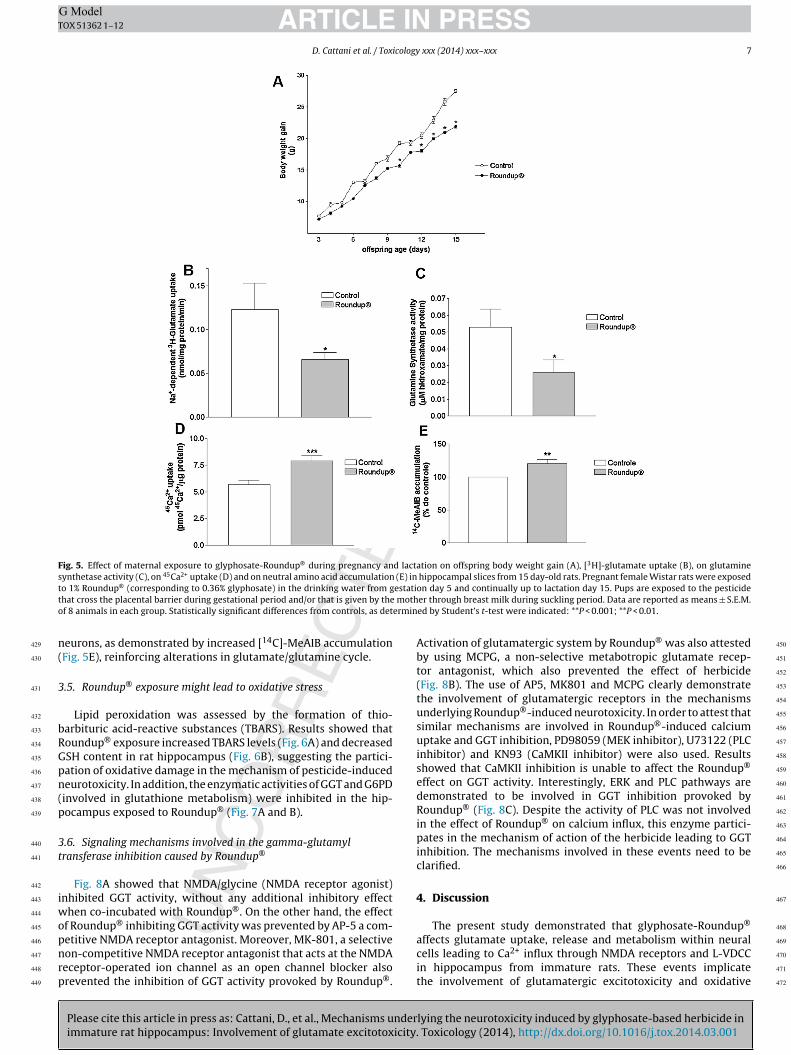
Chronic exposure to Roundup® during pregnancy and lactationdid affect neither dam’s body weight nor daily water consumptionover pregnancy (data not shown). Based on female body weightand on daily water consumption we estimated that each damingested 70 mg/Kg body weight/day. This dose was equivalent to1/2.5 of the limits of NOAEL. Fig. 5A showed that maternal expo-sure to Roundup® (70 mg/Kg body weight) decreased the bodyweight gain of pups from 10- to 15-day old. Moreover, chronicmaternal exposure to Roundup® leads to similar excitotoxic eventsobserved during short-term in vitro treatments in hippocampalcells. Results showed decreased the Na+-dependent glial [3H]-glutamate uptake (Fig. 5B) and glutamine synthetase inhibition(Fig. 5C), suggesting excess of glutamate in synaptic cleft associatedwith decreased neurotransmitter metabolism. Corroborating the
lying the neurotoxicity induced by glyphosate-based herbicide in. Toxicology (2014), http://dx.doi.org/10.1016/j.tox.2014.03.001
glutamatergic excitotoxicity, the chronic exposure to Roundup®
leads to increased 45Ca2+ uptake in hippocampal slices of immaturepups (Fig. 5D). Also, we observed increased glutamine uptake inside
D. Cattani et al. / Toxicology xxx (2014) xxx–xxx 7
Fig. 5. Effect of maternal exposure to glyphosate-Roundup® during pregnancy and lactation on offspring body weight gain (A), [3H]-glutamate uptake (B), on glutaminesynthetase activity (C), on 45Ca2+ uptake (D) and on neutral amino acid accumulation (E) in hippocampal slices from 15 day-old rats. Pregnant female Wistar rats were exposedt estatit motho ermin
n(
3
bRGpn(p
3t
iwopnrp
429
430
431
432
433
434
435
436
437
438
439
440
441
442
443
444
445
446
447
448
449
450
451
452
453
454
455
456
457
458
459
460
461
462
463
464
465
466
467
468
o 1% Roundup® (corresponding to 0.36% glyphosate) in the drinking water from ghat cross the placental barrier during gestational period and/or that is given by thef 8 animals in each group. Statistically significant differences from controls, as det
eurons, as demonstrated by increased [14C]-MeAIB accumulationFig. 5E), reinforcing alterations in glutamate/glutamine cycle.
.5. Roundup® exposure might lead to oxidative stress
Lipid peroxidation was assessed by the formation of thio-arbituric acid-reactive substances (TBARS). Results showed thatoundup® exposure increased TBARS levels (Fig. 6A) and decreasedSH content in rat hippocampus (Fig. 6B), suggesting the partici-ation of oxidative damage in the mechanism of pesticide-inducedeurotoxicity. In addition, the enzymatic activities of GGT and G6PDinvolved in glutathione metabolism) were inhibited in the hip-ocampus exposed to Roundup® (Fig. 7A and B).
.6. Signaling mechanisms involved in the gamma-glutamylransferase inhibition caused by Roundup®
Fig. 8A showed that NMDA/glycine (NMDA receptor agonist)nhibited GGT activity, without any additional inhibitory effect
hen co-incubated with Roundup®. On the other hand, the effectf Roundup® inhibiting GGT activity was prevented by AP-5 a com-
Please cite this article in press as: Cattani, D., et al., Mechanisms underimmature rat hippocampus: Involvement of glutamate excitotoxicity.
etitive NMDA receptor antagonist. Moreover, MK-801, a selectiveon-competitive NMDA receptor antagonist that acts at the NMDAeceptor-operated ion channel as an open channel blocker alsorevented the inhibition of GGT activity provoked by Roundup®.
on day 5 and continually up to lactation day 15. Pups are exposed to the pesticideer through breast milk during suckling period. Data are reported as means ± S.E.M.ed by Student’s t-test were indicated: **P < 0.001; **P < 0.01.
Activation of glutamatergic system by Roundup® was also attestedby using MCPG, a non-selective metabotropic glutamate recep-tor antagonist, which also prevented the effect of herbicide(Fig. 8B). The use of AP5, MK801 and MCPG clearly demonstratethe involvement of glutamatergic receptors in the mechanismsunderlying Roundup®-induced neurotoxicity. In order to attest thatsimilar mechanisms are involved in Roundup®-induced calciumuptake and GGT inhibition, PD98059 (MEK inhibitor), U73122 (PLCinhibitor) and KN93 (CaMKII inhibitor) were also used. Resultsshowed that CaMKII inhibition is unable to affect the Roundup®
effect on GGT activity. Interestingly, ERK and PLC pathways aredemonstrated to be involved in GGT inhibition provoked byRoundup® (Fig. 8C). Despite the activity of PLC was not involvedin the effect of Roundup® on calcium influx, this enzyme partici-pates in the mechanism of action of the herbicide leading to GGTinhibition. The mechanisms involved in these events need to beclarified.
4. Discussion
The present study demonstrated that glyphosate-Roundup®
lying the neurotoxicity induced by glyphosate-based herbicide in Toxicology (2014), http://dx.doi.org/10.1016/j.tox.2014.03.001
affects glutamate uptake, release and metabolism within neuralcells leading to Ca2+ influx through NMDA receptors and L-VDCCin hippocampus from immature rats. These events implicatethe involvement of glutamatergic excitotoxicity and oxidative
Fig. 6. Effect of Roundup® on lipid peroxidation and GSH levels in immature rat hip-pocampus. After preincubation, hippocampal slices were incubated in the presenceor absence of 0.01% Roundup® for 30 min. Data from thiobarbituric acid-reactivesaf
ddpawctb
C
F(pRti
473
474
475
476
477
478
479
480
481
482
483
484
485
486
487
488
489
490
491
492
493
494
495
496
497
498
499
500
501
502
503
504
505
506
507
508
509
510
511
512
513
514
515
516
517
ubstances (TBARS) measurement of lipid peroxidation and GSH levels are reporteds means ± S.E.M. of 8 animals from each group. Statistically significant differencesrom controls, as determined by Student’s t test, are indicated. *P < 0.05, **P < 0.01.
amage in the mechanism of Roundup®-induced neurotoxicityuring development. Exposure to environmental toxicants duringregnancy and suckling periods has the potential to affect embryond fetal development (Brent, 2004). In our experimental model,e induce a maternal exposure to the glyphosate-Roundup® and
onsequently, the offspring are exposed to the pesticide that cross
Please cite this article in press as: Cattani, D., et al., Mechanisms underimmature rat hippocampus: Involvement of glutamate excitotoxicity
he placental barrier during gestational period and/or that is giveny the mother through breast milk during suckling period.
The first demonstration that the herbicide glyphosate may affecta2+ homeostasis was demonstrated by Olorunsogo (1990), which
ig. 7. Effect of Roundup® on the activities of glucose-6-phosphate dehydrogenaseG6PD), and gamma-glutamyl transferase (GGT) in immature rat hippocampus. Hip-ocampal slices were incubated for 30 min in the presence or absence of 0.01%oundup® . Data are reported as means ± S.E.M. of 8 animals from each group. Sta-istically significant differences from controls, as determined by Student’s t test, arendicated. *P < 0.01, **P < 0.001.
518
519
520
521
522
523
524
525
526
527
528
529
530
531
532
533
534
535
536
537
538
539
540
541
542
543
544
PRESS xxx (2014) xxx–xxx
showed increased mitochondrial membrane permeability to pro-tons and Ca2+. In our study we demonstrated that 0.01%. Roundup®
(corresponding to 0.036 g/L glyphosate) induced 45Ca2+ uptake anddecreased cell viability in hippocampal slices from immature rats.It is important to emphasize that the concentration of Roundup®
used in agricultural working activities ranges from 1% to 2%, con-centrations 10,000–20,000 times larger than those used in ourexperimental protocols. In this context, we have recently demon-strated that the same concentrations used in this study were able toincrease calcium uptake and decrease Sertoli cell viability, clearlydemonstrating that Roundup® might affect male reproductive cells(de Liz Oliveira Cavalli et al., 2013). The present study demon-strates that both in vitro (acute) and in vivo (chronic) exposureto Roundup® induced 45Ca2+ uptake in hippocampal cells. Inter-estingly, our results demonstrated that modifications in 45Ca2+
influx were related with cell death at 0.01% Roundup®. However,the higher Roundup® concentration used (0.1% Roundup®, corre-sponding to 0.36 g/L glyphosate) leads to increased cell death whiledecreases 45Ca2+ influx, suggesting that high pesticide doses mightinduce necrotic cell death. Thus, we propose that the decreased cellviability induced by the pesticide might compromise the plasmamembrane activity and permeability, and therefore affects calciumuptake into hippocampal cells.
The Roundup®-induced Ca2+ influx occurs through NMDAreceptor activation and L-VDCC opening. The Roundup®-inducedexcitotoxicity in rat hippocampus also involves the recruitment ofsignal transduction pathways leading to the activation of kinasecascades including ERK. Activation of the ERK pathway mightaccount for neuroprotection under excitotoxic conditions in hip-pocampal neurons (Almeida et al., 2005). Roundup®-induced ERKactivation probably involves a compensatory effect to the exci-totoxic damage caused by the pesticide. In this context, Gomeset al. (2012) demonstrated a BDNF-induced activation of ERKunder excitotoxic condition. The authors suggest that upon exci-totoxic stimulation the kinase activity probably requires multiplecomponents that may be lost and/or redistributed within thecell. Moreover, previous results from our group demonstratedthat Roundup® disrupts male reproductive functions by trigger-ing calcium-mediated cell death in male reproductive cells (de LizOliveira Cavalli et al., 2013).
Astrocytes play a key role in removing glutamate from thesynaptic cleft and metabolizing it to glutamine. Then, glutamine isreleased from these cells and then taken up by neurons, to serve asa glutamate precursor (Danbolt, 2001). On the other hand, Torres etal. (2013) have recently demonstrated that once in astrocytes, glu-tamate is preferentially used as a fuel in the tricarboxylic acid (TCA)cycle instead of being converted into glutamine. They suggestedthat oxidation of glutamate could buffer excitotoxic conditions dueto high glutamate concentrations. Our results demonstrated thateither in vivo or in vitro exposure to Roundup® reduced glutamateuptake and metabolism within glial cells, associated with increasedrelease of this neurotransmitter in the synaptic cleft. Moreover,Roundup® leads to glutamine synthetase, AST and ALT enzymaticactivity inhibition, attesting that either glutamate uptake or itsmetabolism is impaired in hippocampal astrocytes exposed tothe herbicide. Taken together these results demonstrated thatRoundup® might lead to excessive extracellular glutamate levelsand consequently to excitotoxic condition in rat hippocampus.
The glial-neuronal metabolism is controlled by severalmetabolic pathways. Both in astrocytes and in neurons glucose isconverted through glycolysis in pyruvate, which could be convertedto lactate by lactate dehydrogenase, alanine by ALT, oxaloacetate
lying the neurotoxicity induced by glyphosate-based herbicide in. Toxicology (2014), http://dx.doi.org/10.1016/j.tox.2014.03.001
by pyruvate carboxylase or acetyl CoA by pyruvate dehydrogenase.The acetyl CoA enters the TCA cycle and the TCA cycle interme-diates can exit the cycle at the �-ketoglutarate step and thenform glutamate by transaminases such as AST and ALT (Alvestad
D. Cattani et al. / Toxicology xxx (2014) xxx–xxx 9
Fig. 8. Involvement of glutamatergic system and kinase pathways on the mechanism of GGT inhibition by Roundup® in immature rat hippocampus. Hippocampal slices werepre-incubated for 15 min with or without NMDA/gly (NMDA glutamate receptor agonist), or 10 �M AP-5 (a competitive NMDA receptor antagonist), or 10 �M MK-801 (aselective non-competitive NMDA receptor antagonist), or MCPG (a metabotropic glutamate receptor antagonist), 10 �M KN93 (CaMKII inhibitor), or 10 �M PD 98059 (MAPKi ith or
d nt difm 0.01
ecgptichputihprramrri
549
550
551
552
553
554
555
556
557
558
559
560
561
562
563
564
565
566
567
568
569
570
571
572
573
574
575
576
577
578
579
580
581
582
583
584
585
586
nhibitor) or 10 �M U73122 (PLC inhibitor). After that, the slices were incubated wrugs described above. Values are means ± S.E.M. of 8 animals. Statistically significaultiple comparison test are indicated: *p < 0.01 compared with control group. #p <
t al., 2011). Moreover, the glutamate released into the synapticleft is taken up into astrocytes at which it can be converted tolutamine or enter the TCA cycle. Then, glutamine can be trans-orted back to the neurons and regenerate glutamate. Consideringhat Roundup® affects the activity of important enzymes involvedn neural cell metabolism, our results suggested that the herbi-ide causes energetic deficit in addition to excitotoxic damage inippocampus from immature rats. The ineffective glutamate trans-ort into astrocytes supports the decreased glutamate oxidationnder Roundup®-induced neurotoxic condition. On the other hand,he glutamine transport into neurons was increased both aftern vitro and chronic Roundup® exposure, as demonstrated by theigh 14C-MeAIB accumulation observed in herbicide-treated hip-ocampus. Corroborating these findings, Morken et al. (2013) haveecently demonstrated that the transfer of glutamate from neu-ons to astrocytes was much lower in neonatal rat brain than indult one, while transfer of glutamine from astrocytes to gluta-
Please cite this article in press as: Cattani, D., et al., Mechanisms underimmature rat hippocampus: Involvement of glutamate excitotoxicity.
atergic neurons was relatively higher. Corroborating the presentesults, we have recently demonstrated that congenital hypothy-oidism also leads to stimulation in system A transporter whichs associated with decreased glutamate uptake and increased Ca2+
without 0.01% Roundup® for 30 min (incubation) in the presence or absence of theferences from controls, as determined by one-way ANOVA followed by Bonferronicompared with Roundup® or glyphosate group.
influx in rat hippocampus (Cattani et al., 2013). These intercon-nected events compromise glutamate-glutamine cycle accountingfor oxidative stress in hypothyroid hippocampus (Cattani et al.,2013) and cerebral cortex (Zamoner et al., 2008) accounting forglutamate excitotoxicity.
Supporting our findings Pérez-De La Cruz et al. (2008) demon-strated that excitotoxicity and energy deficit caused by theco-administration of quinolinate and 3-nitropropionate lead toexcessive cytoplasmic calcium levels which mediate oxidativedamage in brain synaptosomal membranes. In this context, exces-sive calcium influx might potentiate lethal metabolic pathways,which in turn involves increased formation of reactive oxygenspecies and mitochondrial dysfunction (Rami et al., 1997). More-over, increased calcium influx might augment the release ofexcitatory amino acids (such as glutamate), and hence propagateexcitotoxic cell damage through a positive feedback, further lead-ing cells to death (Pérez-De La Cruz et al., 2008). Taking into
lying the neurotoxicity induced by glyphosate-based herbicide in Toxicology (2014), http://dx.doi.org/10.1016/j.tox.2014.03.001
account these findings, we could ascribe that oxidative damage,demonstrated by increased lipid peroxidation and GSH depletion,is probably dependent on the high intracellular calcium concentra-tions induced by Roundup® in hippocampal slices.
10 D. Cattani et al. / Toxicology xxx (2014) xxx–xxx
Fig. 9. Proposed mechanism underlying hippocampal neurotoxicity of Roundup® . The pesticide causes Ca2+ influx by activating NMDA receptors and L-type voltage-dependentCa2+ channels (L-VDCC) setting off oxidative stress and cell death. The mechanisms underlying Roundup® neurotoxicity involve the activation of Ca2+/calmodulin-dependentprotein kinase II (CaMKII) and extracellular signal-regulated kinase (ERK). Astrocytes play a key role in removing glutamate from the synaptic cleft and metabolizing itto glutamine, which serve as a glutamate precursor in neurons. Our results demonstrated that Roundup® reduced glutamate uptake and metabolism within glial cells,associated with increased release of this neurotransmitter in the synaptic cleft. Moreover, Roundup® leads to glutamine synthetase, aspartate aminotransferase (AST) andalanine aminotransferase (ALT) enzymatic activity inhibition, attesting that either glutamate uptake or its metabolism is impaired in hippocampal astrocytes exposed tothe herbicide. Moreover, Roundup® reduced GSH levels and increased the amounts of thiobarbituric acid reactive species (TBARS), characterizing oxidative damage. Also,exposure to the pesticide decreased the activity of gamma-glutamyl transferase (GGT) and glucose-6-phosphate dehydrogenase, supporting the depletion of GSH. In thiscontext, the GGT inhibition induced by Roundup® could decrease the amino acid availability to GSH de novo synthesis and the decreased activity of G6PD may reduce NADPHl that Rt
iGptcooot(npmTRGpr
pTmsiiMtGD
591
592
593
594
595
596
597
598
599
600
601
602
603
604
605
606
607
608
609
610
611
612
613
614
615
616
617
618
619
620
621
622
623
624
625
626
627
628
629
630
631
632
633
634
635
636
637
638
639
640
641
642
643
evels, necessary to reduce glutathione. Taken together these results demonstratedo excitotoxic condition and energetic deficit in rat hippocampus.
The oxidative damage induced by Roundup® in rat hippocampuss also confirmed by the inhibition of the enzymatic activity of eitherGT or G6PD. It was described that in neonatal brain more glucose isrioritized to pentose phosphate pathway and pyruvate carboxyla-ion than in adult one. These events may have implications for theapacity to protect the neonatal brain against excitotoxicity andxidative stress (Morken et al., 2013). The rate-limiting enzymef the pentose phosphate pathway, G6PD, has been implicated notnly to promote reduced glutathione (GSH) but also enhance oxida-ive stress in specific cellular conditions. In this context, Zhao et al.2012) have demonstrated that G6PD plays a role in either oxidativeeuronal damage or neuroprotection during ischemic reperfusioneriod in CA1 pyramidal neurons. The G6PD enzymatic inhibitionight decrease the NADPH availability to reduce the glutathione.
he GSH depletion, an important marker of oxidative stress, inoundup®-exposed hippocampus and could be a consequence of6PD inhibition. Therefore, Roundup® exposure can decrease therotective action of G6PD against oxidative damage in immatureat brain.
GGT is involved in extracellular breakdown of GSH, providingrecursor amino acids for GSH de novo synthesis (Lee et al., 2004).herefore, the GGT inhibition in Roundup®-exposed hippocampusight cause a decrease in the glutamate reservoir to GSH synthe-
is leading to a decrease in the levels of this important antioxidantn brain. The present study investigated the mechanisms involvedn the inhibition of this enzyme by Roundup® in rat hippocampus.
Please cite this article in press as: Cattani, D., et al., Mechanisms underimmature rat hippocampus: Involvement of glutamate excitotoxicity
oreover, results showed that NMDA receptors and ERK activa-ion participate in the mechanisms underlying the inhibition ofGT activity in this brain structure. Consistent with our findings,ang et al. (2011) demonstrated that after 30 min of treatment
oundup® might lead to excessive extracellular glutamate levels and consequently
with acrolein it was observed GSH depletion and ERK activationin cultured astrocytes. Although CAMKII activation participates inRoundup®-induced Ca2+ influx, this kinase is not involved in GGTmodulation. Moreover, the activation of CaMKII and ERK1/2 con-tributes to the potentiation of Ca2+ response elicited by capsaicinin rat dorsal root ganglion neurons. Results indicate that the poten-tiation is a Ca2+-modulated process that is mediated by intracellularsignaling pathways involving activation of CaMKII and ERK1/2, butnot by activation of PKC or PKA (Zhang et al., 2011). Moreover, inter-actions between Ca2+, CAMKII e ERK1/2 signaling pathways werepreviously demonstrated in neural cells (Ji and Woolf, 2001; Choeand Wang, 2002; Zamoner et al., 2008).
5. Conclusions
The proposed mechanism underlying Roundup®-induced neu-rotoxicity is summarized in Fig. 9. In conclusion, our results showedthat the herbicide Roundup® leads to glutamatergic excitotoxicityand energy deficit in hippocampal cells from immature rats. Themechanisms underlying Roundup® neurotoxicity involve activa-tion of kinase cascades as well as misregulation of glutamatergicsynapses, Ca2+ influx through NMDA and L-VDCC, energetic deficitsand oxidative damage in rat hippocampus. Thus, we propose thatRoundup® induced glutamatergic excitotoxicity which culminatein neural cell death.
lying the neurotoxicity induced by glyphosate-based herbicide in. Toxicology (2014), http://dx.doi.org/10.1016/j.tox.2014.03.001
Funding
This work was supported by grants from Conselho Nacionalde Desenvolvimento Científico e Tecnológico (CNPq-Brazil)/Edital
niversal 14/2011/research grant #479483/2011-6; Fundac ãoe Apoio à Pesquisa Científica e Tecnológica do Estado deanta Catarina (FAPESC) research grant “chamada públicaAPESC 04/2011 – Apoio a infra-estrutura de CT&I para jovensesquisadores” #11,338/2012-7; PGFAR; PPGBQA; Coordenac ão deperfeic oamento de Pessoal de Nível Superior (CAPES-Brazil) andAPES-REUNI.
onflict of interest
Authors declare that there is no conflict of interest that could beerceived as prejudicing the impartiality of the research reported.
ransparency document
The Transparency document associated with this article can beound in the online version.
cknowledgements
We acknowledge FAPESC, CNPq-Brazil, CAPES, CAPES-REUNI,GFAR and PPGBQA for grants and research scholarships. We alsohank INTERCIENTÍFICA (São José dos Campos, SP, Brazil) for theenerous gift of G6PD kits. Daiane Cattani was registered on Phar-acy Postgraduate Program/UFSC-Brazil (PGFAR), Vera Lúcia de
iz Oliveira Cavalli was registered on Biochemistry Postgraduaterogram/UFSCBrazil (PPGBQA).
eferences
lmeida, R.D., Manadas, B.J., Melo, C.V., Gomes, J.R., Mendes, C.S., Grãos, M.M., Car-valho, R.F., Carvalho, A.P., Duarte, C.B., 2005. Neuroprotection by BDNF againstglutamate-induced apoptotic cell death is mediated by ERK and PI3-kinase path-ways. Cell Death Differ. 12, 1329–1343.
lvestad, S., Hammer, J., Qu, H., Håberg, A., Ottersen, O.P., Sonnewald, U., 2011.Reduced astrocytic contribution to the turnover of glutamate, glutamine, andGABA characterizes the latent phase in the kainate model of temporal lobeepilepsy. J. Cereb. Blood Flow Metab. 31, 1675–1686.
stiz, M., de Alaniz, M.J., Marra, C.A., 2012. The oxidative damage and inflammationcaused by pesticides are reverted by lipoic acid in rat brain. Neurochem. Int. 61,1231–1241.
arbosa, E.R., Leiros da Costa, M.D., Bacheschi, L.A., Scaff, M., Leite, C.C., 2001. Parkin-sonism after glycine-derivate exposure. Move. Disord. 16, 565–568.
arlow, B.K., Lee, D.W., Cory-Slechta, D.A., Opanashuk, L.A., 2005. Moduation ofantioxidant defence systems by the environmental pesticide Maneb in dopa-minergic cells. Neurotoxicology 26, 63–75.
enachour, N., Séralini, G.-E., 2009. Glyphosate formulations induce apoptosis andnecrosis in human umbilical, embryonic, and placental cells. Chem. Res. Toxicol.22, 97–105.
ird, R.P., Draper, A.H., 1984. Comparative studies on different methods of malondy-haldehydedetermination. Methods Enzymol. 90, 105–110.
rent, R.L., 2004. Environmental causes of human congenital malformations: thepediatrician’s role in dealing with these complex clinical problems caused by amultiplicity of environmental and genetic factors. Pediatrics 113, 957–968.
attani, D., Goulart, P.B., Cavalli, V.L., Winkelmann-Duarte, E., Dos Santos, A.Q.,Pierozan, P., de Souza, D.F., Woehl, V.M., Fernandes, M.C., Silva, F.R., Gonc alves,C.A., Pessoa-Pureur, R., Zamoner, A., 2013. Congenital hypothyroidism altersthe oxidative status, enzyme activities and morphological parameters in thehippocampus of developing rats. Mol. Cell. Endocrinol. 375, 14–26.
hong, Z.Z., Li, F., Maiese, K., 2005. Oxidative stress in the brain: novel cellular tar-gets that govern survival during neurodegenerative disease. Prog. Neurobiol. 75,207–246.
horfa, A., Bétemps, D., Morignat, E., Lazizzera, C., Hogeveen, K., Andrieu, T., Baron,T., 2013. Specific pesticide-dependent increases in �-synuclein levels in humanneuroblastoma (SH-SY5Y) and melanoma (SK-MEL-2) cell lines. Toxicol. Sci. 133,289–297.
anbolt, N.C., 2001. Glutamate uptake. Prog. Neurobiol. 65, 1–105.ang, T.N., Arseneault, M., Ramassamy, C., 2011. Regulation of redox-sensitive
signaling pathways in rat primary astrocytes following acrolein exposure. J.Alzheimers Dis. 25, 263–277.
Please cite this article in press as: Cattani, D., et al., Mechanisms underimmature rat hippocampus: Involvement of glutamate excitotoxicity.
e Liz Oliveira Cavalli, V.L., Cattani, D., Heinz Rieg, C.E., Pierozan, P., Zanatta, L.,Benedetti Parisotto, E., Wilhelm Filho, D., Mena Barreto Silva, F.R., Pessoa-Pureur,R., Zamoner, A., 2013. Roundup disrupts male reproductive functions by trigger-ing calcium-mediated cell death in rat testis and Sertoli cells. Free Radic. Biol.Med. 65C, 335–346.
PRESS xxx (2014) xxx–xxx 11
El-Shenawy, N.S., 2009. Oxidative stress responses of rats exposed to Roundupand its active ingredient glyphosate. Environ. Toxicol. Pharmacol. 28,379–385.
Gomes, J.R., Costa, J.T., Melo, C.V., Felizzi, F., Monteiro, P., Pinto, M.J., Inácio, A.R.,Wieloch, T., Almeida, R.D., Grãos, M., Duarte, C.B., 2012. Excitotoxicity down-regulates TrkB.FL signaling and upregulates the neuroprotective truncatedTrkB receptors in cultured hippocampal and striatal neurons. J. Neurosci. 32,4610–4622.
Hayden, K.M., Norton, M.C., Darcey, D., Ostbye, T., Zandi, P.P., Breitner, J.C.,Welsh-Bohmer, K.A., Cache County Study Investigators, 2010. Occupationalexposure to pesticides increases the risk of incident AD: the Cache County study.Neurology 74, 1524–1530.
Jenstad, M., Quazi, A.Z., Zilberter, M., Haglerød, C., Berghuis, P., Saddique, N.,Goiny, M., Buntup, D., Davanger, S.S., Haug, F.M., Barnes, C.A., McNaughton,B.L., Ottersen, O.P., Storm-Mathisen, J., Harkany, T., Chaudhry, F.A., 2009. Sys-tem A transporter SAT2 mediates replenishment of dendritic glutamate poolscontrolling retrograde signaling by glutamate. Cereb. Cortex 19, 1092–1106.
Ji, R.R., Woolf, C.J., 2001. Neuronal plasticity and signal transduction in nociceptiveneurons: implications for the initiation and maintenance of pathological pain.Neurobiol. Dis. 8, 1–10.
Kanamori, K., Ross, B.D., 2004. Quantitative determination of extracellular glutamineconcentration in rat brain, and its elevation in vivo by system A transportinhibitor, alpha-(methylamino)isobutyrate. J. Neurochem. 90, 203–210.
Kirby, M.L., Barlow, R.L., Bloomquit, J.R., 2001. Neurotoxicity of the organochlorineinsecticide hepatochlor to murine striatal dopaminergic pathways. Toxicol. Sci.61, 100–106.
Le Couteur, D.G., Mc Lean, A.J., Taylor, M.C., Woodham, B.L., Board, P.G., 1999. Pesti-cides and Parkinson’s disease. Biomed. Pharmacother. 53, 122–130.
Lee, D.H., Blomhoff, R., Jacobs Jr., D.R., 2004. Is serum gamma glutamyltransferase amarker of oxidative stress? Free Radic. Res. 38, 535–539.
Londres, F., 2011. Agrotóxicos no Brasil: um guia para ac ão em defesa da vida. Rio deJaneiro: AS-PTA – Assessoria e Servic os a Projetos em Agricultura Alternativa.
Lowry, O.H., Rosebrough, N.J., Farr, A.L., Randall, R.J., 1951. Protein measurementwith the Folin phenol reagent. J. Biol. Chem. 193, 265–267.
Marc, J., Mulner-Lorillon, O., Boulben, S., Hureau, D., Durand, G., Bellé, R., 2002. Pes-ticide Roundup provokes cell division dysfunction at the level of CDK1/cyclin Bactivation. Chem. Res. Toxicol. 15, 326–331.
Morken, T.S., Brekke, E., Håberg, A., Widerøe, M., Brubakk, A.M., Sonnewald, U.,2013. Neuron–astrocyte interactions, pyruvate carboxylation and the pentosephosphate pathway in the neonatal rat brain. Neurochem. Res..
Negga, R., Stuart, J.A., Machen, M.L., Salva, J., Lizek, A.J., Richardson, S.J., Osborne, A.S.,Mirallas, O., McVey, K.A., Fitsanakis, V.A., 2012a. Exposure to glyphosate- and/orMn/Zn-ethylene-bis-dithiocarbamate-containing pesticides leads to degenera-tion of �-aminobutyric acid and dopamine neurons in Caenorhabditis elegans.Neurotox. Res. 21 (3), 281–290.
Negga, R., Rudd, D.A., Davis, N.S., Justice, A.N., Hatfield, H.E., Valente, A.L., Fields,A.S., Fitsanakis, V.A., 2011. Exposure to Mn/Zn ethylene-bis-dithiocarbamateand glyphosate pesticides leads to neurodegeneration in Caenorhabditis elegans.Neurotoxicology 32, 331–341.
Negga, R., Stuart, J.A., Machen, M.L., Salva, J., Lizek, A.J., Richardson, S.J., Osborne, A.S.,Mirallas, O., McVey, K.A., Fitsanakis, V.A., 2012b. Exposure to glyphosate- and/orMn/Zn-ethylene-bis-dithiocarbamate-containing pesticides leads to degenera-tion of �-aminobutyric acid and dopamine neurons in Caenorhabditis elegans.Neurotox. Res. 21, 281–290.
Olorunsogo, O.O., 1990. Modification of the transport of protons and Ca2+ ions acrossmitochondrial coupling membrane by N-(phosphonomethyl)glycine. Toxico-logy 61, 205–209.
Orlowsky, M., Meister, A., 1963. Gamma-glutamyl-p-nitroanilide: a new con-venient substrate for determination and study of l- and d-gamma-glutamyltranspeptidase activities. Biochim. Biophys. Acta 73, 679–681.
Paganelli, A., Gnazzo, V., Acosta, H., López, S.L., Carrasco, A.E., 2010. Glyphosate-based herbicides produce teratogenic effects on vertebrates by impairingretinoic acid signaling. Chem. Res. Toxicol. 23, 1586–1595.
Patel, S., Singh, V., Kumar, A., Gupta, Y.K., Singh, M.P., 2006. Status of antioxidantdefence system and expression of toxicant responsive genes in striatum ofmaneb- and paraquat-induced Parkinson’s disease phenotype in mouse: mech-anism of neurodegeneration. Brain Res. 1081, 9–18.
Peng, J., Peng, L., Stevenson, F.F., Doctrow, S., Andersen, J.K., 2007. Iron and Paraquatas synergistic environmental risk factors in sporadic Parkinson’s disease accel-erate age-related neurodegeneration. J. Neurosci. 27, 6914–6922.
Pérez-De La Cruz, V., Konigsberg, M., Pedraza-Chaverri, J., Herrera-Mundo, N., Díaz-Munoz, M., Morán, J., Fortoul-van der Goes, T., Rondán-Zárate, A., Maldonado,P.D., Ali, S.F., Santamaría, A., 2008. Cytoplasmic calcium mediates oxidative dam-age in an excitotoxic/energetic deficit synergic model in rats. Eur. J. Neurosci.27, 1075–1085.
Pessoa-Pureur, R., Wajner, M., 2007. Cytoskeleton as a potential target in the neu-ropathology of maple syrup urine disease: insight from animal studies. J. Inherit.Metab. Dis. 30, 664–672.
lying the neurotoxicity induced by glyphosate-based herbicide in Toxicology (2014), http://dx.doi.org/10.1016/j.tox.2014.03.001
Rami, A., Ferger, D., Krieglstein, J., 1997. Blockade of calpain proteolytic activityrescues neurons from glutamate excitotoxicity. Neurosci. Res. 27, 93–97.
Reitman, S., Frankel, S., 1957. A colorimetric method for the determination of serumglutamic oxalacetic and glutamic pyruvic transaminases. Am. J. Clin. Pathol. 28,56–63.
repeated application of capsaicin in rat DRG neurons. Am. J. Physiol. Regul. Integr.Comp. Physiol. 300, R644–R654.
802
803
804
805
806
807
808
809
810
811
812
813
814
815
816
817
818
819
820
821
822
823
824
825
826
827
828
829
830
831
832
833
834
835
ARTICLEOX 51362 1–12
2 D. Cattani et al. / Tox
ichard, S., Moslemi, S., Sipahutar, H., Benachour, N., Séralini, G.E., 2005. Differentialeffects of glyphosate and roundup on human placental cells and aromatase.Environ. Health Perspect. 113, 716–720.
ilva, F.R., Leite, L.D., Barreto, K.P., D’Agostini, C., Zamoner, A., 2001. Effect of 3,5,3′-triiodo-l-thyronine on amino acid accumulation and membrane potential inSertoli cells of the rat testis. Life Sci. 69, 977–986.
utor, B., Luhmann, H.J., 1995. Development of excitatory and inhibitory postsynap-tic potentials in the rat neocortex. Perspect. Dev. Neurobiol. 2, 409–419.
orres, F.V., Hansen, F., Doridio Locks-Coelho, L., Souza, D.O., 2013. Increase of extra-cellular glutamate concentration increases its oxidation and diminishes glucoseoxidation in isolated mouse hippocampus: reversible by TFB-TBOA. J. Neurosci.Res., http://dx.doi.org/10.1002/jnr.23187.
sui, M.T., Chu, L.M., 2003. Aquatic toxicity of glyphosatebased formulations: com-parison between different organisms and the effects of environmental factors.Chemosphere 52, 1189–1197.
Please cite this article in press as: Cattani, D., et al., Mechanisms underimmature rat hippocampus: Involvement of glutamate excitotoxicity
Williams, G.M., Kroes, R., Munro, I.C., 2000. Safety evaluation and risk assessment ofthe herbicide roundup and its active ingredient, glyphosate, for human. Regul.Toxicol. Pharmacol. 31, 117–165.
Zamoner, A., Heimfarth, L., Oliveira Loureiro, S., Royer, C., Mena Barreto Silva, F.R.,Pessoa-Pureur, R., 2008. Nongenomic actions of thyroxine modulate interme-diate filament phosphorylation in cerebral cortex of rats. Neuroscience 156,640–652.
Zamoner, A., Royer, C., Barreto, K.P., Pessoa-Pureur, R., Silva, F.R., 2007. Ionic involve-ment and kinase activity on the mechanism of nongenomic action of thyroidhormones on 45Ca2+ uptake in cerebral cortex from young rats. Neurosci. Res.57, 98–103.
Zhang, X., Daugherty, S.L., de Groat, W.C., 2011. Activation of CaMKII and ERK1/2contributes to the time-dependent potentiation of Ca2+ response elicited by
lying the neurotoxicity induced by glyphosate-based herbicide in. Toxicology (2014), http://dx.doi.org/10.1016/j.tox.2014.03.001
Zhao, G., Zhao, Y., Wang, X., Xu, Y., 2012. Knockdown of glucose-6-phosphate dehy-drogenase (G6PD) following cerebral ischemic reperfusion: the pros and cons.Neurochem. Int. 61, 146–155.