Page 1
Mechanistic considerations on thewavelength-dependent variations of UVRgenotoxicity and mutagenesis in skin: thediscrimination of UVA-signature fromUV-signature mutation
著者 Hironobu Ikehatajournal orpublication title
Photochemical & Photobiological Sciences
volume 17number 12page range 1861-1871year 2018-05-14URL http://hdl.handle.net/10097/00125833
doi: 10.1039/C7PP00360A
Page 2
1
Mechanistic considerations on the wavelength-dependent variations of
UVR genotoxicity and mutagenesis in skin: Discrimination of UVA-
signature from UV-signature mutation
Hironobu Ikehata
Department of Medical Biochemistry, Tohoku University Graduate School of Medicine,
Sendai, Japan.
E-mail: [email protected] ; Tel: +81-22-717-8085; Fax: +81-22-717-8090
Page 3
2
Abstract
Ultraviolet radiation (UVR) predominantly induces UV-signature mutations, C ® T and
CC ® TT base substitutions at dipyrimidine sites, in the cellular and skin genome,
although these UVR-specific mutations show a wavelength-dependent variation in their
sequence-context preference, as evidenced by our in vivo mutation studies of mouse
skin. The C ® T mutation occurs most frequently in the 5’-TCG-3’ context regardless
of the UVR wavelength, but is recovered more preferentially there as the wavelength
increases, resulting in prominent occurrences exclusively at the TCG context in the
UVA wavelength range, which I will designate as a “UVA signature” in this review.
The preference of the UVB-induced C ® T mutation for the sequence contexts shows a
mixed pattern of UVC- and UVA-induced mutations, and a preference pattern similar to
the UVB-induced one is also observed for natural sunlight, in which UVB is the most
genotoxic component. In addition, the CC ® TT mutation hardly occurs at UVA1
wavelengths, although it is detected rarely but constantly in the UVC and UVB ranges.
These wavelength-dependent, sequence-context preferences of the UVR-specific
mutations could be explained by two different photochemical mechanisms of
cyclobutane pyrimidine dimer (CPD) formation. The UV-signature mutations observed
in the UVC and UVB ranges are known to occur mainly through error-free translesion
DNA synthesis (TLS) by DNA polymerase h across deaminated cytosines in CPDs,
which are produced through the conventional singlet/triplet excitation of pyrimidine
bases by the direct absorption of UVC/UVB photon energy in those bases. On the other
hand, a novel photochemical mechanism through the direct absorption of UVA energy
to double-stranded DNA, which is called “collective excitation”, has been proposed for
the UVA-induced CPD formation. The UVA photons directly absorbed by DNA cause
Page 4
3
CPD formation with a sequence context preference different from those caused by the
UVC/UVB-mediated singlet/triplet excitation, producing CPDs preferentially at
thymine-containing dipyrimidine sites, and probably also preferably at methyl CpG-
associated dipyrimidine sites. Cytosine deamination in these CPDs, which is known to
be accelerated for CPDs formed at the TCG context, can lead to the UVA-signature
mutations through the DNA polymerase h-dependent, error-free TLS.
Introduction
Action spectrum analysis of the mouse skin cancer induction by ultraviolet radiation
(UVR), which was performed mainly by Jan C. van der Leun’s group, clearly
demonstrated that the genotoxicity of UVR for mammalian skin depends on the
wavelength, and suggested that, although the UVB component plays a major role in the
genotoxicity, UVA, the longer wavelength components of UVR (320–400 nm), also
makes a small but distinct contribution.1 The genotoxicity of UVR induces mutation in
the skin genome, which can result in the carcinogenesis as evidenced by p53 mutations
in skin cancers in sun-exposed areas of human skin2–5 and those experimentally induced
in mouse skin.6–11 The mutagenicity of UVR is derived from its ability to produce DNA
damage by direct or indirect photochemical reactions with DNA and/or by indirect
oxidative DNA modifications through the formation of reactive oxygen species
(ROS).12 The former reactions produce UVR-specific base photolesions such as
cyclobutane pyrimidine dimers (CPDs) and pyrimidine(6-4)pyrimidone photoproducts
(64PPs) at dipyrimidine sites in DNA.13 The latter oxidative modifications include
single strand DNA breaks and the formation of oxidative base damage such as 8-
hydroxyguanine (8OH-G).14 The contribution of the oxidative DNA modification to the
Page 5
4
UVR genotoxicity has been noticed especially for UVA, where the efficiency of
photolesion production by direct photochemical reactions is reduced by several orders
of magnitude compared to the shorter wavelengths of UVR.12,14–16 However,
quantitative and mechanistic analyses of UVA-induced CPD formation in the last two
decades have provoked a reconsideration on the origin of the UVA genotoxicity.17–25
I have studied UVR-induced mutation spectra in mouse skin using a transgenic
mouse strain with l-phage vector-based, bacterial lacZ-transgenes, which were
developed for mutation analysis, and a variety of UVR sources emitting different
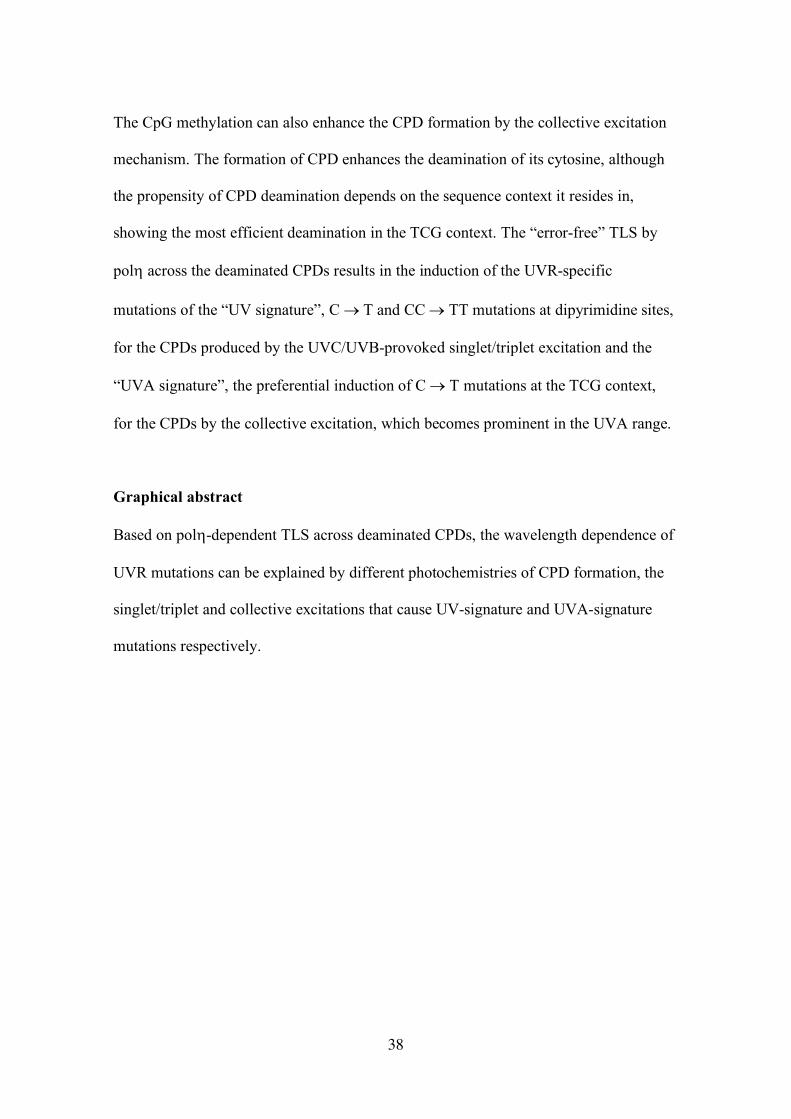
wavelength components from UVC to UVA (Fig. 1A).26–31 In this review, I provide an
updated overview of the wavelength-dependent UVR genotoxicity mainly based on the
mutation spectra obtained by in vivo analyses, and propose a model explaining the
mechanism of wavelength-dependent variations in the mutation spectra by combining
some recent findings in DNA repair, photochemistry and photobiology.
Wavelength dependence of UVR-induced mutation spectra
UVR induces specific types of mutation in DNA as reported for phages,32–34
bacteria,35,36 yeasts,37–39 mammalian cultured cells40–50, and mammalian skin.26–31,51,52
These UVR-specific mutation types include the C ® T transition at dipyrimidine sites
and CC ® TT tandem base substitution, which are called collectively “UV signature”
as discriminative mutations indicating the trace of UVR genotoxic insults.2 All UVR
components, UVC (wavelengths <280 nm), UVB (280–320 nm), UVA2 (320–340 nm)
and UVA1 (340–400 nm), can induce the UV-signature mutations as demonstrated in
our studies,26–31,53 summarized in Fig. 1B and C, although the wavelengths in the UVA1
region hardly induced CC ® TT mutations.29,30 Our studies revealed that the C ® T
Page 6
5
transition at dipyrimidine sites is the dominant type for all UVR components,
comprising 59–84% of total mutations observed after irradiation, demonstrating that
UVR genotoxicity results mostly from DNA photolesions specifically produced by
UVR.54 On the other hand, the influence of UVR-produced ROS is not remarkable or, if
any, minor in the UVR-induced mutation spectra, judging from the contribution of G ®
T transversion, a mutation that can be caused by 8OH-G, one of the representative types
of oxidative DNA damage.55 Only in the sunlight-induced spectrum, the G ® T
mutation was significantly induced, although it was a minor component,28 which might
suggest some contribution of non-UVR wavelengths included in sunlight to the skin
genotoxicity as also observed in other studies with yeast and phage.38,39,56 Some
photodynamic reactions might be relevant. Moreover, it should be noted that UVA1
sources, both the broadband UVA1 lamps and narrowband UVA1 laser, did not induce
oxidative damage-related mutations such as G ® T and G ® C transversions57 at a
remarkable frequency,29,30 although a dose-dependent formation of 8OH-G was
observed in the skin after UVA1 irradiation,29 as observed in cultured cells.15 It is also
known that 8OH-Gs are removed from cellular DNA much faster than CPDs.58 These
observations strongly support that UVR exerts its genotoxicity to the skin mainly
through direct photochemical reactions with DNA, irrespective of its wavelength
component. In addition, the ROS-mediated genotoxicity by UVR should be studied with
caution, especially in in vitro studies, because artificial ingredients in the DNA solvent
or cell/tissue culture media could cause or promote the production of ROS upon UVR
irradiation.17,59 To avoid these artifacts, analyses in vivo such as in the skin would be
preferable. This is one of the reasons I have excluded the cell-based studies from my
consideration of the UVR-induced mutation spectra in this review, although some
Page 7
6
important, contradicting points shown in those studies are discussed below. A more
detailed discussion on the disadvantages of the use of cell-based, in vitro mutation
assays for the study of mammalian UVR-induced mutation spectra has already been
made.60 However, most of the studies with skin mentioned above examined only the
UVR genotoxicity for normal skin after an acute single exposure. Multiple/chronic
UVR exposures or exposures of the skin under pathological conditions could bring a
ROS-mediated genotoxicity in addition to the genotoxicity mediated by direct
photochemical reactions with DNA. Interestingly, it has been demonstrated that CPDs
can be produced by ROS generated from melanin derivatives chemically excited long
after UVR exposure, which suggests that ROS could also induce UV-signature
mutations.61 However, melanocytes usually reside in the dermal layer in mouse skin,
and melanin is poor in the mouse epidermal layer, so that such mutations induced by
ROS-produced CPDs would be difficult to detect with the current in vivo mutation
assay system using transgenic mice.
Mutation induction mechanism by UVR-induced photolesions
The molecular mechanism of the mutagenesis by UVR-specific photolesions has been
studied widely and elucidated fairly well for some aspects.54 The mutation induction by
UVR requires replicative DNA synthesis after irradiation.62–64 CPD and 64PP are both
replication-blocking DNA damage, so that they should be removed by DNA repair
before a replication fork encounters them,65 or should be overcome by some damage
tolerance mechanisms so that the replicational DNA synthesis can be continued over the
damage site, because a failure in replication can lead to cell death.66,67 One of the
damage tolerance pathways could be a recombinational bypass of these photolesions by
Page 8
7
detouring the damage on the template strand using the genetic information of the other,
newly replicated daughter strand.66,67 This pathway would be error-free, but should be
too elaborate to perform the over-damage replication efficiently. Delay in DNA
replication, which leads to delay in cell proliferation, could cause a deficiency in the
recovery of damaged tissues. Another tolerance pathway is translesion DNA synthesis
(TLS), which can pass directly over the lesions on the template strand with the help of
specialized DNA polymerases, TLS polymerases.54,68,69 In the TLS mechanism,
replicative DNA polymerases switch to TLS polymerases upon an encounter with
replication-blocking DNA damage, and the TLS polymerases continue DNA synthesis
opposite the DNA damage, usually ignoring the base-pairing rule of nucleic acids. After
the replication fork has passed across the damage, replicative polymerases take over the
DNA synthesis in place of TLS polymerases and continue DNA replication. Thus, in
the mechanism of damage tolerance by TLS, the DNA replication could be continued
efficiently at the damage site in a manner sufficient to assist the recovery of damaged
tissues, although the DNA synthesis by TLS would usually be error-prone. Actually, it
was suggested that sites with repair-resistant CPDs in the p53 gene are also frequently
mutated sites in human skin cancers.70
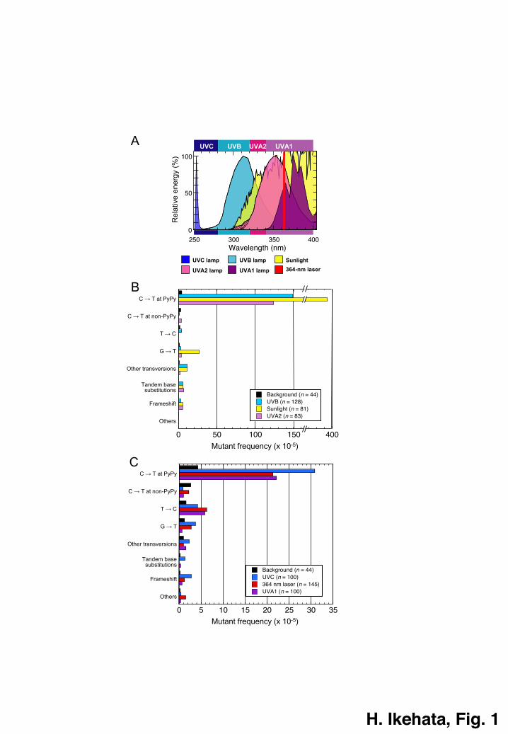
Among the TLS polymerases, however, DNA polymerase h (polh) is exceptional.
Polh can synthesize a daughter strand error-free across a CPD on the template strand,
probably by using the base pairing ability remaining in CPDs,71,72 thus rather
suppressing mutation induction by CPDs. This error-free TLS ability of polh appears
CPD-specific because polh can hardly bypass 64PPs or bypass other types of base
damage less error-free than CPDs.73–76 However, this polh-dependent error-free TLS
itself causes the UVR-specific mutations. It is known that cytosines in CPDs are highly
Page 9
8
prone to deaminate at position 4 and change easily to uracils (or a thymine if the
cytosine is methylated at position 5), which results in the conversion of cytosine-
containing CPDs to uracil or thymine-containing ones.77–80 If a replication fork
encounters such deaminated CPDs, the error-free TLS by polh should insert adenine
opposite the deaminated cytosine, namely uracil or thymine, thus resulting in the
induction of UVR-specific C ® T and CC ® TT mutations (Fig. 2). Since CPD has
been demonstrated to be the main mutagenic UVR photolesion in normal mammalian
cells and skin,81,82 the error-free TLS opposite deaminated CPDs by polh should be the
major pathway in the induction of UVR mutations in repair-proficient cells and skin.
In the absence of polh, UVR can induce mutations in cells and skin at much
higher frequencies than in the presence, although the mutation spectrum still shows the
UV signature predominantly.53,83–86 This polh-independent UVR mutagenesis has been
explained by a mechanistic model called the “two-step model”, in which inserter and
extender DNA polymerases are involved in the TLS.54,87–89 These DNA polymerases
might include polymerase i, k, z, Rev1 as well as h,54,71,90,91 which are TLS
polymerases, and replicative DNA polymerases such as d.92 64PPs and Dewar isomers,
as well as CPDs, could induce UV-signature mutations by this “two-step” mechanism
because the base insertions opposite photolesions by this mechanism is supposed to
occur according to the “A-rule”, in which an adenine is inserted with the base pairing
rule ignored.54,93,94 Although strongly supportive genetic studies have been reported,87–
89 the two-step model for the mutagenesis with UVR photolesions is, however, still
presumptive, awaiting experimental demonstrations by biochemically reconstituted
systems. Another UVR-specific mutation that could be explained by the two-step model
is the triplet mutation, a mutation with multiple base substitutions or frameshifts within
Page 10
9
a three-nucleotide sequence that includes a dipyrimidine sequence.54 The triplet
mutations were detected frequently in UVB-exposed mouse skin deficient in the
nucleotide excision repair,95–98 whereas the same mutations have also been detected in
other systems including mammalian cultured cells and skin cancers, although their
frequencies are variable depending on their repair abilities.99 The multiple base
substitutions and frameshifts occurring around a dipyrimidine site are easy to explain by
multiple misincorporations by inserter and extender DNA polymerases in the two-step
model.54
Variation of sequence context preference of the UVR-specific C ® T mutation by
wavelength
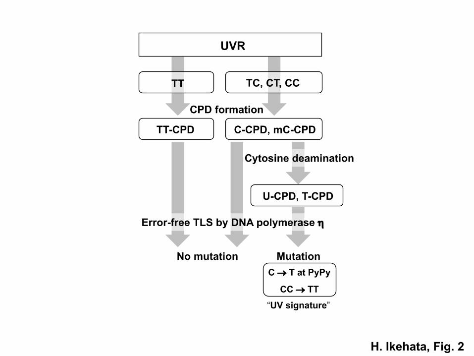
Although the mutation spectrum induced by each component of UVR shows a similar
pattern of UV-signature mutations (Fig. 1B, C), we found that the sequence context
preference of those UVR-specific mutations was remarkably different among UVR
components, as reported in our studies with transgenic mice,30,31 which are summarized
in Fig. 3A. We focused on three-tandem-base sequences in which the UVR-specific C
® T mutation occurs at the center base. There are 12 types of such triplet sequences,
which possess a cytosine base at the center and also include one or two dipyrimidine(s).
We found that UVR-specific C ® T mutations occurred preferably at the 5’-TCG-3’
(TCG) context in the lacZ transgene, 26–31 particularly with exposure to longer
wavelength components of UVR.27,29,30 Although the mutations at the TCG context
were most frequent among all the triplet contexts regardless of the UVR source, their
contribution to the mutation spectrum was moderate with the UVC source but
prominent exclusively with the UVA sources (Fig. 3A).31 Especially, with the UVA1
Page 11
10
sources more than 80% of the UVR-specific C ® T mutations occurred at the TCG
context. In the UVB range, the mutations at the TCG context were fairly conspicuous
but not as prominent as those by UVA, which were intermediate between UVC and
UVA. The distribution of occurrences of the UVR-specific mutation by sunlight was
relatively similar to that by UVB, reflecting the fact that UVB is the component in
sunlight most genotoxic to the skin.1,100–103 Thus, the occurrence of the UVR-specific C
® T mutation at the TCG context becomes conspicuous gradually as the wavelength
increases, finally overwhelming those at the other triplet contexts at UVA1 wavelengths
(Fig. 3A). Based on these observations, I propose that the UVR-specific C ® T
mutation that occurs preferentially at the TCG context should be called the “UVA
signature”. Although we proposed previously to call this type of mutation the “solar-UV
signature”,30,60 which we featured as a kind of the UVR-specific mutation that occurs
preferably at methyl CpG-associated dipyrimidine sites, the context preferences of the
sunlight- and UVB-induced mutations were rather a mixture of those of UVC and UVA,
as shown in Fig. 3A. Thus, “UVA signature” is more appropriate as a designation for
the TCG-preferential UVR-specific mutation.
Since, as mentioned above, UVR mutagenesis occurs in a polh-dependent manner
in normal cells and skin (see Fig. 2), we examined how the defect in polh affects the
TCG preference of the UVR-specific mutation.53 We found that the polh deficiency
made the mutation lose the TCG preference, as shown in Fig. 3B, clearly demonstrating
that the sequence context preference of the mutation depends on the TLS by polh, and
suggesting that the TCG preference of the mutation should reflect the preferable
formation or deamination of CPDs at some specific sequence motifs, which should at
least overlap with the TCG sequence.
Page 12
11
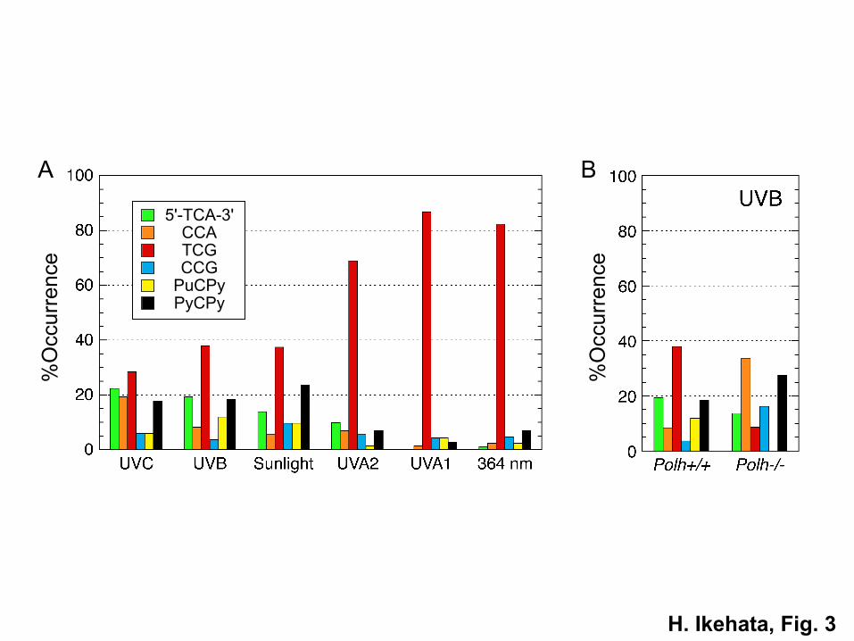
The mechanisms inducing two types of UVR-specific mutation, UV signature and
UVA signature
As shown in Fig. 1, the mutation spectra with UV-signature mutations can be induced
by any components of UVR, whereas the mutation spectra with UVA-signature
mutations, namely the exclusive occurrences at the TCG context of UVR-specific C ®
T mutations, are manifested specifically in the UVA range. Since both signature
mutations are induced by TLS over deaminated CPDs by polh as mentioned above, then
what causes the difference between them? UVA is known to induce CPDs significantly
although not as efficiently as UVC,17,18,21,104 but in a distribution pattern among
dipyrimidine motifs different from those by UVC and UVB.19,20,22 UVA, more
specifically UVA1, produces CPDs of TT dipyrimidines (TT-CPDs) at much higher
frequencies and CPDs of 5’-TC-3’ (TC) and 5’-CT-3’ (CT) dipyrimidines (TC- and CT-
CPDs) at lower frequencies than the shorter UVR components, although it does not
produce detectable amounts of CPDs of CC dipyrimidines (CC-CPDs).20,22
Accordingly, CC ® TT mutations were not detected in our UVA1-induced mutations in
mouse skin.29,30 It was supposed that the mechanism of CPD formation by UVA was
different from that by the shorter UVR, and that a triplet energy transfer to DNA bases
from some endogenous photosensitizers that can be activated by UVA energy would
mediate the CPD formation in the UVA range, because the energy of UVA photons is
not sufficient to directly activate pyrimidine bases to their excited singlet states, which
is necessary to cause photochemical reactions.20,105 However, such photosensitizers
have not been identified in vivo so far, and direct CPD formations in DNA by UVA1
have been demonstrated in experiments with isolated DNA.17,23,106,107 Recently, another
Page 13
12
mechanism by which UVA directly produces CPDs was proposed,108,109 in which the
UVA energy is absorbed directly to double-stranded DNA through “collective excited
states”, which can be followed by redistribution of the energy to pyrimidine bases
leading to CPD formation. On the other hand, UVC, and probably UVB, should
produce CPDs principally through the conventional singlet/triplet excited states induced
by direct absorption of the photon energy to pyrimidine bases,110,111 although some
minor contribution of the collective excitation would also be probable. Thus, UVA and
UVC/UVB could both produce CPDs directly, but through different photochemical
mechanisms (Fig. 4).105
It has been shown that UVB and/or solar UVR produce CPDs preferably at CpG-
associated dipyrimidine sites.112–114 The CpG motif is the target sequence of mammalian
DNA methylation that modifies cytosine to 5-methylcytosine (mC).115 This CpG
preference of CPD formation requires CpG methylation,113,114 and is not observed for
UVC.114,116,117 The CpG-associated dipyrimidine sites are 5’-TCG-3’ and 5’-CCG-3’,
the former of which is also the target context of the UVA-signature mutation. As
mentioned above, UVA produces predominantly TT-CPDs along with small amounts of
TC- and CT-CPDs, in other words, preferentially induces thymine-containing CPDs.
The molecular structure of 5-methylcytosine is similar to that of thymine, which would
raise the possibility that UVA produces CPDs not only from thymine but also from 5-
methylcytosine, probably in the order of dipyrimidine preferences of TT ≥ TmC > TC >
CT ≥ CmC (Fig. 4). Although the preferable CPD formation at 5’-TmCG-3’ and 5’-
CmCG-3’ contexts (TmCG and CmCG) has not been demonstrated for UVA so far, the
methyl CpG (mCpG)-directed CPD formation was much more remarkable after
exposure to sunlight than to UVB with 5 to 15-fold increases by sunlight and 1.7 to 1.8-
Page 14
13
fold by UVB compared to UVC-induced formation,113,114 suggesting some contribution
of the UVA component. If we accept the hypothesis that UVA should produce CPDs
preferably at mCpG-associated dipyrimidine sites, the TCG preference of the UVA-
signature mutation can be easily explained. However, there is one perplexing matter. If
UVA can also produce CPDs at the CmCG context, why don’t the mutations at the
same context contribute remarkably to the UVA-signature mutation?
It has been demonstrated that the propensity of cytosine deamination in CPDs
depends on the sequence context in which the CPD resides.118 CPDs in the CmCG
context are 50-fold slower to deaminate than those in the TCG and TmCG contexts,
which are most prone to deaminate with a half-life of around 6 hours in double-stranded
DNA, as far as examined so far. This difference in the CPD deamination propensity can
explain the poor recoveries of UVR-specific mutations in the CCG context after UVA
exposure that was reported in our studies using mouse skin (Fig. 3A),27,29,30 in which all
the mutation-detected CpG sites in the mutational target lacZ transgene were confirmed
to be fully methylated.26,119 Consequently, the preferential mutation occurrences at the
TCG context characteristic for the UVA-signature mutation can be rationalized by the
preferable CPD formation at mCpG-associated dipyrimidine sites and the context-
dependent propensity of CPD deamination under the mutation mechanism of the error-
free TLS by polh (Fig. 4).
Moreover, the context-dependent CPD deamination affects not only the UVA-
signature, but also the UV-signature mutations. As shown in Fig. 3A, the UVR-specific
mutation was most frequent at the TCG context even in the UVC and UVB ranges,
although their occurrence ratios were not as conspicuous as those in the UVA range. In
these shorter UVR ranges, the mCpG-preferable CPD formation, which should be
Page 15
14
mediated through the collective excitation mechanism, would be less remarkable than in
the longer UVR ranges, probably overwhelmed by abundant CPDs produced by the
singlet/triplet excitation mechanism, which functions dominantly in the UVC and UVB
ranges but almost completely fades out in the UVA1 range. Thus, CPD formation at the
TCG context would not be so prominent in the UVC and UVB ranges as in the UVA
range. However, once CPDs are formed at the TCG context, they should deaminate
efficiently and could cause mutations by the polh-dependent TLS mechanism, resulting
in the distribution of mutation occurrences among the triplet contexts shown in Fig. 3A.
The mutation occurrence distribution observed in the absence of polh (Fig. 3B) might
reflect the distribution of CPD formations among the triplet contexts, if we suppose that
the mutation induction occurs randomly through the TLS over UVR photolesions by
other error-prone TLS polymerases with the mechanism of the two-step model. This,
however, remains to be demonstrated.
The TCG preference of UVR-induced mutations was also demonstrated by an
exome analysis of 74 cancer-related genes in human sun-exposed normal and three
types of cancerous skin tissues, in which the most frequent and overwhelming mutation
was C ® T transitions, which occurred predominantly at the TCG context with fewer
occurrences at the other dipyrimidine-containing triplet contexts, regardless of the skin
tissue type.120 This result corresponds well with our observations on the occurrence
distribution of the sunlight-induced UVR-specific mutations among the triplet contexts
shown in Fig. 3A.28 Thus, the TCG preference of UVR-induced mutations is neither an
experimental artifact nor an observation limited to the lacZ transgene in mouse. It
occurs both in mouse and human, and would occur in other organisms with cytosine
methylation in their genome as far as they possess polh-like TLS polymerases.
Page 16
15
Although an exceptional case was reported for p53 gene mutations in human skin
cancers, which were detected rather more frequently in the CCG context than in TCG,2–
5,30,121 this discrepancy has not been observed for the p53 mutations in mouse skin
cancers,6–11 and can be explained by the poverty of mutable TCG sites in the human p53
gene on the transcribed strand, as discussed in detail previously.30 The lack of mutable
TCG sites in human p53 gene further suggests that the human genome have evolved to
prevent solar UVR from inducing malignant mutations by substituting genetically
important but UVR-vulnerable TCG sites with other genetically equivalent and UVR-
refractory sequences. This evolution would be promoted by the human features of
hairless skin and diurnal activity under the threat of photochemically genotoxic UVR
components in natural sunlight.
Studies inconsistent with the UVA-signature hypothesis
My proposal for the UVA-signature mutation is based on the CPD formation
mechanism through the collective excited state-mediated photochemistry, with which
CPDs should be produced preferably at thymine-containing dipyrimidine sites resulting
in the paucity of CC-CPDs, which becomes evident after the exposure to UVA,
especially UVA1. On the contrary, Rochette et al. reported the significant formation of
CC-CPDs by UVA1 using a ligation-mediated PCR (LMPCR) method.19 However, the
LMPCR method seems to have a tendency to overestimate the amounts of cytosine-
containing CPDs, especially that of CC-CPDs,19,122 compared with other methods such
as chromatographic analyses,123 post-labeling CPD-specific enzymatic cleavage
assays124,125 and HPLC with tandem mass spectrometry (HPLC-MS/ MS).126 Actually,
little CC-CPD formation has been detected in DNA, cells and skin tissues exposed to
Page 17
16
UVA1 with the HPLC-MS/MS, a far more sensitive, direct CPD detection
method.20,22,23 In addition, the LMPCR image (Fig. 1) given in the paper by Rochette et
al. showed distribution patterns of UVA-induced CPD formation among dipyrimidine
sites clearly different from those induced by other UVR sources such as UVC, UVB
and simulated sunlight.19 Although the bands corresponding to cytosine-containing
dipyrimidine sites were easily discernible in the lanes for the shorter UVR sources,
those were hardly distinguishable from the backgrounds in the UVA lanes, which would
reduce the reliability of estimates of the amount of cytosine-containing CPDs for the
UVA lanes.
The paucity of CC-CPDs should also suppress the CC ® TT tandem mutations in
the mutation spectrum induced by UVA, resulting in the lack of such tandem mutations
in the UVA signature. Accordingly, in our studies, the tandem mutations were not
observed in the mutations recovered from the mouse skin exposed to UVA1,29,30
although they were detected after exposure to UVA2 (Fig.1),27 which would indicate
that the singlet/triplet excitation mechanism for CPD formation are still valid in this
wavelength range. Drobetsky et al. studied a UVA-induced mutation spectrum in the
aprt gene using Chinese hamster cells and reported the induction of a unique type of
mutation, T ® G transversion, which they named UVA fingerprint,46 although the
preferable induction of such mutations has not been confirmed in subsequent studies
except for one.127 In the same study, Drobetsky et al. also recovered a few CC ® TT
mutations.46 Kappes et al. reported another UVA-induced mutation spectrum in the hprt
gene using human primary fibroblasts, detecting again a few CC ® TT mutations.50
Since both studies used short-cut filter-equipped UVR sources emitting mainly UVA1,
they suggest that UVA1 could induce the tandem base substitutions, in contradiction to
Page 18
17
my consideration given above. The recoveries of the tandem mutation in these studies
might result from the significant contribution of the UVA2 component to the irradiated
UVA, especially for the former study because they used blacklight lamps,46 which emit
mainly UVA2 wavelengths that might leak through the short-cut filter. Moreover, both
studies were performed with cultured cells, and irradiation to cultured cells often
produces ROS depending on the ingredients of the cultured medium.17,59 It is known
that ROS could induce CC ® TT mutations independently of UVR exposure,128,129
especially in mononucleotidyl cytosine runs.130 Although the mechanism of the ROS-
mediated CC ® TT mutation is unknown, it could have affected the mutation spectra
observed in these cellular studies. In addition, the use of aprt and hprt genes as
mutational markers was not appropriate for the study of UVR-induced mutations in
mammalian cells because both genes are hypomethylated and poor in mutable
dipyrimidine-associated CpG sites, whereas collective excited state-mediated, UVR-
specific mutations are supposed to prefer mCpG sites as supported by our studies and
p53 gene mutations in human and mouse skin cancers. The short size of coding
sequences of both genes (543 and 657 bp) is also disadvantageous for mutation
spectrum studies because of their low variation in sequence contexts (the lacZ transgene
is 3090-bp long). These points have already been discussed in detail in our previous
review.60 Reflecting these situations, the appearance of UV-signature mutations in the
UVA-induced mutation spectra were much less remarkable in both the cellular studies
(27–35%)46,50 than those in our studies with skin (59–68%),29,30 suggesting a much
greater contribution of non-UVR-induced mutations to the spectra of the cellular
studies.
Page 19
18
Conclusion
In my studies with mice, UVC induced the UVR-specific C ® T mutations most
frequently at the TCG context but also at other cytosine-containing dipyrimidine
contexts at comparable frequencies, whereas UVA induced the same mutations
exclusively at the TCG context with rare mutations at the other contexts. The context
preference of UVB-induced mutations showed a mixture between those of UVC and
UVA. Based on the molecular mechanism of UVR mutagenesis that is mediated mainly
through polh-dependent error-free TLS across deaminated CPDs, this wavelength-
dependent context preference of the mutations can be explained by deamination
tendencies of cytosine-containing CPDs and a recently identified/proposed
photochemical mechanism of CPD formation, the collective excitation, which would
justify the discrimination of UVA-induced UVR-specific mutations as the UVA
signature from the UV-signature mutations induced by UVC/UVB, which would be
caused mainly by CPDs formed through the conventional photochemical mechanism of
singlet/triplet excitation.
Conflicts of interest
There are no conflicts of interest to declare.
Acknowledgments
We thank B. Bell for help in editing the manuscript. This study was supported by JSPS
KAKENHI Grant Number JP15H02815 to H. Ikehata.
Page 20
19
References
1 F. R. de Gruijl, H. J. C. M. Sterenborg, P. D. Forbes, R. E. Davies, C. Cole, G.
Kelfkens, H. van Weelden, H. Slaper and J. C. van der Leun, Wavelength
dependence of skin cancer induction by ultraviolet irradiation of albino hairless
mice, Cancer Res., 1991, 53, 53–60.
2 D. E. Brash, J. A. Rudolph, J. A. Simon, A. Lin, G. J. McKenna, H. P. Baden, A. J.
Halperin and J. Pontén, A role for sunlight in skin cancer: UV-induced p53
mutations in squamous cell carcinoma, Proc. Natl Acad. Sci. U. S. A., 1991, 88,
10124–10128.
3 P. Rady, F. Scinicariello, R. F. Wagner Jr. and S. K. Tyring, p53 mutations in basal
cell carcinomas, Cancer Res., 1992, 52, 3804–3806.
4 A. Ziegler, D. J. Leffell, S. Kunala, H. W. Sharma, M. Gailani, J. A. Simon, A. J.
Halperin, H. P. Baden, P. E. Shapiro, S. E. Bale and D. E. Brash, Mutation hotspots
due to sunlight in the p53 gene of nonmelanoma skin cancers, Proc. Natl Acad. Sci.
U. S. A., 1993, 90, 4216–4220.
5 J.-P. Molès, C. Moyret, B. Guillot, P. Jeanteur, J.-J. Guilhou, C. Theillet and N.
Basset-Sèguin, p53 gene mutations in human epithelial skin cancers, Oncogene,
1993, 8, 583–588.
6 S. Kress, C. Sutter, P. T. Strickland, H. Mukhtar, J. Schweizer and M. Schwarz,
Carcinogen-specific mutational pattern in the p53 gene in ultraviolet B radiation-
induced squamous cell carcinomas of mouse skin, Cancer Res., 1992, 52, 6400–
6403.
7 S. Kanjilal, W. E. Pierceall, K. K. Cummings, M. L. Kripke and H. N.
Ananthaswamy, High frequency of p53 mutations in ultraviolet radiation-induced
Page 21
20
murine skin tumors: evidence for strand bias and tumor heterogeneity, Cancer Res.,
1993, 53, 2961–2964.
8 H. J. van Kranen, F. R. de Gruijl, A. de Vries, Y. Sontag, P. W. Wester, H. C. M.
Senden, E. Rozemuller and C. F. van Kreijl, Frequent p53 alterations but low
incidence of ras mutations in UV-B- induced skin tumors of hairless mice,
Carcinogenesis, 1995, 16, 1141–1147.
9 N. Dumaz, H. J. van Kranen, A. de Vries, R. J. W. Berg, P. W. Wester, C. F. van
Kreijl, A. Sarasin, L. Daya-Grosjean and F. R. de Gruijl, The role of UV-B light in
skin carcinogenesis through the analysis of p53 mutations in squamous cell
carcinomas of hairless mice, Carcinogenesis, 1997, 18, 897–904.
10 H. J. van Kranen, A. de Laat, J. van de Ven, P. W. Wester, A. de Vries, R. J. W.
Berg, C. F. van Kreijl and F. R. de Gruijl, Low incidence of p53 mutations in UVA
(365-nm)-induced skin tumors in hairless mice, Cancer Res., 1997, 57, 1238–1240.
11 H. N. Ananthaswamy, A. Fourtanier, R. L. Evans, S. Tison, C. Medaisko, S. E.
Ullrich and M. L. Kripke, p53 mutations in hairless SKH-hr1 mouse skin tumors
induced by a solar simulator, Photochem. Photobiol., 1998, 67, 227–232.
12 J. Cadet, S. Mouret, J.-L. Ravanat and T. Douki, Photoinduced damage to cellular
DNA: direct and photosensitized reactions, Photochem. Photobiol., 2012, 88,
1048–1065.
13 T. Douki, The variety of UV-induced pyrimidine dimeric photoproducts in DNA as
shown by chromatographic quantification methods, Photochem. Photobiol. Sci.,
2013, 12, 1286–1302.
14 J. Cadet, T. Douki and J.-L. Ravanat, Oxidatively generated damage to cellular
DNA by UVB and UVA radiation, Photochem. Photobiol., 2015, 91, 140–155.
Page 22
21
15 C. Kielbassa, L. Roza and B. Epe, Wavelength dependence of oxidative DNA
damage induced by UV and visible light, Carcinogenesis, 1997, 18, 811–816.
16 G. P. Pfeifer, Y. You and A. Besaratinia, Mutations induced by ultraviolet light,
Mutat. Res., 2005, 571, 19–31.
17 Z. Kuluncsics, D. Perdiz, E. Brulay, B. Muel and E. Sage, Wavelength dependence
of ultraviolet-induced DNA damage distribution: involvement of direct or indirect
mechanisms and possible artefacts, J. Photochem. Photobiol. B: Biol., 1999, 49,
71–80.
18 D. Perdiz, P. Gróf, M. Mezzina, O. Nikaido, E. Moustacchi and E. Sage,
Distribution and repair of bipyrimidine photoproducts in solar UV-irradiated
mammalian cells, J. Biol. Chem., 2000, 35, 26732–26742.
19 P. J. Rochette, J.-P. Therrien, R. Drouin, D. Perdiz, N. Bastien, E. A. Drobetsky
and E. Sage, UVA-induced cyclobutane pyrimidine dimers form predominantly at
thymine-thymine dipyrimidines and correlate with the mutation spectrum in rodent
cells, Nucleic Acids Res., 2003, 31, 2786–2794.
20 T. Douki, A. Reynaud-Angelin, J. Cadet and E. Sage, Bipyrimidine photoproducts
rather than oxidative lesions are the main type of DNA damage involved in the
genotoxic effect of solar UVA radiation, Biochemistry, 2003, 42, 9221–9226.
21 A. Besaratinia, T. W. Synold, H. Chen, C. Chang, B. Xi, A. D. Riggs and G. P.
Pfeifer, DNA lesions induced by UV A1 and B radiation in human cells:
comparative analyses in the overall genome and in the p53 tumor suppressor gene,
Proc. Natl Acad. Sci. U. S. A., 2005, 102, 10058–10063.
Page 23
22
22 S. Mouret, C. Baudouin, M. Charveron, A. Favier, J. Cadet and T. Douki,
Cyclobutane pyrimidine dimers are predominant DNA lesions in whole human skin
exposed to UVA radiation, 2006, Proc. Natl Acad. Sci. U. S. A., 103, 13765–13770.
23 S. Mouret, C. Philippe, J. Gracia-Chantegrel, A. Banyasz, S. Karpati, D.
Markovitsi and T. Douki, UVA-induced cyclobutane pyrimidine dimers in DNA: a
direct photochemical mechanism?, Org. Biomol. Chem., 2010, 8, 1706–1711.
24 E. Sage, P.-M. Girard and S. Francesconi, Unravelling UVA-induced mutagenesis,
Photochem. Photobiol. Sci., 2012, 11, 74–80.
25 D. Markovitsi, UV-induced DNA damage: the role of electronic excited states,
Photochem. Photobiol., 2016, 92, 45–51.
26 H. Ikehata, T. Masuda, H. Sakata and T. Ono, Analysis of mutation spectra in
UVB-exposed mouse skin epidermis and dermis: frequent occurrence of C ® T
transition at methylated CpG-associated dipyrimidine sites, Environ. Mol.
Mutagen., 2003, 41, 280–292.
27 H. Ikehata, H. Kudo, T. Masuda and T. Ono, UVA induces C ® T transitions at
methyl CpG-associated dipyrimidine sites in mouse skin epidermis more frequently
than UVB, Mutagenesis, 2003, 18, 511–519.
28 H. Ikehata, S. Nakamura, T. Asamura and T. Ono, Mutation spectrum in sunlight-
exposed skin epidermis: small but appreciable contribution of oxidative stress-
induced mutagenesis, 2004, Mutat. Res., 556, 11–24.
29 H. Ikehata, K. Kawai, J. Komura, K. Sakatsume, L. Wang, M. Imai, S. Higashi, O.
Nikaido, K. Yamamoto, K. Hieda, M. Watanabe, H. Kasai and T. Ono, UVA1
genotoxicity is mediated not by oxidative damage but by cyclobutane pyrimidine
dimers in normal mouse skin, J. Invest. Dermatol., 2008, 128, 2289–2296.
Page 24
23
30 H. Ikehata, J. Kumagai, T. Ono and A. Morita, Solar-UV-signature mutation
prefers TCG to CCG: extrapolative consideration from UVA1-induced mutation
spectra in mouse skin, Photochem. Photobiol. Sci., 2013, 12, 1319–1327.
31 H. Ikehata, T. Mori and M. Yamamoto, In vivo spectrum of UVC-induced mutation
in mouse skin epidermis may reflect the cytosine deamination propensity of
cyclobutane pyrimidine dimers, 2015, Photochem. Photobiol., 91, 1488–1496.
32 J. W. Drake, Properties of ultraviolet-induced rII mutants of bacteriophage T4, J.
Mol. Biol., 1963, 6, 268–283.
33 J. E. LeClerc and N. L. Istock, Specificity of UV mutagenesis in the lac promoter
of M13lac hybrid phage DNA, Nature, 1982, 297, 596–598.
34 R. D. Wood, T. R. Skopek and F. Hutchinson, Changes in DNA base sequence
induced by targeted mutagenesis of lambda phage by ultraviolet light, J. Mol. Biol.,
1984, 173, 273–291.
35 J. H. Miller, Mutagenic specificity of ultraviolet light, J. Mol. Biol., 1985, 182, 45–
68.
36 R. M. Schaaper, R. L. Dunn and B. W. Glickman, Mechanisms of ultraviolet-
induced mutation: mutational spectra in the Escherichia coli lacI gene for a wild-
type and an excision-repair deficient strain, J. Mol. Biol., 1987, 198, 187–202.
37 J. D. Armstrong and B. A. Kunz, Site and strand specificity of UVB mutagenesis in
the SUP4-o gene of yeast, Proc. Natl Acad. Sci. U. S. A., 1990, 87, 9005–9009.
38 B. A. Kunz and J. D. Armstrong, Differences in the mutational specificities of
sunlight and UVB radiation suggest a role for transversion-inducing DNA damage
in solar photocarcinogenesis, Mutat. Res., 1998, 422, 77–83.
Page 25
24
39 S. G. Kozmin, Y. I. Pavlov, T. A. Kunkel and E. Sage, Roles of Saccharomyces
cerevisiae DNA polymerases Polh and Polz in response to irradiation by simulated
sunlight, Nucleic Acids Res., 2003, 31, 4541–4552.
40 J. Hauser, M. M. Seidman, K. Sidur and K. Dixon, Sequence specificity of point
mutations induced during passage of a UV-irradiated shuttle vector plasmid in
monkey cells, Mol. Cell. Biol., 1986, 6, 277–285.
41 P. M. Glazer, S. N. Sarkar and W. C. Summers, Detection and analysis of UV-
induced mutations in mammalian cell DNA using l phage shuttle vector, Proc.
Natl Acad. Sci. U. S. A., 1986, 83, 1041–1044.
42 E. A. Drobetsky, A. J. Grosovsky and B. W. Glickman, The specificity of UV-
induced mutations at an endogenous locus in mammalian cells, Proc. Natl Acad.
Sci. U. S. A., 1987, 84, 9103–9107.
43 S. Keyse, F. Amaudruz and R. M. Tyrell, Determination of the spectrum of
mutations induced by defined-wavelength solar UVB (313-nm) radiation in
mammalian cells by use of a shuttle vector, Mol. Cell. Biol., 1988, 8, 5425–5431.
44 H. C. Hsia, J. S. Lebkowski, P. Leong, M. P. Calos and J. H. Miller, Comparison of
ultraviolet irradiation-induced mutagenesis of the lacI gene in Escherichia coli and
in human 293 cells, J. Mol. Biol., 1989, 205, 103–113.
45 S. Romac, P. Leong, H. Sockett and F. Hutchinson, DNA base sequence changes
induced by ultraviolet light mutagenesis of a gene on a chromosome in Chinese
hamster ovary cells, J. Mol. Biol., 1989, 209, 195–204.
46 E. A. Drobetsky, J. Turcotte and A. Châteauneuf, A role for ultraviolet A in solar
mutagenesis, Proc. Natl Acad. Sci. U. S. A., 1995, 92, 2350–2354.
Page 26
25
47 C. Robert, B. Muel, A. Benoit, L. Dubertret, A. Sarasin and A. Stary, Cell survival
and shuttle vector mutagenesis induced by ultraviolet A and ultraviolet B radiation
in a human cell line, J. Invest. Dermatol., 1996, 106, 721–728.
48 Y. You, C. Li and G. P. Pfeifer, Involvement of 5-methylcytosine in sunlight-
induced mutagenesis, J. Mol. Biol., 1999, 293, 493–503.
49 Y. You and G. P. Pfeifer, Similarities in sunlight-induced mutational spectra of
CpG-methylated transgenes and the p53 gene in skin cancer point to an important
role of 5-methylcytosine residues in solar UV mutagenesis, J. Mol. Biol., 2000,
305, 389–399.
50 U. P. Kappes, D. Luo, M. Potter, K. Schulmeister and T. M. Rünger, Short- and
long-wave UV light (UVB and UVA) induce similar mutations in human skin cells,
J. Invest. Dermatol., 2006, 126, 667–675.
51 A. F. W. Frijhoff, H. Rebel, E. J. Mientjes, M. C. J. M. Kelders, M.-J. S. T.
Steenwinkel, R. A. Baan, A. A. van Zeeland and L. Roza, UVB-induced
mutagenesis in hairless llacZ-transgenic mice, Environ. Mol. Mutagen., 1997, 29,
136–142.
52 M. Horiguchi, K. Masumura, H. Ikehata, T. Ono, Y. Kanke, T. Sofuni and T.
Nohmi, UVB-induced gpt mutations in the skin of gpt delta transgenic mice,
Environ. Mol. Mutagen., 1999, 34, 72–79.
53 H. Ikehata, Y. Chang, M. Yokoi, M. Yamamoto and F. Hanaoka, Remarkable
induction of UV-signature mutations at the 3’-cytosine of dipyrimidine sites except
at 5’-TCG-3’ in the UVB-exposed skin epidermis of xeroderma pigmentosum
variant model mice, DNA Repair, 2014, 22, 112–122.
Page 27
26
54 H. Ikehata and T. Ono, The mechanisms of UV mutagenesis, J. Radiat. Res., 2011,
52, 115–125.
55 A. P. Grollman and M. Moriya, Mutagenesis by 8-oxoguanine: an enemy within,
Trends Genet., 1993, 9, 246–279.
56 S. Yang, W. Hao, A. Ekuni, Y. Fujiwara, T. Ono, N. Munakata, H. Hayatsu and K.
Negishi, Sunlight mutagenesis: changes in mutational specificity during the
irradiation of phage M13mp2, Mutat. Res., 1999, 438, 53–62.
57 K. Kino and H. Sugiyama, UVR-induced G-C to C-G transversions from oxidative
DNA damage, Mutat. Res., 2005, 571, 33–42.
58 A. Besaratinia, S. Kim and G. P. Pfeifer, Rapid repair of UVA-induced oxidized
purines and persistence of UVB-induced dipyrimidine lesions determine the
mutagenicity of sunlight in mouse cells, FASEB J., 2008, 22, 2379–2392.
59 A. Besaratinia, S. Kim, S. E. Bates and G. P. Pfeifer, Riboflavin activated by
ultraviolet A1 irradiation induces oxidative DNA damage-mediated mutations
inhibited by vitamin C, Proc. Natl Acad. Sci. U. S. A., 2007, 104, 5953–5958.
60 H. Ikehata, and T. Ono, Significance of CpG methylation for solar UV-induced
mutagenesis and carcinogenesis in skin, Photochem. Photobiol., 2007, 83, 196–
204.
61 S. Premi, S. Wallisch, C. M. Mano, A. B. Weiner, A. Bacchiocchi, K. Wakamatsu,
E. J. H. Bechara, R. Halaban, T. Douki and D. E. Brash, Chemiexcitation of
melanin derivatives induces DNA photoproducts long after UV exposure, Science,
2015, 347, 842–847.
Page 28
27
62 P. Caillet-Fauquet, M. Defais and M. Radman, Molecular mechanism of induced
mutagenesis. I. in vivo replication of the single-stranded ultraviolet-irradiated
fX174 phage DNA in irradiated host cells, J. Mol. Biol., 1977, 117, 95–112.
63 M. P. Carty, J. Hauser, A. S. Levine and K. Dixon, Replication and mutagenesis of
UV-damaged DNA templates in human and monkey cell extracts, Mol. Cell. Biol.,
1993, 13, 533–542.
64 D. C. Thomas and T. A. Kunkel, Replication of UV-irradiated DNA in human cell
extracts – evidence for mutagenic bypass of pyrimidine dimers, Proc. Natl Acad.
Sci. U. S. A., 1993, 90, 7744–7753.
65 B. Konze-Thomas, R. M. Hazard, V. M. Maher and J. J. McCormick, Extent of
excision repair before DNA synthesis determines the mutagenic but not the lethal
effect of UV radiation, Mutat. Res., 1982, 94, 421–434.
66 R. P. Fuchs, Tolerance of lesions in E. coli: Chronological competition between
translesion synthesis and damage avoidance, DNA Repair, 2016, 44, 51–58.
67 Z. Livneh, I. S. Cohen, T. Paz-Elizur, D. Davidovsky, D. Carmi, U. Swain and N.
Mirlas-Neisberg, High-resolution genomic assays provide insight into the division
of labor between TLS and HDR in mammalian replication of damaged DNA, DNA
Repair, 2016, 44, 59–67.
68 S. Sharma, C. M. Helchowski and C. E. Canman, The roles of DNA polymerase z
and the Y family DNA polymerases in promoting or preventing genome instability,
Mutat. Res., 2013, 743–744, 97–110.
69 S. D. McCulloch and T. A. Kunkel, The fidelity of DNA synthesis by eukaryotic
replicative and translesion synthesis polymerases, Cell Res., 2008, 18, 148–161.
Page 29
28
70 G. P. Pfeifer and G. P. Holmquist, Mutagenesis in the P53 gene, Biochim. Biophys.
Acta, 1997, 1333, M1–M8.
71 J. Yoon, L. Prakash and S. Prakash, Highly error-free role of DNA polymerase η in
the replicative bypass of UV-induced pyrimidine dimers in mouse and human cells.
Proc. Natl Acad. Sci. U. S. A., 2009, 106, 18219–18224.
72 Q. Song, S. M. Sherrer, Z. Suo and J.-S. Taylor, Preparation of site-specific T=mCG
cis-syn cyclobutane dimer-containing template and its error-free bypass by yeast
and human polymerase h, J. Biol. Chem., 2012, 287, 8021–8028.
73 RE. Johnson, S. Prakash and L. Prakash, Efficient bypass of a thymine-thymine
dimer by yeast DNA polymerase, pol η, Science, 1999, 283, 1001–1004.
74 C. Masutani, M. Araki, A. Yamada, R. Kusumoto, T. Nogimori, T. Maekawa, S.
Iwai, F. Hanaoka, Xeroderma pigmentosum variant (XP-V) correcting protein from
HeLa cells has a thymine dimer bypass DNA polymerase activity, EMBO J., 1999,
18, 3491–3501.
75 C. Masutani, R. Kusumoto, S. Iwai, F. Hanaoka, Mechanisms of accurate
translesion synthesis by human DNA polymerase η, EMBO J., 2000, 19, 3100–
3109.
76 S. D. McCulloch, et al Preferential cis-syn thymine dimer bypass by DNA
polymerase η occurs with biased fidelity, Nature, 2004, 428, 97–100.
77 Y. Barak, O. Cohen-Fix and Z. Livneh, Deamination of cytosine-containing
pyrimidine photodimers in UV-irradiated DNA, J. Biol. Chem., 1995, 41, 24174–
24179.
Page 30
29
78 W. Peng and B. R. Shaw, Accelerated deamination of cytosine residues in UV-
induced cyclobutane pyrimidine dimers leads to CC → TT transitions,
Biochemistry, 1996, 35, 10172–10181.
79 Y. Tu, R. Dammann and G. P. Pfeifer, Sequence and time-dependent deamination
of cytosine bases in UVB-induced cyclobutane pyrimidine dimers in vivo, J. Mol.
Biol., 1998, 284, 297–311.
80 A. Burger, D. Fix, H. Liu, J. Hay and R. Bockrath, In vivo deamination of cytosine-
containing cyclobutane dimers in E. coli: a feasible part of UV-mutagenesis, Mutat.
Res., 2003, 522, 145–156.
81 Y. You, D. Lee, J. Yoon, S. Nakajima, A. Yasui and G. P. Pfeifer, Cyclobutane
pyrimidine dimers are responsible for the vast majority of mutations induced by
UVB irradiation in mammalian cells, 2001, J. Biol. Chem., 276, 44688–44694.
82 J. Jans, W. Schul, Y.-G. Sert, Y. Rijksen, H. Rebel, A. P. M. Eker, S. Nakajima, H.
van Steeg, F. R. de Gruijl, A. Yasui, J. H. J. Hoeijmakers and G. T. J. van der
Horst, Powerful skin cancer protection by a CPD-photolyase transgene, Curr. Biol.,
2005, 15, 105–115.
83 A. Stary, P. Kannouche, A. R. Lehmann and A. Sarasin, Role of DNA polymerase
η in the UV mutation spectrum in human cells, J. Biol. Chem., 2003, 278, 18767–
18775.
84 J. Choi and G. P, Pfeifer, The role of DNA polymerase η in UV mutational spectra,
DNA Repair, 2005, 4, 211–220.
85 C. A. Dumstorf, A. B. Clark, Q. Lin, G. E. Kissling, T. Yuan, R. Kucherlapati, W.
G. McGregor and T. A. Kunkel, Participation of mouse DNA polymerase ι in
Page 31
30
strand-biased mutagenic bypass of UV photoproducts and suppression of skin
cancer, Proc. Natl Acad. Sci. U. S. A., 2006, 103, 18083–18088.
86 Q. Gueranger, A. Stary, S. Aoufouchi, A, Faili, A. Sarasin, C.-A. Reynaud and J.-
C. Weill, Role of DNA polymerases h, i and z in UV resistance and UV-induced
mutagenesis in a human cell line, DNA Repair, 2008, 7, 1551–1562.
87 B. Bridges and R. Woodgate, The two-step model of bacterial UV mutagenesis,
Mutat. Res., 1985, 150, 133–139.
88 B. Bridges, The two-step model for Translesion synthesis: then and now, Mutat.
Res., 2001, 485, 61–67.
89 R. Woodgate, Evolution of the two-step model for UV-mutagenesis, Mutat. Res.,
2001, 485, 83–92.
90 Johnson RE, et al (2000) Eukaryotic polymerase ι and z act sequentially to bypass
DNA lesions. Nature 406: 1015–1019.
91 O. Ziv, N. Geacintov, S. Nakajima, A. Yasui and Z. Livneh, DNA polymerase ζ
cooperates with polymerases k and i in translesion DNA synthesis across
pyrimidine photodimers in cells from XPV patients, Proc. Natl Acad. Sci. U. S. A.,
2009, 106, 11552–11557.
92 P. E. M. Gibbs, J. McDonald, R. Woodgate, C. W. Lawrence, The relative roles in
vivo of Saccharomyces cerevisiae Pol η, Pol z, Rev1 protein and Pol23 in the
bypass and mutation induction of an abasic site, T-T (6-4) photoproduct and T-T
cis-syn cyclobutane dimer, Genetics, 2005, 169, 575–582.
93 B. S. Strauss, The ‘A-rule’ of mutagen specificity: a consequence of DNA
polymerase bypass of non-instructional lesions?, Bioessays, 1991, 13, 79–84.
Page 32
31
94 J.-S. Taylor, New structural and mechanistic insight into the A-rule and the
instructional and non-instructional behavior of DNA photoproducts and other
lesions, Mutat. Res., 2002, 510, 55–70.
95 F. Wang, Y. Saito, T. Shiomi, S. Yamada, T. Ono and H. Ikehata, Mutation
spectrum in UVB-exposed skin epidermis of a mildly-affected Xpg-deficient
mouse, Environ. Mol. Mutagen., 2006, 47, 107–116.
96 H. Ikehata, F. Yanase, T. Mori, O. Nikaido, K. Tanaka and T. Ono, Mutation
spectrum in UVB-exposed skin epidermis of Xpa-knockout mice: frequent recovery
of triplet mutations, Environ. Mol. Mutagen., 2007, 48, 1–13.
97 H. Ikehata, Y. Saito, F. Yanase, T. Mori, O. Nikaido and T. Ono, Frequent recovery
of triplet mutations in UVB-exposed skin epidermis of Xpc-knockout mice, DNA
Repair, 2007, 6, 82–93.
98 H. Ikehata, R. Okuyama, E. Ogawa, S. Nakamura, A. Usami, T. Mori, K. Tanaka,
S. Aiba and T. Ono, Influences of p53 deficiency on the apoptotic response, DNA
damage removal and mutagenesis in UVB-exposed mouse skin, Mutagenesis, 2010,
25, 397–405.
99 H. Ikehata, T. Ono, K. Tanaka and T. Todo, A model for triplet mutation formation
based on error-prone translesional DNA synthesis opposite UV photolesions, DNA
Repair, 2007, 6, 658–668.
100 R. B. Setlow, The wavelengths in sunlight effective in producing skin cancer: a
theoretical analysis, Proc. Natl Acad. Sci. U. S. A., 1974, 71, 3363–3366.
101 F. R. de Gruijl and J. C. van der Leun, Estimate of the wavelength dependency of
ultraviolet carcinogenesis in humans and its relevance to the risk assessment of a
stratospheric ozone depletion, Health Phys., 1994, 67, 319–325.
Page 33
32
102 H. Ikehata, S. Higashi, S. Nakamura, Y. Daigaku, Y. Furusawa, Y. Kamei, M.
Watanabe, K. Yamamoto, K. Hieda, N. Munakata and T. Ono, Action spectrum
analysis of UVR genotoxicity for skin: the border wavelengths between UVA and
UVB can bring serious mutation loads to skin, J. Invest. Dermatol., 2013, 133,
1850–1856.
103 H. Ikehata, N. Munakata and T. Ono, Skin can control solar UVR-induced
mutations through the epidermis-specific response of mutation induction
suppression, Photochem. Photobiol. Sci., 2013, 12, 2008–2015.
104 R. M. Tyrrell, Induction of pyrimidine dimers in bacterial DNA by 365 nm
radiation, Photochem. Photobiol., 1973, 17, 69-73.
105 J. Cadet, A. Grand and T. Douki, Solar UV radiation-induced DNA bipyrimidine
photoproducts: formation and mechanistic insights, Top. Curr. Chem., 2015, 356,
249–275.
106 Y. Jiang, M. Rabbi, M. Kim, C. Ke, W. Lee, R. L. Clark, R. A. Mieczkowski and P.
E. Marszalek, UVA generates pyrimidine dimers in DNA directly, Biophys. J.,
2009, 96, 1151–1158.
107 P. M. Girard, S. Francesconi, M. Pozzebon, D. Graindorge, P. J. Rochette, R.
Drouin and E. Sage, UVA-induced damage to DNA and proteins: direct versus
indirect photochemical processes, J. Phys.: Conf. Ser., 2011, 261, 012002.
108 A. Banyasz, I. Vayá, P. Changenet-Barret, T. Gustavsson, T. Douki and D.
Markovitsi, Base pairing enhances fluorescence and favors cyclobutane dimer
formation induced upon absorption of UVA radiation by DNA, J. Am. Chem. Soc.,
2011, 133, 5163–5165.
Page 34
33
109 D. Markovitsi, UV-induced DNA damage: The role of electronic excited states,
Photochem. Photobiol., 2016, 92, 45–51.
110 A. Banyasz, T. Douki, R. Improta, T. Gustavsson, D. Onidas, I. Vayá, M. Perron
and D. Markovitsi, Electronic excited states responsible for dimer formation upon
UV absorption directly by thymine strands: Joint experimental and theoretical
study, J. Am. Chem. Soc., 2012, 134, 14834–14845.
111 R. Improta, Photophysics and photochemistry of thymine deoxy-dinucleotide in
water: A PCM/TD-DFT quantum mechanical study, J. Physic. Chem. B, 2012, 116,
14261–14274.
112 R. Drouin and J.-P. Therrien, UVB-induced cyclobutane pyrimidine dimer
frequency correlates with skin cancer mutational hotspots in p53, Photochem.
Photobiol., 1997, 66, 719–726.
113 S. Tommasi, M. F. Denissenko and G. P. Pfeifer, Sunlight induces pyrimidine
dimers preferentially at 5-methylcytosine bases. Cancer Res., 1997, 57, 4727–4730.
114 P. J. Rochette, S. Lacoste, J.-P. Therrien, N. Bastien, D. E. Brash and R. Drouin,
Influence of cytosine methylation on ultraviolet-induced cyclobutane pyrimidine
dimer formation in genomic DNA, Mutat. Res., 2009, 665, 7–13.
115 S. Grünwald and G. P. Pfeifer, Enzymatic DNA methylation, 1989, Prog. Clin.
Biochem. Med., 9, 61–103.
116 S. Tornaletti, D. Rozek and G. P. Pfeifer, The distribution of UV photoproducts
along the human p53 gene and its relation to mutations in skin cancer, Oncogene,
1993, 8, 2051–2057.
117 P. Monti, A. Inga, G. Scott, A. Aprile, P. Campomenosi, P. Menichini, L. Ottaggio,
S. Viaggi, A. Abbondandolo, P. S. Burns and G. Fronza, 5-methylcytosine at HpaII
Page 35
34
sites in p53 is not hypermutable after UVC irradiation, Mutat. Res., 1999, 431, 93–
103.
118 V. J. Cannistraro and J.-S. Taylor, Acceleration of 5-methylcytosine deamination in
cyclobutane dimers by G and its implications for UV-induced C-to-T mutation
hotspots, J. Mol. Biol., 2009, 392, 1145–1157.
119 H. Ikehata, M. Takatsu, Y. Saito and T. Ono, Distribution of spontaneous CpG-
associated G:C ® A:T mutations in the lacZ gene of MutaTM mice: effects of CpG
methylation, the sequence context of CpG sites, and severity of mutations on the
activity of the lacZ gene product, Environ. Mol. Mutagen., 2000, 36, 301–311.
120 I. Martinocorena, A. Roshan, M. Gerstung, P. Ellis, P. van Loo, S. McLaren, D. C.
Wedge, A. Fullam, L. B. Alexandrov, J. M. Tubio, L. Stebbings, A. Menzies, S.
Widda, M. R. Stratton, P. H. Jones and P. J. Campbell, High burden and pervasive
positive selection of somatic mutations in normal skin, Science, 2015, 348, 880–
886.
121 A. Petitjean, E. Mathe, S. Kato, C. Ishioka, S. V. Tavtigian, P. Hainaut and M.
Olivier, Impact of mutant p53 functional properties on TP53 mutation patterns and
tumor phenotype: lessons from recent developments in the IARC TP53 database,
Hum. Mutat., 2007, 28, 622–629.
122 N. Bastien, J.-P. Therrien and R. Drouin, Cytosine containing dipyrimidine sites
can be hotspots of cyclobutane pyrimidine dimer formation after UVB exposure,
Photochem. Photobiol. Sci., 2013, 12, 1544–1554.
123 R. B. Setlow and W. L. Carrier, Pyrimidine dimers in ultraviolet-irradiated DNA’s,
J. Mol. Biol., 1966, 17, 237–254.
Page 36
35
124 L. K. Gordon and W. A. Haseltine, Quantitation of cyclobutane pyrimidine dimer
formation in double- and single-stranded DNA fragments of defined sequence,
Radiat. Res., 1982, 89, 99–112.
125 D. L. Mitchell, J. Jen and J. E. Cleaver, Sequence specificity of cyclobutane
pyrimidine dimers in DNA treated with solar (ultraviolet B) radiation, Nucleic
Acids Res., 1992, 20, 225–229.
126 T. Douki and J. Cadet, Individual determination of the yield of the main UV-
induced dimeric pyrimidine photoproducts in DNA suggests a high mutagenicity of
CC photolesions, Biochemistry, 2001, 40, 2495–2501.
127 N. S. Ager, G. M. Halliday, R. StC. Barnetson, H. N. Ananthaswamy, M. Wheeler
and A. M. Jones, The basal layer in human squamous tumors harbors more UVA
than UVB fingerprint mutations: a role for UVA in human skin carcinogenesis,
Proc. Natl Acad. Sci. U. S. A., 2004, 101, 4954–4959.
128 T. M. Reid and L. A. Loeb, Tandem double CC ® TT mutations are produced by
reactive oxygen species, Proc. Natl Acad. Sci. U. S. A., 1993, 90, 3904–3907.
129 C. Y. Shin-Darlak, A. M. Skinner and M. S. Turker, A role for Pms2 in the
prevention of tandem CC ® TT substitutions induced by ultraviolet radiation and
oxidative stress, DNA Repair, 2005, 4, 51–57.
130 A. M. Skinner, C. Dan and M. S. Turker, The frequency of CC to TT tandem
mutations in mismatch repair-deficient cells is increased in a cytosine run,
Mutagenesis, 2008, 23, 87–91.
Page 37
36
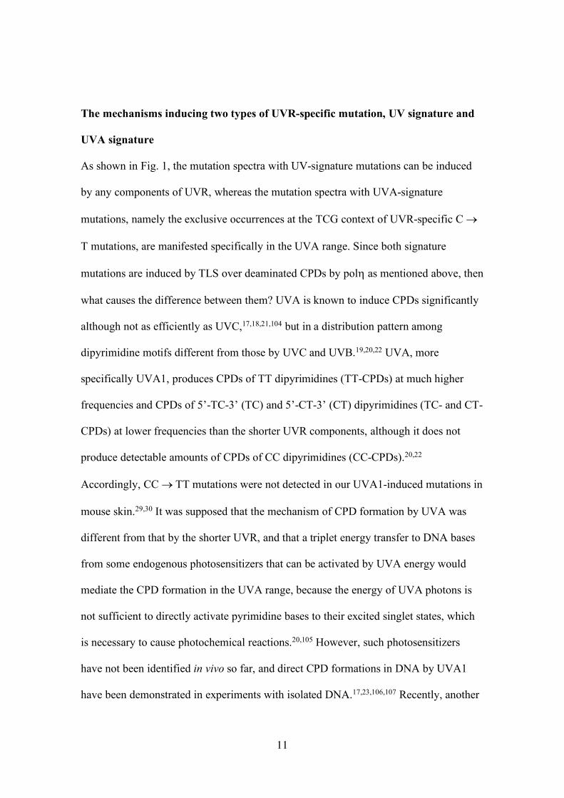
Figure legends
Fig. 1 Mutation spectra in mouse skin epidermis induced by various UVR sources. (A)
Profiles of percent spectral energy outputs of UVR sources used for my studies on
induced mutation spectra in mouse skin. UVC: germicidal lamps (GL15, Hitachi,
Japan);31 UVB: broadband UVB fluorescent lamps (FL20S.E, Toshiba, Japan);26
sunlight: summer noon sunlight in Japan;28 UVA2: blacklight fluorescent lamps
(FL20S.BLB, Toshiba, Japan) with a Mylar filter (the cut-off output is indicated by a
shaded area);27 UVA1: Sellamed 2000 (Sellas, Germany);30 and 364-nm laser (National
Institute for Basic Biology, Japan).29 (B)Mutation spectra induced in mouse epidermis
by UVB, sunlight and UVA2.26–28,53 (C) Mutation spectra induced in mouse epidermis
by UVC, UVA1 and 364-nm laser.29–31 Background is the mutation spectrum in the
epidermis of unirradiated mice.26 The tandem base substitutions are mostly CC ® TT
mutations, but those for the background and UVA1 do not include CC ® TT.26,30 PyPy,
dipyrimidine.
Fig. 2 The mechanism of UVR mutagenesis by error-free TLS across deaminated
CPDs by DNA polymerase h. UVR can produce CPDs at dipyrimidine sites (PyPy): 5’-
TT-3’, 5’-TC-3’, 5’-CT-3’ and 5’-CC-3’ (TT, TC, CT and CC). DNA polymerase h
(polh) can synthesize a DNA strand opposite a CPD on the template strand following
the base pairing rule faithfully. Thus, translesion DNA synthesis (TLS) by polh can
bypass CPDs error-free. However, cytosines in CPDs are unstable and easily deaminate
to produce uracils, or thymines if the cytosine is methylated at position 5, converting a
cytosine or 5-methylcytosine-containing CPD (C-CPD or mC-CPD) to an uracil or
Page 38
37
thymine-containing CPD (U-CPD or T-CPD), which can induce the UV-signature
mutations upon the “error-free” TLS by polh, although polh could bypass CPDs
without inducing mutations if the deamination does not occur, as in the case of thymine
dimer (TT-CPD).
Fig. 3 Sequence context preference of the UVR-specific C ® T mutation. (A) The
distributions of the occurrence ratios (%occurrence) of the UVR-specific mutations
among triplet sequence contexts were compared among UVR sources. The occurrence
ratios were estimated as ratios of the occurrences of the UVR-specific C ® T mutations
at specific triplet contexts (shown in the box) to those at the total triplet contexts
relevant to the mutation, which are cytosine-centered, dipyrimidine-containing three-
base sequences. UVR sources are the same as those in Fig. 1. Pu, purine; Py,
pyrimidine. (B) UVB-induced distributions of the UVR-specific mutation among triplet
contexts were compared between mice with polh proficient (Polh+/+) and deficient
(Polh-/-).
Fig. 4 A model of two independent, but overlapping mechanisms of the UVR-specific
mutations of “UV signature” and “UVA signature”. The shorter (UVC/UVB) and
longer (UVA) UVR components produce CPDs differently through two distinct
photochemical reactions mediated by the singlet/triplet excitation of pyrimidine bases,
which would function at shorter wavelengths up to the UVA2 range, and the collective
excitation of double-stranded DNA, which could work throughout the whole UVR
ranges, with different distributions in dipyrimidine composition: TT > TC > CT > CC
for the former and TT >> TC > CT with CC undetectable for the latter, respectively.
Page 39
38
The CpG methylation can also enhance the CPD formation by the collective excitation
mechanism. The formation of CPD enhances the deamination of its cytosine, although
the propensity of CPD deamination depends on the sequence context it resides in,
showing the most efficient deamination in the TCG context. The “error-free” TLS by
polh across the deaminated CPDs results in the induction of the UVR-specific
mutations of the “UV signature”, C ® T and CC ® TT mutations at dipyrimidine sites,
for the CPDs produced by the UVC/UVB-provoked singlet/triplet excitation and the
“UVA signature”, the preferential induction of C ® T mutations at the TCG context,
for the CPDs by the collective excitation, which becomes prominent in the UVA range.
Graphical abstract
Based on polh-dependent TLS across deaminated CPDs, the wavelength dependence of
UVR mutations can be explained by different photochemistries of CPD formation, the
singlet/triplet and collective excitations that cause UV-signature and UVA-signature
mutations respectively.
Page 40
H. Ikehata, Fig. 1
0 50 100 150 400
C → T at PyPy
C → T at non-PyPy
T → C
G → T
Other transversions
Tandem basesubstitutions
Frameshift
Others
Background (n = 44)UVB (n = 128)Sunlight (n = 81)UVA2 (n = 83)
Mutant frequency (x 10-5)
B
0 5 10 15 20 25 30 35
C → T at PyPy
C → T at non-PyPy
T → C
G → T
Other transversions
Tandem basesubstitutions
Frameshift
Others
Background (n = 44)UVC (n = 100)364 nm laser (n = 145)UVA1 (n = 100)
Mutant frequency (x 10-5)
C
Wavelength (nm)
Rel
ativ
e en
ergy
(%)
250 300 350 400
100
50
0
UVC UVB UVA1UVA2
UVB lamp Sunlight
UVA2 lamp UVA1 lamp
UVC lamp364-nm laser
A
Page 41
H. Ikehata, Fig. 2
UVR
CPD formation
TT-CPD C-CPD, mC-CPD
Cytosine deamination
Error-free TLS by DNA polymerase h
MutationC ® T at PyPy
CC ® TT“UV signature”
U-CPD, T-CPD
No mutation
TT TC, CT, CC
Page 42
H. Ikehata, Fig. 3
5'-TCA-3'CCATCGCCGPuCPyPyCPy
%Occurrence
%Occurrence
A B
Page 43
H. Ikehata, Fig. 4
UVA
Singlet/triplet excitation Collective excitation
CPD formation at:
TT > TC > CT > CC TT >> TC > CT (CC ≈ 0)with preference for mCpG
Cytosine deamination (extremely efficient at TCG)
Error-free TLS by DNA polymerase h
Mutation C ® T at TCGC ® T at PyPy
CC ® TT
“UVA signature”“UV signature”
UVC UVB
Page 44
H. Ikehata, Graphical abstract
UVC UVB
Singlet/triplet excitation Collective excitation
TLS over C-deaminated CPDs by Polh
MutationUV-signature UVA-signature
C-containing CPDs
Based on polh-dependent TLS across deaminated CPDs, the wavelength
dependence of UVR mutations can be explained by different photochemistries
of CPD formation, the singlet/triplet and collective excitations that cause UV-
signature and UVA-signature mutations respectively.
UVC UVB UVA