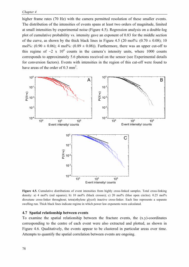
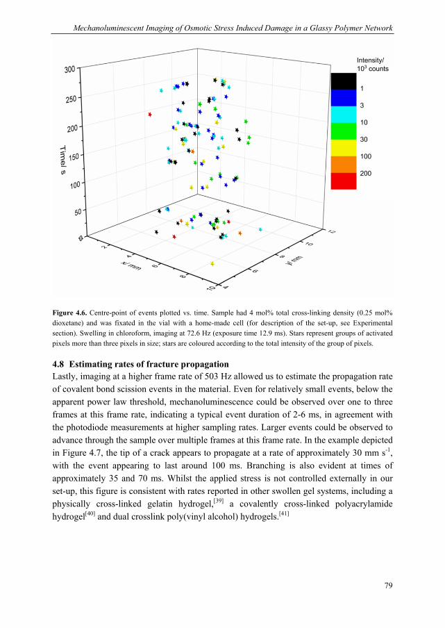
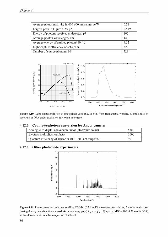
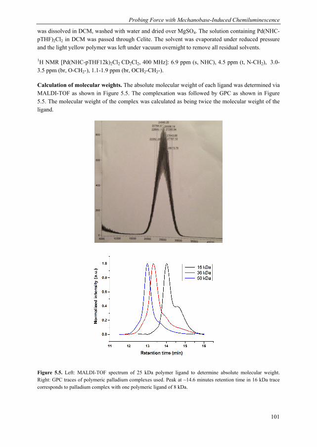
Mechanoluminescent probes in polymers Clough, J.M. Published: 15/11/2016 Document Version Publisher’s PDF, also known as Version of Record (includes final page, issue and volume numbers) Please check the document version of this publication: • A submitted manuscript is the author's version of the article upon submission and before peer-review. There can be important differences between the submitted version and the official published version of record. People interested in the research are advised to contact the author for the final version of the publication, or visit the DOI to the publisher's website. • The final author version and the galley proof are versions of the publication after peer review. • The final published version features the final layout of the paper including the volume, issue and page numbers. Link to publication Citation for published version (APA): Clough, J. M. (2016). Mechanoluminescent probes in polymers Eindhoven: Technische Universiteit Eindhoven General rights Copyright and moral rights for the publications made accessible in the public portal are retained by the authors and/or other copyright owners and it is a condition of accessing publications that users recognise and abide by the legal requirements associated with these rights. • Users may download and print one copy of any publication from the public portal for the purpose of private study or research. • You may not further distribute the material or use it for any profit-making activity or commercial gain • You may freely distribute the URL identifying the publication in the public portal ? Take down policy If you believe that this document breaches copyright please contact us providing details, and we will remove access to the work immediately and investigate your claim. Download date: 26. Apr. 2018
Transcript
Mechanoluminescent probes in polymers
Clough, J.M.
Published: 15/11/2016
Document VersionPublisher’s PDF, also known as Version of Record (includes final page, issue and volume numbers)
Please check the document version of this publication:
• A submitted manuscript is the author's version of the article upon submission and before peer-review. There can be important differencesbetween the submitted version and the official published version of record. People interested in the research are advised to contact theauthor for the final version of the publication, or visit the DOI to the publisher's website.• The final author version and the galley proof are versions of the publication after peer review.• The final published version features the final layout of the paper including the volume, issue and page numbers.
Link to publication
Citation for published version (APA):Clough, J. M. (2016). Mechanoluminescent probes in polymers Eindhoven: Technische Universiteit Eindhoven
General rightsCopyright and moral rights for the publications made accessible in the public portal are retained by the authors and/or other copyright ownersand it is a condition of accessing publications that users recognise and abide by the legal requirements associated with these rights.
• Users may download and print one copy of any publication from the public portal for the purpose of private study or research. • You may not further distribute the material or use it for any profit-making activity or commercial gain • You may freely distribute the URL identifying the publication in the public portal ?
Take down policyIf you believe that this document breaches copyright please contact us providing details, and we will remove access to the work immediatelyand investigate your claim.
ter verkrijging van de graad van doctor aan de Technische Universiteit Eindhoven, op gezag van de rector magnificus prof.dr.ir. F.P.T. Baaijens, voor een commissie aangewezen door het College voor Promoties, in het
openbaar te verdedigen op dinsdag 15 november 2016 om 16:00 uur
door
Jessica Megan Clough
geboren te Luton, Verenigd Koninkrijk
Dit proefschrift is goedgekeurd door de promotoren en de samenstelling van de promotiecommissie is als volgt: voorzitter: prof.dr.ir. J. C. Schouten 1e promotor: prof.dr. R. P. Sijbesma copromotor: dr.ir. S. J. Meskers leden: prof.dr. S. L. Craig (Duke University) prof.dr. C. Creton (ESPCI ParisTech) prof.dr. A. P. H. Schenning adviseur: dr. K. G. Blank (Max Planck Institute Potsdam)
Het onderzoek of ontwerp dat in dit proefschrift wordt beschreven is uitgevoerd in overeenstemming met de TU/e Gedragscode Wetenschapsbeoefening.
For Grandad and Milo
Two energetic players of tug
Cover design: Jessica Clough Mechanoluminescence emission from dioxetane-functionalised silica-filled poly(dimethylsiloxane) (PDMS) upon compression with metal stamps by hand or by drawing on the polymeric surface with a pencil. Recorded with Andor iXon Ultra 888 EM-CCD camera fitted with micro-Nikkor lens (40 mm, f/2.8) at 0.5 fps and electron multiplication factor of 500. Printed by: Gildeprint – the Netherlands A catalogue record is available from the Eindhoven University of Technology Library ISBN: 978-94-6233-417-5 This work has been financially supported by the Council for Chemical Sciences of the Netherlands Organisation for Scientific Research (CW-NOW grant number 726.011.002), by the Ministry of Education, Culture and Science of the Netherlands (Gravity program 024.001.035) and by the NSF (CHE-1124694 and CHE-1508566).
Table of Contents Chapter 1 Mechanochemical Stress-Sensing in Polymers .................................................................. 1
1.1 Mechanochemistry: using force to activate chemical reactions ........................... 2 1.2 Methods for mechanically activating covalent bonds .......................................... 3 1.3 Mechanically activated bond formation ............................................................... 6 1.4 Mechanophores for optical stress-sensing .......................................................... 10
1.5 Aim and outline of this thesis ............................................................................. 17 1.6 References .......................................................................................................... 18
Chapter 2 Photophysical Determination of the Excited State Products from Mechanically Induced Dioxetane Scission................................................................................................................................ 23
2.1 Introduction ........................................................................................................ 24 2.2 Outline of experimental approach: sonication and acceptor selection ............... 25 2.3 Sensitisation of singlet and triplet acceptor dyes................................................ 27 2.4 Kinetic modelling ............................................................................................... 30 2.5 Discussion .......................................................................................................... 32 2.6 Conclusions ........................................................................................................ 34 2.7 Acknowledgements ............................................................................................ 35 2.8 Experimental Details .......................................................................................... 35
2.8.1 Materials .................................................................................................... 35 2.8.2 Sonication tests .......................................................................................... 37 2.8.3 Spectral responsivity of photodiode .......................................................... 37 2.8.4 General form of photodiode response ........................................................ 38 2.8.5 Kinetic modeling ....................................................................................... 39 2.8.6 Luminol calibration ................................................................................... 41
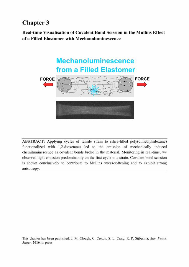
2.9 References .......................................................................................................... 41 Chapter 3 Real-time Visualisation of Covalent Bond Scission in the Mullins Effect of a Filled Elastomer with Mechanoluminescence ............................................................................ 43
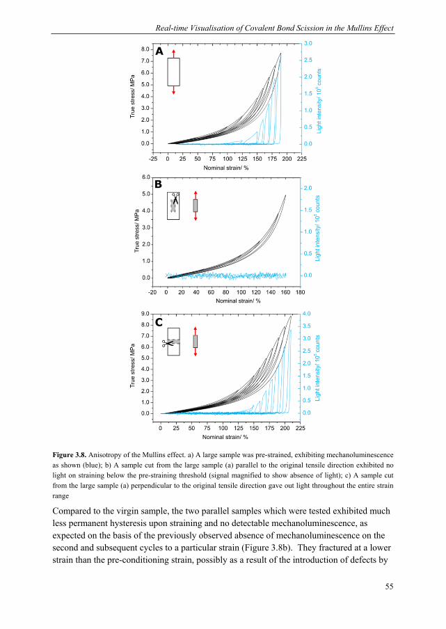
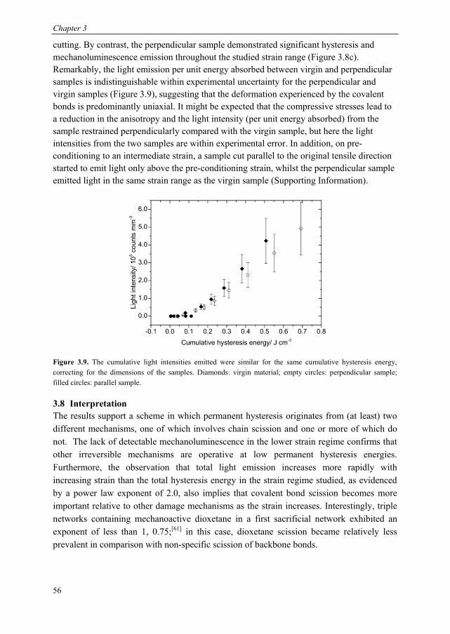
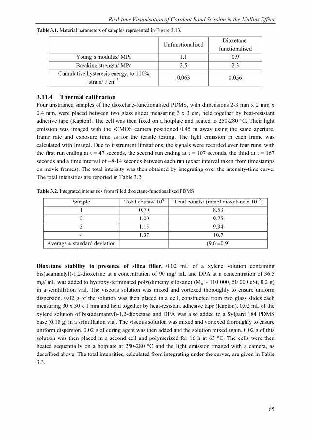
3.1 Introduction ........................................................................................................ 44 3.2 Dioxetane incorporation in silica-filled PDMS .................................................. 47 3.3 Mechanical properties dioxetane-functionalised filled PDMS ........................... 48 3.4 Form of mechanoluminescence on application of tensile cycles ........................ 49 3.5 Relationship between light intensity and hysteresis energy ............................... 52 3.6 Quantification of covalent bonds broken with thermal calibration .................... 53
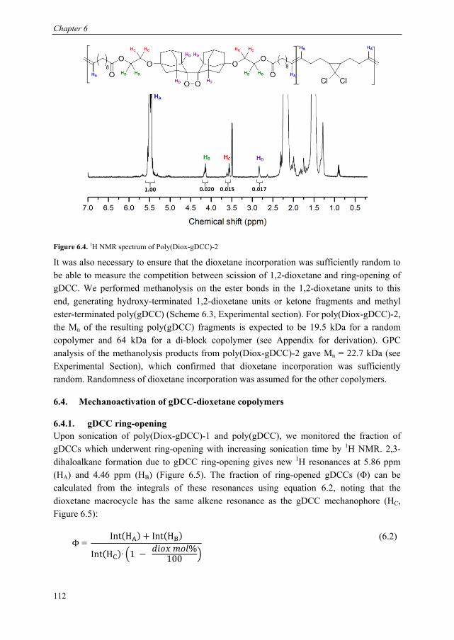
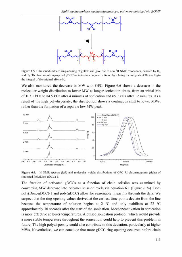
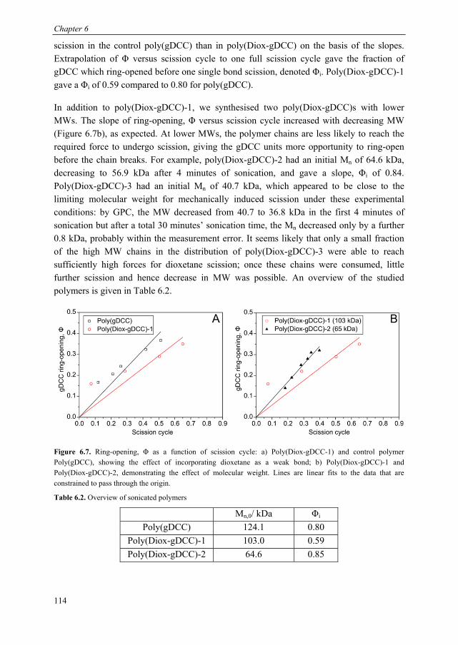
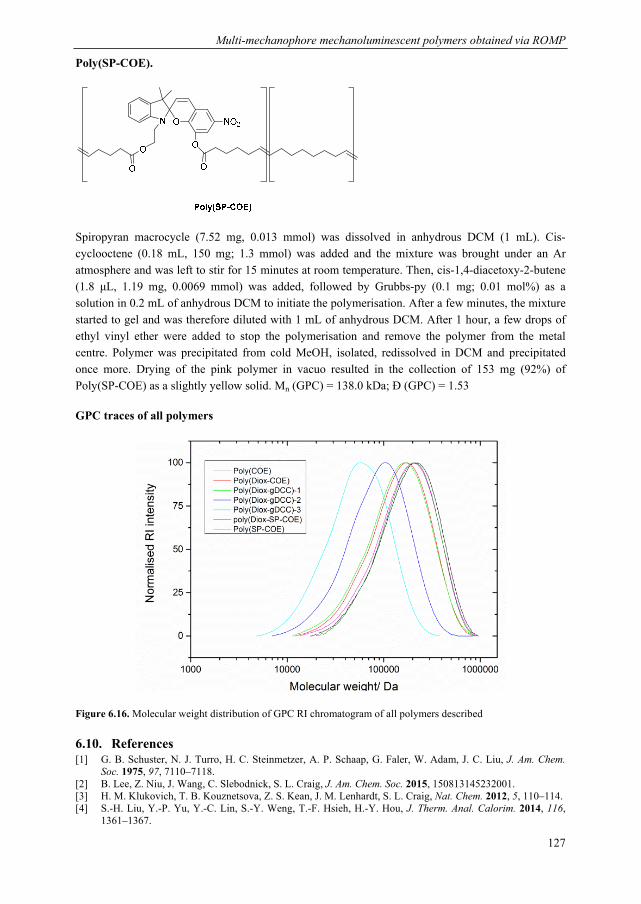
6.5. Calculation of forces at break of gDCC-dioxetane copolymers ....................... 116 6.6. Synthesis of spiropyran-dioxetane copolymers ................................................ 117 6.6. Mechanoactivation of spiropyran-dioxetane copolymers ................................. 118
6.10. References .................................................................................................... 127 Summary .......................................................................................................................... 131 Curriculum Vitae ............................................................................................................. 135 List of Publications .......................................................................................................... 136 Acknowledgements .......................................................................................................... 137
Chapter 1
Mechanochemical Stress-Sensing in Polymers
ABSTRACT: Over the past century, polymers have become ubiquitous in modern life and in many of their applications, they are expected to be able to withstand mechanical stress. Generally, the application of mechanical forces to polymers leads to irreversible damage and deterioration in the material properties, but chemists working in the resurgent field of polymer mechanochemistry have developed approaches to make use of these forces for productive chemistry. In this chapter, some fundamental aspects of polymer mechanochemistry are addressed and examples of mechanoresponsive materials that have emerged from this field are outlined, with a focus on applications in optical stress-sensing.
Parts of this chapter have been published: J. M. Clough, A. Balan, R. P. Sijbesma, Top. Curr. Chem. 2015, 369, 209; R. Göstl, J. M. Clough, R. P. Sijbesma in Mechanochemistry of Materials (Eds.: S. L. Craig, Y. C. Simon), RSC Publishing, to be published
Chapter 1
2
1.1 Mechanochemistry: using force to activate chemical reactions Chemical reactions often require an energetic stimulus to overcome the activation barrier between the starting materials and the products. This stimulus has traditionally been supplied by heat, light or an electric field. In the field of mechanochemistry, chemists use energy from an applied mechanical force to promote useful chemical transformations. Whilst not as well-known, mechanoactivation of chemical bonds has a documented history reaching back to Ancient Greece, when a student of Aristotle in 315 BC described the reduction of the mineral cinnabar to mercury upon grinding with a copper mortar and pestle.[1] In the early 21st century, mechanochemistry is a thriving discipline bridging a broad range of fields, such as mineralogy,[2] inorganic and organic synthesis,[3] polymer science[4] and biochemistry.[5] In particular, polymers present an attractive platform for the study of mechanical effects on chemical bonds: the typical macroscopic forces applied to polymers can lead to the build-up of forces on the molecular level that are sufficient to break even covalent bonds, ensuring plentiful activation; furthermore, polymeric systems offer the potential to pull or push specific nuclei via choice of polymer attachment point, affording a level of control over chemical reactivity that is impossible with more traditional forms of activation.
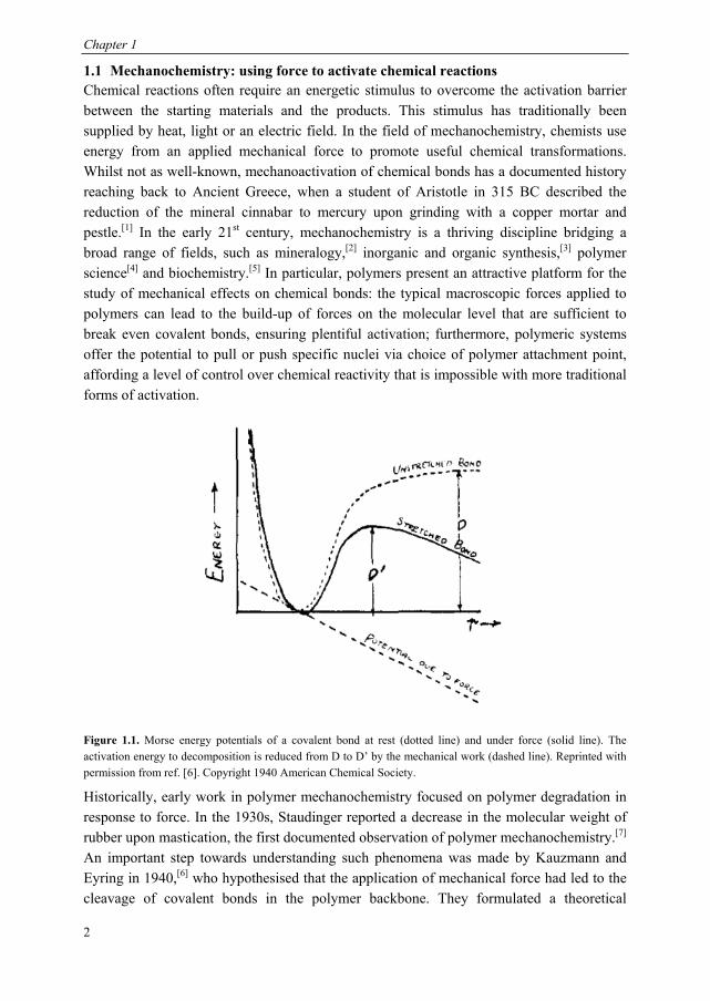
Figure 1.1. Morse energy potentials of a covalent bond at rest (dotted line) and under force (solid line). The activation energy to decomposition is reduced from D to D’ by the mechanical work (dashed line). Reprinted with permission from ref. [6]. Copyright 1940 American Chemical Society.
Historically, early work in polymer mechanochemistry focused on polymer degradation in response to force. In the 1930s, Staudinger reported a decrease in the molecular weight of rubber upon mastication, the first documented observation of polymer mechanochemistry.[7] An important step towards understanding such phenomena was made by Kauzmann and Eyring in 1940,[6] who hypothesised that the application of mechanical force had led to the cleavage of covalent bonds in the polymer backbone. They formulated a theoretical
Mechanochemical Stress-Sensing in Polymers
3
framework to express the effect of mechanical force on the rates of activated chemical reactions, such as covalent bond scission. Under zero force, the potential energy of a
chemical bond is described by a Morse potential, with a dissociation energy, D as
depicted in Figure 1.1. On the application of an external force Fext, the bond is stretched
from its equilibrium position, to a new position, ′ and the potential energy is lowered by
the work of the external force, . The stretched bond is described by a
new Morse potential, ′ , with a decreased activation barrier, D’. Thermally activated
bond scission (TABS) theory postulates that when D is lowered sufficiently by mechanical work, the bond may overcome its barrier to dissociation through thermal fluctuations (kBT ~ 2.5 kJ mol-1 at 25 °C). More refined theoretical approaches have been developed, such as the Bell-Evans and tilted potential models,[8] but they retain the core idea that the potential energy surface of a bond or reaction is altered depending on the total work done by applying a force to the molecule along a particular direction.
The past decade has seen chemists and material scientists beginning to make use of the large macroscopic forces endured by polymers for constructive ends. A flurry of publications has detailed the exploration of many new concepts, such as the incorporation of specific functionalities (“mechanophores”) in the main chain that break selectively on application of force;[9] the use of supramolecular polymers as reversible “force mediators”[10] and the activation of unique mechanochemical reaction pathways, giving rise to reactions that are not promoted by thermal or photochemical activation.[11][12] This chapter first outlines the methods typically used to apply force in a controlled manner to polymeric materials, before discussing the range of constructive responses to these forces developed within this field. We focus on self-reinforcement of materials following damage via mechanochemical processes that lead to the net formation of chemical bonds and early stage detection of damage in polymers with high sensitivity, both of which are particularly relevant to material science applications.
1.2 Methods for mechanically activating covalent bonds Chemists and materials scientists wishing to examine the mechanical reactivity of particular bonds or groups in a polymeric system have a variety of techniques at their disposal, in both solution, the solid state and at the single molecule level (Figure 1.2). They differ significantly in the maximum attainable strain rates and forces. Ultrasound sonication of dilute polymer solutions is popularly employed in the field of polymer mechanochemistry as a first screening for new mechanophores and in more fundamental investigations of mechanoreactivity. Relatively high strain rates of 106-107 s-1 are accessible, which allows mechanical activation to be obtained in polymers of lower molecular weight and with greater scission rates, compared with other solution-based techniques.
Chapter 1
4
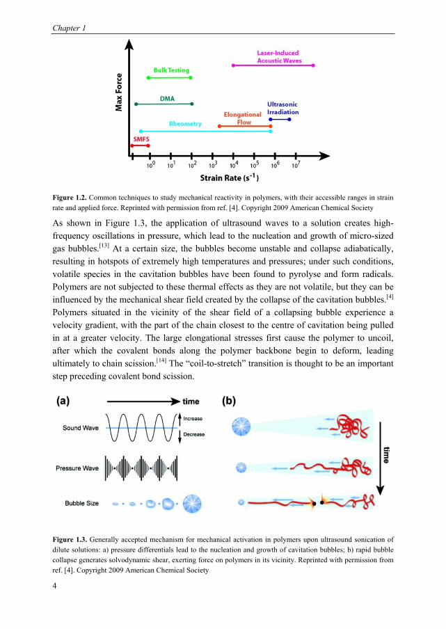
Figure 1.2. Common techniques to study mechanical reactivity in polymers, with their accessible ranges in strain rate and applied force. Reprinted with permission from ref. [4]. Copyright 2009 American Chemical Society
As shown in Figure 1.3, the application of ultrasound waves to a solution creates high-frequency oscillations in pressure, which lead to the nucleation and growth of micro-sized gas bubbles.[13] At a certain size, the bubbles become unstable and collapse adiabatically, resulting in hotspots of extremely high temperatures and pressures; under such conditions, volatile species in the cavitation bubbles have been found to pyrolyse and form radicals. Polymers are not subjected to these thermal effects as they are not volatile, but they can be influenced by the mechanical shear field created by the collapse of the cavitation bubbles.[4] Polymers situated in the vicinity of the shear field of a collapsing bubble experience a velocity gradient, with the part of the chain closest to the centre of cavitation being pulled in at a greater velocity. The large elongational stresses first cause the polymer to uncoil, after which the covalent bonds along the polymer backbone begin to deform, leading ultimately to chain scission.[14] The “coil-to-stretch” transition is thought to be an important step preceding covalent bond scission.
Figure 1.3. Generally accepted mechanism for mechanical activation in polymers upon ultrasound sonication of dilute solutions: a) pressure differentials lead to the nucleation and growth of cavitation bubbles; b) rapid bubble collapse generates solvodynamic shear, exerting force on polymers in its vicinity. Reprinted with permission from ref. [4]. Copyright 2009 American Chemical Society
Mechanochemical Stress-Sensing in Polymers
5
Several fundamental features of mechanical reactivity have emerged from such experiments. Firstly, scission occurs preferentially around the mid-point of the polymer chain, as the solvodynamic forces are greatest at this point.[15] Consequently, chemists seeking to understand the reactivity of a particular bond or group incorporate it at the centre of a polymer chain, where the probability of chain scission and mechanoactivation are highest. This behaviour contrasts with that on thermal activation, in which scission occurs in a random fashion. Secondly, the rate of mechanoactivation exhibits a characteristic dependence upon the degree of polymerisation or alternatively the contour length of the polymer,[16,17] although molecular weight is commonly used to describe this relationship. The greater the chain length of the polymer, the longer the relaxation time of the polymer chain and hence the lower the strain rates required to induce a coil-to-stretch transition, giving greater rates of mechanical activation. Furthermore, below a certain chain length, bond scission and mechanoactivation do not occur to a detectable extent as the strain rates required for chain scission are higher than those which can be provided by sonication.[18] This limiting length, often expressed in terms of the molecular weight for different polymers, Mlim, is empirically derived by plotting rate constants of scission against molecular weight and extrapolating to a rate constant of zero.[19] Incorporation of a weak bond into a polymer backbone significantly increases the rate of scission and decreases Mlim, as observed with weak bonds such as peroxides,[19] azo links[20] and metal-ligand coordination bonds.[21,22] These studies also reported remarkably selectivity for the weakest bond, which underwent scission almost exclusively. Weak bonds like these are often referred to as “mechanophores” when intentionally built into polymer backbones for their latent mechanical reactivity.
Apart from sonication, other solution-based techniques are used to study polymer degradation,[23] of which the best known are cross slots, extensively investigated by Odell and Keller,[24] and contraction flows, as studied by Nguyen and Kausch.[25] The geometry of the flow field in a cross slots set-up creates a region of zero velocity, known as the stagnation point; here, polymers become trapped and experience a large velocity gradient, leading to elongation and chain scission. In contraction flow, a polymer solution is driven through a narrow contraction by a pressure differential, leading to a sudden acceleration of the fluid. The set-up for contraction flow is simpler and can give higher scission yields in a single pass, but cross slots permits more precise control over the strain rates experienced by the polymers. Generally, however, mechanoactivation is not as efficient as under ultrasound sonication, and high MW polymers are required (typically 105-106 Da). For these reasons, these set-ups are not as widely used to study mechanochemical reactivity in polymers. Nevertheless, microfluidic devices capable of creating similar types of shear field have broadened the appeal of flow chemistry to study the mechanical behaviour of macromolecules,[26] particularly in the biophysical field.[27,28]
Mechanoactivation in solid state polymeric materials is achieved predominantly in compression, shear or tension, the last being employed in Chapter 3 of this thesis, but has
Chapter 1
6
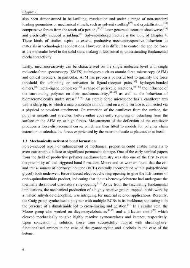
also been demonstrated in ball-milling, mastication and under a range of non-standard loading geometries or mechanical stimuli, such as solvent swelling[29] and crystallisation,[30] compressive forces from the touch of a pen or ,[31,32] laser-generated acoustic shockwaves[33] and electrically induced wrinkling.[34] Solvent-induced fracture is the topic of Chapter 4. These kinds of studies aspire to extend productive mechanoresponsive behaviour to materials in technological applications. However, it is difficult to control the applied force at the molecular level in the solid state, making it less suited to understanding fundamental mechanoreactivity.
Lastly, mechanoreactivity can be characterised on the single molecule level with single molecule force spectroscopy (SMFS) techniques such as atomic force microscopy (AFM) and optical tweezers. In particular, AFM has proven a powerful tool to quantify the force threshold for unbinding or activation in ligand-receptor pairs,[35] hydrogen-bonded dimers,[36] metal-ligand complexes[37] a range of pericyclic reactions,[38–40] the influence of the surrounding polymer on their mechanoactivity,[41–43] as well as the behaviour of biomacromolecules under stress.[44–46] An atomic force microscope has a cantilever arm with a sharp tip, to which a macromolecule immobilised on a solid surface is connected via a physical or covalent attachment. On retraction of the cantilever from the surface, the polymer uncoils and stretches, before either covalently rupturing or detaching from the surface or the AFM tip at high forces. Measurement of the deflection of the cantilever produces a force-displacement curve, which are then fitted to models for polymer chain extension to calculate the forces experienced by the macromolecule at plateaus or at break.
1.3 Mechanically activated bond formation Force-induced repair or enhancement of mechanical properties could enable materials to avert catastrophic failure or significant permanent damage. One of the early seminal papers from the field of productive polymer mechanochemistry was also one of the first to raise the possibility of load-triggered bond formation. Moore and co-workers found that the cis- and trans-isomers of benzocyclobutene (BCB) centrally incorporated within poly(ethylene glycol) both underwent force-induced electrocyclic ring-opening to give the E,E-isomer of ortho-quinodimethide product, indicating that the cis-benzocyclobutene had undergone the thermally disallowed disrotatory ring-opening.[11] Aside from the fascinating fundamental implications, the mechanical production of a highly reactive group, trapped in this work by a maleic anhydride dienophile, was intriguing for material science applications. Recently, the Craig group synthesised a polymer with multiple BCBs in its backbone; sonicating it in the presence of a dimaleimide led to cross-linking and gelation.[47] In a similar vein, the Moore group also worked on dicyanocyclobutanes[48,49] and a β-lactam motif[50] which cleaved mechanically to give highly reactive cyanoacrylates and ketenes, respectively. Upon sonication in solution, these were successfully trapped with chromophore-functionalised amines in the case of the cyanoacrylate and alcohols in the case of the ketene.
Mechanochemical Stress-Sensing in Polymers
7
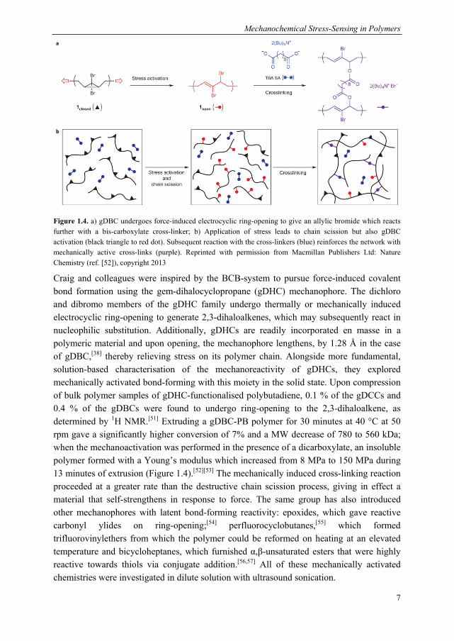
Figure 1.4. a) gDBC undergoes force-induced electrocyclic ring-opening to give an allylic bromide which reacts further with a bis-carboxylate cross-linker; b) Application of stress leads to chain scission but also gDBC activation (black triangle to red dot). Subsequent reaction with the cross-linkers (blue) reinforces the network with mechanically active cross-links (purple). Reprinted with permission from Macmillan Publishers Ltd: Nature Chemistry (ref. [52]), copyright 2013
Craig and colleagues were inspired by the BCB-system to pursue force-induced covalent bond formation using the gem-dihalocyclopropane (gDHC) mechanophore. The dichloro and dibromo members of the gDHC family undergo thermally or mechanically induced electrocyclic ring-opening to generate 2,3-dihaloalkenes, which may subsequently react in nucleophilic substitution. Additionally, gDHCs are readily incorporated en masse in a polymeric material and upon opening, the mechanophore lengthens, by 1.28 Å in the case of gDBC,[38] thereby relieving stress on its polymer chain. Alongside more fundamental, solution-based characterisation of the mechanoreactivity of gDHCs, they explored mechanically activated bond-forming with this moiety in the solid state. Upon compression of bulk polymer samples of gDHC-functionalised polybutadiene, 0.1 % of the gDCCs and 0.4 % of the gDBCs were found to undergo ring-opening to the 2,3-dihaloalkene, as determined by 1H NMR.[51] Extruding a gDBC-PB polymer for 30 minutes at 40 °C at 50 rpm gave a significantly higher conversion of 7% and a MW decrease of 780 to 560 kDa; when the mechanoactivation was performed in the presence of a dicarboxylate, an insoluble polymer formed with a Young’s modulus which increased from 8 MPa to 150 MPa during 13 minutes of extrusion (Figure 1.4).[52][53] The mechanically induced cross-linking reaction proceeded at a greater rate than the destructive chain scission process, giving in effect a material that self-strengthens in response to force. The same group has also introduced other mechanophores with latent bond-forming reactivity: epoxides, which gave reactive carbonyl ylides on ring-opening;[54] perfluorocyclobutanes,[55] which formed trifluorovinylethers from which the polymer could be reformed on heating at an elevated temperature and bicycloheptanes, which furnished α,β-unsaturated esters that were highly reactive towards thiols via conjugate addition.[56,57] All of these mechanically activated chemistries were investigated in dilute solution with ultrasound sonication.
Chapter 1
8
A recent addition to this class of mechanophores is the spirothiopyran moiety, developed by the Weng group.[58] Like the well-known spiropyran, it rapidly isomerises under force, giving the green thiomerocyanine in both solution and the solid state. In addition to mechanochromism, the mechanically generated thiolate can participate in thiol-ene reactions with a bismaleimide, which led to gelation in solution. This work raises the prospect of materials capable of damage-reporting and load-induced self-strengthening.
Figure 1.5. Mechanoactivation of a ruthenium catalyst for ring-opening metathesis polymerisation in the solid state. Reprinted with permission from ref. [61]. Copyright 2013 American Chemical Society
Our group has also been active in the area of force-induced bond creation, making use of a very different strategy. As first demonstrated by Kartikeyan and Piermattei, transition metal catalysts can be incorporated centrally in polymer chains, which upon mechanoactivation create new C—C bonds from appropriate substrates, with turnover numbers exceeding 800.[59] Following initial work on metal–phosphine coordination polymers, our group began investigating the mechanical dissociation of silver(I)-coordination complexes with N-heterocyclic carbene (NHC) functionalized polymers. External force from ultrasound sonication was found to selectively break Ag-NHC bonds, yielding free NHC which catalysed the transesterification of benzyl alcohol and vinyl acetate under sonication. COGEF calculations performed by Groote et al. indicated that 400-500 pN of force is required to break the Ag-NHC bond on the 10 ps timescale of the simulation, significantly lower than the force that is typically required to break covalent bonds (several nN), confirming the greater mechanical lability of this coordination bond compared with covalent bonds.[60] Parallel to this, bis-NHC ruthenium catalysts with pTHF chains proved to be mechanically latent catalysts for metathesis, both in solution upon sonication and also in the solid state under compressive strain, as shown by Jakobs et al.[61] The polymer
Mechanochemical Stress-Sensing in Polymers
9
catalyst and a norbornene monomer were incorporated in a high molecular weight poly(tetrahydrofuran) (pTHF) matrix, which provided physical cross-linking through its crystalline domains, allowing macroscopic forces to be transferred to the metal–ligand bonds (Figure 1.5). Consecutive compressions showed that up to 25% of the norbornene monomer was polymerized after five loading cycles. Similarly, the Binder group reported a copper bis-NHC click catalyst that could be activated on compression in the solid state, as visualised by a fluorescent dye.[62] Lastly, our group has also started to explore organic catalysts for polymerisation. A hexaarylbiimidazole (HABI) motif cleaved to give coloured triphenylimidazolyl radicals capable of initiating secondary radical reactions that led to polymerisation. Like in the spirothiopyran-linked polymers, optical stress-sensing is combined with load-triggered reinforcement.[63]
One last nascent approach to mechanically induced bond-forming is the use of mechanical force to effect the scission of bonds that are not part of the polymer backbone, leading to the release of small molecules, whilst maintaining the overall mechanical integrity of the polymer matrix. The range of accessible reactivities remains limited, but researchers in this fledgling area are starting to make use of these small molecule products for further reactions, including polymerisation. The first example of this type of activation was the mechanochemical generation of an acid, developed by Moore, Diesendruck et al. Inspired by Craig’s gDHC system, they incorporated gem- dichlorotetrahydrocyclopropanated indene into poly(methyl acrylate) matrix (PMA) and showed that compression resulted in ring-opening of the cyclopropane to release the elimination product, 2- chloronaphthalene and HCl.[64] The Boydston group have also described the flex, or bond-bending, activation of an oxanorbornadiene, a Diels–Alder adduct of furan and dimethyl acetylenedicarboxylate.[65] They incorporated the mechanophore into poly(methyl acrylate) (PMA) matrix and showed that the furan derivative could be released under stress applied to the bulk polymer. The main-chain alkene moieties are thus converted into alkynes, not only preserving the overall macromolecular structure but making bonds in the main chain shorter and stronger following the application of mechanical force.
Lastly, in a significant departure from the aforementioned pericyclic-based mechanophores, Diesendruck and colleagues in the Moore group were able to mechanically induce the heterolytic depolymerisation of cyclic and linear poly(ortho-phthalaldehyde) (PPA) to its constituent monomers upon sonication in dilute solution, constituting small molecule release en masse.[66] The reactive ortho-phthalaldehyde (OPA) monomers could then be repolymerised with a chemical initiator, regenerating PPA. The system represents an interesting reimagining of the mechanically induced remodeling concept, inspired in part by the recycling of monomeric building blocks such as amino acids, carbohydrates and nucleic acids in nature. An alternating copolymer of sulphur dioxide and vinyl acetate was also recently found to depolymerise in response to ultrasound sonication.[67]
Chapter 1
10
1.4 Mechanophores for optical stress-sensing The detection of strains and forces is vital in a broad range of scenarios, from the maintenance and failure prevention of man-made construction materials to the study of biological processes. Materials capable of reporting on their own stresses could substantially improve and streamline the screening of critical load-bearing components, for example. The optical feedback pathway is well-suited for this task on account of the high spatiotemporal resolution of light, which permits precise localisation of the damage, and its non-invasive character, which enables the remote visualisation of stress. Additionally, the high energetic resolution of light enables simultaneous detection of different colours.
In polymer mechanochemistry, three different types of optical response to stress have been realised: namely, a change in absorption, fluorescence generation and chemiluminescence generation. For stress-induced changes in absorption, or mechanochromism (or mechanochromogenism if the change is irreversible), a chromophore is generated or altered through mechanical stress,[68] which is readily quantifiable via the Beer-Lambert law if the molar absorptivity ε and the concentration of the corresponding chromophore are known. However, this method is the least sensitive of all, as the measurement relies on the transmission of attenuated light, i.e. a small signal against a noisy background, making it unsuitable for chromophore concentrations below 10-5 M. Mechanically induced fluorescence (mechanofluorochromism) offers much greater sensitivity in the steady-state optical detection of stress.[69] In this technique, a fluorophore is first mechanically generated; then, upon excitation, it may take a higher singlet state Sn, from which it converts to the S1 state; finally, a radiative transition can take place, i.e. a photon of lower energy is emitted, allowing the fluorophore to relax to the S0 state. The absolute intensity of emitted light is easier to detect than the relative intensity of absorbed light, such that fluorophore concentrations of 10-7 M are routinely detectable, decreasing to 10-12 M with confocal microscopy.[70] Quantifying fluorescence signals is not as straightforward, however, as the quantum yield of a fluorophore is influenced by a range of photophysical processes, such as intersystem crossing and internal conversion, and the fluorophore’s microenvironment. Lastly and of the greatest relevance to this thesis, mechanically induced chemiluminescence (mechanoluminescence) is closely related to fluorescence. The crucial difference is that the excited states of chemiluminescence are not populated through the absorption of light, but through a preceding chemical reaction. The detectability lies in the same concentration range as fluorescence but because chemiluminophores only undergo one chemical reaction before they are consumed, the excited state is populated only once for each chemiluminophore (as opposed to continually when visualising fluorescence), rendering chemiluminescence a temporally resolved, non-steady-state method.[71,72] Chemiluminescence is thus able to achieve a higher spatial resolution through the recording of successive emission events.
Mechanochemical Stress-Sensing in Polymers
11
1.4.1 Mechano(fluoro)chromism The first polymer-based mechanochromic reaction reported was based on the azobenzene moiety. The isomerisation from its E- to Z-form produces a colour-change and is also associated with a significant geometrical change that is affected by mechanical stress.[73–75] Reneker and Kim incorporated the thermodynamically stable E-isomer into linear polyurethane chains forming a physically crosslinked network; light-induced isomerisation yielded the unstable Z-isomer which could then be transformed to its E-form by tensile stress, as seen in the altered UV/vis absorption spectrum. However, the difference between colours of E- and Z-isomer is not very pronounced, making azobenzene a non-ideal optical probe for mechanical stress.
Figure 1.6. Examples of spiropyran mechanoactivation by localised compression in bulk PDMS elastomers. Reprinted with permission from ref. [31]. Copyright 2014 American Chemical Society
Perhaps the most successful mechano(fluoro)phore is the spiropyran, which isomerises to purple-coloured, red-fluorescent merocyanine (Figure 1.6). Its force-induced activation was first reported by Moore, White, Sottos and co-workers in 2007 in linear and cross-linked poly(methyl acrylate)[76,77] and it has since become a popularly employed optical probe for mechanical stress,[78] applied to polymers as diverse as poly(methyl methacrylate),[79,80] poly(ε-caprolactone),[81,82] poly(dimethylsiloxane),[31] poly(tert-butyl acrylate)-b-poly(N-isopropyl acrylamide)[83] and supramolecular polymers[84–86] (an example of which is depicted in Figure 1.6). Spiropyran has also been used to address fundamental aspects of the effect of stress on mechanical bond activation, such as those of strain hardening and flow,[87] microphase separation,[45] local temperature,[88] crystallinity[89,90] and plasticity/elasticity,[31,90–92] giving valuable insights into the isomerisation mechanism and fundamental design principles of mechanophores. Moreover, Craig and colleagues
Chapter 1
12
determined with SMFS experiments that the spiropyran to merocyanine isomerisation could be activated by as little as ~240 pN of force on the millisecond timescale of the experiment.[39] Other common covalent mechanophores require forces of 5-10 times this value, retrospectively explaining the extensive applicability of this excellent mechanophore. In a similar manner, spirolactam derivatives also undergo force-induced isomerisation to their fluorescent rhodamine forms in a polyurethane film,[32] as reported by Jia and colleagues.
Covalent bond scission reactions can also give optical feedback on stress distributions.[93] Cycloelimination reactions figure prominently in this area, with the retro Diels-Alder reaction of the anthracene-maleimide adduct being perhaps the best-known. The optically transparent Diels-Alder adduct with interrupted π-conjugation is mechanically transformed to the fully conjugated and fluorescent anthracene via the retro Diels-Alder reaction, which can be followed in absorption or fluorescence emission. The adduct was first employed by Yoshie and colleagues in 2011 in a self-mendable polymer network;[94] its mechanoreactivity was later studied in sonication solutions of linear PMA chains centrally incorporating the adduct by Makarov, Bielawski and co-workers[95] and also as an anchoring motif for polymer hairs attached to silica nanoparticles by the Moore group.[96] These studies followed anthracene release with UV/vis absorption. The fluorescence of anthracene was used to track bond cleavage by Boydston and co-workers, investigating the scission behaviour of star-shaped polymers,[97] as well as by Craig and co-workers,[98] by planarisation of the corresponding Diels-Alder adduct crosslinker in a poly(dimethylsiloxane) network.[31] Very recently, Göstl in our group enhanced the sensing capability of this motif by π-extension of the anthracene unit, generating a fluorophore with high ΦF as well as red-shifted absorption and emission spectra.[99] The fluorescence of anthracene was also used to sense microcracks not via the retro Diels-Alder reaction but by cycloelimination of anthracene dimer crosslinkers in a poly(vinyl alcohol) network.[100] Another force-induced chromo- and fluorogenic cycloelimination reaction was discovered recently by Craig and co-workers.[101] Similar to other cyclobutane mechanophores,[40,102] ultrasonication of a widely transparent coumarin dimer incorporated in the centre of linear poly(methyl acrylate) could induce the [2+2] cycloelimination and generate fluorescent coumarin chain ends.
The chromogenic homolytic cleavage of diarylbibenzofuranones in polymer networks was reported by Otsuka and colleagues, following an approach pioneered by Löwenbein and co-workers in the 1920s.[103–106] Due to rapid thermal radical recombination, their bright blue colour could only be observed initially by exerting force through freezing organic solvent-swollen networks, [30] but incorporation at a high concentration in a physically crosslinked dynamic covalent polyurethane network permitted stress visualisation at room temperature.[107]
In addition to the covalent incorporation of mechanophores, several other approaches have been developed to endow polymeric materials with (fluoro)chromic or (fluoro)chromogenic
Mechanochemical Stress-Sensing in Polymers
13
mechanoresponsivity, including the incorporation of conjugated polymeric units, the dispersion of “aggregachromic” dyes in polymers and the fixation of photonic crystals and cholesteric liquid crystals by polymerisation, which are now discussed in turn.
Conjugated polymers can exhibit mechanically induced fluorochromism and fluorochromogenic transitions, including poly(p-phenylene-ethynylene)[108] and poly(fluorene).[109–111] Poly(diacetylene)s (PDAs) are perhaps the archetypal example in this area.[112–114] π-delocalisation along the alternating ene-yne backbone gives rise to a strong optical absorption from the π-π* transition at ~640 nm, which is generally blue-shifted to ~540 nm following a chromic or chromogenic transition, causing the material’s colour to change from blue to red or yellow. The activated red-state is also strongly fluorescent, whereas in the starting blue-state fluorescence is symmetry-forbidden. It is thought that the backbone of the polymer is formed in a strained state during polymerisation and the stimulated optical changes arise from the relaxation of the backbone into a lower energy conformation via rotation about the C—C bonds. Mechanical force was first used to activate this transition in a polymer by Nallicheri and Rubner.[115] They synthesized segmented polyurethanes containing diacetylenes in their hard blocks which could be polymerized in the solid state; upon the application of tensile stress, the PDAs underwent a chromic phase transition which was reversible up to 350%. Our group employed a similar approach in thermoplastic elastomers of copoly(ether urea).[116] More recently, the chromic transitions of PDA have been activated by a diverse selection of mechanical stimuli, including the mechanical force from an oscillating tuning fork in nanowires of poly(ethylene oxide) (PEO);[117] the swelling of PDMS with aliphatic hydrocarbons[118] and in two-dimensional PDA monolayers, upon changes to the lateral surface pressure of a monolayer in a Langmuir-Blodgett trough[119] and by scratching a monolayer surface with a scanning force microscopy tip.[120,121]
The physical incorporation of chromophores in polymeric matrices has also been applied to the force-sensing quite recently.[122–124] These so-called “aggregachromic” (in most cases more correctly aggregachromogenic) dyes are generally flat, rigid, aromatic molecules, with electron-releasing or electron-withdrawing groups conjugated to the π-system, promoting their self-assembly via π-π and electrostatic interactions into micro- or nano-sized aggregates during processing. The application of force disrupts these supramolecular interactions, breaking up the aggregates and leading to significant optical changes in the material. One of the first and most successful aggregachromic dyes were the oligo(phenylene vinylene)s (OPVs), pioneered by the Weder group. On dispersing OPVs bearing cyano groups in low density polyethylene (LLDPE), the dye molecules formed excimers above a certain concentration of dye, giving rise to a broad excimer emission band at ~644 nm in addition to the monomer emission peaks at 491 nm and 536 nm.[125] The application of tensile strain to these polymer blends greatly reduced the intensity of the excimer band, indicating break-up of the stacks and molecular dispersion of the monomeric dyes. A melt-processed blend of LLDPE and 0.2 wt% 1,4-bis(R-cyano-4-methoxystyryl)-
Chapter 1
14
2,5-dimethoxybenzene (BCMDB) exhibited measurable aggregate disruption at strains as low as 10% and by 500%, the OPV molecules were almost completely dispersed within the matrix.[126] OPVs have since been extended to semi-crystalline PE, [126–128] poly(vinylidene fluorides)[129], polyesters[130,131] and polyurethanes.[132] The Pucci group has worked on bis(benzoxaloyl)stilbene (BBS) and perylene derivatives, which operate on a similar principle. [133–136] Very recently, dyes that display aggregation induced emission (AIE) have just started to be applied as force sensors in polymers.[137] In the free state, dyes in this class undergo intramolecular motions that inhibit their fluorescence, but in the aggregated state, these motions are restricted, allowing fluorescence to occur, giving ‘on’/’off’ switching behaviour. The first systems have made use of cyanostilbene derivatives[138] and tetraphenylethylene,[139] although currently their applicability remains limited.
Photonic materials constitute a very particular group of mechanoresponsive, chromogenic materials. In polymeric systems they are either generated by fixation of photonic crystals through polymerisation or directly by self-assembly of block-copolymers bearing blocks of different dielectric properties. Mechanical deformation of the photonic material then changes the optical path length and thus the stopband’s exclusion wavelength, leading to a colour-change.[140–144] This phenomenon has been demonstrated by the Gong group in a self-assembled poly(dodecylglyceryl itaconate)-b-poly(acrylamide) matrix[145,146] and by Chen and co-workers in self-assembled monodisperse magnetic colloid nanocrystal clusters[147] and other works.[148,149] Cholesteric liquid crystals (CLCs) are in effect 1D photonic bandgap materials and can also respond chromically to force. They consist of rod-like molecules (mesogens) arranged in a helical order, through which circularly polarised light within a particular wavelength range cannot propagate and is instead reflected. CLCs fixated in a polymer were employed to create mechanically tunable lasing materials, as pioneered by Finkelman[150,151] and others.[152–154]
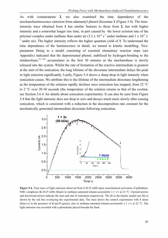
1.4.2 Mechanically induced chemiluminescence Alongside efforts to develop force-induced chromo- or fluorogenic polymeric materials, our group has established mechanically induced chemiluminescence (mechanoluminescence) as an excellent method to detect strain in polymeric materials. In this strategy, thermally stable bis(adamantyl)-1,2-dioxetane is incorporated in a polymer chain or network and upon the application of force, the dioxetane group decomposes via a cycloelimination to yield two ketones under the emission of light (Scheme 1.1). This phenomenon was first demonstrated in 2012 from dioxetane-functionalised poly(methyl acrylate), both in bulk samples placed under tension and in solutions of linear polymers upon sonication.[155] The advantages bestowed by the transient nature of mechanoluminescence were evident even in this first study, allowing both the location and temporal progression of bond scission to be visualised with high sensitivity.
Mechanochemical Stress-Sensing in Polymers
15
Scheme 1.1. Top: thermally induced mechanoluminescence from bis(adamantyl)-1,2-dioxetane, first discovered by Wieringa et al.[156] At 200 °C, bis(adamantyl)-1,2-dioxetane has a half-life of approximately 100 seconds.[157]
Bottom: on incorporating in a polymer, chemiluminescence from bis(adamantyl)-1,2-dioxetane can be induced mechanically, as first reported by Chen et al.[155]
Employing this work as foundation, dioxetane mechanoluminescence shed light on two new approaches to toughening polymer networks, both of which enhance a material’s ability to dissipate strain energy before fracture. The first strategy, devised by the Creton group, entails the incorporation of second and third networks in the first elastomer network via sequential free radical polymerisation of simple acrylates, forming a multiple network elastomer. In single edge notch tests on dioxetane-functionalised multiple networks (with the dioxetane incorporated in the first network), light emission was highly localised at the crack tip for the single network, becoming more intense for the double network and for the triple network, a large yielding zone could be visualised over an extended region ahead of the crack tip (Figure 1.7).[158] Mechanoluminescence thus confirmed the significance of the first and second networks as sacrificial stress-bearers, dissipating energy prior to material failure. Furthermore, the mechanoluminescence traces were rich in information about the yielding zone, revealing its extent and shape, which could be controlled by the extensibility of the second network chains.
A second reinforcement strategy, investigated by the groups of Craig and Sijbesma, was based on the Craig group’s prior study on poly(vinyl pyridine) networks cross-linked with bifunctional van Koten-type palladium or platinum complexes (Figure 1.8a). The pincer complexes coordinate reversibly to the free pyridines, resulting in weak, transient supramolecular interactions which control the bulk dynamic properties of the gel. By varying the central metal ion within the complex or by making small structural changes to the ligands, the lifetime of the supramolecular interaction can be controlled. In such a system, the supramolecular cross-links have a stress-homogeneising effect, boosting the fracture energy, but do not significantly affect the modulus and structure of the material. In a series of such gels studied under compression, the dioxetane mechanophore demonstrated that covalent bond scission was indeed inhibited by the addition of reversible cross-linkers to the network, with higher concentrations of supramolecular cross-linkers delaying the onset of mechanoluminescence to a greater extent (Figure 1.8b).[10] This approach to material design, whereby fracture toughness is improved independently of the elastic modulus, is of particular interest to the development of materials where a high modulus is a disadvantage, for example, in biomaterials.
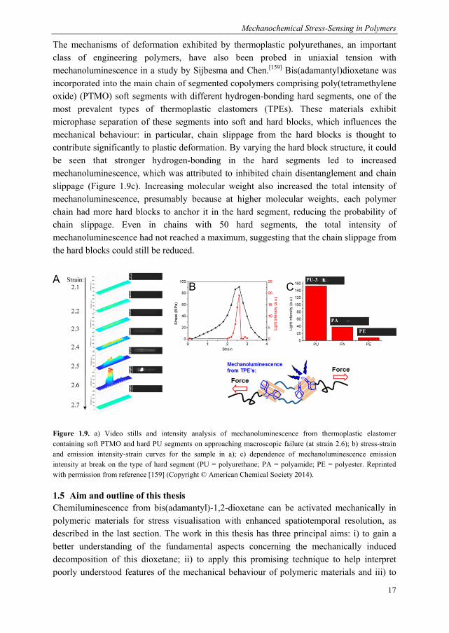
The mechanisms of deformation exhibited by thermoplastic polyurethanes, an important class of engineering polymers, have also been probed in uniaxial tension with mechanoluminescence in a study by Sijbesma and Chen.[159] Bis(adamantyl)dioxetane was incorporated into the main chain of segmented copolymers comprising poly(tetramethylene oxide) (PTMO) soft segments with different hydrogen-bonding hard segments, one of the most prevalent types of thermoplastic elastomers (TPEs). These materials exhibit microphase separation of these segments into soft and hard blocks, which influences the mechanical behaviour: in particular, chain slippage from the hard blocks is thought to contribute significantly to plastic deformation. By varying the hard block structure, it could be seen that stronger hydrogen-bonding in the hard segments led to increased mechanoluminescence, which was attributed to inhibited chain disentanglement and chain slippage (Figure 1.9c). Increasing molecular weight also increased the total intensity of mechanoluminescence, presumably because at higher molecular weights, each polymer chain had more hard blocks to anchor it in the hard segment, reducing the probability of chain slippage. Even in chains with 50 hard segments, the total intensity of mechanoluminescence had not reached a maximum, suggesting that the chain slippage from the hard blocks could still be reduced.
1.5 Aim and outline of this thesis Chemiluminescence from bis(adamantyl)-1,2-dioxetane can be activated mechanically in polymeric materials for stress visualisation with enhanced spatiotemporal resolution, as described in the last section. The work in this thesis has three principal aims: i) to gain a better understanding of the fundamental aspects concerning the mechanically induced decomposition of this dioxetane; ii) to apply this promising technique to help interpret poorly understood features of the mechanical behaviour of polymeric materials and iii) to
Chapter 1
18
extend the available range of mechanoluminescent detection systems, so that lower forces could ultimately be visualised and greater sensitivities obtained.
In Chapter 2, the excited products of the mechanical decomposition pathway of bis(adamantyl)-1,2-dioxetane are characterised by sonicating dilute solutions of poly(methyl acrylate) containing a centrally incorporated dioxetane unit in the presence of either singlet or triplet acceptors. In addition to its fundamental interest, this study provides information needed for relating mechanoluminescence intensity to the numbers of bonds broken in a polymeric material.
Chapters 3 and 4 show the bis(adamantyl)-1,2-dioxetane being applied as a stress-probe to elucidate complex mechanical behaviour of solid state polymeric materials. In Chapter 3, dioxetane is incorporated in silica-filled poly(dimethylsiloxane) to investigate the contribution of covalent bond scission to the much-debated Mullins effect, which refers to the permanent stress-softening on the first extension. It is found that covalent bond scission occurs to a significant extent only upon Mullins-type stress-softening and in a highly anisotropic manner. The presence of other damage mechanisms is also inferred and an approximate calibration is described. Chapter 4 considers the mechanoluminescence induced by the solvent swelling of cross-linked poly(methyl methacrylate). Discrete bursts of light indicate cascades of covalent bond scission, taking place over many milliseconds and involving billions of bonds, the propagation of which can be imaged.
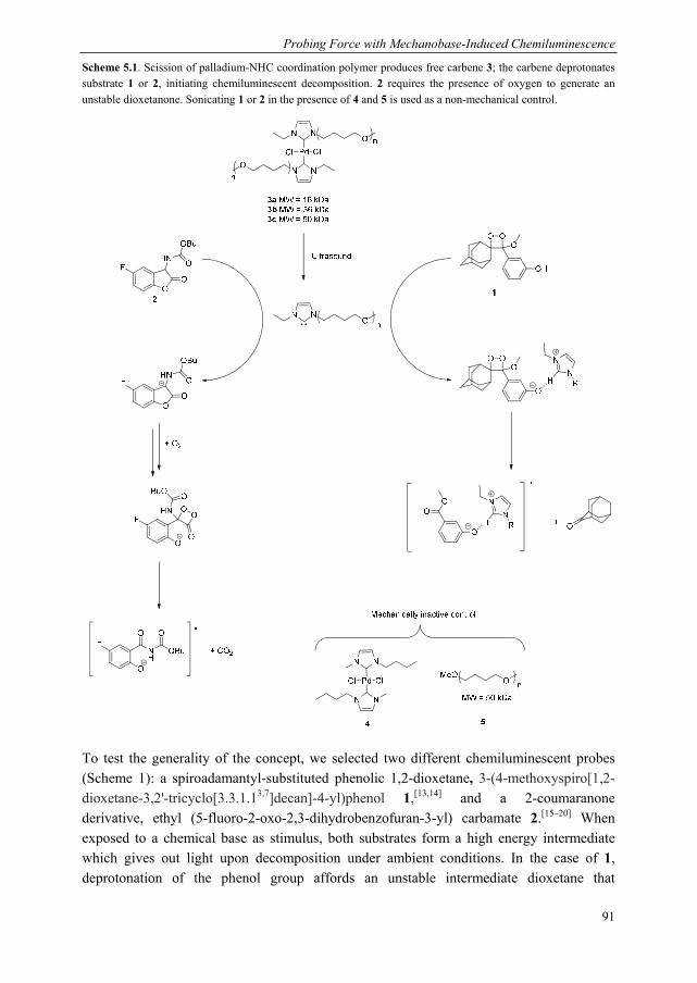
Chapter 5 introduces an alternative system for mechanoluminescence, based on our group’s previous work on the mechanical generation of N-heterocyclic carbenes (NHC), as described above. In this system, the NHC is employed as a base to induce chemiluminescence from latent substrates in solution upon sonication. The generation of chemiluminescence is thus decoupled from the applied force, which suggests the possibility of tuning the activation barrier to mechanoluminescence independently of the dioxetane.
Preliminary work towards characterising the activation barrier to dioxetane decomposition is presented in Chapter 6. Inspired by a recent study from the Craig group, we incorporate dioxetane in a polymer with multiple gDCCs or spiropyran moieties and study the competition between the two mechanoactivation pathways in sonication.
1.6 References [1] L. Takacs, Chem. Soc. Rev. 2013, 42, 7649–7659. [2] P. Baláž, Int. J. Miner. Process. 2003, 72, 341–354. [3] S. L. James, C. J. Adams, C. Bolm, D. Braga, P. Collier, T. Friščić, F. Grepioni, K. D. M. Harris, G. Hyett,
W. Jones, et al., Chem. Soc. Rev. 2011, 41, 413–447. [4] M. M. Caruso, D. A. Davis, Q. Shen, S. A. Odom, N. R. Sottos, S. R. White, J. S. Moore, Chem. Rev. 2009,
109, 5755–5798. [5] D. Keller, C. Bustamante, Biophys. J. 2000, 78, 541–556. [6] W. Kauzmann, H. Eyring, J. Am. Chem. Soc. 1940, 62, 3113–3125. [7] H. Staudinger, W. Heuer, Berichte Dtsch. Chem. Ges. B Ser. 1934, 67, 1159–1164. [8] J. Ribas-Arino, M. Shiga, D. Marx, Angew. Chem. Int. Ed. 2009, 48, 4190–4193. [9] J. Li, C. Nagamani, J. S. Moore, Acc. Chem. Res. 2015, 48, 2181–2190. [10] Z. S. Kean, J. L. Hawk, S. Lin, X. Zhao, R. P. Sijbesma, S. L. Craig, Adv. Mater. 2014, 26, 6013–6018.
Mechanochemical Stress-Sensing in Polymers
19
[11] C. R. Hickenboth, J. S. Moore, S. R. White, N. R. Sottos, J. Baudry, S. R. Wilson, Nature 2007, 446, 423–427.
[12] J. M. Lenhardt, M. T. Ong, R. Choe, C. R. Evenhuis, T. J. Martinez, S. L. Craig, Science 2010, 329, 1057–1060.
[13] G. Cravotto, E. C. Gaudino, P. Cintas, Chem. Soc. Rev. 2013, 42, 7521. [14] M. W. A. Kuijpers, P. D. Iedema, M. F. Kemmere, J. T. F. Keurentjes, Polymer 2004, 45, 6461–6467. [15] J. A. Odell, A. J. Muller, K. A. Narh, A. Keller, Macromolecules 1990, 23, 3092–3103. [16] P. A. May, N. F. Munaretto, M. B. Hamoy, M. J. Robb, J. S. Moore, ACS Macro Lett. 2016, 5, 177–180. [17] M. Schaefer, B. Icli, C. Weder, M. Lattuada, A. F. M. Kilbinger, Y. C. Simon, Macromolecules 2016, 49,
1630–1636. [18] J. Ribas-Arino, M. Shiga, D. Marx, Chem. – Eur. J. 2009, 15, 13331–13335. [19] M. V. Encina, E. Lissi, M. Sarasúa, L. Gargallo, D. Radic, J. Polym. Sci. Polym. Lett. Ed. 1980, 18, 757–
760. [20] K. L. Berkowski, S. L. Potisek, C. R. Hickenboth, J. S. Moore, Macromolecules 2005, 38, 8975–8978. [21] J. M. J. Paulusse, R. P. Sijbesma, Chem. Commun. 2008, 4416–4418. [22] J. M. J. Paulusse, R. P. Sijbesma, Angew. Chem. Int. Ed. 2004, 43, 4460–4462. [23] P. A. May, J. S. Moore, Chem. Soc. Rev. 2013, 42, 7497–7506. [24] J. A. Odell, A. Keller, J. Polym. Sci. Part B Polym. Phys. 1986, 24, 1889–1916. [25] T. Q. Nguyen, H.-H. Kausch, in Macromol. Synth. Order Adv. Prop., Springer Berlin Heidelberg, 1992, pp.
73–182. [26] D. E. Smith, H. P. Babcock, S. Chu, Science 1999, 283, 1724–1727. [27] R. Dylla-Spears, J. E. Townsend, L. Jen-Jacobson, L. L. Sohn, S. J. Muller, Lab. Chip 2010, 10, 1543–1549. [28] A. Karimi, S. Yazdi, A. M. Ardekani, Biomicrofluidics 2013, 7, 21501. [29] C. K. Lee, C. E. Diesendruck, E. Lu, A. N. Pickett, P. A. May, J. S. Moore, P. V. Braun, Macromolecules
2014, 47, 2690–2694. [30] K. Imato, A. Irie, T. Kosuge, T. Ohishi, M. Nishihara, A. Takahara, H. Otsuka, Angew. Chem. Int. Ed. 2015,
54, 6168–6172. [31] G. R. Gossweiler, G. B. Hewage, G. Soriano, Q. Wang, G. W. Welshofer, X. Zhao, S. L. Craig, ACS Macro
Lett. 2014, 3, 216–219. [32] Z. Wang, Z. Ma, Y. Wang, Z. Xu, Y. Luo, Y. Wei, X. Jia, Adv. Mater. 2015, 27, 6469–6474. [33] M. E. Grady, B. A. Beiermann, J. S. Moore, N. R. Sottos, ACS Appl. Mater. Interfaces 2014, 6, 5350–5355. [34] Q. Wang, G. R. Gossweiler, S. L. Craig, X. Zhao, Nat. Commun. 2014, 5, DOI 10.1038/ncomms5899. [35] E. L. Florin, V. T. Moy, H. E. Gaub, Science 1994, 264, 415–417. [36] S. Zou, H. Schönherr, G. J. Vancso, Angew. Chem. Int. Ed. 2005, 44, 956–959. [37] F. R. Kersey, W. C. Yount, S. L. Craig, J. Am. Chem. Soc. 2006, 128, 3886–3887. [38] D. Wu, J. M. Lenhardt, A. L. Black, B. B. Akhremitchev, S. L. Craig, J. Am. Chem. Soc. 2010, 132, 15936–
15938. [39] G. R. Gossweiler, T. B. Kouznetsova, S. L. Craig, J. Am. Chem. Soc. 2015, 137, 6148–6151. [40] J. Wang, T. B. Kouznetsova, Z. Niu, M. T. Ong, H. M. Klukovich, A. L. Rheingold, T. J. Martinez, S. L.
Craig, Nat. Chem. 2015, 7, 323–327. [41] H. M. Klukovich, T. B. Kouznetsova, Z. S. Kean, J. M. Lenhardt, S. L. Craig, Nat. Chem. 2013, 5, 110–114. [42] J. Wang, T. B. Kouznetsova, Z. S. Kean, L. Fan, B. D. Mar, T. J. Martínez, S. L. Craig, J. Am. Chem. Soc.
2014, 136, 15162–15165. [43] J. Wang, T. B. Kouznetsova, Z. Niu, A. L. Rheingold, S. L. Craig, J. Org. Chem. 2015, 80, 11895–11898. [44] M. K. Beyer, H. Clausen-Schaumann, Chem. Rev. 2005, 105, 2921–2948. [45] S. Jiang, L. Zhang, T. Xie, Y. Lin, H. Zhang, Y. Xu, W. Weng, L. Dai, ACS Macro Lett. 2013, 2, 705–709. [46] M. J. Jacobs, K. Blank, Chem. Sci. 2014, 5, 1680–1697. [47] J. Wang, I. Piskun, S. L. Craig, ACS Macro Lett. 2015, 4, 834–837. [48] M. J. Kryger, M. T. Ong, S. A. Odom, N. R. Sottos, S. R. White, T. J. Martinez, J. S. Moore, J. Am. Chem.
Soc. 2010, 132, 4558–4559. [49] M. J. Kryger, A. M. Munaretto, J. S. Moore, J. Am. Chem. Soc. 2011, 133, 18992–18998. [50] M. J. Robb, J. S. Moore, J. Am. Chem. Soc. 2015, 137, 10946–10949. [51] J. M. Lenhardt, A. L. Black, B. A. Beiermann, B. D. Steinberg, F. Rahman, T. Samborski, J. Elsakr, J. S.
Moore, N. R. Sottos, S. L. Craig, J. Mater. Chem. 2011, 21, 8454. [52] A. L. B. Ramirez, Z. S. Kean, J. A. Orlicki, M. Champhekar, S. M. Elsakr, W. E. Krause, S. L. Craig, Nat.
Chem. 2013, 5, 757–761. [53] A. L. Black, J. A. Orlicki, S. L. Craig, J. Mater. Chem. 2011, 21, 8460. [54] H. M. Klukovich, Z. S. Kean, A. L. B. Ramirez, J. M. Lenhardt, J. Lin, X. Hu, S. L. Craig, J. Am. Chem.
Soc. 2012, 134, 9577–9580. [55] H. M. Klukovich, Z. S. Kean, S. T. Iacono, S. L. Craig, J. Am. Chem. Soc. 2011, 133, 17882–17888. [56] Z. S. Kean, A. L. Black Ramirez, Y. Yan, S. L. Craig, J. Am. Chem. Soc. 2012, 134, 12939–12942. [57] Z. S. Kean, Z. Niu, G. B. Hewage, A. L. Rheingold, S. L. Craig, J. Am. Chem. Soc. 2013, 135, 13598–
13604.
Chapter 1
20
[58] H. Zhang, F. Gao, X. Cao, Y. Li, Y. Xu, W. Weng, R. Boulatov, Angew. Chem. Int. Ed. 2016, 55, 3040–3044.
[59] A. Piermattei, S. Karthikeyan, R. P. Sijbesma, Nat. Chem. 2009, 1, 133–137. [60] R. Groote, B. M. Szyja, E. A. Pidko, E. J. M. Hensen, R. P. Sijbesma, Macromolecules 2011, 44, 9187–
9195. [61] R. T. M. Jakobs, S. Ma, R. P. Sijbesma, ACS Macro Lett. 2013, 2, 613–616. [62] P. Michael, W. H. Binder, Angew. Chem. Int. Ed. 2015, 54, 13918–13922. [63] F. Verstraeten, R. Göstl, R. P. Sijbesma, Chem. Commun. 2016, 52, 8608–8611. [64] C. E. Diesendruck, B. D. Steinberg, N. Sugai, M. N. Silberstein, N. R. Sottos, S. R. White, P. V. Braun, J. S.
Moore, J. Am. Chem. Soc. 2012, 134, 12446–12449. [65] M. B. Larsen, A. J. Boydston, J. Am. Chem. Soc. 2013, 135, 8189–8192. [66] C. E. Diesendruck, G. I. Peterson, H. J. Kulik, J. A. Kaitz, B. D. Mar, P. A. May, S. R. White, T. J.
Martínez, A. J. Boydston, J. S. Moore, Nat. Chem. 2014, 6, 623–628. [67] K. Kumar, A. P. Goodwin, ACS Macro Lett. 2015, 4, 907–911. [68] M. Montalti, A. Credi, L. Prodi, M. T. Gandolfi, Handbook of Photochemistry, CRC Press, 2006. [69] J. R. Lakowicz, Principles of Fluorescence Spectroscopy, Springer, New York, 2006. [70] H. S. Rye, J. M. Dabora, M. A. Quesada, R. A. Mathies, A. N. Glazer, Anal. Biochem. 1993, 208, 144–150. [71] S. W. Hell, J. Wichmann, Opt. Lett. 1994, 19, 780. [72] S. W. Hell, Science 2007, 316, 1153–1158. [73] S.-J. Kim, D. H. Reneker, Polym. Bull. 1993, 31, 367–374. [74] S. K. Surampudi, H. R. Patel, G. Nagarjuna, D. Venkataraman, Chem. Commun. 2013, 49, 7519–7521. [75] P. K. Kundu, R. Klajn, ACS Nano 2014, 8, 11913–11916. [76] S. L. Potisek, D. A. Davis, N. R. Sottos, S. R. White, J. S. Moore, J. Am. Chem. Soc. 2007, 129, 13808–
13809. [77] D. A. Davis, A. Hamilton, J. Yang, L. D. Cremar, D. Van Gough, S. L. Potisek, M. T. Ong, P. V. Braun, T.
J. Martínez, S. R. White, et al., Nature 2009, 459, 68–72. [78] J. Li, C. Nagamani, J. S. Moore, Acc. Chem. Res. 2015, 48, 2181–2190. [79] B. A. Beiermann, D. A. Davis, S. L. B. Kramer, J. S. Moore, N. R. Sottos, S. R. White, J. Mater. Chem.
2011, 21, 8443–8447. [80] C. M. Kingsbury, P. A. May, D. A. Davis, S. R. White, J. S. Moore, N. R. Sottos, J. Mater. Chem. 2011, 21,
8381–8388. [81] G. O’Bryan, B. M. Wong, J. R. McElhanon, ACS Appl. Mater. Interfaces 2010, 2, 1594–1600. [82] G. I. Peterson, M. B. Larsen, M. A. Ganter, D. W. Storti, A. J. Boydston, ACS Appl. Mater. Interfaces 2014,
7, 577–583. [83] L.-J. Wang, X.-J. Zhou, X.-H. Zhang, B.-Y. Du, Macromolecules 2015, 2016, 49, 98–104 [84] X. Fang, H. Zhang, Y. Chen, Y. Lin, Y. Xu, W. Weng, Macromolecules 2013, 46, 6566–6574. [85] G. Hong, H. Zhang, Y. Lin, Y. Chen, Y. Xu, W. Weng, H. Xia, Macromolecules 2013, 46, 8649–8656. [86] Y. Chen, H. Zhang, X. Fang, Y. Lin, Y. Xu, W. Weng, ACS Macro Lett. 2014, 3, 141–145. [87] C. M. Degen, P. A. May, J. S. Moore, S. R. White, N. R. Sottos, Macromolecules 2013, 46, 8917–8921. [88] J. R. Hemmer, P. D. Smith, M. van Horn, S. Alnemrat, B. P. Mason, J. R. de Alaniz, S. Osswald, J. P.
Hooper, J. Polym. Sci. Part B Polym. Phys. 2014, 52, 1347–1356. [89] C. K. Lee, B. A. Beiermann, M. N. Silberstein, J. Wang, J. S. Moore, N. R. Sottos, P. V. Braun,
Macromolecules 2013, 46, 3746–3752. [90] J. W. Kim, Y. Jung, G. W. Coates, M. N. Silberstein, Macromolecules 2015, 48, 1335–1342. [91] A.-D. N. Celestine, B. A. Beiermann, P. A. May, J. S. Moore, N. R. Sottos, S. R. White, Polymer 2014, 55,
4164–4171. [92] Q. Wang, G. R. Gossweiler, S. L. Craig, X. Zhao, J. Mech. Phys. Solids 2015, 82, 320–344. [93] J. M. Clough, A. Balan, R. P. Sijbesma, in Polym. Mechanochemistry (Ed.: R. Boulatov), Springer
International Publishing, 2015, pp. 209–238. [94] N. Yoshie, S. Saito, N. Oya, Polymer 2011, 52, 6074–6079. [95] S. S. M. Konda, J. N. Brantley, B. T. Varghese, K. M. Wiggins, C. W. Bielawski, D. E. Makarov, J. Am.
Chem. Soc. 2013, 135, 12722–12729. [96] J. Li, T. Shiraki, B. Hu, R. A. E. Wright, B. Zhao, J. S. Moore, J. Am. Chem. Soc. 2014, 136, 15925–15928. [97] D. C. Church, G. I. Peterson, A. J. Boydston, ACS Macro Lett. 2014, 3, 648–651. [98] S. Billiet, K. De Bruycker, F. Driessen, H. Goossens, V. Van Speybroeck, J. M. Winne, F. E. Du Prez, Nat.
Chem. 2014, 6, 815–821. [99] R. Göstl, R. P. Sijbesma, Chem. Sci. 2016, 7, 370–375. [100] Y.-K. Song, K.-H. Lee, W.-S. Hong, S.-Y. Cho, H.-C. Yu, C.-M. Chung, J. Mater. Chem. 2011, 22, 1380–
1386. [101] Z. S. Kean, G. R. Gossweiler, T. B. Kouznetsova, G. B. Hewage, S. L. Craig, Chem. Commun. 2015, 51,
9157–9160. [102] C. R. Hickenboth, J. S. Moore, S. R. White, N. R. Sottos, J. Baudry, S. R. Wilson, Nature 2007, 446, 423–
427.
Mechanochemical Stress-Sensing in Polymers
21
[103] A. Löwenbein, H. Simonis, Berichte Dtsch. Chem. Ges. B Ser. 1924, 57, 2040–2048. [104] A. Löwenbein, Berichte Dtsch. Chem. Ges. B Ser. 1925, 58, 601–609. [105] A. Löwenbein, R. F. Gagarin, Berichte Dtsch. Chem. Ges. B Ser. 1925, 58, 2643–2644. [106] A. Löwenbein, H. Schmidt, Berichte Dtsch. Chem. Ges. B Ser. 1927, 60, 1851–1861. [107] K. Imato, T. Kanehara, T. Ohishi, M. Nishihara, H. Yajima, M. Ito, A. Takahara, H. Otsuka, ACS Macro
Lett. 2015, 4, 1307–1311. [108] J. Kim, T. M. Swager, Nature 2001, 411, 1030–1034. [109] K. Becker, J. M. Lupton, J. Am. Chem. Soc. 2005, 127, 7306–7307. [110] E. Da Como, K. Becker, J. Feldmann, J. M. Lupton, Nano Lett. 2007, 7, 2993–2998. [111] H. E. Cingil, I. M. Storm, Y. Yorulmaz, D. W. te Brake, R. de Vries, M. A. Cohen Stuart, J. Sprakel, J. Am.
Chem. Soc. 2015, 137, 9800–9803. [112] R. W. Carpick, D. Y. Sasaki, M. S. Marcus, M. A. Eriksson, A. R. Burns, J. Phys. Condens. Matter 2004,
16, R679. [113] M. A. Reppy, B. A. Pindzola, Chem. Commun. 2007, 4317–4338. [114] O. Yarimaga, J. Jaworski, B. Yoon, J.-M. Kim, Chem. Commun. 2012, 48, 2469–2485. [115] R. A. Nallicheri, M. F. Rubner, Macromolecules 1991, 24, 517–525. [116] R. A. Koevoets, S. Karthikeyan, P. C. M. M. Magusin, E. W. Meijer, R. P. Sijbesma, Macromolecules 2009,
42, 2609–2617. [117] H. Feng, J. Lu, J. Li, F. Tsow, E. Forzani, N. Tao, Adv. Mater. 2013, 25, 1729–1733. [118] D.-H. Park, J. Hong, I. S. Park, C. W. Lee, J.-M. Kim, Adv. Funct. Mater. 2014, 24, 5186–5193. [119] Y. Tomioka, N. Tanaka, S. Imazeki, J. Chem. Phys. 1989, 91, 5694–5700. [120] R. W. Carpick, D. Y. Sasaki, A. R. Burns, Langmuir 2000, 16, 1270–1278. [121] A. R. Burns, R. W. Carpick, D. Y. Sasaki, J. A. Shelnutt, R. Haddad, Tribol. Lett. 2001, 10, 89–96. [122] D. R. T. Roberts, S. J. Holder, J. Mater. Chem. 2011, 21, 8256–8268. [123] A. Pucci, G. Ruggeri, J. Mater. Chem. 2011, 21, 8282–8291. [124] F. Ciardelli, G. Ruggeri, A. Pucci, Chem. Soc. Rev. 2013, 42, 857–870. [125] C. Löwe, C. Weder, Adv. Mater. 2002, 14, 1625–1629. [126] B. R. Crenshaw, C. Weder, Chem. Mater. 2003, 15, 4717–4724. [127] J. Kunzelman, B. R. Crenshaw, M. Kinami, C. Weder, Macromol. Rapid Commun. 2006, 27, 1981–1987. [128] B. R. Crenshaw, M. Burnworth, D. Khariwala, A. Hiltner, P. T. Mather, R. Simha, C. Weder,
Macromolecules 2007, 40, 2400–2408. [129] J. Lott, C. Weder, Macromol. Chem. Phys. 2010, 211, 28–34. [130] J. Kunzelman, M. Gupta, B. R. Crenshaw, D. A. Schiraldi, C. Weder, Macromol. Mater. Eng. 2009, 294,
244–249. [131] M. Kinami, B. R. Crenshaw, C. Weder, Chem. Mater. 2006, 18, 946–955. [132] B. R. Crenshaw, C. Weder, Macromolecules 2006, 39, 9581–9589. [133] A. Pucci, M. Bertoldo, S. Bronco, Macromol. Rapid Commun. 2005, 26, 1043–1048. [134] A. Pucci, C. Cappelli, S. Bronco, G. Ruggeri, J. Phys. Chem. B 2006, 110, 3127–3134. [135] A. Pucci, F. D. Cuia, F. Signori, G. Ruggeri, J. Mater. Chem. 2007, 17, 783–790. [136] A. Pucci, G. Ruggeri, J. Mater. Chem. 2011, 21, 8282–8291. [137] J. Mei, N. L. C. Leung, R. T. K. Kwok, J. W. Y. Lam, B. Z. Tang, Chem. Rev. 2015, 115, 11718–11940. [138] S.-J. Yoon, J. W. Chung, J. Gierschner, K. S. Kim, M.-G. Choi, D. Kim, S. Y. Park, J. Am. Chem. Soc. 2010,
132, 13675–13683. [139] Y. Wu, J. Hu, H. Huang, J. Li, Y. Zhu, B. Tang, J. Han, L. Li, J. Polym. Sci. Part B Polym. Phys. 2014, 52,
104–110. [140] J. Ge, Y. Yin, Angew. Chem. Int. Ed. 2011, 50, 1492–1522. [141] E. P. Chan, J. J. Walish, A. M. Urbas, E. L. Thomas, Adv. Mater. 2013, 25, 3934–3947. [142] C. Fenzl, T. Hirsch, O. S. Wolfbeis, Angew. Chem. Int. Ed. 2014, 53, 3318–3335. [143] H. Li, X. Sun, H. Peng, ChemPhysChem 2015, 16, 3761–3768. [144] M. J. Serpe, Y. Kang, Q. M. Zhang, Eds. , Photonic Materials for Sensing, Biosensing and Display Devices,
Springer, Cham, 2016. [145] Y. F. Yue, M. A. Haque, T. Kurokawa, T. Nakajima, J. P. Gong, Adv. Mater. 2013, 25, 3106–3110. [146] Y. Yue, T. Kurokawa, M. A. Haque, T. Nakajima, T. Nonoyama, X. Li, I. Kajiwara, J. P. Gong, Nat.
Commun. 2014, 5, 4659. [147] X.-Q. Wang, C.-F. Wang, Z.-F. Zhou, S. Chen, Adv. Opt. Mater. 2014, 2, 652–662. [148] Y. Cho, S. Y. Lee, L. Ellerthorpe, G. Feng, G. Lin, G. Wu, J. Yin, S. Yang, Adv. Funct. Mater. 2015, 25,
6041–6049. [149] X. Jia, J. Wang, K. Wang, J. Zhu, Langmuir 2015, 31, 8732–8737. [150] H. Finkelmann, S. T. Kim, A. Muñoz, P. Palffy-Muhoray, B. Taheri, Adv. Mater. 2001, 13, 1069–1072. [151] J. Schmidtke, S. Kniesel, H. Finkelmann, Macromolecules 2005, 38, 1357–1363. [152] P. Cicuta, A. R. Tajbakhsh, E. M. Terentjev, Phys. Rev. E 2002, 65, 51704. [153] P. Cicuta, A. R. Tajbakhsh, E. M. Terentjev, Phys. Rev. E 2004, 70, 11703. [154] A. Varanytsia, H. Nagai, K. Urayama, P. Palffy-Muhoray, Sci. Rep. 2015, 5, 17739.
Chapter 1
22
[155] Y. Chen, A. J. H. Spiering, S. Karthikeyan, G. W. M. Peters, E. W. Meijer, R. P. Sijbesma, Nat. Chem. 2012, 4, 559–562.
[156] J. H. Wieringa, J. Strating, H. Wynberg, W. Adam, Tetrahedron Lett. 1972, 13, 169–172. [157]J. C. Hummelen, T. M. Luider, H. Wynberg, Pure Appl. Chem. 1987, 59, 639–650. [158] E. Ducrot, Y. Chen, M. Bulters, R. P. Sijbesma, C. Creton, Science 2014, 344, 186–189. [159] Y. Chen, R. P. Sijbesma, Macromolecules 2014, 47, 3797–3805.
Chapter 2
Photophysical Determination of the Excited State Products from Mechanically Induced Dioxetane Scission
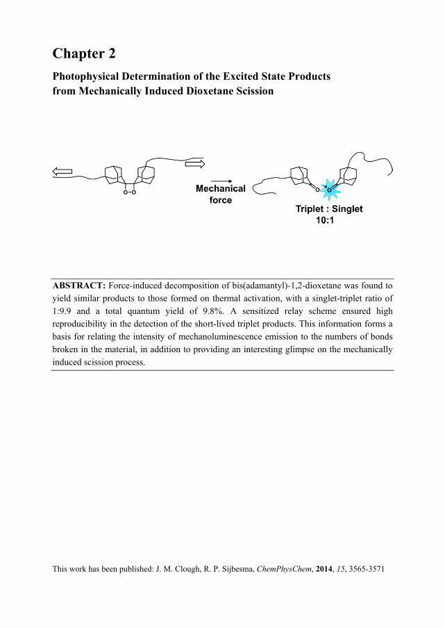
ABSTRACT: Force-induced decomposition of bis(adamantyl)-1,2-dioxetane was found to yield similar products to those formed on thermal activation, with a singlet-triplet ratio of 1:9.9 and a total quantum yield of 9.8%. A sensitized relay scheme ensured high reproducibility in the detection of the short-lived triplet products. This information forms a basis for relating the intensity of mechanoluminescence emission to the numbers of bonds broken in the material, in addition to providing an interesting glimpse on the mechanically induced scission process.
This work has been published: J. M. Clough, R. P. Sijbesma, ChemPhysChem, 2014, 15, 3565-3571
Chapter 2
24
2.1 Introduction A diverse array of important natural processes is mediated by mechanochemical transduction, by which mechanical strain elicits a chemical or electrical response, with examples ranging from hearing[1] and tissue growth[2] to bioluminescence.[3] However, the design of synthetic material systems which respond functionally to mechanical stimuli remains challenging. Recent developments in the burgeoning field of mechanoresponsive materials have made use of selective bond scission in polymers, whereby mechanical force is transferred via the polymer backbone to a responsive unit, or mechanophore, centrally incorporated within the polymer chain. A wide-ranging selection of mechanophores was reviewed in Chapter 1.
In 2012, our group reported the first and to date only chemiluminescent mechanophore, based on bis(adamantyl)-1,2-dioxetane.[4] Luminescence from polymeric networks or linear chains fitted with these dioxetane units can be observed as the four-membered ring breaks open on the application of mechanical force in solution or to solid samples, generating excited state ketones that can take either a singlet or a triplet spin configuration. Our aim in the present work was to inspect the mechanical scission process in greater depth by examining its scission products. Such insights are essential for relating the mechanoluminescence emission more quantitatively to the damage in polymeric materials and could also be useful for the design of more efficient harvesting strategies for the excitation energy of dioxetane mechanoluminescence.
In addition, the thermally induced scission of dioxetanes remains an enigmatic phenomenon, despite having been studied intensively, partly in the pursuit of synthetic analogues to highly efficient bioluminescent systems, such as the firefly luciferin or coelenterazine, found in many marine organisms. Triplet excesses dominate the scission products of most alkyl 1,2-dioxetanes, with the singlet-triplet ratio ranging from 1:0.2 to 1:11 000, in spite of the fact that triplet formation is formally spin-forbidden.[5,6] The triplet formation is such that it cannot be explained by a facile intersystem crossing from directly produced excited singlets. For example, the exceptionally stable bis(adamantyl)-1,2-dioxetane decomposes when heated to 200 °C in a first-order chemiluminescent process with an activation energy of 35 kcal mol-1 and a half-life of less than a few minutes[7] to form singlet and triplet products in a ratio of 1:7.5 and a total chemiexcitation yield of 17%, as determined by Schuster et al. via chemical titration.[8] The vast majority of other dioxetanes are much less stable, although the triplet excesses and total chemiexcitation yields vary widely from dioxetane to dioxetane. By comparison, the singlet-triplet ratio of tetramethyldioxetane, with reported activation energies in the range of 25 to 27.6 kcal mol-
1, is approximately 1:200 in a variety of solvents, with a total chemiexcitation yield of 35%.[9,10] Whilst the exact thermal decomposition mechanism is not yet entirely understood, the results of high-level calculations indicate that the abundance of triplets is intimately linked with the decomposition pathway of these molecules, which is thought to lie between the mechanistic extremes of concerted and stepwise.[11] Determination of the excited state
Photophysical Determination of the Excited State Products
25
products upon mechanical scission is therefore of fundamental interest, especially given that the application of mechanical force has been shown to open up reaction pathways that are forbidden or disfavored thermally. For example, the work by Hickenboth et al.[12] revealed that mechanical activation is able to overcome the Woodward-Hoffman orbital symmetry preferences in the electrocyclic ring-opening of a benzocyclobutene; similarly, trans-gem-difluorocyclopropanes isomerized into the less stable cis-isomer under sonication, as elucidated by Lenhardt et al.[13]
2.2 Outline of experimental approach: sonication and acceptor selection To effect mechanical activation of dioxetane-functionalized poly(methyl acrylate) 1 (Chart 2.1), we employed ultrasonication in solution, which is one of the most effective ways to break covalent bonds in polymers with mechanical force, as described in the Introduction chapter. Sonication generates cavitation bubbles in solution which, upon their collapse, generate a velocity gradient along the polymer coil, leading to stretching of the polymer and ultimately mid-chain scission. Strain rates, defined as the fractional change in length per unit time, accessible by this technique are of the order of 106-107 s-1, resulting in a tensile force in the nanonewton range. The cavitation process, and the mechanochemical efficiency as a consequence, is known to be sensitive to a number of experimental conditions, including temperature, solvent viscosity, dissolved gases and sonication power. Increasing sonication power leads to greater cavitation and mechanochemical efficiency, up to a maximum. Care was therefore taken to ensure that the experimental conditions remained constant throughout our series of experiments.
In principle, the singlet-triplet ratio can be determined from the direct fluorescence and phosphorescence of the excited ketones, but excited adamantanone, in common with other alkyl ketones, is not a very efficient emitter from the singlet state and triplet adamantanones are not known to phosphoresce at room temperature.[7,8] Nevertheless, excited ketones can transfer their excitation energy efficiently to fluorescent or phosphorescent acceptors.[14–16] In the case of adamantanone however, the triplet species is very short-lived and its direct sensitisation of acceptors is low and unreliable, even at the diffusion limit.[16]
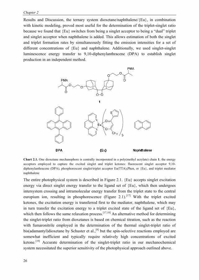
For the current study, we designed a photophysical system capable of monitoring the production of both excited state ketones, including the short-lived triplet ketone, from the mechanical scission of bis(adamantyl)dioxetanes incorporated in poly(methyl acrylate) 1. Our excitation energy acceptors are shown in Chart 2.1. Our luminescent acceptor was the europium complex, Eu(TTA)3Phen, henceforth referred to as {Eu} (TTA = thenoyltrifluoroacetone, Phen = phenanthroline). This dye is readily accessible synthetically, displays good solubility and has a good emission efficiency. In addition to these selection criteria, we found that reliable detection of triplet adamantanone could be obtained via indirect sensitisation of {Eu}, which required the use of a mediator, naphthalene, so there are certain requirements vis-à-vis the energy levels of the acceptor. Specifically, the triplet states of naphthalene must be higher than those of the triplet state of the ligand set of {Eu}, to allow naphthalene to act as a mediator. As described in the
Chapter 2
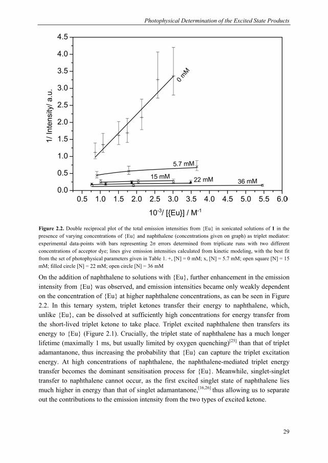
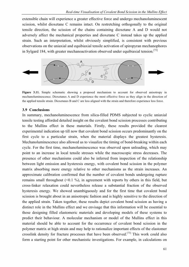
26
Results and Discussion, the ternary system dioxetane/naphthalene/{Eu}, in combination with kinetic modeling, proved most useful for the determination of the triplet-singlet ratio because we found that {Eu} switches from being a singlet acceptor to being a “dual” triplet and singlet acceptor when naphthalene is added. This allows estimation of both the singlet and triplet formation rates by simultaneously fitting the emission intensities for a set of different concentrations of {Eu} and naphthalene. Additionally, we used singlet-singlet luminescence energy transfer to 9,10-diphenylanthracene (DPA) to establish singlet production in an independent method.
Chart 2.1. One dioxetane mechanophore is centrally incorporated in a poly(methyl acrylate) chain 1; the energy acceptors employed to capture the excited singlet and triplet ketones: fluorescent singlet acceptor 9,10-diphenylanthracene (DPA), phosphorescent singlet/triplet acceptor Eu(TTA)3Phen, or {Eu}, and triplet mediator naphthalene
The entire photophysical system is described in Figure 2.1. {Eu} accepts singlet excitation energy via direct singlet energy transfer to the ligand set of {Eu}, which then undergoes intersystem crossing and intramolecular energy transfer from the triplet state to the central europium ion, resulting in phosphorescence (Figure 2.1).[17] With the triplet excited ketones, the excitation energy is transferred first to the mediator, naphthalene, which may in turn transfer the excitation energy to a triplet excited state of the ligand set of {Eu}, which then follows the same relaxation process.[17,18] An alternative method for determining the singlet-triplet ratio from dioxetanes is based on chemical titration, such as the reaction with fumaronitrile employed in the determination of the thermal singlet-triplet ratio of bis(adamantyl)dioxetane by Schuster et al.,[8] but the spin-selective reactions employed are somewhat inefficient and typically require relatively high concentrations of excited ketone.[19] Accurate determination of the singlet-triplet ratio in our mechanochemical system necessitated the superior sensitivity of the photophysical approach outlined above.
Photophysical Determination of the Excited State Products
27
Figure 2.1. Mechanical generation of excited ketones (top) and Jablonski diagram depicting the two principal pathways for energy transfer from the electronically excited products of dioxetane cleavage to {Eu} (bottom): singlet-singlet energy transfer and naphthalene-mediated triplet-triplet transfer.
2.3 Sensitisation of singlet and triplet acceptor dyes Historically, nearly all previous studies of dioxetane cleavage made use of the double reciprocal plot method to determine singlet-triplet ratios. The singlet and triplet sensitized intensities, I at a range of acceptor concentration, [A] for two separate acceptors, a singlet and triplet acceptor respectively, would be measured and plotted in a double reciprocal format, 1/I vs. 1/[A], to give two straight lines; the limiting sensitized intensity would then be found by extrapolating to infinite [A] for each acceptor. The relative yields of triplet and singlet excited states could be calculated simply from taking the ratio of these limiting intensities, with a correction for the difference in emission efficiencies of the two acceptors. However, aside from the inherently large errors in this method, the extremely short lifetime
Chapter 2
28
(~0.15 ns)[8] of triplet state adamantanone precludes such straightforward determination of the limiting triplet intensity. Instead, we devised an alternative method to extract the singlet-triplet ratio. Emission intensities were measured in the ternary system dioxetane/naphthalene/{Eu} at a range of concentrations of naphthalene and {Eu}, and instead of relying on a graphical extrapolation of the limiting intensities, the triplet and singlet formation rates were obtained by fitting these as parameters in a kinetic model that takes all the relevant photophysical processes into account.
First, the formation of singlet excited state adamantanone was established. Upon sonicating a 0.15 mM solution of dioxetane-functionalized PMA 1 in toluene, we observed the direct fluorescence as a low intensity signal. On the addition of singlet acceptor DPA, a significant enhancement of the intensity was observed. Adding the triplet quencher naphthalene had no effect on the direct chemiluminescence, confirming the singlet origin of the direct chemiluminescence; naphthalene also had no effect on the sensitized fluorescence of DPA, indicating that singlet excited ketones were the excitation source. In addition, the photodiode response to extended periods of sonication decayed exponentially as the dioxetane was gradually consumed. From this decay, we extracted a rate coefficient of scission of (7.7 ± 1.0) x 10-4 s-1, which is comparable to those rates previously determined for other mechanically induced polymer scission processes.[20]
Next, the relative formation rates of singlet and triplet excited states were determined by measuring the chemiluminescence intensity from {Eu} in the presence of varying concentrations of naphthalene. The total emission intensities were plotted in a double
reciprocal format, 1 I⁄ vs. 1 Eu⁄ (Figure 2.2). On the addition of {Eu} alone to the
0.15 mM solution of dioxetane-functionalized PMA, enhancement of the chemiluminescence intensity was observed; the results of these mechanoluminescence experiments are shown with crosses as datapoints in Figure 2.2. Whilst {Eu} has previously been employed to detect triplet ketones via triplet-triplet energy transfer, excited adamantanone has a number of features which result in predominantly singlet energy transfer in the absence of naphthalene as a mediator. Firstly, the triplet lifetime of this ketone is very short, having been claimed to be as low as 0.15 ns[8] (by comparison, acetone has a triplet lifetime of 5 μs[21] and the singlet lifetime of adamantanone is 9.12 ns[22]), so the triplet energy acceptor does not compete effectively with the triplet ketone deactivation process at the concentrations at which {Eu} can be dissolved in toluene (< 2mM). Secondly, the singlet lifetime of adamantanone is much longer than the triplet lifetime and lastly, in contrast with most other dioxetanes, the singlet excitation yield is relatively high compared to the triplet excitation yield. Consequently, the sensitized phosphorescence from {Eu} in the absence of a mediator originates predominantly from singlet adamantanone.[23,24] This was borne out by the modeling, which showed that the direct triplet excitation pathway alone (triplet ketone to {Eu}) is insufficient to account for the observed intensities represented by the crosses in Figure 2.2.
Photophysical Determination of the Excited State Products
29
Figure 2.2. Double reciprocal plot of the total emission intensities from {Eu} in sonicated solutions of 1 in the presence of varying concentrations of {Eu} and naphthalene (concentrations given on graph) as triplet mediator: experimental data-points with bars representing 2σ errors determined from triplicate runs with two different concentrations of acceptor dye; lines give emission intensities calculated from kinetic modeling, with the best fit from the set of photophysical parameters given in Table 1. +, [N] = 0 mM; x, [N] = 5.7 mM; open square [N] = 15 mM; filled circle [N] = 22 mM; open circle [N] = 36 mM
On the addition of naphthalene to solutions with {Eu}, further enhancement in the emission intensity from {Eu} was observed, and emission intensities became only weakly dependent on the concentration of {Eu} at higher naphthalene concentrations, as can be seen in Figure 2.2. In this ternary system, triplet ketones transfer their energy to naphthalene, which, unlike {Eu}, can be dissolved at sufficiently high concentrations for energy transfer from the short-lived triplet ketone to take place. Triplet excited naphthalene then transfers its energy to {Eu} (Figure 2.1). Crucially, the triplet state of naphthalene has a much longer lifetime (maximally 1 ms, but usually limited by oxygen quenching)[25] than that of triplet adamantanone, thus increasing the probability that {Eu} can capture the triplet excitation energy. At high concentrations of naphthalene, the naphthalene-mediated triplet energy transfer becomes the dominant sensitisation process for {Eu}. Meanwhile, singlet-singlet transfer to naphthalene cannot occur, as the first excited singlet state of naphthalene lies much higher in energy than that of singlet adamantanone,[16,26] thus allowing us to separate out the contributions to the emission intensity from the two types of excited ketone.
Chapter 2
30
2.4 Kinetic modelling As multiple competing photophysical processes operate in the ternary mixture, the limiting triplet intensity cannot be accurately extracted by a simple linear extrapolation of the double reciprocal plots representation shown in Figure 2.2. Therefore we used kinetic modeling of all the relevant photophysical processes and fitted the formation rates of singlet and triplet adamantanones to the full set of experimental intensity vs. time traces, following the initial temperature stabilisation period of approximately two minutes (see Experimental section). Kinetic modeling has additional advantages, in that fitting provides an estimate of the error in the parameters and that it permits a deeper understanding of how the rate constants and efficiencies affect the emission intensities. Linear extrapolation from double reciprocal plots, on the other hand, requires a number of approximations, whilst the linearisation itself leads to improper treatment of the experimental error.[27]
The kinetic model consisted of the rate constants for the formation of singlet and triplet excited ketones, S* and T* respectively, plus rate constants and efficiencies for all relevant radiative and non-radiative photophysical processes, which are listed in Table 2.1. In order to determine the singlet and triplet formation rate constants, these two parameters were fitted, whilst the other parameters were kept fixed at their literature values. The energy transfer constants were set to diffusion-limited values, accounting for the viscosity increase in the presence of polymer 1[25,28–30] and the reduction in the diffusion rate constant from the attachment of the excited ketones to a polymer chain. We also found it necessary to incorporate a pathway by which triplet excited naphthalene, N* is quenched by other ground state naphthalenes, N. This type of self-quenching is a well-established phenomenon for naphthalene and other longer-lived triplet species.[31–33] The rate constant for this pathway was also made a fitting parameter, as it is known that such quenching processes can be sensitive to the conditions of measurement.
Good agreement between modeled and observed intensities was found. Simulations with the calculated optimum values of the fitted parameters at a single concentration of naphthalene for a range of {Eu} concentrations afforded lines of intensities in the double reciprocal plot of Figure 2.2. These lines show significant curvature, particularly at lower [naphthalene], which results from there being two major activation pathways for {Eu} in the presence of naphthalene: direct singlet excitation and naphthalene-mediated triplet excitation. In the absence of naphthalene, the plot is linear (crosses), as {Eu} is activated primarily only by direct singlet excitation. It can also be seen that increasing the concentration of naphthalene for the same [{Eu}] leads to progressively smaller increases in intensity, which results from a shortening of the lifetime of naphthalene at higher naphthalene concentrations via self-quenching.
Photophysical Determination of the Excited State Products
31
Table 2.1. Rate constants of photophysical processes in kinetic modeling
Fixed rate constants Value Ref.
S* → K + hν 6.5 x 10⁵ s-1 [6,7,34]
T* → K 6.7 x 10⁹ s-1 [8]
S* → T* 1.25 x 10⁸ s-1 [6,35]*
S* + {Eu} → {Eu}* + K 4 x 10⁹ M-1 s-1 [25,28–30]
T* + {Eu} → {Eu}* + K 4 x 10⁹ M-1 s-1 [25,28–30]
T* + N → N* + K 4 x 10⁹ M-1 s-1 [25,28–30]
N* + {Eu} → {Eu}* + N 8 x 10⁹ M-1 s-1 [25,28–30]
{Eu}* → {Eu} + hν 1960 s-1 [36,37]
{Eu}* → {Eu} 3410 s-1 [36,37]
N* → N 1000 s-1 [25]
Fitted parameters† Value
Singlet formation rate 0.7 x 10⁻⁴ s-1
Triplet formation rate 7.1 x 10⁻⁴ s-1
N* + N → 2 N 9.8 x 10⁶ M-1 s-1††
S* = singlet excited state ketone; T* = triplet excited state ketone; K = ground state ketone; {Eu} = europium complex; {Eu}* = triplet excited state europium complex; N = ground state naphthalene; N* = triplet excited state naphthalene. *The intersystem crossing is assumed to represent the primary pathway for non-radiative decay from the singlet excited state ketone.[34,35] †A scaling factor was an additional parameter in the fitting procedure (relative intensities were used in the fitting). ††Comparable with self-quenching rate constant for naphthalene in free isooctane of 2 x 10⁷ M-1 s-1 at 293 K and self-quenching rate constants for other long-lived triplet species[31–33]
The triplet-singlet ratio was derived from two fitted parameters, a singlet and a triplet formation rate constant, assuming each dioxetane decomposition led to the formation of an excited state. The triplet-singlet ratio derived from the best fit parameters for the formation rates is (9.9 ± 0.1) when zero error in the fixed parameters is assumed. The actual error is likely to be larger as a result of errors in the fixed parameters— most notably, in the rate constant of intersystem crossing for adamantanone and the rate constant for the non-radiative decay of triplet adamantanone (see Experimental section). Modeling also revealed that the contribution to the sensitized chemiluminescence from triplets formed by intersystem crossing from the singlets is negligible, thanks in part to a relatively slow intersystem crossing rate compared to other alkyl ketones.[35]
The fitted dioxetane decomposition rate constant (7.8 x 10⁻⁴ s-1) is close to the experimentally derived rate constant (average of experimental results (7.7 ± 1.0) x 10-4 s-1).
Chapter 2
32
As the ratio of formation rate constants is a parameter that is independent of the value of the quantum yield and the scaling factor, these results do not imply that all scission events result in an excited state adamantanone, because the quantum yield forms part of the scaling factor, along with the photosensitivity of the photodiode and the light collection efficiency of the set-up (see Experimental section). Therefore, in order to obtain the absolute excitation yields, we calibrated the chemiluminescence intensity from the mechanically induced dioxetane decomposition against the well-known chemiluminescence of luminol as a standard. The oxidation of luminol in DMSO results in an emission with a known photon yield of 9.75 x 10¹⁴ photons per mL of a luminol solution having an absorbance of 1.0 at 359.5 nm.[38,39] Comparing this with the direct singlet fluorescence, which has a similar spectral range, we were able to ascertain that the absolute singlet yield is (0.9 ± 0.1) %; knowing the triplet-singlet ratio, the absolute triplet yield is therefore (8.9 ± 1.2) %. The absolute yields are comparable with the absolute thermal singlet and triplet yields, 2 % and 15 % respectively (50 % errors reported on individual yields).[8] The effective rates of mechanical generation of singlet and triplet excited state ketones, taking into account of the quantum yields, are therefore 0.069 x 10⁻⁴ s-1 and 0.69 x 10⁻⁴ s-1 respectively.
2.5 Discussion Whilst the singlet-triplet ratio and absolute yields are very similar under thermal and mechanical activation, the mechanical nature of polymer sonochemistry has been well established[20] and is supported by a number of control experiments we performed. Sonicating pure toluene alone, unfunctionalized PMA in toluene or the acceptors, including naphthalene, in toluene, gave out no detectable light, showing that the signal was not the result of sonoluminescence. Sonicating the small molecule bis(adamantyl)-1,2-dioxetane dissolved in toluene alone gave out no detectable light, showing that the thermal effects of sonication are insufficient to induce dioxetane scission. To decompose PMA-incorporated bis(adamantyl)-1,2-dioxetane thermally requires heating to the same elevated temperatures as small molecule bis(adamantyl)-1,2-dioxetane, so the attachment of polymer chains makes negligible difference to the thermal stability of the dioxetane. Sonicating bis(adamantyl)-1,2-dioxetane dissolved with unfunctionalized poly(methyl acrylate) (MW 65 kDa) in toluene also gave out no light, in agreement with the previous observations of Chen et al.[4] Furthermore, Chen et al. also showed that no light could be observed from sonicating a polymer end-functionalized with dioxetane.
The remarkable similarity of the thermal and mechanochemical triplet-singlet ratios indicates that the relative energy barriers to decomposition on the triplet and singlet excited state surfaces, responsible for the partitioning between the two excited states, are little changed by the application of mechanical force. Furthermore, the similarity in the total quantum yields suggests that the partitioning process between excited and ground state products is also relatively unaffected by the application of force. To understand this experimental result, it is instructive to take a closer look at the proposed decomposition pathway under thermal activation, as expounded by workers in the Lindh group and
Photophysical Determination of the Excited State Products
33
others.[40–42] Alkyl-1,2-dioxetanes are thought to decompose by a common thermal mechanism with significant stepwise character, with O—O cleavage preceding C—C cleavage. Torsional motion about the C—C bond and O—O bond lengthening leads to the main transition state on the S0 potential energy surface, at which point the O—O bond breaks. It has been established experimentally that the activation energy for decomposition in the S0 state corresponds to the rate-determining step for the formation of excited state products, for most dioxetanes.[43] In the case of bis(adamantyl)dioxetane, this activation energy is 35 kcal mol-1, which is exceptionally high on account of the “steric lock” posed by the β-hydrogens of the adamantyl groups, depicted in Figure 2.3.[7] The singlet-triplet ratio and quantum yield are determined by events after this transition state, where the ground and excited state surfaces become almost degenerate in a largely flat, feature-less region, which constitutes an entropic trap.[40–42] This allows the ground state biradical-like species to cross to the excited state surfaces. This species must then overcome a second energy barrier, corresponding to C—C bond cleavage, to form excited state products. State-of-the-art simulations conducted for the simplest dioxetane, 1,2-dioxetane, indicated that the flat, degenerate region is important in giving access to the excited states. The ground state surface has no potential energy barrier to decomposition, so it is only the entropic trap which prevents non-luminescent decomposition. It is also postulated that the longer the time the biradical-like species spends in the entropic trap, the greater the quantum yield.[41] Furthermore, the barrier to dissociation on the excited singlet surface was found to be higher than that on the triplet surface, providing a rationale for the greater triplet chemiexcitation, observed for 1,2-dioxetane and most other alkyl dioxetanes.[41,42]
Figure 2.3. MM2 energy-minimized structure of bis(adamantyl)-1,2-dioxetane, with the interacting β-hydrogens involved in the “steric lock” shown in space filling representation.
In the mechanochemical decomposition, the force is highly directional and primarily drives lengthening of the C—C and O—O bonds of the dioxetane. The force lowers the energy of the transition state by releasing the steric lock between the β-hydrogens, which allows the dioxetane to undergo torsional motion that first leads to cleavage of the weak O—O bond, which is also the first step in the thermal process. Increasing the force applied to the dioxetane, for example, by increasing the molecular weight of the polymer, leads to an
Chapter 2
34
increase in the decomposition rate, as seen from the decay in the mechanoluminescence signal. With the steric lock released and the O—O bond broken, the torsional motion continues unimpeded, giving the molecule access to the region of degeneracy in which crossing to the excited states occurs. The near degeneracy of the ground and excited state surfaces following the transition state leads us to expect that the effect of force on the respective surfaces is similar, leaving the partitioning between the ground and excited state surfaces unaffected. Mechanical force tilts the entire set of potential energy surfaces, reducing the lifetime of the biradical in the entropic trap, in line with the modest reduction in total quantum yield compared with the thermal process. However, without a detailed high-level simulation of this phenomenon, this last point remains a supposition. Lastly, as the final dissociation to break the C—C bond involves similar nuclear motions on both the singlet and triplet energy surfaces, the influence of force on the heights of the barriers to dissociation of the biradical species on the triplet and singlet excited surfaces is also similar. Thus, the mechanochemical singlet-triplet ratio produced is similar to the thermal singlet-triplet ratio.
2.6 Conclusions The scission of a novel mechanoluminescent probe designed to investigate the failure mechanisms of polymeric materials, the bis(adamantyl)dioxetane, was studied in detail. In particular, the nature of the scission products was elucidated and found to be strikingly comparable with that obtained upon thermal activation, with a singlet-triplet ratio of 1: 9.9 and a total quantum yield of 9.8 %. To achieve this, we devised a sensitized chemiluminescence approach in which the excited ketones, generated on sonicating dioxetane-functionalised poly(methyl acrylate) in toluene solution, transferred their energy to Eu(TTA)3Phen. In the case of the short-lived triplet ketones, we employed a photophysical relay scheme via naphthalene. Using kinetic modeling of the energy transfer pathways at different concentrations of acceptor and mediator, the relative yield of excited states was obtained by least squares fitting.
Aside from its obvious practical implications, the moderate triplet excess derived is notable for its similarity with that obtained under thermal activation. This quantity allows indirect scrutiny of a particular region of the potential energy surfaces, where intersystem crossing and internal conversion occurs, and its response to the application of mechanical force. We envisage that the opening of the steric lock by the pulling forces produced on sonochemical cavitation permits the dioxetane to undergo torsional motion to cleave without significant changes in the reaction coordinate. We propose that the singlet-triplet ratio is similar to the thermal ratio because it is determined in a region where the potential energy surfaces of ground and excited states are nearly degenerate, and therefore relative rates of formation are influenced to the same extent by the force. Given that most other alkyl-1,2-dioxetanes are thought to decompose thermally in a similar manner, it is probable that the insights revealed here would be widely applicable to other members of this large dioxetane family. Lastly, from a design viewpoint, the finding of a triplet excess suggests that incorporation
Photophysical Determination of the Excited State Products
35
of triplet acceptors, possibly via a supramolecular tethering strategy to position the acceptor within the appropriate distance range for direct Dexter energy transfer from this short-lived triplet, would lead to a ten-fold increase in light yield. Chemically biasing singlet state production, for example, with the use of chemically activated luminophores, represents an alternative route to improved chemiluminescence yields.
2.7 Acknowledgements We thank Benjamin Bory for initial help setting up the photodiode and providing the spectral response of the photodiode. We also thank Stefan Meskers for helpful discussions.
2.8 Experimental Details
2.8.1 Materials Poly(methyl acrylate) containing a central bis(adamantyl) dioxetane was prepared following the published procedure,[4] detailed below, and characterized by 1H NMR and GPC (Figure 2.4). The europium chelate Eu(TTA)3Phen was also synthesized as described in literature[44] and characterized by 1H NMR. 9,10-diphenylanthracene, naphthalene and luminol were purchased from Sigma Aldrich and used as received. Toluene was purchased from Biosolve, dried by storage over molecular sieves and degassed with argon before use. Methane (99.995% v/v, max. 5 ppm water) was supplied by Linde, The Netherlands.
Scheme 2.1. Synthetic route towards bis-initiator for SET-LRP, as in Chen et al.[4]
5,5’/7’-Di(2-hydroxyethylenoxy)adamantylideneadamantane (2). To a solution of 5,5’/7’-dibromoadamantylideneadamantane 1 (1.0 g, 2.35 mmol) in 1,4-dioxane (10 ml) was added ethylene glycol (56 mL, 1 mol). The flask was brought under an Ar atmosphere. After the addition of AgBF4 (2.0 g, 10.27 mmol), the reaction mixture was heated at 85 °C for 3 h. Diethyl ether (150 mL) was added and the mixture was washed with water (2x 200 mL). The organic layer was separated, dried over anhydrous magnesium sulfate, and the solvent evaporated under reduced pressure. The crude product was purified by silica gel chromatography eluting with CHCl3 followed by CHCl3/MeOH (20/1) to afford 2 as a brown solid (0.44 g, 48%). 1H NMR (CDCl3, 400 MHz): δ 3.68 (m, 4H), 3.51 (m, 4H), 3.11 (br, 4H), 2.23-1.57 (m, 24H).
5,5’/7’-bis(α-bromopropionyloxy)-5,5’/7’-dihydroxyethylenoxy-adamantylideneadamantane (3). To a solution of 2 (1.0 g, 2.57 mmol) and triethylamine (2 mL) in dry tetrahydrofuran (10 mL) was added dropwise a dry tetrahydrofuran solution (10 mL) of α-bromopropionyl bromide (1.25 mL, 11.9 mmol) at 0 °C. After stirring at room temperature for 24 h, the solution was poured into water (30
Chapter 2
36
mL) and extracted with diethyl ether (2x 30 mL). The combined organic extracts were dried over anhydrous sodium sulfate and the solvent evaporated. The residue was purified by chromatography on silica gel, eluting with CH2Cl2 followed by CH2Cl2/MeOH (10/1) to afford 3 as a brown oil (1.10 g, 65%). 1H NMR (CDCl3, 400 MHz): δ 4.40 (m, 2H), 4.26 (m, 4H), 3.64 (t, 4H), 3.10 (br, 4H,), 2.22-1.48 (m, 28H).
5,5’/7’-bis(α-bromopropionyloxy)-5,5’/7’-dihydroxyethylenoxy-adamantylideneadamantane 1,2-dioxetane (4). To a solution of 3 (0.3 g, 0.45 mmol) in CH2Cl2 (55 mL) was added methylene blue (30 mg). Oxygen was bubbled through the mixture while irradiating with a 600 Watt high pressure sodium lamp. After 6 h, the reaction was complete according to 1H NMR and the methylene blue was removed with active carbon, followed by filtration. The CH2Cl2 was evaporated under reduced pressure to afford 4 as a yellow oil (0.30 g, 95%). 1H NMR (CDCl3, 400 MHz): δ 4.39 (m, 2H), 4.25 (m, 4H), 3.62 (m, 4H), 2.82-1.52 (m, 32H).
Dioxetane-functionalised poly(methyl acrylate). Dry DMSO was degassed and purged with Ar for 30 min immediately prior to using it for polymerisation. The dioxetane initiator 4 (30 mg, 0.043 mmol) and Me6TREN (40 mg, 0.17 mmol) were weighed into aluminium weighing boats and added to an oven-dried Schlenk tube. Methyl acrylate (3.8 mL) and DMSO (5 mL) were then added to the Schlenk tube. Approximately 10 cm of Cu(0) wire (diameter 1 mm) was wrapped around a stirrer bar and positioned above the solution inside the Schlenk tube with a magnet. The tube was sealed, and submitted to three freeze-pump-thaw cycles. The Schlenk tube was backfilled with Ar and the stirrer bar with the Cu(0) wire allowed to drop into the solution. The reaction mixture was stirred at room temperature in a water bath at 25 °C for 45 minutes. The tube was then opened to air and THF (15 mL) was added to the viscous solution. The reaction mixture was filtered through a plug of basic alumina to remove Cu(0) and precipitated into stirred MeOH. The MeOH was decanted and the polymer was washed 3 times by adding fresh MeOH and decanting the solvent. The polymer was collected and dried under vacuum. The Mn and Mw/Mn values were determined by analytical GPC with polystyrene standards. Dioxetane-functionalised PMA, 1H NMR (CDCl3, 400 MHz): 3.66 (6H, s, -OCH3), 2.31 (2H, m, α-H), 1.94 (1H, m, -CH2 meso diad), 1.68 (2H, m, -CH2 racemic diad), 1.55-1.45 (1H, m, -CH2 meso diad). Very low intensity peak at 2.82 (adamantyl-H)
Figure 2.4. GPC trace of dioxetane-functionalised PMA used in this work in THF. Mw = 60.0 k, PDI = 1.25, where Mw denotes the weight average molecular weight.
Photophysical Determination of the Excited State Products
37
2.8.2 Sonication tests For a typical mechanoluminescence test, a solution of dioxetane-functionalized poly(methyl acrylate) in toluene was made (4 mL, concentration 0.3 mM or 18 mg mL-1) and the appropriate amount of acceptor added. The solution was transferred to a 10 mL double-jacketed glass sonication vessel, with a silicon photodiode (Hanamatsu, diameter of photosensitive area 7 mm) positioned underneath the vessel. The set-up was covered to exclude background light. The reaction vessel was placed under an inert methane atmosphere and cooled to (2.0 ± 0.2) °C with water from a recirculation thermostat bath. The reaction mixture was kept at this temperature for 20 minutes during which it was saturated with methane[45] by bubbling through. The content of molecular oxygen is assumed to be below 1 ppm.[46] Following the saturation period, sonication was initiated. The resulting light emission was recorded with the photodiode, connected to an Agilent 4155C semiconductor analyser. Sonication was carried out using a Sonics VC750 sonication set-up (maximum power 750 W) operating at 20 kHz and 30 % of the maximum amplitude (225 W). A continuous sonication protocol was used for all the mechanoluminescence tests in this work. Temperature measurements showed that for these conditions, the bulk temperature inside the vessel during sonication was constant at (22 ± 2) °C, after a stabilisation period of approximately two minutes. Care was taken to ensure the set-up was as reproducible as possible, with the sonication probe placed approximately 7 mm above the bottom of the sonication flask and the photodiode 5 mm below the outer jacket of the flask.
2.8.3 Spectral responsivity of photodiode
Figure 2.5. a) external quantum efficiency (EQE) as a function of wavelength for the photodiode used. b) emission spectra of DPA (light blue, concentration 10 μM, excitation at 340 nm), {Eu} (red, concentration 10 μM, excitation at 350 nm) and adamantanone (dark blue, concentration 0.1 M, excitation at 350 nm), all in toluene at 25 °C.
The responsivity, R, of the photodiode to the emission from the two acceptors was found from the following equation
λ λ dλ
λ dλ
wherein λ is the spectral responsivity, λ is spectral intensity distribution of the emission from
the acceptor, and ε is the spectral interval over which λ is not equal to zero. The spectral
responsivity of the photodiode, λ , was measured relative to a calibrated silicon solar cell as
reference, giving the plot in Figure 2.5a. The emission spectra, λ , were recorded using an Edinburgh Instruments FLSP920 double-monochromator luminescence spectrometer equipped with a
Chapter 2
38
nitrogen-cooled photomultiplier (Hanamatsu) and corrected for the spectral sensitivity of the detection channel (Figure 2.5b).
2.8.4 General form of photodiode response
Figure 2.6. a) Following an initial rapid decrease in the first two minutes of sonication, the photodiode response is an exponential decay. Trace recorded with 1.32 mM {Eu} in toluene. b) Photodiode response to starting and stopping sonication of the same sample. Starting temperature 2 °C, stabilizing to 22 °C. After sonicating for approximately 4.5 minutes, sonication was stopped for one minute, allowing the solution to cool to 5 °C, then started again, with the temperature stabilizing at 22 °C once more. Trace recorded with 1.09 mM {Eu}, 0.105 M naphthalene in toluene. c) Starting temperature 2 °C, stabilizing to 22 °C after approximately two minutes. After sonicating for 10 minutes, the temperature of the cooling bath water was increased from 2 °C to 22 °C. The very small spike at this point (one datapoint with a 2 points/ second sampling rate) is an electrical artifact. The temperature then increased gradually over 3.5 minutes to reach 40 °C. Trace recorded with 1.34 mM {Eu}, 59.5 mM naphthalene in toluene.
As shown in Figure 2.6a, the photodiode response to extended periods of sonication decayed exponentially as the dioxetane was gradually consumed, with a rate constant of 7.7 x 10⁻⁴ s-1. Additionally, it can be seen that a rapid decay in the signal occurred during the first two minutes of sonication, associated with the temperature increase on starting the sonication. This observation is consistent with mechanical activation, which decreases on increasing temperature because of the increase in the solvent vapor pressure within the cavitation bubbles.[20] The quantum yield of emission from Eu(TTA)3Phen also decreases slightly on increasing the temperature from 2 °C to 22 °C,[47] although that of DPA is temperature-independent in this range. On stopping the sonication, allowing the solution to cool and re-starting the sonication, the photodiode response also displayed the rapid
Photophysical Determination of the Excited State Products
39
initial decay before returning to the same exponential course (Figure 2.6b). Increasing the temperature of the solution during the sonication also led to a decrease in the light emitted, though once the temperature had stabilised at its new value, the trace became exponential once more. In addition, at the higher temperature, the exponential decay of the signal was slower, with the rate constant reduced to 3 x 10⁻⁴ s-1, also characteristic of mechanical activation (Figure 2.6c).
The stabilities of DPA, {Eu} and naphthalene under sonication conditions were also checked with 1H NMR; the emission spectra of DPA and {Eu} also revealed no change before and after sonication.
2.8.5 Kinetic modeling The kinetic simulations were performed in Copasi 4.11 (Build 65). The kinetic model consisted of the following steps (Scheme 2.2). D = dioxetane; S = singlet excited state ketone; T = triplet excited state ketone; K = ground state ketone; Eu = europium complex; Eu* = triplet excited state europium complex; N = ground state naphthalene; N* = triplet excited state naphthalene.
D -> S D -> T S -> K + hν T -> K S -> T S + Eu -> Eu* + K T + Eu -> Eu* + K T + N -> N* + K N* + Eu -> Eu* + N Eu* -> Eu + hν Eu* -> Eu N* -> N N* + N -> 2 N
Scheme 2.2. Photophysical system modeled in Gepasi.
As described in Section 2.4, the rate constants of (3)-(5) and (10)-(13) were taken from literature. The energy transfer steps (6)-(9) were assumed to be diffusion-controlled. Knowing that the rate constant for diffusion-controlled reactions in free toluene[25] is 1.1 x 10¹⁰ M-1 s-1 and accounting for the increased viscosity from the presence of PMA,[25,28–30] the rate constant for energy transfer of (9) was set to 8 x 10⁹ M-1 s-1. For steps (6)-(8), the slower diffusion of the polymer-attached ketone was also taken into account, giving rate constants of energy transfer of 4 x 10⁹ M-1 s-1.
Fitting procedure to obtain the singlet-triplet ratio. The model was fitted to the data after the initial two-minute temperature stabilisation period, as shown in Figure 2.7. The product of the change in the formation of hν, d[hν]/dt (/ M s-1) with a constant scaling factor was used to model the sensitized chemiluminescence emission, fitting to the experimentally obtained intensity vs. time data for all combinations of [{Eu}] and [naphthalene]. The scaling factor contains the photosensitivity of the photodiode (photon to electron conversion efficiency), the light collection efficiency of the set-up itself and the quantum yield of the reaction, which was not specified in the model itself, along with other numerical constants. As the light collection efficiency was not known with precision, the scaling factor was used as a fitting parameter in the simulation.
Chapter 2
40
Figure 2.7. Example of an intensity trace to which the model in Scheme 2.2 was fitted (0.33 mM {Eu}, 0 mM naphthalene). The fitted region is shown in grey (A); a zoom-in of the fitted region and the fitted curve (light grey) (B). The peak below zero at the start of the sonication in (A) results from electrical interference. The polydispersity of the polymer was most likely responsible for the slight deviation from first order kinetics, as seen by B.
The free parameters were the rate constants for (1) and (2), along with the scaling factor. The rate constants for steps (3)-(13) were set to the values given above and the sum of steps (1) and (2) was constrained between 7.5 x 10⁻⁴ s-1 and 7.9 x 10⁻⁴ s-1, as the rate constant for the total dioxetane consumption was found experimentally to be 7.7 x 10⁻⁴ s-1. The fitting was performed using the Evolutionary Programming method (200 populations, population size 20, 1 random number generator; fitting weighted by actual values) in the Parameter Estimation module in Copasi. For the
modeled light intensities, we calculated the value of the reduced chi square, , which is a generally
accepted measure for goodness of fit,[48] to be 1.42, indicating a good fit (Table 2.2). This value is based on a standard deviation of 10%, which was determined in two sets of independent triplicate measurements.
Table 2.2 Parameter estimation in Copasi
Parameter Fitting bounds Fitted value Standard deviation
Coefficient of variation
Scaling factor
2e8 ≤ x ≤ 2e15 5.1798e+010 1.85628e+008 0.358368
k(1)/ s-1 1e-5 ≤ x ≤ 1e-2 7.15197e-005 3.17996e-007 0.444627
k(2)/ s-1 1e-5 ≤ x ≤ 1e-2 0.000706165 2.95988e-006 0.419148
k(13)/ s-1 2e6 ≤ x ≤ 2e8 9.80167e+006 125532 1.28073
Fitted value Sum of squared residuals Chi-squared value
Light intensity 113.1 1.42
Deriving the singlet-triplet ratio. The ratio of the rate constants derived by Copasi for steps (1) and (2) was taken to be the singlet-triplet ratio. The error on the ratio was calculated from an error propagation analysis on the standard deviations provided on the individual rate constants for steps (1) and (2); the errors were assumed to be independent, as there was no negative correlation between them.
Photophysical Determination of the Excited State Products
41
However, the fitting is dependent on the set parameters, the rate constants for steps (3)-(12). The singlet-triplet ratio can be expected to be most sensitive to the rate constants of (4) and (5) (the principal non-radiative decay pathways for the triplet and singlet excited ketones respectively). Changing the value of these parameters by 20 % yielded the following singlet-triplet ratios.
Table 2.3 Dependence of fitted singlet-triplet ratios on value of fixed parameters.
Rate constant (4)/ s-1 Rate constant (5)/ s-1
1.0 x 10⁸
(lit. value - 20%) 1.25 x 10⁸ (lit. value)
1.5 x 10⁸
(lit. value + 20%) 5.4 x 10⁹ (lit. value -
20%) 10.0 7.8 6.4
6.7 x 10⁹ (lit. value) 12.5 9.9 8.1 8.1 x 10⁹ (lit. value +
20%) 15.2 12.0 9.9
Simulation to generate modeled double reciprocal plot. To generate the modeled double reciprocal plot shown in Figure 2.2, the fitted parameters obtained by Copasi and the other set parameters were used to run time course simulations for specified concentrations of {Eu} and naphthalene, which were integrated in order to obtain the total emission intensity. The total intensities were then plotted in the double reciprocal format.
2.8.6 Luminol calibration Photodiode traces of the following form were obtained from the luminol chemiluminescence reaction. It can be seen that the reaction is essentially (~99%) complete within five minutes (Figure 2.8), in accordance with observations in the literature.
Figure 2.8. Photodiode trace of luminol chemiluminescence following injection of KOtBu
To ensure that the set-up was as similar as possible to the one used in the mechanoluminescence experiments, air was bubbled through the solution during the experiment. The number of photons collected was reduced by 7 %, which can be assumed to result from the scattering of the photons by the bubbling away from the light detector. Eight independent runs were conducted and the standard deviation in the total integrated photon yield was found to be 10%.
2.9 References [1] P. G. Gillespie, U. Müller, Cell 2009, 139, 33–44. [2] D. E. Ingber, Circ. Res. 2002, 91, 877–887. [3] S. H. D. Haddock, M. A. Moline, J. F. Case, Annu. Rev. Mar. Sci. 2010, 2, 443–493.
Chapter 2
42
[4] Y. Chen, A. J. H. Spiering, S. Karthikeyan, G. W. M. Peters, E. W. Meijer, R. P. Sijbesma, Nat. Chem. 2012, 4, 559–562.
[5] W. Adam, A. Beinhauer, H. Hauer, in Handb. Org. Photochem., CRC Press, Inc., Boca Raton, Florida, USA, 1989, pp. 271–327.
[6] N. J. Turro, A. Devaquet, J. Am. Chem. Soc. 1975, 97, 3859–3862. [7] J. C. Hummelen, T. M. Luider, H. Wynberg, Pure Appl. Chem. 2009, 59, 639–650. [8] G. B. Schuster, N. J. Turro, H. C. Steinmetzer, A. P. Schaap, G. Faler, W. Adam, J. C. Liu, J. Am. Chem.
Soc. 1975, 97, 7110–7118. [9] W. Adam, W. J. Baader, J. Am. Chem. Soc. 1985, 107, 410–416. [10] P. Lechtken, G. Reissenweber, P. Grubmüller, Tetrahedron Lett. 1977, 18, 2881–2884. [11] G. Zomer, in Chemilumin. Biolumin. (Ed.: A. Roda), Royal Society Of Chemistry, Cambridge, 2010, pp. 51–
90. [12] C. R. Hickenboth, J. S. Moore, S. R. White, N. R. Sottos, J. Baudry, S. R. Wilson, Nature 2007, 446, 423–
427. [13] J. M. Lenhardt, M. T. Ong, R. Choe, C. R. Evenhuis, T. J. Martinez, S. L. Craig, Science 2010, 329, 1057–
1060. [14] N. J. Turro, P. Lechtken, G. Schuster, J. Orell, H. C. Steinmetzer, W. Adam, J. Am. Chem. Soc. 1974, 96,
1627–1629. [15] T. Wilson, D. E. Golan, M. S. Harris, A. L. Baumstark, J. Am. Chem. Soc. 1976, 98, 1086–1091. [16] A. V. Trofimov, R. F. Vasil’ev, K. Mielke, W. Adam, Photochem. Photobiol. 1995, 62, 35–43. [17] G. E. Buono-Core, H. Li, B. Marciniak, Coord. Chem. Rev. 1990, 99 pp. 55–87. [18] M. L. Bhaumik, M. A. El-Sayed, J. Phys. Chem. 1965, 69, 275–280. [19] W. Adam, C.-C. Cheng, O. Cueto, K. Sakanishi, K. Zinner, J. Am. Chem. Soc. 1979, 101, 1324–1325. [20] M. M. Caruso, D. A. Davis, Q. Shen, S. A. Odom, N. R. Sottos, S. R. White, J. S. Moore, Chem. Rev. 2009,
109, 5755–5798. [21] P. J. Wagner, J. Am. Chem. Soc. 1966, 88, 5672–5673. [22] D. R. Charney, J. C. Dalton, R. R. Hautala, J. J. Snyder, N. J. Turro, J. Am. Chem. Soc. 1974, 96, 1407–
1410. [23] A. I. Voloshin, N. M. Shavaleev, V. P. Kazakov, J. Lumin. 2000, 91, 49–58. [24] K. Binnemans, in Handb. Phys. Chem. Rare Earths (Ed.: J.-C.G.B. and V.K.P. Karl A. Gschneidner),
Elsevier, 2005, pp. 107–272. [25] N. J. Turro, V. Ramamurthy, J. C. Scaiano, Modern Molecular Photochemistry of Organic Molecules,
University Science Books, 2010. [26] S. Singh, E. F. Ullman, Chem. Commun. 2003, 1756–1757. [27] M. Badertscher, E. Pretsch, TrAC Trends Anal. Chem. 2006, 25, 1131–1138. [28] G. S. Misra, P. K. Mukherjee, Colloid Polym. Sci. 1980, 258, 464–466. [29] H. Matsuda, H. Inagaki, Bull. Inst. Chem. Res., Kyoto Univ. 1968, 46, 2, 48-61 [30] K. Karunakaran, M. Santappa, J. Polym. Sci. Part -2 Polym. Phys. 1968, 6, 713–721. [31] N. J. Turro, K.-C. Lru, M.-F. Chow, P. Lee, Photochem. Photobiol. 1978, 27, 523–529. [32] T. Takemura, M. Aikawa, H. Baba, Y. Shindo, J. Am. Chem. Soc. 1976, 98, 2205–2210. [33] M. W. Wolf, R. E. Brown, L. A. Singer, J. Am. Chem. Soc. 1977, 99, 526–531. [34] J. C. Dalton, D. M. Pond, D. S. Weiss, F. D. Lewis, N. J. Turro, J. Am. Chem. Soc. 1970, 92, 2564–2566. [35] N.-C. Yang, E. D. Feit, M. H. Hui, N. J. Turro, J. C. Dalton, J. Am. Chem. Soc. 1970, 92, 6974–6976. [36] N. Filipescu, G. W. Mushrush, C. R. Hurt, N. Mcavoy, Nature 1966, 211, 960–961. [37] P. Nockemann, E. Beurer, K. Driesen, R. V. Deun, K. V. Hecke, L. V. Meervelt, K. Binnemans, Chem.
Commun. 2005, 4354–4356. [38] J. Lee, H. H. Seliger, Photochem. Photobiol. 1972, 15, 227–237. [39] D. J. O’Kane, J. Lee, in (Ed.: B.-M. in Enzymology), Academic Press, 2000, pp. 87–96. [40] C. Tanaka, J. Tanaka, J. Phys. Chem. A 2000, 104, 2078–2090. [41] L. De Vico, Y.-J. Liu, J. W. Krogh, R. Lindh, J. Phys. Chem. A 2007, 111, 8013–8019. [42] P. Farahani, D. Roca-Sanjuán, F. Zapata, R. Lindh, J. Chem. Theory Comput. 2013, 9, 5404–5411. [43] H. C. Steinmetzer, A. Yekta, N. J. Turro, J. Am. Chem. Soc. 1974, 96, 282–284. [44] K. Binnemans, P. Lenaerts, K. Driesen, C. Görller-Walrand, J. Mater. Chem. 2004, 14, 191–195. [45] R. Groote, R. T. M. Jakobs, R. P. Sijbesma, ACS Macro Lett. 2012, 1, 1012–1015. [46] I. B. Butler, M. A. A. Schoonen, D. T. Rickard, Talanta 1994, 41, 211–215. [47] V. A. Belyakov, R. F. Vasil’ev, G. F. Fedorova, Russ. Chem. Bull. 1996, 45, 1596–1600. [48] W. H. Press, Numerical Recipes 3rd Edition: The Art of Scientific Computing, Cambridge University Press,
2007.
Chapter 3
Real-time Visualisation of Covalent Bond Scission in the Mullins Effect of a Filled Elastomer with Mechanoluminescence
ABSTRACT: Applying cycles of tensile strain to silica-filled poly(dimethylsiloxane) functionalized with 1,2-dioxetanes led to the emission of mechanically induced chemiluminescence as covalent bonds broke in the material. Monitoring in real-time, we observed light emission predominantly on the first cycle to a strain. Covalent bond scission is shown conclusively to contribute to Mullins stress-softening and to exhibit strong anisotropy.
This chapter has been published: J. M. Clough, C. Creton, S. L. Craig, R. P. Sijbesma, Adv. Funct. Mater. 2016, in press
Chapter 3
44
3.1 Introduction Filled elastomers are ubiquitous engineering polymers demonstrating high tensile strength, deformability and toughness. These remarkable properties are mainly brought about by the addition of a large amount of nano-sized filler particles to the elastomer, but the addition of filler also gives rise to a complex mechanical behavior. Most notably, these materials have mechanical behaviour that depends upon the maximum strain that they have experienced during prior mechanical testing.[1] When that maximum strain is exceeded, they undergo damage (a change in their structure) and absorb energy irreversibly. The resulting history-dependent stress-softening is often referred to as the Mullins effect or “mechanomemory”,[2] as the material appears to “remember” its previous maximum strain. This phenomenon has most often been examined in cyclic uniaxial tension, as first described in a report by Bouasse and Carrière in 1903[3] and depicted schematically in Figure 3.1.
Figure 3.1. Stress-softening, or mechanomemory, in uniaxial tension. On straining from λ = 1 to failure (1), filled elastomers exhibit a characteristic S-shaped stress-strain curve (dotted line). If this material is strained (2) then relaxed (3) at a certain value of strain, λ1, hysteresis is observed (yellow line), as the network absorbs energy and undergoes permanent deformation (shaded grey area). This energy, corresponding to the area bound by the loading and reloading curves to a particular strain on a plot of nominal stress vs. nominal strain, is referred to here as the “hysteresis energy”. On restraining to λ1 for a second or subsequent time, the material follows the reloading curve up to λ1 (4, red line). Straining beyond λ1 (4) and relaxing (5), the material absorbs energy again.
On straining to λ1, the material is not fully elastic but absorbs energy and undergoes a change in mechanical properties, as shown by the shaded area between the loading and reloading curves. Re-straining to λ1 for a second or subsequent time, the material exhibits a lower stress than it did on the first straining and absorbs much less energy; however, on straining beyond λ1 to λ2, the stress response re-joins the curve that would have been
Real-time Visualisation of Covalent Bond Scission in the Mullins Effect
45
obtained upon straining to failure and significant hysteresis is again observed. Phenomenologically, a similar behaviour is exhibited by a disparate array of materials, including thermoplastic elastomers,[4,5] double networks,[6] fibrin and collagen networks,[7] biological tissues[8,9] and shape-memory alloys.[10] All of these systems undergo a change in structure upon straining to large strains, which in turn modifies the mechanical properties on subsequent cycles.
In spite of the technological importance of filled elastomers and the significant research interest that the Mullins effect has generated over the past few decades, its molecular origins remain unresolved. The exact mechanisms vary with the nature of the polymer and filler of the system; nevertheless, some molecular interactions are thought to be generally relevant to understanding the Mullins effect in filled elastomers, as depicted schematically in Figure 3.2 and well-reviewed by Diani et al.[1] Among the most important are covalent cross-links and non-covalent interactions between the filler and the polymer, such as physical adsorptions and hydrogen bonds. Covalent bond-breaking has been demonstrated in ESR experiments indicating the formation of carbon-centred radicals in silica-filled styrene-butadiene rubber (SBR) under tension,[11] but its involvement in the Mullins effect was not shown explicitly with this technique. Some covalent bond scission is also necessary for nanocavity formation, which has been detected with various methods, such as dilatometry,[12] direct optical visualisation[13,14] and SAXS.[15] However, the decreases in cross-linking density resulting from straining carbon black-reinforced SBR, as determined by solvent swelling samples post mortem, are relatively small, leading some to claim that covalent bond scission cannot make a significant direct contribution to the stress-softening.[16,17]
Figure 3.2. Schematic structure of a filled elastomer network. Fillers are aggregates of silica, represented as circles.
Many authors have instead assigned decisive roles to other energy-absorbing processes. In silica-filled PDMS, stress-softening has been ascribed to the detachment of the polymer chains from the filler particles, in a study on the temperature dependence of the mechanical hysteresis curves,[18] but also to polymer disentanglement by others.[19] The rupture of filler aggregates has been scrutinized for its contribution to the mechanical hysteresis,
Chapter 3
46
particularly in carbon black-filled networks, where the level of percolation of the network formed by fillers can be characterized by conductivity measurements.[17,20] Lastly, micro- and mesoscopic changes in the structure of the material have been proposed to account for the Mullins effect, such as the conversion of hard blocks to soft ones under force[21] and force-induced rearrangements in a filler super-network connected by oriented polymer chains.[22] Structural changes at these length scales have been probed with SAXS,[23] AFM[18,24–26] and SEM.[27] It is clear that new experimental techniques are required to separate the contributions to the Mullins effect from the various interactions and assess which are the most significant.
Over the past ten years, approaches have been developed to produce optical responses to mechanical force in polymers, enabling materials to report on the mechanical damage they have sustained. To obtain these properties, functional groups with relatively weak covalent bonds (or mechanophores) are incorporated in the material, which isomerize or break selectively when a force is applied.[28–34] Mechanoresponsivity is thereby achieved without significantly compromising the mechanical integrity of the material. Until now, filled PDMS has received some attention in this area as a platform for mechanophore activation in the linear elastic regime: the Craig group found that a spiropyran, a mechanoresponsive moiety that changes its UV absorption and fluorescence emission under mechanical force, when used as cross-linker, was activated at ~50 % strain in the material.[35] Researchers in the Grzybowski group demonstrated mechanoradical formation in water at similar strains, [36] possibly aided by the lowered rupture force of siloxane bonds in water.[37] However, these approaches have significant drawbacks in addressing the Mullins effect. In particular, mechanoactivation of spiropyran gives an integrated signal in absorption or fluorescence, making it more difficult to record small changes over time. In a parallel line of research, piezoluminescent inorganic crystals have been employed to create mechanoluminescent materials, by creating composites,[38,39] such as in PDMS,[40] or by coating a material with a thin layer of the crystal.[41,42] In these systems, however, it is difficult to relate the mechanoluminescence output with the stresses experienced by the covalent bonds in the polymer chains of the material.
Mechanically induced chemiluminescence, or mechanoluminescence,[29,43] from 1,2-dioxetanes offers a new approach to delineate and quantify the contribution of covalent bond scission in the bulk polymer matrix to the mechanomemory of filled elastomers. In this study, we use commercial components (from Sylgard 184) to prepare silica-filled poly(dimethylsiloxane) (PDMS) networks containing mechanoluminescent dioxetane as an additional cross-linker to establish the role of covalent bond scission in stress-softening. We perform the study by simultaneously recording stress and light intensity when samples are subjected to cyclic tensile testing. We also investigate the role of covalent bond scission in the anisotropy of mechanomemory, which has never been addressed experimentally.
Real-time Visualisation of Covalent Bond Scission in the Mullins Effect
47
3.2 Dioxetane incorporation in silica-filled PDMS A bis(adamantyl)-1,2-dioxetane mechanophore contained within a bis(vinyl) cross-linker (2 wt%) and a diphenylanthracene (DPA) fluorophore (0.5 wt%) were incorporated in silica-filled poly(dimethylsiloxane) (PDMS) networks by mixing them into the pre-polymer/curing agent combination of the Sylgard 184 elastomer kit. DPA serves to boost the quantum yield by accepting excitation energy from the mechanically produced excited state ketones. The singlet excited state adamantanones, in common with most ketones, have a low fluorescence efficiency, but they can transfer their energy more efficiently via Förster resonance energy transfer (FRET) to a fluorescent acceptor, such as DPA. DPA can then emit the energy as fluorescence with a much higher quantum yield than the adamantanone (Figure 3.3a) and a peak wavelength of approximately 420 nm.
The curing process in this material is a platinum-catalysed hydrosilylation reaction (Figure 3.3b). The bulk of the pre-polymer is comprised of vinyl-terminated siloxane oligomers and dimethylvinylated silica filler particles present in a volume fraction of at least 0.16, whilst tetravinyl tetracyclosiloxanes and methylhydrogen siloxane oligomers, incorporated in much lower proportions of approximately 0.5 wt% and 5 wt% respectively, serve to cross-link the network.[44,45] In the elastomer, the mechanophore was incorporated into the network via reaction of its vinyl end groups with the methylhydrogen siloxane oligomers, forming cross-links with a length of 27 bonds. The dioxetane cross-linker provides an excess of vinyl groups relative to the optimum stoichiometry of the Sylgard mixture. The silica filler is composed of ~100 nm aggregated spherical silica particles, the individual particles being ~10 nm in size.[19] Reinforcement originates from a combination of covalent attachments between the siloxane oligomers and the fillers (formed via hydrosilylation with the surface vinyl groups on the silica) and hydrogen-bonding between the silanol groups on the silica and the backbone of the siloxanes. The dioxetane-functionalized PDMS networks had good mechanical properties, including a Young’s modulus of (0.92 ± 0.1) MPa (calculated in the linear elastic region, <20 % strain) and an ultimate tensile strength of (2.4 ± 0.7) MPa, within the generally expected ranges for silica-filled PDMS. The dioxetane material has a Young’s modulus ~20 % lower than the unfunctionalised PDMS (see Supporting Information), as a result of the change to stoichiometry from the additional cross-linker, although the breaking strength and hysteresis energy are comparable.
On pulling a sample by hand to fracture, light was readily observable by eye. No light was observed from mechanically inactive control samples with bis(adamantyl)-1,2-dioxetane (without reactive vinyl functionalities) dissolved within the PDMS network, supporting the mechanical origin of the luminescence at break from mechanically active samples. Furthermore, on heating mechanically active samples, thermally induced chemiluminescent decomposition only occurred significantly at temperatures above 150 °C. These control experiments indicate that mechanical transduction of force is required to induce the chemiluminescence of bis(adamantyl)-1,2-dioxetane when it is covalently embedded in the PDMS network. Furthermore, 1H NMR of samples that were heated at 60 °C overnight
Chapter 3
48
showed that the dioxetane did not decompose significantly under the conditions of network formation.
Figure 3.3. a) Photophysical scheme. 9,10-Diphenylanthracene (DPA) accepts excitation energy from the mechanically formed excited state adamantanones via Förster resonance energy transfer (FRET), then releases energy as fluorescence, peak emission λ ~420 nm. b) Synthesis of silica-filled PDMS networks via a platinum-catalysed hydrosilylation reaction.
3.3 Mechanical properties dioxetane-functionalised filled PDMS Cycles of uniaxial tensile stress were applied to a rectangular sample at an initial strain rate of 0.1 s-1, increasing the maximum nominal strain on each successive cycle by 50 %, 25 % or 10 % (smaller intervals were used at higher strains). The resulting stress-strain curves are displayed in Figure 3.4 and show the characteristic stress-strain behaviour of a filled elastomer, with an approximately linear elastic regime up to 50 % strain. At higher strains, the Mullins effect is manifest: the material exhibits significantly lower stresses on re-straining below the maximum previously applied strain, indicating some damage and irreversible dissipation of energy. This energy, which we will define as the area bound by the loading and reloading curves to a particular strain, will be referred to throughout as the (permanent) hysteresis energy. It is important to note that this hysteresis is smaller than the hysteresis between loading and unloading, a widely reported phenomenon in filled
Real-time Visualisation of Covalent Bond Scission in the Mullins Effect
49
elastomers.[1] The difference is indicative of viscoelasticity. After a time interval of one week, the samples did not exhibit significant recovery in strain, suggesting that the strain recovery due to viscoelasticity is complete in the time before the next cycle begins (during unloading and in the interval between cycles, ~1-2 minutes), in line with previous reports describing recovery in silica-filled PDMS.[19] Furthermore, upon repeated cycling to a fixed strain, the second and consecutive cycles exhibit much less permanent residual deformation at zero stress. For a series of cycles increasing by 10 % beyond the previous strain maximum, the energy absorbed on the second and third cycles was 17 % and 3 % of the energy absorbed on the first cycle respectively. The permanent hysteresis exhibited on the first cycle represents approximately 40 % of the area bound by the loading and unloading curves on average, although its exact proportion is dependent upon the maximum strain in the cycle and the strain interval by which the maximum strain is increased. As a result of the stress softening, the small-strain modulus of the material decreases by (26 ± 3) % of its original value upon straining to 150 %. Lastly, the tensile experiments presented here were performed at one, relatively high (initial) strain rate, 0.1 s-1, on account of the low mechanoluminescence signal-to-noise ratio at lower strain rates. Furthermore, the Mullins-type hysteresis behaviour and elastic modulus for silica-filled PDMS have been previously reported to have a weak dependence on the strain rate,[46–49] so this aspect was not addressed any further in this study.
3.4 Form of mechanoluminescence on application of tensile cycles To analyse bond scission upon failure in detail, we monitored the mechanoluminescence emission with a camera during the application of tensile strain, as shown in Figure 3.4. The light intensity was integrated over the sample for each 0.1 s time interval and is plotted in blue against strain. Several key features of the covalent bond scission processes are immediately apparent from this plot. Firstly, there is a strain threshold of approximately 100-120 % strain, below which very little light was detected. At lower strains, the material does experience some permanent damage, as indicated by the fact that there was permanent hysteresis in the tensile cycles at these strains, yet no detectable light was produced. At strains below this threshold, it seems that softening is dominated by other mechanisms not involving covalent chain scission, such as rupture and reformation of the physical adsorptions or hydrogen bonds binding the polymer chains and the filler. Above this threshold, the mechanoluminescence intensity increased with applied strain, so that the light emitted in each 10 % increment of strain increased as the maximum strain of the cycle increased.
Chapter 3
50
Figure 3.4. Top: true stress (black) and light emission intensity (blue) on straining dioxetane-functionalised silica-filled PDMS through consecutive tensile cycles to progressively higher strains. Bottom: image stills from camera recording of mechanoluminescence over the tensile cycle to 190 %, at a frame rate of 10 s-1, alongside intensity analysis showing homogeneity of light emission over the rectangular sample. Each count represents the detection of ~1.5 photons on the camera sensor.
Most strikingly, the material only emitted significant light when it encountered a strain which it had not experienced in a prior tensile cycle. For example, on straining to 160 %, significant light was only emitted above 150 %, the maximum strain of the previous cycle. Straining a sample multiple times to a strain equal to the maximum strain previously applied produced insignificant amounts of light, in agreement with the small amounts of energy absorbed on the second and third cycles, described above. Furthermore, after leaving the samples for an extended period (~1 week) the mechanical properties were not recovered and mechanoluminescence was not emitted below the maximum all-time strain (in agreement with previous literature reports concerning recovery in silica-filled
Real-time Visualisation of Covalent Bond Scission in the Mullins Effect
51
PDMS).[19] The history-dependence of the mechanoluminescence strongly implicates polymer chain scission in the Mullins effect.
Figure 3.5. a) True stress-strain (black) and light intensity-nominal strain (blue) graphs for tensile cycles to 180% and 190% strain. b) Close-up. (1): Once the sample is strained to above the maximum strain of the previous cycle, light starts to be emitted. (2): On straining further, the light emission intensity exceeds the maximum intensity in the previous cycle.
On closer inspection of the emission signal, it can be seen that the light emission began immediately upon exceeding the highest maximum previous strain (Figure 3.5b, dashed line 1), indicating the activation of covalent bond scission. The light intensity reached the maximum intensity exhibited on the previous cycle on straining (4.2 ± 0.2) % beyond the highest maximum previous strain (Figure 3.5b, dashed line 2; average taken over series of cycles increasing by 10 % strain). This strain interval was not related to a trivial issue, such as the permanent set (although the interval decreased to ~2.5 % on correcting for the permanent set). Interestingly, a small amount of light was emitted at the start of each unloading curve, corresponding to ~10 % of the light emitted during the loading curve. The time over which the intensity decreases (~0.4-0.5 s) is longer than the time in which the upper crosshead of the tensile tester reverses (~0.15 s), as can be seen from Figure 3.6, indicating that the light emission upon unloading cannot be attributed to instrumental inertia. Furthermore, intensity analysis of the movie frames shows that the intensity
Chapter 3
52
increases as long as the sample continues to lengthen (Supporting Information). Possibly, a delay in force transmission to the dioxetanes, mediated by the filler particles, is responsible for the small but significant amount of mechanoluminescence upon unloading. It is notable that mechanoluminescence upon unloading has not been observed in the other polymeric materials studied with this technique.
Figure 3.6. a) Light intensity and nominal strain vs. time for tensile cycle to 190%. b) Close-up of peak in strain and light emission. Light emission: blue line, filled circles; strain: black line, open squares
3.5 Relationship between light intensity and hysteresis energy To examine the relationship between the amount of covalent bond scission occurring in the material and the degree of damage in further detail, the total light emission emitted in a series of cycles up to a certain strain (cumulative light intensity) and the total energy absorbed by the sample in those cycles up to that strain (cumulative hysteresis energy) for the tensile cycles performed on three separate samples were plotted against the maximum strain of that cycle (Figure 3.7a). As discussed above, we define the hysteresis energy as the area between the first and second loading curves to a particular nominal strain on a plot of nominal stress vs. nominal strain, as shown by the grey shaded area in Figure 3.1. The cumulative hysteresis energy is the sum of the hysteresis energies from consecutive cycles up to a particular strain. Noteworthy features of this graph include firstly a hysteresis
Real-time Visualisation of Covalent Bond Scission in the Mullins Effect
53
energy threshold for mechanoluminescence of (0.082 ± 0.012) J cm-3; this threshold value represents the ~120 % strain required for mechanoluminescence noted above. It is worth noting that when samples are repeatedly stretched to the same strain, the energy absorbed on the second and successive cycles falls below this energy threshold. A log-log plot of the same data gives a straight line with an exponent of 2.0 (Figure 3.7b), indicating that covalent bond scission becomes more important as the amount of damage increases relative to the other mechanisms participating in the Mullins effect, within the strain regime studied.
Figure 3.7. a) Cumulative light intensity and cumulative hysteresis energy vs. maximum nominal strain applied to sample. Each data-point represents the average cumulative light intensity (open circles) or cumulative hysteresis energy (filled circles) for the maximum strain of the cycle for three tensile samples cut from the same PDMS sheet; error bars represent one standard deviation. Total light emission intensities corrected for differences in sample volume. b) Double logarithmic plot of cumulative light intensity vs. cumulative hysteresis energy. The light intensity varies with the total hysteresis energy to a power of 2.0.
3.6 Quantification of covalent bonds broken with thermal calibration The total number of bonds broken in the cyclic straining experiments was estimated by comparing the mechanically induced light output with the amount of light produced when all of the dioxetane bonds in the sample were activated thermally. To this end, unstrained
Chapter 3
54
tensile samples were heated to 250-280 °C until light emission had stopped (for details, see Supporting Information). The light emission was imaged with the same shutter speed, integration time and aperture as in the tensile tests. Comparison of the thermally induced light emission with the mechanoluminescence obtained on the tensile cycling experiment with a maximum strain of 190 %, depicted in Figure 3.4, allowed us to calculate that (0.03 ± 0.01) % of the dioxetane bonds were broken during the entire series of cycles. This estimate took account of the fact that the quantum yield of the mechanoluminescence is approximately 50 % of the thermoluminescence quantum yield.[43] However, we consider the value of 0.03 % to be a lower bound, because we observed that that the quantum yield of the thermoluminescence above 200 °C is higher by a factor of five in the absence of silica filler particles (see Supporting Information). We assume that the reduction in quantum yield is a thermal effect that does not occur to the same extent at room temperature, which is partly supported by reports of degradation in silica-filled PDMS starting to occur at ~200 °C.[50] However, if the silica-induced reduction in quantum yield is also effective at room temperature, the figure for % mechanochemical dioxetane scission may be up to five times higher.
3.7 Anisotropy in covalent bond scission Having observed the strongly history-dependent nature of the mechanoluminescence emission, we decided to investigate an unusual and poorly understood feature of the mechanomemory effect, namely its anisotropy. First reported by Mullins[20] and exhibited by many filled elastomers,[19,51–53] a sample cut perpendicularly from a pre-strained sample behaves like the virgin material in uniaxial tension, its mechanical properties seemingly unchanged. Varying the angle at which the sample is cut from the pre-strained sample gives varying degrees of hysteresis.[52] The anisotropy is also evident under other modes of deformation.[52] More recently, several phenomenological models have been developed to account for this effect,[51,53–57] but these (by definition) are not directly concerned with the sources of stress-softening: generally, experimental data is fitted by means of a damage parameter which covers all possible physical mechanisms. Nevertheless, several authors have commented on the need for more physical data to validate current models and to help formulate more accurate models in the future.[58,59] Some of the physical interpretations of anisotropy which have been proposed until now even exclude covalent bond scission as a significant contributor.[19,60] We anticipated therefore that it would be of great interest to examine this effect with our covalent bond scission probe.
We studied this aspect of mechanomemory with mechanoluminescence in two separate sets of experiments. In the first, a large sample was pre-conditioned to a relatively high strain of 190 % via several tensile cycles, exhibiting the stress-strain behaviour and mechanoluminescence shown in Figure 3.8a. Smaller samples were then cut from the original large sample, one set parallel and the other perpendicular to the original tensile direction. The same sequence of strain cycles was applied to the smaller samples and the resulting mechanoluminescence recorded (Figure 3.8b and c).
Real-time Visualisation of Covalent Bond Scission in the Mullins Effect
55
Figure 3.8. Anisotropy of the Mullins effect. a) A large sample was pre-strained, exhibiting mechanoluminescence as shown (blue); b) A sample cut from the large sample (a) parallel to the original tensile direction exhibited no light on straining below the pre-straining threshold (signal magnified to show absence of light); c) A sample cut from the large sample (a) perpendicular to the original tensile direction gave out light throughout the entire strain range
Compared to the virgin sample, the two parallel samples which were tested exhibited much less permanent hysteresis upon straining and no detectable mechanoluminescence, as expected on the basis of the previously observed absence of mechanoluminescence on the second and subsequent cycles to a particular strain (Figure 3.8b). They fractured at a lower strain than the pre-conditioning strain, possibly as a result of the introduction of defects by
Chapter 3
56
cutting. By contrast, the perpendicular sample demonstrated significant hysteresis and mechanoluminescence emission throughout the studied strain range (Figure 3.8c). Remarkably, the light emission per unit energy absorbed between virgin and perpendicular samples is indistinguishable within experimental uncertainty for the perpendicular and virgin samples (Figure 3.9), suggesting that the deformation experienced by the covalent bonds is predominantly uniaxial. It might be expected that the compressive stresses lead to a reduction in the anisotropy and the light intensity (per unit energy absorbed) from the sample restrained perpendicularly compared with the virgin sample, but here the light intensities from the two samples are within experimental error. In addition, on pre-conditioning to an intermediate strain, a sample cut parallel to the original tensile direction started to emit light only above the pre-conditioning strain, whilst the perpendicular sample emitted light in the same strain range as the virgin sample (Supporting Information).
Figure 3.9. The cumulative light intensities emitted were similar for the same cumulative hysteresis energy, correcting for the dimensions of the samples. Diamonds: virgin material; empty circles: perpendicular sample; filled circles: parallel sample.
3.8 Interpretation The results support a scheme in which permanent hysteresis originates from (at least) two different mechanisms, one of which involves chain scission and one or more of which do not. The lack of detectable mechanoluminescence in the lower strain regime confirms that other irreversible mechanisms are operative at low permanent hysteresis energies. Furthermore, the observation that total light emission increases more rapidly with increasing strain than the total hysteresis energy in the strain regime studied, as evidenced by a power law exponent of 2.0, also implies that covalent bond scission becomes more important relative to other damage mechanisms as the strain increases. Interestingly, triple networks containing mechanoactive dioxetane in a first sacrificial network exhibited an exponent of less than 1, 0.75;[61] in this case, dioxetane scission became relatively less prevalent in comparison with non-specific scission of backbone bonds.
Real-time Visualisation of Covalent Bond Scission in the Mullins Effect
57
Some existing molecular-level models of the Mullins effect in filled elastomers propose such a combination of the damage mechanisms, with at least one involving covalent bond scission. Blanchard and Parkinson[62] and Bueche[63] ascribed Mullins stress-softening to the rupture of non-covalent interactions and covalent bond scission. In their scheme, stress-softening at lower strains is brought about by the rearrangement of chains on the fillers, facilitated by the rupture and reformation of hydrogen bonds and/ or physical adsorption bonds, which break more easily than the covalent bonds (shown schematically in Figure 3.10). On straining further, the lengths of the polymer chains connecting the filler particles along the tensile direction increase and homogenize, the shorter chains experiencing a greater force than the longer ones on account of their lower extensibility. Covalent rupture in the polymer matrix would come into play as the polymer chains connecting the filler particles reach the limit of their extensibility in the tensile direction. The contemporary interpretation is more complex. If the distance between particles increases in the tensile direction, in the direction perpendicular to the straining direction, the thickness of the sample is reduced, inducing very high local shear stresses and a highly non-affine deformation of the chains (i.e. chain deformation is not proportional to the macroscopic deformation) as filler particles are pushed together.[64] It is likely that forcing undeformable particles together in the perpendicular direction also leads to covalent bond rupture. In essence, a uniaxial macroscopic deformation applied to a nano-filled elastomer (and ours is close to the percolation limit at a volume fraction of 0.16) leads to a very heterogeneous level of stretch of the chains and in particular very high local stresses between particles which could cause bond breakage also by cavitation. These high local stresses cannot be fully uniaxial since bonds break upon unloading i.e. a macroscopic unloading can lead to a local loading of the molecules. The sensitivity to the stretching direction that is observed, however (Figure 3.8), suggests that the applied local stress field, while heterogeneous, is mainly oriented in the tensile direction.
Mechanoluminescence also provided a wealth of temporal information regarding covalent bond scission within each tensile cycle. In each cycle, covalent bond scission began as soon as the previous maximum strain was exceeded, although there was a delay, corresponding to a strain difference of (4.2 ± 0.2) %, before covalent bond scission was occurring to the same extent at the maximum strain of the previous cycle. Possibly, reversible non-covalent filler-polymer interactions that can reform in a different location upon unloading in each cycle could first accommodate some of the applied strain in the new cycle. In addition, a small amount of bond-breaking was observed to occur upon unloading because of the non-uniaxial local deformation field created by the filler structure. As the structure is modified irreversibly during loading, the unloading path leads to other regions of high stress, dependent upon the filler reorganisation and viscoelasticity, leading to the scission of different covalent bonds. This difference in loading path has been clearly shown in the cavitation study of Zhang et al. on filled SBR elastomers.[23]
Chapter 3
58
Figure 3.10. Bueche-type mechanism for mechanoluminescence response from a filled elastomer, combining rupture of non-covalent interactions and covalent bond scission. Adamantyl groups of dioxetanes not represented for clarity. (1): Applying a low strain, the weaker physical adsorption interactions between the filler and the polymer network break first. (2): On unloading, the adsorptions reform, although the length of polymer chain between the fillers is homogenized, leading to a stress-softening. (3): On applying a higher strain to the same material, the polymer chains connecting the fillers begin to reach the limit of their extensibility. This leads to either complete rupture of all the physisorptions between a filler particle and a length of polymer chain or (4) covalent bond scission, giving rise to mechanoluminescence. FRET to DPA not represented.
During a typical series of cycles to 190 % strain, only a small fraction of the dioxetane bonds are broken, ~0.03 %, or 1016 dioxetane bonds cm-3. Regardless of whether there is preferential scission of the weak dioxetane bonds (see below), this represents a tiny fraction of all cross-links, which nevertheless accompanies a decrease in modulus of 40%. This result is in line with the inhomogeneous nature of the stress distribution in filled elastomeric networks: a wide distribution of polymer chain lengths connects the filler particles, as described above and the fillers themselves are not evenly dispersed but are
Real-time Visualisation of Covalent Bond Scission in the Mullins Effect
59
more fractal structures, leading to local stress concentrations as discussed above. The energy required for thermal decomposition of bis(adamantyl)dioxetane at zero force is 150 kJ mol-1,[65] in comparison to the average bond dissociation energies for C—C, Si—C (in the cross-links) and Si—O (in the polymer main chain) of 350, 360 and 450 kJ mol-1
respectively. Therefore, we may expect that when straining a perfectly homogeneous system, the dioxetane bonds would break significantly more often than the other cross-links in the PDMS material. In the real, inhomogeneous network, the preference is less strong, but the arguments we put forward below show that to a first approximation, scission of the dioxetane cross-links contributes significantly to the observed effects.
The activation energy of the dioxetane is 150 kJ mol-1, but the energy that can be stored under strain is significantly smaller because the geometry of the transition state is altered by force so as to reduce the elongation from equilibrium bond length to critical bond length, and hence reduce the work that the force needs to perform to break the bond.[66] Based on the experimental activation energy for mechanically activated bond scission in PDMS (151 kJ mol-1)[67] relative to its thermal bond dissociation energy (450 kJ mol-1), we use a value of 50 kJ mol-1, one third of the dioxetane thermal activation energy. When the dioxetane breaks under strain, the energy stored in that bond is irretrievably lost. A calculation (Supporting Information) shows that the energy dissipated upon breaking 0.03 % of the dioxetane cross-links by straining to 190 % is 1.2 x10⁻⁴ J, corresponding to 0.16 % of the permanent hysteresis energy absorbed by that strain. However, when a network is strained, not only the dioxetane ring in a strand stores potential energy: all the bonds in the strand between cross-links are charged with potential energy, which they subsequently lose when the dioxetane breaks. In DFT studies, Saitta et al. found that a polyethylene chain could sustain on average 68 kJ mol-1 per C—C bond before rupturing at 18% strain;[68] Hanson et al. calculated that polyisoprene could store 335 kJ mol-1 per monomer unit prior to scission at 40 % strain, corresponding to 84 kJ mol-1 per C—C bond.[69] In the current system, the dioxetane cross-linker connects two silicon atoms of the matrix via 26 additional bonds; assuming each of these bonds can store as much potential energy as the dioxetane, 50 kJ mol-1 dioxetane bond scission dissipates an additional 3.2 x 10⁻³ J. Under these assumptions, dioxetane scission and the concomitant relaxation of the cross-linkers release 4.4 % of the total hysteresis energy. This calculation further assumes that dioxetane scission releases energy only from within its own cross-linker. Scission could potentially enable other parts of the network to relax, releasing additional strain energy. In any case, dioxetane scission releases more energy than from the mechanically induced decomposition of the dioxetane moiety alone.
Given that dioxetanes represent a small fraction of the bonds in filled PDMS but contribute significantly to the hysteresis energy, the assumption that the weak bond breaks preferentially appears to hold in this case. Whilst dioxetanes are weaker than normal covalent bonds, they are still considerably stronger than the non-covalent interactions present in these materials, so we do not expect the addition of dioxetane to significantly
Chapter 3
60
increase the prevalence of covalent bond scission at the expense of other energy-absorbing mechanisms. In addition, the dioxetane’s location in a short cross-linker may further increase its likelihood of undergoing scission. Nevertheless, only a tiny fraction of the total number of PDMS bonds would need to break to account for the Mullins hysteresis. This analysis, whilst taking a simplified view of the system, shows that covalent bond scission in the bulk matrix does make an important contribution to the Mullins effect in silica-filled PDMS. As mentioned above, this does not necessarily imply that covalent bond scission in the polymer matrix brings about the Mullins effect; rather, the discrepancy between energy dissipated directly by bond breakage and hysteresis energy suggests that the covalent bond scission does not dissipate a lot of energy per se but rather allows a larger relative motion of fillers (in a specific direction controlled by the macroscopic deformation field) which does in turn induce a much larger viscous dissipation than if those covalent bonds had not been broken. To the best of our knowledge this is the first time that such an insight has been shown.
It is worth considering the findings of our semi-quantitative analysis in comparison with those of Grzybowski et al. They concluded that 1016 cm-3 covalent bonds (of Si—O, Si—C or C—C) were broken in tubes of filled PDMS subjected to 5 minutes of 60 % compressive strain, corresponding to the absorption of 10 % of the mechanical strain energy input. Whilst our own calculations indicate that 190 % (tensile) strain is required to activate 1016 cm-3 dioxetane groups, our findings do not necessarily contradict those of Grzybowski et al. Aside from the potential differences in mechanoactivation under compression and tension, bond scission processes are not homogeneous throughout the network. For example, lower rupture forces have been calculated for Si—O bonds in siloxane oligomers attached to silica surfaces;[70,71] it is therefore possible that the scission processes on the surface of the silica are activated at lower strains than those in the bulk PDMS matrix. The synthetic protocol here permits dioxetane incorporation only in the bulk.
Lastly, we demonstrate unambiguously that the deformation mechanisms in the Mullins effect lead to a strong degree of anisotropy in the observed strain-induced covalent bond scission in these materials. This observation is significant in that it potentially helps to exclude some previous molecular interpretations put forward to account for the anisotropy which discount covalent bond scission as a significant contributor. For example, Papkov[60] advocated a mechanism combining rearrangement of chains on the surface of the filler particles with irreversible displacement of the filler in the polymer matrix, based on a comparison of the calorimetric output with changes in internal energy. More recently, Hanson et al.[19] attributed the anisotropy in silica-filled PDMS to disentanglement of polymer chains, disregarding covalent bond scission as a significant contributor on the basis that it would lead to an overall degradation of mechanical properties in all directions. The experiments performed here suggest otherwise. One can imagine a scenario shown in Figure 3.11, in which various dioxetane bonds are aligned to greater or lesser degrees with the applied strain. Those dioxetanes which are best aligned (A and D) or connected to a less
Real-time Visualisation of Covalent Bond Scission in the Mullins Effect
61
extensible chain will experience a greater effective force and undergo mechanoluminescent scission, whilst dioxetane C remains intact. On restretching orthogonally to the original tensile direction, the scission of the chains containing dioxetane A and D would not adversely affect the mechanical properties and dioxetane C instead takes up the applied strain. Such an interpretation, whilst obviously simplified, is consistent with previous observations on the uniaxial and equibiaxial tensile activation of spiropyran mechanophores in Sylgard 184, with greater mechanoactivation observed under equibiaxial tension.[72]
Figure 3.11. Simple schematic showing a proposed mechanism to account for observed anisotropy in mechanoluminescence. Dioxetanes A and D experience the most effective force as they align in the direction of the applied tensile strain. Dioxetanes B and C are less aligned with the strain and therefore experience less force.
3.9 Conclusions In summary, mechanoluminescence from silica-filled PDMS subjected to cyclic uniaxial tensile testing afforded detailed insight on the covalent bond scission processes contributing to the Mullins effect in these materials. Firstly, these results provided the clearest experimental indication up till now that covalent bond scission occurs predominantly on the first cycle to a particular strain, when the material displays the greatest hysteresis. Mechanoluminescence also allowed us to visualize the timing of bond-breaking within each cycle. For the first time, mechanoluminescence was observed upon unloading, which may point to an increase in local tensile stresses while the macroscopic stress decreases. The presence of other mechanisms could also be inferred from inspection of the relationship between light emission and hysteresis energy, with covalent bond scission in the polymer matrix absorbing more energy relative to other mechanisms as the strain increases. An approximate calibration confirmed that the number of covalent bonds undergoing rupture remains small throughout (<0.1 %), in agreement with reports by others in this field, but cross-linker relaxation could nevertheless release a substantial fraction of the observed hysteresis energy. We showed unambiguously and for the first time that covalent bond scission is brought about in an anisotropic fashion and is highly sensitive to the direction of the applied strain. Taken together, these results depict covalent bond scission as having a distinct role in the Mullins effect and we envisage that this information will be essential to those designing filled elastomeric materials and developing models of these systems to predict their behaviour. A molecular mechanism or model of the Mullins effect in this material should be able to account for the occurrence of covalent bond scission in the polymer matrix at high strain and may help to rationalize important effects of the elastomer crosslink density for fracture processes that have been observed.[73] This work could also form a starting point for other mechanistic investigations. For example, in calculations on
Chapter 3
62
siloxane elastomers attached to silica, the bonds in the covalent attachment were found to be more likely to break than the bonds in the main polymer,[70] in agreement with experimental observations in solution of cleavage of a Diels-Alder adduct covalently attached to silica;[74] conversely, others have predicted the existence of glassy shells of polymer around the filler particles in filled elastomers,[22,75,76] in which mobility and plastic deformation would be limited. It would therefore be of great interest to determine whether bond scission occurs closer to the fillers or in the bulk matrix. The detailed interplay between rupture of non-covalent and covalent interactions could also be probed with selective functionalisation of the silica filler particles to tailor the average density of hydrogen-bonding sites available on the surface of the fillers. Above all, we hope that this work stimulates further interest in the use of mechanoluminescence and other force-probes to build up a better molecular picture of the processes involved in the fascinating mechanical behaviour of this important class of materials.
3.10 Acknowledgements Greg Gossweiler provided experimental assistance in the early stages of the project.
3.11 Experimental details
3.11.1 Chemicals and solvents Unless otherwise stated, all starting materials and reagents were purchased from Aldrich or Acros and used without further purification. 5,5’/7’-(2- hydroxyethylenoxy) adamantylideneadamantane 1,2-dioxetane (3) was prepared according to literature procedure.[29] Bis(adamantyl)-1,2-dioxetane was also prepared following a literature procedure.[77] All reactions were performed under argon atmosphere unless otherwise specified, and all glassware was oven-dried before use. The solvents for synthesis were dried before use, if necessary, employing an MBraun MB-SPS-800 solvent purification system. Basic alumina (0.063-0.200 mm, activity stage 1 from Merck Millipore) was used for column chromatography.
Synthetic route to dioxetane cross-linker for PDMS (1)
Scheme 3.1. Synthetic route towards dioxetane cross-linker for PDMS (1). Reaction conditions: (i) 5 wt% methylene blue, 600W sodium lamp, O2, CH2Cl2, 8 h, r.t, as described in reference 2 (ii) 2.6 eq. pentenoic anhydride, 2 eq. 4-dimethylaminopyridine, DCM, 3 h, r.t.
To compound (3) (0.48 g, 1.14 mmol, 1.0 equiv) and 4-dimethylaminopyridine (DMAP) (0.19 g, 1.56 mmol, 1.4 eq.) in 10 mL DCM was added 4-pentenoic anhydride (0.55 mL, 3.0 mmol, 2.6 equiv). After stirring for 3 hours, the reaction was quenched with MeOH (1 mL) and stirred for an additional 10 min. The crude reaction mixture was passed through a plug of basic alumina and eluted from the column with DCM. Concentration of the solution in vacuo yielded a viscous yellow oil, which was dried overnight under high vacuum yield to give (1) as a yellow oil (0.47 g, 0.8 mmol, 70%)
Real-time Visualisation of Covalent Bond Scission in the Mullins Effect
Set of peaks associated with each carbon environment due to presence of three isomers and impurities given above
MALDI-TOF: [M+Na]+ calculated for C34H48O8Na 607.325, found 607.324
Polymer samples. Sylgard® 184 Base (1.5 g) was added to a 20 mL scintillation vial followed by 0.15 mL xylene solution of (1) at a concentration of 200 mg/ mL and 9,10-diphenylanthracene (DPA) at a concentration of 50 mg/ mL. The solution was mixed to homogeneity with a vortex until the dioxetane was completely dispersed (a faint yellow colour remains). To this mixture was added 0.15 g curing agent and the mixture was further mixed extensively with a vortex. The solution was then poured onto PTFE, taking care to ensure it was evenly spread over a 6 x 6 cm area and then placed under vacuum to remove air bubbles for ~10 mins. The mixture was then polymerized under nitrogen at 65 °C for 16-24 hours. After polymerisation, the resulting material was clear, with a slightly blue colour from the DPA. Rectangular samples were cut from the material with a blade (dimensions 9 x 4 x 0.4 mm, except in anisotropy experiments). In the anisotropy experiments, the large sample had dimensions 25 x 15 x 0.4 mm and the smaller samples cut from it 9 x 5 x 0.4 mm.
Dioxetane stability to polymerisation conditions.
The stability of the dioxetane to the polymerisation conditions was tested as follows. Firstly, bis(adamantyl)-1,2-dioxetane, synthesized according to the literature procedure,[77] was dissolved thoroughly into 1 g PDMS base as a 0.1 mL xylene solution at a concentration of 200 mg/ mL, then 0.1 g curing agent added as described above. The network was polymerized on PTFE at 65 °C for 16 hours and allowed to swell in heptane for 3 hours to extract the small molecules from the network. Both the bis(adamantyl)-1,2-dioxetane and the ketone are soluble in heptane. The PDMS sample was removed and the solvent evaporated in vacuo. The residue from the solute was then examined with
proton NMR, as shown in Figure 3.12.
Chapter 3
64
Figure 3.12. 1H NMR showing stability of dioxetane to polymerisation conditions. A: dioxetane following polymerisation and B: prior to polymerisation.
3.11.2 Optomechanical tests Tensile tests were carried out on a Zwick tensile testing machine with clamps having a maximum load capacity of 100 N at an initial strain rate of 0.1 s-1. A sCMOS pco.edge monochrome camera fitted with a Nikon AF NIKKOR 50 mm 1:1.4D lens was set up on a tripod facing the tensile sample to record the mechanoluminescence. The distance from the sample to the camera was 0.45 m, the minimum focusing distance of the lens. To minimize the background light, the set-up was covered by a box and light-excluding fabric. All of the videos were recorded at the maximum aperture in the global shutter mode with shooting rate of 10 fps and exposure time of 95 ms. The total intensities of each frame were calculated from the 16-bit movies with ImageJ. All the film intensities were corrected for the background signal.
3.11.3 Mechanical properties of dioxetane-functionalised PDMS 1 g of PDMS base was added to 0.1 g curing agent, along with 0.1 mL xylene. The sample was cured as described above. A representative stress-strain curve from this material is given below in Figure 3.13, along with the material parameters in Table 3.1.
Figure 3.13. Stress-strain curves of plain silica-filled PDMS (no dioxetane, grey) and with dioxetane covalently incorporated (black). Recorded at an initial strain rate of 10% s-1. Sample dimensions 5 x 10 x 0.3 mm.
Real-time Visualisation of Covalent Bond Scission in the Mullins Effect
65
Table 3.1. Material parameters of samples represented in Figure 3.13.
Unfunctionalised Dioxetane-
functionalised
Young’s modulus/ MPa 1.1 0.9
Breaking strength/ MPa 2.5 2.3
Cumulative hysteresis energy, to 110% strain/ J cm-3
0.063 0.056
3.11.4 Thermal calibration Four unstrained samples of the dioxetane-functionalised PDMS, with dimensions 2-3 mm x 2 mm x 0.4 mm, were placed between two glass slides measuring 3 x 3 cm, held together by heat-resistant adhesive tape (Kapton). The cell was then fixed on a hotplate and heated to 250-280 °C. Their light emission was imaged with the sCMOS camera positioned 0.45 m away using the same aperture, frame rate and exposure time as for the tensile testing. The light emission in each frame was calculated with ImageJ. Due to instrument limitations, the signals were recorded over four runs, with the first run ending at t = 47 seconds, the second run ending at t = 107 seconds, the third at t = 167 seconds and a time interval of ~8-14 seconds between each run (exact interval taken from timestamps on movie frames). The total intensity was then obtained by integrating over the intensity-time curve. The total intensities are reported in Table 3.2.
Table 3.2. Integrated intensities from filled dioxetane-functionalised PDMS
Sample Total counts/ 109 Total counts/ (mmol dioxetane x 1012)
1 0.70 8.53
2 1.00 9.75
3 1.15 9.34
4 1.37 10.7
Average ± standard deviation (9.6 ±0.9)
Dioxetane stability to presence of silica filler. 0.02 mL of a xylene solution containing bis(adamantyl)-1,2-dioxetane at a concentration of 90 mg/ mL and DPA at a concentration of 36.5 mg/ mL was added to hydroxy-terminated poly(dimethylsiloxane) (Mn ~ 110 000, 50 000 cSt, 0.2 g) in a scintillation vial. The viscous solution was mixed and vortexed thoroughly to ensure uniform dispersion. 0.02 g of the solution was then placed in a cell, constructed from two glass slides each measuring 30 x 30 x 1 mm and held together by heat-resistant adhesive tape (Kapton). 0.02 mL of the xylene solution of bis(adamantyl)-1,2-dioxetane and DPA was also added to a Sylgard 184 PDMS base (0.18 g) in a scintillation vial. The viscous solution was mixed and vortexed thoroughly to ensure uniform dispersion. 0.02 g of curing agent was then added and the solution mixed again. 0.02 g of this solution was then placed in a second cell and polymerized for 16 h at 65 °C. The cells were then heated sequentially on a hotplate at 250-280 °C and the light emission imaged with a camera, as described above. The total intensities, calculated from integrating under the curves, are given in Table 3.3.
Chapter 3
66
Table 3.3. Integrated intensities for filled and unfilled PDMS
Sample Total counts/ 1010
Filled 1.18
Unfilled 6.34
Calculation of % Mullins energy absorbed by covalent bond scission. The following series of tensile cycles shown in Table S3 were performed on a sample of volume 0.15 cm-3 containing the dioxetane cross-linker at a concentration of 0.051 mmol cm-3. The calculation used to extract the energy released through breaking covalent bonds as a percentage of the total energy absorbed by the material is given in Table S4.
Table 3.4. Mullins energy of a series of tensile cycles and the mechanoluminescence emitted
Maximum strain of cycle 100x hysteresis energy/ J cm-3 Total counts/ 106
50 0.6 0
100 3.7 0.2
125 3.9 2.5
150 8.2 5.3
160 5.4 6.2
170 6.7 9.5
180 9.5 16.3
190 12.6 23.6
Sum 50.2 63.7
Table 3.5. Calculation to obtain % hysteresis energy absorbed by the scission of dioxetane bonds
Total counts mechanoluminescence/ (109 x mmol dioxetane) 8.26
% dioxetane scission, accounting for quantum yield and thermal stability to filler 0.032
moles dioxetane broken mechanically in set of cycles in Table S3 x 109 2.5
number of dioxetane bonds broken/ 1015 1.5
dioxetane decomposition energy/ kJ mol-1 50
energy released by dioxetane scission x 104/ J 1.2
% hysteresis energy into dioxetane bond scission 0.16
3.11.5 Light intensity profiles during mechanical testing To examine the timing of the light emission in the cycle, the average value of the pixels in each row across the sample perpendicular to the tensile direction was taken and plotted against the length of the sample to generate the profiles shown in C-E in Figure 3.14. The profiles exhibit a sharp increase in intensity at the positions of the two crossheads, which define the ends of the tensile sample. In the case of the fixed crosshead, the intensities of the profiles exhibit a sharp increase at approximately the same position; at the mobile crosshead, the position of the intensity increase shifts to progressively longer lengths as the strain increases, as shown in the close-up in D. On unloading, the position of the
intensity increase shifts to progressively shorter lengths.
Real-time Visualisation of Covalent Bond Scission in the Mullins Effect
67
Figure 3.14. A: % strain and integrated light intensity for one cycle plotted against time. Blue and red boxes mark the individual frames for which the intensity profiles are plotted. B: Close-up of strain peak. C: Intensity profiles from eleven movie frames in which significant light emission occurred during the ascent in strain, marked by blue dashed box in A. D: Close-up of mobile end of sample, showing increase in length of sample as light intensity increases. E: Intensity profiles from four movie frames showing light emission on unloading, marked by red dashed box in A. F: Close-up of mobile end of sample, showing decrease in length of sample as light intensity decreases. All the profiles were smoothed by a 10-point average and corrected for background.
3.12 References [1] J. Diani, B. Fayolle, P. Gilormini, Eur. Polym. J. 2009, 45, 601. [2] K. M. Schmoller, A. R. Bausch, Nat. Mater. 2013, 12, 278. [3] H. Bouasse, Z. Carrière, Ann. Fac. Sci. Toulouse 1903, 5, 257. [4] C. P. Buckley, C. Prisacariu, C. Martin, Polymer 2010, 51, 3213. [5] H. J. Qi, M. C. Boyce, Mech. Mater. 2005, 37, 817. [6] R. E. Webber, C. Creton, H. R. Brown, J. P. Gong, Macromolecules 2007, 40, 2919. [7] S. Münster, L. M. Jawerth, B. A. Leslie, J. I. Weitz, B. Fabry, D. A. Weitz, Proc. Natl. Acad. Sci. 2013, 110,
12197. [8] E. Peña, J. A. Peña, M. Doblaré, Int. J. Solids Struct. 2009, 46, 1727. [9] G. Franceschini, D. Bigoni, P. Regitnig, G. A. Holzapfel, J. Mech. Phys. Solids 2006, 54, 2592. [10] J. Ma, I. Karaman, Y. I. Chumlyakov, Scr. Mater. 2010, 63, 265.
Chapter 3
68
[11] N. Suzuki, M. Ito, F. Yatsuyanagi, Polymer 2005, 46, 193. [12] G. Gee, J. Stern, L. R. G. Treloar, Trans. Faraday Soc. 1950, 46, 1101. [13] A. N. Gent, B. Park, J. Mater. Sci. 1984, 19, 1947. [14] A. N. Gent, P. B. Lindley, Proc. R. Soc. Lond. Math. Phys. Eng. Sci. 1959, 249, 195. [15] H. Zhang, A. K. Scholz, J. de Crevoisier, F. Vion-Loisel, G. Besnard, A. Hexemer, H. R. Brown, E. J.
Kramer, C. Creton, Macromolecules 2012, 45, 1529. [16] E. M. Dannenberg, J. J. Brennan, Rubber Chem. Technol. 1966, 39, 597. [17] G. Kraus, C. W. Childers, K. W. Rollmann, J. Appl. Polym. Sci. 1966, 10, 229. [18] F. Clément, L. Bokobza, L. Monnerie, Rubber Chem. Technol. 2001, 74, 847. [19] D. E. Hanson, M. Hawley, R. Houlton, K. Chitanvis, P. Rae, E. B. Orler, D. A. Wrobleski, Polymer 2005,
46, 10989. [20] L. Mullins, Rubber Chem. Technol. 1948, 21, 281. [21] L. Mullins, N. R. Tobin, Rubber Chem. Technol. 1957, 30, 555. [22] Y. Fukahori, Rubber Chem. Technol. 2007, 80, 701. [23] H. Zhang, A. K. Scholz, F. Vion-Loisel, Y. Merckel, M. Brieu, H. Brown, S. Roux, E. J. Kramer, C. Creton,
Macromolecules 2013, 46, 900. [24] G. Bar, M. Ganter, R. Brandsch, L. Delineau, M.-H. Whangbo, Langmuir 2000, 16, 5702. [25] O. Lame, Macromolecules 2010, 43, 5881. [26] F. Clément, A. Lapra, L. Bokobza, L. Monnerie, P. Ménez, Polymer 2001, 42, 6259. [27] L. B. Tunnicliffe, A. G. Thomas, J. J. C. Busfield, J. Microsc. 2012, 246, 77. [28] D. A. Davis, A. Hamilton, J. Yang, L. D. Cremar, D. Van Gough, S. L. Potisek, M. T. Ong, P. V. Braun, T.
J. Martínez, S. R. White, J. S. Moore, N. R. Sottos, Nature 2009, 459, 68. [29] Y. Chen, A. J. H. Spiering, S. Karthikeyan, G. W. M. Peters, E. W. Meijer, R. P. Sijbesma, Nat. Chem.
2012, 4, 559. [30] A. L. B. Ramirez, Z. S. Kean, J. A. Orlicki, M. Champhekar, S. M. Elsakr, W. E. Krause, S. L. Craig, Nat.
Chem. 2013, 5, 757. [31] Z. S. Kean, Z. Niu, G. B. Hewage, A. L. Rheingold, S. L. Craig, J. Am. Chem. Soc. 2013, 135, 13598. [32] A. Piermattei, S. Karthikeyan, R. P. Sijbesma, Nat. Chem. 2009, 1, 133. [33] R. Groote, B. M. Szyja, E. A. Pidko, E. J. M. Hensen, R. P. Sijbesma, Macromolecules 2011, 44, 9187. [34] R. T. M. Jakobs, S. Ma, R. P. Sijbesma, ACS Macro Lett. 2013, 2, 613. [35] G. R. Gossweiler, G. B. Hewage, G. Soriano, Q. Wang, G. W. Welshofer, X. Zhao, S. L. Craig, ACS Macro
Lett. 2014, 3, 216. [36] H. T. Baytekin, B. Baytekin, B. A. Grzybowski, Angew. Chem. Int. Ed. 2012, 51, 3596. [37] E. M. Lupton, F. Achenbach, J. Weis, C. Bräuchle, I. Frank, J. Phys. Chem. B 2006, 110, 14557. [38] C.-N. Xu, T. Watanabe, M. Akiyama, X.-G. Zheng, Appl. Phys. Lett. 1999, 74, 2414. [39] N. Terasaki, H. Zhang, H. Yamada, C.-N. Xu, Chem. Commun. 2011, 47, 8034. [40] S. M. Jeong, S. Song, K.-I. Joo, J. Kim, S.-H. Hwang, J. Jeong, H. Kim, Energy Environ. Sci. 2014, 7, 3338. [41] C. N. Xu, T. Watanabe, M. Akiyama, X. G. Zheng, Appl. Phys. Lett. 1999, 74, 1236. [42] N. Terasaki, C.-N. Xu, IEEE Sens. J. 2013, 13, 3999. [43] J. M. Clough, R. P. Sijbesma, ChemPhysChem 2014, 15, 3565. [44] Sylgard 184 Elastomer Curing Agent MSDS, Dow Corning MSDS rev 2002/01/18. [45] Sylgard 184 Elastomer Base MSDS, Dow Corning, MSDS rev 2005/05/09. [46] J. S. Bergström, M. C. Boyce, J. Mech. Phys. Solids 1998, 46, 931. [47] M. Kaliske, H. Rothert, Int. J. Solids Struct. 1998, 35, 2057. [48] N. Gil-Negrete, A. Alonso, Constitutive Models for Rubber VIII; CRC Press, 2013. [49] F. Schneider, T. Fellner, J. Wilde, U. Wallrabe, J. Micromechanics Microengineering 2008, 18, 65008. [50] M. Liu, J. Sun, Q. Chen, Sens. Actuators Phys. 2009, 151, 42. [51] F. Laraba-Abbes, P. Ienny, R. Piques, Polymer 2003, 44, 821. [52] G. Machado, G. Chagnon, D. Favier, Mech. Mater. 2012, 50, 70. [53] J. Diani, M. Brieu, P. Gilormini, Int. J. Solids Struct. 2006, 43, 3044. [54] J. Diani, M. Brieu, J.-M. Vacherand, A. Rezgui, Mech. Mater. 2004, 36, 313. [55] C. Miehe, S. Göktepe, J. Mech. Phys. Solids 2005, 53, 2231. [56] Y. Merckel, J. Diani, M. Brieu, J. Caillard, Mech. Mater. 2013, 57, 30. [57] M. H. B. M. Shariff, Int. J. Solids Struct. 2014, 51, 4357. [58] C. O. Horgan, R. W. Ogden, G. Saccomandi, Proc. R. Soc. Lond. Math. Phys. Eng. Sci. 2004, 460, 1737. [59] M. Itskov, E. Haberstroh, A. E. Ehret, M. C. Vohringer, Kaut. Gummi Kunstst. 2006, 59, 93 [60] V. S. Papkov, Y. K. Godovskii, A. F. Bulkin, A. A. Zhdanov, G. L. Slonimskii, K. A. Andrianov, Polym.
Mech. 1975, 11, 329. [61] E. Ducrot, Y. Chen, M. Bulters, R. P. Sijbesma, C. Creton, Science 2014, 344, 186. [62] A. F. Blanchard, D. Parkinson, Ind. Eng. Chem. 1952, 44, 799. [63] F. Bueche, J. Appl. Polym. Sci. 1960, 4, 107. [64] T. A. Witten, M. Rubinstein, R. H. Colby, J. Phys. II 1993, 3, 17. [65] J. C. Hummelen, T. M. Luider, H. Wynberg, Pure Appl. Chem. 2009, 59, 639.
Real-time Visualisation of Covalent Bond Scission in the Mullins Effect
69
[66] J. Ribas-Arino, M. Shiga, D. Marx, Angew. Chem. Int. Ed. 2009, 48, 4190. [67] A. Ghatak, K. Vorvolakos, H. She, D. L. Malotky, M. K. Chaudhury, J. Phys. Chem. B 2000, 104, 4018. [68] A. M. Saitta, M. L. Klein, J. Chem. Phys. 1999, 111, 9434. [69] D. E. Hanson, R. L. Martin, J. Chem. Phys. 2009, 130, 64903. [70] E. M. Lupton, F. Achenbach, J. Weis, C. Bräuchle, I. Frank, Phys. Rev. B 2007, 76, 125420. [71] D. E. Hanson, J. Chem. Phys. 2000, 113, 7656. [72] Q. Wang, G. R. Gossweiler, S. L. Craig, X. Zhao, Nat. Commun. 2014, 5, 4899. [73] S. Mzabi, D. Berghezan, S. Roux, F. Hild, C. Creton, J. Polym. Sci. Part B Polym. Phys. 2011, 49, 1518. [74] J. Li, T. Shiraki, B. Hu, R. A. E. Wright, B. Zhao, J. S. Moore, J. Am. Chem. Soc. 2014, 136, 15925. [75] J. Berriot, H. Montes, F. Lequeux, D. Long, P. Sotta, Macromolecules 2002, 35, 9756. [76] S. Merabia, P. Sotta, D. R. Long, Macromolecules 2008, 41, 8252. [77] E. W. Meijer, H. Wynberg, J. Chem. Educ. 1982, 59, 1071.
Chapter 3
70
Chapter 4
Mechanoluminescent Imaging of Osmotic Stress Induced Damage in a Glassy Polymer Network
ABSTRACT: The sorption of chloroform by poly(methyl methacrylate) (PMMA) incorporating bis(adamantyl)-1,2-dioxetane units in cross-linkers resulted in mechanoluminescence, demonstrating that solvent ingress caused covalent bond scission. At higher cross-linking densities, the light emission took the form of hundreds of millisecond-long bursts, apparently chaotic in intensity, ranging from 106-109 of photons each. Camera imaging indicated a relatively slow propagation of bursts through the material, suggesting covalent bond scission took place predominantly in the swollen phase. The implications of these observations for the mechanism of sorption are discussed.
Chapter 4
72
4.1 Introduction Many polymers are exposed to solvent in their end-use applications, for example, in sealants,[1] coatings,[2] membranes,[3–5] controlled release systems, especially for pharmaceuticals[6,7] and tissue engineering,[6,8,9] or to generate complex architectures, as in microlithography.[10–12] The sorption of solute by polymeric materials encompasses a broad range of physical phenomena, such as dissolution, diffusion, swelling and relaxation, with concomitant stress build-up and deformation or even failure of the polymeric matrix.[13–15] In particular, glassy polymers provide not only entropic resistance to swelling, as described in Flory-Rehner theory, but additional viscous resistance from limited chain mobility.[16–18] In these materials, solvent uptake is controlled by polymer relaxation processes, which gives rise to a sharp solvent front in the material and a linear relationship between solvent uptake and time. Such behaviour is often referred to as Case II sorption. The resulting osmotic pressures in these systems have been predicted to be of the order of 10 MPa or greater. [19,20] However, whilst much experimental and theoretical study has been devoted to understanding the physical processes involved in the swelling of polymeric materials, particularly regarding the sharp solvent front in glassy polymers,[21–28] little is known about the forces experienced by the polymer chains at the molecular level. A better description of the mechanical effects at these length scales would permit finer control over polymeric responses to solvent ingress, for example, to tune the release time of a drug from a polymeric carrier, to create nano-structures in membranes with greater accuracy or to mimic the mechanical response of natural structural tissues.[29]
In the past ten years, new approaches to visualizing molecular stress distributions in polymeric materials have emerged from the field of polymer mechanochemistry, as described in the Introduction chapter. By directing the large forces that accumulate along polymer chains, chemists in this area have designed materials to give productive mechanical responses, such as reporting or repairing damage that the materials have themselves sustained. To obtain these properties, functional groups with relatively weak covalent bonds, or mechanophores, are incorporated in the material, which isomerize or break selectively when a force is applied. Mechanoresponsivity is thereby achieved without significantly compromising the mechanical integrity of the material.
The mechanical response of polymeric systems to solvent sorption has received some prior attention in this line of research. The first studies found no evidence of mechanical activation of covalent mechanophores: neither scission of disulfide bonds[30] nor Bergman cyclisation of ene-diynes[31] were found to occur on swelling polymer networks cross-linked with these mechanophores. The first swelling-induced mechanoresponse was recorded by the Moore group, from bulk poly(methyl methacrylate) (PMMA) samples incorporating the mechanofluorochromic spiropyran, which undergoes an electrocyclic ring-opening to produce the strongly coloured and fluorescent merocyanine under the influence of mechanical force.[32] In this study, Lee et al. found that the fluorescence intensity of the spiropyran-functionalised PMMA correlated strongly with the swelling
Mechanoluminescent Imaging of Osmotic Stress Induced Damage in a Glassy Polymer Network
73
degree of the sample. Most recently, swelling polyelectrolytes tethered to a surface via a Diels-Alder adduct induced its retro-cycloaddition, with the resulting polymer detachment followed in situ by quartz crystal microbalance (QCM) measurements.[33] However, these approaches have significant drawbacks in monitoring and mapping the stresses brought about by solvent-polymer interactions. In particular, mechanoactivation of spiropyran gives an integrated signal in absorption or fluorescence, making it more difficult to record small changes over time. In addition, the equilibrium between spiropyran and merocyanine is sensitive to the polarity of the environment, which can be influenced by the presence of solvent. QCM meanwhile provides no spatial information.
The 1,2-dioxetane mechanophore is a highly sensitive force probe for polymeric materials which could overcome these limitations to provide a more detailed picture of swelling-induced reactivity. In this study, we incorporate bis(adamantyl)-1,2-dioxetane as a cross-linker in PMMA and study the covalent bond scission processes upon solvent ingress by monitoring the mechanoluminescence with either a photodiode or a sensitive camera. We also investigate the physical mechanisms leading to solvent-induced covalent bond-breaking and macroscopic fracture by systematically varying the cross-linking density and examining the distributions of bond-breaking events in magnitude and time.
4.2 Swelling properties of PMMA networks PMMA exhibits a strong response to swelling in some organic solvents, in common with other glassy polymers. To examine the swelling properties of the dioxetane-functionalised PMMA networks (for preparation details, see Experimental section), flat, square samples (5 mm x 5 mm x 0.5 mm) were allowed to swell in a selection of solvents for approximately one hour, with brief intervals during which the samples were removed from the solvent to be weighed. The increase in swelling ratio with time in different solvents measured in this way is plotted in Figure 4.1 for a sample cross-linked with 1 mol% of tetra(ethylene glycol) diacrylate. Polar protic and apolar solvents, such as water and toluene respectively, gave little measurable swelling. In polar aprotic solvents, such as acetone, ethyl acetate, acetonitrile and tetrahydrofuran (THF), the samples took up approximately half their weight in solvent after half an hour. Chloroform swelled the samples to the greatest extent (even after accounting for the higher molar mass of the solvent) and also at the greatest rate, with the sample taking up almost four times its weight in solvent after approximately 10 minutes. On this basis, most of the experiments reported here were conducted in chloroform. 20 mol% samples of the same size took up less than half their weight in solvent after 5 minutes before becoming too fragile to weigh. In contrast to the report of Lee et al.,[32] imbibing chloroform did not cause our 1 mol% samples to fracture macroscopically, although samples with higher cross-linking densities (>4 mol%) exhibited damage on the macroscopic level: these samples were whitened, possibly from crazing, and many fractured into a few macroscopic pieces (for photographs of the samples at different stages of swelling, see Experimental section). This difference is possibly related to either the
Chapter 4
74
larger size of the samples in the study of Lee et al. (27.5 x 8 x 0.75 mm), or the differing swelling geometry, both of which strongly influence the swelling degree and kinetics.
Figure 4.1. Swelling ratios of poly(methyl methacrylate) networks (cross-linking density 1 mol%, cross-linker tetra(ethylene glycol) diacrylate) over time upon penetration of different solvents: chloroform (filled circle); acetonitrile (filled square); acetone (open diamond); tetrahydrofuran (filled triangle); toluene (open square).
4.3 General form of mechanoluminescent response to solvent ingress Having established which solvents elicit the greatest osmotic response from PMMA, we took a sample (dimensions as described above, 5 mm x 5 mm x 0.5 mm) with a total cross-linking density of 5 mol%, dioxetane concentration 0.75 mol% and 0.32 mol% 9,10-diphenylanthracene (DPA). DPA was added to boost the overall quantum yield, in the same way as described in the previous two chapters. Injecting chloroform onto the sample in a vial in a darkened room led to the emission of mechanoluminescence visible to the naked eye. “Firework”-like flashes of light could be observed over several minutes, following an initial induction time of approximately four minutes and ceasing entirely after ten minutes.
To quantify the intensity of the light emission, we performed the swelling tests on top of a photodiode as a photodetector. Although the photodiode provided only a total intensity read-out, it nevertheless allowed us to rapidly assess the effect of network characteristics on the solvent-induced mechanoluminescence. An example of the photocurrent signal that we obtained in this set-up is shown in Figure 4.2a. The photodiode measurements confirmed the observations made by eye regarding the nature of the emission, namely that it is comprised of discrete bursts of light, which we will also refer to as “events”. The length of time during which light emission occurs also corresponded well to the time taken by the sample to reach its maximum swelling ratio, as determined from the gravimetric tests described above. Otherwise, the events appear to be chaotic in both time and intensity. Control photodiode measurements with a PMMA network containing molecularly
Mechanoluminescent Imaging of Osmotic Stress Induced Damage in a Glassy Polymer Network
75
dissolved bis(adamantyl)-1,2-dioxetane gave out no measurable light emission, demonstrating that the observed activation from covalently incorporated dioxetanes is genuinely mechanical in nature.
Figure 4.2. a) Black line: Photocurrent recorded on swelling PMMA (0.75 mol% dioxetane cross-linker, 5 mol% total cross-linking density, 0.32 mol% DPA) with chloroform vs. time from injection of solvent. Red line: swelling of control sample containing molecularly dissolved dioxetane and DPA. Sampling rate 1 Hz; each peak corresponds to one point at this sampling rate. b) Photocurrent from one burst, or event, recorded at a sampling rate of 1000 Hz, each point corresponding to intensity measured during a 1 ms interval. The oscillations in the baseline result from electrical interference.
At greater read-out rates, it was possible to resolve individual bursts temporally with the photodiode (Figure 4.2b). We were able to determine that the length of the individual bursts was of the order of a few milliseconds, by measuring at a read-out rate of 1000 Hz. Furthermore, on the basis of the photodiode specifications (see Experimental details), the largest of the bursts was estimated to emit in the order of 7.3 x 10⁸ photons; the intensities of the smaller bursts were found to be better resolved with camera imaging, which indicated that the smallest events distinguishable from the noise were approximately two orders of magnitude smaller than the largest events. Given that the efficiency of the mechanical production of singlet state excited states is 0.9%[34] and the efficiency of singlet-singlet energy transfer is approximately 20% based on a previously reported R0-value of 0.23 (for excited state acetone to DPA in a polystyrene film),[35] the largest events recorded corresponded to the breaking of 3.8 x 10¹¹ dioxetane moieties. The total light emitted in the run shown in Figure 4.2a corresponded to the scission of 9.1 x 10¹² dioxetane moieties, or equivalently 15 pmol or 1 out of every 8 x10⁴ dioxetane groups incorporated in the sample. These figures also take into account of the light-capture efficiency of the photodiode, which was calculated as 32 % of the mechanoluminescence emitted from the size of the photoactive area and the sample-to-sensor distance.
4.4 Effect of cross-linking density The effect of cross-linking density in the PMMA network was then studied by varying the amount of a non-functional cross-linker, which contains a tetra(ethylene glycol) spacer, and keeping the amount of dioxetane cross-linker constant (Figure 4.3). A sharp transition in the amount of mechanoluminescence and number of bursts, or events, was observed on
Chapter 4
76
increasing the cross-linking density. At lower cross-linking densities (1 and 0.5 mol%), events occurred infrequently and at irregular intervals. At 4-5 mol% cross-linking density, far more mechanoluminescence was emitted and a strong temporal clustering of events was seen. Increasing cross link density above this threshold led to an increase in the total amount of light emitted and an increase in the number of events. An increase in the length of induction time was also observed, which may be related to both the lower swelling rate at higher cross-linking densities and also to the incorporation of a significant amount of tetra(ethylene glycol), which would be expected to lower the glass transition temperature of the network. Incorporating cross-linkers with an octa(ethylene glycol) spacer gave even longer onset times before significant mechanoluminescence could be observed, which would also provide support for the influence of glass transition temperature on the onset time (see Experimental details). Tripling the % dioxetane cross-linker incorporated whilst keeping the total cross-linking density constant led to an approximate tripling of the intensities of the events (see Experimental details).
Figure 4.3. Effect of variation in cross-linking density of PMMA on solvent-induced mechanoluminescence. Swelling with chloroform; 1 Hz photodiode read-out rate; 0.25 mol% dioxetane cross-linker incorporation (tetra(ethylene glycol) inactive cross-linker). Total cross-linking densities: 0.5 mol% (red); 1 mol% (green); 5 mol% (dark blue); 10 mol% (light blue); 20 mol% (pink).
Swelling PMMA with the other polar aprotic solvents listed in Figure 4.1 gave little or no mechanoluminescence even at high cross-linking densities, comparable with the sporadic emission seen at low cross-linking densities in chloroform. The swelling in these solvents was therefore not studied any further. Interestingly, swelling dioxetane cross-linked
Mechanoluminescent Imaging of Osmotic Stress Induced Damage in a Glassy Polymer Network
77
networks of poly(ethyl methacrylate) and poly(hexyl methacrylate) gave no measurable light emission with any of the solvents studied. These materials have glass transition temperatures (Tg) of 66 °C and -5 °C respectively, in comparison with the Tg of PMMA, 125 °C.[36] Even after equilibration with a swelling solvent, swollen PMMA can remain glassy at 20 °C.[37,38] Glassiness appears to be a requirement for mechanoluminescence (and by extension, covalent bond scission) on solvent uptake. Furthermore, a sample of high molecular weight linear PMMA (100 kDa), containing a single dioxetane group close to the mid-point of each polymer chain, also gave no light on swelling on chloroform, the solvent which elicited the greatest response from cross-linked PMMA. Possibly, molecular entanglements are insufficiently restrictive of mobility to give rise to covalent bond scission.
4.5 Camera imaging of solvent-induced fracture events Imaging the solvent-induced mechanoluminescence with a sensitive camera, containing a charge-coupled device sensor connected to an electron-multiplying register (EMCCD), gave information on the locations of the covalent bond scission events, along with enhanced photosensitivity and temporal resolution. The light from swelling samples was imaged in a very similar geometry to the photodiode measurements: facing the surface of the slab with the largest area, from the bottom. A macro lens with a short focusing distance was used to ensure good spatial resolution and to capture the most amount of light. Examples of the fracture events that could be imaged with the camera are shown in Figure 4.4: a broad range of event sizes and intensities were observed. MATLAB was used to quantify the event intensities from the movie frames (for description of script, see Experimental section).
Figure 4.4. Examples of fracture events, scale bar 1 mm. Swelling in chloroform, imaging at 72.6 Hz (exposure time 12.9 ms). Original 16-bit movie frames are represented in 8 bits for display in this graphic (data analysis performed on 16-bit data).
4.6 Intensity distribution of fracture events We used this more sensitive technique first to try to discern any underlying statistical patterns in the chaotic nature of the solvent-induced mechanoluminescence at higher cross-linking densities (>4 mol%). It was qualitatively evident from the photodiode measurements on these samples that the intensities of the individual events could not be characterized by a normal distribution: there are a small number of large events and many more smaller events, which appear to merge to give a broad hump in intensity. Recording at
Chapter 4
78
higher frame rates (70 Hz) with the camera permitted resolution of these smaller events. The distribution of the intensities of events spans at least two orders of magnitude, limited at small intensities by experimental noise (Figure 4.5). Regression analysis on a double-log plot of cumulative probability vs. intensity gave an exponent of 0.83 for the middle section of the curve, as shown by the thick black lines in Figure 4.5 (20 mol%: (0.70 ± 0.08); 10 mol%: (0.90 ± 0.06); 4 mol%: (0.89 ± 0.08)). Furthermore, there was an upper cut-off to this regime of ~2 x 10⁴ counts in the camera’s intensity units, where 1000 counts corresponds to approximately 5.6 photons received on the sensor (see Experimental details for conversion factors). Events with intensities in the region of this cut-off were found to have areas of the order of 0.3 mm2.
Figure 4.5. Cumulative distributions of event intensities from highly cross-linked samples. Total cross-linking density: a) 4 mol% (red squares); b) 10 mol% (black crosses); c) 20 mol% (blue open circles). 0.25 mol% dioxetane cross-linker throughout; tetra(ethylene glycol) inactive cross-linker. Each line represents a separate swelling run. Thick black lines indicate regime in which power law exponents were calculated.
4.7 Spatial relationship between events To examine the spatial relationship between the fracture events, the (x.y)-coordinates corresponding to the centre of each event were also extracted and plotted, as shown in Figure 4.6. Qualitatively, the events appear to be clustered in particular areas over time. Attempts to quantify the spatial correlation between events are ongoing.
Mechanoluminescent Imaging of Osmotic Stress Induced Damage in a Glassy Polymer Network
79
Figure 4.6. Centre-point of events plotted vs. time. Sample had 4 mol% total cross-linking density (0.25 mol% dioxetane) and was fixated in the vial with a home-made cell (for description of the set-up, see Experimental section). Swelling in chloroform, imaging at 72.6 Hz (exposure time 12.9 ms). Stars represent groups of activated pixels more than three pixels in size; stars are coloured according to the total intensity of the group of pixels.
4.8 Estimating rates of fracture propagation Lastly, imaging at a higher frame rate of 503 Hz allowed us to estimate the propagation rate of covalent bond scission events in the material. Even for relatively small events, below the apparent power law threshold, mechanoluminescence could be observed over one to three frames at this frame rate, indicating a typical event duration of 2-6 ms, in agreement with the photodiode measurements at higher sampling rates. Larger events could be observed to advance through the sample over multiple frames at this frame rate. In the example depicted in Figure 4.7, the tip of a crack appears to propagate at a rate of approximately 30 mm s-1, with the event appearing to last around 100 ms. Branching is also evident at times of approximately 35 and 70 ms. Whilst the applied stress is not controlled externally in our set-up, this figure is consistent with rates reported in other swollen gel systems, including a physically cross-linked gelatin hydrogel,[39] a covalently cross-linked polyacrylamide hydrogel[40] and dual crosslink poly(vinyl alcohol) hydrogels.[41]
Chapter 4
80
Figure 4.7. A propagating crack from a fixated sample. Sample had 4 mol% total cross-linking density (0.25 mol% dioxetane) and was fixated in the vial with a home-made cell. Imaging at 503.4 Hz (exposure time 1.81 ms). Each dot represent groups of activated pixels more than three pixels in size and are coloured according to the total intensity of the group of pixels. The entire series appears to belong to one crack, or event.
4.9 Discussion Solvent-induced mechanoluminescence demonstrated unambiguously that osmotic stresses can result in covalent bond scission in cross-linked PMMA. For significant covalent bond scission to occur, it appeared to be necessary that the matrix is both glassy and that the network contains permanent (covalent) cross-links. The solvent front in glassy polymers is much sharper than the typical Fickian concentration profile observed in rubbery networks, as a result of the greater conformational resistance to diffusion posed by the glassy matrix. Covalent bond scission is most likely initiated by the high stresses at the diffusion front, predicted to be of the order of 10 MPa. Permanent cross-links may increase the likelihood that the network will deform by covalent bond cleavage, rather than by disruption of inter-chain interactions, or disentanglement.[9] Earlier work on the swelling of poly(styrene) networks cross-linked with divinyl benzene attributed osmotically induced macroscopic fracture to stresses associated with the sharp diffusion front.[13] There was also a “quiet period” observed at higher cross-linking densities, preceding the main fracture events. This may imply that a certain swelling ratio or degree of plasticisation must be reached before covalent bond scission can be initiated: others have suggested that a degree of plasticisation, which can be described by a Fickian profile, is necessary before a Case II
Mechanoluminescent Imaging of Osmotic Stress Induced Damage in a Glassy Polymer Network
81
diffusion front can form in the material. At higher cross-linking densities, the initial plasticisation is likely to be slower, giving longer induction times. We note however that initiation times can be influenced by surface effects.
Figure 4.8. Schematic showing a) the structure of a craze formed under tensile stress, with aligned polymer chains within the fibrils and microvoids between the fibrils and b) the propagation of a crack by fibril breakdown.
An approximate calibration revealed that 109-1011 dioxetane bonds undergo scission in each fracture event. For 1 x 10¹¹ dioxetanes to break, as in the largest single events, a fracture surface of 10-100 mm2 would need to be created, which is somewhat larger than expected on the basis of the size of our samples and our observations of the swelling process. The fact that large numbers of bonds are involved suggests that the bond scission initiated at the solvent front occurs via crazing, a type of local plastic deformation exhibited by many polymer glasses. Crazes usually nucleate from existing flaws in the material and resemble cracks optically, with a length of the order of 0.1 mm and diameter of several micron, but in contrast to cracks, crazes remain load-bearing because bundles of polymers, or fibrils, 5 – 30 nm in diameter, are drawn out of the matrix to span the void between the two craze/bulk interfaces.[42] Forming fibrils requires extensive disentanglement or covalent bond scission, leading to substantial increases (x100) in fracture toughness;[43,44] then, for a craze to become a crack, additional disentanglement or scission must occur to break the fibrils (Figure 4.8). Permanent cross-links make disentanglement more difficult and therefore usually inhibit crazing, but local reductions in Tg as a result of solvent sorption can dramatically lower the stress required for crazing.[45–51] The void channels of existing crazes also provide pathways for the solvent to reach the highly stressed polymer chains at the tip of the craze, and the fibrils themselves, being relatively thin, are readily plasticised,
Chapter 4
82
favouring craze (and crack) propagation. A second, competing mode of local plastic deformation in polymer glasses is the orientation of short chain segments to form shear deformation zones, which proceeds without the creation of voids and therefore does not necessarily require the breakdown of entanglements or bond scission.[52] The competition between shear deformation and crazing is strongly dependent on the polymer glass and the experimental conditions.[53,54] A study of the sorption of Freon 113 (Cl2FCCClF2) by polystyrene networks found that solvent diffusion led to the creation of both shear deformation zones and crazes, with shear deformation favoured at higher cross-linking densities.[55] The intensity of the mechanoluminescence from our system shows that it is likely that crazing forms at least a part of the response of PMMA to osmotic stress and increasingly so at higher cross-linking densities.
In the highly cross-linked samples, hundreds of such fracture events could be observed, apparently chaotic in time and intensity. The numbers of bonds involved in the individual fracture events could not be described by a normal distribution, but instead spanned two orders of magnitude, with a high degree of skew in the distribution towards rupture events involving smaller numbers of bonds. Similar behaviour has been reported in the fracture of other brittle media, which in some cases was characterised as a power law distribution[56,57] [57,58] and in others as log-normals or stretched exponentials.[59,60] Fitting a power law to the cumulative distribution of intensities, an exponent of 0.83 is obtained over at least one decade of intensity, which is similar to the value of 0.8 found for classic stick-slip behaviour in, for example, the distribution of earthquake magnitudes.[61] A power-law fit carries the implication that the events are scale-free, i.e. that the fracture events can involve any number of bonds, subject to cut-offs defined by the physical limits of the system under study. In our data, there is an upper cut-off, possibly defined by the size of the sample being swollen, and a lower cut-off, which originates from the resolution of the image. Furthermore, crazes are initiated at stress-concentrating microscopic flaws in the bulk or surface defects, so even if the sensitivity and resolution of our measurement system were improved, it seems unlikely that the fracture behaviour could be described by a power law below these length scales.
A diverse range of physical interpretations have been proposed for power-law behaviour, but the concept of self-organised criticality (SOC) is perhaps the most relevant to our system, having been invoked to describe fracture processes in earlier studies. SOC describes the ability of some dynamic systems on slow driving to progress spontaneously towards a critical state, regardless of their starting state; perturbations lead to sudden avalanches (as opposed to gradual changes), the magnitudes of which follow a power-law distribution. Here, slow driving from the moving solvent front leads to avalanches of bond-breaking. Nevertheless, log-normal and stretched exponential distributions could also describe the observed skew towards smaller events. These distributions express mechanisms in which the effects of independent variables are multiplicative, such as the existing distribution of defects and the distribution of stresses at solvent front.
Mechanoluminescent Imaging of Osmotic Stress Induced Damage in a Glassy Polymer Network
83
Lastly, camera imaging provided direct spatial information, about the size and length of the fracture events and their position. Further analysis of the spatial correlation between separate events over time is underway to establish whether an event in one area makes an event in the same area more likely to occur, in an analogy to the aftershocks of earthquakes. We were also able to estimate fracture propagation rates with this technique, which were found to be relatively slow for glassy materials. Crack propagation in PMMA has been reported to proceed at velocities ranging from 10-10 to 100 m s-1,[42] depending on the local stresses at the crack tips, as described by the stress intensity factor, KI. The relatively slow propagation rate would suggest that the cracks experience a stress intensity factor that is less than the critical value required for fast propagation, KIC.
4.10 Conclusions Mechanoluminescence demonstrated that osmotic stress was sufficient to induce covalent bond scission, particularly in highly cross-linked systems with strongly swelling solvents. The covalent bond scission took place in catastrophic cascades involving the cleavage of 109-1011 dioxetane moieties, on a millisecond timescale consistent with damage in the partly swollen region, initiated at a sharp, relaxation-controlled diffusion front. The numbers of bonds involved, as deduced from a relative calibration, suggested that yielding occurred at least in part by crazing. Preliminary analysis of the distribution of event intensities suggested that a power law with an exponent of 0.83 could be fitted over approximately one decade of intensity. We will next address how the intensity distribution can be best described and test the assignment experimentally. For example, to examine whether a power law is operative in our system, we would expect the exponent to be insensitive to changes in geometry of the sample. Future spatial correlation analysis may hold interest for damage prediction in these systems. Lastly, our experiments at higher frame rates highlighted the potential of this technique to estimate the propagation rate of covalent bond scission cascades. We envisage that this feature could be fruitfully exercised in more controlled set-ups.[40]
4.11 Acknowledgements A group of Master’s students contributed to the initial development of this project: Jiangmiao Yuan, Sisi Tang, Tom Heijmans, Yong Niu and He Wu. Prof. Jasper van der Gucht helped with the development of the MATLAB script. We would also like to thank Prof. van der Gucht, Prof. Thijs Michels and Prof. Costantino Creton for helpful discussions.
4.12 Experimental details
4.12.1 Materials Unless otherwise stated, all starting materials were obtained from commercial suppliers and used without purification. AIBN was precipitated from methanol and dried under vacuum. Methyl methacrylate monomer and diacrylate cross-linkers were filtered through a plug of basic alumina to remove the inhibitors. Dioxetane bis(acrylate) cross-linker was synthesised as previously reported.[62] Bis(adamantyl)-1,2-dioxetane was also prepared following a literature procedure.[63] All
Chapter 4
84
polymerisation reactions were performed under a nitrogen atmosphere. Thin layer chromatography (TLC) was conducted on silica gel 60 F254 (Merck, 0.2 mm). Column chromatography was carried out on silica gel, basic alumina, or neutral alumina (0.063-0.2 mm). NMR spectra were recorded on a 400 MHz (100 MHz for 13C) Varian Mercury VX spectrometer at room temperature using residual protonated solvent signals as internal standards (1H: δ(CDCl3) = 7.26 ppm; 13C: δ(CDCl3) = 77.16 ppm).
4.12.2 Film preparation In a typical experiment AIBN (40 mg, 0.244 mmol, 0.0261 equiv.), dioxetane cross-linker (12.4 mg, 0.0234 mmol, 0.0025 equiv.), diphenylanthracene (10 mg, 0.0302 mmol, 0.00323 equiv), tetra(ethylene glycol) diacrylate (TEGDA) (e.g., 19.1 μL, 0.00701 mmol, 0.0075 equiv. for 1%; 121 μL, 0.444 mmol, 0.0475 equiv. for 5%) and methyl methacrylate (MMA) (1 mL, 9.35 mmol, 1 equiv.) were combined in a vial closed with a septum and flushed with argon for 3-4 minutes. Once dissolved, the reaction mixture was transferred to an unsealed Teflon mold, 2 cm x 7 cm x 5 mm, in an oven under a nitrogen atmosphere and allowed to flush with nitrogen for several minutes, after which a glass plate was placed on top of the mold. The reaction mixture was then heated for 6h at 65 °C. The resulting films were clear, with a slight yellow colour from the cross-linker and they had a slight blue fluorescence under ambient light from the DPA.
4.12.3 Solvent swelling tests Small pieces (5 mm x 5 mm x 0.5 mm) were carefully cut from the films and placed in a small glass vial. The photodiode measurements were conducted with a Hamamatsu S2281-01 photodiode, photoactive area 100 mm2, spectral response given below. In these experiments, the glass vial was fixed directly on top of the photodiode with tape and covered with aluminium foil to exclude light. Solvent was injected via plastic tubing from a syringe. The photocurrent was recorded using an Agilent semiconductor parameter analyser (4155 series). Read-out rates are reported with individual experiments.
The imaging experiments were performed with an Andor iXon Ultra 888 camera containing an EM-CCD sensor and fitted with a Micro Nikkor 40mm f/2.8 macro lens that was fixed manually in its largest aperture (minimum focusing distance 0.185 m). The EM gain was set to 1000. Frame transfer and cropped sensor modes were used to increase frame rates. The frame rate settings are reported with individual experiments. For the intensity analysis, the glass vial was clamped 10 cm above the top of the lens of the camera so that the sample vial with a diameter of 1 cm occupied an area of 512x512 pixels in the final images. Solvent was injected; light from the environment was then excluded. Due to the limitations in the memory of the computer (64 GB RAM), a recording run of 20 000 frames was started after 5-10 minutes of swelling (depending on the cross-linking density) in the period of greatest event activity, using the photodiode measurements as a guide to the timing of the events.
To examine spatial localisation and fracture propagation, the sample was fixated in a home-made cell consisting of a wire ring and cotton string wound around the ring to make two layered webs. The sample was placed in between the two layers. The fixation was checked before and after recording mechanoluminescence.
Photographs of a sample undergoing chloroform sorption can be found in Figure 4.9.
Mechanoluminescent Imaging of Osmotic Stress Induced Damage in a Glassy Polymer Network
85
Figure 4.9. Stages of swelling PMMA with 4 mol% cross-linking density (0.25 mol% dioxetane) in chloroform. 0 mins: dry sample; 2.5 mins, in solvent: the sample can be seen from its slight blue fluorescent colour, as a result of the incorporation of DPA; 2.5 mins, solvent removed: with the solvent removed, it can be seen more clearly that the sample, whilst swollen, does not display obvious signs of damage; 6 mins, in solvent: the photograph was taken just after the sample had broken into two pieces, causing the two fragments to move to opposite sides of the vial. Sometimes, a ‘pop’ can be heard, suggesting glassy fracture; 9 mins, solvent removed: the sample fragmented into a few macroscopic pieces; 1 hour: little change was seen compared with the photograph at 9 minutes; dried: the solvent was removed and the sample allowed to dry in air for 1 hour. The sample exhibits whitening, indicating the presence of additional flaws.
4.12.4 MATLAB script for image analysis A correction for the background is applied by subtracting the median value of the frame. A threshold of three standard deviations is applied to the corrected image to generate a black and white mask: all the pixels with values below this threshold are set to 0 and those above set to 1. The script then counts the number of active (white) pixels in a 3x3 square around each active pixel. If the number of active pixels, including the central pixel, is greater than 4, the central pixel is set to 1 or otherwise 0. This step removes the shot noise introduced by electron multiplication (which introduces bright pixels, but in clusters of 4 pixels or fewer). However, most genuine events are comprised of a few separate but closely positioned groups of connected pixels that should be considered as belonging to one event. To connect the groups, a dilate function using a disk (radius 30 pixels) as a structure element is applied to the mask. For every active pixel, all the pixels within a radius of 30 are set to 1. This black and white mask is now applied to the original image (after background correction), generating an image in which only the pixels around each event are active (not 0). The intensity of each event is calculated by summing over the pixel intensities within the area.
4.12.5 Calculation of dioxetane bonds broken from photodiode measurements Photodiode sensitivity increases approximately linearly with wavelength in the region of diphenylanthracene emission, according to
≅ 8 10 0.14
Chapter 4
86
Average photosensitivity in 400-600 nm range/ A/W 0.21
Largest peak in Figure 4.2a/ pA 22.19
Energy of photons received at detector/ pJ 105
Average photon wavelength/ nm 440
Average energy of emitted photon/ 10-19 J 4.52
Light-capture efficiency of set-up/ % 32
Number of source photons/ 106 729
Figure 4.10. Left: Photosensitivity of photodiode used (S2281-01), from Hamamatsu website. Right: Emission spectrum of DPA under excitation at 340 nm in toluene.
4.12.6 Counts-to-photons conversion for Andor camera Analogue-to-digital conversion factor (electrons/ count) 5.01
Electron multiplication factor 1000
Quantum efficiency of sensor in 400 – 600 nm range/ % 90
4.12.7 Other photodiode experiments
Figure 4.11. Photocurrent recorded on swelling PMMA (0.25 mol% dioxetane cross-linker, 5 mol% total cross-linking density, non-functional crosslinker containing poly(ethylene glycol) spacer, MW = 700, 0.32 mol% DPA) with chloroform vs. time from injection of solvent.
Mechanoluminescent Imaging of Osmotic Stress Induced Damage in a Glassy Polymer Network
87
Figure 4.12. Reducing the dioxetane content from 0.75 mol% (as in Figure 4.2a) to 0.25 mol% led to an approximately proportional decrease in the amount of light. Photocurrent recorded on swelling PMMA (0.25 mol% dioxetane cross-linker, 5 mol% total cross-linking density, non-functional crosslinker containing tetra(ethylene glycol) spacer, 0.32 mol% DPA) with chloroform vs. time from injection of solvent.
4.13 References [1] A. S. M. International, S. Lampman, Characterisation and Failure Analysis of Plastics, ASM International,
2003. [2] E. P. M. van Westing, G. M. Ferrari, J. H. W. de Wit, Corros. Sci. 1994, 36, 957–977. [3] B. Bolto, T. Tran, M. Hoang, Z. Xie, Prog. Polym. Sci. 2009, 34, 969–981. [4] G. Gebel, Polymer 2000, 41, 5829–5838. [5] R. M. Hodge, G. H. Edward, G. P. Simon, Polymer 1996, 37, 1371–1376. [6] N. A. Peppas, J. Z. Hilt, A. Khademhosseini, R. Langer, Adv. Mater. 2006, 18, 1345–1360. [7] R. W. Korsmeyer, R. Gurny, E. Doelker, P. Buri, N. A. Peppas, Int. J. Pharm. 1983, 15, 25–35. [8] C. L. Bell, N. A. Peppas, Int. J. Pharm. 1996, 134, 167–172. [9] B. A. Miller-Chou, J. L. Koenig, Prog. Polym. Sci. 2003, 28, 1223–1270. [10] S. Gourianova, J. Fuhrmann, Macromolecules 2004, 37, 1825–1830. [11] O. Rahmanian, C.-F. Chen, D. L. DeVoe, Langmuir 2012, 28, 12923–12929. [12] P. Zavala-Rivera, K. Channon, V. Nguyen, E. Sivaniah, D. Kabra, R. H. Friend, S. K. Nataraj, S. A. Al-
Muhtaseb, A. Hexemer, M. E. Calvo, et al., Nat. Mater. 2012, 11, 53–57. [13] T. Alfrey, E. F. Gurnee, W. G. Lloyd, J. Polym. Sci. Part C Polym. Symp. 1966, 12, 249–261. [14] L. M. Robeson, Polym. Eng. Sci. 2013, 53, 453–467. [15] V. A. Kefalas, J. Appl. Polym. Sci. 1995, 58, 711–717. [16] N. L. Thomas, A. H. Windle, Polymer 1980, 21, 613–619. [17] C.-Y. Hui, K.-C. Wu, R. C. Lasky, E. J. Kramer, J. Appl. Phys. 1987, 61, 5129–5136. [18] C.-Y. Hui, K.-C. Wu, R. C. Lasky, E. J. Kramer, J. Appl. Phys. 1987, 61, 5137–5149. [19] N. L. Thomas, A. H. Windle, Polymer 1982, 23, 529–542. [20] G. C. Sarti, Polymer 1979, 20, 827–832. [21] S. Bhattacharya, D. K. Sharma, S. Saurabh, S. De, A. Sain, A. Nandi, A. Chowdhury, J. Phys. Chem. B
2013, 117, 7771–7782. [22] T. P. Gall, R. C. Lasky, E. J. Kramer, Polymer 1990, 31, 1491–1499. [23] M. Ercken, P. Adriaensens, D. Vanderzande, J. Gelan, Macromolecules 1995, 28, 8541–8547. [24] O. J. McGarel, R. P. Wool, J. Polym. Sci. Part B Polym. Phys. 1987, 25, 2541–2560. [25] D. F. Stamatialis, M. Sanopoulou, J. H. Petropoulos, Macromolecules 2002, 35, 1021–1027. [26] D. v. Bagrov, A. y. Yarysheva, E. g. Rukhlya, L. m. Yarysheva, A. l. Volynskii, N. f. Bakeev, J. Microsc.
2014, 253, 151–160. [27] M. Shibayama, Polym. J. 2011, 43, 18–34. [28] H. Cornélis, R. G. Kander, Polym. Eng. Sci. 1996, 36, 869–878. [29] M. Arifuzzaman, Z. L. Wu, R. Takahashi, T. Kurokawa, T. Nakajima, J. P. Gong, Macromolecules 2013, 46,
9083–9090. [30] K. N. Plunkett, M. L. Kraft, Q. Yu, J. S. Moore, Macromolecules 2003, 36, 3960–3966. [31] C. R. Hickenboth, J. D. Rule, J. S. Moore, Tetrahedron 2008, 64, 8435–8448. [32] C. K. Lee, C. E. Diesendruck, E. Lu, A. N. Pickett, P. A. May, J. S. Moore, P. V. Braun, Macromolecules
2014, 47, 2690–2694.
Chapter 4
88
[33] B. Lyu, W. Cha, T. Mao, Y. Wu, H. Qian, Y. Zhou, X. Chen, S. Zhang, L. Liu, G. Yang, et al., ACS Appl. Mater. Interfaces 2015, 7, 6254–6259.
[34] J. M. Clough, R. P. Sijbesma, ChemPhysChem 2014, 15, 3565–3571. [35] N. J. Turro, I. E. Kochevar, Y. Noguchi, M.-F. Chow, J. Am. Chem. Soc. 1978, 100, 3170–3177. [36] Wiley-VCH, Ullmann’s Polymers and Plastics, 4 Volume Set: Products and Processes, John Wiley & Sons,
2016. [37] D. R. G. Williams, P. E. M. Allen, Van Tan Truong, Eur. Polym. J. 1986, 22, 911–919. [38] C. B. Lin, K. S. Liu, S. Lee, J. Polym. Sci. Part B Polym. Phys. 1991, 29, 1457–1466. [39] T. Baumberger, C. Caroli, D. Martina, Nat. Mater. 2006, 5, 552–555. [40] A. Livne, G. Cohen, J. Fineberg, Phys. Rev. Lett. 2005, 94, 224301. [41] K. Mayumi, J. Guo, T. Narita, C. Y. Hui, C. Creton, Extreme Mech. Lett. 2016, 6, 52–59. [42] W. Döll, in Crazing Polym. (Ed.: H.H. Kausch), Springer Berlin Heidelberg, 1983, pp. 105–168. [43] C. S. Henkee, E. J. Kramer, J. Mater. Sci. 1986, 21, 1398–1404. [44] A. M. Donald, E. J. Kramer, J. Polym. Sci. Polym. Phys. Ed. 1982, 20, 899–909. [45] I. D. Graham, J. G. Williams, E. L. Zichy, Polymer 1976, 17, 439–442. [46] E. H. Andrews, G. M. Levy, Polymer 1974, 15, 599–607. [47] R. P. Kambour, C. L. Gruner, E. E. Romagosa, J. Polym. Sci. Polym. Phys. Ed. 1973, 11, 1879–1890. [48] R. P. Kambour, C. L. Gruner, E. E. Romagosa, Macromolecules 1974, 7, 248–253. [49] R. P. Kambour, C. Chu, R. W. Avakian, J. Polym. Sci. Part B Polym. Phys. 1986, 24, 2135–2144. [50] Y.-W. Mai, J. Mater. Sci. 1986, 21, 904–916. [51] D. M. Bigg, R. I. Leininger, C. S. Lee, Polymer 1981, 22, 539–542. [52] R. A. C. Deblieck, D. J. M. van Beek, K. Remerie, I. M. Ward, Polymer 2011, 52, 2979–2990. [53] E. J. Kramer, L. L. Berger, in Crazing Polym. Vol 2 (Ed.: H.-H. Kausch), Springer Berlin Heidelberg, 1990,
pp. 1–68. [54] A. M. Donald, E. J. Kramer, J. Mater. Sci. 1982., 17, 1871–1879. [55] P. Miller, E. J. Kramer, J. Mater. Sci. 1990, 25, 1751–1761. [56] C. Maes, A. Van Moffaert, H. Frederix, H. Strauven, Phys. Rev. B 1998, 57, 4987–4990. [57] A. Garcimartín, A. Guarino, L. Bellon, S. Ciliberto, Phys. Rev. Lett. 1997, 79, 3202–3205. [58] T. Mäkinen, A. Miksic, M. Ovaska, M. J. Alava, Phys. Rev. Lett. 2015, 115, 55501. [59] A. Clauset, C. R. Shalizi, M. E. J. Newman, SIAM Rev. 2009, 51, 661–703. [60] M. E. J. Newman, Contemp. Phys. 2005, 46, 323–351. [61] P. Bak, K. Christensen, L. Danon, T. Scanlon, Phys. Rev. Lett. 2002, 88, 178501. [62] Y. Chen, A. J. H. Spiering, S. Karthikeyan, G. W. M. Peters, E. W. Meijer, R. P. Sijbesma, Nat. Chem.
2012, 4, 559–562. [63] E. W. Meijer, H. Wynberg, J. Chem. Educ. 1982, 59, 1071. [64] G. R. Fulmer, A. J. M. Miller, N. H. Sherden, H. E. Gottlieb, A. Nudelman, B. M. Stoltz, J. E. Bercaw, K. I.
Goldberg, Organometallics 2010, 29, 2176–2179.
Chapter 5
Probing Force with Mechanobase-Induced Chemiluminescence
ABSTRACT: Mechanophores capable of releasing N-heterocyclic carbene (NHC), a strong base, are combined with triggerable chemiluminescent substrates to give a novel system for mechanically induced chemiluminescence. The mechanophores are palladium bis-NHC complexes, centrally incorporated in poly(tetrahydrofuran) (pTHF). Chemiluminescence is induced from two substrates, adamantyl phenol dioxetane (APD) and a coumaranone derivative, upon sonication of dilute solutions of the polymer complex and either APD or the coumaranone. Control experiments with a low molecular weight Pd complex showed no significant activation and the molecular weight dependence of the coumaranone emission supports the mechanical origin of the activation. The development of this system is a first step towards mechanoluminescence at lower force thresholds and catalytic mechanoluminescence.
This chapter has been published: J. M. Clough, A. Balan, T. L. J. van Daal, R. P. Sijbesma, Angew. Chem. Int. Ed. Engl. 2016, 55, 1445-1449
Chapter 5
90
5.1 Introduction Developing molecular force probes for the high sensitivity detection of small forces would enable the study of a broad array of mechanical phenomena, particularly in soft materials and biological systems. In the past decade, polymer mechanochemistry has opened up many new mechanoresponsive behaviours,[1–8] of which mechanically induced fluorescence[6,7] and mechanoluminescence[8] have emerged as valuable indicators for stress in polymeric materials. Whilst the former is now a highly established force-sensitive transformation, mechanoluminescence is preferred in situations where sensitivity and time resolution are critical, as no excitation signal is required to visualise the signal and light is emitted directly on bond scission. The analytical scope of mechanoluminescence now spans a diverse selection of polymers, as described in the Introduction.
Despite this progress, only one luminescent mechanophore has been developed to date, the bis(adamantyl)dioxetane, and its mechanical reactivity is ultimately determined by the strength of the dioxetane bond, which has an activation energy barrier to decomposition of approximately 150 kJ mol-1 (at zero force).[9] It would be desirable to lower the force required to activate mechanoluminescence, in part so that mechanical processes operative at lower forces may be characterized. However, direct synthetic modification of the dioxetane structure is far from trivial: thermally more labile dioxetanes are generally more difficult to synthesize and may not necessarily be mechanically more labile.
We set out to develop an alternative approach to mechanoluminescence which obviates the need for complex synthetic modification of the dioxetane mechanophore. The rich array of existing substrates for highly efficient, chemically triggerable chemiluminescence served as our source of inspiration.[10] We envisaged that it would be possible to activate such substrates with a mechanochemically generated trigger. In the course of our group’s research, complexes of transition metals and polymers end-functionalized with N-heterocyclic carbene ligands have proven to be excellent latent sources of carbenes,[4,5,11] which are both highly basic and nucleophilic. In the present work we make use of their high basicity to deprotonate a substrate. The primary advantage of such a system, in which the mechanophore is decoupled from the chemiluminescent emission, is that transition metal complexes with thermal stabilities similar to weak covalent bonds are expected to have lower stability under force by virtue of the longer C-M bond with a wider potential well.[12] This prediction can be rationalized by representing force with a ‘tilted potential energy surface’, on which the barrier for scission of the bond with the wider potential well decreases more. Additionally, much higher quantum yields are achievable with the precursor chemiluminescent substrates than with alkyl-substituted dioxetanes such as the bis(adamantyl)dioxetane, boosting sensitivity.[10]
Probing Force with Mechanobase-Induced Chemiluminescence
91
Scheme 5.1. Scission of palladium-NHC coordination polymer produces free carbene 3; the carbene deprotonates substrate 1 or 2, initiating chemiluminescent decomposition. 2 requires the presence of oxygen to generate an unstable dioxetanone. Sonicating 1 or 2 in the presence of 4 and 5 is used as a non-mechanical control.
To test the generality of the concept, we selected two different chemiluminescent probes (Scheme 1): a spiroadamantyl-substituted phenolic 1,2-dioxetane, 3-(4-methoxyspiro[1,2-dioxetane-3,2'-tricyclo[3.3.1.13,7]decan]-4-yl)phenol 1,[13,14] and a 2-coumaranone derivative, ethyl (5-fluoro-2-oxo-2,3-dihydrobenzofuran-3-yl) carbamate 2.[15–20] When exposed to a chemical base as stimulus, both substrates form a high energy intermediate which gives out light upon decomposition under ambient conditions. In the case of 1, deprotonation of the phenol group affords an unstable intermediate dioxetane that
Chapter 5
92
decomposes within minutes[21] (by comparison, the TBDMS-protected derivative of the phenol has a half-life of 4 years[22]) with the concomitant emission of blue light with a high excitation yield (total quantum yield 12% in acetonitrile).[23] Chemiluminescence of the other probe, coumaranone 2, is triggered by deprotonation of the lactone at the α-position. Subsequent reaction with oxygen generates an unstable dioxetanone intermediate in situ, which decomposes to give out bright blue light.[20]
5.2 Palladium complexes as mechanobases Metal NHC complexes are well known as thermally latent catalysts and bases.[24] Our group has reported on the use of silver(I) and ruthenium(II) bis-NHC complexes centrally incorporated in poly(tetrahydrofuran) (pTHF) as mechanically latent catalysts for transesterification[11] and metathesis reactions.[5,25] The study at hand makes use of newly developed palladium bis-NHC complexes 3a-c, which exhibit greater thermal stability than the silver complexes.[26,27] Despite their thermal stability, palladium bis-NHC complexes are highly susceptible to mechanochemical scission. The complex with the highest molecular weight studied, 3c (weight-average molecular weight 50 kDa) decomposed with a scission rate constant of 1.0 x 10⁻³ s-1 upon continuous sonication under air in toluene, as determined by analysis of GPC traces from aliquots taken at regular intervals from the sonicated solution. On lowering the molecular weight to 16 kDa, as in 3a, the rate constant decreased to 8.3 x 10⁻⁵ s-1 (Figure 5.1). Plotting the rate constants measured under these conditions against molecular weight gave a low limiting molecular weight for mechanochemical chain scission of 3 of ~ 13 kDa (Figure 5.6, Experimental Section).
Figure 5.1. GPC traces of 3c (initial weight-average MW = 50 kDa; PDI = 1.2, black dotted line) subjected to continuous sonication under air for 60 minutes at 22 °C in toluene in the presence of coumaranone 2. The GPC trace after one hour of sonication (light grey dotted line) indicates that the molecular weight has decreased to half the initial molecular weight, supporting scission at the centrally incorporated palladium complex
Probing Force with Mechanobase-Induced Chemiluminescence
93
5.3 Mechanically initiated chemiluminescence from a coumaranone derivative To obtain mechanically induced chemiluminescence, the palladium complexes were first coupled with base-sensitive coumaranone 2. Subjecting a toluene solution of mechanically active palladium bis-carbene polymer (50 kDa) 3c (0.2 mM) and coumaranone 2 (2 mM) to continuous sonication under air led to the emission of light from the solution which is faintly observable by eye in a darkened room. We followed the mechanoluminescence intensity in real time with a photodiode placed underneath the flask; the resulting time-intensity traces are shown in Figure 5.2. The intensity of the chemiluminescence reaches its maximum after a few minutes, before decaying over the course of approximately one hour. Support for the mechanochemical origin of the luminescent signal comes from experiments with 3a-c, with increasing molecular weight of the complex. At identical molar concentrations of complex and chemiluminescent substrate, the maximum emission intensity increases with the molecular weight of the complex, reflecting molecular weight-dependent scission rates established by GPC on the solutions (see Figure 5.1 and Experimental Section). The higher scission rates compete better against recoordination of the NHC to palladium, providing a greater initial “burst” in the concentration of free carbene.
Figure 5.2. Time traces of light emission observed upon sonochemical activation of 0.2 mM solutions of palladium-NHC complexes 3a-c or 4 and 5 in the presence of coumaranone 2 (2 mM) in air-saturated toluene at 22 °C. The light emission was recorded with a photodiode placed beneath the flask.
In contrast to this, when we sonicated a solution in which polymeric 3 was replaced with small molecule bis-NHC palladium complex 4 together with unfunctionalised pTHF 5, only very weak background emission with a constant intensity was observed. Emission with similarly low intensity was observed when a blank toluene solution or toluene solutions of coumaranone 2 alone were sonicated in the presence of air. We attribute this low intensity signal (which is approximately 100 times smaller than observed from sonicating coumaranone 2 in the presence of polymeric Pd-complex 3c) to sonoluminescence,[28,29] and we conclude that the absence of signal above the background in the presence of low
Chapter 5
94
molecular weight 4 and unfunctionalised pTHF 5 demonstrates the mechanochemical origin of the higher intensity transient light emission in the presence of 3. Whilst being sensitive to mechanical force, the system is thermally very stable. At room temperature, no change was observed in the 1H NMR spectrum of 2 in the presence of 3 over the course of a month, and no light could be observed from coumaranone 2 when a toluene solution with small molecule complex 4 was heated at 100 °C in toluene.
5.4 Mechanically initiated chemiluminescence from an adamantyl phenol dioxetane The chemiluminescence of adamantyl phenol dioxetane 1 was also readily induced by mechanically liberated NHC from 3. When 1 (0.25 mM) was sonicated in a 1:1 mixture of toluene and acetonitrile with a polymeric palladium complex with a molecular weight of 50 kDa 3c (0.25 mM), we observed significant light emission, which was clearly observable by eye in a darkened room and easily imaged with a consumer-level camera (Figure 5.3, inset). In this case, oxygen is not required for chemiluminescence from 1, so the sonication could be run under methane, which is known to minimize the production of sonochemical impurities that deactivate the mechanically produced NHCs.[30] As a result of the high chemiluminescence intensity, we were able to inspect the spectroscopic details of the mechanoluminescent emitter. The emission spectrum of the mechanoluminescence under the conditions described above was found to be very similar to the emission spectrum obtained by inducing the chemiluminescence of 1 with a non-mechanical base, 1,8-diazabicycloundec-7-ene (DBU), as shown in Figure 5.3. This observation strongly suggests that the chemiluminescent decomposition of 1 is responsible for the observed mechanoluminescence.
Figure 5.3. Normalised emission spectra of mechanoluminescence from adamantyl phenol dioxetane 1 (0.25 mM) initiated by mechanically liberated NHC from scission of 3c (0.25 mM) (lower curve, black) and of chemiluminescence of 1 initiated by (non-mechanical) 1,8-diazabicycloundec-7-ene (DBU, 50 mM) (upper curve, grey). Both spectra were recorded in 1:1 toluene:acetonitrile, with an intensified CCD camera in combination with a spectrograph. Each spectrum represents the average of twenty sequentially recorded spectra and was corrected for the wavelength-dependent sensitivity of the detection system with a tungsten halogen source. Inset: photo of mechanoluminescence from 1 induced by mechanical scission of 3c after 40 minutes of sonication, whilst still sonicating (a brief flash from an external light source was applied during the exposure to illuminate the set-up).
Probing Force with Mechanobase-Induced Chemiluminescence
95
As with coumaranone 2, we also examined the time dependence of the mechanoluminescence emission from adamantyl phenol dioxetane 1 (Figure 5.4). The time-intensity trace obtained from 1 has similar features to those from 2, but with higher intensity and a somewhat longer rise time, in part caused by the lower scission rate of the polymer complex under methane than under air (3.2 x 10⁻⁴ s-1 under methane and 1 x 10⁻³ s-
1 under air). The higher intensity reflects the higher quantum yield of 1. To understand the time dependence of the luminescence in detail, we turned to kinetic modelling. Two-parameter fitting to a model consisting of essential elementary reaction steps (see Appendix) indicated that the deprotonated phenol, stabilized by hydrogen-bonding to the imidazolium,[31–34] accumulates in the first 30 minutes as the mechanobase is slowly released into the system. Whilst the rate of formation of the reactive intermediate is greatest at the start of the sonication, the long lifetime of the dioxetane intermediate delays the peak in light emission significantly. Lastly, Figure 5.4 shows a sharp drop in light intensity when sonication ceases. We attribute this to the lifetime of the intermediate dioxetane lengthening as the temperature of the solution rapidly declines once sonication has stopped, from 22 °C to 2 °C over 20-30 seconds (the temperature of the solution returns to that of the coolant, see Section 5.6.4. for details about sonication experiments). It can also be seen from Figure 5.4 that the light intensity does not drop to zero and decays much more slowly after ceasing sonication, which is consistent with a reduction in the decomposition rate constant for the mechanically generated intermediate dioxetane following sonication.
Figure 5.4. Time trace of light emission observed from 1 (0.25 mM) upon sonochemical activation of palladium-NHC complexes 3c (0.25 mM) (black) in methane-saturated toluene:acetonitrile 1:1 v/v at 22 °C. Upward arrows and downward arrows indicate the start and end of sonication respectively. The fit to the kinetic model (see SI) is shown by the red line overlaying the experimental data. The inset shows the control experiments with 1 alone (blue) or in the presence of 4 and 5 (green), also in methane-saturated toluene:acetonitrile 1:1 v/v at 22 °C. The light emission was recorded with a photodiode placed beneath the flask.
Chapter 5
96
As for coumaranone 2, 1 was found to be stable to the conditions of sonication by 1H NMR. Sonicating a solution of 1 in 1:1 acetonitrile:toluene under methane did not produce detectable light emission. Sonicating a toluene solution of 1, small molecule palladium complex 4 and unfunctionalised pTHF under methane gave out no detectable light, confirming the mechanical origin of the luminescence with the polymeric palladium complex.
5.5 Conclusions In conclusion, we have demonstrated that mechanically generated base effectively induces chemiluminescence from two substrates, with polymeric palladium carbene complexes acting as a latent source of base. The time-intensity traces obtained were found to be consistent with the kinetics of the mechanobase production and chemiluminescent decomposition. Control experiments with the small molecule palladium complex and the dependence of the peak intensity on the molecular weight of the mechanobase substantiates the mechanical origin of the light emission. We envisage that this system or its future variants will be useful not simply as a means of generating light mechanically, but to probe bond scission processes with much greater sensitivity. Currently we are pursuing the development of complexes with greater mechanical lability and a system by which chemiluminescence is generated not stoichiometrically, but catalytically. Alongside this, we have also made progress with the synthesis of more labile acridine-based dioxetanes for direct mechanochemical coupling, as in the original bis(adamantyl-1,2-dioxetane) system, which will be reported on in due course. Together these different approaches would form a diversified toolbox for temporally resolved stress-sensing in polymeric materials.
5.6 Experimental Section
5.6.1 Analytical instrumentation NMR spectra were recorded on a 400 MHz (100 MHz for 13C) Varian Mercury VX spectrometer or on a 200 MHz Varian Mercury+ spectrometer at room temperature using residual protonated solvent signals as internal standards[35] (1H: δ(CDCl3) = 7.26 ppm, δ(CD2Cl2) = 5.32 ppm, δ((CD3)2SO) = 2.50 ppm; 13C: δ(CDCl3) = 77.16 ppm, δ (CD2Cl2) = 53.84 ppm, δ((CD3)2SO) = 39.52 ppm). Matrix-assisted laser desorption/ionisation time-of-flight mass spectrometry (MALDI-TOF MS) was performed on a Autoflex Speed MALDI-MS instrument (Bruker, Bremen, Germany) equipped with a 355 nm Nd:YAG smartbeam laser. MALDI-TOF MS experiments were performed by spotting samples on a MTP 384 target ground steel plate using an α-cyano-4-hydroxycinnamic acid (CHCA) (Fluka, Switzerland) matrix. Samples were 1:1 premixed with CHCA in 50/50 acetonitrile/water supplemented with 0.1% v/v trifluoroacetic acid (TFA). Mass spectra were acquired in reflector positive ion mode by summing spectra from 500 selected laser shots. The MS spectra were calibrated with cesium triiodide of known masses. Gel permeation chromatography (GPC) was carried out on a Shimadzu Prominence-i LC-2030C 3D system equipped with refractive index and UV/vis detectors (Shimadzu RID-10A and Shimadzu SPD-M10A photodiode array detector respectively) using Polymer Laboratories PL Gel 5 μm MIXED-C and MIXED-D columns. TLC was performed on Merck Silica Gel 60 F254 TLC plates with a fluorescent indicator employing a 254 nm UV-lamp for visualisation. Luminescence spectroscopy was performed on a PerkinElmer LS 50B fluorescence spectrometer at room temperature. Chemiluminescence emission spectra were measured using an
Probing Force with Mechanobase-Induced Chemiluminescence
97
intensified CCD camera in combination with a spectrograph and corrected for the wavelength dependent sensitivity of the detection system using a tungsten halogen source. Photographs of the chemiluminescence taken with a Canon 100D camera and 50mm F1.8 lens on a tripod (ISO 3200, exposure time 5s, aperture F1.8). Sonochemical irradiation experiments were carried out with a Sonics VCX 500 W ultrasonic processor purchased from Sonics & Materials Inc.
5.6.2 Chemicals and Solvents Solvents and commercial starting materials were used as supplied. Monomers were purified prior to polymerisation by passing over a short column of inhibitor remover. The solvents for synthesis were dried before use, if necessary, employing an MBraun MB-SPS-800 solvent purification system. Silica gel for chromatography (0.040–0.063 mm, 60 Å) was used for column chromatography. Toluene and acetonitrile for sonication experiments were purchased from Biosolve and dried by storage over molecular sieves. Methane for sonication experiments (99.995% v/v, max. 5 ppm water) was supplied by Linde, The Netherlands.
5.6.3 Synthetic procedures and characterisation data Adamantyl phenol dioxetane 1 was prepared following a similar route to that in the literature,[14] via a McMurry coupling as described below. Coumaranone derivative 2 was prepared in accordance with a literature protocol. Poly(tetrahydrofuran) with a centrally incorporated palladium bis-NHC complex 3
was prepared following the procedure detailed below.
Methyl 3-{[dimethyl(2-methyl-2-propanyl)silyl]oxy}benzoate (4). A solution of methyl-3-hydroxybenzoate (5.0 g, 32.9 mmol) in DMF was cooled to 0°C. First imidazole (3.6 g, 52.9 mmol) was added and then chloro(dimethyl)(2-methyl-2-propanyl)silane (7 g, 46.4 mmol) was added. The mixture was allowed to warm to r.t. and stirred overnight. To the reaction mixture, 40 mL of 2:1 ethyl acetate/ hexane was added and the mixture was cooled to 0°C and 15 mL of water was added. The aqueous layer was separated and extracted twice with 30 mL of 1:1 ethyl acetate-hexane. The
Chapter 5
98
combined organics were washed twice with 25 mL water, once with 25 mL brine and dried over MgSO4. The solvent was evaporated and the crude product was purified by column chromatography (silica; hexane/ethyl acetate, 9:1; Rf = 0.88) to give 7.4 g (27.8 mmol, 85%) of pure (4) as a colourless oil.
{3-[Methoxy(tricyclo[3.3.1.13,7]dec-2-ylidene)methyl]phenoxy}(dimethyl)(2-methyl-2-propanyl)silane (5). To TiCl3 (5 g, 32.4 mmol) suspended in dry THF (10 mL) at 0°C was added a suspension of LiAlH4 (2 M, THF; 8.55 mL, 17.1 mmol) dropwise under an argon atmosphere. The resulting black mixture was allowed to warm to r.t. and dry Et3N (2.85 ml, 20.5 mmol) was added and the mixture was refluxed for 30 minutes. A solution of 2-adamantanone (576.8 mg, 3.84 mmol) and (4) (855.1 mg, 3.21 mmol) in dry THF (10 ml) was added dropwise over 5 minutes. The resulting reaction mixture was left to reflux for 5 hours under argon. The mixture was cooled down to r.t. and poured into water (200 mL) and Et2O (200 mL). The aqueous phase was extracted 3 times with 200 mL Et2O, the combined organics were dried over MgSO4 and the solvent was evaporated. The crude product was purified by column chromatography (silica; cyclohexane; Rf = 0.18) to give 432.1 mg (1.12 mmol, 35%) of pure (5) as a slightly yellow oil.
3-[Methoxy(tricyclo[3.3.1.13,7]dec-2-ylidene)methyl]phenol (6). To a solution of (2) (100 mg, 0.26 mmol) in dry THF (1 mL) was added TBAF (1.0 M, THF; 0.26 ml, 0.26 mmol). The mixture was left to stir for 1 hour at r.t. under an argon atmosphere. The mixture was filtered over silica with CH2Cl2 and used directly in the next reaction.
3-(4-Methoxyspiro[1,2-dioxetane-3,2'-tricyclo[3.3.1.13,7]decan]-4-yl)phenol, or adamantyl phenol dioxetane (1). (6) (0.26 mmol) was dissolved in CH2Cl2 (10 mL) and polymer bound Rose Bengal (107.8 mg, 10.78 μmol) was added. The mixture was cooled down to −78°C and irradiated with a 600W Sodium lamp for 2 hour whilst bubbling oxygen trough the solution. When the reaction reached full conversion the polymer bound Rose Bengal was filtered off and the solvent was removed in vacuo. If over-oxidation occurred, purification was performed by column chromatography (silica; CH2Cl2; Rf = 0.3) to give 29.5 mg (0.1 mmol, 75%) of pure (1) as off-white crystals.
Probing Force with Mechanobase-Induced Chemiluminescence
99
Synthetic route to coumaranone (2)
Scheme 5.3. Synthetic route to coumaranone (2), performed as reported.[18] Reaction conditions: i) 1 equiv. glyoxylic acid monohydrate, 9:1 acetic acid-concentrated sulphuric acid, 30 mins, r.t.; ii) 1.2 equiv. p-fluorophenol, 48h, r.t.; iii) acetic anhydride, 30 mins, 130 °C. 40% yield over all steps.
2-((Butoxycarbonyl)amino)-2-(5-fluoro-2-hydroxyphenyl)acetic acid (7). N-butyl carbamate (3.51 g, 0.03 mol) and glyoxylic acid monohydrate (2.76 g, 0.03 mol) were dissolved in 50 mL 9:1 acetic acid-concentrated sulphuric acid and left stirring at r.t. for 30 mins. p-Fluorophenol (3.92 g, 0.035 mol) was then added and the solution left stirring for a further 48h. The reaction mixture was poured into 250 mL cold water and filtered to obtain the crude product as an off-white solid precipitate. The crude product was used directly in the following reaction. 1H NMR indicated that some lactonisation
to give the final product (2) had already occurred.
Butyl (5-fluoro-2-oxo-2,3-dihydrobenzofuran-3-yl)carbamate, or coumaranone (2). The crude product of (7) was dissolved in 10 mL acetic anhydride and heated at reflux for 30 mins. The reaction mixture was allowed to cool and kept at -30 °C overnight. On filtering white crystals of (2) were
collected, washed with diethyl ether and dried under vacuum (3.2 g, 40% yield).
1H NMR (200MHz, DMSO-d6, 25 °C): δ 8.34 (1H, d, NH), 8.03 (1H, d)*, 7.19 (3H, m, ArH), 5.33 (1H, d, CH), 3.93 (2H, m, CH2), 3.80 (2H, m)*, 1.50 (2H, m, CH2), 1.30 (2H, m, CH2), 1.24 (2H, br)*, 0.88 (3H, t, CH3), 0.69 (3H, br)* (* Corresponds to a minority rotamer, present in 1:0.15 majority:minority rotamer ratio; peaks coalesce at 75 °C). 13C NMR (400MHz, DMSO-d6, 25 °C): 174.26, 160.50, 158.11, 156.26, 149.83, 149.82, 128.59, 128.50, 116.35, 116.10, 112.18, 112.10,
Chemiluminescence spectrum of coumaranone (2) on initiating with DBU ([coumaranone] = 0.2 mM,
[DBU] = 20 mM in toluene) agreed with literature reference.[20] λmax = 420 nm
Chapter 5
100
Synthetic route to palladium bis-NHC complexes (3)
Scheme 5.4. Synthetic route to mechanically responsive NHC-Pd complexes (i) DTBP (ii) 1-ethyl imidazole, 20 min (iii) ion exchange resin, MeOH (iv) NaOtBu, THF – 4Å MS, 1h (v) Pd(PhCN)2Cl2, 8h (vi) C4H9I, THF (vii) ion exchange resin, MeOH (viii) PdCl2, Cs2CO3, dioxane, 5h.
α-(N-ethylimidazolium)-ω-methoxy poly(tetrahydrofuran), PEtImx. Polymer salts precursor to ligand NHC-pTHF were synthesized via cationic ring-opening polymerisation of tetrahydrofuran (THF). THF (100 mL) and DTBP (200 µL, 0.92 mmol) were added to methyl triflate (100 µL, 0.91 mmol) inside a Schlenk round-bottom flask under Ar to initiate the polymerisation. After stirring for a defined time (3 h for 50 kDa; 2 h for 36 kDa; 1 h for 16 kDa), the polymerisation was terminated by the addition of N-ethylimidazole (200 µL, ca. 2.1 mmol). After 20 mins, the solution was diluted to approximately one quarter of its initial volume and precipitated in water (400 mL) overnight at ambient temperature. The white polymer was washed with water, dissolved in diethyl ether (200 mL), dried over MgSO4 and precipitated overnight at –30 °C. The white powder was then filtered and washed with cold Et2O, yielding the ligands as a white powder. Ion exchange of the anion to chloride was carried out by stirring the polymer with Dowex® exchange resin in methanol for 2–3 hours. Then, the resin was removed by filtration, the methanol evaporated in vacuo and the residue precipitated in Et2O at -30oC again. In order to remove traces of solvents, ligands were left under vacuum at ambient temperature overnight prior to use.
Pd(II)–NHC polymer complexes, Pd(NHC-pTHF)2Cl2 (3). PEtImx (400 mg) was dissolved in THF (10 ml) and stirred over 4Å molecular sieves for 30 mins under Ar. NatOBu (2.5 eq.) was added in one portion and solution stirred for another 30 mins. Then, Pd(PhCN)2Cl2 (0.5 eq.) was added and ligand exchange yielded the desired polymer-functionalised mechanophores. The crude product was filtered over alumina eluting with THF and concentrated under reduced pressure. Then, the complex
Probing Force with Mechanobase-Induced Chemiluminescence
101
was dissolved in DCM, washed with water and dried over MgSO4. The solution containing Pd(NHC-pTHF)2Cl2 in DCM was passed through Celite. The solvent was evaporated under reduced pressure and the light yellow polymer was left under vacuum overnight to remove all residual solvents.
Calculation of molecular weights. The absolute molecular weight of each ligand was determined via MALDI-TOF as shown in Figure 5.5. The complexation was followed by GPC as shown in Figure 5.5. The molecular weight of the complex was calculated as being twice the molecular weight of the ligand.
Figure 5.5. Left: MALDI-TOF spectrum of 25 kDa polymer ligand to determine absolute molecular weight. Right: GPC traces of polymeric palladium complexes used. Peak at ~14.6 minutes retention time in 16 kDa trace corresponds to palladium complex with one polymeric ligand of 8 kDa.
Chapter 5
102
Figure 5.6. Rate constants of scission for polymeric complexes 3 under air in the presence of coumaranone 2, determined by Gaussian fitting to the GPC traces given above.
Model complex, Pd(NHC-MeBu)2Cl2. To a solution of 1-butyl-3-methylimidazolium chloride (70.60 mg, 0.404 mmol, 1.00 equiv.) in dry DCM (4 mL) was added a scoop of 4 Å molecular sieves and silver(I)oxide (0.36 g, 1.6 mmol, 3.8 equiv.). The flask was covered in darkness and left stirring overnight. To the suspension was added a solution of Bis(acetonitrile)dichloropalladium(II) (52.43 mg, 0.202 mmol, 0.50
equiv.) in dry DCM (6 mL) dropwise in 10 minutes, the resulting suspension was stirred covered in darkness over the weekend. The solution was filtered over a filter paper and filtered over a plug of silica with EtOAc, solvent was removed in vacuo to yield the pure product as slightly off white crystalline powder (50.0 mg, 0.110 mmol, 55%)
5.6.4 Sonication tests In general, a solution of 3 (0.25 mM, 5 mL) was made to which the chemiluminescent substrate 1 or 2 was added and dissolved. For mechanoluminescence tests with 1, the solvent was a 1:1 mixture of acetonitrile and toluene and the concentration of 1 was 0.25 mM. For mechanoluminescence tests with 2, the solvent was pure toluene and 2 was added to give a concentration of 2 mM. The solution of 3 and either 1 or 2 was transferred to a 10 mL double-jacketed glass sonication vessel, with a silicon photodiode (Hanamatsu, diameter of photosensitive area 7 mm) positioned underneath the vessel. The set-up was covered to exclude background light. The reaction vessel was placed under an inert methane atmosphere[30] and cooled to (2.0 ± 0.2) °C with water from a recirculation thermostat bath. The reaction mixture was kept at this temperature for 20 minutes during which it was saturated with methane in the case of 1 or by air in the case of 2 by bubbling through. Following the saturation period, sonication was initiated. The resulting light emission was recorded with the photodiode, connected to an Agilent 4155C semiconductor analyser. Sonication was carried out using a Sonics
Probing Force with Mechanobase-Induced Chemiluminescence
103
VC750 sonication set-up operating at 20 kHz and 30 % of the maximum amplitude. A continuous sonication protocol was used for all the mechanoluminescence tests in this work. Temperature measurements showed that for these conditions, the bulk temperature inside the vessel during sonication was constant at (22 ± 2) °C, after a stabilisation period of approximately two minutes. Care was taken to ensure the set-up was as reproducible as possible, with the sonication probe placed approximately 3 mm above the bottom of the inside of the sonication flask and the photodiode 5 mm
below the outer jacket of the flask.
5.6.5 Reproduciblity of chemiluminescence response
Figure 5.7. Five runs with 1 (0.25 mM) and 50 kDa 3 (0.25 mM) in 1:1 toluene:acetonitrile.
In Figure 5.7, the intensities of five runs are normalized by peak intensity to show the similarity in the form of mechanoluminescence response; actual peak intensities vary by ±40% (calculated relative to the average peak intensity). In four of the runs, sonication was stopped at 3600 seconds. The noise in the black curve is a sonication artifact.
5.6.6 Kinetic modeling The kinetic simulations were performed in Copasi 5.11 (Build 65). For the mechanobase-induced
chemiluminescent decomposition of 1, the kinetic model consisted of the following steps.
(1) Mechanically induced scission; recoordination PMP ↔ BP + PM
(2) Deprotonation BP + DH → D-HBP
(3) Dioxetane decomposition D-HBP → E* + AdO
(4) Light emission E* → E + L
(5) Dioxetane quenching DH + PM → X + PM
The polymeric palladium complex is represented as PMP and the initial dioxetane substrate DH. Upon mechanical scission, mechanobase, BP and the unsaturated metal complex, PM are released. BP
Chapter 5
104
deprotonates DH to form the deprotonated dioxetane, D and imidazolium end-capped polymer, HBP, which together form a complex, D-HBP. In non-polar solvents such as toluene, there is a strong hydrogen-bonding interaction between the phenolate and the imidazolium, leading to the formation of an ion pair.[32,37] D-HBP decomposes to form the excited state of the ester, E* and adamantanone, AdO. E* relaxes to its ground state, E with the emission of light, L. A better fit at longer sonication times was found by introducing a quenching reaction, in which the unsaturated metal complex, PM quenches the initial dioxetane, DH. There are previous reports of dioxetane quenching by transition metals.[38] Quenching seems most likely to occur through the coordinatively unsaturated metal centre, as the dioxetane substrate was stable to the conditions of sonication and also the presence of the saturated starting complex (as described in the Results sections). In the fitting, the rate constants k(3) and k(5) were fitted (Table 5.2), whilst the remaining parameters were kept fixed with the values
given in Table 5.1. The fit obtained is given in Figure 5.4.
Table 5.1. Fixed rate constants in fitting to model described in the scheme above.
Rate constant Value Comments
k(1) 3.0 x 10⁻⁴ s-1 Scission rate determined by GPC analysis
k(-1) 1 x 10⁹ M-1 s-1 Diffusion-controlled
k(2) 1 x 10⁹ M-1 s-1 Diffusion-controlled; negligible reprotonation as large difference in pKa of phenol and NHC
k(4) 1 x 10⁸ s-1 Decay of singlet excited state (fast)
Table 5.2. Fitted rate constants in fitting to model.
Rate constant Value Comments
k(3) 4.9 x 10⁻⁴ Decomposition of phenolate-imidazolium complex
k(5) 19.2 M-1 s-1 Quenching of dioxetane by coordinatively unsaturated palladium
5.7 References [1] M. B. Larsen, A. J. Boydston, J. Am. Chem. Soc. 2013, 135, 8189–8192. [2] A. L. B. Ramirez, Z. S. Kean, J. A. Orlicki, M. Champhekar, S. M. Elsakr, W. E. Krause, S. L. Craig, Nat.
Chem. 2013, 5, 757. [3] C. E. Diesendruck, G. I. Peterson, H. J. Kulik, J. A. Kaitz, B. D. Mar, P. A. May, S. R. White, T. J.
Martínez, A. J. Boydston, J. S. Moore, Nat. Chem. 2014, 6, 623–628. [4] A. Piermattei, S. Karthikeyan, R. P. Sijbesma, Nat. Chem. 2009, 1, 133–137. [5] R. T. M. Jakobs, S. Ma, R. P. Sijbesma, ACS Macro Lett. 2013, 2, 613–616. [6] D. A. Davis, A. Hamilton, J. Yang, L. D. Cremar, D. V. Gough, S. L. Potisek, M. T. Ong, P. V. Braun, T. J.
Martínez, S. R. White, et al., Nature 2009, 459, 68–72. [7] C. K. Lee, B. A. Beiermann, M. N. Silberstein, J. Wang, J. S. Moore, N. R. Sottos, P. V. Braun,
Macromolecules 2013, 46, 3746–3752. [8] Y. Chen, A. J. H. Spiering, S. Karthikeyan, G. W. M. Peters, E. W. Meijer, R. P. Sijbesma, Nat. Chem.
2012, 4, 559–562. [9] J. C. Hummelen, T. M. Luider, H. Wynberg, Pure Appl. Chem. 2009, 59, 639–650. [10] M. Matsumoto, J. Photochem. Photobiol. C Photochem. Rev. 2004, 5, 27–53. [11] R. Groote, L. van Haandel, R. P. Sijbesma, J. Polym. Sci. Part Polym. Chem. 2012, 50, 4929–4935. [12] J. Ribas-Arino, M. Shiga, D. Marx, Angew. Chem. Int. Ed. 2009, 48, 4190–4193. [13] A. P. Schaap, T.-S. Chen, R. S. Handley, R. DeSilva, B. P. Giri, Tetrahedron Lett. 1987, 28, 1155–1158. [14] E. Bastos, L. Ciscato, D. Weiss, R. Beckert, W. Baader, Synthesis 2006, 2006, 1781–1786. [15] G. J. Lofthouse, H. Suschitzky, B. J. Wakefield, R. A. Whittaker, B. Tuck, J. Chem. Soc. [Perkin 1] 1979,
1634–1639. [16] B. Matuszczak, J. Für Prakt. ChemieChemiker-Ztg. 1998, 340, 20–25.
Probing Force with Mechanobase-Induced Chemiluminescence
105
[17] B. Matuszczak, Monatshefte Für Chem. Chem. Mon. n.d., 127, 1291–1303. [18] S. Schramm, D. Weiss, I. Navizet, D. Roca-Sanjuan, H. Brandl, R. Beckert, H. Görls, Ark. - Online J. Org.
Chem. 2013, iii, 174–188. [19] S. Schramm, L. F. M. L. Ciscato, P. Oesau, R. Krieg, J. F. Richter, I. Navizet, D. Roca-Sanjuán, D. W. and
R. Beckert, ARKIVOC 2015, 2015 [20] L. F. M. L. Ciscato, F. H. Bartoloni, A. S. Colavite, D. Weiss, R. Beckert, S. Schramm, Photochem.
Photobiol. Sci. 2014, 13, 32–37. [21] T. Wakasugi, K. Fujimori, M. Matsumoto, Chem. Lett. 2002, 31, 762–763. [22] A. P. Schaap, R. S. Handley, B. P. Giri, Tetrahedron Lett. 1987, 28, 935–938. [23] A. V. Trofimov, K. Mielke, R. F. Vasil’ev, W. Adam, Photochem. Photobiol. 1996, 63, 463–467. [24] S. Naumann, M. R. Buchmeiser, Macromol. Rapid Commun. 2014, 35, 682–701. [25] R. T. M. Jakobs, R. P. Sijbesma, Organometallics 2012, 31, 2476–2481. [26] N. Marion, S. P. Nolan, Acc. Chem. Res. 2008, 41, 1440–1449. [27] H. Lebel, M. K. Janes, A. B. Charette, S. P. Nolan, J. Am. Chem. Soc. 2004, 126, 5046–5047. [28] J. Rooze, E. V. Rebrov, J. C. Schouten, J. T. F. Keurentjes, Ultrason. Sonochem. 2013, 20, 1–11. [29] G. L. Sharipov, A. M. Abdrakhmanov, Russ. Chem. Bull. 2011, 59, 1680–1685. [30] R. Groote, R. T. M. Jakobs, R. P. Sijbesma, ACS Macro Lett. 2012, 1, 1012–1015. [31] J. K. W. Chui, T. Ramnial, J. A. C. Clyburne, Comments Inorg. Chem. 2003, 24, 165–187. [32] J. A. Cowan, J. A. C. Clyburne, M. G. Davidson, R. L. W. Harris, J. A. K. Howard, P. Küpper, M. A. Leech,
S. P. Richards, Angew. Chem. Int. Ed. 2002, 41, 1432–1434. [33] M. Movassaghi, M. A. Schmidt, Org. Lett. 2005, 7, 2453–2456. [34] N. Watanabe, Y. Matsumoto, M. Matsumoto, Tetrahedron Lett. 2005, 46, 4871–4874. [35] G. R. Fulmer, A. J. M. Miller, N. H. Sherden, H. E. Gottlieb, A. Nudelman, B. M. Stoltz, J. E. Bercaw, K. I.
Goldberg, Organometallics 2010, 29, 2176–2179. [36] M. R. Gyton, M. L. Cole, J. B. Harper, Chem. Commun. 2011, 47, 9200–9202. [37] N. Kuhn, C. Maichle-Mößmer, M. Steimann, Z. Für Naturforschung B 2009, 64, 835–839. [38] W. R. G. Baeyens, Luminescence Techniques in Chemical and Biochemical Analysis, CRC Press, 1990.
Chapter 5
106
Chapter 6
Multi-mechanophore mechanoluminescent polymers obtained via ROMP
ABSTRACT: We report the preparation and mechanoactivation in sonication of multimechanophore copolymers containing 1,2-dioxetane moieties with either gem-dichlorocyclopropane (gDCC), which ring-opens under force, or mechanofluorochromic spiropyran. We study the competition between the activation of both mechanophores with the aim of elucidating the relative mechanical strength of the dioxetane mechanophore.
Chapter 6
108
6.1 Introduction The bis(adamantyl)-1,2-dioxetane has been used in a range of polymeric systems as a luminescent stress-reporting unit, as discussed elsewhere in this thesis. However, the (relative) mechanical strength of this mechanophore has never been characterised; generally, studies of this kind are rarely performed. In 1975, Schuster and Turro determined a value of 150 kJ/ mol for the decomposition energy of the bis(adamantyl)-1,2-dioxetane under thermal activation,[1] compared with 350 kJ/ mol for the average C—C bond. Decomposition energies under force do not necessarily correlate with those at zero force, however: the potential energy surface of a molecule under force can be highly sensitive to the direction of the applied force. Knowledge of the force for activation of the dioxetane stress-probe could help to form more quantitative descriptions of the level of stress built up in polymeric materials.
In a recent study, Craig and co-workers developed a novel methodology to determine the relative mechanical strength of several “weak” scissile bonds, namely the carbon-nitrogen bond of an azobisdialkylnitrile, the carbon-sulphur bond of a thioether and the carbon-oxygen bond of a benzylphenylether (Figure 6.1).[2] When these bonds are mechanically activated, they undergo scission, dividing the chain into two halves.
In this strategy, the scissile weak bond of interest is incorporated in a polymer bearing non-scissile gDCC mechanophores, which undergo ring-opening at a force of 1.33 nN in SMFS experiments.[3] Mechanoactivation (in sonication) creates a competition between scission of the weak bond and ring-opening of the gDCC mechanophore (Figure 6.1): gDCCs in a particular chain continue to open until the weak bonds in that chain experience sufficiently high forces to cleave. In such an experiment the amount of gDCC ring-opening, as compared with the amount of weak bond scission, will be a measure of the relative mechanical strength of the weak bond. The weaker the scissile bond, the fewer gDCCs open prior to chain scission.
By exploiting this mechanophore competition strategy, they found that the mechanical strengths, which represent a kinetic stability, do not follow the same pattern as their thermodynamic decomposition energies. Thermodynamically, the C-N bond of an azobisdialkylnitrile (100-125 kJ/ mol)[4,5] is the weakest bond, followed by the C-O bond of a benzylphenylether (218-226 kJ/ mol)[6,7] and the C-S bond of a thioether (297-310 kJ/ mol)[8,9] is the strongest bond. Mechanically, the benzylphenylether was found to have a greater mechanical strength than the thioether. The origin of its greater mechanical stability was ascribed to poor mechanochemical coupling to the ether bond, as a result of rehybridisation during bond elongation. In addition to presenting a new methodology for the determination of the relative mechanical strengths of “weak” covalent bonds, this study provided further evidence that the mechanical strength of a bond does not necessarily correlate with its thermodynamic bond strength.
Multi-mechanophore mechanoluminescent polymers obtained via ROMP
109
Figure 6.1. Schematic representation of the multimechanophore approach of Lee et al. to determine the relative mechanical strength of several scissile “weak” bonds. Weak bonds studied: a) azobisdialkylnitrile; b) thioether and c) benzylphenylether. Adapted with permission from ref. [2]. Copyright 2015 American Chemical Society.
Inspired by the aforementioned study of Craig and co-workers, we employed a similar strategy to study the relative mechanical strength of the bis(adamantyl)-1,2-dioxetane, represented schematically in Figure 6.2. In this chapter, we report the synthesis of copolymers containing gDCC and dioxetane and the characterisation of their mechanoresponse with three methods: by the extent of gDCC ring-opening, degradation of the molecular weight of the polymer and the emission of mechanoluminescence. Preliminary conclusions regarding the strength of the dioxetane are drawn.
Figure 6.2. When subjected to ultrasonication, elongation of polymers containing both gDCC and bis(adamantyl)-1,2-dioxetane leads to activation of the non-scissile gDCC followed by mechanoluminescent scission of the 1,2-dioxetane bond.
Additionally, we explored the behaviour of multimechanophore polymers containing the bis(adamantyl)-1,2-dioxetane and the well-known mechanofluorochromophore, spiropyran, with a view to using the competition between spiropyran ring-opening and dioxetane scission as a measure of the dioxetane’s mechanical strength (Figure 6.3). Spiropyran undergoes a force-induced electrocyclic ring-opening to give the strongly coloured, fluorescent merocyanine with a force for activation of 240 pN, determined by SMFS.[10] Like the gDCC, spiropyran is a non-scissile mechanophore, but it offers the additional advantage that it displays strong UV-Vis and fluorescence signals upon activation, which
Chapter 6
110
would improve sensitivity. In contrast to gDCC, the spiropyran-merocyanine system is reversible and dynamic, particularly in solution, implying that the incorporation of a weak bond such as the dioxetane can be characterised by its effect on the position of the equilibrium. Upon sonication, merocyanine absorption first increases then stabilises as it reaches the mechanostationary state. Herein we report the effect of a weak bond on this equilibrium state.
Figure 6.3. When subjected to ultrasonication, elongation of polymers containing both spiropyran and bis(adamantyl)-1,2-dioxetane leads to the creation of a mechanostationary equilibrium favouring the open merocyanine, which is strongly coloured and fluorescent, and mechanoluminescent scission of the 1,2-dioxetane bond.
6.2 Scission cycle concept In the following work, we make use of the scission cycle concept to compare relative amounts of chain scission for different polymers. Introduced by Craig and co-workers, the polymer scission cycle (SC) is a measure of the generation of daughter chains from a parent chain upon sonication.[11] The polymer scission cycle is calculated from the number-average molecular weights at a series of time intervals in the sonication experiment, using eq. 6.1.
ln , ln ,
ln 2
(6.1)
After one scission cycle, a single polymer chain is cut in half, giving two daughter chains. In the next scission cycle, the two daughter chains are cut into four granddaughter chains etc. Consequently, for a distribution of polymer chains one scission cycle represents a two-fold decrease of the number-average molecular weight. It is a useful means of normalising individual sonication experiments.
6.3 Synthesis and characterisation of gDCC-dioxetane copolymers To synthesise copolymers of bis(adamantyl)-1,2-dioxetane macrocycle with gDCC-COE, we employed entropy-driven ring-opening metathesis copolymerisation (ED-ROMP) initiated with Grubbs-Py (Scheme 6.1). ED-ROMP permits the (co)polymerisation of large,
Multi-mechanophore mechanoluminescent polymers obtained via ROMP
111
strainless macrocycles, in contrast to traditional ROMP, under relatively mild conditions.[12,13] Such polymerisations are thermodynamically controlled and consequently give broader MW distributions than standard ROMPs, with Ð typically around 2.[14]
Scheme 6.1. General synthesis of Poly(Diox-gDCC)
Lee et al. incorporated 10 mol% of the weak bond units in their poly(gDCC)s, but we found that 10 mol% 1,2-dioxetane in the feed resulted in very low MW polymers (22 kDa) with only 5 mol% incorporation by 1H NMR. This could be caused by intramolecular coordination of 1,2-dioxetane to the metal centre of the Grubbs catalyst in the catalytic cycle, leading to catalyst deactivation, or an intrinsic difference in reactivity between the 1,2-dioxetane macrocycle and gDCC-COE.
Control experiments using COE in place of the gDCC monomer indicated that it was possible to incorporate 1 mol% of 1,2-dioxetane in a poly(COE) backbone (see Experimental Details), suggesting that the deactivation occurred to a lesser degree when a lower amount of 1,2-dioxetane was incorporated in the polymer. Polymerising gDCC with 1 mol% of 1,2-dioxetane indeed gave polymers with a higher molecular weight, although the reproducibility in molecular weight was rather poor, with three different MWs obtained from the same monomer:catalyst ratio (Table 6.1). Nonetheless, this provided us with the opportunity to study the effect of molecular weight on the competition between chain scission and gDCC ring-opening. Alongside the copolymers, we also synthesised a control polymer poly(gDCC).
Table 6.1. Overview of synthesised poly(Diox-gDCC)
Monomer : catalyst
ratio Mn Ð
Dioxetane incorporation/ mol%
Poly(Diox-gDCC)-1 1000:1 103 1.69 0.55
Poly(Diox-gDCC)-2 1000:1 64.6 1.66 1.7
Poly(Diox-gDCC)-3 1000:1 40.7 1.64 1.6
Poly(gDCC) 1000:1 125 1.63 0
Analysis of the polymers by 1H NMR showed that poly(Diox-gDCC)-2 and poly(Diox-gDCC)-3 contained 1.7 mol% and 1.6 mol% respectively, somewhat more than 1 mol%, based on the integral of 1,2-dioxetane resonance HD in the 1H NMR (Figure 6.4), whilst the dioxetane loading in poly(Diox-gDCC)-1 was calculated to be 0.55 mol%.
Chapter 6
112
Figure 6.4. 1H NMR spectrum of Poly(Diox-gDCC)-2
It was also necessary to ensure that the dioxetane incorporation was sufficiently random to be able to measure the competition between scission of 1,2-dioxetane and ring-opening of gDCC. We performed methanolysis on the ester bonds in the 1,2-dioxetane units to this end, generating hydroxy-terminated 1,2-dioxetane units or ketone fragments and methyl ester-terminated poly(gDCC) (Scheme 6.3, Experimental section). For poly(Diox-gDCC)-2, the Mn of the resulting poly(gDCC) fragments is expected to be 19.5 kDa for a random copolymer and 64 kDa for a di-block copolymer (see Appendix for derivation). GPC analysis of the methanolysis products from poly(Diox-gDCC)-2 gave Mn = 22.7 kDa (see Experimental Section), which confirmed that dioxetane incorporation was sufficiently random. Randomness of dioxetane incorporation was assumed for the other copolymers.
6.4. Mechanoactivation of gDCC-dioxetane copolymers
6.4.1. gDCC ring-opening Upon sonication of poly(Diox-gDCC)-1 and poly(gDCC), we monitored the fraction of gDCCs which underwent ring-opening with increasing sonication time by 1H NMR. 2,3-dihaloalkane formation due to gDCC ring-opening gives new 1H resonances at 5.86 ppm (HA) and 4.46 ppm (HB) (Figure 6.5). The fraction of ring-opened gDCCs (Φ) can be calculated from the integrals of these resonances using equation 6.2, noting that the dioxetane macrocycle has the same alkene resonance as the gDCC mechanophore (HC, Figure 6.5):
Φ = Int H Int H
Int H · 1%
100
(6.2)
Multi-mechanophore mechanoluminescent polymers obtained via ROMP
113
Figure 6.5. Ultrasound-induced ring-opening of gDCC will give rise to new 1H NMR resonances, denoted by HA and HB. The fraction of ring-opened gDCC moieties in a polymer is found by relating the integrals of HA and HB to the integral of the original alkene Hc.
We also monitored the decrease in MW with GPC: Figure 6.6 shows a decrease in the molecular weight distribution to lower MW at longer sonication times, from an initial Mn of 103.1 kDa to 84.5 kDa after 4 minutes of sonication and 65.7 kDa after 12 minutes. As a result of the high polydispersity, the distribution shows a continuous shift to lower MWs, rather than the formation of a separate low MW peak.
Figure 6.6. 1H NMR spectra (left) and molecular weight distributions of GPC RI chromatograms (right) of sonicated Poly(Diox-gDCC)-1.
The fraction of activated gDCCs as a function of chain scission was examined by converting MW decrease into polymer scission cycle via equation 6.1 (Figure 6.7a). Both poly(Diox-gDCC)-1 and poly(gDCC) allow for reasonable linear fits through the data. We suspect that the ring-opening values derived at the earliest time-points deviate from the line because the temperature of solution begins at 2 °C and only stabilises at 22 °C approximately 30 seconds after the start of the sonication. Mechanoactivation in sonication is more effective at lower temperatures. A pulsed sonication protocol, which would provide a more stable temperature throughout the sonication, could help to prevent this problem in future. The high polydispersity could also contribute to this deviation, particularly at higher MWs. Nevertheless, we can conclude that more gDCC ring-opening occurred before chain
Chapter 6
114
scission in the control poly(gDCC) than in poly(Diox-gDCC) on the basis of the slopes. Extrapolation of Φ versus scission cycle to one full scission cycle gave the fraction of gDCC which ring-opened before one single bond scission, denoted Φi. Poly(Diox-gDCC)-1 gave a Φi of 0.59 compared to 0.80 for poly(gDCC).
In addition to poly(Diox-gDCC)-1, we synthesised two poly(Diox-gDCC)s with lower MWs. The slope of ring-opening, Φ versus scission cycle increased with decreasing MW (Figure 6.7b), as expected. At lower MWs, the polymer chains are less likely to reach the required force to undergo scission, giving the gDCC units more opportunity to ring-open before the chain breaks. For example, poly(Diox-gDCC)-2 had an initial Mn of 64.6 kDa, decreasing to 56.9 kDa after 4 minutes of sonication, and gave a slope, Φi of 0.84. Poly(Diox-gDCC)-3 had an initial Mn of 40.7 kDa, which appeared to be close to the limiting molecular weight for mechanically induced scission under these experimental conditions: by GPC, the MW decreased from 40.7 to 36.8 kDa in the first 4 minutes of sonication but after a total 30 minutes’ sonication time, the Mn decreased only by a further 0.8 kDa, probably within the measurement error. It seems likely that only a small fraction of the high MW chains in the distribution of poly(Diox-gDCC)-3 were able to reach sufficiently high forces for dioxetane scission; once these chains were consumed, little further scission and hence decrease in MW was possible. An overview of the studied polymers is given in Table 6.2.
Figure 6.7. Ring-opening, Φ as a function of scission cycle: a) Poly(Diox-gDCC-1) and control polymer Poly(gDCC), showing the effect of incorporating dioxetane as a weak bond; b) Poly(Diox-gDCC)-1 and Poly(Diox-gDCC)-2, demonstrating the effect of molecular weight. Lines are linear fits to the data that are constrained to pass through the origin.
Table 6.2. Overview of sonicated polymers
Mn,0/ kDa Φi
Poly(gDCC) 124.1 0.80
Poly(Diox-gDCC)-1 103.0 0.59
Poly(Diox-gDCC)-2 64.6 0.85
Multi-mechanophore mechanoluminescent polymers obtained via ROMP
115
6.4.2. Mechanoluminescence Recording the light emission upon sonicating these polymers with a photodiode placed beneath the flask gave intensity vs. time traces of a similar form, as can be seen from Figure 6.8. The light intensity increased very rapidly over the first 30 seconds to reach its maximum, then decayed over time, with the rate of decay appearing to decrease with time. The effect of MW is discernible in these mechanoluminescence traces. Normalising to the initial light intensity (Figure 6.8b), the initial rate of decay in light intensity increased on increasing MW: the forces experienced by longer polymer chains are larger and hence exhibit a greater rate of scission. The total light intensities, shown in Table 6.3, were obtained by integrating under the un-normalised intensity-time curves from the start of sonication to the time at which the intensity had reached 10% of its maximum value. Poly(Diox-gDCC)-1, the highest MW polymer, gave the lowest total intensity, resulting from its lower dioxetane incorporation. However, on scaling for the dioxetane incorporation, the amount of light per mol% dioxetane incorporated was the highest for the series. Poly(Diox-gDCC)-2 and -3 may contain a significant fraction of chains which have an insufficient MW (or DP) to undergo scission. We stress though that the mol% dioxetane incorporated is not a particularly precise value.
Figure 6.8. a) Measured mechanoluminescence upon ultrasonication of Poly(Diox-gDCC)s; b) Normalised mechanoluminescence curves.
Table 6.3. Overview of synthesised poly(Diox-gDCC)
Mn Dioxetane
incorporation / mol%
Total intensity/ 103
counts
(Total intensity/ dioxetane)/ 103 counts/ mol%
Poly(Diox-gDCC)-1 103 0.55 111 202
Poly(Diox-gDCC)-2 64.6 1.7 251 148
Poly(Diox-gDCC)-3 40.7 1.6 222 139
Chapter 6
116
6.5. Calculation of forces at break of gDCC-dioxetane copolymers In the previous section, values of Φi were calculated for a polymer containing only gDCC (MW 125 kDa, contour length 5200 atoms) and a polymer of comparable MW and contour length loaded with 1 mol% scissile dioxetane units (MW 100 kDa, contour length 4100 atoms), of 0.51 and 0.32 respectively. These figures represent the proportion of the polymer chain which experiences a sufficient force for ring-opening. A simple model can be constructed to relate these quantities to the force distribution along the backbone. Assuming the chain becomes fully elongated during sonication, the force distribution along the chain will take a parabolic form (Figure 6.9). The force peaks in the middle, where a weak bond will break.
Figure 6.9. Theoretical force distribution along a polymer backbone that is elongated by ultrasonication. The force at which the polymer chain breaks is represented by Fbreak. The force that is required for gDCC ring-activation on the timescale of activation by ultrasonication is represented by Fopen.
In a previous study, the force required to activate the gDCC mechanophore was determined by Craig and co-workers to be 1.33 nN from SMFS experiments,[3] which acts on a significantly different time scale (strain rate 3 μm s-1) to ultrasonication (typical strain rates 106-107 s-1). Extrapolating to the timescale of ultrasonication by fitting to a cusp model gives Fopen = 2.04 nN.[2] The fraction of the polymer chain that experiences a force high enough to activate the gDCC, Φi can be related to the forces required for ring-opening, Fopen and the force for scission of the weak bond, Fbreak (equation 6.3) with knowledge of the (assumed) quadratic form of the force distribution. Fbreak can therefore be calculated, as both Fopen and Φi are known.
Φ i= Fbreak- Fopen
Fbreak
(6.3)
Multi-mechanophore mechanoluminescent polymers obtained via ROMP
117
This analysis permits quantitative comparison of the forces required to break polymers containing different mechanophores. For poly(Diox-gDCC), the force at break was found to be 3.1 nN, compared to 5.7 nN for control poly(gDCC). Our value for poly(gDCC) agrees with Lee et al., who reported a value of 5.4 nN for a poly(gDCC) of Mn 128 kDa. We emphasise that these values are reasonable estimates for the forces that are required to break the polymers, although they depend on a number of assumptions. First of all, the polymer chains are assumed to be fully elongated prior to scission, which may not be the case for longer polymer chains. Secondly, activation without scission and multiple scissions of a single chain are not accounted for in this model, but they cannot be ruled out entirely. As a result of the polydispersity of the polymers, it is likely that a fraction of chains are so low in molecular weight that they will experience a force that is too low for scission to occur, yet high enough for gDCC-ring-opening, artificially increasing Φ. Likewise, polymer chains in the highest MW fraction may break to give daughter chains which are still of a high enough MW to break a second time. Lastly, it is worth noting that whilst the low dioxetane loading means that a dioxetane is unlikely to be exactly in the middle of the chain, the force-at-break will be underestimated by only a few % as a consequence.
6.6. Synthesis of spiropyran-dioxetane copolymers For the synthesis of copolymers of 1,2-dioxetane with spiropyran, ED-ROMP was employed, as for our Poly(Diox-COE) and Poly(Diox-gDCC) polymers. The spiropyran-containing macrocycle was kindly provided by the Craig group at Duke University. In this case, 1 mol% of 1,2-dioxetane and 1 mol% spiropyran were copolymerised with cis-cyclooctene to give Poly(Diox-SP-COE), Mn = 131 kDa and Ð=1.64 (Scheme 6.2). A control polymer containing only spiropyran (1 mol%) was also synthesised, Poly(SP-COE), Mn = 138 kDa and Ð = 1.53.
Scheme 6.2. a) Synthesis of Poly(Diox-SP-COE); b) Synthesis of Poly(SP-COE)
Chapter 6
118
6.6. Mechanoactivation of spiropyran-dioxetane copolymers
6.6.1. Mechanochromism Upon sonication of Poly(Diox-SP-COE) and Poly(SP-COE) in THF, we observed an almost instantaneous colour transition of the sonicated solution from slightly yellow to purple, indicative of spiropyran conversion to merocyanine. Measuring the UV-Vis absorption spectra with increasing sonication time permitted determination of the increase in merocyanine formation over time (Figure 6.10a-b). In both polymers, merocyanine absorption increased over the first 5 minutes of sonication (Figure 6.10c). By this time, the system appears to reach the “mechanostationary state”, in which the rates of mechanically induced ring-opening and thermally induced ring-closing are equal. However, after 15 minutes of sonication, the absorption seems to decrease again in both polymers. Over time, mechanically induced scission leads to MW degradation and the shorter chains experience less force, leading to a decrease in the rate of mechanically induced ring-opening and a shift in the equilibrium towards the closed spiropyran. Significantly, it appears that the decrease in merocyanine absorption is greater in the polymer containing dioxetane, indicating a greater rate of mechanical scission in this polymer, as expected. The UV-Vis spectra also support this interpretation: after 15-minutes of sonication, the decrease in merocyanine absorption is greater in poly(Diox-SP-COE) than in Poly(SP-COE).
Figure 6.10. Normalised UV-Vis absorption spectra of a) sonicated Poly(Diox-SP-COE) and b) control Poly(SP-COE) with increasing sonication time; c) time-dependence of normalised merocyanine absorption at λ=585 nm after sonication of Poly(Diox-SP-COE) and Poly(SP-COE); d) polymer scission cycles of Poly(Diox-SP-COE) and Poly(SP-COE) at various time periods of ultrasonication.
Multi-mechanophore mechanoluminescent polymers obtained via ROMP
119
GPC analysis also confirmed that poly(Diox-SP-COE) underwent chain scission at a greater rate than the control. Conversion of the Mn values measured after sonication to the polymer scission cycle demonstrated that poly(Diox-SP-COE) had undergone more scission than poly(SP-COE) at each time point (Figure 6.10d).
6.6.2. Mechanoluminescence Finally, we measured the mechanoluminescence of poly(Diox-SP-COE) upon sonication and compared this with the control poly(Diox-COE). An overview of the results is given in Figure 6.11. From the curves it is immediately clear that the addition of spiropyran to poly(Diox-COE) does not inhibit the emission of luminescence upon sonication, as poly(Diox-SP-COE) also shows mechanoluminescence. However, the peak intensity as well as the integrated area are significantly reduced, to 44% of the poly(Diox-COE) intensity. This effect cannot be explained by a difference in 1,2-dioxetane content, as both polymers contain 1 mol% of 1,2-dioxetane by 1H NMR. Nevertheless, upon normalisation by the peak intensity, the luminescence from poly(Diox-SP-COE), Mn=131 kDa, decays at a greater rate than the luminescence from poly(Diox-COE), Mn=109 kDa, which is expected on the basis of the higher MW of the former polymer.
Figure 6.11. a) Measured mechanoluminescence upon ultrasonication of Poly(Diox-SP-COE) and Poly(Diox-COE); b) normalised mechanoluminescence curves.
6.7. Discussion and conclusions A range of copolymers of gDCC and dioxetane of different MWs were synthesised and their mechanoresponse to ultrasound sonication in dilute solution was tracked in three ways: via chain scission by GPC; mechanically induced ring-opening by 1H NMR and mechanoluminescence with a photodiode. Incorporation of as little as 1 mol% of 1,2-dioxetane in copolymers with gDCC led to a significant reduction in the force at break upon sonication, supporting mechanochemical selectivity for the weakest bond, the dioxetane. To obtain an estimate of the mechanical strength of the dioxetane mechanophore relative to other weak scissile bonds, a first step would be to synthesise gDCC copolymers with similar levels of weak bond incorporation to the dioxetane copolymer and analyse their mechanoactivation. The effect of polydispersity and the relationship between the degree of
Chapter 6
120
incorporation and the force at break also merit further attention to extend the applicability of this technique for bond strength determination. In particular, the GPC curves show a decreasing polydispersity with sonication time, which we intend to address in future work.
The preliminary results with spiropyran-dioxetane system suggest that the strength of mechanophores may be characterised by their effect on the mechanostationary state, but our attempts were hindered by the relatively fast reversion of the merocyanine to the spiropyran upon cessation of sonication, which occurred on a timescale of seconds when exposed to ambient light in a THF solution (Figure 6.12, Experimental details). Further work must improve the accuracy of the UV-Vis absorption measurements, possibly with a continuous flow system.[15]
6.8. Acknowledgements This work was performed with Master’s student Davey Hoekstra. The spiropyran macrocycle was provided by Greg Gossweiler from Duke University.
6.9. Experimental Details
6.9.1. Materials Solvents and commercial reagents were purchased from Sigma-Aldrich, Merck, Biosolve or Actu-All and were used as received unless stated otherwise. Anydrous DCM and anhydrous THF were freshly taken from an MBRAUN Solvent Purification System (MB SPS-800). Deuterated solvents were obtained from Cambridge Isotope laboratories. Column chromatography was performed manually with silica gel, particle size 40-63 μm (VWR), with neutral alumina, particle size 63-200 μm (Merck), or with basic alumina, particle size 63-200 μm (Merck). Bis(adamantyl)-1,2-dioxetane[16] and gDCC-COE[17] were synthesised according to literature procedures.
6.9.2. Instrumentation NMR spectra were recorded at room temperature on a 400 MHz (100 MHz for 13C) Varian Mercury VX spectrometer or on a 400 MHz (100 MHz for 13C) Bruker Avance III HD spectrometer. Residual solvent signals were used as internal standards (1H: δ(CDCl3) = 7.26 ppm, 13C : δ(CDCl3) = 77.16 ppm). Matrix-assisted laser desorption/ionisation time-of-flight mass spectrometry (MALDI-TOF MS) was performed on a Autoflex Speed MALDI-MS instrument (Bruker, Bremen, Germany) equipped with a 355 nm Nd:YAG smartbeam laser. MALDI-TOF MS experiments were performed by spotting samples on a MTP 384 target ground steel plate using an α-cyano-4-hydroxycinnamic acid (CHCA) (Fluka, Switzerland) matrix. Samples were 1:1 premixed with CHCA in 50/50 acetonitrile/water supplemented with 0.1% v/v trifluoroacetic acid (TFA). Mass spectra were acquired in reflector positive ion mode by summing spectra from 500 selected laser shots. The MS spectra were calibrated with cesium triiodide of known masses.
6.9.3. Ultrasonication experiments A homemade, double-jacketed glass reactor with a volume of 10 mL was used in the sonication experiments. A Sonics & Materials 20 kHz, 0.5” diameter titanium alloy ultrasound probe with half-wave extension (parts 630-0220 and 630-0410) was operated using a Sonics & Materials VC750 power supply. The temperature inside the sonication flask was maintained at 22 °C with a Lauda E300 cooling bath at a temperature of 2 °C. For the individual sonication experiments, 4 mL of
Multi-mechanophore mechanoluminescent polymers obtained via ROMP
121
polymer dissolved in dry toluene at a concentration of 2 mg/mL was added to the pre-cooled sonication flask. Solutions were bubbled with saturation gas (CH4) via teflon tubing for 15 minutes prior to sonication as well as during the sonication. Toluene was removed after sonication and residues were dissolved in THF for GPC analysis. During mechanoluminescence measurements, the sonication set-up was covered in darkness to exclude background light. 9,10-Diphenylanthracene (DPA) was added to the polymer solutions at a concentration of 50 mM to increase the quantum yield om mechanoluminescence. For the control experiment, unfunctionalised bis(adamantyl)-1,2-dioxetane was added to the Poly(COE) solution. Mechanoluminescence intensity was measured with a silicon photodiode (Hanamatsu, diameter of photosensitive area 7 mm) connected to a Keysight B2981 Femto/Picoammeter.
6.9.4. UV-activation of polymers A solution of each polymer was prepared at a concentration of 2 mg/mL. A glass vial was charged with 1 mL of this solution. Subsequently the vial was irradiated with UV light (254 nm) in a UV chamber for 5 minutes. Afterwards an aliquot (0.3 mL) was transferred to a quartz cuvette that was covered in aluminium foil and taken to the UV-Vis spectrometer ASAP. UV-Vis absorption measurements were performed at 2 °C.
Spiropyran reversibility. Exposure of the sonicated polymer solutions to ambient light resulted in a quite rapid decrease in merocyanine intensity, as can be seen from Figure 6.12. Apparently, reversion of merocyanine to spiropyran occurs readily at room temperature in THF, since in both cases most of the purple colour has disappeared after 4 minutes. This shows that it is important to keep the samples in the dark between mechanochemical activation and the measurement of UV-Vis absorption.
Figure 6.12. Reversion of activated polymers to the spiropyran state upon exposure to ambient light. Pictures were taken continuously from a single sample.
6.9.5. Syntheses of small molecules 10-undecenoic anhydride. 10-undecenoic acid (2.2 g; 11.9 mmol) was dissolved in anhydrous DCM
(40 mL). To the resulting solution was added N,N’-dicyclohexylcarbodiimide (DCC) (1.3 g; 6.3 mmol). Reaction flask was brought under an argon atmosphere
and stirred overnight. The reaction mixture was filtered to remove the dicyclohexylurea byproduct that precipitates during reaction. Solvent was removed under reduced pressure and mixture was taken up in Et2O (50 mL). Mixture was filtered once more and solvent was removed under reduced pressure. Purification by column chromatography (silica, n-heptane/EtOAc 9:1) gave 1.3 g (3.7 mmol; 62%) of pure 1 as a white cloudy oil.
Bis(adamantyl)-1,2-dioxetane acyclic diene. A round bottom flask was charged with dioxetane diol 4 (170 mg, 0.40 mmol) and DMAP (226 mg, 1.85 mmol). Anhydrous DCM (20 mL) was added and the flask was brought under Ar atmosphere. To the solution was then added 10-undecenoic anhydride 1 as a solution in 5 mL of anhydrous DCM. The resulting mixture was stirred overnight at room
temperature. Afterwards, the mixture was concentrated in vacuo and taken up in 80 mL of Et2O. The resulting precipitate was removed by filtration, after which the filtrate was washed with H2O (3x 60 mL). The organic phase was isolated, dried over MgSO4 and evaporated to dryness. Purification by column chromatography (neutral alumina, n-heptane/EtOAc 2:1) gave 291 mg (0.386 mmol, 94%) of pure diene 5 as a yellow oil.
Bis(adamantyl)-1,2-dioxetane macrocycle. A round bottom flask was charged with anhydrous DCM (170 mL). Diene 5 (270 mg, 0.36 mmol) was added as a solution in 5 mL of anhydrous DCM. The resulting solution was heated to 40 °C while sparging with Ar over the course of 15 minutes. Then Grubbs catalyst G2 (30 mg, 0.036 mmol, 10 mol%) was added as a solution in 2 mL of anhydrous DCM. The mixture was then stirred at 40 °C overnight while sparging with argon. Afterwards, mixture was allowed to cool to room temperature and ethyl vinyl ether (1 mL) was added to quench the catalyst. Mixture was concentrated in vacuo. Purification by column
Further purification of bis(adamantyl)-1,2-dioxetane macrocycle. Bis(adamantyl)-1,2-dioxetane macrocycle 6 (76 mg, 0.10 mmol) was dissolved in anhydrous THF (5 mL). Flask was placed under Ar atmosphere. p-Hydrazinobenzoic acid (75 mg, 0.60 mmol) was added and solution was heated to 60 °C. Mixture was stirred for 2.5 hours. Conversion of adamantanone impurity was checked by 1H NMR. Residual undissolved p-hydrazinobenzoic acid was filtered off. Purification of macrocycle 6 by column chromatography (basic alumina, n-heptane/EtOAc 2:1) gave 40 mg (0.06 mmol; 55 %) of pure bis(adamantyl)-1,2-dioxetane macrocycle as a clear yellow oil.
Multi-mechanophore mechanoluminescent polymers obtained via ROMP
Figure 6.13. 1H NMR of purified bis(adamantyl)-1,2-dioxetane macrocycle 6.
6.9.6. Polymer syntheses Grubbs-py catalyst. A dry Schlenk tube was flushed with Ar for 10 minutes. Grubbs II catalyst (148
mg, 0.17 mmol) was added, followed by anhydrous toluene (1.0 mL). To the resulting red solution was added pyridine (1.2 mL). Upon addition, the solution immediately turned green. Mixture was stirred for another 15 minutes and was then precipitated into cold pentane (5 mL). Filtration followed by drying under vacuum gave 43 mg (0.06 mmol, 35) of Grubbs-py as a green solid.
Poly(Diox-COE).
To a dry round bottom flask was added bis(adamantyl)-1,2-dioxetane macrocycle (12.5 mg; 0.016 mmol) as a solution in anhydrous DCM (0.9 mL). Cis-cyclooctene (0.18 mL, 153 mg, 0.14 mmol) was added and the solution was placed under Ar atmosphere. After stirring under Ar for 15 minutes, Grubbs-py (1.11 mg, 0.1 mol%) was added as a solution in anhydrous DCM (0.1 mL). Reaction mixture became very viscous after addition of catalyst, therefore 1 mL of anhydrous DCM was added to allow stirring of the mixture. After 1 hour a few drops of ethyl vinyl ether were added to stop the polymerisation. Mixture was left to stir for 15 more minutes. Polymer was precipitated from cold
Chapter 6
124
MeOH, isolated, redissolved in DCM and precipitated in cold MeOH once more. Filtration followed by drying under vacuum gave 141 mg (85%) of Poly(Diox-COE) as a white solid. Mn (GPC): 109.2 kDa; Ð (GPC): 1.64
Figure 6.14. 1H NMR of Poly(Diox-COE) containing 1 mol% dioxetane
Poly(COE).
To a dry round bottom flask was added anhydrous DCM (2.1 mL) followed by cis-cyclooctene (0.29 mL, 250 mg, 2.3 mmol). Solution was put under Ar atmosphere and left to stir for 15 minutes. Afterwards, cis-1,4-diacetoxy-2-butene (1.8 μL, 0.165 mg, 0.00023 mmol) was added to the mixture. Grubbs-py (0.165 mg, 0.01 mol%) was added as a solution in anhydrous DCM (0.2 mL) to initiate polymerisation. After 1 hour a few drops of ethyl vinyl ether were added to stop the polymerisation. Mixture was left to stir for 15 more minutes. Polymer was precipitated from cold MeOH, isolated, redissolved in DCM and precipitated in cold MeOH once more. Filtration followed by drying under vacuum gave 229 mg (92%) of Poly(Diox-COE) as a white solid. Mn (GPC): 138.5 kDa; Ð (GPC): 1.59
Poly(gDCC). To a dry round-bottom flask was added gDCC-COE (518 mg; 2.7 mmol) followed by anhydrous DCM (2.5 mL). The resulting solution was brought under an Ar atmosphere and stirred for 15 minutes. Then, Grubbs-py (2.5 mg, 0.0034 mmol) was added as a green solution in 0.5 mL of anhydrous DCM. The mixture immediately turned yellow upon addition. The
mixture was stirred at room temperature for 3 hours, after which a few drops of ethyl vinyl ether were added to stop the polymerisation and remove the polymer from the metal centre. Polymer was precipitated in cold MeOH, isolated, redissolved in DCM and precipitated 4 more times. Drying in
Multi-mechanophore mechanoluminescent polymers obtained via ROMP
125
vacuo resulted in the collection of 315 mg (61%) of Poly(gDCC) as a greyish gum-like solid. Mn (GPC): 125.4 kDa; Ð (GPC): 1.63
Poly(Diox-gDCC)
General procedure towards Poly(Diox-gDCC). gDCC-COE was freshly purified over a plug column (silica, pentane) just before polymerisation. Bis(adamantyl)-1,2-dioxetane macrocycle and gDCC-COE were dissolved in anhydrous DCM in a round-bottom flask and brought under an Ar atmosphere and left to stir at room temperature for 15 minutes. After that time, Grubbs-py was added, dissolved in a small amount of anhydrous DCM, to initiate the polymerisation. After 1 hour at room temperature, a few drops of ethyl vinyl ether were added to quench the polymerisation and the mixture was left to stir for 15 minutes. The polymer was then precipitated from cold MeOH, isolated, dried, redissolved in DCM and precipitated once more. After drying in vacuo, Poly(Diox-gDCC) was received as an off-white gum-like solid.
Poly(Diox-gDCC)-1. Bis(adamantyl)-1,2-dioxetane macrocycle (12.5 mg, 0.016 mmol), gDCC-COE (255 mg, 1.33 mmol) and Grubbs-py (0.95 mg) in anhydrous DCM (1.4 mL) were polymerised according to general procedure to yield 76 mg (28%) of Poly(Diox-gDCC)-1. Mn (GPC): 103.1 kDa; Ð (GPC): 1.69
Poly(Diox-gDCC)-2. Bis(adamantyl)-1,2-dioxetane macrocycle (10 mg, 0.013 mmol), gDCC-COE (250 mg, 1.3 mmol) and Grubbs-py (0.96 mg, 0.00132 mmol) in anhydrous DCM (1.3 mL) were polymerised according to general procedure to yield 168 mg (65%) of Poly(Diox-gDCC)-2. Mn (GPC): 64.6 kDa; Ð (GPC): 1.66
Poly(Diox-gDCC)-3. Bis(adamantyl)-1,2-dioxetane macrocycle (6.0 mg, 0.0079 mmol), gDCC-COE (150 mg, 0.79 mmol) and Grubbs-py (0.57 mg, 0.00078 mmol) in anhydrous DCM (0.9 mL) were polymerised according to general procedure to yield 103 mg (66%) of Poly(Diox-gDCC)-3. Mn (GPC): 40.7 kDa; Ð (GPC): 1.64
Methanolysis of Poly(Diox-gDCC)-2
Scheme 6.3. Methanolysis of Poly(Diox-gDCC)-2 to confirm random copolymer structure
Poly(Diox-gDCC)-2. (40 mg) was dissolved in anhydrous DCM (17 mL). The flask was brought under an Ar atmosphere, then tetrabutylammonium hydroxide was added as a 1M solution in MeOH (1 mL). Upon addition, the mixture became slightly yellow and cloudy. The mixture was stirred at r.t.
Chapter 6
126
overnight. Afterwards, the solution was washed with HCl (4M, 2x 20 mL) and H2O (3x 20 mL) to remove the bis(adamantyl)-1,2-dioxetane units. Organic phase was isolated and dried over MgSO4. The solvent was removed in vacuo, which yielded a clear waxy solid (35 mg). A small part of this solid was dissolved in THF and submitted for GPC analysis (Figure 6.15).
Figure 6.15. Molecular weight distribution of GPC RI chromatogram of poly(Diox-gDCC)-2 (blue line) and its methanolysis product (red line)
Poly(Diox-SP-COE).
A dry round bottom flask was charged with SP macrocycle (7.54 mg, 0.013 mmol). Then, bis(adamantyl)-1,2-dioxetane macrocycle (10 mg; 0.013 mmol) was added as a solution in 1 mL of anhydrous DCM, followed by cis-cyclooctene (0.18 mL, 150 mg; 1.3 mmol). The reaction mixture was brought under an Ar atmosphere and stirred for 15 minutes at room temperature. Then, Grubbs-py (0.1 mg, 0.01 mol%) was added as a solution in 0.2 mL of anhydrous DCM to initiate the polymerisation. After a few minutes, the mixture started to gel and was therefore diluted with 1 mL of anhydrous DCM. After 1 hour, a few drops of ethyl vinyl ether were added to stop the polymerisation and remove the polymer from the metal centre. Polymer was precipitated from cold MeOH, isolated, redissolved in DCM and precipitated once more. Drying of the pink polymer in vacuo resulted in the collection of 153 mg (92%) of Poly(Diox-SP-COE) as a slightly yellow solid. Mn (GPC) = 130.7 kDa; Ð (GPC) = 1.64
Multi-mechanophore mechanoluminescent polymers obtained via ROMP
127
Poly(SP-COE).
Spiropyran macrocycle (7.52 mg, 0.013 mmol) was dissolved in anhydrous DCM (1 mL). Cis-cyclooctene (0.18 mL, 150 mg; 1.3 mmol) was added and the mixture was brought under an Ar atmosphere and was left to stir for 15 minutes at room temperature. Then, cis-1,4-diacetoxy-2-butene (1.8 μL, 1.19 mg, 0.0069 mmol) was added, followed by Grubbs-py (0.1 mg; 0.01 mol%) as a solution in 0.2 mL of anhydrous DCM to initiate the polymerisation. After a few minutes, the mixture started to gel and was therefore diluted with 1 mL of anhydrous DCM. After 1 hour, a few drops of ethyl vinyl ether were added to stop the polymerisation and remove the polymer from the metal centre. Polymer was precipitated from cold MeOH, isolated, redissolved in DCM and precipitated once more. Drying of the pink polymer in vacuo resulted in the collection of 153 mg (92%) of Poly(SP-COE) as a slightly yellow solid. Mn (GPC) = 138.0 kDa; Ð (GPC) = 1.53
GPC traces of all polymers
Figure 6.16. Molecular weight distribution of GPC RI chromatogram of all polymers described
6.10. References [1] G. B. Schuster, N. J. Turro, H. C. Steinmetzer, A. P. Schaap, G. Faler, W. Adam, J. C. Liu, J. Am. Chem.
Soc. 1975, 97, 7110–7118. [2] B. Lee, Z. Niu, J. Wang, C. Slebodnick, S. L. Craig, J. Am. Chem. Soc. 2015, 150813145232001. [3] H. M. Klukovich, T. B. Kouznetsova, Z. S. Kean, J. M. Lenhardt, S. L. Craig, Nat. Chem. 2012, 5, 110–114. [4] S.-H. Liu, Y.-P. Yu, Y.-C. Lin, S.-Y. Weng, T.-F. Hsieh, H.-Y. Hou, J. Therm. Anal. Calorim. 2014, 116,
1361–1367.
Chapter 6
128
[5] C. Sun, H. Zhao, D.-C. Fang, Z. Li, J. Mol. Struct. THEOCHEM 2004, 679, 89–94. [6] D. A. Pratt, M. I. de Heer, P. Mulder, K. U. Ingold, J. Am. Chem. Soc. 2001, 123, 5518–5526. [7] N. Matsunaga, D. W. Rogers, A. A. Zavitsas, J. Org. Chem. 2003, 68, 3158–3172. [8] J. A. Hawari, D. Griller, F. P. Lossing, J. Am. Chem. Soc. 1986, 108, 3273–3275. [9] J. M. Nicovich, K. D. Kreutter, C. A. Van Dijk, P. H. Wine, J. Phys. Chem. 1992, 96, 2518–2528. [10] G. R. Gossweiler, T. B. Kouznetsova, S. L. Craig, J. Am. Chem. Soc. 2015, 137, 6148–6151. [11] J. M. Lenhardt, A. L. Black Ramirez, B. Lee, T. B. Kouznetsova, S. L. Craig, Macromolecules 2015, 48,
6396–6403. [12] Z. Xue, M. F. Mayer, Soft Matter 2009, 5, 4600–4611. [13] P. Hodge, S. D. Kamau, Angew. Chem. Int. Ed. 2003, 42, 2412–2414. [14] H. Sawada, J. Macromol. Sci. Part C 1969, 3, 313–338. [15] P. A. May, N. F. Munaretto, M. B. Hamoy, M. J. Robb, J. S. Moore, ACS Macro Lett. 2016, 5, 177–180. [16] E. W. Meijer, H. Wynberg, J. Chem. Educ. 1982, 59, 1071. [17] R. Okazaki, M. Ooka, N. Tokitoh, N. Inamoto, J. Org. Chem. 1985, 50, 180–185.
Multi-mechanophore mechanoluminescent polymers obtained via ROMP
129
Appendix: MW calculations for random and block copolymer methanolysis
Methanolysis occurs at every 1,2-dioxetane unit or run of 1,2-dioxetane units. With 1 mol% of 1,2-dioxetane incorporation, assuming a random distribution of monomers, the average run number is given by:
2 ∙ ∙ 2 ∙ 0.99 ∙ 0.01 0.0198
Where the run number represents the number of pure dioxetane or pure gDCC sequences per 100 monomer units: in this case, there are almost 2 runs for every 100 monomers.
Every run of gDCC is followed by a gDCC-dioxetane linkage. Since there are as many dioxetane runs as gDCC runs, the probability of one gDCC being followed by a dioxetane (P2,gDCC-diox) is:
, 0.0099
This represents the number of pure gDCC runs per 100 monomer units (i.e., 1). Furthermore, this is equal to the probability of one dioxetane being followed by a gDCC (P2,diox-gDCC).
, 0.0099
The number-average length of a gDCC or dioxetane sequence is the total number (or proportion) of monomer units divided by the number of runs of that monomer type present. Hence, the average run lengths of both units (lgDCC and ldiox):
,
.
.100
,
.
.1.01
Therefore, the average molecular weight for poly(gDCC) that remains after methanolysis is thus:
, ∙ 340.5 100 ∙ 191 368.6 19.5
In the case of a block copolymer, the number-average molecular weight can be related to the numbers of each monomer and the molecular weight of the monomers, as follows:
∙ ∙
, ∙ 184.3 64.5 725 184.3 63.9
184.3 Da is the molecular weight of the terminal groups of the gDCC segments, as shown in Scheme 6.3.
Reference: H. J. Harwood, W. M. Ritchey, J. Polym. Sci. B, 1964, 2, 601
130
Summary
131
Summary Mechanoluminescent Probes in Polymers
Mechanical force can supply the activation energy required for a chemical transformation, in an analogous way to more traditional stimuli such as heat, light or electricity. The term “mechanochemistry” refers to the activation of a chemical reaction by mechanical force. Polymeric materials display a great range of distinct responses to mechanical stress, including mechanically induced homolytic bond dissociation. Over the past fifteen years, chemists have sought to direct the large forces which accumulate along polymer backbones with the use of strategically placed “weak” bonds, or mechanophores. This approach has been employed to investigate how polymers respond to stress at the molecular level and to develop new mechanoresponsive polymeric materials, capable of stress-reporting and/ or mechanically induced damage repair. This thesis focuses on mechanically induced chemiluminescence, or mechanoluminescence, as a means of visualising and quantifying damage in polymeric materials in real time. The bis(adamantyl)-1,2-dioxetane mechanophore, first reported by our group in 2012, features predominantly in four of the chapters. The work performed with this mechanophore has two main aims: firstly, to deepen the insights obtainable from mechanoluminescence and secondly, to use this probe to address current issues in polymer material science. Alongside this, we present new approaches to mechanoluminescence which make use of chemically latent luminophores decoupled from the polymer network. In order to relate the amount of mechanoluminescence detected to the numbers of bonds broken inside the material, it is necessary to know the chemiexcitation efficiency of the chemiluminescent mechanophore. The work in Chapter 2 forms the basis for this quantification with the bis(adamantyl)-1,2-dioxetane mechanophore. Poly(methyl acrylate) (PMA) containing central bis(adamantyl)-1,2-dioxetane units were sonicated in solution in the presence of either singlet or triplet acceptor dye. It was found that mechanically dioxetanes decompose to give a singlet-triplet ratio of 1:9.9, with a total quantum yield of 9.8%. A sensitized relay scheme ensured high reproducibility in the detection of the short-lived triplet products. In addition to providing useful quantitative information, the mechanical scission products were found to be notable for their similarity to those under thermal activation.
In Chapter 3, this knowledge is employed to examine the contribution of covalent bond scission to a much-debated feature of the mechanical behaviour of filled elastomers, namely the Mullins effect, in which a material undergoes irreversible softening on the first extension. Strain-induced light emission from mechanoluminescent bis(adamantyl)-1,2-dioxetane cross-linkers in silica-filled poly(dimethylsiloxane) (PDMS) could be observed only when the strain experienced by the sample exceeded the previous maximum value. The amount of light increased in a power law relationship with hysteresis energy, with an
Summary
132
exponent of 2.0, indicating that covalent bond scission became progressively more important as an energy-absorbing mechanism at higher strains, relative to other (non-covalent) mechanisms. Calibration of the light intensity indicated that upon straining to 190%, less than 0.1 % of the dioxetane moieties break. Mechanoluminescence could be observed at the start of unloading in each cycle, suggesting that compressive stresses resulting from filler rearrangement could induce covalent bond scission even as the macroscopic strain decreased. Nevertheless, pre-strained material still emitted light on straining perpendicularly, but not on straining parallel to the original direction, demonstrating that the strain field experienced by the cross-linkers is to a large degree uniaxial.
Chapter 4 applies mechanoluminescence to investigate another complex stress state in a solid state polymeric material. The ingress of chloroform into highly cross-linked poly(methyl methacrylate) (PMMA) networks functionalised with dioxetane was observed to result in the emission of hundreds of bursts of light over an equilibration period, with each burst lasting approximately a few milliseconds and corresponding to the emission of ~106 – 109 photons. The lack of mechanoluminescence from swelling rubbery networks implies that a sharp diffusion front, typically found in glassy polymers where solvent diffusion becomes relaxation-limited, is required to initiate covalent bond scission. The large numbers of bonds involved in the fracture events point to crazing as a deformation mechanism, which would also be consistent with the observed propagation rates of the fracture events, although characterisation of smaller events may be restricted by the noise floor. Work is ongoing to investigate the apparently “heavy-tailed” distribution of intensities of the luminescence events and to characterise the clustering of events in a quantitative fashion.
In Chapter 5, mechanophores capable of releasing N-heterocyclic carbene (NHC), a strong base, were combined with triggerable chemiluminescent substrates to give a novel system for mechanically induced chemiluminescence. The mechanophores were palladium bis-NHC complexes, centrally incorporated in poly(tetrahydrofuran) (pTHF). Chemiluminescence was induced from two substrates, adamantyl phenol dioxetane (APD) and a coumaranone derivative, upon sonication of dilute solutions of the polymer complex and either APD or the coumaranone. Control experiments with a low molecular weight Pd-complex showed no significant activation and the molecular weight dependence of the coumaranone emission supports the mechanical origin of the activation. The development of this system is a first step towards mechanoluminescence at lower force thresholds and catalytic mechanoluminescence. Chapter 6 returns to the bis(adamantyl)-1,2-dioxetane mechanophore. A macrocyclic dioxetane monomer for ring-opening metathesis polymerisation (ROMP) was copolymerised with monomers containing either gem-dichloropropane (gDCC), which undergo a permament mechanically induced electrocyclic ring-opening reaction to give the
Summary
133
2,3-dichloroalkene, or spiropyran, which undergoes a reversible electrocyclic ring-opening to form the strongly coloured merocyanine. Upon sonication in dilute solution, competition between the ring-opening gDCCs and the scissile dioxetane mechanophores allowed us to calculate a force-at-break of approximately 3.1 nN for the polymer containing 1 mol% dioxetane mechanophore, compared to 5.7 nN for the polymer containing gDCC only. In addition to demonstrating that the dioxetane is indeed mechanically more labile than the average C—C bond of a polymer backbone, this information is valuable for understanding how much mechanical energy dioxetanes can store. By modelling the decrease in polydispersity of the polymer upon sonication, we hope to derive a more accurate value for the relative strength of the dioxetane mechanophore.
134
Curriculum Vitae
135
Curriculum Vitae
Jessica Clough was born on 16th March 1990 in Luton, UK and grew up in nearby Leighton Buzzard. After finishing her secondary education in 2008 at Vandyke Upper School, she began her studies in Natural Sciences at the University of Cambridge. Whilst an undergraduate, she undertook summer research placements at Imperial College London and Cambridge Display Technology Ltd. In her final year, she completed a research project in the group of Prof. Jeremy Sanders on the self-assembly of naphthalene diimide derivatives in water. She received her MSci degree with first class honours. In September 2012, she began her PhD research on
mechanically induced chemiluminescence in the Macromolecular and Organic Chemistry group at Eindhoven University of Technology under the supervision of Prof. Rint Sijbesma. As part of her PhD, she also spent six weeks in the group of Prof. Steve Craig at Duke University (USA). The most important results are presented in this dissertation.
List of Publications
136
List of Publications Peer-reviewed journal contributions and book chapters
R. Göstl, J. M. Clough, R. P. Sijbesma, “Optical Sensing of Stress in Polymers” in Mechanochemistry of Materials (Eds.: S. L. Craig, Y. C. Simon), RSC Publishing, to be published in 2016
J. M. Clough, C. Creton, S. L. Craig, R. P. Sijbesma, “Covalent Bond Scission in the Mullins Effect of a Filled Elastomer: Real-time Visualization with Mechanoluminescence”, Adv. Funct. Mater. 2016, in press, doi:10.1002/adfm.201602490
J. M. Clough, A. Balan, T. L. J. van Daal, R. P. Sijbesma, “Probing Mechanical Force with Mechanobase-Induced Chemiluminescence”, Angew. Chem. Int. Ed. Engl. 2016, 55 (4), 1445-1449
J. M. Clough, A. Balan, R. P. Sijbesma, “Mechanochemical Reactions Reporting and Repairing Bond Scission in Polymers”, Top. Curr. Chem. 2015, 369, 209-238
J. M. Clough, R. P. Sijbesma, “Dioxetane Scission Products Unchanged by Mechanical Force”, ChemPhysChem 2014, 15 (16), 3565-3571
N. Ponnuswamy, F. B. L. Cougnon, J. M. Clough, G. D. Pantoş, J. K. M. Sanders, “Discovery of an Organic Trefoil Knot”, Science 2012, 338 (1608), 783-785
Manuscripts in preparation
J. M. Clough, J. van der Gucht, R. P. Sijbesma, “Mechanoluminescent Imaging of Osmotic Stress Induced Damage in a Polymer Glass”
Conference Contributions
Warwick Polymer Conference, July 2016, Warwick, UK*
Dutch Polymer Days, March 2016, Lunteren, the Netherlands*
Third Belgian-German Macromolecular meeting, February 2016, Houffalize, Belgium*
Chemistry as Innovating Science (CHAINS), December 2015, Veldhoven, the Netherlands*
Fifth International Conference on Self-Healing Materials, June 2015, Durham NC, USA (poster award by popular vote)†
Dutch Polymer Days, March 2015 and March 2013, Lunteren, the Netherlands†
Chemistry as Innovating Science (CHAINS), November 2014, Veldhoven, the Netherlands†
NWO Study group meeting, Molecules: Synthesis and Properties, November 2013, Lunteren, the Netherlands†
Gordon Research Conference and Seminar on Polymer Chemistry, June 2013, Mount Holyoke College MA, USA†
Contributions: *oral; †poster
Acknowledgements
137
Acknowledgements I would first of all like to thank my promotor, Prof. Rint Sijbesma, for giving me the
opportunity to work on this beautiful project. Your broad scientific expertise and critical eye have been vital to shaping the work in this thesis and my own development as a scientist. My PhD has not been without its difficult periods, during which I was prone to losing faith, but your enthusiasm and patience helped me to persevere and see challenges in a more constructive way. Je hebt me ook vaak geholpen mijn Nederlands te vebeteren, in het bijzonder mijn kennis van uitdrukkingen en chemische terminologie. Mijn hartelijke dank voor alles.
I am grateful to Prof. Steve Craig, Prof. Costantino Creton, Dr. Stefan Meskers, Dr. Kerstin Blank and Prof. Albert Schenning for kindly agreeing to be a part of my defence committee and providing detailed criticism on the work in this thesis.
I was privileged to have had the opportunity to collaborate with the groups of Prof. Craig and Prof. Creton. Steve, I would like to thank you especially for hosting me in your lab for six weeks. My short visit to your group was not only an enjoyable and enlightening experience, but formed the basis of Chapters 3 and 6. Costantino, I am indebted to you for your insights from the materials perspective. Our discussions deepened my understanding of the work in Chapters 3 and 4.
Prof. Jasper van der Gucht and Prof. Thijs Michels, thank you very much for your perceptive suggestions regarding the distribution analysis in Chapter 4; I saw that work in a completely different light as a result of my discussions with you. Jasper, thank you as well for your assistance with the image analysis.
I would also like to thank my co-promotor, Dr. Meskers. Stefan, you were always the one who I could turn to with photochemistry questions, particularly whilst I was still trying to figure out the singlet-triplet ratio.
A number of fellow PhD students and undergraduate students contributed to the work in this thesis. Dr. Greg Gossweiler, thank you very much for being my guide at Duke during my visit and for kindly providing the spiropyran macrocycle. Dr. Abidin Balan, thank you for the collaboration on the mechanobase-induced chemiluminescence project. I still drop by the little shop on Kruisstraat for vegetarian çiğ köfte; eating it reminds me of the snack breaks we used to take during the winter evenings spent by the sonicator in the early stages of that project. I would also like to thank Dr. Benjamin Bory for technical assistance with the photodiode measurements in Chapter 2.
My thanks also go to my Master’s students, Tom van Daal and Davey Hoekstra. I felt lucky to have two hard-working and affable students. Tom, you started as my Bachelor student when I was just a few months into my PhD and came back for a very nice Master’s project on mechanobase-induced chemiluminescence; now in your own PhD you hope to take this a step further by making the process catalytic. I admire your knack for synthesis and enjoy our chats, whether they are about oxidation procedures or the best places to buy baggy trousers. Thank you very much for agreeing to be my paranimf. Davey, in the face of an experimentally and conceptually challenging project, you remained determined and upbeat. Your efforts helped make Chapter 6 of this thesis a reality. Het was ook prettig om mijn Nederlands met jullie allebei te oefenen, heel erg bedankt daarvoor! I wish you both the very best of success in your PhD work and future careers.
The research in this thesis was supported by several SMO staff members, who I would like to thank: Xianwen Lou, Ralf Bovee and Joost van Dongen, our analytical team; the many people who have taken care of the NMR machines; Jolanda Spiering, for assisting with some of the larger-scale synthesis; Joke Rediker and Marjo van Hoof for taking care of all things administrative; Hans Damen for chemical ordering; Christien Sanders, onze
Acknowledgements
138
vriendelijke schoonmaakster en Henk Eding voor zijn vele verschillende taken, vooral de koffie en de taart. Joke, hartelijk bedankt voor de uitnodigingen tot je ontzettend mooie atelier. Ik heb daar veel plezier met het printen en verven op stof gehad.
My time as a PhD student would not have been as productive or enjoyable without all of the talented scientists and lively characters who have been a member of the Sijbesma group in the past four years. Xiao, my dear friend and officemate, you started your PhD just six months after me and we shared much of the PhD experience, as well as learning to drive at the same time. You are spirited and outgoing, but I could talk to you if there was something on my mind. I will surely miss having my Chinese sister around when I leave Eindhoven. Thank you too for being my paranimf. Bao, I envy your skills in the kitchen; the birthday dinner you made us was delicious. Berry, you were always kind and willing to help others. Ik genoot ook van onze oefenpraatjes in het Nederlands, voordat we daarmee moesten stoppen vanwege de druk van onze promotiewerk. Eveline, your circus-themed party last year was great fun! A true testament to your organisational skills as a former scout. Jody, you can be the wild extrovert at the centre of a party, but we shared thoughtful discussions over the years. It was nice too to meet your family and all the Chihuahuas. Marcos, your sunny outlook on life lightened the tougher days at work, and your dedication to the “Temple of Iron” encouraged me to start my own exercise regime. Marko, I wish you and your student lots of success in applying mechanoluminescence to your hydrogels (something I wish I had had more time to try). Robert, the formidable postdoc and cheeky-faced vegan: I was inspired by both your research efficiency and animal-friendly cooking. Samaneh, one of the co-founders of the Floor 3 ladies’ nights, I enjoyed our dinners out together very much and hope we can arrange another one soon. Subham, the newest member of our group, it was nice to meet you and your family. Hans and Yanwu, you brought a different perspective to our group meetings, and I was grateful to learn more about polymer science from you both. Yanwu, it was also lovely to meet your family at the Chinese New Year party. I would also like to thank Yulan, Erik, Ramon, Bob, Marcel and Gajanan, from the early days of my PhD. My special thanks go to Yulan, the first person to create mechanoluminescence with polymer mechanochemistry. I was very grateful for your patient synthetic guidance in my first year.
I have always shared my office with at least one person from the Chemical Biology group and I would like to thank Pauline, Sebastian and, briefly, Olga and Maarten for their company over the years. In particular, Pauline, thank you for your patience when you and Xiao were helping me learn how to ride a bike.
During my PhD, I met lots of new people in Eindhoven outside of the lab. Through Xiao, I came to know many members of the SFD group and through Davide, the Italian PSN community and friends. Thank you all for the many “gezellig” social events, at house parties, meals out, barbecues, bars, Game of Thrones nights and card game sessions. Müge, I remember our chats in the Dutch lessons and at the coffee corner fondly, dankjewel.
Davide, you have been so caring and supportive whilst I’ve been writing this thesis, grazie mille. I can’t wait to see where our next adventures will take us.
Lastly, I would like to thank my family in the UK for their love, confidence and support. It comforts me to know that no matter how far away from you I might be, you will always be there.