57
Theme: Medical and biological basics of parasitism. Protozoa as parasites of a human being
| Date post: | 22-Jan-2018 |
| Category: |
Health & Medicine |
| Upload: | eneutron |
| View: | 218 times |
| Download: | 0 times |
Theme:
Medical and biological basics of parasitism. Protozoa as parasites of a
human being
All animals are in constant interaction with other organisms. These interactions can be divided into two basic types: intra-specific interactions and inter-specific interactions.
Intra-specific interactions are those that occur between organisms of the same species.
Inter-specific interactions are those that take place between different species of organism. They are next kind of interactions for this type:

Mutualism: Relationships are those in which both species benefit from the association in terms of their growth and survival.
Symbiosis: Relationships of any two organisms living in close association, commonly one living in or on the body of the other.
Commensalism: Most definitions indicate that one species benefits from the association and the other is unharmed.
Parasitism: A close relationship in which one organism, the parasite, is dependent on another organism, the host, feeding at its expense during the whole or part of its life.
Parasite lives upon or within another living organism (host) at whose expense it obtains some advantage.
DIFFERENT KINDS OF PARASITES:Ectoparasite – a parasitic organism that lives on the
outer surface of its host (on skin, hair).Endoparasite – parasite that lives inside the body of it’s
host.Obligate Parasite - this parasite is completely
dependent on the host during a segment or all of its life cycle.
Facultative parasite – an organism that exhibits both parasitic and non-parasitic modes of living and hence does not absolutely depend on the parasitic way of life, but is capable of adapting to it if placed on a host.
Temporary parasite – organism that visits the host only for feeding and then leaves it.
Permanent parasite – parasite that lives in or on its host without leaving.

Sponsored
Medical Lecture Notes – All Subjects
USMLE Exam (America) – Practice
Host is an organism that harbours or nourishes another organism (parasite).
DIFFERENT KINDS OF HOSTS:Definitive host (final host) is a host that harbours the
adult or sexually mature parasite.Intermediate host that harbours the immature or
asexual stages of the parasite. Paratenic (transport) host – a host that serves as a
temporary refuge and vehicle for reaching an obligatory host, usually the definitive host, i.e. it is not necessary for the completion of the parasites life cycle.
Reservoir host – a host that makes the parasite available for the transmission to another host and is usually not affected by the infection.

Life cycle of parasites is the route followed by a
parasite from the time of entry to the host to exit,
including the extracorporeal (outside the host) life. It
can either be simple, when only one host is
involved, or complex, involving one or more
intermediate hosts. A parasite’s life cycle consists
of two common phases one phase involves the
route a parasite follows inside the body.

Invasive diseases are caused by parasitic animals. Protozoan diseases are caused by Protozoa. • Anthroponotic diseases are characteristic for humans.• Anthropozoonotic diseases are characteristic for humans and animals.
The main ways of agent transmission of invasive diseases:
•contagion (by skin contact, sexual contact); •alimentary or faecal-oral transmission (ingestion of raw or undercooked food or use of drinking water containing the infective stage of the parasite); •by blood (by bite of vector containing the infective stage, blood transfusion);•congenital (through the placenta).

Parasitology is the science of parasitism and parasites.
Medical Parasitology is the science or study of parasites of humans.
Medical Parasitology consists of:Medical Protistology is the study of human parasites
of Protozoa. Medical Helminthology is the study of human
parasitic worms of Trematoda, Cestoda, Nematoda.Medical Arachnoentomology is the study of
parasites of Arthropoda.
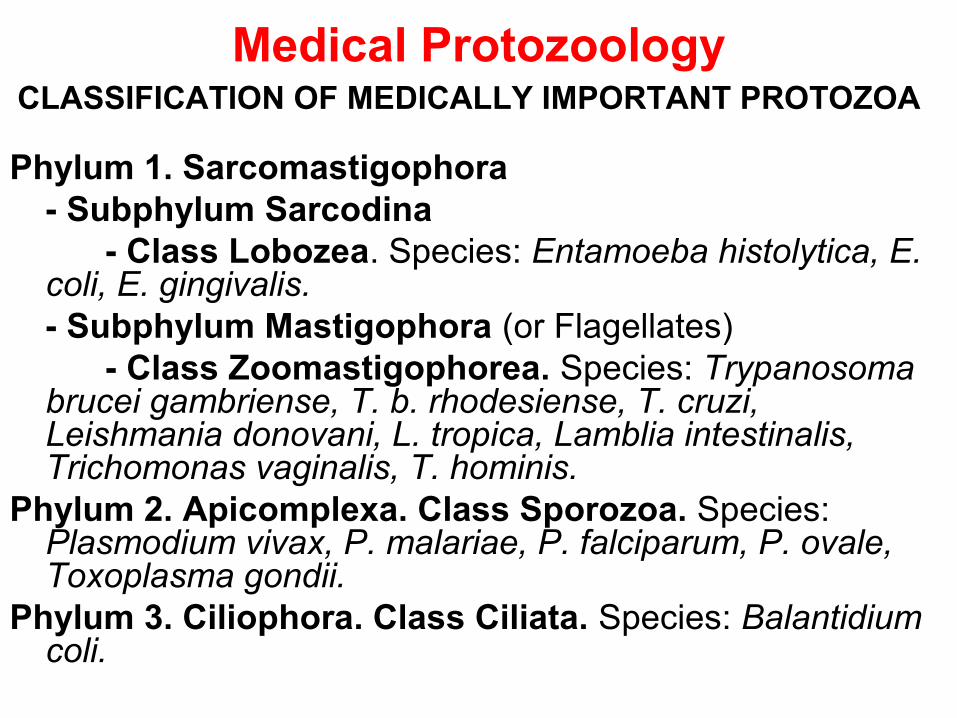
Medical ProtozoologyCLASSIFICATION OF MEDICALLY IMPORTANT PROTOZOA
Phylum 1. Sarcomastigophora - Subphylum Sarcodina
- Class Lobozea. Species: Entamoeba histolytica, E. coli, E. gingivalis.- Subphylum Mastigophora (or Flagellates)
- Class Zoomastigophorea. Species: Trypanosoma brucei gambriense, T. b. rhodesiense, T. cruzi, Leishmania donovani, L. tropica, Lamblia intestinalis, Trichomonas vaginalis, T. hominis.
Phylum 2. Apicomplexa. Class Sporozoa. Species: Plasmodium vivax, P. malariae, P. falciparum, P. ovale, Toxoplasma gondii.
Phylum 3. Ciliophora. Class Ciliata. Species: Balantidium coli.
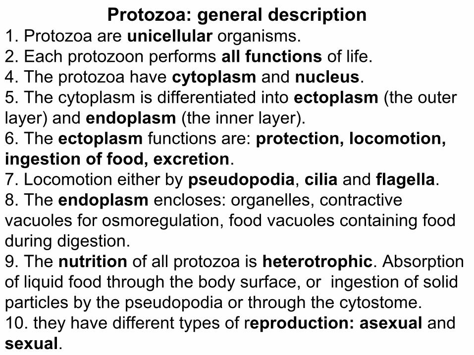
Protozoa: general description1. Protozoa are unicellular organisms. 2. Each protozoon performs all functions of life.4. The protozoa have cytoplasm and nucleus. 5. The cytoplasm is differentiated into ectoplasm (the outer layer) and endoplasm (the inner layer).6. The ectoplasm functions are: protection, locomotion, ingestion of food, excretion.7. Locomotion either by pseudopodia, cilia and flagella.8. The endoplasm encloses: organelles, contractive vacuoles for osmoregulation, food vacuoles containing food during digestion.9. The nutrition of all protozoa is heterotrophic. Absorption of liquid food through the body surface, or ingestion of solid particles by the pseudopodia or through the cytostome.10. they have different types of reproduction: asexual and sexual.
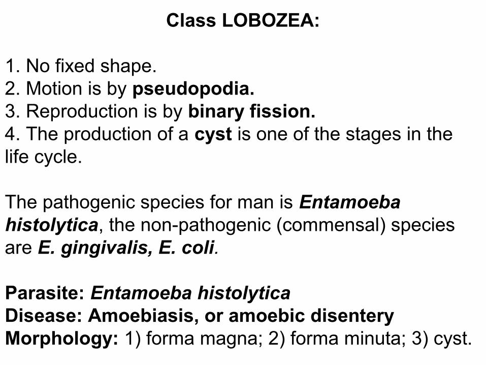
Class LOBOZEA:
1. No fixed shape.2. Motion is by pseudopodia.3. Reproduction is by binary fission.4. The production of a cyst is one of the stages in the life cycle.
The pathogenic species for man is Entamoeba histolytica, the non-pathogenic (commensal) species are E. gingivalis, E. coli. Parasite: Entamoeba histolyticaDisease: Amoebiasis, or amoebic disenteryMorphology: 1) forma magna; 2) forma minuta; 3) cyst.
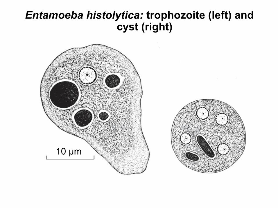
Entamoeba histolytica: trophozoite (left) and cyst (right)
10 μm
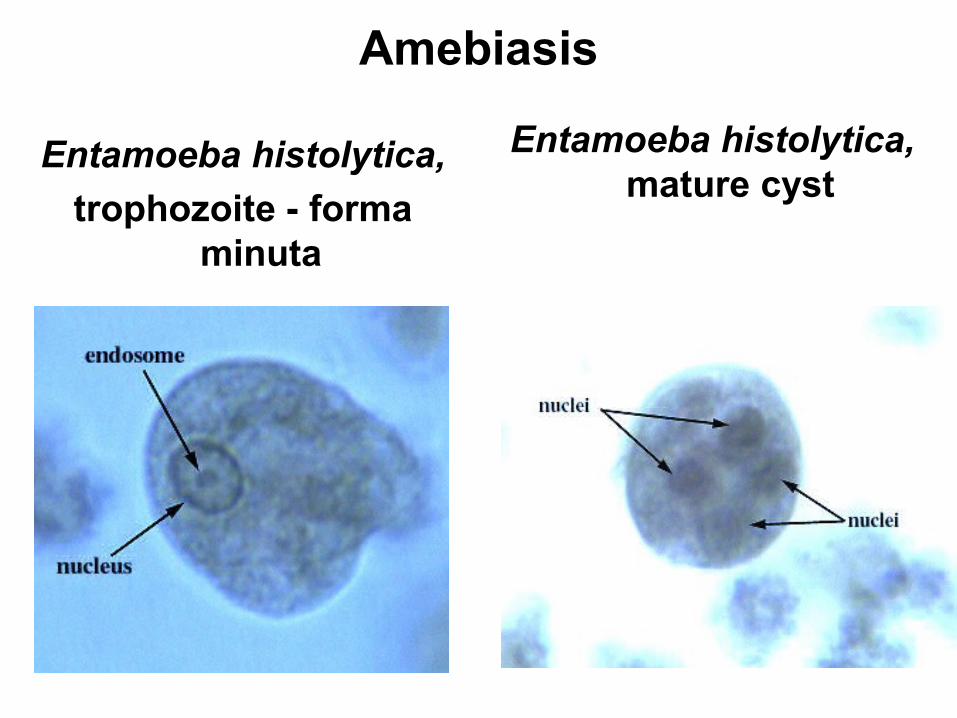
Amebiasis
Entamoeba histolytica,
trophozoite - forma minuta
Entamoeba histolytica, mature cyst
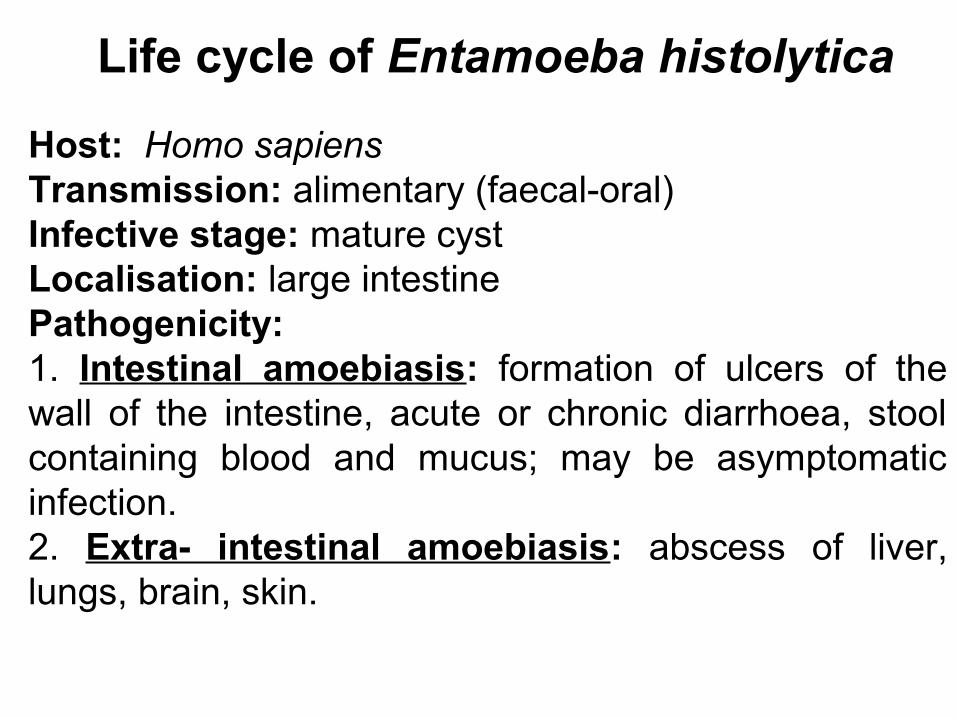
Life cycle of Entamoeba histolytica
Host: Homo sapiensTransmission: alimentary (faecal-oral)Infective stage: mature cystLocalisation: large intestinePathogenicity: 1. Intestinal amoebiasis: formation of ulcers of the wall of the intestine, acute or chronic diarrhoea, stool containing blood and mucus; may be asymptomatic infection. 2. Extra- intestinal amoebiasis: abscess of liver, lungs, brain, skin.

Life cycle of Entamoeba histolytica
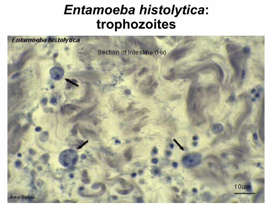
Entamoeba histolytica:trophozoites

Amebic Colitis:Severe dysentery with multiple ulcers in the large
bowel, and a bloody diarrhea
Amoebiasis
Laboratory diagnosis: Fresh stools are examined under the microscope. E. histolytica (forma magna and cysts with 4 nuclei) can be demonstrated in the stools. Prevention: Treatment of patients and asymptomatic cyst carriers; protection of foodstuffs and water from flies and contamination with faeces; the staff of catering establishments must be examined for cysts carriage; health education of the population.
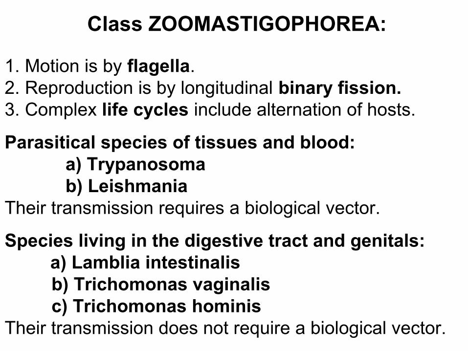
Class ZOOMASTIGOPHOREA:
1. Motion is by flagella. 2. Reproduction is by longitudinal binary fission.3. Complex life cycles include alternation of hosts.
Parasitical species of tissues and blood: a) Trypanosoma b) LeishmaniaTheir transmission requires a biological vector.
Species living in the digestive tract and genitals: a) Lamblia intestinalis
b) Trichomonas vaginalisc) Trichomonas hominis
Their transmission does not require a biological vector.
Parasites: Trypanosoma brucei gambiense and Trypanosoma brucei rhodesienseDisease: African trypanosomiasis, or sleeping sicknessGeographical distribution: Western and Central AfricaTransmission: by bite of infected tsetse flies (Glossina palpalis)Reservoir hosts of T.b.gambiense are: man, domestic pig, cattle, dog, antelopeReservoir hosts of T.b.rhodesiense are: antelope, lion, hyenaLocalisation: blood, lymph nodes, cerebrospinal fluid, brain, muscles
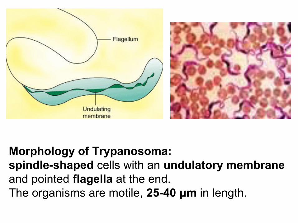
Morphology of Trypanosoma: spindle-shaped cells with an undulatory membrane and pointed flagella at the end.The organisms are motile, 25-40 μm in length.
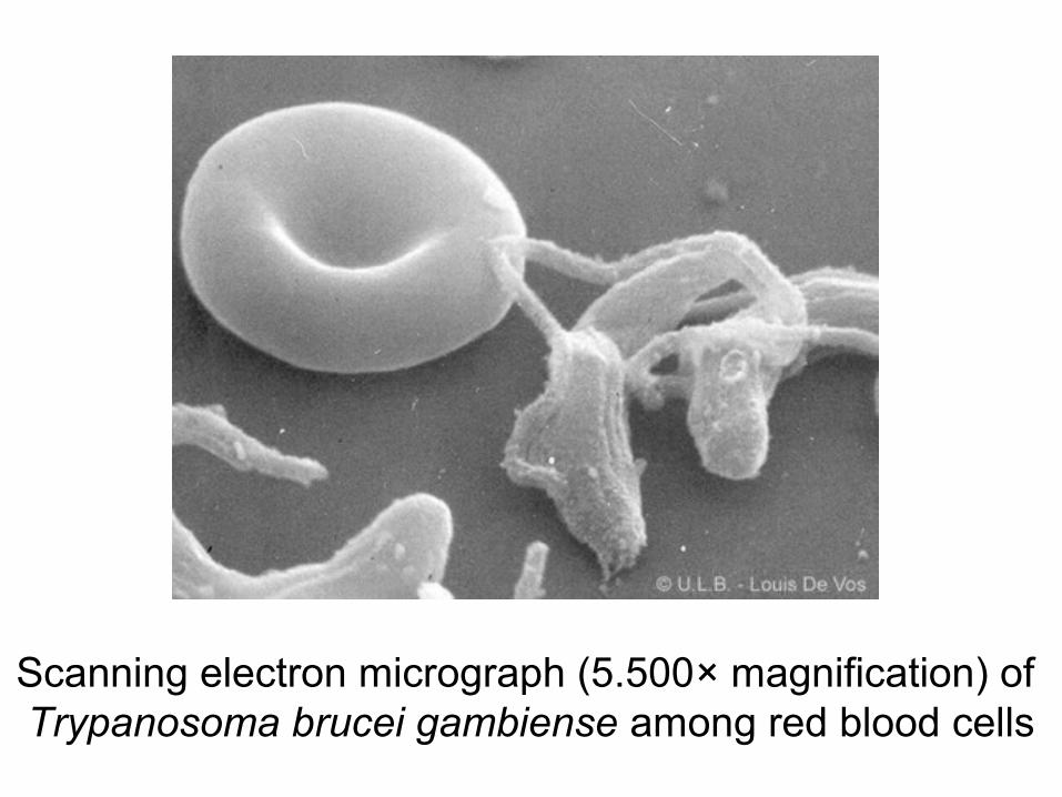
Scanning electron micrograph (5.500× magnification) of Trypanosoma brucei gambiense among red blood cells
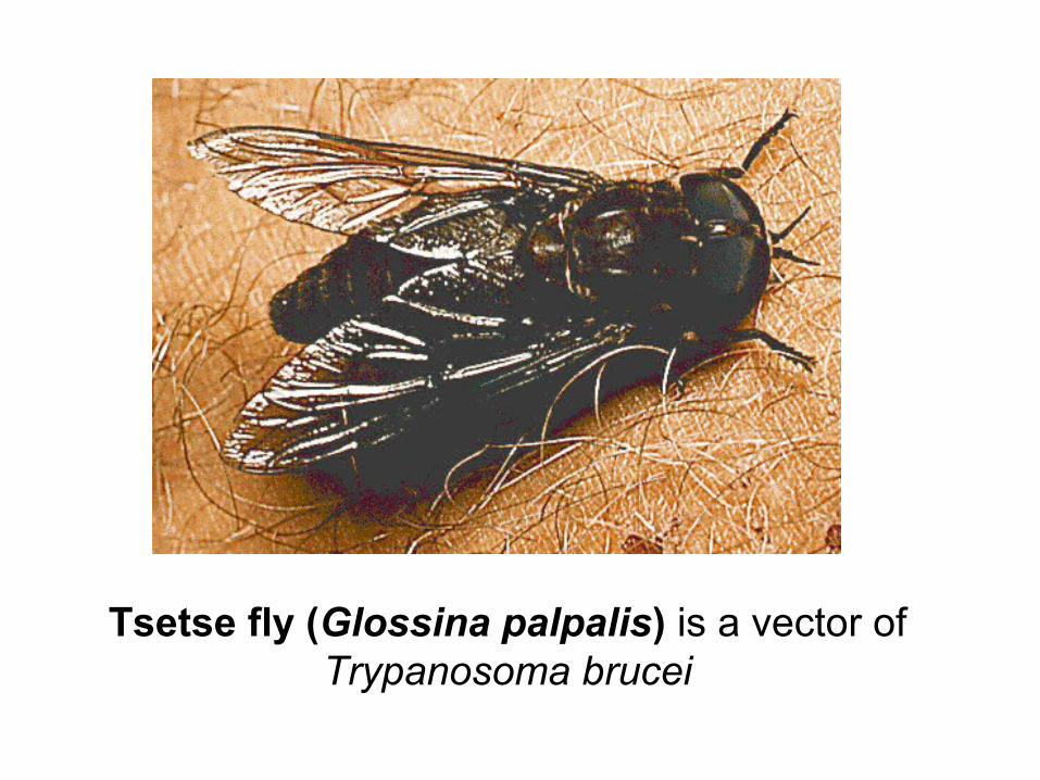
Tsetse fly (Glossina palpalis) is a vector of Trypanosoma brucei

Life cycle of Trypanosoma brucei
Pathogenicity:
1. From the place of bite trypanosomes have spread in the blood and lymphatics, where they multiply.
2. There is perivascular infiltration with chronic inflammation, leading to meningoencephalitis.
3. The patient suffers from fever, rash, headache, lymphadenopathy, oedema of the brain. There are alternating periods of fever and apparent recovery. This is followed by depression and progressive lethargy.
4. Rhodesien form develops within weeks to months, Gambian form develops within years. The disease becomes chronic and persists for months and even years.
Parasite: Trypanosoma cruziDisease: American trypanosomiasis or “Chagas’ disease” (discovered in 1909 by C. Chagas in Brazil)Geographical distribution: Southern and Central AmericaTransmission: 1) by bite of infected bug species of family Triatomidae; 2) congenital; 3) by blood transfusionReservoir hosts: armadillos, opossums, rodents, monkeys, dogs, catsLocalisation: blood (in acute phase), cells of lymph nodes, spleen, liver, brain, muscles

Bug of family Triatomidae is vector of Trypanosoma cruzi

T. cruzi in a cardiac muscle

Clinical manifestation:
fever, oedema of the face, and enlargement of the thyroid glands, lymph nodes, spleen, and liver, heart alterations

Parasite: Leishmania tropicaDisease: Cutaneus leishmaniasisGeographical distribution: Asia, Africa, EuropeTransmission: by sand fly vector - Phlebotomus sergenti (in Iran, Iraq, and India); Phlebotomus papatasi (in southern France, Italy, and certain Mediterranean islands)Hosts: man, dogs, wild rodentsLocalisation: cells of skin
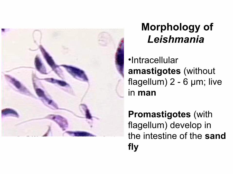
Morphology of Leishmania
•Intracellular amastigotes (without flagellum) 2 - 6 μm; live in man
Promastigotes (with flagellum) develop in the intestine of the sand fly
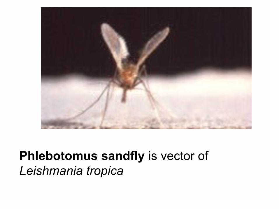
Phlebotomus sandfly is vector of Leishmania tropica

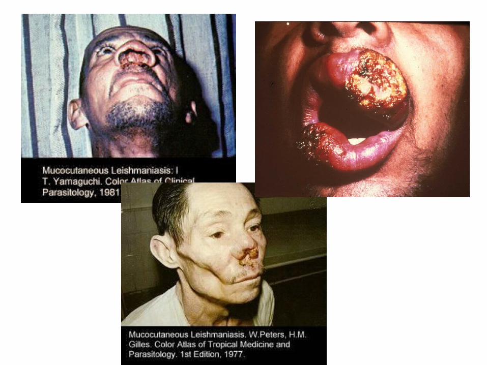
Clinical manifestation: development of a cutaneous papule that evolves into a nodule, breaks down to form an indolent ulcer, and heals, leaving a depressed scar.
Laboratory diagnosis: detection of the Leishmania parasites in cells of skin. Prevention: early diagnosis, extermination of sandflies and dogs and rodents infected with leishmaniasis, and vaccination.
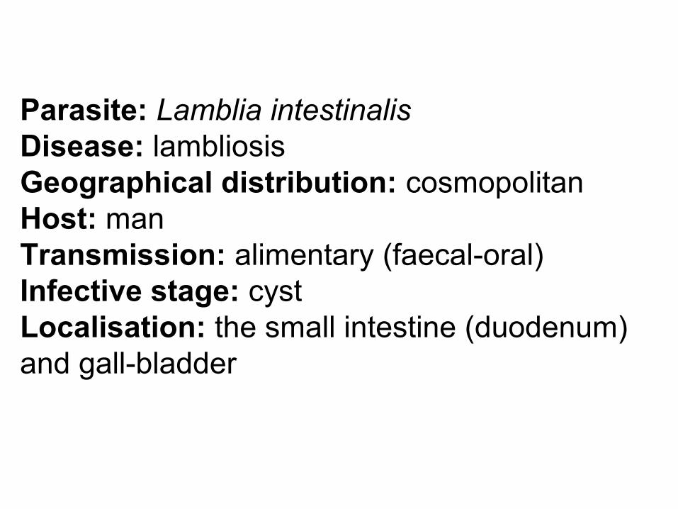
Parasite: Lamblia intestinalisDisease: lambliosisGeographical distribution: cosmopolitanHost: manTransmission: alimentary (faecal-oral)Infective stage: cystLocalisation: the small intestine (duodenum) and gall-bladder
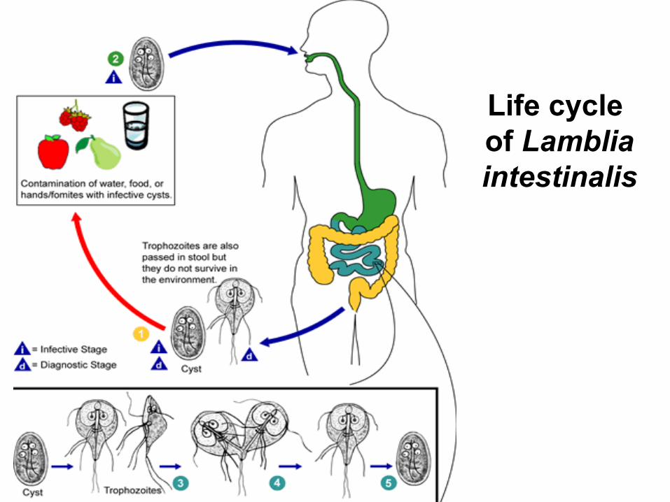
Life cycle of Lamblia intestinalis

Lamblia intestinalis
Morphology: Trophozoites are symmetrical, pear-shaped organisms with 2 nuclei. The body is 10-18 μm with four pairs of flagella. Cysts are oval-shaped which are 10 - 14 micro;m and have 4 nuclei.
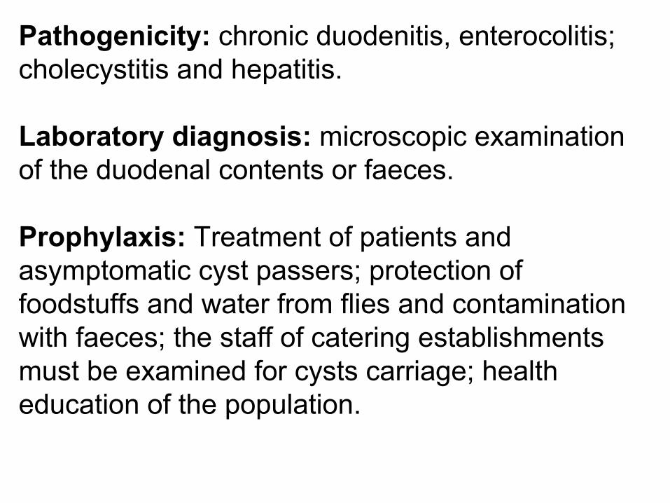
Pathogenicity: chronic duodenitis, enterocolitis; cholecystitis and hepatitis.
Laboratory diagnosis: microscopic examination of the duodenal contents or faeces.
Prophylaxis: Treatment of patients and asymptomatic cyst passers; protection of foodstuffs and water from flies and contamination with faeces; the staff of catering establishments must be examined for cysts carriage; health education of the population.
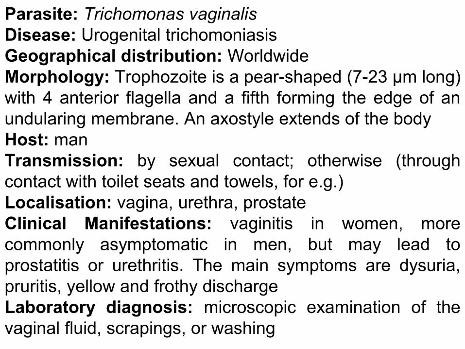
Parasite: Trichomonas vaginalisDisease: Urogenital trichomoniasisGeographical distribution: WorldwideMorphology: Trophozoite is a pear-shaped (7-23 μm long) with 4 anterior flagella and a fifth forming the edge of an undularing membrane. An axostyle extends of the bodyHost: manTransmission: by sexual contact; otherwise (through contact with toilet seats and towels, for e.g.)Localisation: vagina, urethra, prostateClinical Manifestations: vaginitis in women, more commonly asymptomatic in men, but may lead to prostatitis or urethritis. The main symptoms are dysuria, pruritis, yellow and frothy dischargeLaboratory diagnosis: microscopic examination of the vaginal fluid, scrapings, or washing

Trichomonas vaginalis
Class Sporozoa:
1. lack locomotory organelles;2. complex life cycles (sexual and asexual phases);3. alternation of hosts;4. the pathogenic species for man are: Plasmodium
vivax, P. malariae, P. falciparum, P. ovale, Toxoplasma gondii.
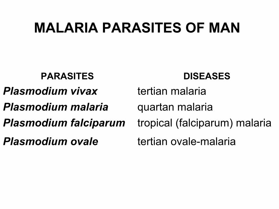
MALARIA PARASITES OF MAN
PARASITES DISEASES
Plasmodium vivax tertian malaria
Plasmodium malaria quartan malaria
Plasmodium falciparum tropical (falciparum) malaria
Plasmodium ovale tertian ovale-malaria

Geographical distribution of malaria: in parts of Africa, Asia, Turkey, the West Indies,
Central and South America, Oceania
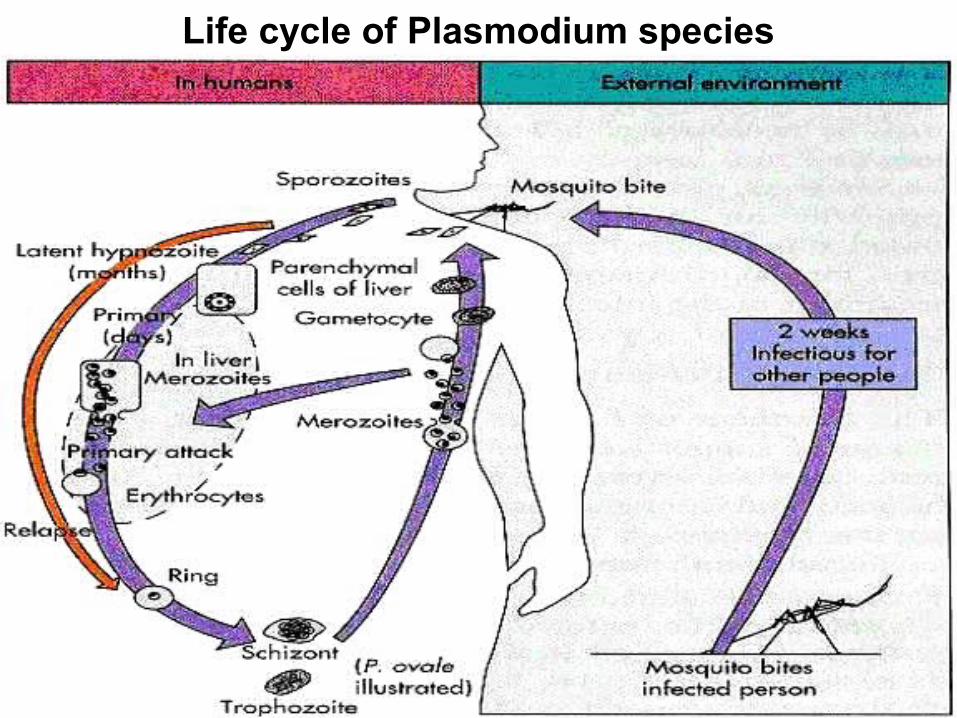
Life cycle of Plasmodium species

LIFE CYCLE OF THE MALARIA PARASITEExoerythrocytic schizogony (liver phase)
1.Mosquito bites man, takes blood meal and injects sporozoites from its salivary gland into the blood.
2.Sporozoites travel through blood to the liver, multiply asexually to form merozoites, which upon liver cell rupture, are released into the bloodstream and infect erythrocytes.
Erythrocytic schizogony (blood phase)1.Merozoites enter the erythrocytes, forming a ring-like trophozoite; mature
trophozoites asexually divide to form schizonts. 2.Schizont develops into merozoite daughter cells, then lyses the
erythrocytes membrane, leading to periodic paroxysms of disease due to resultant parasitemia. P. ovale, P. vivax, P. falciparum - membrane lysis in 48 hours, P. malariae - membrane lysis in 72 hours.
3.Some merozoites develop into macrogametocyte and microgametocyte. Sporogony
1.Mosquito ingests gametocytes with blood meal. 2.Gametocytes enter mosquito gut. 3.Zygote, formed from fertilization, invades gut wall to form an oocyst within
24 hours following ingestion 4.Sporozoites are formed, released into the stomach, migrate to salivary
glands, then injected into human with blood meal.
MALARIA PARASITES OF MAN
Intermediate host: Homo sapiensDefinitive host: Anopheles mosquitoTransmission: by bite of female Anopheles mosquito Infective stage for man: sporozoiteInfective stage for mosquito: gametocyteLocalisation: liver, bloodPrevention: chemoprophylaxis and personal protective measures against the mosquito vector (Anopheles).
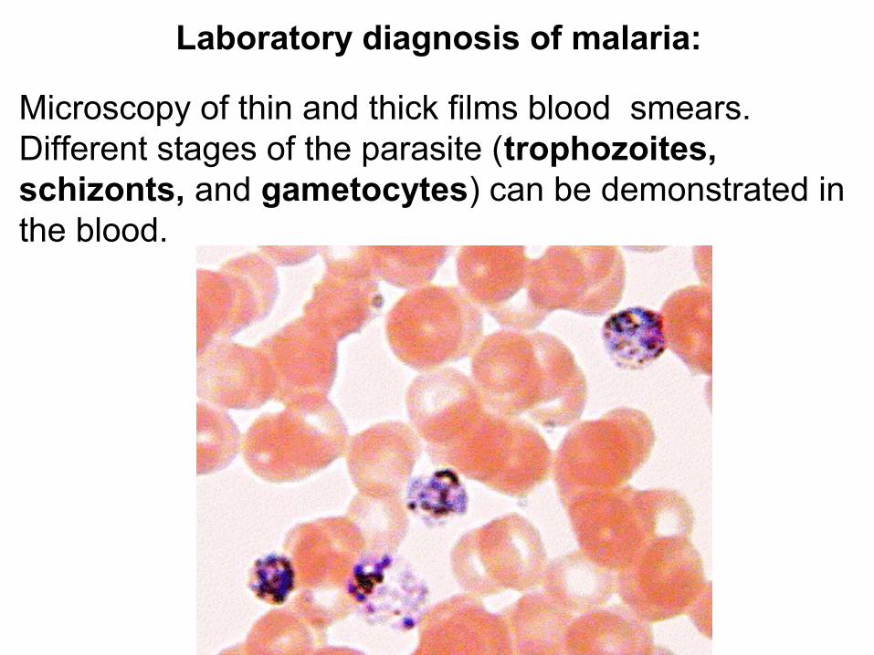
Laboratory diagnosis of malaria:
Microscopy of thin and thick films blood smears. Different stages of the parasite (trophozoites, schizonts, and gametocytes) can be demonstrated in the blood.

Parasite: Toxoplasma gondii Disease: toxoplasmosis Intermediate hosts: birds and mammals, including
humans Definitive hosts: cats Localisation: brain, eyes, skeletal and cardiac
muscles, liver, and lungs Transmitted to humans by: 1) ingestion of undercooked infected meat (cysts
and pseudocysts); 2) contamination of food or drink with infected cat
faeces (oocyts); 3) transplacental (congenital)
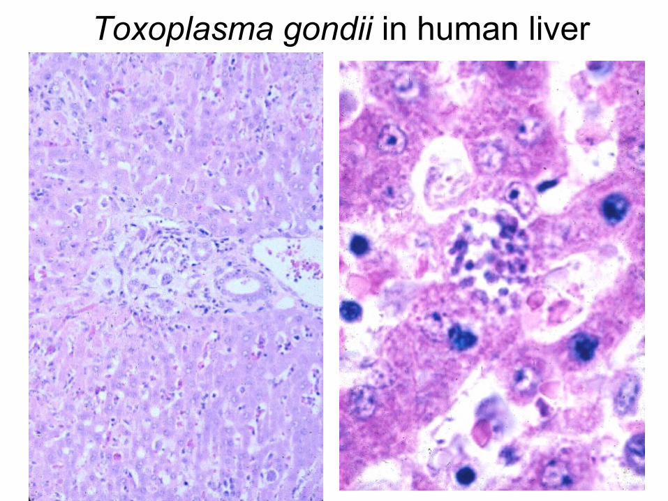
Toxoplasma gondii in human liver

lung heart muscle

Life cycle of Toxoplasma gondii
Prevention of toxoplasmosis:
washing of hands before meals and after handling animals and animal products;the prohibition of preparing food from insufficiently cooked meat products, in particular liver;all women with a history of spontaneous abortion must be examined by laboratory methods for prevention of congenital toxoplasmosis.
Class Ciliata
1. Move by cilia, which are numerous and cover most of the body
2. Have 2 nuclei, macronucleus containing vegetative chromatin and micronucleus containing generative chromatin
3. Reproduce by transverse binary fission, and sometimes by conjugation
Parasite: Balantidium coliDisease: BalantidiasisGeographical distribution: WorldwideMorphology: The trophozoite is 75-200 μm, oval, with cilia. Cyst is 30-60 μmHosts: man, domestic hogTransmission: alimentary (faecal-oral)Localisation: large intestineClinical Manifestations: colitis, ulcers and abscesses of colon, diarrhoea, blood and mucus in the stoolLaboratory diagnosis: microscopic examination of the faecesPrevention: protection of foodstuffs and water from contamination with swine faeces and observation of individual hygiene when talking care of the domestic hog
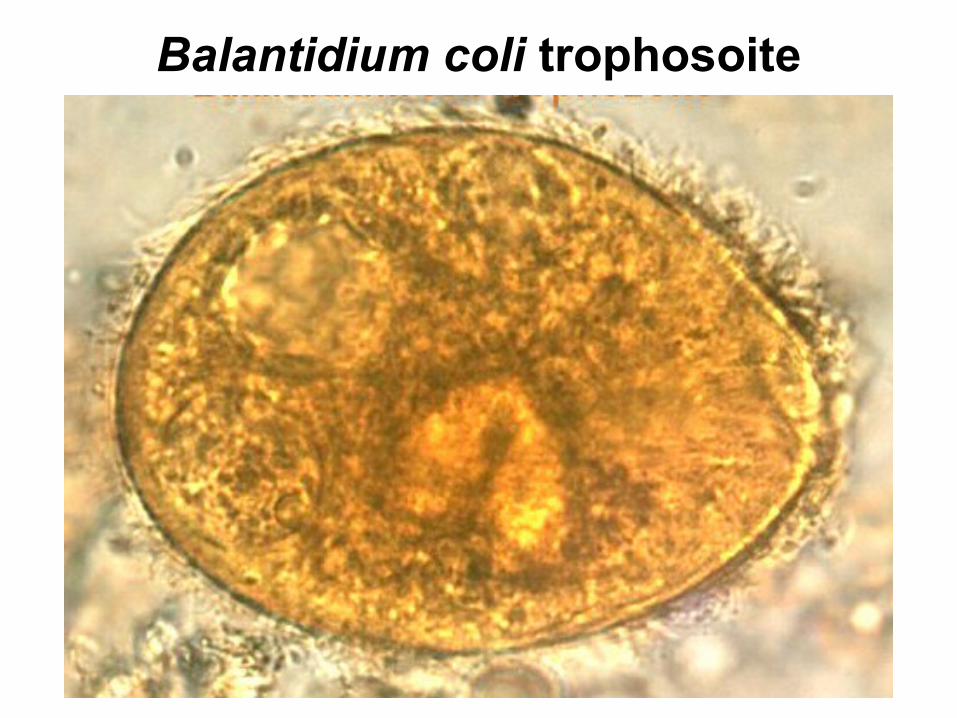
Balantidium coli trophosoite

The lecture is over,
thanks for your attention


