HAL Id: hal-00195036 https://hal.archives-ouvertes.fr/hal-00195036 Submitted on 8 Dec 2007 HAL is a multi-disciplinary open access archive for the deposit and dissemination of sci- entific research documents, whether they are pub- lished or not. The documents may come from teaching and research institutions in France or abroad, or from public or private research centers. L’archive ouverte pluridisciplinaire HAL, est destinée au dépôt et à la diffusion de documents scientifiques de niveau recherche, publiés ou non, émanant des établissements d’enseignement et de recherche français ou étrangers, des laboratoires publics ou privés. Medical image computing and computer-aided medical interventions applied to soft tissues. Work in progress in urology Jocelyne Troccaz, Michael Baumann, Peter Berkelman, Philippe Cinquin, Vincent Daanen, Antoine Leroy, Maud Marchal, Yohan Payan, Emmanuel Promayon, Sandrine Voros, et al. To cite this version: Jocelyne Troccaz, Michael Baumann, Peter Berkelman, Philippe Cinquin, Vincent Daanen, et al.. Medical image computing and computer-aided medical interventions applied to soft tissues. Work in progress in urology. Proceedings of the IEEE, Institute of Electrical and Electronics Engineers, 2006, 94 (9), pp.1665-1677. <hal-00195036>
Transcript
HAL Id: hal-00195036https://hal.archives-ouvertes.fr/hal-00195036
Submitted on 8 Dec 2007
HAL is a multi-disciplinary open accessarchive for the deposit and dissemination of sci-entific research documents, whether they are pub-lished or not. The documents may come fromteaching and research institutions in France orabroad, or from public or private research centers.
L’archive ouverte pluridisciplinaire HAL, estdestinée au dépôt et à la diffusion de documentsscientifiques de niveau recherche, publiés ou non,émanant des établissements d’enseignement et derecherche français ou étrangers, des laboratoirespublics ou privés.
Medical image computing and computer-aided medicalinterventions applied to soft tissues. Work in progress in
urologyJocelyne Troccaz, Michael Baumann, Peter Berkelman, Philippe Cinquin,Vincent Daanen, Antoine Leroy, Maud Marchal, Yohan Payan, Emmanuel
Promayon, Sandrine Voros, et al.
To cite this version:Jocelyne Troccaz, Michael Baumann, Peter Berkelman, Philippe Cinquin, Vincent Daanen, et al..Medical image computing and computer-aided medical interventions applied to soft tissues. Work inprogress in urology. Proceedings of the IEEE, Institute of Electrical and Electronics Engineers, 2006,94 (9), pp.1665-1677. <hal-00195036>
2Urology department, La Pitié Salpêtrière Hospital, Paris
3Radiotherapy Department, Grenoble Hospital
4Urology Department, Grenoble Hospital
Abstract
Until recently, Computer-Aided Medical Interventions (CAMI) and Medical Robotics have
focused on rigid and non deformable anatomical structures. Nowadays, special attention is
paid to soft tissues, raising complex issues due to their mobility and deformation. Mini-
invasive digestive surgery was probably one of the first fields where soft tissues were handled
through the development of simulators, tracking of anatomical structures and specific
assistance robots. However, other clinical domains, for instance urology, are concerned.
Indeed, laparoscopic surgery, new tumour destruction techniques (e.g. HIFU, radiofrequency,
or cryoablation), increasingly early detection of cancer, and use of interventional and
diagnostic imaging modalities, recently opened new challenges to the urologist and scientists
involved in CAMI. This resulted in the last five years in a very significant increase of research
and developments of computer-aided urology systems. In this paper, we propose a description
of the main problems related to computer-aided diagnostic and therapy of soft tissues and give
a survey of the different types of assistance offered to the urologist: robotization, image
fusion, surgical navigation. Both research projects and operational industrial systems are
discussed.
Keywords
Computer-aided surgery, medical robotics, medical image registration, urology.
1. Introduction
1.1 A short introduction to urology
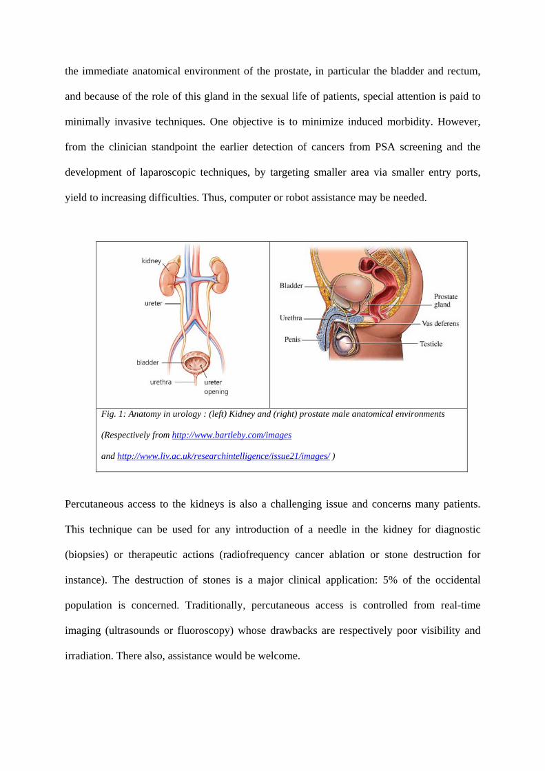
Urology concerns the exploration, diagnostic and medical or surgical treatment of both the
urinary apparatus of men and women and the genital apparatus of men. The organs of interest
are the bladder, kidney, ureter, urethra and, for men, the prostate, penis and testicles (see Fig.
1). Pathologies include among others: lithiases (stones), cancers, traumas, stenoses,
incontinence, infectious diseases, malformations and sterility. Urologic surgery also includes
kidney transplantation. The major targets for robot or image-guided assistance are the prostate
and the kidneys as detailed below.
Prostate cancer is one the most common malignancy among men. [Parkin01] reports year
2000 cancer statistics: 543000 cases and 204000 deaths were attributed to prostate cancer
worldwide. Its detection is based on digital rectal examination (DRE) and Prostate Specific
Antigen (PSA) rating and is confirmed through the anatomo-pathologic analysis of biopsies.
Treatments include watchful waiting, surgery (laparoscopic or conventional radical
prostatectomy), chemotherapy, and destruction of the tumour using different physical agents
including radiotherapy (radiation by external beams), brachytherapy (radiation by implanted
radioactive seeds), HIghly Focused Ultrasound, radiofrequency, and cryoablation. Because of
the immediate anatomical environment of the prostate, in particular the bladder and rectum,
and because of the role of this gland in the sexual life of patients, special attention is paid to
minimally invasive techniques. One objective is to minimize induced morbidity. However,
from the clinician standpoint the earlier detection of cancers from PSA screening and the
development of laparoscopic techniques, by targeting smaller area via smaller entry ports,
yield to increasing difficulties. Thus, computer or robot assistance may be needed.
Fig. 1: Anatomy in urology : (left) Kidney and (right) prostate male anatomical environments
(Respectively from http://www.bartleby.com/images
and http://www.liv.ac.uk/researchintelligence/issue21/images/ )
Percutaneous access to the kidneys is also a challenging issue and concerns many patients.
This technique can be used for any introduction of a needle in the kidney for diagnostic
(biopsies) or therapeutic actions (radiofrequency cancer ablation or stone destruction for
instance). The destruction of stones is a major clinical application: 5% of the occidental
population is concerned. Traditionally, percutaneous access is controlled from real-time
imaging (ultrasounds or fluoroscopy) whose drawbacks are respectively poor visibility and
irradiation. There also, assistance would be welcome.
1.2 Dealing with soft tissues
Because urology deals with soft tissues, it is a perfect illustration of the difficulty to directly
apply the computer-assistance know-how from bony structures to mobile and deformable
tissues. Mobility and deformations have different origins:
- Intrinsic origin: some organs intrinsically move or are deformed to perform natural
physiological activity such as breathing or cardiac rhythm. A foetus organ also has an
intrinsic mobility due to foetal motion. Heart beating is quite predictable whilst foetal
motion is not.
- Anatomical environment: other organs move or are deformed because of their
anatomical environment. This is typically the case for the kidney that is moved up and
down according to the diaphragmatic activity during breathing; this is also true for the
prostate which position and orientation depend on the bladder and rectal filling, and in
a lesser way, breathing.
- Patient position: the position and shape of some structures depend on the patient
posture or position relatively to gravity. For example, the prostate position partly
depends on the flexion of patient’s legs.
- External action: finally, the therapeutic (needle insertion for instance) or diagnostic
(e.g. ultrasound examination) action may move and deform the organ of interest. This
is the case for the kidney and even more for the prostate, especially when an
endorectal ultrasonic exam is performed.
Very often, the motion and deformation of an organ has multiple sources. For instance, the
prostate moves and/or is deformed due to: patient breathing, patient posture, bladder and
rectum natural or artificial filling, insertion of a needle, oedema from multiple needle
insertions.
In the case of the prostate, several groups worldwide paid special attention in the mid-nineties
to motion of the gland in the context of radiotherapy; since most intra-treatment localization
approaches were based on X-Ray data where the prostate is not directly visible, it was
important to quantify prostate motion with respect to bony structures. [VanHerk95] performed
separate CT/MR bone and prostate registrations to determine prostate mobility; rigid chamfer
matching on segmented surfaces was used; [Balter95] used implanted prostate fiducials and
X-ray images to perform a similar study. More recently deformation was studied specially in
the context of imaging involving intrarectal probes or coils (see for instance [Hirose02]).
In order to be able to handle these anatomic changes several issues must be solved: models
must be designed when the motion and/or deformation are predictable and repeatable;
tracking capabilities must be developed based on intra-operative sensing (images, signals
such as ECG); real-time re-planning may be necessary for the guiding system (for instance a
robot) to adapt to these changes; finally robots should be synchronized to those motions and
deformations in a discrete or continuous way. This raises very challenging robustness and
safety issues. One important characteristic of those anatomic changes is their time scale with
respect to the duration of the action to be performed. Consequently, different strategies may
be selected: localization just before the action or tracking during the whole action. In the
following sections, the way those questions have been solved in the case of urological targets
will be analyzed.
2. Robotics and urology
Historically, urology was one of the first clinical domains where a robot was used for patients.
At the time – the late eighties – where most people dealt with neurosurgery or orthopaedics
applications of robotics, the London Clinic and the Imperial College of London developed
PROBOT [Davies91]: a robot for the transurethral resection of the adenomatous prostate, i.e.
the removal from the inside of the gland of extra-tissues compressing the urethra. The first
test on a patient started in April 1991. After a feasibility study on 5 patients, a pre-clinical
series with 40 patients was undertaken. Several versions of this system where developed; the
first prototype was based on a PUMA 560 (from Unimation Inc.) connected to a passive
frame. This frame is an elegant solution to safety issues since it constrains the tool movement
inside a cone related to the task to be executed. The current system consists of a passive robot
positioning a motorized frame with 3 degrees of freedom (dof) – conical motion plus
translation of the resectoscope. [Shah01] reports the difficult task of automatically controlling
this robot for resection monitoring from the real-time intra-operative ultrasound images.
Indeed, because soft tissues move and deform, two types of strategies may be used in robot
control. The ideal approach would be to continuously and automatically close the robot
control loop using intra-operative information about the organ motion. To our knowledge,
such a solution has not yet been developed for urology. However in radiotherapy, where the
tool is outside the body and the planning is rather simple (beam orientation with respect to.
the patient and duration of radiation), organ tracking ability was introduced. In [Coste05] the
motions of intra-body implanted fiducials are correlated to the motions of infra-red on-body
markers for tracking breathing movements this process is however rather invasive.
[Sawada04] proposes a non invasive solution based on real-time image correlation for the
detection of a pre-defined stage in the breathing cycle (full expiration for instance); this
information is used for respiratory-gated radiotherapy treatment. The other and much simpler
approach is to tele-operate robots: in that case the user closes the loop between robot motion
and real-time image information. Such an approach is particularly interesting when operative
planning is too complex to be explicitly defined. Intermediate solutions consist in adding
motion tracking abilities to tele-operated robots (see [Ginhoux05]) or to close the loop from
imaging data in a more discrete way for simple tasks (see section 2.2).
2.1 Tele-operated robots
2.1.1 Endoscope holders
The first FDA1 approved medical robot, AESOP (from Computer Motion Inc.) [Sackier94]
had a significant clinical and industrial success. Two thousand AESOP were sold to around
five hundred hospitals between years 1994 and 2000. AESOP has a SCARA architecture with
4 active and 2 passive (pivot rotation) dof; this tele-manipulator is voice controlled. Many
other robotic endoscope holders have been developed in the academic and industrial tracks.
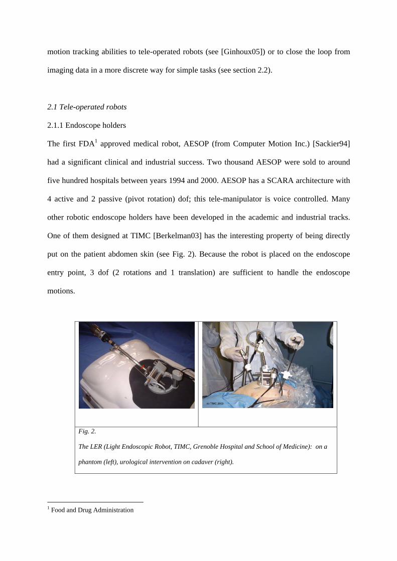
One of them designed at TIMC [Berkelman03] has the interesting property of being directly
put on the patient abdomen skin (see Fig. 2). Because the robot is placed on the endoscope
entry point, 3 dof (2 rotations and 1 translation) are sufficient to handle the endoscope
motions.
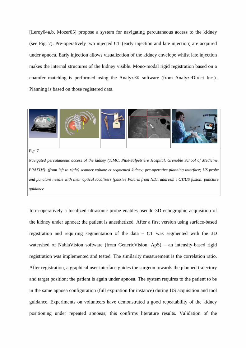
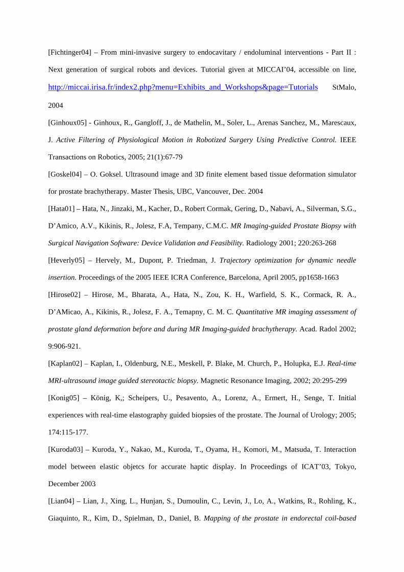
Fig. 2.
The LER (Light Endoscopic Robot, TIMC, Grenoble Hospital and School of Medicine): on a
phantom (left), urological intervention on cadaver (right).
1 Food and Drug Administration
As compared to AESOP and to most of the other systems which are positioned on the
operating room (OR) table, floor or ceiling, this very compact system follows the patient
motions and is very easy to install. It weights 625g; it is voice controlled and completely
sterilizable. Interesting evolutions of robotic endoscope holders deal with automatically
control of robots from image information in order to track organs or instruments during the
surgery (see [Voros06] for instance).
2.1.2 Tele-surgery robots
Based on the robotic endoscope holders experience, instrument holders have naturally been
designed resulting in the so-called tele-surgery robots. ZEUS, an evolution of the AESOP, is
composed of 3 separated 4 dof arms (one endoscope holder and two instrument holders).
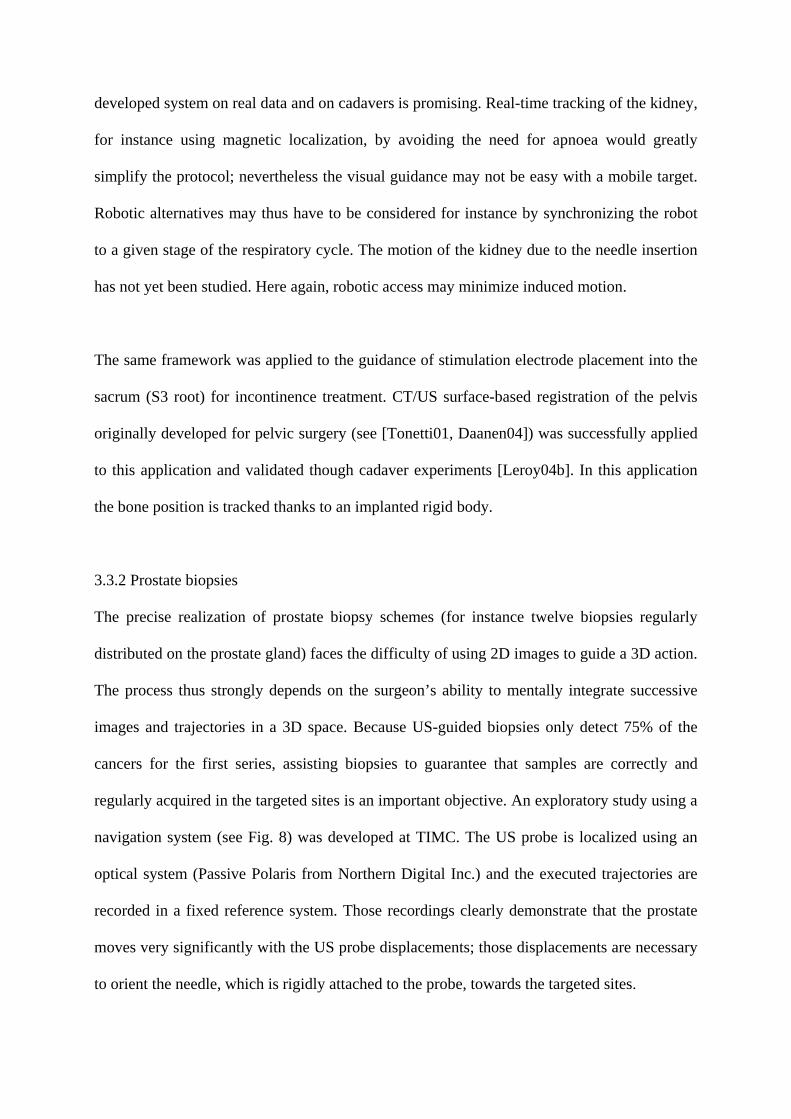
Another system, the DaVinci (from Intuitive Surgical Inc.), is composed of 3 or 4 arms
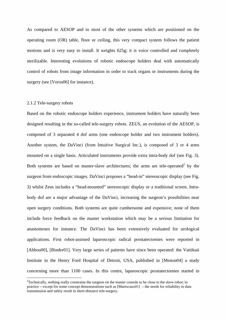
mounted on a single basis. Articulated instruments provide extra intra-body dof (see Fig. 3).
Both systems are based on master-slave architectures; the arms are tele-operated2 by the
surgeon from endoscopic images. DaVinci proposes a “head-in” stereoscopic display (see Fig.
3) whilst Zeus includes a “head-mounted” stereoscopic display or a traditional screen. Intra-
body dof are a major advantage of the DaVinci, increasing the surgeon’s possibilities near
open surgery conditions. Both systems are quite cumbersome and expensive; none of them
include force feedback on the master workstation which may be a serious limitation for
anastomoses for instance. The DaVinci has been extensively evaluated for urological
applications. First robot-assisted laparoscopic radical prostatectomies were reported in
[Abbou00], [Binder01]. Very large series of patients have since been operated: the Vattikuti
Institute in the Henry Ford Hospital of Detroit, USA, published in [Menon04] a study
concerning more than 1100 cases. In this centre, laparoscopic prostatectomies started in 2Technically, nothing really constrains the surgeon on the master console to be close to the slave robot; in practice – except for some concept demonstrations such as [Marescaux01] – the needs for reliability in data transmission and safety result in short-distance tele-surgery.
October 2000 and the DaVinci assistance was introduced in March 2001. A study comparing
conventional/laparoscopic/robot-assisted laparoscopic procedures showed clear advantages of
the robotic series on many points including shorter hospital stays, reduced pain, reduced
blood loss, better PSA control, reduced positive margins, better continence, and less
impotence. Another advantage of robot assistance is a reduction of the learning curve for
laparoscopic procedures; [Alhering03] reports an improvement factor of about 10.
Fig. 3.
The DaVinci robot (Intuitive Surgical Device Inc., http://www.intuitivesurgical.com):the master-slave system (left
and middle); and one of its articulated instruments showing intra-body dof (right)
Laparoscopic radical prostatectomy is probably one of the domains were the robotic clinical
added-value was so clearly demonstrated. Other applications of such robots to urology are
reported in full details in [UroCLin04]. Each time complex dissections, microsurgery or intra-
corporal suturing are necessary, the robot may be a precious assistant.
Several research projects in the world aim at developing competitive smaller and/or cheaper
solutions with articulated intra-body instruments and endoscopes and master station offering
force feedback. Planning tools are also developed in order to optimize the entry ports
positioning, enabling both target access and collision-free motion of the robots (see for
instance [Coste04]).
2.2 Image-guided robots
Many gestures in urology are carried under interventional radiology: the diagnostic or
therapeutic tool is moved under control of an imaging modality. Ultrasounds or fluoroscopy
enable continuous control: the operator can see in real-time the tool position and the anatomy;
CT or MRI allow asynchronous control: for instance, a needle is positioned, a control image
is taken and the needle position is corrected if necessary, and so on. This idea has been
exploited to control from medical images robots performing simple tasks such as a linear tool
insertion.
2.2.1 Prostate biopsies and brachytherapies
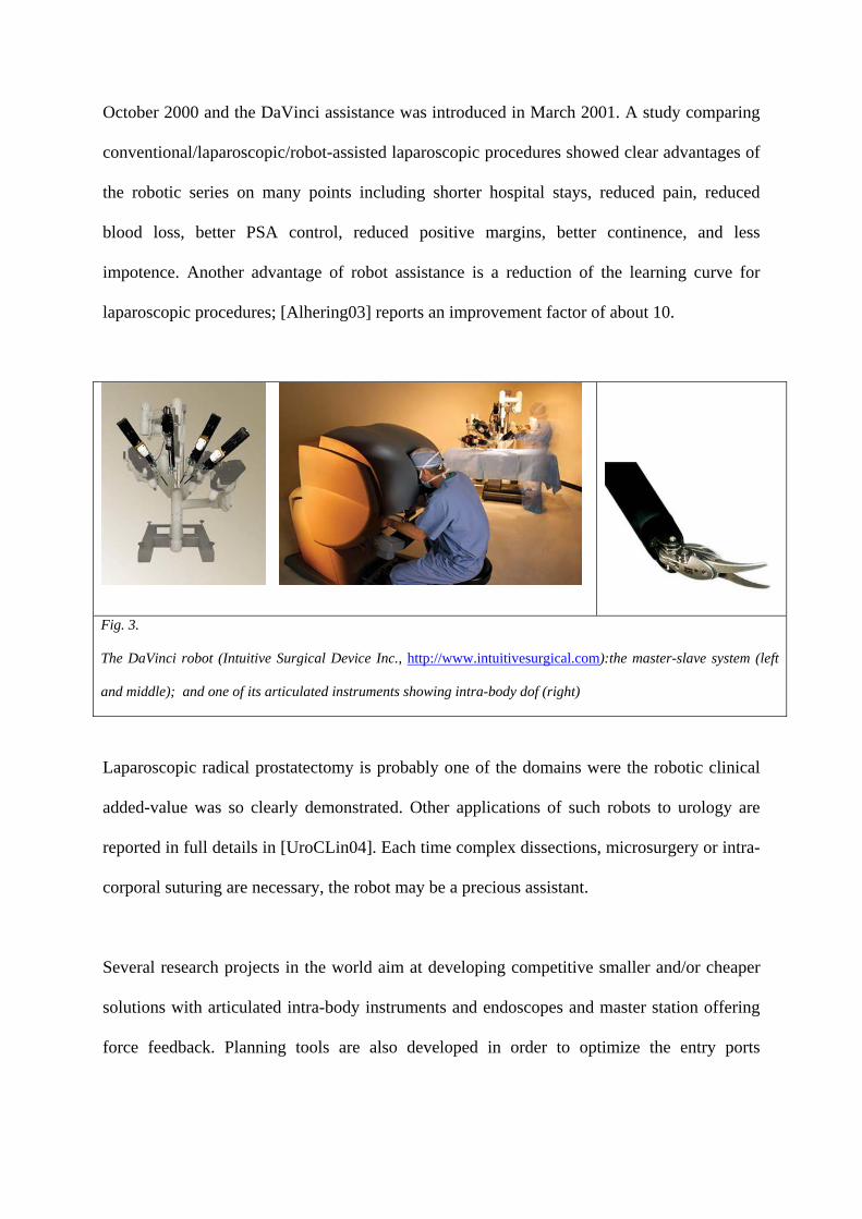
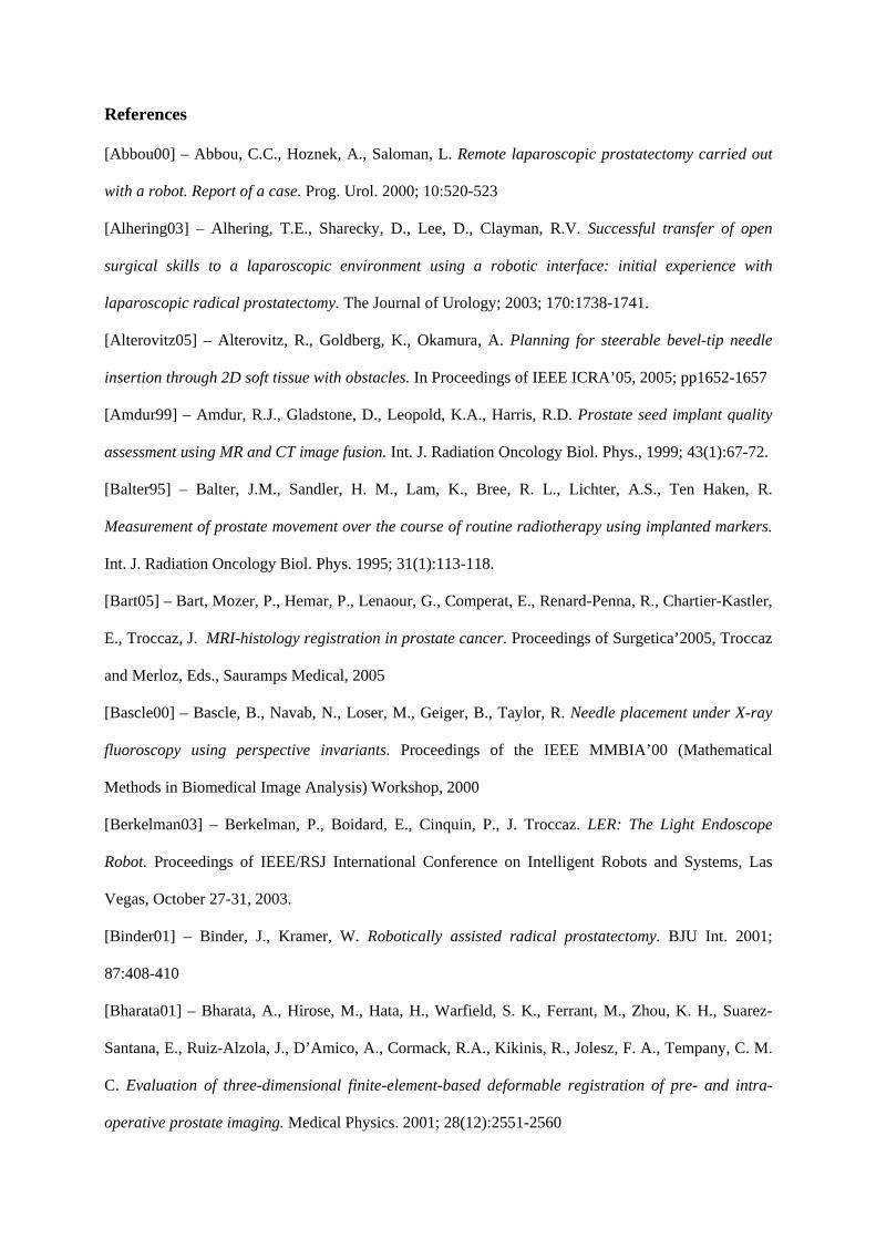
From a technical viewpoint prostate biopsies and brachytherapies (see figure 4) are rather
similar; they both consist in inserting needles in the prostate, either for tissue sampling or for
radioactive seed placement, through transperineal or transrectal access, under imaging control
– most often TransRectal UltraSound imaging (TRUS).
Fig. 4.
US-guided transrectal biopsy (left) and transperineal brachytherapy (right) operating principles.
From http://www.uropage.com/index.htm
However, each biopsy makes use of a single ultrasonic (US) image in which the needle is
visible whilst brachytherapy is based on a volume of images: often parallel axial US images
acquired every 5mm. Brachytherapy is based on a careful patient-specific dose planning
whilst biopsies are generally performed following a predefined global scheme (for instance
sextant or 11-core protocols). Needle insertion is slow and manual during brachytherapies
whilst biopsies are very rapidly performed using a biospy gun. As demonstrated by
[Heverly05] in a different medical context, increasing needle velocity results in minimizing
the displacement and deformation of the tissue. Thus automation may have a positive impact
in terms of gesture accuracy. Moreover, prostate brachytherapy is based on the use of a
template (a stereotactic grid) rigidly connected to the US probe. This restrains needle
trajectories to lines parallel to the probe axis and results in potential collisions of the needles
with the pelvic bone. Again, using a robot may enable various trajectory directions. [Wei04]
proposes to use a general purpose 6 dof robot for needle positioning and insertion. [Davies04]
develops a special-purpose robot mimicking the conventional procedure (trajectories parallel
to the US probe axis); a rotational dof for the needle is added to reduce the needle flexion
during tissue penetration. Those systems are still laboratory test beds. [Phee05] describes a
pre-clinical evaluation of a specific 9 dof system (positioning platform plus biopsy robot) for
transperineal prostate biopsies. A 3D prostate geometric model of the prostate is
approximated from series of close parallel US images enabling planning of the biopsies.
2.5mm accuracy is reported; those performances require very careful patient preparation and
US probe handling. None of these systems really considers prostate motion and deformation
during the procedure.
Another approach consists in performing transrectal prostate biopsies or brachytherapies with
an intra-rectal robot under MRI control [Susil03]. Although conventional MR imaging (1.5T
with endorectal antenna and T2 sequence) enables physicians to see precisely the prostate
anatomy, using such a modality for biopsies is probably restricted to the few cases where US-
guided biopsies are not possible or not successful. Let us remind that in the United States
(resp. in France) about 106 (resp. 105) series of diagnostic biopsies are performed each year).
In [Susil03], conventional MRI is used. The robot is inserted in the patient rectum and has
three dof to reach the target defined on the MRI data: translation in the rectum, rotation
around its main axis and progression of the needle. Thanks to its design, the robot does not
disturb the magnetic field and includes two coils; one used as part of the imaging sensor and
the other one as a position sensor. After validation on dogs, the robot is being clinically tested
for transrectal biopsies and brachytherapies [Fichtinger04]; 2mm accuracy is reported; this
remaining error is probably mainly due to the prostate motion and deformation during needle
insertion. [Chinzei00] proposes another robot for prostate biopsy or brachytherapy inside an
open interventional MR system. Interventional MRI (0.5T) requires an additional
conventional MRI exam which makes the procedure even more complex (see 3.2.1).
2.2.2 Percutaneous renal access
The purpose is to assist percutaneous access to the kidney. Since 1996 a robot named PAKY
(Percutaneous Access of the KidneY) is developed by the Johns Hopkins groups in Baltimore
(MD, USA). The robot has seven passive dof used to position a 3 active dof structure (2 for
orientation and 1 for translation of the needle). Fluoroscopy is used for needle alignment and
control during insertion. During the procedure, the patient is in apnoea in order to keep the
kidney in a constant position. [Cadeddu98] reports in vitro and in vivo experiments. In
[Su02], for 23 patients, no significant difference is reported between the manual and robotic
procedure in terms of precision, rapidity, number of attempts, complications. One advantage
of the robotic procedure lies in the absence of irradiation of the human operator. [Bascle00]
proposes a visual servoing approach from two fluoroscopic views enabling the automatic
placement of the needle to a given target and entry point. This system was also applied to CT-
guided transperineal prostate biopsies through a single entry point.
3. Image-based urology
3.1 Image processing
Many papers propose tools to assist the segmentation of urologic images especially for the
prostate where TRUS images have been paid close attention. Segmentation may be 2D, 2.5D
(the segmentation of a given slice is used to help the segmentation of the following parallel
one) or 3D. Most successful approaches make use of active contours and/or statistical models.
However, for 2D images close to the prostate extremities, existing tools may not be robust
enough due to the poor quality of those images. Other works concern the automatic
segmentation of CT and MRI images of urological targets (kidney in particular). Because this
problem is very vast and not typical of interventional systems, no details are given here.
[Shao03] and [Zhu06] present good reviews of work concerning the image processing of
prostate TRUS images.
3.2 Image fusion
MR and US imaging are probably the most used imaging modalities for prostate diagnosis
and therapy. The interventional nature of US is counterbalanced by their traditional
drawbacks: patient dependence, intra- and inter-operator variability, medium quality due to
speckle, artefacts, etc. Conventional MRI using external or transrectal coils clearly show the
prostate zonal anatomy which is useful for biopsy planning, whilst open MRI (also called
iMRI for interventional MRI) enables near real-time control. This is why several research
groups implemented fusion algorithm to benefit from complementary advantages of these
modalities. Other imaging modalities are used such as CT imaging or histology sections; here
also image fusion may be very useful. This paragraph describes different studies on multi-
modality fusion dedicated to prostate imaging.
3.2.1 MRI/iMRI fusion
The Surgical Planning Laboratory (SPL) and Harvard Medical School have developed a
navigation system for transperineal prostate biopsies under iMRI. Because of a lower
intensity magnetic field, iMRI does not clearly show the prostate anatomy. This is why a pre-
operative MRI acquisition is performed (external pubic antenna, 1.5T, T2 FSE sequence) on
which surgical planning is possible. Intra-operatively, iMRI data are collected (external pubic