41
LOGO SPECT Imaging Single-Photon Emission Computed Tomography

LOGO
SPECT ImagingSingle-Photon Emission Computed Tomography
LOGO
Group 4
�Christopher Emerson
�Caterina Pette
�Douglas Wright

LOGO
Contents
Advantages / Disadvantages
Medical / Non-Medical Applications
History
Modern Systems
Pricing
U.S. SPECT Statistics

LOGO
Advantages
� Provides Physiological Information though Functional Imaging
� Metabolic Activity
� Blood Flow
� Intrinsic Lesion Localization though Radiopharmaceutical Use
� Radio-labeled ligans migrate to specific imaging site
� 3-D Imaging
� Hybrid Imaging Systems provide Increased Spatial Resolution
� SPECT/CT
http://xa.yimg.com/kq/groups/15914941/74581872/name/EJNMMI-SPECT%25EF%2580%25A2CT-Review.pdf,“Advantages and disadvantages of PET and SPECT in a busy clinical practice” Timothy M. Bateman, MD

LOGO
Disadvantages
� Gamma Emissions Harmful due to Ionization Potential
� Non-Hybrid Devices have Poor Spatial Resolution� Tissue boundaries are ill-determined
� Long Scan Time� Upwards of 30-40 minutes
� Inconveniences certain patient populations
� Use of Possible Allergens� Radiopharmaceuticals could induce allergic reactions
� Intrinsic Reliance on Radiopharmaceuticals� Severe supply shortages can halt imaging
http://www.massgeneral.org/imaging/services/procedure.aspx?id=2255http://blue.regence.com/trgmedpol/radiology/rad44.html

LOGO
Medical Applications
Myocardial Perfusion Imaging
Brain Perfusion
Thyroid Function
Renal Function
Bone Imaging
Myocardial Perfusion Imaging
LOGO
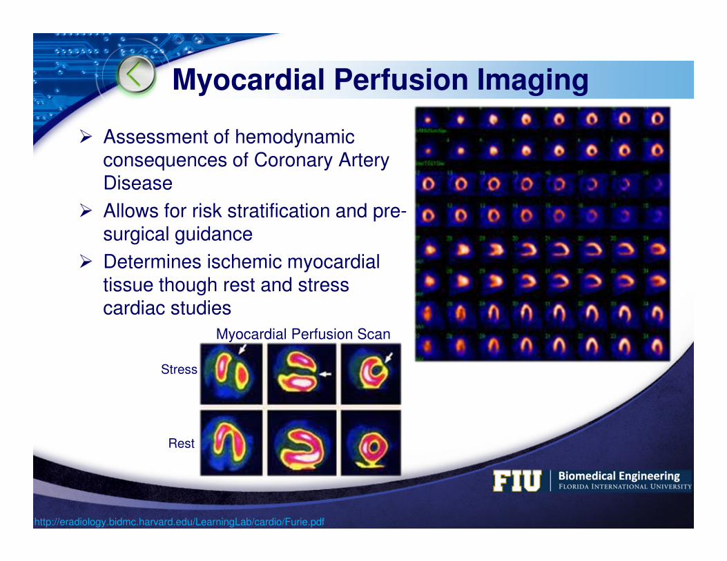
Myocardial Perfusion Imaging
� Assessment of hemodynamic consequences of Coronary Artery Disease
� Allows for risk stratification and pre-surgical guidance
� Determines ischemic myocardial tissue though rest and stress cardiac studies
Myocardial Perfusion Scan
Stress
Rest
http://eradiology.bidmc.harvard.edu/LearningLab/cardio/Furie.pdf

LOGO
Brain Perfusion
� Pre-surgical localization of epileptic seizure origin
� Technetium-99m labeled ECD or HMPAO injected at time of seizure
� Localize to region of increased blood flow
� Images during and after seizure are compared to identify seizure origin
http://my.clevelandclinic.org/Documents/Epilepsy_Center/PET%20and%20SPECT%20-%20Wu.pdf

LOGO
Thyroid Function
� Thyroid tumor causes increased metabolic activity
� Localization achieved through the use of Iodine-123 and Iodine-131
� Iodine-123: Routine testing for hyperthyroidism and nodules
� Iodine-131: Whole-body imaging for detection of metastasis
� SPECT-CT shows metastasis is localized in tip of transverse process of the sixth thoracic vertebra
A. Whole-body SPECTB. Diagnostic CTC. Fusion Image
http://www.ncbi.nlm.nih.gov/pubmed/1418597

LOGO
Renal Function
� Renal lesions can result in elevated levels of norepinehprine, epinephrine, dopamine, cathecholamines, and VMA
� Localization achieved through the use of Indium-111 Pentetreotide
� SPECT scans revealed large right suprarenal lesion
http://www.med.harvard.edu/JPNM/TF00_01/Sept19/WriteUp.html

LOGO
Bone Imaging
� Early Detection of Osteonecrosis of the Femoral Head
http://jnm.snmjournals.org/content/43/8/1006/F1.expansion.html
A. Whole-body scintigraphy
B. Bone SPECT
C. MRI Scan
� A & B show “cold” areas in both femoral heads while C shows normal findings

LOGO
Small Animal SPECT & SPECT/CT Imaging
� Used to determine drug efficacy in pre-clinical trials
http://jnm.snmjournals.org/content/49/10/1651/F5.expansion.html

LOGO
Non-Medical Applications
� Detection of nuclear waste
� Gamma cameras used to study the environmental behavior of nuclear waste
� Helps scientists monitor and control the spread of radioactivity
� Fe(III)-reducing bacteria immobilizes technetium in sediment
http://www.bloomberg.com/news/2011-08-02/tepco-reports-second-deadly-radiation-reading-at-fukushima-plant.html

LOGO
Non-Medical Applications (cont.)
� Detection of nuclear emission� Fukushima Nuclear Power Plant� Gamma cameras can be used to image radiation readings of wrecked
nuclear power plant� A photograph shows a gamma camera image of an area around the
main exhaust of Tokyo’s nuclear power plant detecting 5 Svt/hr.
http://www.bloomberg.com/news/2011-08-02/tepco-reports-second-deadly-radiation-reading-at-fukushima-plant.html
Tokyo Electric Power Co.'s (Tepco) Fukushima Dai-Ichi nuclear power station in Fukushima, Japan, on
Monday, Aug. 1, 2011.
Soil Sampling
LOGO
History
< 1950s
1950s – 1960s
1960s – 1970s
> 1970s
Why the Delay?
LOGO
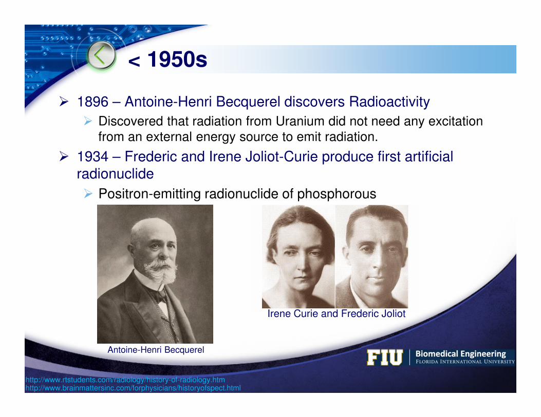
< 1950s
� 1896 – Antoine-Henri Becquerel discovers Radioactivity
� Discovered that radiation from Uranium did not need any excitation from an external energy source to emit radiation.
� 1934 – Frederic and Irene Joliot-Curie produce first artificial radionuclide
� Positron-emitting radionuclide of phosphorous
http://www.brainmattersinc.com/forphysicians/historyofspect.html
Antoine-Henri Becquerel
Irene Curie and Frederic Joliot
http://www.rtstudents.com/radiology/history-of-radiology.htm

LOGO
< 1950s
� 1943 – Gyorgy Hevesy develops radioactive tracers
� Used to study the metabolic processes in plants and animals
� 1946 – Oak Ridge National Laboratory began production of radionuclides for medical use
http://www.brainmattersinc.com/forphysicians/historyofspect.html
Gyorgy Hevesy
Oak Ridge National Laboratory
http://www.rtstudents.com/radiology/history-of-radiology.htm

LOGO
1950s – 1960s
� 1950 – Benedict Cassen assembled the first automated scanning system
� Motor-driven scintillation detector coupled to a printer
� 1957 – Hal Oscar Anger develops Anger Camera
� Sodium-Iodine scintillation crystal
� Vacuum tube photomultipliers
http://jnm.snmjournals.org/content/12/8/573.full.pdfhttp://www.ncbi.nlm.nih.gov/pubmed/11155815
Benedict Cassen Hal Oscar Anger

LOGO
1960s – 1970s� Development of generator system to produce
Technetium-99m� Most utilized element in Nuclear Medicine� Employed in a wide variety of Nuclear
Medicine studies� Late 1963 – David E. Kuhl and Roy Q. Edwards
introduce emission and transmission tomography� Later developed into Single Photon Emission
Computed Tomography (SPECT)
� Used 32 photon detectors � Images were frequently distorted and not of
diagnostic quality
http://www.news-medical.net/health/History-of-Nuclear-Medicine.aspxhttp://www.uclimaging.be/ecampus/etu_med/option_2011/opt_2011_3120_mn.pdf
David Edmund Kuhl

LOGO
> 1970s
� Late 1970s – Most organs of the body could be visualized with nuclear medicine procedures� Liver, spleen, brain tumor localization, and gastrointestinal tract
� 1980s – Radiopharmaceuticals were designed for diagnosing heart disease and cancer
� 1980s – Development of Monoclonal Antibodies � Act as cell-specific ligans that, when tagged with a radioisotope, localize
to a specific region of the body� Can detect cancerous cells before anatomical changes occur
http://interactive.snm.org/index.cfm?PageID=1107http://www.news-medical.net/health/History-of-Nuclear-Medicine.aspx

LOGO
Radionuclide Specificity
www.themegallery.com

LOGO
Why the Delay?
� The development of SPECT was slow due to a number of factors:
� Image analysis algorithms were not advanced enough
� Limited number of radionuclides available as tracers
� Device size limited SPECT use outside of hospitals
� Physicians were inexperienced reading SPECT images
� Better late than never!
� Advances in mathematics, innovations in radionuclide and liganddevelopment, and the advent of multi-modality imaging systems laid the foundation for modern SPECT and SPECT/CT devices
http://www.imagingeconomics.com/issues/articles/MI_2004-06_02.asp
I wonder what they exSPECT me to see from this…

LOGO
Modern Systems
Leading Device Manufacturers
Brivo NM 615
Discovery NM 630
Discovery NM/CT 670
Ventri
Discovery NM 750b

LOGO
Leading Device Manufacturers
�GE Healthcare
�Siemens Healthcare
�Philips Healthcare
�Toshiba Medical Systems
LOGO
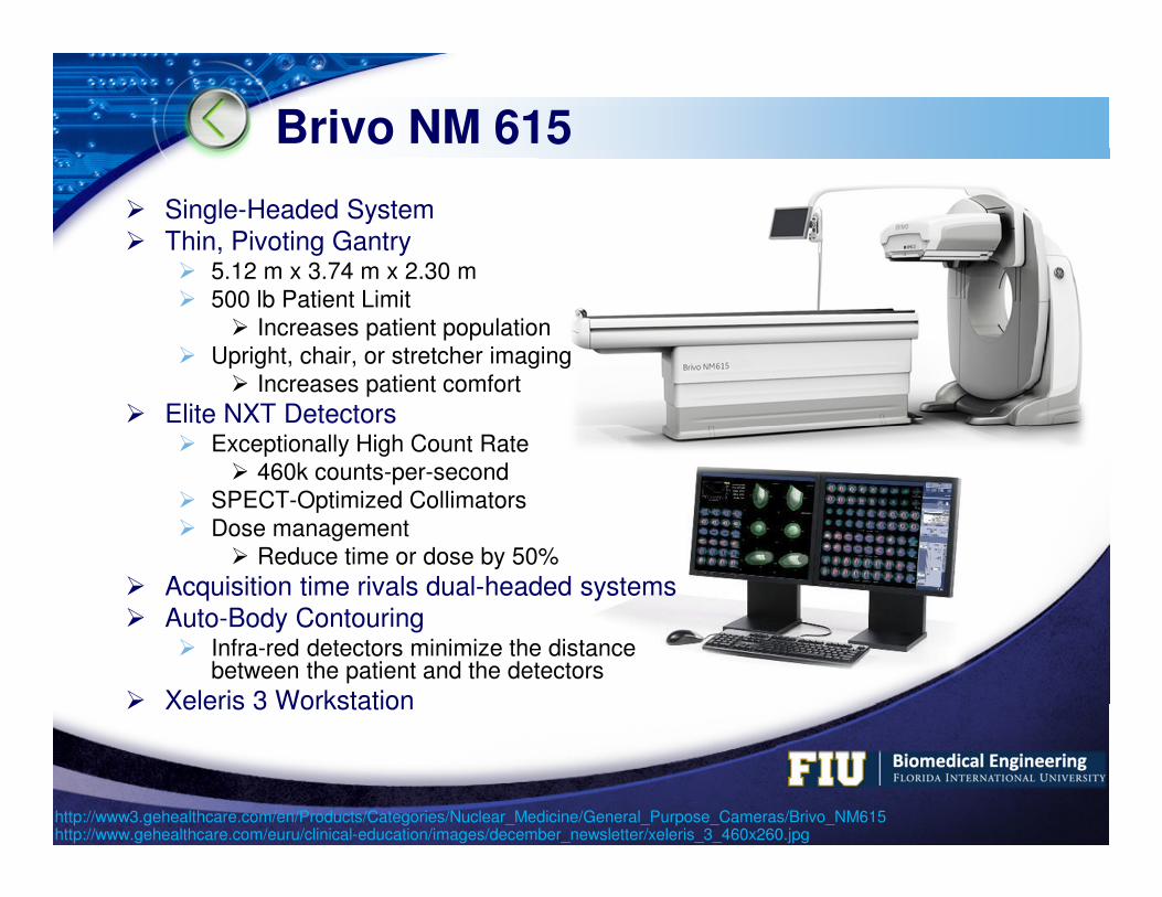
Brivo NM 615
� Single-Headed System� Thin, Pivoting Gantry
� 5.12 m x 3.74 m x 2.30 m� 500 lb Patient Limit
� Increases patient population� Upright, chair, or stretcher imaging
� Increases patient comfort
� Elite NXT Detectors� Exceptionally High Count Rate
� 460k counts-per-second� SPECT-Optimized Collimators� Dose management
� Reduce time or dose by 50%
� Acquisition time rivals dual-headed systems� Auto-Body Contouring
� Infra-red detectors minimize the distance between the patient and the detectors
� Xeleris 3 Workstation
http://www3.gehealthcare.com/en/Products/Categories/Nuclear_Medicine/General_Purpose_Cameras/Brivo_NM615http://www.gehealthcare.com/euru/clinical-education/images/december_newsletter/xeleris_3_460x260.jpg

LOGO
Brivo NM 615
http://www3.gehealthcare.com/~/media/Downloads/Product/Product-Categories/Nuclear-Medicine/General-Purpose-Scanners/Brivo-NM615/GEHealthcare-Brochure_Brivo-NM-615.pdf?Parent={83A02FFC-6F7B-48B3-AB0F-AC550678E0B0}

LOGO
Discovery NM 630
http://www3.gehealthcare.com/en/Products/Categories/Nuclear_Medicine/General_Purpose_Cameras/Discovery_NM_630
� Upgrade from Brivo NM 615
� Dual-Headed System
� Cut imaging time or dose in half, again.
� 180°or 90°Orientations
LOGO
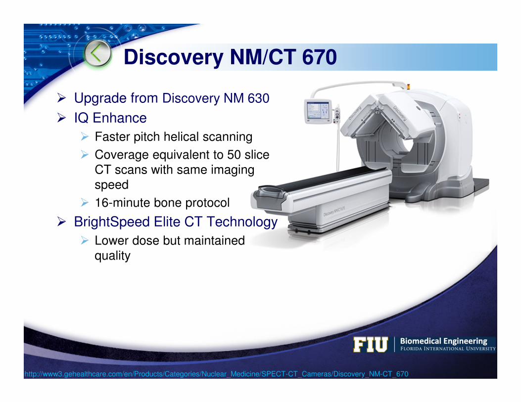
Discovery NM/CT 670
� Upgrade from Discovery NM 630
� IQ Enhance
� Faster pitch helical scanning
� Coverage equivalent to 50 slice CT scans with same imaging speed
� 16-minute bone protocol
� BrightSpeed Elite CT Technology
� Lower dose but maintained quality
http://www3.gehealthcare.com/en/Products/Categories/Nuclear_Medicine/SPECT-CT_Cameras/Discovery_NM-CT_670
LOGO
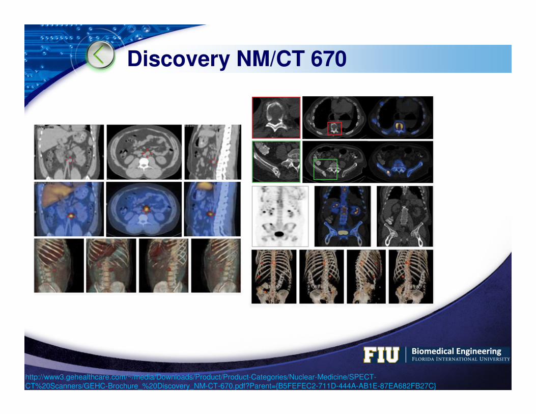
Discovery NM/CT 670
http://www3.gehealthcare.com/~/media/Downloads/Product/Product-Categories/Nuclear-Medicine/SPECT-CT%20Scanners/GEHC-Brochure_%20Discovery_NM-CT-670.pdf?Parent={B5FEFEC2-711D-444A-AB1E-87EA682FB27C}

LOGO
Ventri
� Originally Designed for Cardiac Imaging� Restricted Imaging Range
� Now offers Neurological Imaging
� Smaller Office Footprint
� Less expensive
� Similar Technology as Larger Devices
http://www3.gehealthcare.com/en/Products/Categories/Nuclear_Medicine/Cardiac_Cameras/Ventri

LOGO
Ventri
http://www3.gehealthcare.com/en/Products/Categories/Nuclear_Medicine/Xeleris_3/Evolution_for_Cardiac

LOGO
Discovery NM 750b
� Dedicated Breast Imaging� Solid-state Cadmium Zinc Telluride
Detectors� 3 times the imaging sensitivity of
conventional NaI gamma cameras
� Degradation-free Uniformity across entire Field-of-View
� Overcomes Breast Density challenges� Single or Dual-Head Configuration
� MLO = Mediolateral Oblique View
� CC = Cranio – Caudal View
http://www3.gehealthcare.com/en/Products/Categories/Nuclear_Medicine/Discovery_NM-CT_750b
LOGO
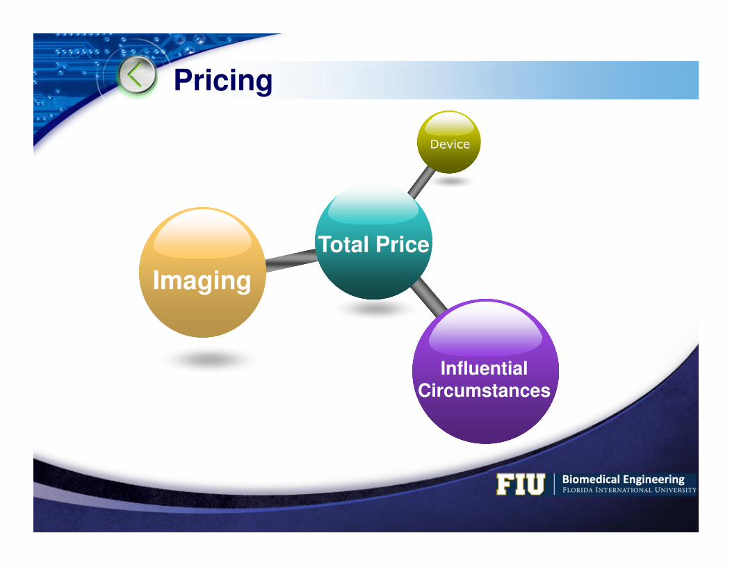
Pricing
Total Price
Device
Imaging
InfluentialCircumstances

LOGO
Imaging
� Base Imaging Cost
� Radiopharmaceuticals Used
� Interpretation by Radiologist
Organ Price
Bone Imaging Scan: $585.20Radiopharmaceutical: $62.35
Liver / Spleen Imaging Scan: $993.30Radiopharmaceutical: $77.90
Brain Imaging Scan: $809.75
Renal Imaging Scan: $996.08
Joint Imaging Scan: $1016.00
Myocardial Perfusion Imaging Scan: $2902.00
http://outofpocket.com/OOP/Default.aspx?q=SPECT&cx=015166729515697606531%3a3sy7u0rpz2o&cof=FORID%3a9
LOGO
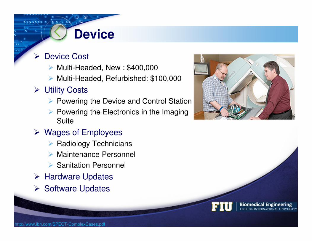
Device
� Device Cost
� Multi-Headed, New : $400,000
� Multi-Headed, Refurbished: $100,000
� Utility Costs
� Powering the Device and Control Station
� Powering the Electronics in the Imaging Suite
� Wages of Employees
� Radiology Technicians
� Maintenance Personnel
� Sanitation Personnel
� Hardware Updates
� Software Updates
http://www.ibh.com/SPECT-ComplexCases.pdf
LOGO
Influential Circumstances
� Age of Imaging System
� Recently Purchased� Hospital may charge more till it breaks even
� Post Breaking-Even� Hospital may charge less since device has
“paid-for-itself”
� Neighboring Hospital Competition
� Rich Competition� May charge less to attract more customers
� Poor Competition� May charge more to capitalize on pseudo-
monopoly
� Pending Lawsuits
LOGO
U.S. SPECT Statistics
Imaging Locations and Frequency
Installed Devices and Patient Waiting Times
Projected Purchases
LOGO
Imaging Locations and Frequency
� ~ 17 million Nuclear Medicine procedures performed at 7,230 different sites in 2010� Procedure frequency decreased by 0.5% each year from 2007 to 2010
� 87% of procedures conducted in nonhospital locations are cardiovascular studies
� 47% of procedures conducted in hospital locations are cardiovascular studies� More likely to be conducting other procedures including bone scans, liver,
renal, respiratory, infection/abscess, and tumor localization studies.
� 1/3 of Nuclear Imaging sites are physician office locations� Includes cardiology offices, multispecialty clinics, and imaging centers
� ~ 25% of imaging sites provide neurology applications� Projected to grow to 1/3 of sites by 2013
� Driven by development of radiopharmaceuticals to address Parkinson's disease
http://www.auntminnie.com/index.aspx?sec=vdp&sub=def&pag=dis&itemId=96830http://184.107.144.35/pressreleases/imv-nuclear-medicine-report-shows-nuclear-medicine-procedure-volume-flat-from-2007-to-2010.html
LOGO
Installed Devices and Patient Waiting Times
� Dual-head SPECT installations comprise 64% of the gamma camera installed base� Down from 70% in 2008
� SPECT/CT camera installed base increased from 2% of the installed base in 2008 to 9% in 2011
� 69% of the SPECT/CT and SPECT camera installed base are considered to be general purpose
� 31% are dedicated cardiac cameras
� Patient waiting times for nuclear imaging procedures have decreased� Waiting times of 1+ days for scheduled outpatient procedures
decreased from 77% of the sites in 2003 to 43% of the sites in 2011
http://184.107.144.35/pressreleases/imv-nuclear-medicine-report-shows-nuclear-medicine-procedure-volume-flat-from-2007-to-2010.htmlhttp://www.auntminnie.com/index.aspx?sec=vdp&sub=def&pag=dis&itemId=96830
LOGO
Projected Purchases
� Estimated replacement time of 12.8 years for a typical gamma camera
� 85% of purchases are replacements
� 1 in 6 planned camera purchases through 2013 will be from physician offices
� ~ 50% are Dual-head SPECT cameras
� 33.33% are SPECT/CT systems
� Gaining momentum
http://184.107.144.35/pressreleases/imv-nuclear-medicine-report-shows-nuclear-medicine-procedure-volume-flat-from-2007-to-2010.htmlhttp://www.auntminnie.com/index.aspx?sec=vdp&sub=def&pag=dis&itemId=96830

LOGO


