57
MEDICAL MANAGEMENT OF ACUTE STROKE Dr Ganesh Subramanian, Stroke Physician
MEDICAL MANAGEMENT
OF
ACUTE STROKE
Dr Ganesh Subramanian, Stroke Physician

Objectives
To able to: Explain the Bamford classification of stroke, describing the prognostic difference
between each stroke type
Describe the acute management of stroke, with particular attention to examination,
investigations, consideration/initiation of antiplatelet therapy, anticoagulation,
thrombolysis, thrombectomy, blood pressure control, statins therapy
List immediate non-pharmacological measures in management of stroke such as
assessment of swallow, rehabilitative and nursing care
Outline measures undertaken in secondary stroke prevention
Outline methods of evaluating and managing patients with carotid stenosis
Outline medical (and surgical) management for TIA
Outline the commonest causes of disability in people with impaired mobility

What is Stroke/TIA?
Acute onset
Focal
Neurological deficit(s)
24 hours or more
Vascular origin
But....

Stroke Mimics
Migraine
Space-occupying lesions
Seizure
Syncope
Metabolic disturbance
Peripheral neuropathy
Cervical spine pathologies
Transient global amnesia
Psychiatric conditions

Why is stroke important?
Common
111000 first or recurrrent strokes/ year
High morbidity
Commonest long term neurological disability
High mortality
3rd commonest killer, 1/3 of all strokes
Expensive
£8+ billions/year (direct, informal care, loss of productivity)
Highly treatable
Largely preventable

Assessment (Pre-Hosp)
Time is brain!!!

Assessment (A&E)
ROSIER Has there been loss of consciousness: Y (-1) N (0)
Has there been seizure activity: Y (-1) N (0)
Is there a new onset (or waking from sleep)?
Asymmetric facial weakness :Y (+1) N (0)
Asymmetric arm weakness: Y (+1) N (0)
Asymmetric leg weakness: Y (+1) N (0)
Speech disturbance: Y (+1) N (0)
Visual field defect: Y (+1) N (0)
Stroke is likely if total score > 0
Scores of < / = 0 have low probability of stroke but not excluded

Assessment (1)
History taking – very similar in the main to any
other condition
PMH
Drug Hx
Family Hx
Social Hx – very important
When?
How?
What?

What can be affected?
Usually negative symptoms
Motor
Speech
Vision
Sensation
Coordination
Conscious level
Memory
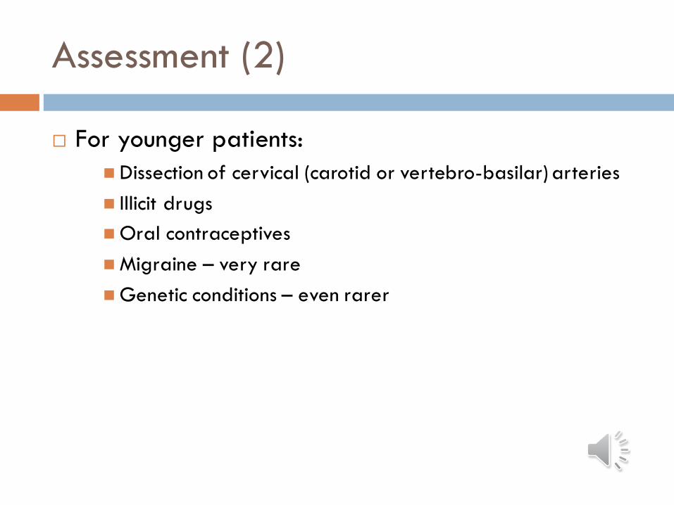
Assessment (2)
For younger patients:
Dissection of cervical (carotid or vertebro-basilar) arteries
Illicit drugs
Oral contraceptives
Migraine – very rare
Genetic conditions – even rarer

Assessment (3)
Clinical examination
General inspection
GCS
ABC
CVS – murmur, bruit, DVT, BP, pulse, HF, SBE, dissection
Resp – O2 sats, RR, pneumonia
Gastro – mass
Neuro – UMN/LMN, Speech, Sensory, Cerebellar, CN

NIHSS
Validated, reliable, reproducible
Provides insight to location of stroke
Provides insight to severity of stroke
Help identify those who will benefit from
thrombolysis
Assessment (4)

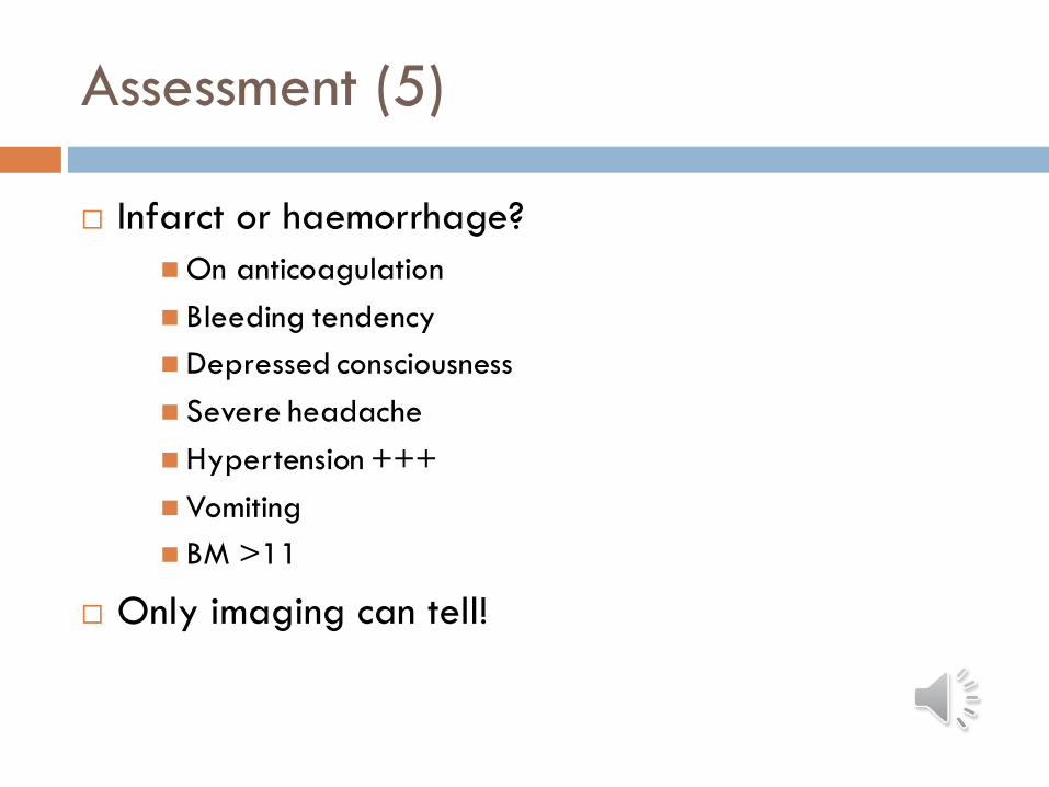
Assessment (5)
Infarct or haemorrhage?
On anticoagulation
Bleeding tendency
Depressed consciousness
Severe headache
Hypertension +++
Vomiting
BM >11
Only imaging can tell!

Investigation (1)
Bloods
FBC, U+E, LFT, TFT
Glucose
Lipids
Coagulation
ESR
Other bloods
Thrombophilia screen
Vasculitic screen
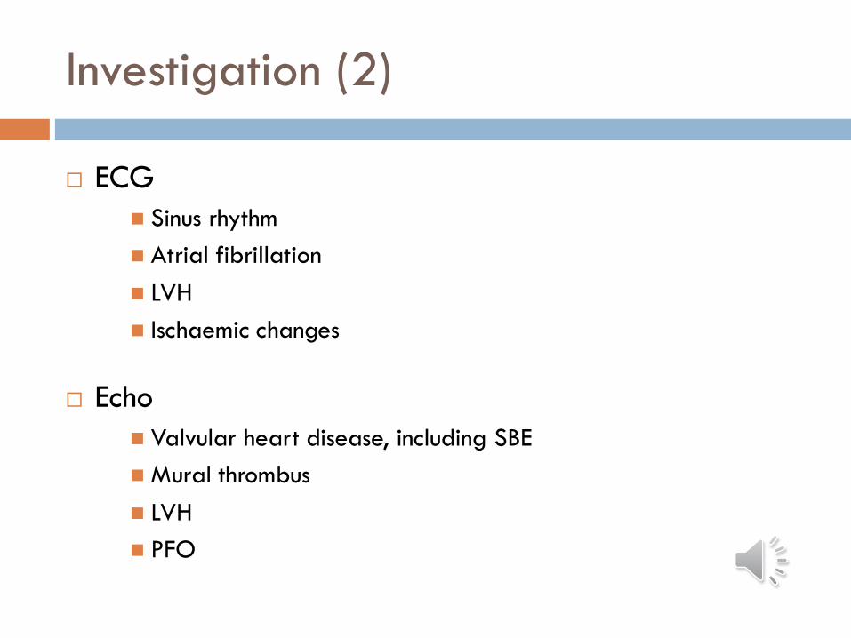
Investigation (2)
ECG
Sinus rhythm
Atrial fibrillation
LVH
Ischaemic changes
Echo
Valvular heart disease, including SBE
Mural thrombus
LVH
PFO

Investigation (3)
Neuroimaging
Objectives:
Define arterial territory
Define pathology
Exclude stroke mimics
Guide further investigations
Aids treatment strategies
Aids prognostication

CT Brain (1)
Easily accessible
Quick, 256 slices
Sensitive for bleeding
High radiation burden 2mSv=100 CXR

CT Brain (2)
RCP guideline
All strokes to be scanned within 12 hrs (of which 50% within
an hour)
Indications for urgent scan
? Thrombolysis/thrombectomy
On anticoagulants
Bleeding tendency
Unexplained progressive/fluctuating symptoms
Depressed conscious level
Suspicion of SAH/Head injury

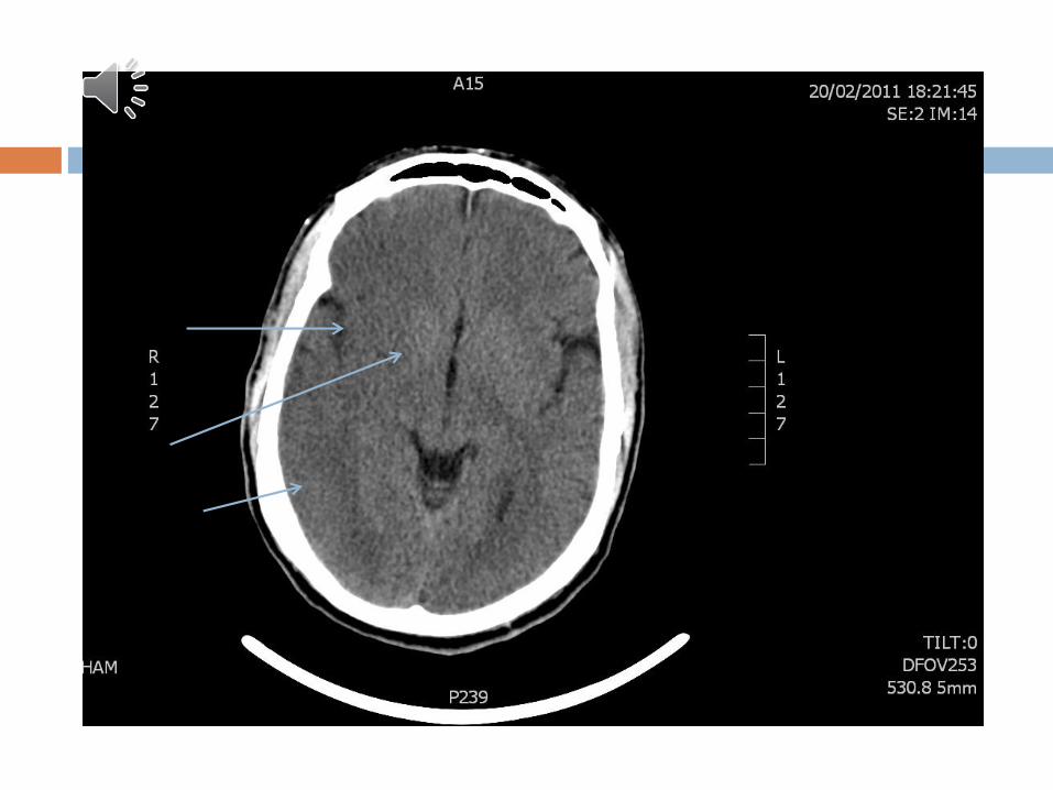
CT Brain (3)
Critical role = to exclude haemorrhage
Early signs of infarct
Hyperdense MCA
Loss of grey-white differentiation
Sulcal effacement
Loss of insular ribbon

MRI brain
Less accessible
Longer procedure
Contraindications
More detailed, better images
Define pathologies and arterial supplies
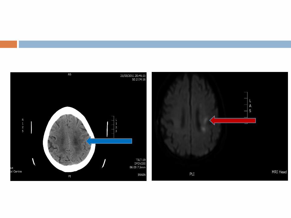
DWI
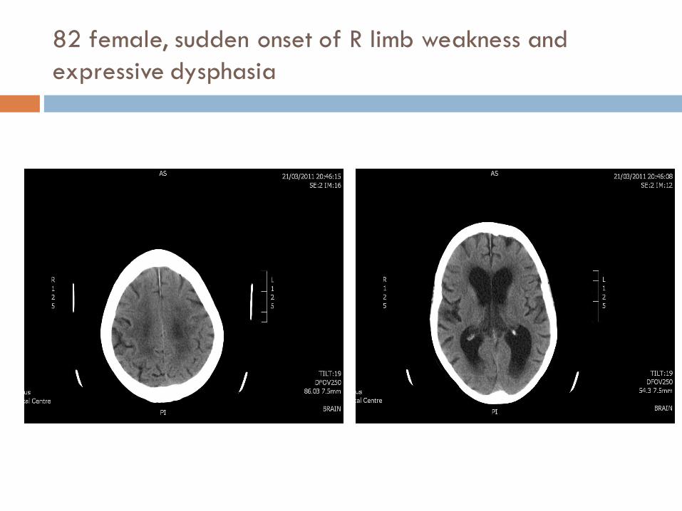
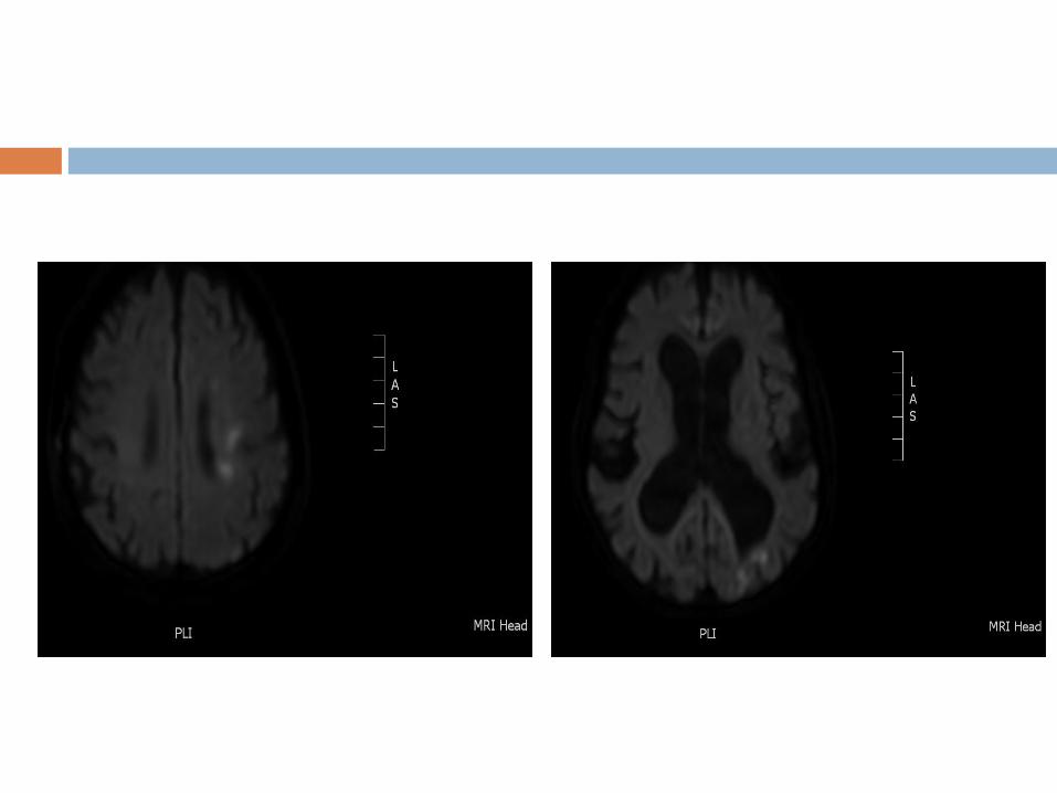
82 female, sudden onset of R limb weakness and
expressive dysphasia

Carotid Imaging
Carotid Doppler
CTA
MRA
NASCET >50%
ECST >70%
Carotid endarectomy asap (within 12 weeks)
Classifications (1)
Left or right
Ischaemic or haemorrhagic
Frontal, parietal, temporal or occipital
ACA, MCA or PCA
TOAST
Oxford
Classifications (2)
Trial of Org 10172 in Acute Stroke Treatment
(TOAST)
Large-artery atherosclerosis
Cardioembolic
Small vessel occlusion
Stroke of other determined aetiology
Stroke of undetermined aetiology

Classifications (3)
Oxford (Bamford) classification
LAC – pure motor, pure sensory, mixed, ataxic-hemiparetic
PAC
TAC
POC

Outcome of Stroke
TACS PACS LACS POCS
30 Days Dead 40 5 5 5
Dependent 55 40 30 30
Independent 5 55 65 65
1 Year Dead 60 15 10 20
Dependent 35 30 30 20
Independent 5 55 60 60

Where do you manage?
? EAU
? General medical ward
? Health care of elderly ward
? Somewhere else

Stroke Unit (1)
A discrete area in hospital
Staffed by specialist stroke MDT
Access to equipment for monitoring & rehabilitating
patients
Regular MDM for goal setting

Stroke Unit (2)
Stroke Unit Trialists’ Collaboration. Organised inpatient (stroke unit) care for
stroke. Cochrane Database of Systematic Reviews 2007;CD000197.
NSF for the Elderly (Standard 5): “all stroke patients should be admitted to
organised stroke units”
RCP/NICE guidelines (2016 and 2019 respectively)
More likely to
Receive measures to reduce aspiration
Receive early nutrition
Shorter LOS
Less death
More likely to discharge independent
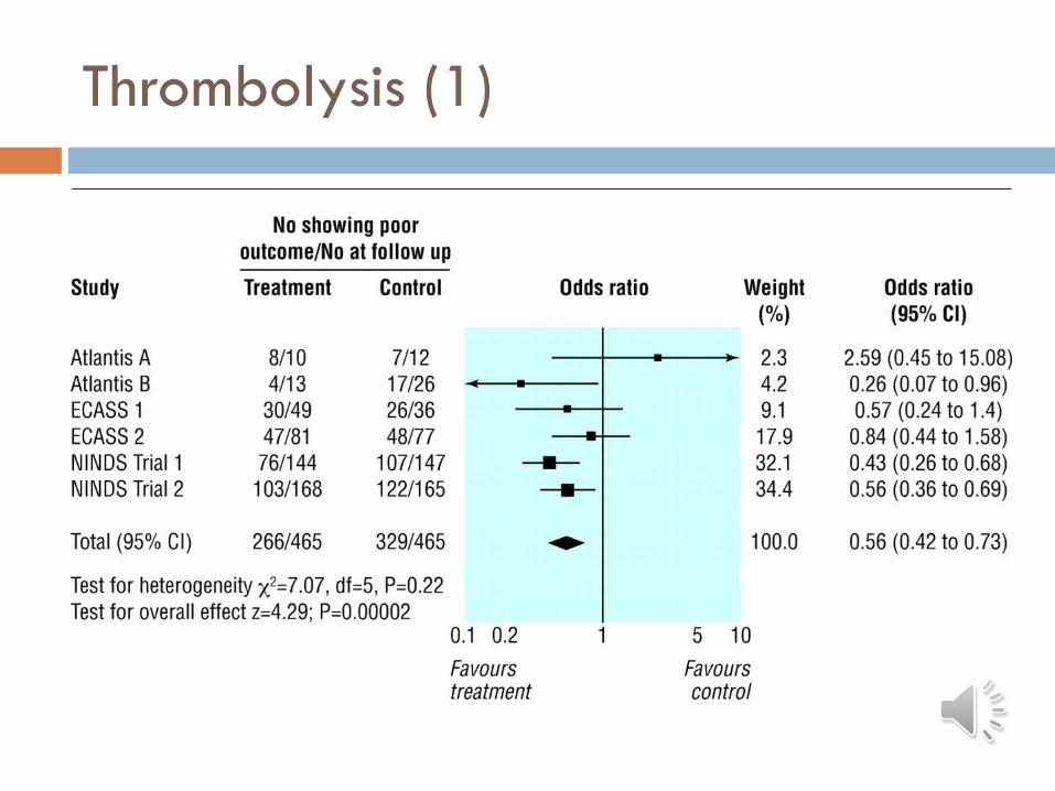
Thrombolysis (1)

Thrombolysis (2)
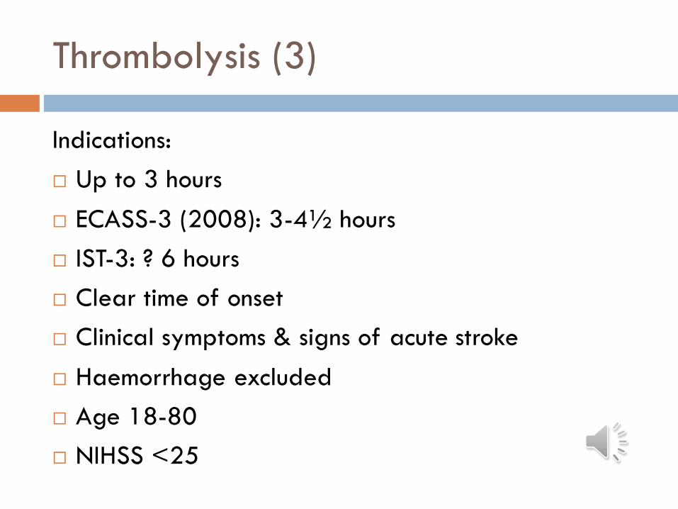
Thrombolysis (3)
Indications:
Up to 3 hours
ECASS-3 (2008): 3-4½ hours
IST-3: ? 6 hours
Clear time of onset
Clinical symptoms & signs of acute stroke
Haemorrhage excluded
Age 18-80
NIHSS <25
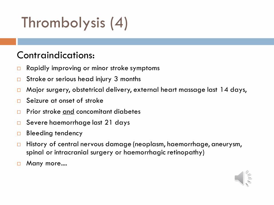
Thrombolysis (4)
Contraindications: Rapidly improving or minor stroke symptoms
Stroke or serious head injury 3 months
Major surgery, obstetrical delivery, external heart massage last 14 days,
Seizure at onset of stroke
Prior stroke and concomitant diabetes
Severe haemorrhage last 21 days
Bleeding tendency
History of central nervous damage (neoplasm, haemorrhage, aneurysm, spinal or intracranial surgery or haemorrhagic retinopathy)
Many more....

Thrombolysis
Alteplase
0.9mg/kg, up to max 90mg
Dilute with sterile water 1mg/ml
10% bolus
90% over 1 hour
Risk of anaphylaxis <1.5%


DAWN trial
49% of thrombectomy patients had an mRS of 0, 1, or
2 at 90 days compared to 13% of control patients
No significant difference in stroke related deaths at
90 days, but significant (P = 0.04) reduction in
neurological deterioration in thrombectomy group

DEFUSE Trial

Antiplatelets
Clopidogrel 75mg
Aspirin 300mg + Dipyridamole MR 200mg
Aspirin PR
Risks & benefits
Side effects
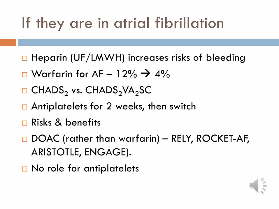
If they are in atrial fibrillation
Heparin (UF/LMWH) increases risks of bleeding
Warfarin for AF – 12% 4%
CHADS2 vs. CHADS2VA2SC
Antiplatelets for 2 weeks, then switch
Risks & benefits
DOAC (rather than warfarin) – RELY, ROCKET-AF,
ARISTOTLE, ENGAGE).
No role for antiplatelets


Dos and Don’ts
Do
Give aspirin after CT
Start statins
IPC stockings (for DVT prevention)
Don’ts
Start new antihypertensives for ischaemic strokes unless advised by a Stroke specialist
Start LMWH for DVT prophylaxis
Test swallowing

Physiological parameters
Oxygen
Glucose
Blood pressure
Pulse
Temperature

ANY DETERIORATION IN THE
FIRST 48 HOURS, CONSIDER
CT BRAIN (MAY BE A
CANDIDATE FOR
HEMICRANIECTOMY)

What about non-pharmacological
measures?
Nursing – from time 0 – rehab, pressure areas,
swallow screen
Speech therapist – swallow within 24 hrs (full
assessment if necessary), speech within 72 hrs
Physiotherapy – within 24 hrs
Occupational therapy – within 72 hrs
Psychology/Dietician/Orthoptist/Orthotist if
necessary
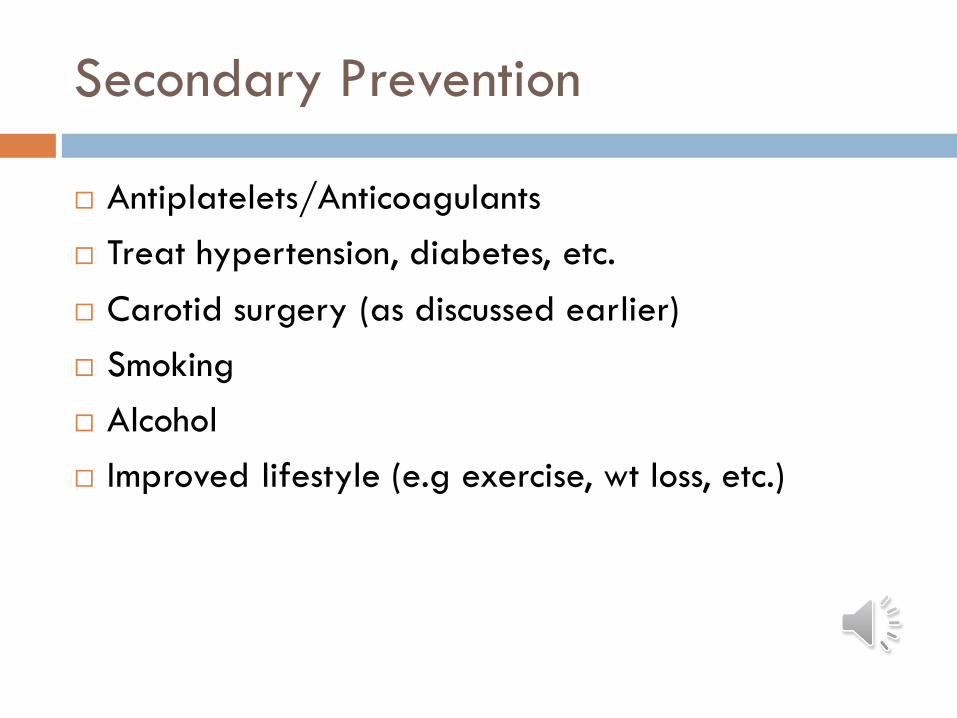
Secondary Prevention
Antiplatelets/Anticoagulants
Treat hypertension, diabetes, etc.
Carotid surgery (as discussed earlier)
Smoking
Alcohol
Improved lifestyle (e.g exercise, wt loss, etc.)
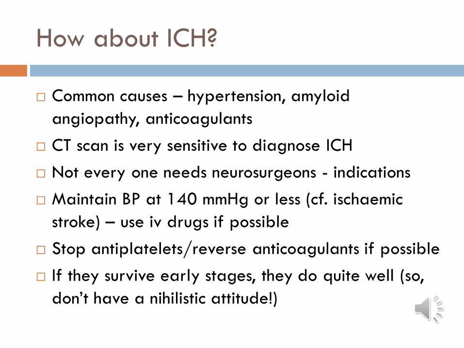
How about ICH?
Common causes – hypertension, amyloid
angiopathy, anticoagulants
CT scan is very sensitive to diagnose ICH
Not every one needs neurosurgeons - indications
Maintain BP at 140 mmHg or less (cf. ischaemic
stroke) – use iv drugs if possible
Stop antiplatelets/reverse anticoagulants if possible
If they survive early stages, they do quite well (so,
don’t have a nihilistic attitude!)
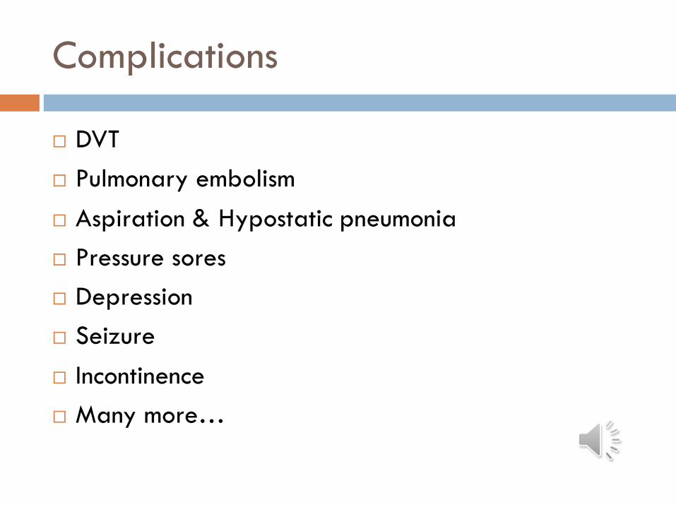
Complications
DVT
Pulmonary embolism
Aspiration & Hypostatic pneumonia
Pressure sores
Depression
Seizure
Incontinence
Many more…

Objectives
I am now able to… Explain the Bamford classification of stroke, describing the prognostic difference
between each stroke type
Describe the acute management of stroke, with particular attention to examination,
investigations, consideration/initiation of antiplatelet therapy, anticoagulation,
thrombolysis, thrombectomy, blood pressure control, statins therapy
List immediate non-pharmacological measures in management of stroke such as
assessment of swallow, rehabilitative and nursing care
Outline measures undertaken in secondary stroke prevention
Outline methods of evaluating and managing patients with carotid stenosis
Outline medical (and surgical) management for TIA
Outline the commonest causes of disability in people with impaired mobility

Thank you


