This publication is copyright under the Berne Convention and the Universal Copyright Convention. This publication is posted on this site with the permission of the British
Institute of Radiology for personal use only. No part of this publication may be reproduced, or stored on a retrieval system, or transmitted in any form or by any
means without the prior permission of the copyright holder.
BIR Published by The British Institute of Radiology
Manual on the Acute Radiation Syndrome C1
Compendium
In the case of accidental or suspected exposure of humans to external, penetratingtotal body irradiation (TBI) or large volume partial body irradiation (PBI), immediateand specialised care is required. Prior to the initiation of appropriate therapeuticprocedures it is necessary to assess the state and probable outcome of radiationaccident victims in the shortest time possible. To organise the medical managementof these patients appropriately it is necessary to anticipate the clinical course and todecide what needs to be examined and what should be documented. Co-ordinationof the multiple tasks required in such an emergency might be difficult. Thereforethis compendium is designed to guide the medical management of persons acciden-tally exposed to ionising radiation.
This compendium is not meant to replace the main document1. It is recommendedthat it be used in combination with the main document, where the scientific andpathophysiological background for an understanding of the complex interactions ofthe acute radiation syndrome (ARS) are provided in more detail.
This compendium is a useful supplement, providing short definitions, keywordsand brief overviews in the form of figures or tables on the following subjects:
� Definition of acute racute racute racute racute radiaadiaadiaadiaadiation syndrtion syndrtion syndrtion syndrtion syndrome ome ome ome ome (ARS)� The most crcrcrcrcritical oritical oritical oritical oritical orgggggan systems an systems an systems an systems an systems in which effects can be expected after acute
exposure to ionising radiation� Description and terminology of the rrrrresponse caesponse caesponse caesponse caesponse catetetetetegggggororororory (RC) concey (RC) concey (RC) concey (RC) concey (RC) conceptptptptpt� Procedures for estaestaestaestaestabbbbblishing lishing lishing lishing lishing the organ specific gggggrrrrradingadingadingadingading, the gggggrrrrrading codeading codeading codeading codeading code
and the corresponding RCRCRCRCRC� List of observable signs and symptoms signs and symptoms signs and symptoms signs and symptoms signs and symptoms reflecting the clinical manifestation
of ARS and the severity of damage� CrCrCrCrCritical phases itical phases itical phases itical phases itical phases in the medical management of ARS (trtrtrtrtriaiaiaiaiagggggeeeee, diadiadiadiadiagnostic stegnostic stegnostic stegnostic stegnostic stepspspspsps
and recommended frfrfrfrfrequencequencequencequencequency of ey of ey of ey of ey of examinaxaminaxaminaxaminaxaminationtiontiontiontion)� Therapeutic principlesTherapeutic principlesTherapeutic principlesTherapeutic principlesTherapeutic principles� TTTTTherherherherheraaaaapeutic and institutional lepeutic and institutional lepeutic and institutional lepeutic and institutional lepeutic and institutional levvvvvels of carels of carels of carels of carels of care e e e e according to the RC concept
At the end of this compendium a “Patient Accompanying Documentation Sheet”(PPPPPADSADSADSADSADS) is provided, which will aid the essential task of documenting systematicallya patient’s condition after a radiation accident.
The ARS is a composite of characteristic signs, symptoms and health impair-ments after TBI or large volume PBI. These develop as a result of damage to earlyreacting organ systems and are manifest within 60 days. A prodromal phase (firstweek after exposure) can be distinguished from a manifest illness phase.
The most critical organ systems in which effects can be expected after acuteexposure to ionising radiation are the:
� Neurovascular system (N)� Haematopoietic system (H)� Cutaneous system (C)� Gastrointestinal system (G)
1Fliedner TM, Friesecke I, Beyrer K. Medical management ofradiation accidents. Manual on the acute radiation syndrome.London: The British Institute of Radiology, 2001.
Compendium
Medical Management of Radiation AccidentsC2
Ni Hi Ci Gi
Grading code
RC=?xd
Response category
Hi
Grading (organ specific)
Ni
Ci
Gi
N = Neurovascular system
H = Haematopoietic system
C = Cutaneous system
G = Gastrointestinal system
i = Severity index 1–4
xd = Time point (x) at which RC was established; measured in days (d) after beginning of exposure.
NauseaVomitingAnorexia
Fatigue syndromeFever
HeadacheHypotension
Neurological deficitsCognitive deficits
Lymphocyte patternGranulocyte pattern
Thrombocyte patternBlood loss
InfectionErythema
Sensation/itchingSwelling and oedema
BlisteringDesquamation Ulcer/necrosis
Hair lossOnycholysis
DiarrhoeaAbdominal cramps/
pain
Symptoms
N
H
C
G
N2
Index letter for the organ system, e.g. neurovascular system
Severity index to describe the extent of damage
N2 H3 C1 G2 RC=32d
An RC equal to 3 was determined on the second day after exposure
Example:
The aim of the RC approach is to assess the damage to the critical organ systemsas a function of time after radiation exposure using indicators of effect, i.e. observ-able clinical signs and symptoms. These reflect the clinical manifestation and theseverity of the damage to the accident victim. This assessment will be the basis forassigning a patient to different RCs, each of which requires specific therapeuticstrategies.
Grading
Describes the extent of damage to a specific organ system by assessing relevantsymptoms that manifest in the course of ARS, using semi-quantitative criteria (seebelow).
Grading code
Term resulting from the combination of the organ specific grading, providing aweighted description of the major radiation reactions in the victim.
Response category (RC)
Interpretation of the overall state and outcome of the radiation accident victim,based on the grading and grading code. This is useful as a basis for decision-makingin the medical management as it assigns patients to different therapeutic and institu-tional levels of care. It is also meant to facilitate the comparison of intra- and inter-individual data on a national as well as an international level. An initial RC resultingfrom the prodromal phase can be distinguished from an epicritic RC, which summa-rises the clinical course retrospectively at day 60.
The following figure depicts the terminology used for the RC concept.Terminology
The steps for establishing the organ specific grading, the grading code and thecorresponding RC are:
1. Assess each observable symptom according to the list “Signs and symptoms”(see below).
2. Take the maximum of the degree of severity (1–4) found in any of the organspecific symptoms to determine the damage to the individual organ system(maximum approach) and attach this number as an index to the initial of theorgan system.
3. Proceed in this way for all the critical organ systems.4. Combine each organ specific grading to the grading code.5. The highest organ specific severity index of the grading code determines the
RC at a certain time point.6. Repeat steps 1–5 at certain intervals (see “Frequency of examination” below).
Enter all the above information directly onto PADS (see second part of theCompendium).
The observable signs and symptoms reflecting the clinical manifestation of ARSand the semiquantitative criteria for assessing the degree of severity of these symptomsin a standardised way are listed below.
neurological deficit; able to perform normal activity
easily detectable neurological deficit, no significant interference with normal activity
prominent neurological deficit, significant interference with normal activity
life threatening neurological signs, loss of consciousness
Cognitive deficits minor loss of memory, reasoning and/or judgement
moderate loss of memory, reasoning and/or judgement
major intellectual impairment since accident
complete memory loss and/or incapable of rational thought
HR, heart rate; BP, blood pressure. aFatigue: self-recognised state of overwhelming, sustained exhaustion and decreased capacity for physical and mental work—not relieved by rest. Typical descriptions are drained, finished off, lethargic, beaten, exhausted or worn out, prostration, drowsiness. Components are physical, cognitive, emotional/affective.
bNeurological deficits: reflex status including reflexes of the eye, ophthalmoscopy (oedema of papilla), fainting, dizziness, ataxia and other motor signs, sensory signs.
involvement Hair loss thinning, not striking patchy, visible complete and most
likely reversible complete and most
likely irreversible Onycholysis absent partial Ø complete Changes in the skin pigmentation may also occur. However, given the lack of reference data describing
depigmentation or hyperpigmentation, this symptom is not included in the grading. Nevertheless it should be recorded systematically, as it may be helpful in future radiation accidents.
Ø, not defined. aThe extent of the skin area affected is decisive and should be documented for all skin changes.
bOnly for penetrating irradiation.
Manual on the Acute Radiation Syndrome C5
Compendium
Four essential issues must be considered in patient management after a radiationaccident:
� the assessment of the severity of damage� the decision on the kind of hospital� the provision of appropriate therapeutic interventions� the evaluation of the patient’s prognosis
The following figure shows the “workflow” that will guide the management ofthe radiation accident victim.
Critical phasesCritical phasesCritical phasesCritical phasesCritical phasesin the in the in the in the in the ARSARSARSARSARS
* If, owing to additional trauma, immediate invasive intervention is necessary (e.g. surgery), this should be performed as fast as possible prior to or in parallel with the diagnostic phase. As there is a risk of pancytopenia, surgical measures should be definitive.
• Grading based specific therapeutic approaches
• RC based general therapeutic approaches
DiagnosisDiagnosis
Triageprimary*/extended
Triageprimary*/extended
Laboratory tests
Physical examination
Case history
Additional diagnostic measures
Do
cum
entatio
n
Tri
age
ph
ase
Dia
gn
ost
ic p
has
eT
her
apeu
tic
ph
ase
Organ specific gradingOrgan specific grading
Grading codeGrading code
RC (initial, epicritic)
RC (initial, epicritic)
TherapyTherapy
Ref
erra
l
�
�
�
Referral?
Acute TBI/ PBI
Radiation accident
Workflow
Information flow
Feed back
Compendium
Medical Management of Radiation AccidentsC6
ObservationInterrogation
InspectionExamination
Laboratory tests
VomitingDiarrhoeaFatigue syndromeConsciousness
Erythema
FeverNeurological deficits Lymphocyte changes
Granulocyte changes
Diarrhoea VomitingConsciousness
Routine historyChief complaintsPast medical historyPast medicationAllergies
Accident description
Assessment of overall health conditionExternal trauma, burns
Routine physical exam. (head to toe)EEGECGImaging testsDosimetry
FeverNeurological deficitsHypotensionSensation/itchingCognitive deficitsInfection Blood loss
Subsequent radiation specific information
Diagnosis
Triage is an important initial phase after the radiation accident.PrPrPrPrPrimarimarimarimarimary y y y y triage (as known from any other emergency situation)
� check vital signs and symptoms� check necessity of surgical intervention
ExtendedExtendedExtendedExtendedExtended triage
� perform a preliminary assessment of radiation induced effects, which willdetermine subsequent treatment options
� check the necessity of decontamination/decorporation� referral to primary care institution
In the diagnostic phase information is collected which is necessary for the assess-ment of the patient’s health status as a function of time after exposure to ionisingradiation. Usually the diagnostic phase is composed of:
Observation, interrogation and inspection are easy to perform. All provide goodinformation on the medical history of the patient as well as on the nature of theaccident. Physical examinations and laboratory tests (including additional diagnos-tic measures) complete the diagnostic phase and allow the grading, grading codeand RC to be obtained.
The following figure summarises the different medical tasks to be performed.
DiagnosisDiagnosisDiagnosisDiagnosisDiagnosis
TTTTTrrrrriaiaiaiaiagggggeeeee
Manual on the Acute Radiation Syndrome C7
Compendium
By simply observing the patient, valuable information is gained on the generalphysical state as well as the presence and extent of any external trauma or burns thatrequire special attention. Early radiation specific information can be gained onsymptoms such as vomiting, diarrhoea and consciousness.
Interrogation is one of the major sources of information. By taking a detailedhistory the doctor learns about the patient’s main current complaints as well as hisrole in the accident and his past medical history, which might influence the clinicalcourse. In addition, the patient’s answers enable his intellectual capacity and cogni-tive function to be assessed.
Early radiation specific information can be gained on symptoms such as vomiting,diarrhoea, consciousness and fatigue but now—in contrast to the doctor observingthem—from the patient’s viewpoint.
A witness statement might provide reliable information during this phase.Furthermore, a schematic drawing of the patient’s location during the accident mightprove useful.
A thorough inspection of the entire integument from head to toe is necessary,both to detect early reactions of the skin and to provide a baseline to help recognisea delayed onset of cutaneous signs. In addition to a detailed written description,colour photography is extremely helpful in documenting the findings and theirchanges over time.
A thorough physical examination provides valuable information on all organ sys-tems, this being necessary to complete the clinical picture of the patient. Again, thisinformation constitutes a baseline inventory for the assessment and interpretation offuture developments in the patient’s course.
Using basic technical equipment, blood pressure, temperature, neurologic status,etc. can be assessed quantitatively and incorporated into the RC concept.
Many laboratory tests are routinely used to supplement and verify diagnosis. Bloodcounts are extremely important as routine screening for the early grading. In addition,blood samples should be taken for the following tests:
� HLA typing, in case transplantation therapy may be required (within 24 h,hold at 4 °C)
� Cytogenetic tests: indicators of effect and repair for the assessment ofgenotoxic changes
reconstitution and the efficacy of therapy (will be of importance in the futureas automated reticulocyte counting machines become used more frequently)
Blood smears and bone marrow examinations have to be performed to detectmitotically connected abnormalities and to determine the pattern of degenerationand the onset of haematopoietic regeneration, respectively. Also, stem cell tests arehelpful for a quantitative assessment of stem cells and the frequency of stem celldamage.
Interleukin 8 (IL-8), procalcitonin (PCT) and C-reactive protein (CRP) should beassessed in addition to microbiological colonisation tests from blood cultures, otherbody fluids or the skin to detect early signs of infections and to act accordingly.
Observation
Interrogation
Laboratory tests
Inspection
Examination
Compendium
Medical Management of Radiation AccidentsC8
Electrolyte loss and fluid loss need to be assessed for possible replacement. Inaddition, functional tests of liver, kidney, metabolism, the endocrine system, etc.should be performed. As exposure to TBI or large volume PBI will most likely alsoaffect the reproductive system, semen analysis should be done (if feasible) as wellas laboratory tests on luteinising hormone (LH), follicle stimulating hormone (FSH),testosterone and prolactin.
Additional information can be obtained by different imaging studies, which canbe used either to evaluate the patient’s present state or to establish referenceinformation for follow-up examinations.
� chest X-ray (status of the lung, early detection of ARDS, etc.)� abdominal X-ray (e.g. in case of suspected ileus)
If available, CT or MRI is useful in assessing the extent of oedema, inflammatoryreactions, necrosis/atrophy or the depth of ulcers. Furthermore, MRI might be help-ful in detecting gut fistulas. Ultrasound is useful in the assessment of the organs inthe abdominal cavity, as well as in the detection of skin thickness, density and depthof ulcers using 7.5 MHz and higher. Also thermography, capillary microscopy,profilometry, bone scintigraphy and histology are known to be useful in the diagnosisof skin lesions.
Electroencephalography (EEG) is valuable in the assessment of changes in brainelectrical activity, as slowing of the EEG waves is an indicator of high dose exposure.
Electrocardiography (ECG), part of the routine diagnostic inventory, providesbasic information on the cardiovascular system.
The results of physical and biological dosimetry are meaningful for the generalassessment of the clinical course and the probability of late effects. They will not beof much help in the initial clinical management of a patient as the results will usuallynot be available until several days after exposure. For biological dosimetry it isimportant to obtain relevant material for examination (e.g. blood samples forchromosomal analysis) as soon as practicable after the accident.
According to the RC approach, the first examination should be done as soon aspossible after the radiation accident. It will give critical information on the patient’sfurther clinical course. Each repeated examination will improve the reliability of thediagnosis (RC). However, the frequency of examination will depend on the severityof damage and the individual clinical performance of the patient (see table below).
High technology diagnostic methods are not necessary in the first instance forassessing the radiation induced damage to a patient. Therefore, they are not takeninto account in the following recommendations, given as a rule of thumb for the RCconcept.
Complete system review every 24 h (for 6 days). Thereafter once weekly. Final assessment at day 60 post exposure.
RC 2, moderate damage
Without clinical complications such as bleeding, infections, etc., complete system review every 24 h (for 6 days). Thereafter once weekly.
With clinical complications, complete system review every 12 h (until stabilisation of symptoms). Thereafter once weekly.
Final assessment at day 60 post exposure.
Manual on the Acute Radiation Syndrome C9
Compendium
In accordance with the grading, the grading code and corresponding RCs, thefollowing treatment concepts can be suggested. They are explained in more detailbelow.
However, therapeutic measures should be adapted to the state of health of thepatient and in particular to the extent of damage to different organs and organ systemsby radiation exposure. Furthermore, individual contraindications and possible sideeffects have to be taken in account when prescribing any of the following medication.
Anti-emetic therapyDrug therapy with anti-emetics of low effectiveness (such as antihistamines) orhigh effectiveness (such as 5-HT
3-antagonists, dopamine-D2-antagonists).
If this is not sufficiently effective, glucocorticoids might be indicated (caveat:check contraindications). Furthermore, it is possible to combine these withneuroleptics (e.g. butyrophenone, phenothiazine, benzodiazepines).
Analgesic therapy (in accordance to WHO schemes)Level I: non-steroidal anti-inflammatory drugs (except aspirin).Level II: low effect opiates.Level III: high effect opiates.When level III opiates are not sufficiently effective it might be useful to combinethem with corticosteroids and neuroleptics.
Brain oedema therapyMedication with corticosteroids (dexamethasone).
Low dose scheme (20–40 mg initially, followed by daily doses of 2–4 mg)High dose scheme (40–100 mg initially, followed by slow dose reduction)
In addition mannitol (20%) and diuretic drugs i.v.Artificial ventilation as well as neurosurgical intervention might be required.
Without clinical complications such as bleeding, infections, unconsciousness, etc., complete system review every 12 h (for about 6 days). Thereafter once daily (up to day 30). Only if signs of recovery are seen can the intervals be extended (examination once weekly).
With clinical complications, complete system review every 6 h (until stabilisation of symptoms). Thereafter proceed as described for RC 3 without clinical complications.
Final assessment at day 60 post exposure.
RC 4, serious damage
Complete system review every 6 h (for about 3 days, in case of uncertainties or clinical complications for about 6 days). Thereafter examination once daily. Only if signs of recovery can be seen and no additional complications arise can the intervals be extended (examination every 2 or 3 days or once weekly).
Final assessment at day 60 post exposure.
Compendium
Medical Management of Radiation AccidentsC10
Adapted nutrition (including electrolyte and fluid replacement)Diet adaptation (small portions).Hypercaloric food.Parenteral nutrition (peripheral or central i.v. application of prefabricated completesolutions, or individual schemes), including adequate substitution of vitaminsand trace elements.Electrolyte and fluid replacement according to laboratory assessed loss.
Antibiotic treatmentSpecific antibiotic therapy as early as possible, according to microbiological tests.If not available: broad spectrum penicillin plus third generation cephalosporin ormonotherapy with one of the latest carbapenem antibiotics (caveat: check formost recent recommendations).In case of life-threatening exposure, additional gastrointestinal decontaminationaccording to common schemes.In febrile patients not responding to antibiotics, fungal infection must be suspected,which requires systemic antifungal therapy.In addition, antiviral therapy might be indicated for herpes simplex orcytomegalovirus infection.Furthermore, for severely injured patients the use of a protective environment hasbeen described as being effective.
Skin treatmentIn the prodromal stage basic therapy is usually required with linoleic creams orlotio alba, as well as non-atrophogenic steroids and antihistamines.Later it is important to apply topical or systemic steroids, tetrachlorodecaoxide(TCDO), thrombocytic growth factors, hydrocolloid dressings, antibioticprophylaxis and analgesics.In the chronic or late stage of cutaneous symptoms, retinoids, interferon gamma,superoxide dismutase, pentoxifylline and alpha-tocopherol might have to beapplied.
Further approachesAccording to the patient’s general state, physical exercises, occupational therapy,etc. to help overcome fatigue.Psychological and educational interventions, occupational therapy and physio-therapy might be helpful in the treatment of impaired cognitive functions.In the case of seizures, anticonvulsive drugs (oral or i.v., occasional or permanent).In the case of hypotension, initially the application of sympathomimetics mightbe helpful (but check for the underlying cause).In the case of abdominal cramps, analgesic treatment can be combined withantispasmodics.In general, for the acute and subacute phases, treatment is directed against neuro-hormonal mediators, and loperamide, which has both antimotility and antisecretoryactivity, seems to be the drug of choice.Furthermore, attempts should be made to protect the gut mocosa and prevententry of either endogenous and/or exogenous agents. To this end, elementary diets,with particular reference to glutamine, cholestyramine to chelate bile acids,probiotics and sulcralfate, may be of use.
Manual on the Acute Radiation Syndrome C11
Compendium
In immune compromised patients, blood component treatment might be followedby graft versus host disease. To prevent this, leukocyte depletion is recommendedfor blood component therapy (filtration or irradiation).
Thrombocyte concentratesPlatelet substitution is recommended on the basis of the patient’s individualmedical situation and threshold values. Substitution is indicated if:
• Close monitoring possible, no other complications, no bleeding: threshold forsubstitution is 10 × 109/l• Close monitoring not possible, increased risk or manifest bleeding: thresholdfor substitution is 20 × 109/l• Additional trauma, surgery, mass transfusions, cerebral oedema: thresholdfor substitution is 50 × 109/l
Granulocyte concentratesUsually neither indicated nor efficient. Might be useful only in the case of septic
ulcerations.Highly related to increased risk of cytomegalovirus infection.
Erythrocyte concentratesAnaemia is not usually a direct effect of irradiation. Without strict indication
there is a risk of overtransfusion, which might negatively influence the regenerationof the erythropoietic system.
Transfusion required if Hb < 10 g/dl in patients with known coronary heart dis-ease, or clinical situation with decreased intracerebral perfusion.
The field of cytokine stimulation is changing rapidly. To date several growth factorsare in clinical trial and use, respectively. The most effective growth factors aregranulocyte-colony stimulating factor (G-CSF), granulocyte–macrophage colonystimulating factor (GM-CSF) and thrombopoetin (TPO). Several combinations ofthese agents have been tested in clinical trials, but a final recommendation cannot begiven, although the combination of TPO and G-CSF seems to be promising. However,TPO should be administered very early after the exposure, i.e. within 24 h, to bemost effective.
In addition to haematopoietic cytokines, several growth factors appear to promotethe restoration of the gastrointestinal epithelium and/or the surrounding tissue. IL-11and keratinocyte growth factor (KGF) are currently being investigated. However,growth factor therapy for the gastrointestinal syndrome is not yet in clinical use andmuch work remains to be carried out to show that it yields clear therapeutic benefitsfor radiation accident victims.
If it turns out that spontaneous haematopoietic recovery is impossible, stem celltransplantation must be considered, the effectiveness and feasibility of which dependson the individual situation. Sources of haematopoietic stem cells are:
1. Bone marrow2. Peripheral blood3. Umbilical cord blood
In a radiation accident situation, most likely allogeneic stem cell transplantation(SCT) will take place.
Stimulation
Substitution
Stem celltransplantation
Compendium
Medical Management of Radiation AccidentsC12
Stem cells should preferably be obtained in the following order of priority:
1. From a human leukocyte antigen (HLA) identical sibling2. From other HLA-identical members of the family3. From an HLA-identical unrelated donor
The SCT has to follow the institutional protocols of the hospital and thereshould be interdisciplinary collaboration with the transplantation unit.
When surgical interventions are required, they have to follow the specific rulesfor surgery of an irradiated patient. Owing to the risk of pancytopenia, surgicalinterventions should be carried out as early as possible after the irradiation, or ata time when the risk of bleeding or infection can be controlled.
With regard to the treatment of skin lesions, puncture of blisters, excision ofulcers or fibrotic tissue, primary wound closure, split or full thickness skin graftsor vascularised flaps may be indicated. If basal and squamous cell carcinomasoccur in the late effect phase after exposure to ionising radiation, they should beexcised.
Furthermore, surgical interventions according to common rules should beconsidered for the treatment of ileus, gut fistulas, or—if at all possible—brainoedema.
The following figure gives a synopsis of the RC-dependent therapeutic andinstitutional levels of care for radiation accident victims.
Accident Information (As this information is the same for all victims, this section can be copied and attached to the PADS of the other patients)
Accident location:
Address:
City: State: Zip:
Telephone: Fax:
Proprietor of the radiation source:
Beginning of accident (date and time):
End of accident (date and time):
Number of persons involved:
General accident description:
Source of irradiation:
Reactor accident
Nuclear explosion
Sealed source
Unsealed source
Critical chain reaction
Other:
Additional comments:
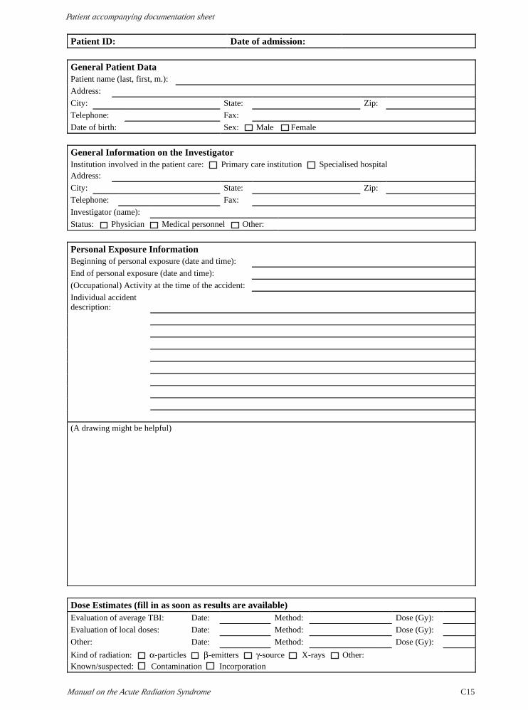
Manual on the Acute Radiation Syndrome C15
Patient accompanying documentation sheet
Patient ID: Date of admission:
General Patient Data Patient name (last, first, m.):
Address:
City: State: Zip:
Telephone: Fax:
Date of birth: Sex: Male Female
General Information on the Investigator Institution involved in the patient care: Primary care institution Specialised hospital Address:
City: State: Zip:
Telephone: Fax:
Investigator (name):
Status: Physician Medical personnel Other:
Personal Exposure Information Beginning of personal exposure (date and time):
End of personal exposure (date and time):
(Occupational) Activity at the time of the accident:
Individual accident description:
(A drawing might be helpful)
Dose Estimates (fill in as soon as results are available) Evaluation of average TBI: Date: Method: Dose (Gy):
Evaluation of local doses: Date: Method: Dose (Gy):
Other: Date: Method: Dose (Gy):
Kind of radiation: α-particles β-emitters γ-source X-rays Other:
Known/suspected: Contamination Incorporation
PADS
Medical Management of Radiation AccidentsC16
Pre-accident History (to be elaborated as usual covering all organ systems!) Information of special interest concerning the ARS (uk = unknown)
CNS Psychiatric disorders: uk no yes, if yes, please specify:
Neurological disorders: uk no yes, if yes, please specify:
Neurovascular disorders: uk no yes, if yes, please specify:
Malignancies: uk no yes if yes, please specify:
Others: uk no yes if yes, please specify:
Haematopoietic system Leukaemia: uk no yes, if yes, please specify:
MDS: uk no yes, if yes, please specify:
Other malignancies: uk no yes, if yes, please specify:
Others: uk no yes, if yes, please specify:
Skin Scars: uk no yes, if yes, please specify:
Rash: uk no yes, if yes, please specify:
Mycotic diseases: uk no yes, if yes, please specify:
Allergic diseases: uk no yes, if yes, please specify:
Malignancies: uk no yes, if yes, please specify:
Others: uk no yes if yes, please specify:
GIT Related diseases: uk no yes, if yes, please specify:
Malignancies: uk no yes, if yes, please specify:
Others: uk no yes, if yes, please specify:
Additional Past Health Information (including date of first diagnosis) Other organ systems
Lung:
Heart:
Vascular system:
Liver:
Bone and skeleton:
Endocrine system:
Eyes:
Others:
Malignancies
Allergies
Past hospitalisations
Habits
Tobacco:
Alcohol:
Others:
Former occupation
Social history
Manual on the Acute Radiation Syndrome C17
Patient accompanying documentation sheet
Family History (questions of special interest)
Number of siblings: Sisters: Brothers:
Number of children: Daughters: Sons:
Cardiovascular diseases: no yes: mother father brother sister others:
if yes, please specify:
Malignancies: no yes: mother father brother sister others:
if yes, please specify:
Metabolic disorders: no yes: mother father brother sister others:
if yes, please specify:
Haematological disorders: no yes: mother father brother sister others:
if yes, please specify:
Others: no yes: mother father brother sister others:
if yes, please specify:
Medication Past medication:
Current medication:
Post exposure Chief complaints and timing of symptoms on admission Date/time Complaint Description
Vital signs on admission Date/time Sign Description
Blood pressure
Heart rate
Respiratory rate
Temperature
Others
Radiation related health impairments of other organ systems Description Consultation
Lung no yes
Heart no yes
Eyes no yes
Liver no yes
Bone and skeleton no yes
Endocrine system no yes
Lymph nodes no yes
Mucous membranes no yes
Salivary glands no yes
Others no yes
PADS
Medical Management of Radiation AccidentsC18
Use the following template to document ARS symptoms as a function of time according to the “Checklist” of ARS specific clinical symptoms of the four early reacting organ systems. Copy as required!
Patient ID: Beginning of exposure: Examiner:
Date and time of examination
N Degree of severity
Degree of severity
Degree of severity
Degree of severity
Degree of severity
Degree of severity
Degree of severity
Degree of severity
Nausea Vomiting Anorexia Fatigue syndrome Fever Headache Hypotension Neurological deficits Cognitive deficits Maximum Grading N
H Degree of severity
Degree of severity
Degree of severity
Degree of severity
Degree of severity
Degree of severity
Degree of severity
Degree of severity
Lymphocyte changes Granulocyte changes Thrombocyte changes Infection Blood loss Maximum Grading H
C Degree of severity
Degree of severity
Degree of severity
Degree of severity
Degree of severity
Degree of severity
Degree of severity
Degree of severity
Erythema Sensation/itching Swelling/oedema Blistering Desquamation Ulcer/necrosis Hair loss Onycholysis Maximum Grading C
G Degree of severity
Degree of severity
Degree of severity
Degree of severity
Degree of severity
Degree of severity
Degree of severity
Degree of severity
Frequency (stool) Consistency (stool) Mucosal loss/d (stool) Bleeding/d (stool) Abdominal cramps/pain Maximum Grading G
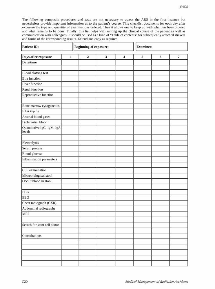
The following composite procedures and tests are not necessary to assess the ARS in the first instance but nevertheless provide important information as to the patient’s course. This checklist documents for each day after exposure the type and quantity of examinations ordered. Thus it allows one to keep up with what has been ordered and what remains to be done. Finally, this list helps with writing up the clinical course of the patient as well as communication with colleagues. It should be used as a kind of “Table of contents” for subsequently attached stickers and forms of the corresponding results. Extend and copy as required!
Patient ID: Beginning of exposure: Examiner:
Days after exposure 1 2 3 4 5 6 7
Date/time
Blood clotting test
Bile function
Liver function
Renal function
Reproductive function
Bone marrow cytogenetics
HLA typing
Arterial blood gases
Differential blood
Quantitative IgG, IgM, IgA levels
Electrolytes
Serum protein
Blood glucose
Inflammation parameters
CSF examination
Microbiological stool
Occult blood in stool
ECG
EEG
Chest radiograph (CXR)
Abdominal radiographs
MRI
Search for stem cell donor
Consultations
Manual on the Acute Radiation Syndrome C21
Patient accompanying documentation sheet
PERSONAL NOPERSONAL NOPERSONAL NOPERSONAL NOPERSONAL NOTESTESTESTESTES