+ All Categories
Home > Documents > Medical Microbiology Detection of disease: –Signs & Symptoms –Traditional Microbiological...

Medical Microbiology Detection of disease: –Signs & Symptoms –Traditional Microbiological...

Date post: 14-Dec-2015
Category:
Upload: theodore-cradduck
View: 219 times
Download: 0 times
Share this document with a friend
Popular Tags:
15
Medical Microbiology • Detection of disease: – Signs & Symptoms – Traditional Microbiological Identification • Physiological Characteristics • Microscopy Techniques – Biotechnology (e.g. PCR) – Immunological • Serological testing by agglutination • Fluorescent Antibodies • Enzyme Linked Immuno-Absorbance (ELISA) Assay • Western Blots for ELISA confirmation • Selected “Nasties”:
Transcript

Medical Microbiology• Detection of disease:

– Signs & Symptoms– Traditional Microbiological Identification

• Physiological Characteristics• Microscopy Techniques

– Biotechnology (e.g. PCR)– Immunological

• Serological testing by agglutination• Fluorescent Antibodies• Enzyme Linked Immuno-Absorbance (ELISA) Assay• Western Blots for ELISA confirmation

• Selected “Nasties”:

Agglutination Serological Test

Figure 18.6

Fluorescence Microscopy

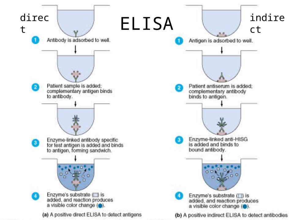
ELISAdirect indirect

Yersinia pestis

Black Death

Taxonomy

• Member of the Enterobacteriaceae family

Yersinia is a Gram-negative coccobacilli

Target Tissues• This disease direct effects the lymph nodes which can

be found in the groin, neck, and armpits and cause them to enlarge and suppurate.

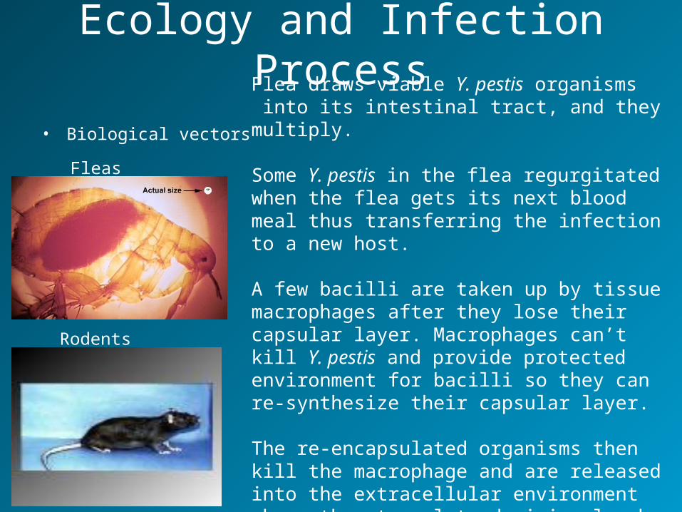
Ecology and Infection Process

• Biological vectors

Fleas

Rodents

Flea draws viable Y. pestis organisms into its intestinal tract, and they multiply.

Some Y. pestis in the flea regurgitated when the flea gets its next blood meal thus transferring the infection to a new host.

A few bacilli are taken up by tissue macrophages after they lose their capsular layer. Macrophages can’t kill Y. pestis and provide protected environment for bacilli so they can re-synthesize their capsular layer.

The re-encapsulated organisms then kill the macrophage and are released into the extracellular environment where they travel to draining lymph nodes.

Symptoms Bubonic Plaguebacteria infect lymph nodes

• Bubos

Fever HeadacheVomiting Blood

Diagnostic Tests

• Take smear from blood or feces for bubonic plague

> bacteria has “safety pin” appearance

Can also use FA (fluorescent-antibody) testAll plague bacilli have unique diagnostic envelope glycoprotein called the Fraction 1 (F1) antigen

Treatments and Preventive Measures

7- to 10-day course of antimicrobic therapy • streptomycin • chloramphenicol • tetracycline

Vaccine:

Y. pestis organisms grown in artificial media, inactivated with

formaldehyde, and preserved in 0.5% phenol. The vaccine contains

trace amounts of beef-heart extract, yeast extract, agar, and

peptones and peptides of soya and casein.

Control of rat populations concurrent with elimination of their flea

prevent spread of the plague to humans.

Epidemiology: Transmission BubonicInfected Rodent Fleas Humans

Can also enter through breaks in skin when handling infected animal

Prevalence and Distribution in Global Human and Animal Populations

1000- 3000 cases reported

annually across the world

•Africa (most cases)•Asia•Northeastern Brazil•Andes Mountain Regions•US (19-40 cases a year mostly in Western areas such as New Mexico and Arizona)

MortalityBubonic Plague

Untreated 50- 60% mortality rateTreated 5 – 20% mortality rate

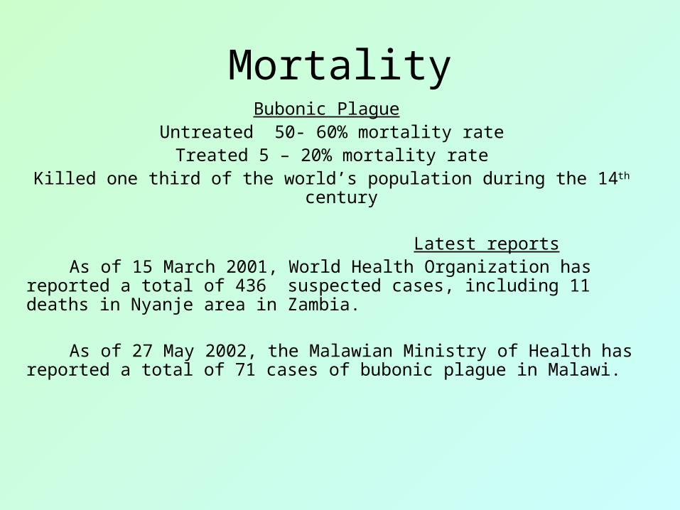
Killed one third of the world’s population during the 14th century

Latest reports As of 15 March 2001, World Health Organization has reported a total

of 436 suspected cases, including 11 deaths in Nyanje area in Zambia.

As of 27 May 2002, the Malawian Ministry of Health has reported a total of 71 cases of bubonic plague in Malawi.

Latest research EVOLUTION: A single gene change in a relatively benign

recent ancestor of the bacterium that causes bubonic plague played a key role in the evolution of the deadly disease from a germ that causes a mild human stomach illness acquired via contaminated food or water to the flea-borne agent of the "Black Death.”

GENETICS: Research on three genes, hemin storage (hms)

genes, in Y. pestis that change it from a harmless, long-term inhabitant in the flea midgut to one that amasses in its foregut.

PREVENTION: Current prevention measures include dusting

family pets with insecticides to prevent the spread of the Yersinia pestis organism from the native prairie dog populations


Recommended