35
Medical Mycology Dr. Hala Al Daghistani

Medical Mycology Dr. Hala Al Daghistani

FAre eukaryotes that grow in two
basic forms, a yeasts and molds (or
moulds).
Growth in the mold form occurs by
production of multicellular filamentous
colonies. These colonies consist of
branching tubules called hyphae
The mass of intertwined hyphae that
accumulates during active growth is a
mycelium.
In the reproductive phase, fungi may
undergo either asexual or sexual
reproduction. Asexual reproduction
involves the generation of spores;
sexual reproduction requires specific
cellular structures that are used for
taxonomic differentiation
Classification of Mycoses The clinical nomenclatures used for the mycoses are based on the
A. Site of the infection Mycoses are classified as
1- Superficial (are generally limited to the outer layers of the skin and hair.
2- Cutaneous (are located deeper in the epidermis, hair and nails.
3- Subcutaneous (involve the dermis, subcutaneous tissues and muscle). infections (generally originating in the lungs and other organs).
Route of acquisition of the pathogen . B 1. Exogenous (routes of entry for exogenous fungi include airborne, cutaneous)
2. Endogenous (endogenous infection involves colonization by a member of the normal flora or reactivation of a previous infection).
C. Type of virulence exhibited by the fungus. 1. Primary pathogens can establish infections in normal hosts.
2. Opportunistic pathogens cause disease in individuals with compromised host defense mechanisms.

Fungal Morphology
Hypae (threads)
making up a mycelium
Yeasts
Many pathogenic
fungi are dimorphic,
forming hyphae at
ambient temperatures
but yeasts at body
temperature.
There are five types of mycoses to describe, in two main
categories:
Skin mycoses 1. Superficial mycoses
2. Cutaneous mycoses
3. Subcutaneous mycoses
Systemic mycoses Systemic mycoses due to primary (usually dimorphic) pathogens
Systemic mycoses due to opportunistic pathogen.

Superficial Mycoses
Superficial Mycoses include the following
fungal infections and their etiological agent:
• Pityriasis versicolor النخالية المبرقشة(Malassezia
furfur)
• Tinea nigra السعفة السوداء (Phaeoannellomyces
werneckii)
• Black piedra البيصرية السوداء (Piedraia hortae),
• White piedra البيصرية البيضاء (Trichosporon beigelii)
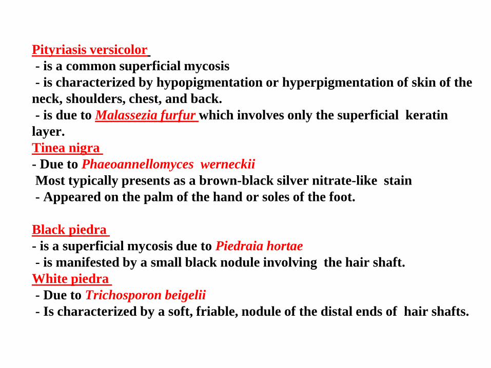
versicolor Pityriasis
- is a common superficial mycosis
- is characterized by hypopigmentation or hyperpigmentation of skin of the
neck, shoulders, chest, and back.
which involves only the superficial keratin furfur Malasseziais due to -
layer.
nigra Tinea
- Due to Phaeoannellomyces werneckii
Most typically presents as a brown-black silver nitrate-like stain
- Appeared on the palm of the hand or soles of the foot.
piedraBlack
- is a superficial mycosis due to Piedraia hortae
- is manifested by a small black nodule involving the hair shaft.
piedraWhite
- Due to Trichosporon beigelii
- Is characterized by a soft, friable, nodule of the distal ends of hair shafts.
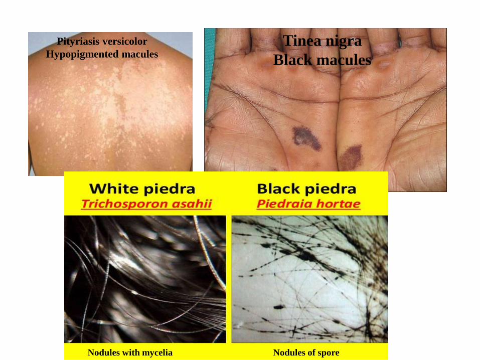
Pityriasis versicolor
Hypopigmented macules
Tinea nigra
Black macules
Nodules with mycelia Nodules of spore
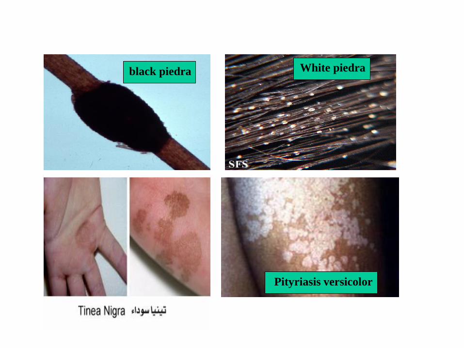
black piedra White piedra
Pityriasis versicolor
Cutaneous mycoses (Dermatophytes) - There are three genera of fungi that commonly cause disease in the
on-living tissues of skin, hair, or nails of people and animals, by growing
in a zone just above where the protein keratin is deposited.
These three genera are
Microsporum
Trichophyton
Epidermophyton
-These fungi all have the ability to degrade keratin and grow as non-
invasive saprotrophs on skin and its appendages
- Their growth causes irritation and inflammation of underlying
epithelial cells, this being an allergic reaction that may result in death of
these cells
- Contagious-direct or indirect contact
Cutanous Mycosis: Obligate Parasitic Fungi (dermatophytes),
attack the outer surface of humans
• Soil fungi: thermal dimorphic, adaptations to human body
• Opportunistic saprobes: attack people with compromised immune
systems
• Secrete keratinase, an enzyme that degrades keratin.
• Infection is transmitted by direct contact or contact with infected hair
(hair salon) or cells (shower floors).
– Cause common tinea (ringworm), which is a very common fungal
infection of the skin. Tinea is often called "ringworm" because the
rash is circular, with a ring-like appearance
– Grow only on humans
– Reservoir not in soil or animals
– Reservoir in carpets and upholstery for up to two years
Cutanous mycosis is classified as
a. Dermatophytoses (caused by the genera Epidermophyton,
Microsporum, and Trichophyton)
b. Dermatomycoses (the most common of which are Candida spp.)
- The Dermatophytoses are characterized by an anatomic site-
specificity according to genera. for example
*Epidermophyton spp infects only skin and nails, but does not
infect hair shafts and follicles
*Microsporum spp. infect hair and skin, but do not involve nails.
*Trichophyton spp. may infect hair, skin, and nails.

Dermatomycoses

Subcutaneous Mycoses Involves dermis, subcutaneous tissues and may be the muscle. There are
three general types of subcutaneous mycoses:
a. Chromoblastomycosis الفطريات االصطباغية
b. Mycetoma (chronic inflammation of the tissues)
c. Sporotrichosis. (a chronic fungal infection producing nodules and
ulcers in the lymph nodes and skin).
All appear to be caused by traumatic inoculation of the etiological fungi into the
subcutaneous tissue.
* Chromoblastomycosis is a subcutaneous mycosis characterized by
Verrucoid lesions of the skin افة ثالولية. It is believed to originate in minor trauma
to the skin, usually from thorns or splinters
- symptoms often do not appear for years. It is generally limited to the
subcutaneous tissue, with no involvement of bone, tendon, or muscle, the lower
extremities.
- The most common causes of Chromoblastomycosis are Fonsecaea
pedrosoi, Fonsecaea compacta, Cladosporium carionii.
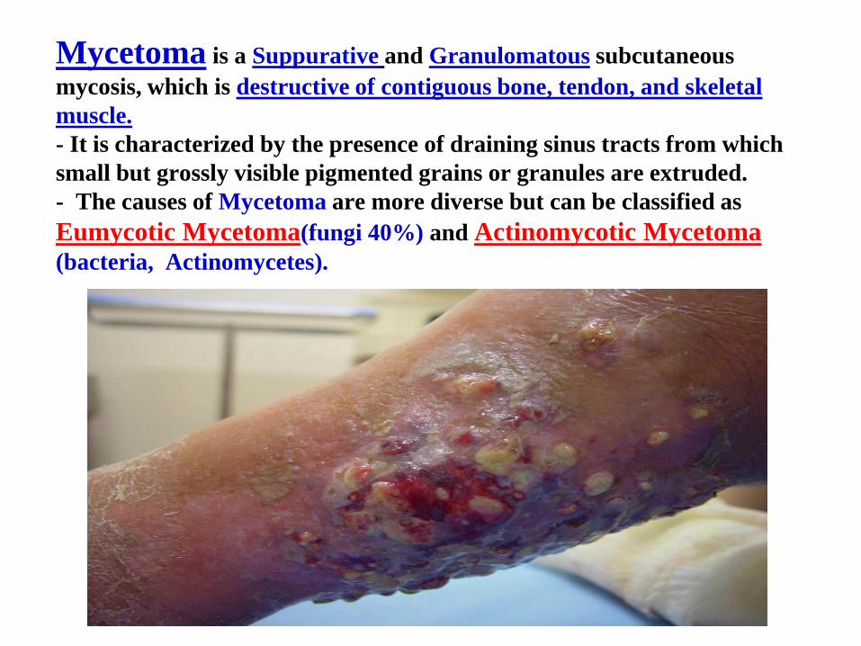
Mycetoma is a Suppurative and Granulomatous subcutaneous
mycosis, which is destructive of contiguous bone, tendon, and skeletal
muscle.
- It is characterized by the presence of draining sinus tracts from which
small but grossly visible pigmented grains or granules are extruded.
- The causes of Mycetoma are more diverse but can be classified as
Eumycotic Mycetoma(fungi 40%) and Actinomycotic Mycetoma (bacteria, Actinomycetes).

Sporotrichosis
- This infection is due to Sporothrix schenckii and involves the
subcutaneous tissue at the point of traumatic inoculation.
- The infection usually spreads along cutaneous lymphatic
channels of the extremity involved.

Deep Mycoses Primary versus Opportunistic mycoses
pathogenic fungi are able to establish infection in a normal PrimaryThe -
pathogens require a compromised host in Opportunistic host; whereas,
order to establish infection (e.g., Cancer, Organ transplantation, Surgery,
and AIDS).
respiratory Primary deep pathogens usually gain access to the host via the -
. tract
, Respiratory tractOpportunistic fungi causing deep mycosis invade via the -
Intravascular devices., or Alimentary tract
Primary systemic fungal pathogens include:
- Coccidioides immitis (Coccidioidomycosis )
- Histoplasma capsulatum (cave disease)
- Blastomyces dermatitidis (Blastomycosis)

Originate in lungs, phagocytosis by macrophages, spread to
many organs.
Most primary infections are inapparent.
Progression may produce pulmonary symptoms or ulcerative
lesions.
Host responses produce formation of fibrous tissue, granulomas
and calcified lesions.
Normally found in soil, infect via inhalation
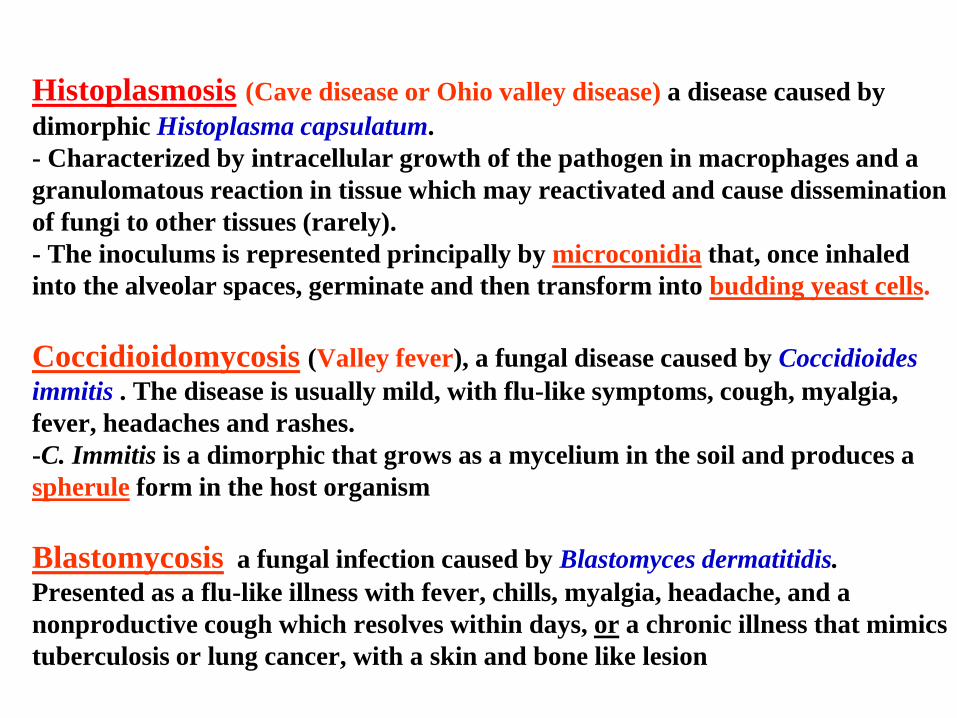
Histoplasmosis (Cave disease or Ohio valley disease) a disease caused by
dimorphic Histoplasma capsulatum.
- Characterized by intracellular growth of the pathogen in macrophages and a
granulomatous reaction in tissue which may reactivated and cause dissemination
of fungi to other tissues (rarely).
- The inoculums is represented principally by microconidia that, once inhaled
into the alveolar spaces, germinate and then transform into budding yeast cells.
Coccidioidomycosis (Valley fever), a fungal disease caused by Coccidioides
immitis . The disease is usually mild, with flu-like symptoms, cough, myalgia,
fever, headaches and rashes.
-C. Immitis is a dimorphic that grows as a mycelium in the soil and produces a
spherule form in the host organism
Blastomycosis a fungal infection caused by Blastomyces dermatitidis.
Presented as a flu-like illness with fever, chills, myalgia, headache, and a
nonproductive cough which resolves within days, or a chronic illness that mimics
tuberculosis or lung cancer, with a skin and bone like lesion

Opportunistic Fungal pathogens include:
Cryptococcus neoformans
Candida spp. (Candidiasis)
Aspergillus spp. (Aspergillosis)
Penicillium marneffei
Zygomycetes (Zygomycosis)
Trichosporon beigelii.
-These organisms generally have a low potential for virulence but
can produce severe disease involving a variety of body tissues.
- Mycotic disease is often a consequence of predisposing factors
including
1. Age
2. Stress
3. Other pathologic conditions (e.g. cancer, diabetes, AIDS).
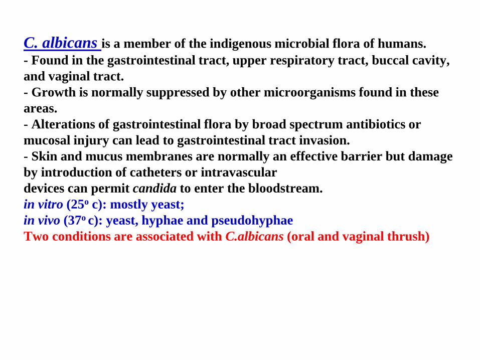
C. albicans is a member of the indigenous microbial flora of humans.
- Found in the gastrointestinal tract, upper respiratory tract, buccal cavity,
and vaginal tract.
- Growth is normally suppressed by other microorganisms found in these
areas.
- Alterations of gastrointestinal flora by broad spectrum antibiotics or
mucosal injury can lead to gastrointestinal tract invasion.
- Skin and mucus membranes are normally an effective barrier but damage
by introduction of catheters or intravascular
devices can permit candida to enter the bloodstream.
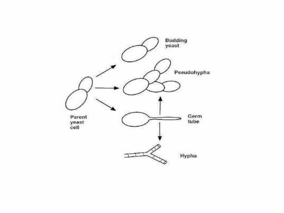
in vitro (25o c): mostly yeast;
in vivo (37o c): yeast, hyphae and pseudohyphae
Two conditions are associated with C.albicans (oral and vaginal thrush)

Vaginal Candidiasis (vaginal thrush) is the most common
clinical infection. Local factors such as pH and glucose concentration
(under hormonal control) are of prime importance in the occurrence of
vaginal candidiasis. In mouth: normal saliva reduces adhesion
(lactoferrin that produce by various secretory fluids has antimicrobial
activity, is also protective). Risk factor for candidiasis
1.Post-operative status
2.Cytotoxic cancer
3.Chemotherapy
4. Antibiotic therapy
5. Burns
6. Drug abuse
7. GIT damage.

Some of the tissue responses to fungi infection may be due to the
Mycotoxins, which are fungal metabolites that are toxic to the host.
Some fungi have a variety of virulence factors including
LPS-like endotoxins
Hemolysins
Steroid-like toxins that affect the nervous system
Aspergillus produces a toxin called Aflatoxin that has a strong association
with liver cancer.
Aflatoxin:When contaminated food is processed, Aflatoxins enter the
general food supply where they have been found in human foods as well as
in feedstocks for agricultural animals.
- At least 14 different Aflatoxins are produced (b1 is the most toxic) .
- Animals fed contaminated food can pass Aflatoxin into eggs, milk
products, and meat.
Children are particularly affected by Aflatoxin exposure, which leads to
stunted growth, delayed development, liver damage, and liver cancer.
Adults have a higher tolerance to exposure, but are also at risk.
Aflatoxins are among the most carcinogenic substances known
Host defenses Host defenses against the fungi include nonspecific and specific factors
include Nonspecific defenses
1. the skin (lipids, fatty acids, normal flora)
2. Internal factors (mucous membranes, ciliated cells, macrophages)
3. Blood components
4. Temperature
5. Genetic
6. Hormonal factors.
include both Specific defenses
A. Humoral Immunity (antibodies may be protective (e.g. antitoxins or
opsonins).
B. Cell-mediated Immunity. Generally, cell-mediated defenses are probably
more important. It is usually T-cell mediated and persons with compromised
cell-mediated defenses generally show more disseminated disease

EPIDEMIOLOGY
Dermatophytes may be communicated from person to person by
combs, towels, etc.
Candida is a member of the normal vaginal flora; candidiasis is
often associated with diabetes.
In some cases of mycosis, Occupation seems an important
contributor. For example, Sporothrix is normally found in woody
plants; hence, agricultural workers acquire disease more often.
Similarly, Histoplasma is often found in bird or bat excret; hence
caves workers or persons involved in community clean up may
acquire more often.

DIAGNOSIS
Clinical: For the dermatophytes, appearance of the lesions is
usually diagnostic. For systemic mycoses, the epidemiology and
symptomology are useful
Samples include: scrapings of scale, hair which has been pulled
out from the roots, brushings from an area of scaly scalp, nail
clippings, or skin scraped from under a nail, skin biopsy, moist
swab from a mucosal surface (inside the mouth or vagina) in a
special transport medium.
Laboratory: Treatment of skin scrapings with 10% potassium
hydroxide can reveal hyphae or spores. Most fungi can be grown
on Sabouraud's dextrose agar but they are often very difficult
to speciate.
Skin testing for a delayed hypersensitivity response is useful for
epidemiologic purposes but often not for diagnosis.

Germ tube test Is a screening test which is used to differentiate candida
albicans from other yeast.
When candida is grown in human or sheep serum at 37°c for
3-4 hours, they forms a germ tube, which can be detected
with a wet films as filamentous outgrowth extending from
yeast cells. It is positive for candida albicans .
CONTROL
Sanitary: Control by sanitary means is difficult, but the incidence
of communicable disease can be reduced by good hygiene.
Immunological: No vaccines are currently available.
Chemotherapeutic: Many antifungals are available but some are
to the host and must be used with caution. very toxic
Topical powders and creams often contain azole derivatives
(miconazole, clotrimazole, econazole) are useful against superficial
dermatophytes.
Sporotrichosis may be treated using potassium iodide
Systemic infections are generally treated by miconazole,
Fluconazole or ketoconazole.