192
The Maryland Medical Protocols for Emergency Medical Services Providers Effective July 1, 2016 Maryland Institute for Emergency Medical Services Systems

The Maryland
Medical Protocols for Emergency Medical Services Providers
Effective July 1, 2016
Maryland Institute for Emergency Medical Services Systems

Edition Date July 1, 2016
I. GENERAL INFORMATION
A. GENERAL PROVISIONS The goal of prehospital emergency medical services is to deliver a viable patient to appropriate definitive care as soon as possible. Optimal prehospital care results from a combination of careful patient assessment, essential prehospital emergency medical services, and appropriate medical consultation. The Maryland Medical Protocols were developed to standardize the emergency patient care that EMS providers, through medical consultation, deliver at the scene of illness or injury and while transporting the patient to the closest appropriate hospital. These pro-tocols will help EMS providers anticipate and be better prepared to give the emergency patient care ordered during the medical consultation. Maryland has highly trained and dedicated basic and advanced life support personnel who may need on-line medical consultation only for complicated or extended resuscita-tive patient care. These protocols are a form of “standing orders” for emergency patient care intervention in a patient who has a life-threatening illness or injury. It remains the responsibility of the EMT, CRT-(I), or paramedic to obtain on-line medical consultation when appropriate. If it is genuinely impossible or inappropriate (i.e., when rendering emergency care to a patient who has a life-threatening injury or medical condition) to obtain on-line medical consultation, the EMT/CRT-(I)/paramedic may render emergency patient care in accordance with these protocols in an effort to save a patient’s life or limb. Whenever such emergency life-saving patient care is rendered, the EMT/CRT-(I)/paramedic must document the treatment rendered and the reason on-line medical con-sultation could not be obtained on the Patient Care Report (PCR), the equivalent of the MAIS runsheet, and on an additional narrative. In addition, the “exceptional call” area on the PCR must be marked, and the provider must immediately notify the EMS Juris-diction. The EMS Jurisdiction must notify the State EMS Medical Director within 5 days of the incident. This general provision applies throughout these protocols. Requests for additions, deletions, or exceptions must be submitted through the State EMS Medical Director’s Office of the Maryland Institute for Emergency Medical Services Systems. Unless otherwise specified, a mandate with a stated year but no date shall be interpreted as taking effect on the protocol implementation date for that year.
THE GENERAL PATIENT CARE SECTION AND THE ALGORITHMS MUST BE FOLLOWED IN THE SPECIFIC SEQUENCE NOTED. FOR ALL OTHER TREATMENT PROTOCOLS, THE LETTER AND NUMERICAL OUTLINE FORMAT IS STRICTLY FOR RAPID AND UNIFORM REFERENCE AND DOES NOT IMPLY OR DIRECT A MANDATORY SEQUENCE FOR PATIENT CARE.
1

2Edition Date July 1, 2016
IF AN EMERGENCY MEDICAL RESPONDER IS DISPATCHED AS AN EMS UNIT, OR FOR PURPOSES RELATED TO MEDICAL ASSISTANCE, OXYGEN AND AED TREATMENT MAY BE UTILIZED, WHEN APPROPRIATE AND APPLICABLE, PROVIDED THE EMERGENCY MEDICAL RESPONDER IS JURISDICTIONALLY AUTHORIZED TO USE AN AED AND/OR THE EMERGENCY MEDICAL RESPONDER HAS BEEN EDUCATED AND TRAINED TO PROVIDE OXYGEN AND/OR AED THERAPY. THE EMERGENCY MEDICAL RESPONDER SHALL DOCUMENT ALL PATIENT CARE.

Edition Date July 1, 201613
D. MARYLAND TRAUMA AND SPECIALTY REFERRAL CENTERS
Trauma Centers
Primary Adult Resource Center• R Adams Cowley Shock Trauma Center (UM), Baltimore
Level I Trauma Center• The Johns Hopkins Hospital Adult Trauma Center, Baltimore
Level II Trauma Centers• Johns Hopkins Bayview Medical Center, Baltimore• Prince George’s Hospital Center, Cheverly• Sinai Hospital of Baltimore• Suburban Hospital (JHM), Bethesda
Level III Trauma Centers• Meritus Medical Center, Hagerstown• Peninsula Regional Medical Center, Salisbury• Western Maryland Regional Medical Center, Cumberland
Out-of-State Centers• Christiana Care Health System, Wilmington, DE• MedStar Washington Hospital Center, Washington, DC
Specialty Referral Centers
Eye Trauma• Wilmer Eye Institute at The Johns Hopkins Hospital, Baltimore
Hand/Upper Extremity Trauma• The Curtis National Hand Center for Treatment of the Hand and Upper Extremity/
Union Memorial Hospital (MedStar), BaltimoreHyperbaric Medicine
• Center for Hyperbaric Medicine/R Adams Cowley Shock Trauma Center (UM), Baltimore
Neurotrauma (Head and Spinal Cord Injuries)• Neurotrauma Center/R Adams Cowley Shock Trauma Center (UM), Baltimore
Pediatric Trauma• Pediatric Trauma Center at The Johns Hopkins Children’s Center, Baltimore• Pediatric Trauma Center at Children’s National Health System, Washington, DC
Burns• Baltimore Regional Burn Center at Johns Hopkins Bayview Medical Center,
Baltimore• Burn Center at MedStar Washington Hospital Center, Washington, DC• Pediatric Burn Center at The Johns Hopkins Children’s Center, Baltimore• Pediatric Burn Center at Children’s National Health System, Washington, DC

14Edition Date July 1, 2016
Specialty Referral Centers
Perinatal Referral Centers• Anne Arundel Medical Center, Annapolis• Franklin Square Medical Center (MedStar), Baltimore• Frederick Memorial Hospital, Frederick• Greater Baltimore Medical Center, Towson• Holy Cross Hospital, Silver Spring• Howard County General Hospital (JHM), Columbia• Johns Hopkins Bayview Medical Center, Baltimore• Mercy Medical Center, Baltimore• Prince George’s Hospital Center, Cheverly• Saint Agnes Hospital, Baltimore• Saint Joseph Medical Center (UM), Baltimore• Shady Grove Adventist Hospital, Gaithersburg• Sinai Hospital of Baltimore• The Johns Hopkins Hospital, Baltimore• University of Maryland Medical Center, Baltimore
Primary Stroke• Anne Arundel Medical Center, Annapolis• Atlantic General Hospital, Berlin• Baltimore Washington Medical Center (UM), Glen Burnie• Calvert Memorial Hospital, Prince Frederick• Carroll Hospital Center, Westminster• Charles Regional Medical Center (UM), La Plata• Franklin Square Medical Center (MedStar), Baltimore• Frederick Memorial Hospital, Frederick• Good Samaritan Hospital (MedStar), Baltimore• Greater Baltimore Medical Center, Baltimore• Harbor Hospital (MedStar), Baltimore• Harford Memorial Hospital (UMUCH), Havre De Grace• Holy Cross Hospital, Silver Spring• Howard County General Hospital (JHM), Columbia• Johns Hopkins Bayview Medical Center, Baltimore• Mercy Medical Center, Baltimore• Meritus Medical Center, Hagerstown• Midtown Campus (UM), Baltimore• Montgomery Medical Center (MedStar), Olney• Northwest Hospital, Baltimore• Peninsula Regional Medical Center, Salisbury• Saint Agnes Hospital, Baltimore• Saint Joseph Medical Center (UM), Baltimore• Saint Mary’s Hospital (MedStar), Leonardtown• Shady Grove Adventist Hospital, Rockville• Shore Medical Center at Easton (UMSRH)
MARYLAND TRAUMA AND SPECIALTY REFERRAL CENTERS (Continued)

15 Edition Date July 1, 2016
MARYLAND TRAUMA AND SPECIALTY REFERRAL CENTERS (Continued)
Primary Stroke (Continued)• Sinai Hospital of Baltimore• Southern Maryland Hospital (MedStar), Clinton• Suburban Hospital (JHM), Bethesda• Union Hospital of Cecil County, Elkton• Union Memorial Hospital (MedStar), Baltimore• Upper Chesapeake Medical Center (UMUCH), Bel Air• Washington Adventist Hospital, Takoma Park• Western Maryland Regional Medical Center, Cumberland
Comprehensive Stroke• The Johns Hopkins Hospital, Baltimore• University of Maryland Medical Center, Baltimore
Cardiac Interventional• Anne Arundel Medical Center, Annapolis• Baltimore Washington Medical Center (UM), Glen Burnie• Bayhealth Kent General, Dover, DE• Carroll Hospital Center, Westminster• Christiana Care Health System, Newark, DE• Franklin Square Medical Center (MedStar), Baltimore• Frederick Memorial Hospital, Frederick• Holy Cross Hospital, Silver Spring• Howard County General Hospital (JHM), Columbia• Johns Hopkins Bayview Medical Center, Baltimore• MedStar Washington Hospital Center, Washington, DC• Meritus Medical Center, Hagerstown• Nanticoke Memorial Hospital, Seaford, DE • Peninsula Regional Medical Center, Salisbury• Prince George’s Hospital Center, Cheverly• Saint Agnes Hospital, Baltimore• Saint Joseph Medical Center (UM), Baltimore• Shady Grove Adventist Hospital, Rockville• Sinai Hospital of Baltimore• Southern Maryland Hospital (MedStar), Clinton• Suburban Hospital (JHM), Bethesda• The Johns Hopkins Hospital, Baltimore• Union Memorial Hospital (MedStar), Baltimore• University of Maryland Medical Center, Baltimore• Upper Chesapeake Medical Center (UMUCH), Bel Air• Washington Adventist Hospital, Takoma Park• Western Maryland Regional Medical Center, Cumberland

16Edition Date July 1, 2016
E. PROTOCOL KEY
1. Basic Life Support Level Care
2. Advanced Life Support Level Care
3. Requires Medical Consultation
4. Pediatric CareNOTE: ALL PROVIDERS (BLS and ALS) SHOULD CHECK ALL PEDIATRIC SECTIONS FOR NECESSARY CARE.
5. Caution/Warning/Alert

17 Edition Date July 1, 2016
F. PROTOCOL USAGE FLOW DIAGRAM
General PatientCare Section
Refer to SpecificProtocols
LEGENDResponse
Scene Arrival+
Size Up
PatientApproach
PersonalProtectiveEquipment
InitialAssessment
History+
Physical Exam
Presumed Dead On Arrival
DNR/MOLSTPalliative
CareProtocol
Detailed +Ongoing
Assessment
Determine and Provide Care Accordingto Treatment Protocol
Disposition: Determine ReceivingFacility + Mode of Transportation
Transport the Patient whenAppropriate
Communications:Consult / Notify Receiving Facility
Transfer of Care / Rendezvous:Transfer Patient to Receiving Facility
Complete Documentation
YES
NO
WithholdResuscitation
OPTION
A/B
A
B
YES
Assign Clinical Priority
Procedures
Pharmacology
Inability toCarry Out
Physician’sOrders
ExtraordinaryCare
Termination ofResuscitation
Efforts
NO
Pronouncement of Death
18Edition Date July 1, 2016
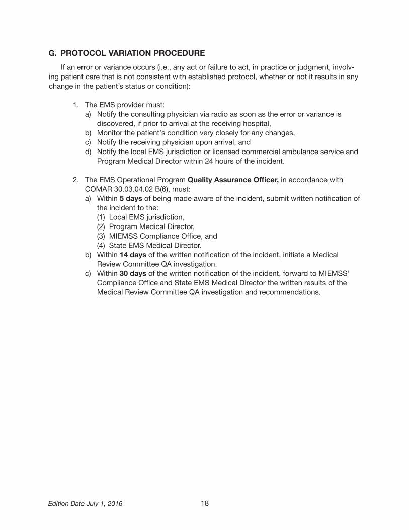
If an error or variance occurs (i.e., any act or failure to act, in practice or judgment, involv-ing patient care that is not consistent with established protocol, whether or not it results in any change in the patient’s status or condition):
1. The EMS provider must:a) Notify the consulting physician via radio as soon as the error or variance is
discovered, if prior to arrival at the receiving hospital,b) Monitor the patient’s condition very closely for any changes,c) Notify the receiving physician upon arrival, andd) Notify the local EMS jurisdiction or licensed commercial ambulance service and
Program Medical Director within 24 hours of the incident.
2. The EMS Operational Program Quality Assurance Officer, in accordance with COMAR 30.03.04.02 B(6), must:a) Within 5 days of being made aware of the incident, submit written notification of
the incident to the:(1) Local EMS jurisdiction,(2) Program Medical Director,(3) MIEMSS Compliance Office, and(4) State EMS Medical Director.
b) Within 14 days of the written notification of the incident, initiate a Medical Review Committee QA investigation.
c) Within 30 days of the written notification of the incident, forward to MIEMSS’ Compliance Office and State EMS Medical Director the written results of the Medical Review Committee QA investigation and recommendations.
G. PROTOCOL VARIATION PROCEDURE

19 Edition Date July 1, 2016
Occasionally a situation may arise in which a physician’s order cannot be carried out; e.g., the provider feels the administration of an ordered medication would endanger the patient, a medication is not available, or a physician’s order is outside the protocol. If this occurs:
1. The EMS provider must:
a) Immediately notify the consulting physician as to the reason the order cannot be carried out.
b) Document on the patient care report what was ordered, the time it was ordered, and the reason the order could not be carried out.
c) As soon as practical following the call, notify the local EMS jurisdiction of the incident.
2. Public Service EMS Operational Programs must:
a) Within 5 days of being made aware of the incident, submit written notification of the incident through the local EMS jurisdiction and Program Medical Director to the Regional Medical Director with a copy to the State EMS Medical Director. The MIEMSS Regional EMS Administrator shall be notified at the discretion of the Regional Medical Director.
b) Within 14 days of the written notification of the incident, initiate a QA investiga-tion under the authority of the Medical Review Committee.
c) Within 30 days of the written notification of the incident, forward to MIEMSS’ Compliance Office and State EMS Medical Director written results of the Medi-cal Review Committee QA investigation and recommendations.
3. Licensed Commercial Programs must:
a) Within 5 days of being made aware of the incident, submit written notification of the incident through the commercial Program Medical Director to the Director of the State Office of Commercial Ambulance Licensing and Regulation with a copy to the State EMS Medical Director.
b) Within 14 days of the written notification of the incident, initiate a QA investiga-tion under the authority of the Medical Review Committee.
c) Within 30 days of the written notification of the incident, forward to the Program Medical Director and to the Director of the State Office of Commercial Ambu-lance Licensing and Regulation and State EMS Medical Director written results of the Medical Review Committee QA investigation and recommendations.
H. INABILITY TO CARRY OUT PHYSICIAN ORDER

20Edition Date July 1, 2016
Rarely, a physician providing on-line medical consultation may direct a prehospital provider to render care that is truly life-saving and is not explicitly listed within the protocols.
1. ALL of the following criteria MUST be present for prehospital providers to proceed with an order under this section:
a) During the consultation, both the consulting physician and the provider must acknowledge and agree that the patient’s condition and extraordinary care are not addressed elsewhere within these medical protocols and that the order is absolutely necessary to maintain the life of the patient.
b) The provider must feel capable of correctly performing the care directed by the consulting physician, based on the instructions given by the consulting physician.
c) When such an order is carried out, the consulting physician and the provider must immediately notify the State EMS Medical Director (via SYSCOM, 800-648-3001) of the extraordinary care situation. In addition, the provider must fax docu-mentation of the rationale for extraordinary care within 24 hours to the State EMS Medical Director at 410-706-0853. Attendance at a subsequent review meeting shall be required.
d) The prehospital provider must inform the consulting physician of the effect of the treatment and notify the receiving physician of the treatment upon arrival at the hospital (if the receiving physician is different than the consulting physician). The prehospital provider must also notify his/her BLS/ALS Program Medical Director within 24 hours.
e) The public service local EMS jurisdiction and the Program Medical Director must then submit written notification of the incident to the Regional Medical Director with a copy to the State EMS Medical Director within 5 days of the incident.
f) The commercial ambulance company and the Program Medical Director must submit written notification of the incident to the Director of the State Office of Commercial Ambulance Licensing and Regulation and the State EMS Medical Director within 5 days of the incident.
I. PHYSICIAN ORDERS FOR EXTRAORDINARY CARE NOT COVERED BY MARYLAND PROTOCOL

27 Edition Date July 1, 2016
II. GENERAL PATIENT CARE (GPC)
A. RESPONSEReview the dispatch information and select appropriate response.
B. SCENE ARRIVAL AND SIZE-UP1. Consider Body Substance Isolation (BSI).
2. Consider Personal Protective Equipment (PPE).
3. Evaluate the scene safety.
4. Determine the number of patients.
5. Consider the need for additional resources.
C. PATIENT APPROACH1. Determine the Mechanism of Injury (MOI)/Nature of Illness (NOI).
2. If appropriate, begin triage and initiate Mass Casualty Incident (MCI) procedures.
D. INITIAL ASSESSMENT
CORRECT LIFE-THREATENING PROBLEMS AS IDENTIFIED.STABILIZE CERVICAL SPINE WHEN APPROPRIATE.
FOR PEDIATRIC PATIENTS, CONSIDER USING THE PEDIATRIC ASSESSMENT TRIANGLE.
1. Assess mental statusa) Alertb) Responds to Verbal stimulic) Responds to Painful stimulid) Unresponsive
2. Airwaya) Open and establish airway using appropriate adjunct.b) Place patient in appropriate position.c) Suction airway as needed, including tracheostomy tubes.
Appearance Work ofBreathing
Circulation to Skin

28Edition Date July 1, 2016
IF A PATENT AIRWAY CANNOT BE ESTABLISHED, THE PATIENT MUST BE TRANSPORTED TO THE NEAREST APPROPRIATE HOSPITAL-BASED EMERGENCY DEPARTMENT OR DESIGNATED FREESTANDING MEDICAL FACILITY. ONCE THE PATIENT PRESENTS TO THE HOSPITAL OR DESIGNATED FREESTANDING MEDICAL FACILITY FOR TREATMENT OF AN EMERGENCY CONDITION, TREATMENT AND TRANSFER DECISIONS ARE THE RESPONSIBILITY OF THE HOSPITAL UNDER APPLICABLE LAW. THE PROVIDER SHOULD STAND BY TO BE AVAILABLE FOR AND ASSIST WITH TRANSFER OF THE PATIENT IF THE HOSPITAL DETERMINES SUCH A TRANSFER IS APPROPRIATE. – – – – – – – – – – – – – – – – – – – – – – – – – – – – – – – – – – – – – – – –
IN INFANTS AND YOUNG CHILDREN, INSPIRATORY STRIDOR IS AN INDICATION OF UPPER AIRWAY FOREIGN BODY OR PARTIAL AIRWAY OBSTRUCTION. REQUEST ALS RENDEZVOUS. TRANSPORT THE PATIENT RAPIDLY AND CAUTIOUSLY AND HAVE FOREIGN BODY AIRWAY REMOVAL EQUIPMENT READY FOR IMMEDIATE USE IN CASE THE PATIENT’S AIRWAY BECOMES OBSTRUCTED.
3. Breathing a) Determine if breathing is adequate. Assess oxygen saturation (SpO2) with por-
table pulse oximeter (required on all transport units since 2012).(1) If patient’s ventilations are not adequate, provide assistance with 100% oxy-
gen using Bag-Valve-Mask (BVM).(i) For all ages except neonates, 1 breath every 5 seconds (8–12
breaths/min) (manually-activated positive pressure oxygen delivery device is not recommended for this group)
(ii) For a neonate, 1 breath every 3 seconds (higher rates may be required)
(2) The decision to oxygenate will be based on the patient’s clinical condition.(i) SpO2 greater than or equal to 94% is considered normoxia in adults
and children. Supplemental oxygen is not needed if SpO2 greater than or equal to 94% unless the patient is in respiratory distress, acutely dyspneic, or suffering from suspected CO poisoning. Patients in severe respiratory distress may benefit from high flow oxygen from a nonre-breather (NRB). Note: Respiratory distress is present if the patient has retractions, nasal flaring, wheezing, stridor, or difficulty speaking.
(ii) Unless in respiratory distress, avoid administration of high flow oxygen to patients presenting with the following conditions: (a) STEMI/Angina (b) CVA/stroke (c) Post arrest
(iii) CO exposure: Apply 100% oxygen via NRB mask. Maintain SpO2 at 100%.

29 Edition Date July 1, 2016
(3) If available, utilize EtCO2 waveform monitoring in intubated patients (required on all ALS transport units for advanced airway management since 2015).
(4) Consider carbon monoxide measurement, if available.
b) Hyperventilate the head-injured patient only if signs/symptoms of herniation are present, including posturing, loss of pupillary light response, dilation of one or both pupils, vomiting, hypertension, bradycardia, and/or irregular respirations.(1) If hyperventilating, use the following rates (NEW ’16)
Adult (including adolescent greater than 13 years): 20 breaths per minute Child (1-12 years of age): 30 breaths per minute Infant (less than 1 year of age): 35 breaths per minute
(2) If hyperventilating, use EtCO2 monitoring if available.
NEVER WITHHOLD OXYGEN FROM A PATIENT IN RESPIRATORY DISTRESS!
DEVICE FLOW RATE CONCENTRATION
Nasal Cannula 2–6 lpm 24–44%
Venturi Mask Variable 24–60%
Partial Rebreather Mask 6–10 lpm 35–60%
Simple Face Mask 6–10 lpm 35–60%
Pocket Mask 12–15 lpm 50–60%
Non-Rebreather Mask 12–15 lpm 80–100%
Bag-Valve-Mask 12–15 lpm 90–100%
Percent O2 Saturation Ranges General Patient Care
94–100% Normal Give oxygen as necessary
91–93% Mild Hypoxia Give oxygen as necessary
86–90% Moderate Hy-poxia
Give 100% oxygenAssisting Ventilations if necessary
less than or equal to 85%
Severe Hypoxia Give 100% oxygenAssist Ventilations If indicated, Intubate
INACCURATE OR MISLEADING SpO2 READINGS MAY OCCUR IN THE FOLLOWING PATIENTS: HYPOTHERMIC, HYPOPERFUSION (SHOCK), CO POISONING, HEMOGLOBIN ABNORMALITY, ANEMIA, AND VASOCONSTRICTION.

30Edition Date July 1, 2016
4. Circulation ONCE CONFIRMED PULSELESS, HIGH-QUALITY CONTINUOUS CPR WITH FREQUENT PROVIDER ROTA-TION IS AN ESSENTIAL COMPONENT IN THE SUCCESSFUL RESUSCITATION OF THE ARRESTED PATIENT. THIS MAY BE ACCOMPLISHED THROUGH MANUAL OR MECHANICAL MEANS AS APPROPRIATE.
PERFORM CPR WHILE PREPARING FOR RHYTHM ANALYSIS AND DEFIBRILLATION. – – – – – – – – – – – – – – – – – – – – – – – – – – – – – – – – – – – – – – – –
a) Assess pulse.(1) Patients from one hour after birth (newly born) up to those who have not
reached their 13th birthday (NEW ’16):(a) If pulse is absent, use AED/manual defibrillator or begin CPR.(b) If patient is symptomatic with poor perfusion (unresponsive or
responds only to painful stimuli) and pulse is less than 60 bpm:(i) Ventilate for 30 seconds.(ii) If after 30 seconds the pulse is less than 60 bpm, begin CPR.
(c) If pulse greater than 60 bpm, continue assessment.(2) Patients 13 years of age or older:
(a) If pulse is absent, use AED/manual defibrillator or begin CPR.(b) If pulse is present, continue assessment.
b) Assess for and manage profuse bleeding.c) Assess skin color, temperature, and capillary refill.
5. Disabilitya) Perform Mini-Neurologic Assessment (Pulse/Motor/Sensory).b) Spinal protection
(1) The provider shall determine the appropriate method to use in spinal protection of the patient. Infant or child car seats may NOT be used as a spinal immobiliza-tion device for the pediatric patient.
(2) Patients who have a blunt trauma with a high-energy mechanism of injury that has potential to cause spinal cord injury or vertebral instability and one or more the following should receive spinal protection. (a) Midline spinal pain, tenderness, or deformity(b) Signs and symptoms of new paraplegia or quadriplegia(c) Focal neurological deficit(d) Altered mental status or disorientation(e) Distracting injury
– – – – – – – – – – – – – – – – – – – – – – – – – – – – – – – – – – – – – – – – In addition to the above indicators for adults, the below apply to children who have not yet reached their 15th birthday.
(f) Neck pain or torticollis(g) High impact diving incident or high risk motor vehicle crash (head on
collision, rollover, ejected from the vehicle, death in the same crash, or speed greater than 55 mph)
(h) Substantial torso injury(i) Conditions predisposing to spine injury
(3) If NO to all of the above, transport as appropriate.
IF PATIENT IS UNABLE TO COMMUNICATE OR APPROPRIATELY RESPOND TO THE ABOVE QUESTIONS, APPLY SPINAL PROTECTION PROTOCOL.

31 Edition Date July 1, 2016
6. Exposure To assess patient’s injuries, remove clothing as necessary, considering condition and environment.
7. Assign Clinical Prioritya) Priority 1 — Critically ill or injured person requiring immediate attention; unstable
patients with life-threatening injury or illness. b) Priority 2 — Less serious condition yet potentially life-threatening injury or illness, re-
quiring emergency medical attention but not immediately endangering the patient’s life.
c) Priority 3 — Non-emergent condition, requiring medical attention but not on an emergency basis.
d) Priority 4 — Does not require medical attention.e) In the event of a multiple casualty incident, the Simple Triage And Rapid Treatment
(START and/or JumpSTART) technique will be instituted for rapid tagging and sort-ing of patients into priority categories for both treatment and transport.
8. Normal Vital Signs Chart
AGE ESTIMATEDWEIGHT
HEARTRATE
RESPIRATORYRATE
SYSTOLICB/P
Premature Less than 3 kg 160 Greater than 40 60
Newborn 3.5 kg 130 40 70
3 mo. 6 kg 130 30 90
6 mo. 8 kg 130 30 90
1 yr. 10 kg 120 26 90
2 yrs. 12 kg 115 26 90
3 yrs. 15 kg 110 24 90
4 yrs. 17 kg 100 24 90
6 yrs. 20 kg 100 20 95
8 yrs. 25 kg 90 20 95
10 yrs. 35 kg 85 20 100
12 yrs. 40 kg 85 20 100
14 yrs. 50 kg 80 18 110
ADULT Greater than 50 kg 80 18 120

32Edition Date July 1, 2016
HIS
TOR
Y A
ND
PH
YS
ICA
L E
XA
MIN
ATI
ON
TRA
UM
A P
ATI
EN
TM
ED
ICA
L P
ATI
EN
T
CO
NS
IDE
R A
LS,
PE
RF
OR
M I
NT
ER
VE
NT
ION
S,
AN
D T
RA
NS
PO
RT.
D C A P B T L S
Sig
nific
ant M
OI
Rap
id T
raum
aA
sses
smen
t
H
ead
Cre
pita
tion
C
hest
Cre
pita
tion
Res
pira
tion
Par
adox
ical
Mot
ion
Bre
ath
Sou
nds
A
bdom
enR
igid
ityD
iste
ntio
n
Pel
vis/
GU
Pai
n on
Mot
ion
Blo
od,U
rine,
Fec
es
Ext
rem
ities
Pul
se/M
otor
/Sen
sory
P
oste
rior
Bas
elin
e V
ital S
igns
Obt
ain
SA
MP
LE H
isto
ry
Sig
ns &
Sym
ptom
s
A
llerg
ies
Med
icat
ions
Per
tinen
t His
tory
Las
t Ora
l Int
ake
Eve
nts
Prio
r
Non
-Sig
nific
ant M
OI
Det
erm
ine
Chi
efC
ompl
aint
Per
form
Foc
used
Exa
min
atio
n o
f the
Inju
red
Site
and
Are
asC
ompa
tible
with
Giv
enM
OI
Bas
elin
e V
ital S
igns
Obt
ain
SA
MP
LE H
isto
ry
Sig
ns &
Sym
ptom
s
A
llerg
ies
Med
icat
ions
Per
tinen
t His
tory
Las
t Ora
l Int
ake
Eve
nts
Prio
r
Unr
espo
nsiv
e P
atie
nt
Rap
id P
hysi
cal
Exa
min
atio
n
H
ead
N
eck
JVD
Med
ical
Ale
rt D
evic
e
Che
stB
reat
h S
ound
s
Abd
omen
Rig
idity
Dis
tent
ion
P
elvi
s/G
UB
lood
,Urin
e,F
eces
E
xtre
miti
esM
SP
Med
ical
Ale
rt D
evic
e
Pos
terio
r
Bas
elin
e V
ital S
igns
Obt
ain
His
tory
of E
piso
de
O
nset
Pro
voca
tion
Qua
lity
Rad
iatio
n
S
ever
ity
T
ime
Obt
ain
SA
MP
LE H
isto
ry
Res
pons
ive
Pat
ient
Obt
ain
His
tory
of E
piso
de
O
nset
Pro
voca
tion
Qua
lity
Rad
iatio
n
S
ever
ity
Tim
e
Bas
elin
e V
ital S
igns
Obt
ain
SA
MP
LE H
isto
ry
S
igns
& S
ympt
oms
A
llerg
ies
M
edic
atio
ns
Per
tinen
t His
tory
La
st O
ral I
ntak
e
Eve
nts
Prio
r
Foc
used
Phy
sica
l Exa
mD
CA
PB
TLS
Che
ck a
reas
sug
gest
edby
MO
I and
SA
MP
LE.
D C A P B T L S
D C A P B T L S

33 Edition Date July 1, 2016
DE
TAIL
ED
AN
D O
NG
OIN
G A
SS
ES
SM
EN
TS
DE
TAIL
ED
EX
AM
INA
TIO
NO
NG
OIN
G A
SS
ES
SM
EN
T
HE
AD
Sca
lp &
Cra
nium
Cre
pita
tion
Eye
s Dis
colo
ratio
nE
qual
ityF
orei
gn B
odie
sB
lood
in A
nter
ior
Cha
mbe
r E
ars
& N
ose
Flu
id D
rain
age
or B
leed
ing
Dis
colo
ratio
n M
outh Te
eth
& F
orei
gn B
odie
sS
wel
ling
or L
acer
atio
nsB
reat
h O
dor
Dis
colo
ratio
nN
EC
K Jugu
lar
Vei
n D
iste
ntio
nTr
ache
a P
ositi
onC
repi
tatio
nC
HE
ST
Par
adox
ical
Mot
ion
Bre
ath
Sou
nds
Cre
pita
tion
AB
DO
ME
NR
igid
ityD
iste
ntio
nP
ELV
IS/G
UP
ain
on M
otio
nE
XT
RE
MIT
IES
Pul
se, M
otor
, Sen
sory
Cap
illar
y R
efill
PO
ST
ER
IOR
ME
DIC
AL
PATI
EN
T
RE
PE
AT
IN
ITIA
L A
SS
ES
SM
EN
T
R
eass
ess
AV
PU
R
eass
ess
Airw
ay
Mon
itor
Bre
athi
ng
Rea
sses
s C
ircul
atio
n
M
onito
r S
kin
C
onfir
m C
linic
al P
riorit
y
RE
PE
AT
& R
EC
OR
D V
ITA
L S
IGN
S
RE
PE
AT
FO
CU
SE
DA
SS
ES
SM
EN
TE
spec
ially
Chi
ef C
ompl
aint
or
Inju
ries
CH
EC
K A
LL
INT
ER
VE
NT
ION
S
A
ssur
e O
xyge
n A
dequ
acy
C
heck
Ble
edin
g
Che
ck In
terv
entio
ns
Che
ck fo
r Tre
ndin
g
Sta
ble
Pt.-
Eve
ry 1
5 M
in.
U
nsta
ble
Pt.-
Rec
omm
end
E
very
5 M
in.
TRA
UM
A P
ATI
EN
T
RE
PE
AT
IN
ITIA
L A
SS
ES
SM
EN
T
R
eass
ess
AV
PU
R
eass
ess
Airw
ay
Mon
itor
Bre
athi
ng
Rea
sses
s C
ircul
atio
n
M
onito
r S
kin
C
onfir
m C
linic
al P
riorit
y
RE
PE
AT
& R
EC
OR
D V
ITA
L S
IGN
S
RE
PE
AT
RA
PID
TR
AU
MA
AS
SE
SS
ME
NT
CH
EC
K A
LL
INT
ER
VE
NT
ION
S
A
ssur
e O
xyge
n A
dequ
acy
C
heck
Ble
edin
g
Che
ck N
eck
Sta
biliz
atio
n
Che
ck In
terv
entio
ns
Che
ck fo
r Tre
ndin
g
Sta
ble
Pt.-
Eve
ry 1
5 M
in.
U
nsta
ble
Pt.-
Rec
omm
end
E
very
5 M
in.
D C A P B T L S
CO
NS
IDE
R A
LS,
PE
RF
OR
M I
NT
ER
VE
NT
ION
S,
AN
D T
RA
NS
PO
RT.

34Edition Date July 1, 2016

35 Edition Date July 1, 2016
E. HISTORY AND PHYSICAL EXAMINATION/ASSESSMENT
1. Conduct a Focused Examination/Detailed Examination/Ongoing Assessment.
2. Collect and transport documentation related to patient’s history (example: Emer-gency Information Form, Medic Alert, EMS DNR/MOLST, or jurisdictional form).
3. Obtain an EKG when appropriate.
F. TREATMENT PROTOCOLS1. Refer to ALL appropriate protocols.
2. Patients who have had an impaled conducted electrical weapon used on them will be transported to the nearest appropriate facility without dart removal (exception: Tactical EMS). ANY conducted electrical weapon dart impalement to the head, neck, hands, feet, or genitalia must be stabilized in place and evaluated by a physi-cian. An assessment must be conducted to determine if the patient meets Excited Delirium Syndrome. (NEW ’16)
3. Providers may assist the patient or primary caregiver in administering the patient’s prescribed rescue medication.a) BLS providers may assist with the administration of the patient’s fast-acting
bronchodilator MDI and sublingual nitroglycerin.b) ALS providers may administer the patient’s prescribed benzodiazepine for sei-
zures, Factor VIII or IX for Hemophilia A or B, or reestablish IV access for con-tinuation of an existing vasoactive medication.
c) Providers should obtain on-line medical direction to administer other prescribed rescue medications not specifically mentioned in The Maryland Medical Protocols for EMS Providers (e.g., Solucortef for adrenal insufficiency). The rescue medica-tion must be provided by the patient or caregiver and the label must have the patient’s name and the amount of medication to be given.
DO NOT ADMINISTER ORAL MEDICATIONS (EXCEPT GLUCOSE PASTE) TO PATIENTS WITH AN ALTERED MENTAL STATUS. – – – – – – – – – – – – – – – – – – – – – – – – – – – – – – – – – – – – – – – –
4. For pediatric patientsa) Pediatric section of the treatment protocol will be used for children who have
not reached their 15th birthday (trauma) or their 18th birthday (medical), except as otherwise stated in the treatment protocol. (NEW ’16)
b) Medication dosing(1) Pediatric doses apply to patients weighing less than 50 kg.(2) For pediatric patients equal to or greater than 50 kg, utilize adult dosing.
c) The developmental age of the infant/child must be considered in the communi-cation and evaluation for treatment. Destination consideration: For those patients who are 18 years of age or older who receive spe-
cialized care at a pediatric facility, consider medical consultation with a Pediatric Base Station for patient destination. (NEW ’16)
d) Infants and children must be properly restrained prior to and during transport.e) When appropriate, family members should remain with pediatric patients.

36Edition Date July 1, 2016
G. COMMUNICATIONS (NEW ’16)
1. Communications with and through EMRC/SYSCOM are recorded. In addition, as part of the quality assurance and quality improvement process, communications with hospitals are frequently recorded. Therefore, you should assume that all your communications among EMS providers, hospitals, public safety communications centers, and EMRC/SYSCOM are being recorded.
2. All Priority 1 patients require on-line medical consultation through EMRC on a re-corded line (radio or phone).
ANY PATIENT WHOM THE PROVIDER IDENTIFIES AS MEETING ANY “SPECIALTY” ALERT (E.G., TRAUMA, STEMI ALERT, STROKE ALERT, SEPSIS ALERT) REQUIRES AN ON-LINE MEDICAL CONSULTATION THROUGH EMRC ON A RECORDED LINE (RADIO OR PHONE).
3. All Priority 2 patients who have persistent symptoms or need further therapeutic intervention(s) require on-line medical consultation through EMRC on a recorded line (radio or phone).
4. Notification (“information only call” that can be through EOC or EMS communica-tion system following local standard operating procedures) should be made to the receiving hospital for Priority 2 or Priority 3 patients whose symptoms have resolved and whose vital signs are within normal limits.
ON-LINE MEDICAL CONSULTATION MAY BE OBTAINED AT ANY TIME FOR ANY PATIENT, IF DESIRED BY THE PREHOSPITAL EMS PROVIDER. PEDIATRIC AND SPECIALTY CONSULTATION IS ENCOURAGED FOR TRAUMA AND MEDICAL PATIENTS. CONSULTATION WITH PEDIATRIC AND SPECIALTY CENTERS SHALL OCCUR SIMULTANEOUSLY WITH A BASE STATION CONSULT.
5. If medical consultation is genuinely unavailable, or if the time necessary to initiate consultation significantly compromises patient care, the provider shall proceed with additional protocol directed care, so long as transport will not be significantly delayed. “Exceptional Call” must be indicated on the Patient Care Report (PCR).
6. Core essentials for communications:a) Assigned patient priority (1 to 4)b) Agec) Chief complaintd) Provider impressione) Pertinent patient signs and symptoms (e.g., HR, RR, BP, Pulse Ox, and GCS) (be
specific–do not use within normal limits or stable in description)f) Pertinent physician findingsg) ETAIn addition, for specialty center patients:Traumah) Patient Trauma Decision Tree Category (Alpha, Bravo, Charlie, Delta)i) Number of victims if more than onej) Describe mechanism Strokek) Last known well timel) Specific neurological findings (sensory, motor, cognitive) m) Upon positive assessment using the Cincinnati Stroke Scale, a STROKE alert
shall be made and the LAMS score will be included in the consult.STEMIn) 12-Lead interpretationo) Duration of symptoms
CONSIDER ACTIVATION OF THE GO-TEAM FOR SERIOUSLY INJURED PATIENTS WHO REQUIRE A PROLONGED EXTRICATION AND WHO MEET THE INDICATIONS FOR GO-TEAM ACTIVATION.

37 Edition Date July 1, 2016
H. REASSESSMENT1. Reassess unstable patients frequently (recommended every 5 minutes).2. Reassess stable patients at a minimum of every 15 minutes.3. Reassess patients being discharged to home or long-term care at the beginning and
end of the transport or more frequently, at the provider’s discretion.
I. DISPOSITION1. Destination
a) Priority 1 patients shall be triaged according to Maryland Medical Protocols to the closest appropriate hospital-based emergency department, designated trauma, or designated specialty referral center. Critically unstable patients in need of immediate life-saving interventions that cannot be provided in the field shall, with the approval of EMS system medical consultation, be diverted to the closest facility (including freestanding medical facility) capable of immediately providing those interventions.
b) Priority 2 patients shall be triaged according to the Maryland Medical Protocols to the closest appropriate hospital-based emergency department, designated trauma or designated specialty referral center unless otherwise directed by EMS system medical consultation.
c) Stable Priority 3 or 4 patients who do not need a time-critical intervention may also be transported to the local emergency department or freestanding medical facility.
d) Patients Under Investigation (PUI) for an Emerging Infectious Disease (EID) at a residence should be transported directly to an Assessment Hospital unless total transport time is no longer than 45 minutes greater than transport to the nearest Frontline Hospital ED. If transport time is longer than 45 minutes greater than transport to the nearest Frontline Hospital ED, the patient must be transported to the closest appropriate Frontline hospital. Priority 1 and Priority 2 patients with unresolved symptoms that cannot be managed outside the hospital should be taken to the closest Frontline Hospital. Receiving hospital notification of all sus-pected PUI patients should be done as early as possible to allow for hospital staff to prepare. Helicopter transport is NOT indicated for the PUI patient. (NEW ’16)
e) For Priority 2 and Priority 3 patients not meeting a specialty center destination care protocol, the EMS provider should ask if the patient has had a hospital ad-mission (inpatient service) within the last 30 days. If the answer is yes, the EMS provider should transport (repatriate) the patient to that hospital as long as that hospital is not more than 15 additional minutes further than nearest hospital (or greater if allowed for by the EMS Operational Program). (NEW ’16)
7. Mass Casualty Incident (MCI) Communicationsa) When a local jurisdiction declares an MCI, it is extremely important to maximize
patient care resources and reserve EMS communications for emergent situations. Except for extraordinary care interventions, EMS providers may perform all skills and administer medications within protocol during a declared MCI. When the MCI condition is instituted, the Exceptional Call box must be checked on the PCR.
b) During an MCI, the EMS Officer-in-Charge (OIC) shall designate an EMS Communi-cator who shall establish appropriate communications.
c) Reference the Multiple Casualty Incident/Unusual Incident Protocol.

38Edition Date July 1, 2016
2. Mode of transport (air, land, water)a) Medevac patients with indications for specialty referral center should be flown to
the appropriate type of specialty center if not more than 10–15 minutes further than the closest trauma center. (Patients with an airway, breathing, or circulatory status who would be jeopardized by going an additional 10–15 minutes should go to the closest trauma center.)
b) Consider utilization of a helicopter when the patient’s condition warrants trans-port to a trauma or specialty referral center and the use of a helicopter would re-sult in a clinically significant reduction in time compared with driving to a trauma/specialty center.
ALL REQUESTS FOR SCENE HELICOPTER TRANSPORTS SHALL BE MADE THROUGH SYSCOM. FOR TRAUMA DECISION TREE CATEGORY CHARLIE OR DELTA, RECEIVING TRAUMA CENTER MEDICAL CONSULTATION IS REQUIRED WHEN CONSIDERING WHETHER HELICOPTER TRANSPORT IS OF CLINICAL BENEFIT. c) If the time of arrival at the trauma or specialty referral center via ground unit is less
than 30 minutes, there will generally not be a benefit in using the helicopter, espe-cially for Trauma Decision Tree categories Charlie and Delta.
d) Refer to the Trauma Decision Tree when considering use of aeromedical transport. Provide SYSCOM with the patient’s category (Alpha, Bravo, Charlie, or Delta).
e) On-line medical direction should be obtained from the local trauma center and the specialty referral center when transport to the specialty center would require more than 10–15 minutes additional transport time.(1) Pediatric Trauma Patients: Indications as per the pediatric section of the
Trauma Protocols.(2) Spinal Trauma Patients: Indications as per Spinal Protection Protocol.(3) Burn Patients: Indications as per Burn Protocol. Special note:
Isolated burn patients without airway injury or other associated trauma should normally be flown to a burn center, regardless of the location of the closest trauma center.
(4) Hand Injury Patients: Indications as per Hand Trauma Protocol. Special note: Medevac patients with appropriate indications for hand center referral should normally be flown to the hand center, regardless of the loca-tion of the closest trauma center.
3. StatusEvaluate the need for emergent versus non-emergent transportation.
DO NOT WAIT ON-SCENE FOR ADVANCED LIFE SUPPORT. ATTEMPT TO RENDEZVOUS EN ROUTE TO THE HOSPITAL.

39 Edition Date July 1, 2016
J. TRANSFER OF CARE/RENDEZVOUS AND TRANSITION OF PATIENT CARE ALS TO BLSThe ALS provider-patient relationship is established when the ALS provider initiates patient assessment and
1. ALS medication(s)* is/are administered or2. ALS procedure(s)* is/are performed or3. UponALSproviderassessmentofthepatientthereispotentialriskofdeterioration.
* Based on the medication or procedure as listed in the protocol pages 182–185
ALS providers may only terminate their EMS provider-patient relationship when they are assured that the patient will continue to receive care at the same or greater levels, or when they have documented with on-line medical direction that the patient’s condition has improved and that patient care may be transferred safely to an EMS provider with a lower scope of practice. BLS providers have the right to decline the transition of patient care. When con-sensus between the providers cannot be gained, ALS shall get on-line medical direction. Providers will relay assessment findings and treatment provided to the individual(s) as-suming responsibility for the patient(s).
L. CONFIDENTIALITYPatient confidentiality must be maintained at all times.
M. PROFESSIONAL CONDUCTAll patients should be treated with dignity and respect in a calm and reassuring manner.
K. DOCUMENTATIONA Patient Care Report (PCR) will be completed and delivered to the receiving facility as soon as possible, ideally upon transfer of care. If this is not immediately possible, providers must provide documentation of the patient’s prehospital care on a template and in a format provid-ed or approved by MIEMSS for inclusion in the patient care record before leaving the receiv-ing facility, then deliver the completed PCR within 24 hours after dispatch, in compliance with COMAR 30.03.04.04. Only the unit that pronounces death will select the “Dead on Scene” option in the PCR (eMEDS®) and thus all other units will report “Operational Support Only.” If no interventions are performed, the highest level EMS provider on scene will pronounce death and document “Dead on Scene.” If BLS care was rendered by a BLS unit and then termination of resuscita-tion and pronouncement of death occurred, the BLS unit will select “Dead at Scene with BLS Intervention” option on the eMEDS® PCR. If ALS care was rendered by an ALS unit and then termination of resuscitation and pronouncement of death occured, the ALS unit will select “Dead at Scene with ALS Intervention” option on the eMEDS® PCR.

41 Edition Date July 1, 2016
III. TREATMENT PROTOCOLS
FOR ALL TREATMENT PROTOCOLS, THE LETTER AND NUMERICAL OUTLINE FORMAT IS STRICTLY FOR RAPID AND UNIFORM REFERENCE AND DOES NOT IMPLY OR DIRECT A MANDATORY SEQUENCE FOR PATIENT CARE. HOWEVER, THE GENERAL PATIENT CARE SECTION AND THE ALGORITHMS DO HAVE A SPECIFIC SEQUENCE TO BE FOLLOWED.
A. ABUSE/NEGLECT1. Initiate General Patient Care.
ALL HEALTH CARE PROVIDERS ARE OBLIGATED BY LAW TO REPORT CASES OF SUSPECTED CHILD OR VULNERABLE ADULT ABUSE OR NEGLECT TO EITHER THE LOCAL POLICE OR SOCIAL SERVICE AGENCIES. DO NOT INITIATE REPORT IN FRONT OF THE PATIENT, PARENT, OR CAREGIVER. DO NOT CONFRONT OR BECOME HOSTILE TO THE PARENT OR CAREGIVER.
2. PresentationThe patient may present with patterned burns or injuries suggesting intentional in-fliction, such as injuries in varying stages of healing, injuries scattered over multiple areas of the body, fractures, or injuries inconsistent with stated cause of injury. The patient, parent, or caregiver may respond inappropriately to the situation. Malnutri-tion or extreme lack of cleanliness of the patient or environment may indicate ne-glect. Signs of increased intracranial pressure (bulging fontanels and altered mental status in an infant) may also be seen.
3. Treatmenta) Stabilize injuries according to protocol.
b) Discourage patient from washing if sexual abuse is suspected.
c) Document the following information on the PCR:(1) All verbatim statements made by the patient, the parent, or caregiver shall
be placed in quotation marks, including statements made about the manner of the injuries.
(2) Any abnormal behavior of the patient, parent, and/or caregiver
(3) The condition of the environment and other residents present

42Edition Date July 1, 2016
A. ABUSE/NEGLECT (Continued)
(4) The time the police/welfare agency was notified and the name of the person notified
(5) The name of the receiving health care provider (RN, PA, MD) and any state-ments made
d) Treat injuries according to presentation.
4. Continue General Patient Care.

43 Edition Date July 1, 2016
B. ALTERED MENTAL STATUS: SEIZURES
1. Initiate General Patient Care.
2. Presentation Seizures are a neuromuscular response to an underlying cause such as: epilepsy, hypoxia, hypoglycemia, hypoperfusion, head injury, CVA, alcohol or drug abuse. Consider recent history of possible illness, infection, fever, or stiff neck.
DO NOT ATTEMPT TO FORCE ANY DEVICE INTO THE PATIENT’S MOUTH IF THE PATIENT IS STILL SEIZING.
3. Treatment
a) If the patient is still seizing:
(1) DO NOT RESTRAIN.
(2) Protect patient from further injury.
(3) Consider cause of seizure activity.
b) When seizure activity has stopped:
(1) Identify and treat injuries.
(2) If patient is a known diabetic, glucose paste (10–15 grams) should be administered between the gum and cheek. Consider single additional dose of glucose paste if not improved after 10 minutes.
c) Use glucometer and treat accordingly.
d) Consider midazolam. (NEW ’16)If patient has no IV or IO in place, administer midazolam 5 mg IN or IM. If IV/IO is already in place: 0.1 mg/kg in 2 mg increments SLOW IVP over 1–2 minutes per increment with maximum single dose 5 mg.
REDUCE BY 50% FOR PATIENTS 69 YEARS OR OLDER.
If IV unavailable, 5 mg IM may be administered. Additional doses up to a maximum total dose 10 mg require medical consulta-tion for all providers. If patient seizures are refractory to treatment, consider IO administration of mid-azolam. If midazolam is not available, consider diazepam. 2.5 mg increments SLOW IVP/IM (IM requires all providers to obtain medical consultation).

44Edition Date July 1, 2016
B. ALTERED MENTAL STATUS: SEIZURES (Continued)
Maximum total dose 10 mg If patient is in status, consider IO administration of diazepam. If suspected severe nerve agent exposure, providers may administer midazolam 5 mg IM or diazepam (CANA) without medical consultation.
e) Establish IV access with LR.
f) If patient is pregnant, actively seizing, consider magnesium sulfate 4 grams IV/IO over 10 minutes.
g)
If seizures persist, consult for second dose of magnesium sulfate.
IF PATIENT IS PREGNANT, USE MIDAZOLAM FOLLOWED BY MAGNESIUM SULFATE. MEDICAL CONSULTATION REQUIRED FOR PREGNANT PATIENTS WHO MAY REQUIRE LARGER DOSES OF MIDAZOLAM TO CONTROL SEIZURES.
IF, FOLLOWING ADMINISTRATION OF MAGNESIUM SULFATE, PATIENT EXHIBITS SIGNS OF TOXICITY, CONSIDER ADMINISTRATION OF CALCIUM CHLORIDE. CONSIDER CALCIUM CHLORIDE 500 MG IVP FOR RESPIRATORY DEPRESSION, DECREASED REFLEXES, FLACCID PARALYSIS, AND APNEA FOLLOWING MAGNESIUM SULFATE ADMINISTRATION. MEDICAL CONSULTATION REQUIRED. – – – – – – – – – – – – – – – – – – – – – – – – – – – – – – – – – – – – – – – –
h) If the patient is still seizing:
(1) DO NOT RESTRAIN.
(2) Protect from further injury.
(3) Consider underlying cause of seizure.
i) When seizure activity has stopped:
(1) Identify and treat any injuries.
(2) If patient is a known diabetic, glucose paste (10–15 grams) should be ad-ministered between the gum and cheek. Consider single additional dose of glucose paste if not improved after 10 minutes.
j) Use glucometer and treat accordingly.
k) The paramedic may assist patients with the administration of their prescribed benzodiazepine.

45 Edition Date July 1, 2016
B. ALTERED MENTAL STATUS: SEIZURES (Continued)
l) Consider midazolam for seizures lasting greater than 10 minutes. (NEW ’16) If patient has no IV or IO in place: Administer midazolam 0.2 mg/kg IN or IM. Maximum total dose 5 mg. If IV or IO is already in place: Administer midazolam 0.1 mg/kg in 2 mg increments SLOW IVP over 1–2 minutes. Maximum total dose 5 mg.
m) If patient is pregnant, actively seizing, consider magnesium sulfate 4 grams IV/IO over 10 minutes.
n) Establish IV/IO access with LR.
o) Administer fluid bolus, if appropriate 20 mL/kg of LR IV/IO.
FOR A CHILD ACTIVELY SEIZING, ADMINISTER MIDAZOLAM IN/IM AND RESERVE IO FOR LIFE-THREATENING ILLNESS.
p) Additional doses up to a maximum total dose 5 mg require medical consulta-tion for all providers.
If patient’s seizures are refractory to treatment, consider IO administration of midazolam. If midazolam is not available, consider diazepam for seizures lasting greater than 10 minutes (paramedic may perform without consult for pa-tients with active seizures). Up to 0.2 mg/kg rectal Maximum total dose 10 mg OR0.1 mg/kg in 2.5 mg increments SLOW IVP/IO/IM (IM requires all providers to obtain medical consultation.) Maximum total dose 5 mg If suspected severe nerve agent exposure, providers may administer mid-azolam as above or diazepam (CANA) without medical consultation.
4. Continue General Patient Care.

46Edition Date July 1, 2016
C. ALTERED MENTAL STATUS: UNRESPONSIVE PERSON
1. Initiate General Patient Care.
2. Presentation Patients may exhibit confusion, focal motor sensory deficit, unusual behavior, unre-sponsiveness to verbal or painful stimulus.
ALCOHOL CAN CAUSE ALTERED MENTAL STATUS BUT IS NOT COMMONLY A CAUSE OF TOTAL UNRESPONSIVENESS TO PAIN.
3. Treatmenta) Obtain pulse oximetry, if available.
b) Administer glucose paste (10–15 grams) between the gum and cheek. Consider single additional dose of glucose paste if not improved after 10 minutes.
c) If patient has respiratory depression with decreased LOC, constricted pupils, and provider suspects an opioid/narcotic overdose, Administer naloxone. 2 mg intranasal atomizer (Divide administration of the dose equally between the nares to a maximum of 1 mL per nare.)
d) If patient has respiratory depression with decreased LOC, constricted pupils, and provider suspects an opioid/narcotic overdose, Administer naloxone 0.4–2 mg IVP/IO (titrated)/IM/IN (If delivery device is available—divide administra-tion of the dose equally between the nares to a maximum of 1 mL per nare) Maximum single dose 0.4–2 mg (NEW ’16)
e) Establish IV access with LR. Administer fluid bolus, if appropriate. 20 mL/kg of LR IV
f) Titrate to a systolic pressure of 100 mmHg.
g) Consider obtaining blood sample using closed system.
h) Use glucometer and treat accordingly.
i) Consider an additional dose of naloxone. (NEW ’16)
j) Consider additional fluid administrationMaximum 2,000 mL without medical consultation.

47 Edition Date July 1, 2016
C. ALTERED MENTAL STATUS: UNRESPONSIVE PERSON (Continued)
– – – – – – – – – – – – – – – – – – – – – – – – – – – – – – – – – – – – – – – –
k) Obtain pulse oximetry if available.
l) Administer glucose paste (10–15 grams) between the gum and cheek. Consider single additional dose of glucose paste if not improved after 10 minutes.
m) If patient has respiratory depression with decreased LOC, constricted pu-pils, and provider suspects an opioid/narcotic overdose, Administer naloxone 28 days to 4 years (NEW ’16): Administer naloxone 0.8–1 mg intranasal atomizer (Divide administration of the dose equally between the nares to a maximum of 1 mL per nare.) 5 years to adult (NEW ’16): Administer naloxone 2 mg intranasal atomizer (Di-vide administration of the dose equally between the nares to a maximum of 1 mL per nare.)
n) If patient has respiratory depression with decreased LOC, constricted pu-pils, and provider suspects an opioid/narcotic overdose, Administer naloxone. 0.1 mg/kg IVP/IO (titrated)/IM/IN (If delivery device is available—divide admin-istration of the dose equally between the nares to a maximum of 1 mL per nare) (NEW ’16)Maximum single dose 0.4–2 mg
o) Consider repeating naloxone. (NEW ’16)
p) Establish IV/IO access with LR.If age-related vital signs and patient’s condition indicate hypoperfusion, ad-
minister initial fluid bolus of 20 mL/kg LR IV/IO. If patient’s condition does not improve, administer the second bolus of fluid at 20 mL/kg LR IV/IO. ORFor volume-sensitive children administer initial fluid bolus of 10 mL/kg LR IV/IO. If patient’s condition does not improve, administer the second bolus of fluid at 10 mL/kg LR IV/IO. Volume-sensitive children include: neonates (birth to 28 days) (NEW ’16), children with congenital heart disease, chronic lung disease, or chronic renal failure.
(1) Consider obtaining blood sample using closed system.
q) Use glucometer and treat accordingly.
r)
Third and subsequent fluid boluses at 20 mL/kg IV/IO except in volume-sensitive children, then bolus at 10 mL/kg.
4. Continue General Patient Care.

48Edition Date July 1, 2016
D. APPARENT LIFE-THREATENING EVENT (ALTE)
– – – – – – – – – – – – – – – – – – – – – – – – – – – – – – – – – – – – – – – –
1. Initiate General Patient Care.
2. Presentation An episode in an infant or child less than 2 years old that is frightening to the ob-server and is characterized by some combination of the following:a) Apnea (central or obstructive)b) Skin color change: cyanosis, erythema (redness), pallor, plethora (fluid overload) c) Marked change in muscle toned) Choking or gagging not associated with feeding or a witnessed foreign body
aspiration
MOST PATIENTS WILL APPEAR STABLE AND EXHIBIT A NORMAL PHYSICAL EXAM UPON ASSESSMENT BY RESPONDING FIELD PERSONNEL. HOWEVER, THIS EPISODE MAY BE THE SIGN OF UNDERLYING SERIOUS ILLNESS OR INJURY. FURTHER EVALUATION BY MEDICAL STAFF IS REQUIRED AND IT IS ESSENTIAL TO TRANSPORT ALL PATIENTS WHO EXPERIENCED ALTE.
3. Treatmenta) Perform an initial assessment utilizing the Pediatric Assessment Triangle.b) Obtain a description of the event including nature, duration, and severity.c) Obtain a medical history with emphasis on the following conditions:
(1) Known chronic diseases(2) Evidence of seizure activity(3) Current or recent infections(4) Gastroesophageal reflux(5) Recent trauma(6) Medications (current or recent)
d) Apply oxygen.e) Be prepared to assist with ventilation if this type of episode occurs again
during transport.f) Assess environment for possible causes.
g) Place patient on cardiac monitor.h) Consider establishing IV/IO access with LR.
IF THE PARENT OR GUARDIAN REFUSES MEDICAL CARE OR TRANSPORT, PROVIDER SHALL CONTACT A PEDIATRIC BASE STATION PHYSICIAN.
4. Continue General Patient Care.

49 Edition Date July 1, 2016
E. BEHAVIORAL EMERGENCIES
1. Initiate General Patient Care.
2. PresentationBehavior or actions that indicate the patient’s mental function is disturbed and may pose a threat to oneself or to others (suicide, threat of violence, or psychosis).
THE PROVIDER SHOULD RECOGNIZE CRITICAL INCIDENT STRESS AS A STATE OF EMOTIONAL DISTRESS THAT DOES NOT NECESSARILY POSE A THREAT TO ONESELF OR OTHERS (E.G., DEATH IN THE FAMILY, BYSTANDERS AT A CRASH SCENE, OR REACTION TO VIOLENCE). THE PREHOSPITAL CARE PROVIDER SHOULD NOT BE PLACED IN ANY PHYSICAL JEOPARDY OR ASSUME ANY LAW ENFORCEMENT FUNCTIONS, ESPECIALLY WHEN WEAPONS AND/OR ACTS OF VIOLENCE ARE INVOLVED! LAW ENFORCEMENT SHOULD BE REQUESTED ON ALL CALLS INVOLVING POTENTIALLY VIOLENT PATIENTS.
3. Treatmenta) When considering the prehospital use of restraints, a law enforcement officer
should apply the device and accompany the provider and the patient in the ambulance.
b) For interfacility transport, a physician order must be obtained for physical re-straint.
c) Implement SAFER model.
(1) Stabilize the situation by containing and lowering the stimuli.
(2) Assess and acknowledge the crisis.
(3) Facilitate the identification and activation of resources (chaplain, family, friends, or police).
(4) Encourage patient to use resources and take actions in his/her best interest.
(5) Recovery or referral—leave patient in care of responsible person or professional or transport to appropriate facility.

50Edition Date July 1, 2016
E. BEHAVIORAL EMERGENCIES (Continued)
d) Establish IV access with LR, if appropriate.e) Consider Chemical Restraint.
4. Continue General Patient Care.

51 Edition Date July 1, 2016
F. CARDIAC EMERGENCIES: CARDIAC GUIDELINES
1. The following algorithmic and standard formatted sections pertain to cardiac emer-gencies. Several guidelines apply to all algorithms when assessing and treating cardiac patients. These guidelines are:
a) When the patient’s condition changes, indicating the transition to a new treatment algorithm, the new treatment shall take into account prior therapy (e.g., previously administered medications).
b) As BLS/ALS guidelines indicate, definitive airway control is preferable; if this can be achieved, along with other initial interventions, then the earlier the better. However, defibrillation is more important initially if the patient can be ventilated without intubation.
c) Cardiac Arrest: Immediately start CPR and apply AED or manual defibrillator as soon as pos-sible; shock if indicated. The goal is to defibrillate as soon after stopping CPR as possible (ideally for manual defibrillator, in less than 5 seconds). After single shock, immediately restart CPR (do not perform pulse or EKG rhythm check) for 2 minutes, then assess for pulse and rhythm and apply single shock if indicated. Repeat this sequence of single shocks and 2 minutes of CPR.
SOME AEDs MAY NOT CURRENTLY DELIVER THE REFERENCED JOULE SETTING NOR ALLOW FOR SINGLE SHOCK DELIVERY. THE AED INDUSTRY WILL BE IMPLEMENTING THESE CHANGES OVER TIME, WHICH WILL BE A MULTI-YEAR PROCESS. WHEN IN DOUBT FOLLOW THE AED PROMPT.
d) If unable to initiate an IV or perform endotracheal intubation within 5 minutes, continue with appropriate care and transport the patient as soon as possible to the appropriate hospital. Further attempts to initiate IV therapy or endotracheal intubation should be accomplished while en route to the receiving hospital.
e) Only in a pediatric or neonatal arrest situation, naloxone, atropine, and epineph-rine, can be administered via the ET route. Medications administered for pediat-ric patients via the endotracheal tube route shall be 2–2.5 times the IV dose for naloxone, and atropine, and ten times the IV dose for epinephrine (1:1,000). All ET medications shall be diluted in 5 mL of LR for pediatric patients. (NEW ’16)

52Edition Date July 1, 2016
2. UNIVERSAL ALGORITHM FOR ADULTEMERGENCY CARDIAC CARE FOR BLS
UnresponsiveNot Breathing
Pulse?
Supportventilation
ALS &transport
Begin CPRAttach AED ASAP
Analyzeshockablerhythm?
Defibrillate 1 timeResume CPR immediatelyfor 2 minutes
YES NO
YES NO
Resume CPR immediatelyfor 2 minutes

53 Edition Date July 1, 2016
3. UNIVERSAL ALGORITHM FOR ADULTEMERGENCY CARDIAC CARE FOR ALS
Assess Responsiveness
Breathing
Pulse
Begin CPR
Too Slow Too Fast
VF/VT Present on Monitor
Not Responsive:Call for DefibrillatorAssess Breathing
IntubateConfirm Tube PlacementConfirm Ventilations
GO TOTACHYCARDIA
ALGORITHM
GO TOBRADYCARDIA
ALGORITHM
GO TO
VT/VF
ALGORITHM
Oxygen as needed VENTILATE as needed
IV with LRCardiac Monitor
Vital SignsHistory & Physical
Detailed Assessment
Determine Rhythm &Possible Cause
GO TO ASYSTOLEALGORITHM
GO TO PEAALGORITHM
Electrical Activity?
Suspected Cause
Pulmonary Edema/CHFSee Protocol
Chest PainSee Protocol
Dysrhythmia
Assess Circulation If unconscious and no trauma, place in recovery position
Responsive:Observe
Treat as Indicated
YES
YES
YES
YES
NO
NO
NO
NO

54Edition Date July 1, 2016
4. UNIVERSAL ALGORITHM FOR PEDIATRIC (LESS THAN 18 YEARS OF AGE)EMERGENCY CARDIAC CARE FOR BLS
(If less than 1 hour old, refer to Newly Born Protocol)(NEW ’16)
UnresponsiveNot Breathing
Pulse?
Supportventilation
ALS &transport
Begin CPRAttach AED with
pediatric capability
Analyzeshockable rhythm?
Defibrillate 1 timeResume CPR immediatelyfor 2 minutes
YES NO
YES NO
Resume CPR immediatelyfor 2 minutes

55 Edition Date July 1, 2016
5. UNIVERSAL ALGORITHM FOR PEDIATRIC (LESS THAN 18 YEARS OF AGE) EMERGENCY CARDIAC CARE FOR ALS
(If less than 1 hour old, refer to Newly Born Protocol) (NEW ’16)
Assess Responsiveness
Breathing
Pulse
Begin CPRAttach AED with pediatric capability
100 compressions/minute
100% oxygen
GO TO PEDIATRIC CARDIAC ARREST ALGORITHM
Too FastToo Slow
Not Responsive: Call for Defibrillator Assess Breathing
GO TO PEDIATRICTACHYCARDIA
ALGORITHM
GO TO PEDIATRICBRADYCARDIA
ALGORITHM
Oxygen as needed VENTILATE as needed
Cardiac monitorVital signsIV with LR
History & PhysicalDetailed Assessment
Suspected Cause
Altered Mental Status: See Protocol
Respiratory DistressAllergic Reaction or Anaphylaxis: See
Protocol, as appropriateAsthma/COPD: See Protocol
Pulmonary Edema/CHF: See Protocol
Dysrhythmia
Assess CirculationIf unconscious with adequate
respiratory rate and effort and no trauma, place in recovery position
Responsive:Observe
Treat as Indicated
YES
YES
NO
NO

56Edition Date July 1, 2016
G. CARDIAC EMERGENCIES: BRADYCARDIA
1. Initiate General Patient Care.
2. PresentationPatient may present with a slow heart rate and chest pain, shortness of breath, decreased level of consciousness, hypotension, hypoperfusion, pulmonary congestion, congestive heart failure, and/or acute myocardial infarc-tion.
3. Treatmenta) Place patient in position of comfort.
b) Assess and treat for shock, if indicated.
c) Continuously monitor airway and reassess vital signs every 5 minutes.
d) Establish IV access with LR.
e) If patient is hemodynamically unstable: initiate transcutaneous pacing (TCP).
f) If TCP is unsuccessful or not available, administer atropine: 0.5–1 mg IVP Atropine should be given in repeat doses in 3–5 minute intervals up to a total of 0.04 mg/kg.
g) Consider dopamine 2–20 mcg/kg/min
h) If patient is hemodynamically stable and in Type II, second-degree AV Block or third-degree AV Block:
(1) Consider/prepare for TCP.
(2) If patient develops discomfort with TCP Administer opioid per Pain Management Protocol. ORConsider midazolam 0.1 mg/kg in 2 mg increments SLOW IVP over 1–2 minutes per increment with maximum single dose 5 mg. (Reduce by 50% for patients 69 years or older.)
i) Refer to appropriate algorithm.

57 Edition Date July 1, 2016
(a) - Serious signs and symptoms must be related to the slow rate. Signs and symptoms may include chest pain, shortness of breath, decreased level of consciousness, hypotension, hypoperfusion, pulmonary congestion, CHF, and/or AMI.
(b) - Do not delay TCP while awaiting IV or atropine to take effect if the patient is symptomatic.
(c) - Denervated transplanted hearts will not respond to atropine. Go at once to TCP.
(d) - Atropine shall be given in repeat doses in 3–5 minute intervals up to a total of 0.04 mg/kg. Consider shorter intervals in severe clinical conditions.
Medical consultation required to administer atropine in AV block at the His-Purkinje level (Type II AV block and new third-degree block with wide QRS complexes).
(e) - Never treat third-degree AV block or ventricular escape beats with amiodarone. (NEW ’16)
(f) -
In the presence of Mobitz II and third-degree AV block, medical consultation is required for atropine administration.
(g) - Requires medical consultation for administration of dopamine. Adults: titrate to systolic BP 100 mmHg or medical consultation directed BP. IV infusion pump is preferred.
GENERALPATIENT CARE
Type II, Second-degree AV Blockor
Third-degree AV Block (e,f)
Bradycardia - (Less than 60 bpm)
Serious Signs or Symptoms (a,b)
Prepare for possible Transcutaneous Pacing should patientdevelop serious signs or symptoms.
Observe
Transcutaneous Pacemaker (b)
Atropine 0.5–1 mg IVP (c,d,f)
Dopamine 2–20 mcg/kg/min. (g)Medical Consultation Required
4. ADULT BRADYCARDIA ALGORITHM
YES
YES
NO
NO

58Edition Date July 1, 2016
(a) - Hemodynamically unstable is defined as a systolic blood pressure less than 60 in neonates (patients less than 28 days old), less than 70 in infants (patients less than 1 year of age), and less than [70 + (2 x years) = systolic BP] for patients greater than 1 year of age.
(b) - Neonates (birth to 28 days) (NEW ’16), epinephrine ET 0.03 mg/kg (1:10,000) dilute with 1 mL. (c) - Volume infusion for neonates and volume-sensitive children, 10 mL/kg; for infant and child 20 mL/kg.
(d) - Sodium Bicarbonate, 1 mEq/kg with medical consultation. See sodium bicarbonate.
(e) - Calcium chloride, 20 mg/kg (0.2 mL/kg) SLOW IVP/IO (50 mg/min). Max dose 1 gram.
5. PEDIATRIC BRADYCARDIA ALGORITHM(If less than 1 hour old, refer to Newly Born Protocol) (NEW ’16)
ObserveSupport ABCs
Hemodynamically unstable? (a)
Begin CPR if HR less than 60 with poor perfusion despite oxygenation and ventilation
Bradycardia persists?
Identify and treat underlying causes
Possible causes of bradycardia(Parenthesis) = Possible Therapies and Treatments
Hypovolemia (Volume Infusion) (c)Hypoxia (Ventilation) Hydrogen ion (acidosis) (d)Hypo-/hyperkalemia (d,e)Hypoglycemia (Glucometer Protocol)Hypothermia (Warming)Toxins (d,e)Tamponade, cardiac Tension pneumothorax (NDT) Thrombus Trauma
Pacer Age-Related Rates (NEW ’16)
Start pacemaker at age-appropriate heart rate: Infant (less than 1 year): 120 beats per minute Child (1 through 12 years): 100 beats per minute
Adult/Adolescent (13 years and greater): 80 beats per minute
YESNO
NO YES
Epinephrine (b)IV/IO 0.01 mg/kg (1:10,000)ET 0.1 mg/kg (1:1,000),
Dilute in 5 mL; Repeat every 3–5 minutes
AtropineIV/IO 0.02 mg/kg,
Maximum single dose 0.5 mg,ET 0.04–0.06 mg/kg,
Dilute in 5 mLRepeat once
Consider Transcutaneous Pacing
If pulseless arrest developsgo to Cardiac Arrest
Algorithm

59 Edition Date July 1, 2016
H. CARDIAC EMERGENCIES: TACHYCARDIA
1. Initiate General Patient Care.
2. Presentation Patient may present with chest pain, shortness of breath, decreased level of con-sciousness, low blood pressure, hypoperfusion, pulmonary congestion, congestive heart failure, and/or acute myocardial infarction.
3. Treatmenta) Place patient in position of comfort.
b) Assess and treat for shock, if indicated.
c) Continuously monitor airway and reassess vital signs every 5 minutes.
d) Establish IV access with LR.
e) Verify presence of pulse.
f) If no pulse present, treat as pulseless VF/VT.
g) If patient is hemodynamically unstable with a ventricular rate greater than 150, prepare for immediate cardioversion.
h) If patient is hemodynamically stable, identify rhythm and proceed to appropriate algorithm.
– – – – – – – – – – – – – – – – – – – – – – – – – – – – – – – – – – – – – – – –
i) Place patient in position of comfort.
j) Assess and treat for shock, if indicated.
k) Continuously monitor airway and reassess vital signs every 5 minutes.
l) Establish IV access with LR.
m) Verify presence of pulse.
n) If no pulse present, treat as pulseless VF/VT.

60Edition Date July 1, 2016
H. CARDIAC EMERGENCIES: TACHYCARDIA (Continued)
o) If patient is hemodynamically unstable with a ventricular rate greater than 220 for an infant or 180 for a child, prepare for immediate cardioversion.
p) If patient is hemodynamically stable, identify rhythm and proceed to appropriate algorithm.
4. Continue General Patient Care.

61 Edition Date July 1, 2016
(a) - Unstable condition must be related to the tachycardia. Signs and symptoms may include chest pain, shortness of breath, decreased level of consciousness, hypotension, hypoperfusion, pulmonary congestion, CHF, and/or AMI.
(b) - Consider sedation (midazolam with medical consultation). However, overall patient status, including BP, may affect ability to administer sedative.
(c) - Consider calcium chloride 500 mg IVP for hypotension induced by diltiazem. Medical consultation required. If rate does not slow in 15 minutes, administer a second dose of diltiazem (15–25 mg over 2 minutes). Medical consultation required.
(d) - Be prepared for up to 40 seconds of asystole.(e) - If irregular, DO NOT administer amiodarone or adenosine. Cardiovert if unstable.(f) - If Torsades de Pointes, administer magnesium sulfate (1–2 grams IV/IO over 2 minutes).
5. ADULT TACHYCARDIA ALGORITHM (NEW ’16)
GENERALPATIENT CARE
Narrow QRSSVT
Valsalva maneuvers
BP?
Normal orelevated
Low or unstable
Monitor & transport
Adenosine (d)6 mg rapid IVP
Adenosine (d)6 mg rapid IVP
Adenosine (d)12 mg rapid IVP
Repeat X 1 in 1–2 Min.
Adenosine 12 mg Rapid IVP (d)Repeat X 1 in 1–2 Min.
Amiodarone 150 mg over 10 minutes
Repeat if necessary
Amiodarone 150 mg over 10 minutes
Repeat if necessary
Wide QRS regular monomorphic complex tachycardia (e)
Wide QRS regular polymorphic OR ventricular
tachycardia (e, f)
Atrial fibrillationor
Atrial flutter
Medicalconsultation
Diltiazem10–20 mg over
2 min. (c)
Unstable with serious signs and symptoms and ventricular rate greater than 150 bpm? (a)
PREPARE FOR IMMEDIATE CARDIOVERSION (b)
SYNCHRONIZED CARDIOVERSION (b)
YESNO

62Edition Date July 1, 2016
6. PEDIATRIC TACHYCARDIA ALGORITHM(If less than 1 hour old, refer to the Newly Born Protocol) (NEW ’16)
Identify and treat underlying causes
Evaluate QRS duration
Narrow(less than or equal to 0.09 seconds)
Wide regular(greater than 0.09 seconds)
Possible VT
Hemodynamically unstable? (b)
Cardiovert0.5 J/kg (c) (d)
Probablesupraventriculartachycardia (a)
Probable sinustachycardia
Cardiovert 1 J/kg
Consider vagal manuevers
Consider adenosine (e)
Consider (c) (d)cardioversion
Identify and treat underlying
cause
Cardiovert 2 J/kg
IV/IO access
Amiodarone (f)
YES NO
Consider adenosine (e)
Amiodarone (f)
(c) - If calculated joules setting is lower than cardioversion device is able to deliver, use the lowest joules setting possible or obtain medical consultation.
(d) - Consider sedation (midazolam with medical consultation). However, overall patient status, including BP, may affect ability to administer sedative.
(e) - Adenosine: 0.1 mg/kg rapid IV/IO, maximum 6 mg. Second and third doses 0.2 mg/kg rapid IV/IO, maximum single dose 12 mg. Be prepared for up to 40 seconds of asystole. (Contraindicated in polymorphic or irregular wide complex tachycardia)
(f) -
Amiodarone: 5 mg/kg IV/IO over 20 minutes. Obtain 12-lead EKG prior to administration of amiodarone. (NEW ’16)
(g) If Torsades de Pointes, administer magnesium sulfate (25 mg/kg IV/IO to a maximum of 2 grams over 2 minutes).
(a) - Ventricular Heart Rates in excess of: Infant 220 bpm or Pediatric 180 bpm
(b) - Hemodynamically unstable is defined as a systolic blood pressure less than 60 in neonates (patients from birth to 28 days old) (NEW ’16), less than 70 in infants (patients less than 1 year of age), less than [70 + (2 x years) = systolic BP] for patients greater than 1 year of age, altered mental status with hypoperfusion evidenced by delayed capillary refill, pallor, or peripheral cyanosis.

63 Edition Date July 1, 2016
I. CARDIAC EMERGENCIES: CARDIAC ARREST
1. Initiate General Patient Care.
2. Presentation Patient must be unconscious, apneic, and pulseless.
3. Treatmenta) Perform CPR.
HIGH-QUALITY CONTINUOUS CPR WITH FREQUENT PROVIDER ROTATION IS AN ESSENTIAL COMPONENT IN THE SUCCESSFUL RESUSCITATION OF THE ARRESTED PATIENT. THIS MAY BE ACCOMPLISHED THROUGH MANUAL OR MECHANICAL MEANS AS APPROPRIATE. PERFORM CPR WHILE PREPARING FOR RHYTHM ANALYSIS AND DEFIBRILLATION.
b) Utilize AED as appropriate.c) Transport
(1) If no shock indicated, consider Termination of Resuscitation Protocol or transport immediately.
(2) If shock indicated, defibrillate and resume CPR. Consider Termination of Resuscitation Protocol or transport ASAP.
(3) If ROSC, refer to ROSC Protocol.(4) If no ROSC, consider Termination of Resuscitation Protocol or transport to
the closest appropriate facility.
d) Identify rhythm and treat according to appropriate algorithm.e) If no ROSC, consider Termination of Resuscitation Protocol or transport to the
closest appropriate facility.f) If ROSC, refer to ROSC Protocol.
– – – – – – – – – – – – – – – – – – – – – – – – – – – – – – – – – – – – – – – –
g) Perform CPR.h) Utilize AED as appropriate (see AED Procedure).i) If no shock indicated, continue CPR and transport ASAP.j) If shock indicated, defibrillate, continue CPR, and transport ASAP.
For patients who have not reached their 18th birthday:k) Identify rhythm and treat according to appropriate algorithm.l) If no ROSC, transport to the closest appropriate facility.m) If ROSC, perform 12-Lead EKG and transport the patient to Children’s National
Health System or Johns Hopkins Children’s Center by ground or medevac. If arrival time is greater than 30 minutes to either of these destinations, transport to the clos-est appropriate ED.

64Edition Date July 1, 2016
4. ADULT ASYSTOLE ALGORITHM
- Continue CPR - Intubate O2 (90–100%) - Establish IV access with LR - Confirm asystole in more than one lead
Consider possible causes of asystole.(Parenthesis) = Possible Therapies and Treatments
Hypovolemia (Volume Infusion) (c)Cardiac Tamponade (Volume Infusion) (c)Tension Pneumothorax (Needle Decompression Thorocostomy–NDT) Massive Pulmonary EmbolismMassive AMIDrug Overdose (a,b)Hypoxia (Ventilation)Hypothermia (Warming)Acidosis (a)Hyperkalemia (a,b)
Consider PossibleCauses
Epinephrine 1 mg IVP - Repeat every 3–5 minutes
(a) - Sodium bicarbonate 1 mEq/kg, with medical consultation. See sodium bicarbonate.
(b) - Calcium chloride, 0.5–1 gram IVP, with medical consultation. See calcium chloride.
(c) -
Volume infusion is 20 mL/kg.
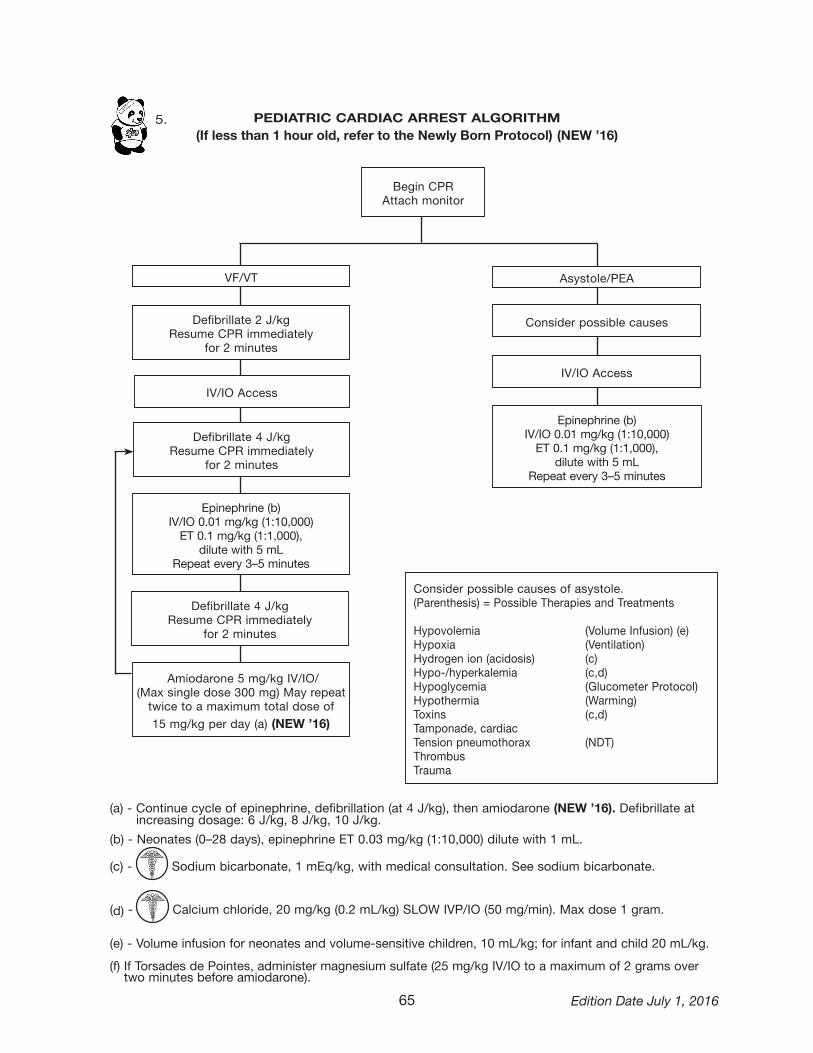
65 Edition Date July 1, 2016
5. PEDIATRIC CARDIAC ARREST ALGORITHM(If less than 1 hour old, refer to the Newly Born Protocol) (NEW ’16)
(a) - Continue cycle of epinephrine, defibrillation (at 4 J/kg), then amiodarone (NEW ’16). Defibrillate at increasing dosage: 6 J/kg, 8 J/k g, 10 J/kg.
(b) - Neonates (0–28 days), epinephrine ET 0.03 mg/kg (1:10,000) dilute with 1 mL.
(c) - Sodium bicarbonate, 1 mEq/kg, with medical consultation. See sodium bicarbonate.
(d) - Calcium chloride, 20 mg/kg (0.2 mL/kg) SLOW IVP/IO (50 mg/min). Max dose 1 gram.
(e) - Volume infusion for neonates and volume-sensitive children, 10 mL/kg; for infant and child 20 mL/kg.
(f) If Torsades de Pointes, administer magnesium sulfate (25 mg/kg IV/IO to a maximum of 2 grams over two minutes before amiodarone).
Begin CPRAttach monitor
Asystole/PEA
Consider possible causes
IV/IO Access
IV/IO Access
VF/VT
Amiodarone 5 mg/kg IV/IO/ (Max single dose 300 mg) May repeat twice to a maximum total dose of
15 mg/kg per day (a) (NEW ’16)
Epinephrine (b)IV/IO 0.01 mg/kg (1:10,000)ET 0.1 mg/kg (1:1,000),
dilute with 5 mL Repeat every 3–5 minutes
Epinephrine (b)IV/IO 0.01 mg/kg (1:10,000)ET 0.1 mg/kg (1:1,000),
dilute with 5 mLRepeat every 3–5 minutes
Consider possible causes of asystole.(Parenthesis) = Possible Therapies and Treatments
Hypovolemia (Volume Infusion) (e)Hypoxia (Ventilation) Hydrogen ion (acidosis) (c)Hypo-/hyperkalemia (c,d)Hypoglycemia (Glucometer Protocol)Hypothermia (Warming)Toxins (c,d)Tamponade, cardiac Tension pneumothorax (NDT) Thrombus Trauma
Defibrillate 2 J/kgResume CPR immediately
for 2 minutes
Defibrillate 4 J/kgResume CPR immediately
for 2 minutes
Defibrillate 4 J/kgResume CPR immediately
for 2 minutes

66Edition Date July 1, 2016
Epinephrine 1 mg IVP. Repeat every 3–5 minutes.
Consider possible causes of PEA.(Parenthesis) = Possible Therapies and Treatments
Hypovolemia (Volume Infusion) (c)Cardiac Tamponade (Volume Infusion) (c)Tension Pneumothorax (Needle Decompression Thorocostomy–NDT) Massive Pulmonary EmbolismMassive AMIDrug Overdose (a,b)Hypoxia (Ventilation)Hypothermia (Warming)Acidosis (a)Hyperkalemia (a,b)
Continue CPR
Intubate
IV with LR
Consider PossibleCauses
Includes:- EMD - Idioventricular Rhythms- Pseudo EMD - Ventricular Escape Rhythms- Brady-asystolic Rhythms - Post-defibrillation Idioventricular Rhythms
6. ADULT PULSELESS ELECTRICAL ACTIVITY(PEA) ALGORITHM
(a) - Sodium bicarbonate 1 mEq/kg, with medical consultation. See sodium bicarbonate.
(b) - Calcium chloride, 0.5–1 gram IVP, with medical consultation. See calcium chloride.
(c) -
Volume infusion is 20 mL/kg.

67 Edition Date July 1, 2016
7. VENTRICULAR FIBRILLATIONPULSELESS VENTRICULAR TACHYCARDIA (NEW ’16)
(a) - Sodium bicarbonate 1 mEq/kg, with medical consultation. See sodium bicarbonate.
(b) - If Torsades de Pointes is present, give magnesium sulfate 1–2 grams IV/IO over 2 minutes before amiodarone. (NEW ’16)
Perform CPR until defibrillator is attachedVF/VT present on monitor
Defibrillate 1 timeResume CPR immediately
for 2 minutes
Confirm Rhythm
Defibrillate 1 timeResume CPR immediately
for 2 minutes
Intubate
IV with LR
Epinephrine1 mg IVP
Repeat every 3–5 minutes
Defibrillate 1 timeResume CPR immediately
for 2 minutes
(NEW ’16) Amiodarone300 mg IV/IO push
May repeat once 150 mg IV/IO push (a) (b)
Defibrillate 1 timeResume CPR immediately
for 2 minutes
Persistent orRecurrentVF/VT
Return of SpontaneousCirculation
GO TO ROSC PROTOCOL
PEAGO TO PEAALGORITHM
AsystoleGO TO ASYSTOLE
ALGORITHM

68Edition Date July 1, 2016
J. RETURN OF SPONTANEOUS CIRCULATION (ROSC) (NEW ’16)
1. Initiate General Patient Care.
2. Presentation Patients revived from non-traumatic cardiac arrest.
3. Treatmenta) Verify presence of carotid pulse. If absent, go to Cardiac Arrest Protocol.
FREQUENTLY REASSESS FOR PRESENCE OF PULSE. IF ANY DOUBT AS TO PRESENCE OF PULSE, REINITIATE CHEST COMPRESSIONS AND RETURN TO APPROPRIATE ALGORITHM FOR CARDIAC ARREST.
b) If apneic, or inadequate respirations, continue to support ventilations. Other-wise, use supplemental oxygen as needed.
c) Reassess vital signs. Treat any abnormalities in accordance with relevant algo-rithms.
d) If patient 18 years of age or older and comatose (GCS less than 8), initiate Neu-roprotective Induced Hypothermia Protocol.
e) Rendezvous with ALS or transport to nearest ED.
f) Identify rhythm and treat according to appropriate algorithm.g) Obtain 12-lead EKG.h) Establish IV/IO access, if not yet obtained.i) Treat hypotension
(1) If lungs are clear, consider fluid bolus. 20 mL/kg LR IV. Titrate to SBP of 100 mmHg.
(2) With medical consultation, consider dopamine infusion.(a) Adjust infusion rate in accordance with blood pressure and clinical re-
sponse. (b) Adult: Administer 2–20 mcg/kg/min IV/IO drip titrated to BP of 100
systolic or medical consultation selected BP; initial infusion rate 2–5 mcg/kg/min.
(c) Pediatric: Administer 2–20 mcg/kg/min IV/IO drip titrated to age specific BP or medical consultation selected BP; initial infusion rate is 2 mcg/kg/min.
j) Reassess need for intubation, if not yet performed.k) Evaluate for contributing causes. For example,
(1) If chest pain currently or prior to arrest, see Chest Pain Protocol.(2) If signs and symptoms of CHF, follow Pulmonary Edema/CHF Protocol.

69 Edition Date July 1, 2016
J. RETURN OF SPONTANEOUS CIRCULATION (ROSC) (continued)
l) If VF or VT during arrest and amiodarone not yet given, consider Amiodarone 150 mg IV/IO over ten minutes.(a)
m) Initiate transport to appropriate facility. Most patients should go to a Cardiac Interventional Center. Consider helicopter transport. (1) Except as under (2) below, most patients should be transported to a Cardiac
Interventional Center. (2) Transport to nearest ED
(a) If obvious non-cardiac cause for arrest (e.g., drowning, asphyxiation, opiate overdose), (If cause for arrest is in any way uncertain, patient must be transported to Cardiac Interventional Center, except as under b and c below)OR
(b) If transport time to Cardiac Interventional Center is more than 45 minutes greater than transport time to nearest ED,OR
(c) With medical consultation, if patient’s clinical instability will not allow for safe transport to Cardiac Interventional Center due to transport time.
(3) Medical Consultation may assist with determination of destination. – – – – – – – – – – – – – – – – – – – – – – – – – – – – – – – – – – – – – – – –(4) Except as under (5) below, most pediatric patients should be transported
to Children’s National Health System or Johns Hopkins Children’s Center. Consider helicopter transport.
(5) Transport to nearest ED(a) If transport time to Children’s National Health System or Johns Hopkins
Children’s Center is more than 30 minutes greater than transport time to nearest ED,OR
(b) With medical consultation, if patient’s clinical instability will not allow for safe transport to one of the above centers due to transport time.
ALL POST-CARDIAC ARREST PATIENTS ARE PRIORITY 1, AND REQUIRE MEDICAL CONSULTATION. PEDIATRIC PATIENTS REQUIRE CONSULTATION WITH A PEDIATRIC BASE STATION AND MAY ASSIST IN DESTINATION DETERMINATION.
4. Continue General Patient Care.
(a) Presence of a perfusing sinus rhythm is necessary for the administration of amiodarone for the
ROSC patient post VF/VT conversion.

70Edition Date July 1, 2016
K. TERMINATION OF RESUSCITATION (Medical and Traumatic)
IF ANY DOUBT EXISTS, INITIATE RESUSCITATION AND TRANSPORT
1. PURPOSE This protocol is designed to guide the provider in determining a futile resuscita-
tion and managing the patient after this determination.
2. PROCEDUREa) Exclusions to this protocol.
(1) If arrest is believed to be secondary to hypothermia or submersion, treat according to appropriate protocol and transport to the nearest appropriate facility.
(2) If patient is pregnant, treat according to appropriate protocol and transport to the nearest appropriate facility.
(3) If patient has not reached his/her 18th birthday, treat according to appropri-ate protocol and transport to the nearest appropriate facility.
b) Medical Arrest(1) EMS providers may terminate resuscitation without medical consult when all
three criteria are met.(a) The arrest was not witnessed by an EMS provider (and patient is unre-
sponsive, pulseless, and apneic). AND(b) There is no shockable rhythm identified by an AED or there is
asystole or PEA on a manual cardiac monitor. AND(c) There is no return of spontaneous circulation (ROSC) prior to decision
to terminate resuscitation despite appropriate field EMS treatment that includes 15 minutes of minimally-interrupted EMS CPR. OR
(2) EMS providers may terminate resuscitation with medical consult when there is no ROSC prior to decision to terminate resuscitation despite appropriate field EMS treatment that includes 15 minutes of minimally-interrupted CPR in the presence of an arrest witnessed by an EMS provider or the presence of a shockable rhythm.
c) Trauma Arrest(1) EMS providers may terminate resuscitation without medical consult when
both criteria are met. (If medical etiology is suspected, use “Medical Arrest” above.)(a) There are no signs of life. AND(b) The patient is in asystole. OR
(2) EMS providers may terminate resuscitation with medical consult when both criteria are met in either blunt or penetrating trauma.
(a) Blunt(i) There are no signs of life. AND(ii) The patient is in a rhythm other than asystole and there is no ROSC
despite 15 minutes of appropriate treatment that includes 15 minutes of minimally-interrupted CPR.

71 Edition Date July 1, 2016
K. TERMINATION OF RESUSCITATION (Medical and Traumatic) (Continued)
(b) Penetrating(i) There are no signs of life. AND(ii) The patient is in a rhythm other than asystole and there is no ROSC.
If less than 15 minutes from a trauma center, transport the patient. If transport time exceeds 15 minutes, consult.
THERE ARE SOME CAUSES OF TRAUMATIC CARDIOPULMONARY ARREST (E.G., PENETRATING TRAUMA) THAT MAY BE REVERSED IF APPROPRIATELY AND EMERGENTLY MANAGED. THEREFORE, EMS PROVIDERS SHOULD FOLLOW APPROPRIATE PROTOCOLS FOR TRAUMATIC ARREST INCLUDING APPROPRIATE AIRWAY MANAGEMENT AND CONSIDERATION FOR BILATERAL NEEDLE DECOMPRESSION THORACOSTOMY. HOWEVER, EVEN WITH THE APPLICATION OF THESE MANEUVERS, ASYSTOLE AND PULSELESSNESS FOR GREATER THAN 10 MINUTES ARE INDEPENDENT PREDICTORS OF MORTALITY.
d) Pronouncement of Death in the Field Protocol.

72Edition Date July 1, 2016
TERMINATION OF RESUSCITATION ALGORITHM

73 Edition Date July 1, 2016
L. PRONOUNCEMENT OF DEATH IN THE FIELD
1. PURPOSEThis protocol is designed to guide the EMS provider in pronouncing death in the field. Health General Article §5-202 provides that:
a) An individual is dead if, based on ordinary standards of medical practice, the individual has sustained either:(1) Irreversible cessation of circulatory and respiratory functions; or(2) Irreversible cessation of all functions of the entire brain, including the brain
stem.
2. INDICATIONSEMS providers may pronounce the death of a patient when one or more of the fol-lowing criteria has been met.a) Decapitationb) Rigor mortisc) Decompositiond) Dependent lividity
e) Pulseless, apneic patient in a multi-casualty incident where system re-sources are required for the stabilization of living patients
f) Pulseless, apneic patient with an injury not compatible with life (with the exception of an obviously pregnant female where resuscitation attempts should be initiated and the patient transported to the nearest appropriate facility)
g) The EMS provider has terminated resuscitation per the Termination of Resuscitation Protocol.
3. PROCEDUREa) Confirm that the patient is unresponsive, pulseless, and apneic.b) The patient who meets criteria in 2.e may be “black” tagged during triage (by a
BLS or ALS provider), but asystole must be confirmed by ALS provider before a formal pronouncement of death.
c) The patient who meets criteria in 2.f must be confirmed to be in asystole by ALS provider before a formal pronouncement of death. If the condition of the remains precludes obtaining a cardiac rhythm to confirm asystole (e.g., incineration, se-vere disruption of the torso, etc.), this must be documented on the patient care report.
d) Document the exact time and location of the pronouncement of death.e) Notify law enforcement and follow local jurisdictional policies or, if death is pro-
nounced during transport, deliver patient to emergency department and follow hospital policies.

74Edition Date July 1, 2016
M. EMS DNR/MOLST
THE FOLLOWING SECTION IS ABSTRACTED FROM THE ORIGINAL MARYLAND EMERGENCY MEDICAL SERVICES DO NOT RESUSCITATE PROGRAM 2ND REVISION (07/01/98). THE PAGE (pg.) AND THE CHAPTER (ch.) NUMBERS HAVE BEEN APPENDED TO THE FOLLOWING CHAPTER TITLES FOR EASY REFERENCE. BECAUSE THIS ABSTRACT IS CONDENSED FROM THE ORIGINAL DOCUMENT, SOME CHAPTER NUMBERS OR LETTERS WERE INTENTIONALLY LEFT OUT. PLEASE REFER TO THE ORIGINAL MARYLAND EMS/DNR DOCUMENT FOR FURTHER INFORMATION. AS OF JANUARY 1, 2002, A COPY OF THE MARYLAND EMS DNR ORDER FORM CAN BE ACCEPTED IN LIEU OF THE ORIGINAL. AS OF OCTOBER 1, 2011, THE MARYLAND MOLST FORM CAN BE ACCEPTED IN LIEU OF THE MARYLAND EMS/DNR FORM.
1. PREFACE EMS/DNR Order or MOLST forms, bracelets, and necklaces will recog-nize three patient options for care prior to arrest: (pg. 15 ch. A)a) Option A (ALS) (MOLST A1)—Maximal (Restorative) Care (with intubation) Be-
fore Arrest, then DNR b) Option A (DNI) (MOLST A2)—Comprehensive Efforts to Prevent Arrest But Do
Not Intubate, then DNR c) Option B (BLS) (MOLST B)—Limited (Palliative) Care Only Before
Arrest, then DNR
2. VALID EMS/DNR or MOLST BRACELET WITH INSERT or AUTHORIZED METAL EMBLEM HAS THE SAME EFFECT AS THE FORM. (pg. 17 ch. D)a) Typically only one EMS/DNR device is needed to initiate the EMS/DNR Protocol. b) EMS providers should only request a second instrument (e.g., a bracelet when a
form has already been presented) if there is reason to question the validity of the first produced notification device.
3. RECIPROCITY (pg. 19 ch. E)a) A standardized EMS/DNR Order from another state may be honored.b) Out-of-state EMS/DNR Orders shall be followed to the full extent that is permis-
sible by the Maryland Medical Protocols for Emergency Medical Services Pro-viders. If there is misunderstanding with family members or others present at the scene or if there are other concerns about following the out of state EMS/DNR Order, contact online medical direction for assistance. (NEW ’16)
c) See chart in “EMS/DNR Program” booklet for how other states will treat Mary-land devices.
4. ORAL EMS/DNR ORDERS (pg. 19 ch. G)a) EMS providers may follow an oral EMS/DNR Order directly from a Maryland-
licensed physician (MD or DO) or nurse practitioner who is physically present “on-site.” EMS shall not accept orders from private physician attendings or nurse practitioner by telephone.
b) EMS providers may follow an oral EMS/DNR Order from a Maryland- licensed physician “on-line” via the EMS Communications System (e.g., radio or telephone consult that is routed through a public service access point (PSAP) for audio recording).

75 Edition Date July 1, 2016
M. EMS DNR/MOLST (Continued)
5. ACCEPTABLE AND UNACCEPTABLE EMS/DNR ORDERS (pg. 19 ch. H)a) The following are acceptable for implementing the EMS/DNR Protocol:
(1) Original Maryland EMS/DNR Order Form (2) Copy of the Maryland EMS/DNR Order Form(3) Other State EMS/DNR Order Form(4) Maryland EMS/DNR Bracelet Insert(5) Medic Alert DNR Bracelet or Necklace(6) Oral DNR Order from EMS System Medical Consultation(7) Oral DNR Order from other on-site physician or nurse practitioner(8) Maryland MOLST Form(9) Maryland MOLST Bracelet
b) The following are not acceptable for implementing the EMS/DNRProtocol:(1) Advance directives without an EMS/DNR Order(2) Facility-specific DNR orders(3) Notes in medical records(4) Prescription pad orders(5) DNR stickers(6) An oral request from someone other than a physician or nurse
practitioner (7) An oral order from an attending physician or nurse practitioner who is not on site (8) Any other device or instrument not listed above as acceptable
6. VALIDITY OF EARLIER VERSIONS OF EMS/DNR ORDERS (pg. 22 ch. K)a) Older versions of EMS/DNR Orders — i.e., initial version (1995 and first revision,
4/1/96) — continue to be valid and need not be updated unless the patient or authorized decision maker wishes to take advantage of new features available in the newer forms.
b) EMS providers should treat older versions of EMS/DNR order (pre 7/1/98) as “Op-tion B (BLS) - Limited (Palliative) Care Only Before Arrest, Then DNR.”
7. REVOCATION OF AN EMS/DNR ORDER (pg. 24 ch. M)a) An EMS/DNR Order may be revoked at any time by:
(1) Physical cancellation or destruction of all EMS/DNR Order devices; or(2) An oral statement by the patient made directly to emergency medical servic-
es personnel requesting only palliative care or resuscitation. If the patient revokes an EMS/DNR order orally, the EMS/DNR Order notification devices do not need to be destroyed. EMS providers should thoroughly document the circumstances of the revocation. An oral revocation by a patient is only good for the single response or transport for which it was issued.
b) An authorized decision-maker, other than the patient, cannot revoke an EMS/DNR Order orally. Because of the difficulty in identifying authorized decision makers in emergent situations, it is incumbent upon an authorized decision maker who has authority to revoke an EMS/DNR Order to either destroy or with-hold all EMS/DNR Order devices, if they wish resuscitation for the patient.

76Edition Date July 1, 2016
M. EMS DNR/MOLST (Continued)
c) Section 5-610 of the Health Care Decision Act (Health General Article, Anno-tated Code of Maryland) makes willful concealment, cancellation, defacement, obliteration, or damage of an advance directive (including EMS/DNR Orders), without the patient’s or authorized decision maker’s consent, a misdemeanor subject to a fine not exceeding $10,000, imprisonment not exceeding one year, or both.
8. ANTICIPATED LOCATIONS FOR EMS/DNR ORDER FORMS: (pg. 25 ch. N) EMS personnel shall be directed to look for an EMS/DNR Order in the following places:a) About a patient’s wrist, hung from a necklace, or safety-pinned to a
patient’s clothing. b) At medical facilities, in the patient’s chart.c) In residences and domicile facilities, by the bedside, behind the patient’s bed-
room door, or on the refrigerator door. d) In schools and educational institutions, in the nurse’s office, health room, or with
the student’s attendant caregiver/aide. e) Family or caregivers will be expected to retrieve the original EMS/DNR Order
prior to the ambulance’s arrival.
9. IDENTIFICATION OF PATIENT (pg. 25 ch. O)a) If the patient is able, the patient can self-identify during the initial
assessment. b) If the patient is unable to communicate, then family, caregivers, or
bystanders can identify the patient for EMS providers. c) If an EMS/DNR vinyl bracelet with insert or metal emblem (bracelet or necklace)
is attached to a patient (on wrist, pendant from neck, pinned to clothing, etc.) the patient’s identity can be reasonably assumed by EMS providers.
d) If an EMS/DNR vinyl bracelet insert or metal emblem (bracelet or necklace) is found detached from the patient, EMS personnel must treat it as an EMS/DNR Order form and identify the subject of the EMS/DNR Order as the pa-tient. A valid bracelet insert alone, without the vinyl bracelet, is a valid EMS/DNR Order so long as EMS providers confirm the patient’s identity (pg. 17 ch. D).
e) If EMS personnel are unable to ascertain with reasonable certainty, when required to do so, that the subject of the EMS/DNR Order is the patient, they may resuscitate the patient.
10. HEALTH PROVIDER/EMS PERSONNEL IMMUNITY (pg. 26 ch. R)a) General immunity provisions, such as Good Samaritan immunity for
volunteers and sovereign immunity for government employees, may apply under specific circumstances.

77 Edition Date July 1, 2016
M. EMS DNR/MOLST (Continued)
b) In addition to other immunity that may be provided for in law, the Health Care Decisions Act provides the following specific immunity in cases involving the provision, withdrawal, or withholding of care that may be life-sustaining in na-ture: (1) EMS providers are not subject to criminal prosecution or civil liability or
deemed to have engaged in unprofessional conduct as determined by the appropriate licensing, registering, or certifying authority as a result of with-holding or withdrawing any health care under authorization obtained in accordance with the Health Care Decisions Act. See HG (5-609(a)(1)).
(2) EMS providers providing, withholding, or withdrawing treatment under authorization obtained under the Health Care Decisions Act do not incur liability arising out of any claim to the extent the claim is based on lack of consent or authorization for the action.See HG (5-609(a)(2)).
(3) EMS providers providing treatment because they reasonably believe that an EMS/DNR order, other than a bracelet, is not valid, do not incur liability aris-ing out of any claim to the extent the claim is based on lack of consent or authorization for the action. See HG (5-608(d)).
11. EMS/DNR MEDICAL PROTOCOLS (pg. 29 ch. T)a) DISPATCH
(1) Option B EMS/DNR patients (7/98 version) or patients with older version EMS/DNR orders (pg. 22 ch K) only require a BLS response. Once the on-scene BLS provider has determined the need for additional pain control, an ALS Rendezvous may be requested. Medevac requests are not appropriate for these patients.
(2) Option A or A (DNI) EMS/DNR patients (7/98 version) who are not in arrest may require a range of responses from BLS through the highest echelon of response available. This will depend on the information available to dispatch and the service requested. The response complement in these cases will be dictated by local standard operating procedures (SOP).
(3) If a dispatch center is unclear whether the DNR order is an EMS/DNR order or is unclear about the pre-arrest patient care option selected (A, A (DNI), or B), the dispatch center shall dispatch the appropriate resources based on the information available.
(4) In the absence of knowledge to the contrary, information from medical professionals at a health care facility about the EMS/DNR status of a patient may be presumed to be reliable.

78Edition Date July 1, 2016
M. EMS DNR/MOLST (Continued)
b) PERFORM LIMITED PATIENT ASSESSMENT Vital signs:(1) Check for absence of a palpable pulse.(2) Check for absence of spontaneous respirations in an unresponsive patient. (3) Check for a valid EMS/DNR Order or MOLST form; vinyl bracelet insert worn
either on the wrist, as a necklace, or pinned to clothing; or for a metal em-blem (bracelet or necklace).
c) RESUSCITATE/DO NOT RESUSCITATE CRITERIA(1) If an EMS/DNR Order is not present, revoked, or otherwise void, the EMS
provider shall treat and, if necessary, transport the patient.(2) If an EMS/DNR Order is not present, but the EMS provider believes that re-
suscitation or further resuscitation is futile, they may initiate the Termination of Resuscitation Protocol.
(3) If a valid EMS/DNR order is found and the patient is in cardiac or respiratory arrest, no resuscitative measures shall be initiated.
(4) If the patient is conscious and able to communicate that he/she revokes the EMS/DNR orally directly to EMS providers, EMS providers shall treat and, if necessary, transport the patient.
(5) If the EMS/DNR patient (Option A, A (DNI), or B) arrests, withhold or with-draw further resuscitation and provide support to the family and caregivers. Consider notifying appropriate personnel.
d) OPTION A (MOLST A1) – MAXIMAL (RESTORATIVE) CARE PROTOCOL(1) When Option A - “Maximal (Restorative) Care (with intubation) Before Arrest,
then DNR” is selected on an EMS/DNR Order or MOLST form, the patient shall receive the full scope of restorative interventions permissible under the Maryland EMS Medical Protocols (including Continuous Positive Airway Pressure (CPAP), cardiac monitoring, synchronized cardioversion for pulse-present ventricular or supraventricular tachycardia, cardiac pacing for pulse-present symptomatic bradycardia, insertion of IVs, and drug therapy), in an attempt to forestall cardiac or respiratory arrest.
(2) This option was requested primarily by long-term care facilities for their patients who are on DNR orders for potentially prolonged periods of time. Many of these patients are less concerned about palliation of pain and more concerned about the quality of life after a stroke or heart attack. The primary medical conditions seen in the field necessitating this option have been the desire to administer dextrose for diabetic emergencies and epinephrine for anaphylactic reactions in patients who, upon arrest, are not to be resusci-tated.

79 Edition Date July 1, 2016
M. EMS DNR/MOLST (Continued)
(3) If, despite these efforts, the patient becomes pulseless or stops breathing spontaneously, EMS providers shall then withhold or withdraw cardiopulmo-nary resuscitation including, but not limited to, no CPR, no cardiac pacing, no defibrillation, withdrawal of active ventilatory assistance upon cardiac arrest, and withholding or withdrawal of drug therapy (e.g., chemical resusci-tation).
e) OPTION A (DNI) (MOLST A2) – COMPREHENSIVE EFFORTS TO PREVENT ARREST BUT DO NOT INTUBATE, THEN DNR(1) Option A (DNI) is exactly the same as Option A, which may include limited
ventilatory support by CPAP or BiPAP, but Do Not Intubate.(2) Therefore, inappropriate care for “Option A (DNI) – Comprehensive Efforts to
Prevent Arrest but Do Not Intubate, then DNR” would be nasal or oral intu-bation.
IF MAXIMAL CARE IS SELECTED AND THE PATIENT’S CONDITION REQUIRES ALS, AN ALS UNIT SHOULD BE REQUESTED IF FEASIBLE GIVEN THE LOCATION OF THE INCIDENT RELATIVE TO THE NEAREST APPROPRIATE FACILITY, THE AVAILABILITY OF AN ALS UNIT, AND ITS ABILITY TO ARRIVE OR RENDEZVOUS IN A MEDICALLY APPROPRIATE PERIOD OF TIME.
f) OPTION B (MOLST B)– PALLIATIVE CARE PROTOCOL(1) Supportive Care for Control of Signs and Symptoms
(a) Respiratory distress(i) Open the airway using non-invasive means (e.g., chin lift, jaw thrust,
finger sweep, nasopharyngeal airway, oropharyngeal airway, and Heimlich maneuver, but no laryngoscopy, no Magill forceps, no cricothyroidotomy, and no tracheostomy).
(ii) Administer O2 as follows:a. If the patient is not on a ventilator and would benefit from oxygen
therapy, provide passive oxygen via nasal cannula or non-rebreath-er mask (but no positive pressure oxygen via ambu bag, demand valve, or ventilator).
b. If the patient is found on an outpatient ventilator and is not in cardiac arrest, maintain ventilatory support during transport to the hospital.
c. If the patient is found on an outpatient ventilator and is in cardiac arrest, contact on-line medical direction to consult about discon-necting the ventilator.
(iii) Maintain an open airway by non-invasive means (e.g., chin lift, jaw thrust, finger sweep, nasopharyngeal airway, oropharyngeal airway, and Heimlich maneuver, but no laryngoscope, no Magill forceps, no cricothyroidotomy, and no tracheostomy).
(iv) Suction as necessary.(v) Position for comfort.

80Edition Date July 1, 2016
M. EMS DNR/MOLST (Continued)
(b) External bleeding(i) Standard treatment (direct pressure with dressing, tourniquet)(ii) No IVs
(c) Immobilize fractures using skills and devices that minimize pain.(d) Uncontrolled pain or other symptoms (e.g., severe nausea)
(i) Allow patient, family, or health care providers (other than the prehos-pital provider) to administer patient’s prescribed medications. Such health care providers administering medication will not have to ac-company the patient to the hospital.
(ii) Patient controlled analgesia (PCA) systems for pain medication de-livery and other patient-controlled medication (PCM) systems shall be left in place in DNR patients and monitored to the extent possible according to the provider’s level of certification or licensure.
(iii) For the patient with significant pain and/or pain with a prolonged transport, opioid may be administered.
(e) Existing IV lines may be in place and if so, shall be monitored to the ex-tent possible according to the provider’s level of certification and licen-sure.
(2) Inappropriate Care for a Palliative Care Patient(a) Cardiac monitoring, including 12-lead EKG, pacing, cardioversion, and
defibrillation (b) Initiation of IV therapy (except for morphine and fentanyl administration
for pain control as in 1 (d) (iii)) (NEW ’16)(c) EMS-initiated medications (except oxygen, and morphine or fentanyl
administration for pain control as in 1 (d) (iii)) (NEW ’16)(d) CPR (e) Intubation (alternative airway device, endotracheal, nasotracheal, or
gastric tube) (f) Active ventilatory assistance, unless on an outpatient ventilator
(pg. 32 ch. 5)
g) TRANSPORT (1) Upon request of the patient, family, or caregivers and in lieu of transport to a
hospital-based emergency department, EMS providers may transport Option B EMS/DNR patients who require transportation for pain control or symptom management or respite care to a specified inpatient hospice facility.
(2) A current list of those facilities is available from the MIEMSS Program Development Office 410-706-4367 (4DNR). The receiving status of a particular facility can be ascertained from EMRC (24 hours a day) by EMS radio, EMSTEL, or red phone, or by calling 800-492-3805.

81 Edition Date July 1, 2016
M. EMS DNR/MOLST (Continued)
(3) The State EMS Board may authorize additional facilities under 6.2.2 or 6.2.4 (pp. 35-36), if recognized in the future by DHMH in accordance with 42 CFR 418.98 and 42 CFR 418.100. EMS jurisdictions and commercial ambulance services will be notified by MIEMSS of any facilities that become eligible and elect to receive patients by ambulance, become ineligible, or elect to discontinue their participation.
(4) Take a copy of EMS/DNR Order or MOLST form, vinyl bracelet with insert, or metal emblem (bracelet or necklace) to the hospital with the patient. If returning the patient from a previous transport, be sure to request a copy of the EMS/DNR Order form, vinyl bracelet with insert, or metal emblem (brace-let or necklace) from the staff (see pg. 20 ch H2 and the “EMS/DNR Order Retrieval Strategies” on pg. 58 of the EMS/DNR program booklet).
h) COMMUNICATIONS(1) Consultation requirements for Option A EMS/DNR patients shall be dictated
by the Maryland EMS Medical Protocols in accordance with the patient’s medical needs. EMS providers shall notify the hospital of the patient’s EMS/DNR status (i.e., Option A) and the identity of patient’s physician or nurse practitioner.
(2) No consultation is required for the Option B EMS/DNR patients. The receiv-ing hospital or inpatient hospice facility should be notified to expect the patient and prepare accordingly. Also make the hospital or inpatient facility aware of the patient’s EMS/DNR status (i.e., Option B) and the identity of the patient’s physician or nurse practitioner.
(3) If there is misunderstanding with family members or others present at the scene or if there are other concerns about following the EMS/DNR Order and the patient’s condition permits, contact the physician or nurse practitioner signing the order, or the patient’s hospice program, or on-line medical direc-tion for assistance.
i) DOCUMENTATION (1) If possible, make or retain a copy of the EMS/DNR Order or MOLST form
and attach it to the official copy of the call runsheet that is kept by the EMS service. Having a copy of the EMS/DNR Order or MOLST form can significantly reduce documentation requirements. Encourage sending facilities to provide you with a copy of the EMS/DNR order or MOLST form, in addition to an original of the order, with the patient’s transfer documents.

82Edition Date July 1, 2016
M. EMS DNR/MOLST (Continued)
(2) If the EMS/DNR Protocol is initiated:(a) Document, in the narrative section:
(i) Who gave you the EMS/DNR Order or MOLST form (as an applicable person physically providing the written order, name of on-site physi-cian or nurse practitioner, or name of on-line medical direction physi-cian) or
(ii) Where the EMS/DNR Order or MOLST form was found; (b) Document the EMS/DNR order number, the effective date of the order,
the name of the patient, the patient’s date of birth, and the name of the physician, nurse practitioner, or physician assistant who signed the order;
(c) Document the time the EMS/DNR Protocol was initiated; (d) Document any care rendered; (e) If the patient arrests while under your care, document the time the
patient lost spontaneous respirations or palpable pulse, if able to deter-mine, and
(f) If the patient arrests while under your care, document the chain of cus-tody until the body is out of custody of EMS.
(3) If resuscitation protocols are initiated, document:(a) Care rendered as per normal practice;(b) The reason the EMS/DNR Protocol was not initiated, if relevant (e.g., un-
able to find EMS/DNR Order, EMS/DNR is not or does not appear to be valid, patient request);
(c) If resuscitation was started because there was reasonable doubt as to the validity of an EMS/DNR Order;(i) The EMS/DNR Order number, the effective date of the order, the
name of the patient, the patient’s date of birth, and the name of the physician, nurse practitioner, or physician assistant signing the order; and
(ii) Who gave you the EMS/DNR Order or where the EMS/DNR Order or MOLST form was found.
(4) Transfer any EMS/DNR Order or MOLST form to the appropriate authorities (e.g., to hospital or in-patient hospice personnel of the facility where the patient was transferred or, if the patient is deceased, to the physi-cian/police/medical examiner). If possible at the receiving facility, and if not already done, make a copy of the EMS/DNR Order or MOLST form. DO NOT RETAIN an original EMS/DNR Order or MOLST form.

83 Edition Date July 1, 2016
M. EMS DNR/MOLST (Continued)
(5) If a copy of the EMS/DNR Order or MOLST form is available to EMS provid-ers, it shall be attached to the official copy of the call runsheet that is re-tained by the EMS service.
(6) A vinyl bracelet with insert or metal emblem (bracelet or necklace) shall be left where found on the patient. Bracelets or metal emblems shall not be removed without the permission of the patient or the patient’s authorized decision maker and, when possible, shall be returned with the patient to the sending facility (see pg.16 ch. C of the EMS/DNR Program booklet).
j) PATIENT DISPOSITION IF NOT TRANSPORTED If the EMS/DNR Protocol is implemented and the patient is not transported be-cause the patient arrested at the response site, EMS personnel shall: (1) Follow local operational procedures for handling deceased patients (see
“How to Best Tell the Worst News” on pp. 105–106 of the EMS/DNR program booklet);
(2) Do not remove an EMS/DNR vinyl bracelet or metal emblem (bracelet or necklace) from the deceased patient;
(3) Law enforcement personnel or a representative of the medical examiner’s office needs to be notified only in the case of sudden or unanticipated death that occurs:(a) By violence(b) By suicide(c) As a result of an accident(d) Suddenly, if the deceased was in apparent good health, or(e) In any suspicious or unusual manner.

84Edition Date July 1, 2016
N. EMS DNR Flowchart
EMS/DNR Order Presented:
1. Maryland EMS/DNR Order Form
2. Other State EMS/DNR Order Form
3. Maryland EMS/DNR Bracelet Insert
4. Medic Alert DNR Bracelet or Necklace
5. Oral DNR Order from medical consultation
6. Oral DNR Order from other on-site physician or nurse practitioner
7. Maryland MOLST form
8. Maryland MOLST Bracelet Insert
If spontaneous respirations are ABSENT, OR
palpable pulse is ABSENT, OR
patient meets “Pronouncement of Death” criteria:
DO NOT ATTEMPT RESUSCITATION
If OPTION “A” or “A (DNI)”:
Treat in accordance with
all Maryland Protocols
If patient loses spontaneous res-
pirations or palpable pulse,
withdraw resuscitative
efforts.
If OPTION “B”:
Treat in accordance with
Maryland Palliative Care
Protocol
If spontaneous respirations AND
palpable pulse are PRESENT:
DETERMINE DNR CARE OPTION “A” OR “B”

85 Edition Date July 1, 2016
O. CARDIAC EMERGENCIES: CHEST PAIN/ACUTE CORONARY SYNDROME
1. Initiate General Patient Care.
2. Presentation Chest discomfort that may radiate to the arm, shoulders, jaw, or back. Generally described as a crushing pain or toothache. May be accompanied by shortness of breath, sweating, nausea, or vomiting.
ACUTE CORONARY SYNDROME (ACS) IS DEFINED AS PATIENTS PRESENTING WITH ANGINA OR ANGINAL EQUIVALENTS SUCH AS SHORTNESS OF BREATH; CHEST, EPIGASTRIC, ARM, OR JAW PAIN OR DISCOMFORT; DIAPHORESIS; AND/OR NAUSEA.
3. Treatmenta) Place patient in position of comfort.
b) Assist patient with administration of patient’s own prescribed nitroglycerin. May be repeated in 3–5 minutes if chest pain persists, blood pressure is greater than 90 mmHg, and pulse is greater than 60 bpm. Maximum three doses total (pa-tient and EMT assisted).
c) Assess and treat for shock if indicated.
d) Continuously monitor airway and reassess vital signs every 5 minutes.
e) Consider aspirin 324 mg or 325 mg chewed, if acute myocardial infarction is suspected.
NITROGLYCERIN IS CONTRAINDICATED FOR ANY PATIENT HAVING TAKEN MEDICATION FOR PULMONARY ARTERY HYPERTENSION (E.G., ADCIRCA™ OR REVATIO™) OR ERECTILE DYSFUNCTION (E.G., VIAGRA™, LEVITRA™, OR CIALIS™) WITHIN THE PAST 48 HOURS. MEDICAL CONSULTATION IS REQUIRED TO OVERRIDE THIS CONTRAINDICATION. IF THE PATIENT’S BLOOD PRESSURE DROPS MORE THAN 20 mmHg AFTER ADMINISTRATION OF NITROGLYCERIN, OBTAIN MEDICAL CONSULTATION BEFORE FURTHER ADMINISTRATION.
f) Additional doses of nitroglycerin require medical consultation.
g) Establish IV access with LR.
h) Shall perform a 12-lead EKG for patients with ACS. (If trained, providers may perform a 15-lead EKG.)
i) If patient has a prescription or previous history of nitroglycerin use, administer nitroglycerin: 0.4 mg SL. May be repeated if symptoms persist, and BP is greater than 90 mmHg and pulse is greater than 60 bpm, to a maximum dose of 1.2 mg.

86Edition Date July 1, 2016
O. CARDIAC EMERGENCIES: CHEST PAIN/ACUTE CORONARY SYNDROME (Continued)
j) If patient does not have a prescription or previous history of nitroglycerin use, an IV must be established prior to administration; then administer nitroglycerin as above.
k) If IV cannot be established, nitroglycerin may be administered with medical consultation.
l) Identify rhythm and treat according to appropriate algorithm.
m) Administer additional doses of nitroglycerin.
n) Administer opioid per Pain Management Protocol.
– – – – – – – – – – – – – – – – – – – – – – – – – – – – – – – – – – – – – – – –
CONSULT A PEDIATRIC BASE STATION FOR CHILDREN (WHO HAVE NOT REACHED THEIR 18TH BIRTHDAY) WITH CHEST PAIN WITH ASSOCIATED DYSRHYTHMIAS, CARDIAC DISEASE, OR BLUNT CHEST TRAUMA.
4. Continue General Patient Care.

87 Edition Date July 1, 2016
P. CARDIAC EMERGENCIES: HYPERKALEMIA (RENAL DIALYSIS/FAILURE OR CRUSH SYNDROME)
1. Initiate General Patient Care.
2. Presentation Certain conditions may produce an elevated serum potassium level that can cause hemodynamic complications.
3. Treatment a) Patients must meet the following criteria:
(1) Suspected hyperkalemia patient(a) Renal dialysis/failure with poor or non-functioning kidneys or(b) Crush syndrome or patients with functional kidneys by historyAND
(2) Hemodynamically unstable renal dialysis patients or patients suspected of having an elevated potassium with bradycardia and wide QRS complexes.
b) Place patient in position of comfort.
c) Assess and treat for shock, if indicated.
d) Continuously monitor airway and reassess vital signs every 5 minutes.
e) Establish IV access with LR.
f) Initiate Bradycardia Protocol.
g) Consider calcium chloride 0.5–1 gram SLOW IVP over 3–5 minutes. Maximum dose 1 gram or 10 mL.
h) Consider sodium bicarbonate 50 mEq IV over 5 minutes.
i) Consider albuterol 20 mg (high dose) via nebulizer (if available).
FLUSH IV WITH 5 ML OF LR BETWEEN CALCIUM AND SODIUM BICARBONATE ADMINISTRATION.
j) Crush syndrome or patients with functional kidneys by history
Consider sodium bicarbonate 50 mEq SLOW IV over 5 minutes and then initiate drip of sodium bicarbonate 100 mEq in 1,000 mL to run over 30–60 minutes (reserve for patient suspected of crush syndrome or patients with functional kidneys by history).

88Edition Date July 1, 2016
P. CARDIAC EMERGENCIES: HYPERKALEMIA (Continued)
– – – – – – – – – – – – – – – – – – – – – – – – – – – – – – – – – – – – – – – –
k) Place patient in position of comfort.
l) Assess and treat for shock, if indicated.
m) Continuously monitor airway and reassess vital signs every 5 minutes.
n) Establish IV access with LR.
o) Initiate Bradycardia Protocol.
p) Administer calcium chloride 20 mg/kg (0.2 mL/kg) slow IVP/IO (50 mg/min) Maximum dose 1 gram or 10 mL.
q) Consider albuterol via nebulizer(1) For patients 2 years of age or greater, administer albuterol 2.5 mg.(2) For patients less than 2 years of age, administer albuterol 1.25 mg.
FLUSH IV WITH 5 ML OF LR BETWEEN CALCIUM AND BICARBONATE ADMINISTRATION.
r) Crush syndrome or patients with functional kidneys by history
Consider sodium bicarbonate 1 mEq/kg IV over 5 minutes. Maximum dose 50 mEq. (Reserve for patient suspected of crush syndrome or pa-tients with functional kidneys by history.)
4. Continue General Patient Care.

89 Edition Date July 1, 2016
Q. CARDIAC EMERGENCIES: IMPLANTABLE CARDIOVERTER DEFIBRILLATOR (ICD) MALFUNCTION
1. Initiate General Patient Care.
2. Presentation An implantable cardioverter defibrillator (ICD) is a device that delivers an internal de-fibrillation (shock) whenever the patient’s heart rhythm/rate exceeds defined limits. EMS providers may encounter ICD devices that are appropriately or inappropriately delivering shock therapy. Internal shocks cause patient discomfort but DO NOT pose a danger to EMS personnel even when in direct contact with patient receiving an internal shock.
3. Treatment a) Place patient in position of comfort.b) Assess and treat for shock if indicated.c) Continuously monitor airway and reassess vitals every 5 minutes.
IF PATIENT IS IN CARDIAC ARREST, PERFORM CPR AND USE AED AS APPROPRIATE DESPITE THE PATIENT’S ICD, WHICH MAY OR MAY NOT BE DELIVERING SHOCKS.
d) Establish IV access with LR.e) Monitor cardiac rhythm and treat according to appropriate algorithm(s).f) ICD deactivation: Patient must meet the following criteria:
(1) Three or more distinct shocks and(2) Obvious device malfunction with an EMS provider-witnessed inappropriate
shock (e.g., alert patient in atrial fibrillation with rapid ventricular rate or SVT)g) Place an EMS donut magnet directly over device. Magnet placed directly over
will deactivate device and shocks will not be delivered. After defibrillator is deactivated, tape magnet firmly in place and treat according to the appropriate algorithm(s).
IF THE PATIENT HAS A COMBINATION ICD AND PACEMAKER, DEACTIVATING THE ICD MAY OR MAY NOT DEACTIVATE THE PACEMAKER.
h) Regardless of the decision to deactivate the ICD device, be prepared to manage the underlying rhythm (e.g., treat wide complex tachycardia with cardioversion or amiodarone per protocol as appropriate). (NEW ’16)

90Edition Date July 1, 2016
Q. CARDIAC EMERGENCIES: IMPLANTABLE CARDIOVERTER DEFIBRILLATOR (ICD) MALFUNCTION (Continued)
IF PATIENT BECOMES UNSTABLE OR IN THE EVENT OF A RHYTHM CHANGE WHERE A SHOCK IS DESIRED, REMOVE THE MAGNET TO REACTIVATE THE ICD. IF REACTIVATION DOES NOT OCCUR, USE MANUAL DEFIBRILLATOR IN ACCORDANCE WITH TACHYCARDIA PROTOCOL. CONTINUE CHEST COMPRESSIONS FOR PEDIATRIC PATIENTS WHO REMAIN POORLY PERFUSED DESPITE PACEMAKER CAPTURE.
i) If ICD deactivation indications are questionable or deactivation is unsuccessful (or a donut magnet is not available) and undesired shocks continue, medications may be administered for patient comfort.(1) Administer opioid per Pain Management Protocol.
OR(2) Midazolam 0.1 mg/kg SLOW IVP/IN/IM/IO. Maximum single dose is 5 mg.
(Paramedic may perform without consult.) IN administration max 1 mL per nare IM administration requires all providers to obtain consultation
j) Transport to the closest appropriate facility.
– – – – – – – – – – – – – – – – – – – – – – – – – – – – – – – – – – – – – – – –
Consult a Pediatric Base Station for children (who have not reached their 18th birthday) with an ICD device delivering shock therapy or malfunctioning.
k) If ICD deactivation indications are questionable or deactivation is unsuccessful (or a donut magnet is not available) and undesired shocks continue, medications may be administered for patient comfort.(1) Administer opioid per Pain Management Protocol.
OR(2) Midazolam 0.1 mg/kg SLOW IV/IO over 1–2 minutes. Maximum single IV/
IN/IO dose 2 mg. Maximum total dose 5 mg. IN administration max 1 mL per nare. If IV cannot be established, administer 0.2 mg/kg IM. Max single IM dose is 5 mg. (IM requires all providers to obtain medical consultation.) Maximum total dose 5 mg.
l) Transport to the closest appropriate facility.
4. Continue General Patient Care.

91 Edition Date July 1, 2016
R. CARDIAC EMERGENCIES: ST ELEVATION MYOCARDIAL INFARCTION (STEMI)
1. Initiate General Patient Care.
2. Presentation
ACUTE CORONARY SYNDROME (ACS) IS DEFINED AS PATIENTS PRESENTING WITH ANGINA OR ANGINAL EQUIVALENTS SUCH AS SHORTNESS OF BREATH; CHEST, EPIGASTRIC, ARM, OR JAW PAIN OR DISCOMFORT; DIAPHORESIS; AND/OR NAUSEA.
Inclusion Criteria: Patient presents with Acute Coronary Syndrome (ACS) symptoms and has one of the following in a diagnostic quality EKG:a) Greater than 1 mm of ST elevation in two or more contiguous limb leadsb) Greater than 1.5 mm of ST elevation in two or more precordial leads (in women)c) Greater than 2 mm of ST elevation in two or more precordial leads (in men)d) Anterior, Inferior, or Lateral MI: ST elevation greater than 1 mm in two or more
contiguous leads andQRS complex is narrower than 0.12 seconds; (if wider than 0.12, you are unable to diagnose as STEMI) OR
e) Posterior MI: ST depression greater than 1 mm in V1 and V2 with an R/S ratio of greater than or equal to one andQRS complex is narrower than 0.12 seconds; (if wider than 0.12, you are unable to diagnose as STEMI)
IF PATIENT MEETS ABOVE STEMI CRITERIA, THIS PATIENT IS A PRIORITY 1 PATIENTAND REQUIRES NOTIFICATION OF THE NEAREST DESIGNATED CARDIAC INTERVENTIONAL CENTER AS SOON AS POSSIBLE TO ALLOW FOR HOSPITAL PREPARATION. DURING THE CONSULTATION WITH THE RECEIVING FACILITY, THE PROVIDER SHALL USE THE VERBIAGE, “STEMI ALERT” AS THE UNIVERSAL METHOD OF NOTIFYING THE FACILITY THAT THE PATIENT MEETS THE STEMI INCLUSION CRITERIA. DETECTION OF RIGHT VENTRICULAR AND POSTERIOR WALL INFARCTION IS IMPORTANT, AS APPROXIMATELY 40% OF PATIENTS WITH INFERIOR WALL INFARCTIONS HAVE RIGHT VENTRICULAR AND/OR POSTERIOR WALL INVOLVEMENT, WHICH PREDISPOSES THEM TO MORE COMPLICATIONS AND INCREASED MORTALITY.

92Edition Date July 1, 2016
R. CARDIAC EMERGENCIES: ST ELEVATION MYOCARDIAL INFARCTION (STEMI) (Continued)
Consider the following presentations as indicative of increased cardiovascular risk and request guidance from the closest appropriate EMS Base Station or Cardiac lnterventional Center.
a) Left bundle branch block (LBBB): LBBB is rare in the setting of acute myocardial infarction and often indicates underlying cardiovascular disease. LBBB is more likely to signal a myocardial infarction if the one of the follow-ing conditions are met:1) Patient presents in cardiogenic shock2) EKG shows excessive ST segment elevation greater than 5 mm3) EKG shows ST segment deviation (elevation or depression) in the same direction as the QRS complex. This concept is known as inappropriate concordance.
b) Wellens’ Wave: Biphasic T waves or deeply inverted T waves in precordial leads (V2-V3, +/-V4).
c) ST segment elevation in Lead aVR: Multilead ST segment depression with coexisting ST segment elevation in lead aVR.
d) Hyperacute T waves: Peaked, broad-based T waves
3. Treatment
a) Follow Chest Pain Protocol for nitrate, aspirin, and pain management.
b) If patient meets above STEMI criteria, this patient is a Priority 1 patient and requires a medical consult.
c) If a patient meets one of the above condition sets for STEMI inclusion criteria, the patient shall be transported to the closest Cardiac Interventional Center by air or ground as long as the delivery time is not more than 45 minutes greater than trans-port to the nearest ED. (1) When indicated and based on the EMS provider’s report, the Base Station
physician at the receiving Cardiac Interventional Center will activate its Car-diac Interventional Team.
(2) The receiving ED physician will determine if the patient can bypass the ED and go directly to the cardiac catheterization lab to meet the cardiac inter-ventional team.
(3) If the patient cannot be delivered to a Cardiac Interventional Center within the allotted time, complete the Fibrinolytic Therapy Checklist for STEMI.(a) If the patient meets all of the criteria for fibrinolytic therapy, transport to
the nearest ED.(b) If the patient does not meet all of the criteria for fibrinolytic therapy, con-
sult with the nearest Cardiac Interventional Center and the nearest ED to determine the most appropriate receiving facility.

93 Edition Date July 1, 2016
R. CARDIAC EMERGENCIES: ST ELEVATION MYOCARDIAL INFARCTION (STEMI) (Continued)
d) If patient does not have EKG ST elevations greater than 1 mm in two contiguous leads, the patient shall be transported to the closest appropriate facility.
e) If a patient presents with IWMI, obtain a tracing of V4R to rule out right ventricu-lar involvement. If ST elevation noted in V4R, withhold nitrates. The triad of RVMI often includes clear lung sounds, hypotension, and JVD. 40% of IWMI have right ventricular involvement. If hypotensive with clear lung sounds, administer 250–500 mL of LR. For additional bolus, perform medical consultation.
– – – – – – – – – – – – – – – – – – – – – – – – – – – – – – – – – – – – – – – –
CONSULT A PEDIATRIC BASE STATION FOR CHILDREN WITH ST ELEVATIONS WHO HAVE NOT REACHED THEIR 18TH BIRTHDAY.
Fibrinolytic Therapy Checklist for STEMI
Use this checklist if a STEMI patient cannot be delivered to a Cardiac Interventional Center within 45 minutes greater than transport to the nearest ED. All of the “YES” boxes and all of the “NO” boxes must be checked
before a patient should be transported to the nearest emergency department.
INCLUSION CRITERIA(All of the “YES” boxes must be checked)
YES ❑ 18 years of age or older ❑ Signs and symptoms of STEMI ❑ Patient cannot be delivered to a Cardiac Interventional Center within 45 minutes greater than transport to
the nearest ED
EXCLUSION CRITERIA(If any of the“NO” are unchecked, provider must consult with a Cardiac Interventional Center and
nearest ED to determine most appropriate receiving facility.)
PATIENT HAS NO: ❑ Active internal bleeding (e.g., GI or urinary
bleeding within the last 21 days) ❑ Known bleeding disorder ❑ Within 3 months of intracranial surgery, serious
head trauma, or stroke
❑ Within 14 days of major surgery or serious trauma ❑ History of intracranial hemorrhage ❑ Witnessed seizure at onset ❑ History of cancer of the brain

94Edition Date July 1, 2016
S. SUDDEN INFANT DEATH SYNDROME (SIDS)
– – – – – – – – – – – – – – – – – – – – – – – – – – – – – – – – – – – – – – – –
1. Initiate General Patient Care.
2. PresentationThe unexpected arrest of an apparently healthy infant in which resuscitation is unsuccessful and there is no attributable cause of death. The infant is often discovered by a caretaker in the early morning hours after having been uneventfully laid down to sleep the night before.
3. Treatment a) Perform an initial patient assessment, assign a treatment priority, and perform
CPR, if indicated.
RIGOR MORTIS MAY BE PRESENT (SEE PRONOUNCEMENT OF DEATH IN THE FIELD PROTOCOL).
b) Move patient to the transport unit.
c) Establish communications and obtain medical direction.
d) If physician consultation is genuinely unavailable, monitor cardiac rhythm and treat according to the appropriate algorithm(s).
e) Transport quickly to the closest appropriate facility.
SIDS IS ONE OF THE LEADING CAUSES OF DEATH IN THE 1–12 MONTH AGE GROUP AND SEEMS TO PEAK AT 2 TO 4 MONTHS OF AGE. HOW YOU INTERACT WITH THE FAMILY MAY HAVE A SIGNIFICANT IMPACT ON HOW THEY DEAL WITH THE LOSS OF THE INFANT. BE CAUTIOUS OF STATEMENTS OR ACTIONS THAT MAY BE JUDGMENTAL. SPECIAL ATTENTION SHOULD BE PAID TO THE CONDITION OF THE INFANT, INCLUDING THE PRESENCE OF ANY MARKS OR BRUISES, AND TO PRESERVATION OF THE ENVIRONMENT, INCLUDING ANY BED CLOTHING AND THE CONDITION OF THE ROOM. RIGOR MORTIS MAY BE PRESENT (SEE PRONOUNCEMENT OF DEATH IN THE FIELD PROTOCOL).
4. Continue General Patient Care.

95 Edition Date July 1, 2016
T. ENVIRONMENTAL EMERGENCIES: COLD EMERGENCIES (FROSTBITE)
1. Initiate General Patient Care.
2. Presentation Exposure to cold environment (not necessarily outdoors). Frostbite usually af-fects the feet first followed by the hands, face, and/or ears. The skin initially appears reddened, then turns mottled, bluish, white and/or gray with continued freezing of the flesh. Pain persists during initial stages followed by numbness.
3. Treatment a) Remove patient from cold environment.
b) Handle potential frostbitten areas gently.
c) Cover lightly with gauze.
d) Protect from further heat loss.
DO NOT RUB THE AFFECTED AREAS, AS THIS WILL CAUSE MORE DAMAGE TO THE FROZEN TISSUE.
e) Establish IV access with LR.
f) Administer opioid per Pain Management Protocol.
PEDIATRIC SECTION ON NEXT PAGE

96Edition Date July 1, 2016
T. ENVIRONMENTAL EMERGENCIES: COLD EMERGENCIES (FROSTBITE) (Continued)
– – – – – – – – – – – – – – – – – – – – – – – – – – – – – – – – – – – – – – – –
g) Remove patient from cold environment.
h) Handle potential frostbitten areas gently.
i) Cover lightly with gauze.
j) Protect from further heat loss
k) Establish IV/IO access with LR, if appropriate.
l) Administer opioid per Pain Management Protocol.
4. Continue General Patient Care.

97 Edition Date July 1, 2016
U. ENVIRONMENTAL EMERGENCIES: COLD EMERGENCIES (HYPOTHERMIA)
1. Initiate General Patient Care.
2. Presentationa) Mild to moderate hypothermia (90°–95° F)
Core body temperature (if available) less than 95° F but greater than 90° F. Patient may present with a history of exposure to cold, altered level of con-sciousness, shivering, stiffness of muscles, stumbling or staggering gait, cool or cold skin, mottled or pale skin, absent or difficult to detect respira-tory effort and/or peripheral pulses, respiratory and/or cardiac arrest.
b) Severe hypothermia (less than 90° F)
c) Core body temperature (if available) less than 90° F. Patient may present with any of the symptoms listed above except shivering.
HANDLE ALL HYPOTHERMIC PATIENTS CAREFULLY. ROUGH HANDLING MAY PRECIPITATE CARDIAC ARREST. IF HYPOTHERMIA IS SUSPECTED AND THE PATIENT DOES NOT HAVE INJURIES INCOMPATIBLE WITH LIFE, THE PATIENT SHOULD BE RESUSCITATED.
3. Treatment a) Remove the patient from the cold environment.b) Avoid further heat loss by removing wet clothing, replacing with dry
blankets and insulating material. Use a thermal type blanket and special atten-tion to covering the patient’s head.
c) PASSIVELY rewarm patient within a warm environment.d) If available, administer warmed oxygen.
ADMINISTER SHOCK(S) WITH THE AED IF INDICATED.
e) For further AED shocks, obtain medical consultation.

98Edition Date July 1, 2016
U. ENVIRONMENTAL EMERGENCIES: COLD EMERGENCIES (HYPOTHERMIA)(Continued)
f) Monitor EKG closely.
g) Establish IV access with LR, if appropriate.
h) Identify rhythm and treat according to appropriate algorithm.
CONSIDER, WITH MEDICAL CONSULTATION, CONTINUED CARDIOPULMONARY ARREST PROTOCOLS WITH LONGER MEDICATION INTERVALS.
4. Continue General Patient Care.

99 Edition Date July 1, 2016
V. ENVIRONMENTAL EMERGENCIES: DEPRESSURIZATION
1. Initiate General Patient Care.
2. PresentationHistory of SCUBA, breathing in a pressurized environment, or altitude cham-ber usage with sudden depressurization. Patients may present with any of the following symptoms: fatigue and itching, pain, vertigo, focal weakness, visual disturbances, speech difficulty, marbled rash, numbness, tingling, confusion, seizure, and/or cardiac arrest.
CONSIDER TRANSPORT TO HYPERBARIC MEDICINE SPECIALTY CENTER. AEROMEDICAL TRANSPORT MAY BE APPROPRIATE FOR PATIENTS WITH BAROTRAUMA. FOR ADDITIONAL INFORMATION CONCERNING SCUBA INJURIES, CONTACT THE DIVING ALERT NETWORK VIA EMRC 1-800-648-3001.
3. Treatment a) Remove patient from water.
b) Protect patient from and/or treat for hypothermia.
c) Establish IV access with LR.
4. Continue General Patient Care.

100Edition Date July 1, 2016
W. ENVIRONMENTAL EMERGENCIES: HAZARDOUS MATERIALS EXPOSURE
1. Initiate General Patient Care.
2. Presentation Exposure to a known or unknown hazardous material. Patient may present with a wide array of signs and symptoms due to the variables of substance exposure. Any patient who is exposed to a hazardous material is considered contaminated until the patient is decontaminated thoroughly.
3. Treatment
DO NOT ENTER THE SCENE UNLESS PROPERLY TRAINED AND EQUIPPED TO DO SO. PROPER LEVELS OF PERSONAL PROTECTIVE EQUIPMENT (PPE) ARE TO BE WORN BY ALL PERSONNEL, DEPENDING ON THE MATERIAL INVOLVED AND THE ZONE OCCUPIED. IT IS ESSENTIAL TO HAVE THE EMS PROVIDER IN CHARGE NOTIFY EMRC AND POTENTIAL RECEIVING HOSPITALS OF A HAZARDOUS MATERIALS EVENT IN WHICH THEY MAY BE CONSULTED. NOTIFY EMRC/RECEIVING HOSPITALS ABOUT THE FIRST PATIENT’S ETA, THE NUMBER OF VICTIMS, AND THE TYPE OF HAZARDOUS MATERIAL AS SOON AS INFORMATION BECOMES AVAILABLE.
a) Transport of patients even after decontamination will be by ground units only.
THE USE OF AEROMEDICAL TRANSPORT IS CONTRAINDICATED FOR ANY POTENTIALLY CONTAMINATED PATIENT
b) Triage and decontaminate if indicated.
c) Protect the patient from the environment and ensure the patient is not/does not become hypothermic.
d) Establish IV access with LR in a clean area if medication administration is antici-pated.
e) Consider antidote to specific agent if available.
f) Consider antibiotic specific to agent in mass casualty incident, if available.

101 Edition Date July 1, 2016
W. ENVIRONMENTAL EMERGENCIES: HAZARDOUS MATERIALS EXPOSURE(Continued)
g) Medical Follow -UpAll public safety personnel who come into close contact with hazardous materials should receive an appropriate medical examination, post-incident, based on information from the designated poison control center. This should be completed within 48 hours of the incident and compared with the findings of any recent pre-incident examination. Personnel who routinely respond to hazardous materials emergencies should have periodic pre-incident exami-nations. Personnel should be advised of possible latent symptoms at the time of their exams.
4. Continue General Patient Care.

102Edition Date July 1, 2016
X. ENVIRONMENTAL EMERGENCIES: HEAT-RELATED EMERGENCIES
1. Initiate General Patient Care
2. Presentationa) Heat Cramps: Moist, cool skin temperature, cramps, normal to slightly elevat-
ed temperature
b) Heat Exhaustion: Moist, cool skin, cramps, weakness, dizziness, normalto elevated temperature, nausea
c) Heat Stroke: Hot, dry skin (25% of patients will still be moist), seizures,altered mental status, dilated pupils, rapid heart rate, or arrhythmia
3. Treatment a) Remove patient from hot environment.
b) Cool patient as appropriate.
DO NOT GIVE ANYTHING BY MOUTH TO A PATIENT WITH AN ALTERED MENTAL STATUS.
c) If patient is fully conscious and not nauseated, give electrolyte-rich fluid by mouth if available. (NEW ’16)
d) If heat stroke, aggressively cool patient and place patient in semi-fowler’s posi-tion.
e) Establish IV access with LR.
f) Administer fluid bolus, if appropriate. 20 mL/kg of LR IV Titrate to a systolic pressure of 100 mmHg.
4. Continue General Patient Care.

103 Edition Date July 1, 2016
Y. ENVIRONMENTAL EMERGENCIES: NEAR-DROWNING
1. Initiate General Patient Care.
2. PresentationConfirmed or suspected near drowning, altered level of consciousness, dyspnea, cyanosis, vomiting, seizures, or cardiopulmonary arrest.
3. Treatment a) Remove patient from water.
ABDOMINAL THRUSTS ARE CONTRAINDICATED, UNLESS THE PATIENT HAS A FOREIGN BODY AIRWAY OBSTRUCTION. ALL NEAR-DROWNING VICTIMS SHOULD BE TRANSPORTED EVEN IF THEY APPEAR UNINJURED OR APPEAR TO HAVE RECOVERED. ENTER WATER ONLY IF TRAINED AND AS A LAST RESORT. (REACH, THROW, ROW, GO WITH ASSISTANCE)
b) Protect from and/or treat for hypothermia.
c) Establish IV access with LR.
d) Identify rhythm and treat according to appropriate algorithm. – – – – – – – – – – – – – – – – – – – – – – – – – – – – – – – – – – – – – – – –
e) Protect from and/or treat for hypothermia.
f) Establish IV/IO access with LR.
g) Identify rhythm and treat according to appropriate algorithm.
IF THE PARENT OR GUARDIAN REFUSES MEDICAL CARE OR TRANSPORT, PROVIDER SHALL CONTACT A PEDIATRIC BASE STATION PHYSICIAN. (NEW ’16)
4. Continue General Patient Care.

104Edition Date July 1, 2016
Z. ENVIRONMENTAL EMERGENCIES: OVERPRESSURIZATION
1. Initiate General Patient Care.
2. PresentationHistory of SCUBA, breathing in a pressurized environment and altitude cham-ber or exposure to blast concussion waves. Patients may present with any of the following symptoms: fatigue and itching, pain, vertigo, visual disturbances, dyspnea, bleeding from any body orifice, hearing difficulty, speech difficulty, numbness, tingling, confusion, seizure, and/or cardiac arrest.
ASSOCIATED INJURIES MAY MAKE ASSESSMENT AND COMMUNICATION DIFFICULT. SYMPTOMS MAY BE SLOW TO PRESENT. AEROMEDICAL TRANSPORT MAY BE APPROPRIATE FOR PATIENTS WITH BAROTRAUMA. FOR ADDITIONAL INFORMATION CONCERNING SCUBA INJURIES, CONTACT THE DIVING ALERT NETWORK VIA EMRC 1-800-648-3001.
3. Treatment a) Treat associated trauma.
b) Establish IV access with LR.
c) Administer fluid bolus, if appropriate. 20 mL/kg of LR IV Titrate to a systolic pressure of 100 mmHg.
4. Continue General Patient Care.

105 Edition Date July 1, 2016
AA. HYPERBARIC THERAPY PROTOCOL
1. Initiate General Patient Care.
2. Presentationa) Patients involved in a closed space fire and/or explosion incident with exposure
to products of combustion or toxic gas inhalation are more likely to have carbon monoxide toxicity.
b) Patients with a recent history of scuba diving exhibiting signs of decompression complications.
3. INDICATIONS FOR REFERRAL TO A HYPERBARIC MEDICINE SPECIALTY CENTERa) Patients presenting with altered mental status or nausea with vomiting, seizures,
loss of consciousness, or marked dyspnea in the face of suspected carbon monoxide or toxic inhalation with or without minor burns should be considered for transport to the hyperbaric specialty center. Patients in closed space inci-dents are more likely to manifest these symptoms.
b) Patients experiencing pain, paralysis, respiratory distress, altered mental status with a history of scuba diving in the last 48 hours.
4. CONTRAINDICATIONS FOR REFERRAL TO A HYPERBARIC MEDICINE SPECIALTY CENTERa) Patients who meet the criteria for referral to a burn center.b) Patients with injuries that meet the criteria for a trauma center.
PATIENTS WITH BURNS AND TRAUMA SHOULD BE REFERRED TO THE NEAREST APPROPRIATE TRAUMA CENTER, NOT A BURN CENTER. WHILE TIME, DISTANCE, AND PROXIMITY ARE ALL FACTORS TO BE CONSIDERED IN THE TRIAGE DECISION, THE TRAUMA DECISION TREE SHOULD BE USED TO DETERMINE WHO SHOULD BE TRANSPORTED TO THE NEAREST APPROPRIATE TRAUMA CENTER AND WHEN THE TRANSPORT SHOULD OCCUR. CHILDREN WHO MEET INCLUSION BASED ON THE TRAUMA DECISION TREE AND WHO HAVE NOT REACHED THEIR 15TH BIRTHDAY SHOULD BE TRANSPORTED TO A PEDIATRIC TRAUMA CENTER.
5. Treatment a) Remove patient from toxic environment or eliminate source of toxic gas.b) Administer as high a concentration of oxygen as possible.

106Edition Date July 1, 2016
AA. HYPERBARIC THERAPY PROTOCOL (Continued)
c) Establish IV access with LR.
(1) If hypoperfusion exists, initiate IV LR fluid therapy 20 mL/kg bolus in un-burned area, if possible. Titrate to a systolic pressure of 100 mmHg.
(2) Obtain medical consultation to initiate an IV in an area of burn,if unable to obtain an IV in unburned area.
(3) Consider additional fluid administration.(Max 2,000 mL without medical consultation)
– – – – – – – – – – – – – – – – – – – – – – – – – – – – – – – – – – – – – – – –
d) Establish IV/IO access with LR.
(1) If age-related vital signs and patient’s condition indicates hypoperfusion, ad-minister initial fluid bolus of 20 mL/kg LR IV/IO in unburned area, if possible. If patient’s condition does not improve, administer the second bolus of fluid at 20 mL/kg LR IV/IO.
(2) Obtain medical consultation to initiate an IV in an area of burn,if unable to obtain an IV in unburned area.
6. Transportationa) Priority 1 Patients (immediate threat to life)
(1) Consider air transportation if the patient will ARRIVE at theappropriate receiving facility more quickly than could be accomplished by ground transportation.
(2) The provider should consider all of the following:(a) Time for helicopter response(b) Patient turnover (loading time)(c) Flight time to appropriate facility(d) Weather conditions
b) Priority 2 Patients (no immediate threat to life) Consider air transport if drive time is greater than 30 minutes.
7. Continue General Patient Care.

107 Edition Date July 1, 2016
BB. NAUSEA AND VOMITING
1. Initiate General Patient Care.
2. Presentation Patients presenting with nausea and/or vomiting due to underlying injury, medical condition, active motion sickness, or medication side effect/complication. Under certain injury or medical conditions, vomiting or intense nausea can compli-cate the existing injury or medical condition. Preventative administration of an anti-nausea/anti-emetic should be considered (e.g., penetrating eye injury, high risk for aspiration, side effects of opioid administration). (NEW ’16)
3. Treatment a) Place patient either in position of comfort or in left lateral position if not prevent-
ed by spinal protection or packaging. b) Perform acupressure on P6 point either digitally or with commercial wrist band.
c) Establish IV access with LR, if appropriate.d) Administer fluid bolus, if appropriate.
20 mL/kg of LR IV Titrate to a systolic pressure of 100 mmHg.
e) Adult (NEW ’16): 8 mg SLOW IV over 2–5 minutes OR 4–8 mg IM OR 8 mg ODTMay repeat once without medical consultation.
For third repeat dose to a patient with maximum total dose of 24 mg.
– – – – – – – – – – – – – – – – – – – – – – – – – – – – – – – – – – – – – – – –
f) Establish IV access with LR, if appropriate.g) If age-related vital signs and patient’s condition indicate
hypoperfusion, administer initial fluid bolus of 20 mL/kg LR IV/IO.
h) Pediatric (NEW ’16):For patients 28 days – 12 years old: 0.1 mg/kg SLOW IV over 2–5 minutes For patients 13–18 years of age: 8 mg ODT OR 8 mg SLOW IV over 2–5 minutes OR If no IV: 0.1 mg/kg IM (with max single dose of 8 mg); May repeat once without medical consultation.
For third repeat dose to a patient with maximum total dose of 0.3 mg/kg or 24 mg, whichever is lower.
4. Continue General Patient Care.

Edition Date July 1, 2016
CC. NON-TRAUMATIC SHOCK: HYPOPERFUSION
1. Initiate General Patient Care.
2. PresentationThe body responds in various ways to a state of inadequate blood flow to meet the oxygen demands of the cells. A patient may exhibit an altered mental sta-tus; cool, clammy skin; diaphoresis; dilated pupils; a rapid, weak pulse; shallow, labored respirations; general weakness; and/or a decreasing pulse pressure.
3. Treatment a) Continue General Patient Care.
b) Establish IV access with LR.
(1) If lungs are clear, administer fluid bolus. 20 mL/kg of LR IV Titrate to a systolic pressure of 100 mmHg.
(2) If rales are present, administer fluid bolus. Maximum of 250 mL of LR IV Titrate to a systolic pressure of 100 mmHg. More fluid requires medical consultation.
c) Consider dopamine (2–20 mcg/kg/min).Titrate to a systolic pressure of 100 mmHg.
d) Consider additional fluid administration. Maximum Dose 2,000 mL without medical consultation.
108

109 Edition Date July 1, 2016
CC. NON-TRAUMATIC SHOCK: HYPOPERFUSION (Continued) – – – – – – – – – – – – – – – – – – – – – – – – – – – – – – – – – – – – – – – –
e) The pediatric patient may present hemodynamically unstable or with hypoperfu-sion evidenced by hypotension and signs such as altered mental status, delayed capillary refill greater than 2 seconds, pallor, and/or peripheral cyanosis. Hypo-tension is defined as a systolic blood pressure less than 60 in neonates (patients birth to 28 days of age) (NEW ’16), less than 70 in infants (patients less than 1 year of age), less than [70 + (2 x years) = systolic BP] for patients greater than 1 year of age.
f) Continue General Patient Care.
g) Establish IV/IO access with LR.If age-related vital signs and patient’s condition indicate hypoperfusion, administer initial fluid bolus of 20 mL/kg LR IV/IO. If patient’s condition does not improve, administer the second bolus of fluid at 20 mL/kg LR IV/IO. ORFor volume-sensitive children administer initial fluid bolus of 10 mL/kg LR IV/IO. If patient’s condition does not improve, administer the second bolus of fluid at 10 mL/kg LR IV/IO. Volume-sensitive children include: neonates (birth to 28 days) (NEW ’16), children with congenital heart disease, chronic lung disease, or chronic renal failure.
h) Third and subsequent fluid boluses at 20 mL/kg IV/IO.
i) Consider dopamine.2–20 mcg/kg/min IVP/IOTitrate to age-specific vital signs.
4. Continue General Patient Care.

110Edition Date July 1, 2016
DD. OBSTETRICAL/GYNECOLOGICAL EMERGENCIES: CHILDBIRTH ALGORITHM
1. Initiate General Patient Care.
2. Presentation Patient presents pregnant, with contractions and/or pain, accompanied by bleeding or discharge, crowning during contraction, the feeling of an impending bowel move-ment, and/or a rock-hard abdomen.
3. Treatment
(Continued on next page)
Pre-Arrival Information
Excessive Bleeding?
Seizures
Baby’s Head Presents?
Amniotic Sac Broken?
Support Head
Suction mouth then nose; if meconium present, multiple suction attempts should be made.
Hand/Foot Presents?
Puncture Sac
Left Lateral Position
Absorb BleedingTreat for Shock
TransportLeft Lateral PositionMaintain Body Temp.
Have Suction Ready (d)
Deliver BodySupport Baby’s Wt.Form V to Open
Airway
Feetor
Butt Presents?
Cord Presents? Position Mother Face Down & Butt Up
Wrap CordKeep Moist
Insert Gloved Hand to Lift Baby (a,b)
YES
YES
YES
YES
YES
YES
YES
NO
NONO
NO

111 Edition Date July 1, 2016
DD. OBSTETRICAL/GYNECOLOGICAL EMERGENCIES: CHILDBIRTH ALGORITHM (Continued)
(a) - Keep presenting part of baby off the cord. Monitor and attempt to maintain the pulse in the cord.
(b) - Position of mother:
(c) - Uterine massage is performed with the heel of the hand applying firm pressure from the pubis toward the umbilicus only. This massage is continued until bleeding diminishes. Transport rapidly.
(d) - Go to Seizure Protocol: Consider midazolam.
4. Continue General Patient Care.
Deliver Remainder of Baby
Breathing?
Multiple Birth?
Consider UterineMassage (c)
GO TO BEGINNING
GO TO NEWLY BORNPROTOCOL
Clamp and Cut Cord
Deliver Placenta
Excessive Bleeding?
Transport to
Medical Facility
Reconsider ALS,Priority Transport
& Notify Receiving Facility
Shoulders Delivered? Assist Upper then Lower Shoulder
YES
YES
YES
YES
YES
YES
NO NO
NO
NO
NO
NO

112Edition Date July 1, 2016
EE. NEWLY BORN PROTOCOL
1. Initiate General Patient Care.
2. Presentation This protocol applies to the infant within the first hour after delivery. (NEW ’16)
Dry, Warm, Position, Stimulate
Suction if non-vigorous or obvious airway obstruction
If Apnea/Gasping, HR is less than 100 or central cyanosisVentilate with BVM @ 40–60 breaths/min using room air for thefirst minute (40-60 breaths) before connecting to 100% oxygen
HR less than 60 after 30 seconds of BVM120 compressions/minute
with 3:1 compressions: ventilations AED NOT INDICATED FOR
NEWLY BORN
ALS Care for RhythmManagement &
TreatmentMedications(ALS Only)
UNIVERSAL ALGORITHM FOR THE NEWLY BORN FOR BLS (NEW ’16)
(NEW ’16)

113 Edition Date July 1, 2016
3. UNIVERSAL ALGORITHM FOR NEWLY BORN FOR ALS (NEW ’16)
Dry, Warm, Position, Stimulate
IV/IO with LR (f)Epinephrine IV/IO 0.01 mg/kg(1:10,000) Neonates (0–28 days), Epinephrine ET 0.03 mg/kg (1:10,000) dilute with 1 mL Repeat every 3–5 minutesConsider intubation (a)Consider causes (b)
Reassess
Reassess
Administer supplemental O2
Monitor IV/IO with LR if poor perfusion (f)
Medical consult
Monitor and maintain body temperatureTransport
Monitor and maintain body temperatureTransport
Respiratory Rate Slow/Gasping, Absent
Position airwayVentilate with BVM @ 40-60 breaths/min using room air for first minute (a)
Respirations Spontaneous with Good Effort
Evaluate Heart Rate
Heart Rate 60–100Heart Rate less than 60
Perform CPR 120 compressions/minute with 3:1 compressions: ven-tilations on 100% oxygen Consider intubation (a)
Support ventilations with BVM at a rate of 40–60 breaths/min. Use room air for an additional 30 sec-onds before connecting to 100% oxygen
Reassess respiratory rate and effortRemain on room air Monitor SpO2 (a)Evaluate skin colorAPGAR at 1 min, repeat at 5 mins
Heart Rate greater than 100
Assess respirations

114Edition Date July 1, 2016
EE. NEWLY BORN PROTOCOL (Continued)
(a) - Acceptable Target SpO2 after Birth 1 min – 60-65% 2 min – 65-70% 3 min – 70-75% 4 min – 75-80% 5 min – 80-85% 10 min – 85-95%
(b) - Consider possible causes of depressed newborn. (Parenthesis = possible therapies and treatments) Respiratory depression (c, d) Hypoglycemia (e) (Threshold for treatment = 30 mg/dL) Hypothermia (Warming) Hypovolemia (Volume infusion – see “f”, below)
(c) - Premature infants less than 32 weeks gestation will likely require ongoing BVM ventilations due to immature lungs.
(d) - Naloxone 0.1 mg/kg ET/IV/IO
(e) - D10W 2–4 mL/kg IV/IO (D10W is prepared by mixing one part of D50W with four parts LR.)
(f) - Volume infusion is 10 mL/kg.
4. Apgar Chart
APGAR Chart
SIGN 0 1 2
MUSCLE TONE LIMP SOME FLEXION ACTIVE,(ACTIVITY) GOOD FLEXION
PULSE ABSENT LESS THAN GREATER THAN 100/MIN 100/MIN
REFLEX IRRITABILITY* NO RESPONSE SOME GRIMACE COUGH, CRY(GRIMACE) OR AVOIDANCE OR SNEEZE
COLOR BLUE, PALE PINK BODY, BLUE PINK(APPEARANCE) HANDS/FEET
RESPIRATIONS ABSENT SLOW/IRREGULAR, CRYING, RHYTHMIC INEFFECTIVE EFFECTIVE
*Nasal or Oral Suction Catheter Stimulus

115 Edition Date July 1, 2016115
FF. OBSTETRICAL/GYNECOLOGICAL EMERGENCIES: VAGINAL BLEEDING
1. Initiate General Patient Care.
2. PresentationUnusually heavy vaginal bleeding as a result of possible pregnancy, miscarriage, postpartum bleeding, or sexual assault. Patient may exhibit the signs and symptoms of hypoperfusion.
3. Treatment a) Place absorbent pads underneath patient.
b) Treat for hypoperfusion.
c) If post-partum bleeding, consider uterine massage from pubis toward umbilicus only.
d) Reconsider ALS.
PRODUCTS OF CONCEPTION SHOULD BE BROUGHT TO THE HOSPITAL! DO NOT PULL CONCEPTUAL PRODUCTS FROM VAGINAL OPENING WITHOUT MEDICAL CONSULTATION!
e) Establish IV access with LR, if appropriate.
f) Administer fluid bolus, if appropriate. 20 mL/kg of LR IV Titrate to a systolic pressure of 100 mmHg.
g) Consider additional fluid administration. Maximum dose 2,000 mL without medical consultation.
4. Continue General Patient Care.

116Edition Date July 1, 2016
GG. OVERDOSE/POISONING: ABSORPTION
1. Initiate General Patient Care.
2. PresentationPatient may exhibit any of the following: nausea, vomiting, diarrhea, altered mental status, abdominal pain, rapid heart rate, dyspnea, seizures, arrhythmias, sweating, tearing, defecation, constricted/dilated pupils, rash, or burns to the skin.
3. Treatment a) Remove patient from the toxic environment by appropriately trained
personnel using proper level PPE.
b) Identify agent and mechanism of exposure.
c) Decontaminate as appropriate.
d) If patient has respiratory depression with decreased LOC, constricted pu-pils, and provider suspects an opioid/narcotic overdose, Administer naloxone. 2 mg intranasal atomizer (Divide administration of the dose equally between the nares to a maximum of 1 mL per nare.) Consider additional doses of naloxone. (NEW ’16)
e) If patient has respiratory depression with decreased LOC, constricted pupils, and provider suspects an opioid/narcotic overdose, Administer naloxone 0.4–2 mg IVP/IO (titrated)/IM/IN (If delivery device is available–divide administration of the dose equally between the nares to a maximum of 1 mL per nare) Maximum single dose 0.4–2 mg (NEW ’16)
f) Consider repeating naloxone. (NEW ’16)
g) Establish IV access with LR in a clean area, if appropriate.
h) If organophosphate poisoning, consider atropine2–4 mg IV or IM every 5–10 minutes.
i) Consider antidote to specific agent if available.
j) Consider antibiotic specific to agent in mass casualty incident, if available.

117 Edition Date July 1, 2016
GG. OVERDOSE/POISONING: ABSORPTION (Continued)
- - - - - - - - - - - - - - - - - - - - - - - - - - - - - - - - - - - - - - - - - -
k) If patient has respiratory depression with decreased LOC, constricted pu-
pils, and provider strongly suspects an opioid/narcotic overdose, Administer naloxone. 28 days to 4 years (NEW ’16): Administer naloxone 0.8–1 mg IN atomizer (Divide administration of the dose equally between the nares to a maximum of 1 mL per nare.) 5 years to adult (NEW ’16): Administer naloxone 2 mg IN atomizer (Divide ad-ministration of the dose equally between the nares to a maximum of 1 mL per nare.) Consider additional doses of naloxone. (NEW ’16)
l) Remove patient from the toxic environment by appropriately trained personnel using proper level PPE.
m) Identify agent and mechanism of exposure.
n) Decontaminate as appropriate.
o) Establish IV access with LR in a clean area, if appropriate.
p) If patient has respiratory depression with decreased LOC, constricted pu-pils, and provider strongly suspects an opioid/narcotic overdose, Administer naloxone. 0.1 mg/kg SLOW IVP/IO/IN (Divide administration of the IN dose equally be-tween nares to a maximum of 1 mL per nare.) Maximum single dose 2 mg ET dose 0.2–0.25 mg/kg
q) If organophosphate poisoning, consider atropine0.02 mg/kg IV/IO or IM every 5–10 minutes.
r) Consider antidote to specific agent if available.
s) Consider antibiotic specific to agent in mass casualty incident,if available.
4. Continue General Patient Care.

118Edition Date July 1, 2016
HH. OVERDOSE/POISONING: INGESTION
1. Initiate General Patient Care.
2. Presentation Patient may exhibit any of the following: nausea, vomiting, diarrhea, altered mental status, abdominal pain, rapid or slow heart rate, dyspnea, seizures, arrhythmias, chemical burns around or inside the mouth, or abnormal breath odors.
3. Treatment
DO NOT GIVE ANYTHING BY MOUTH WITHOUT MEDICAL CONSULTATION! POISON INFORMATION CENTER RECOMMENDATIONS SHOULD BE SOLICITED IN CONJUNCTION WITH MEDICAL CONSULTATION, BUT MEDICATION ORDERS CAN ONLY BE ACCEPTED FROM AN APPROVED BASE STATION.
a) Identify substance and amount ingested.
b) Consider activated charcoal without Sorbitol 1 gram/kg PO.
c) If patient has respiratory depression with decreased LOC, constricted pupils, and provider suspects an opioid/narcotic overdose, Administer naloxone. 2 mg IN atomizer (Divide administration of the dose equally between the nares to a maximum of 1 mL per nare.) Consider additional doses of naloxone. (NEW ’16)
d) If patient has respiratory depression with decreased LOC, constricted pupils, and provider suspects an opioid/narcotic overdose, Administer naloxone. 0.4–2 mg SLOW IVP/IO (titrated)/IM/IN (If delivery device is available–divide admin-istration of the dose equally between the nares to a maximum of 1 mL per nare.) (NEW ’16)Titrate to adequate respiratory effort.
e) Establish IV access with LR in a clean area, if appropriate.
f)
If dystonic, extrapyramidal, or mild allergic reaction,consider diphenhydramine. 25 mg IV or IM

119 Edition Date July 1, 2016
HH. OVERDOSE/POISONING: INGESTION (Continued)
g)
If beta-blocker overdose, consider glucagon.1 mg every 5 minutes IVP
h)
If calcium channel blocker overdose, consider calcium chloride.0.5–1 gram SLOW IVP (50 mg/min)
CALCIUM CHLORIDE IS CONTRAINDICATED IN A CALCIUM CHANNEL BLOCKER OVERDOSE PATIENT TAKING DIGOXIN.
i) If organophosphate poisoning, consider atropine.2–4 mg IVP or IM every 5–10 minutes
j) If tricyclic overdose, consider sodium bicarbonate.1 mEq/kg IVP bolus initially with 0.5 mEq/kg at 10 minute intervals
k) Consider antidote to specific agent if available.
l) Consider antibiotic specific to agent in mass casualty incident, if available.
m) Identify substance and amount ingested.
n) Consider activated charcoal without Sorbitol 1 gram/kg PO.
- - - - - - - - - - - - - - - - - - - - - - - - - - - - - - - - - - - - - - - - - -
o) If patient has respiratory depression with decreased LOC, constricted pupils,
and provider suspects an opioid/narcotic overdose, Administer naloxone. 28 days to 4 years (NEW ’16): Administer naloxone 0.8–1 mg IN atomizer (Divide administration of the dose equally between the nares to a maximum of 1 mL per nare.) 5 years to adult (NEW ’16): Administer naloxone 2 mg IN atomizer (Divide ad-ministration of the dose equally between the nares to a maximum of 1 mL per nare.) Consider additional doses of naloxone. (NEW ’16)

120Edition Date July 1, 2016
HH. OVERDOSE/POISONING: INGESTION (Continued)
p) If patient has respiratory depression with decreased LOC, constricted pu-pils, and provider suspects an opioid/narcotic overdose, Administer naloxone. 0.1 mg/kg SLOW IVP/IO (titrate)/IN (Divide administration of the IN dose equally between nares to a maximum of 1 mL per nare.) (NEW ’16)Maximum single dose 2 mg ET dose 0.2–0.25 mg/kg
q) Establish IV/IO access with LR in a clean area, if appropriate.
r) If dystonic, extrapyramidal, or mild allergic reaction, consider diphenhydramine 1 mg/kg IVP/IO or IM. Maximum single dose 25 mg
s) If beta-blocker overdose, consider glucagon.1 mg IVP (25–40 kg) 0.5 mg IVP (less than 25 kg) Every 5 minutes as necessary
t) If calcium channel blocker overdose, consider calcium chloride.20 mg/kg (0.2 mL/kg) SLOW IVP/IO (50 mg/mL) Maximum dose 1 gram or 10 mL
CALCIUM CHLORIDE IS CONTRAINDICATED IN A CALCIUM CHANNEL BLOCKER OVERDOSE PATIENT TAKING DIGOXIN.
u) If organophosphate poisoning, consider atropine.0.02 mg/kg IVP/IO or IM Maximum single dose 2 mgMay be repeated every 5–10 minutes
v) If tricyclic overdose, consider sodium bicarbonate.1 mEq/kg diluted 1:1 SLOW IVP/IO
w) Consider antidote to specific agent if available.
x) Consider antibiotic specific to agent in mass casualty incident, if available.
4. Continue General Patient Care.

121 Edition Date July 1, 2016
II. OVERDOSE/POISONING: INHALATION
1. Initiate General Patient Care.
2. PresentationPresentation may vary depending on the concentration and duration of expo-sure. Symptoms may include, but are not limited to, the following: nausea, vomiting, diarrhea, altered mental status, abnormal skin color, dyspnea, seizures, burns to the respiratory tract, stridor, sooty sputum, known exposure to toxic or irritating gas, sweating, tearing, constricted/dilated pupils, and/or diz-ziness.
PULSE OXIMETRY MAY NOT BE ACCURATE FOR TOXIC INHALATION VICTIMS! PATIENTS PRESENTING WITH ALTERED MENTAL STATUS OR NAUSEA WITH VOMITING, SEIZURES, LOSS OF CONSCIOUSNESS, OR MARKED DYSPNEA IN THE FACE OF SUSPECTED CARBON MONOXIDE OR TOXIC INHALATION WITH OR WITHOUT MINOR BURNS SHOULD BE CONSIDERED FOR TRANSPORT TO THE HYPERBARIC SPECIALTY CENTER. PATIENTS IN CLOSED SPACE INCIDENTS ARE MORE LIKELY TO MANIFEST THESE SYMPTOMS.
3. Treatment a) Remove patient from the toxic environment by appropriately trained
personnel using proper level PPE.
b) Identify agent and mechanism of exposure.
c) Decontaminate as appropriate.
d) Consider obtaining blood sample using closed system, if indicated.
e) Establish IV access with LR in a clean area, if appropriate.
f) If organophosphate poisoning, consider atropine2–4 mg IVP or IM every 5–10 minutes.
g) Consider antidote to specific agent if available.
h) Consider antibiotic specific to agent in mass casualty incident, if available.

122Edition Date July 1, 2016
II. OVERDOSE/POISONING: INHALATION (Continued)
- - - - - - - - - - - - - - - - - - - - - - - - - - - - - - - - - - - - - - - - - - -
i) Remove patient from the toxic environment by appropriately trained personnel
using proper level PPE.
j) Identify agent and mechanism of exposure.
k) Decontaminate as appropriate.
l) Establish IV/IO access with LR in a clean area, if appropriate.
m) If organophosphate poisoning, consider atropine.0.02 mg/kg IV/IO or IM every 5–10 minutes.
n) Consider antidote to specific agent if available.
o) Consider antibiotic specific to agent in mass casualty incident, if available.
4. Continue General Patient Care.

123 Edition Date July 1, 2016
JJ. OVERDOSE/POISONING: INJECTION
1. Initiate General Patient Care.
2. PresentationPatient may exhibit any of the following: local pain, puncture wounds, reddening skin, local edema, numbness, tingling, nausea, vomiting, diarrhea, altered mental status, seizures, muscle twitching, hypoperfusion, metal-lic or rubbery taste.
3. Treatment
a) Identify markings (insects, bites, needlestick, etc.).
b) Do not apply distal and/or proximal constricting bands for a poisonous snakebite to an extremity. Do remove any jewelry on the affected extremity.
c) Assist patient experiencing moderate to severe allergic reaction symptoms or mild symptoms with a history of life-threatening allergic reaction with the pa-tient’s prescribed or EMS service’s epinephrine (1:1,000) 0.3 mg in 0.3 mL IM or patient’s prescribed fast-acting bronchodilator.
IF THE SNAKE IS DEAD, AND IF IT IS PRACTICAL, DELIVER IT WITH ITS HEAD INTACT. DEAD SNAKES STILL BITE!
d) Immobilize extremity.
e) Apply cool packs for relief of pain only.
f) If patient has respiratory depression with decreased LOC, constricted pu-pils, and provider suspects an opioid/narcotic overdose, Administer naloxone. 2 mg IN atomizer (Divide administration of the dose equally between the nares to a maximum of 1 mL per nare.) Consider additional doses of naloxone. (NEW ’16)
g) Establish IV access with LR; administer 20 mL/kg bolus in uninjured extremity. Titrate to a systolic pressure of 100 mmHg.
h) If patient has respiratory depression with decreased LOC, constricted pupils, and provider suspects an opioid/narcotic overdose, Administer naloxone. 0.4–2 mg SLOW IVP/IO (titrate)/IM/IN (If delivery device is available–divide administration of the dose equally between the nares to a maximum of 1 mL per nare.) (NEW ’16)Titrate to adequate respiratory effort.

124Edition Date July 1, 2016
JJ. OVERDOSE/POISONING: INJECTION (Continued)
i) If organophosphate poisoning, consider atropine.2–4 mg IVP or IM every 5–10 minutes.
j) Consider antidote to specific agent if available.
k)
Consider antibiotic specific to agent in mass casualty incident, if available.
- - - - - - - - - - - - - - - - - - - - - - - - - - - - - - - - - - - - - - - - - -
l) Identify markings (insects, bites, needlestick, etc.).
m) Do not apply distal and/or proximal constricting bands for a poisonous snakebite to an extremity. Do remove any jewelry on the affected extremity.
n) Assist patient experiencing moderate to severe allergic reaction symptoms or mild symptoms with a history of life-threatening allergic reaction with the pa-tient’s prescribed or EMS service’s epinephrine (1:1,000) 0.15 mg in 0.15 mL IM or patient’s prescribed fast-acting bronchodilator.
o) If patient has respiratory depression with decreased LOC, constricted pupils, and provider suspects an opioid/narcotic overdose,Administer naloxone. 28 days to 4 years (NEW ’16): Administer naloxone 0.8–1 mg IN atomizer (Divide administration of the dose equally between the nares to a maximum of 1 mL per nare.) 5 years to adult (NEW ’16): Administer naloxone 2 mg IN atomizer (Divide admin-istration of the dose equally between the nares to a maximum of 1 mL per nare.) Consider additional doses of naloxone. (NEW ’16)
p) Establish IV access with LR; administer 20 mL/kg bolus in uninjured extremity. Titrate to a systolic pressure of 100 mmHg.
q) I f patient has respiratory depression with decreased LOC, constricted pu-pils, and provider suspects an opioid/narcotic overdose, Administer naloxone. 0.1 mg/kg SLOW IVP/IO (titrate)/IN (Divide administration of the IN dose equally between nares to a maximum of 1 mL per nare.) (NEW ’16)Maximum single dose 2 mg ET dose 0.2–0.25 mg/kg
r) If organophosphate poisoning, consider atropine.0.02 mg/kg IV/IO or IM every 5–10 minutes
s) Consider antidote to specific agent if available.
t) Consider antibiotic specific to agent in mass casualty incident, if available.
4. Continue General Patient Care.

125 Edition Date July 1, 2016
KK. OVERDOSE/POISONING: STIMULANT TOXICITY
1. Initiate General Patient Care.
2. Presentationa) Moderate toxicity:
- Patient exhibits chest pain, hypertension, supraventricular tachycardia, moder-ate anxiety, respiratory distress, and/or hallucinations
b) Moderate to severe toxicity: - Includes the symptomatology described above along with severe agitation, seizures, and hyperthermia
3. Treatment
a) Ensure scene is secure and safe from paraphernalia.
b) Initiate patient care.
c) Identify amount, route, and time the stimulant was introduced into the body if possible.
d) Establish IV access with LR. Consider blood draw if possible.
e) Consider midazolam.0.1 mg/kg in 2 mg increments SLOW IVP over 1–2 minutes per increment with maximum single dose 5 mg (Reduce by 50% for patients 69 years or older) If IV unavailable, 2 mg IN or 5 mg IM may be administered. IN administration max 1 mL per nare Larger doses may be needed to treat stimulant toxicity. Additional doses require medical consultation.
f) Initiate Chest Pain Protocol and treat accordingly with unstable angina or sus-pected MI.
SUPRAVENTRICULAR TACHYCARDIA (SVT) MAY RESOLVE WITH THE ADMINISTRATION OF MIDAZOLAM. TREATING SVT DUE TO STIMULANT TOXICITY WITH ADENOSINE WILL NOT WORK SINCE THE SUBSTANCE CAUSING THE SVT WILL STILL BE IN THE SYSTEM AND CAUSE REFRACTORY SVT AFTER THE ADENOSINE HAS WORN OFF.

126Edition Date July 1, 2016
KK. OVERDOSE/POISONING: STIMULANT TOXICITY (Continued)
- - - - - - - - - - - - - - - - - - - - - - - - - - - - - - - - - - - - - - - - - - - - -
g) Establish IV access with LR.
h) Consider midazolam 0.1 mg/kg in 2 mg increments SLOW IVP over 1–2 minutes with maximum single dose of 5 mg. If IV unavailable, administer 0.2 mg/kg IN to a maximum single dose of 2 mg or 0.2 mg/kg IM to maximum single dose of 5 mg. IN administration max 1 mL per nare Additional doses require medical consultation.
4. Continue General Patient Care.

127 Edition Date July 1, 2016
LL. EXCITED DELIRIUM SYNDROME (ExDS) (NEW ’16)
1. Initiate General Patient Care2. Presentation:
a) Excited delirium syndrome (ExDS) is a potentially life-threatening condition in which a person is in a psychotic and extremely agitated state. Mentally, the sub-ject is unable to process rational thoughts or to focus his/her attention. Physical-ly, the body’s systems are functioning at such a high rate that they begin to shut down and fail. When these two factors occur at the same time, a person can act erratically enough that he/she becomes a danger to self and to the public.
b) History of present illness often includes:(1) Ingestion of a stimulant or hallucinogenic drug(2) Drug/alcohol withdrawal(3) Psychiatric patient who is off of medication
c) Signs and symptoms: ExDS is characterized as having a minimum of bizarre and aggressive behavior and one of the above history. The more signs and symp-toms the patient exhibits, the more likely the patient is to have ExDS and the higher the risk for complications.(1) Tachycardia(2) Hypertension(3) High body temperature(4) Dilated pupil(5) Incoherent or nonsensical speech(6) Rapid or inconsistent breathing patterns(7) Paranoia(8) Skin changes:
(a) Hot/dry skin (in the anticholinergic patient) (b) Profuse sweating (in the cocaine/MDMA/methamphetamine patient)
(9) Shivering(10) Inappropriate removal of clothing(11) Patients who present after receiving multiple TASER or other less lethal
energy by law enforcement
MANY LIFE-THREATENING MEDICAL EMERGENCIES PRESENT WITH SIMILAR SIGNS OF EXDS. EXAMPLES INCLUDE HYPOGLYCEMIA, HYPOXIA, SEIZURES, HEAD INJURIES, AND SEPSIS. EMS PROVIDERS MUST ALWAYS ASSESS FOR THE POSSIBILITY OF OTHER EMERGENCY MEDICAL CAUSES FOR THE PATIENT’S PRESENTATION.
ANOTHER KEY SYMPTOM THAT OCCURS JUST PRIOR TO THE ONSET OF SUDDEN DEATH IN A PATIENT EXPERIENCING EXDS IS “INSTANT TRANQUILITY.” THIS SYMPTOM IS NOTED WHEN A PATIENT WHO HAS BEEN VERY VIOLENT AND AGITATED SUDDENLY BECOMES QUIET AND LETHARGIC. THIS IS A SIGN OF IMMINENT CARDIOPULMONARY ARREST. PATIENTS WHO HAVE UNDERGONE PERIODS OF PROLONGED PHYSICAL STRUGGLE WITHOUT SEDATION WITH BENZODIAZEPINES ARE AT HIGH RISK FOR CARDIAC ARREST. ALL EFFORTS MUST BE MADE BY ALS PROVIDERS TO EXPEDITIOUSLY ADMINISTER MIDAZOLAM TO THE AGITATED AND STRUGGLING EXDS PATIENT.

128Edition Date July 1, 2016
LL. EXCITED DELIRIUM SYNDROME (ExDS) (Continued)
3. Treatment (BLS)a) Ensure scene is secure and safe.b) Initiate patient care.
(1) Obtain a measured temperature, as these patients often have severe hyper-thermia.
(2) If possible, attempt to identify the amount, route, and time of any substance ingested.
(3) Suspected ExDS patients with evidence of head injury or traumatic mecha-nism of injury should receive Spinal Protection Protocol.
c) Patients displaying signs of ExDS do not have medical capacity to refuse care.(1) If a suspected ExDS patient resists the delivery of care, ALS resources, EMS
supervisors (where available), and law enforcement shall be requested to facilitate the treatment and transport of the patient in a safe and effective manner.
(2) Patients who exhibit violent behavior shall require a police officer to accom-pany the patient during transport. Appropriate physical restraint procedures should be utilized per Restraint Protocol.
PATIENTS DISPLAYING SIGNS AND SYMPTOMS OF EXDS SHALL BE TREATED AND TRANSPORTED AT THE ADVANCED LIFE SUPPORT LEVEL. ALS CARE AND TREATMENT WILL BE GUIDED BY THE SIGNS AND SYMPTOMS THAT THE PATIENT IS EXHIBITING, AS WELL AS POSSIBLE OCCULT INJURIES THAT MAY HAVE OCCURRED WHILE THE INDIVIDUAL WAS BEING SUBDUED. THE APPROPRIATE LIFESAVING TREATMENT FOR EXDS IS THE ADMINISTRATION OF BENZODIAZEPINES, FLUID RESUSCITATION, AND DECREASING HYPERTHERMIC CORE BODY TEMPERATURE.
PATIENTS WHO HAVE RECEIVED MULTIPLE ROUNDS OF ENERGY FROM CONDUCTED ELECTRICAL WEAPONS (INCLUDING T.A.S.E.R) AND ARE DISPLAYING SIGNS OF EXDS ARE AT HEIGHTENED RISK FOR SUDDEN CARDIAC DEATH. THESE PATIENTS SHOULD BE TREATED WITH BENZODIAZEPINES AND CLOSELY MONITORED FOR ANY EVIDENCE OF HEMODYNAMIC COLLAPSE.
d) Establish IV/IO access. Consider blood draw if possible.e) Administer 20 mL/kg IV fluid bolus LR if tachycardiac and/or hyperthermic.f) Check glucometer and treat accordingly.g) Administer midazolam.
(1) Administer midazolam in 2 mg increments (SLOW IV/IO push over 1–2 min-utes).
(2) May be repeated twice to a maximum total IV dose of 6 mg prior to consult. (3) Reduce by 50% for patients 69 years or older.(4) If IV/IO unavailable or unsafe to obtain, administer 2 mg increments IN
(1 mL per nare)(5) If IV/IO/IN administration routes are not possible, administer 5 mg IM. (6) Multiple doses may be required to achieve therapeutic effect.
Additional doses require medical consultation.

129 Edition Date July 1, 2016
LL. EXCITED DELIRIUM SYNDROME (ExDS) (Continued)
h) Consider the administration of cold packs to the groin, neck, and axilla for pa-tients displaying evidence of hyperthermia.
PATIENTS DISPLAYING SIGNS AND SYMPTOMS OF EXDS SHOULD NOT RECEIVE HALDOL AND/OR BENADRYL FOR CHEMICAL RESTRAINT. THESE MEDICATIONS MAY WORSEN AN ANTICHOLINERGIC CRISIS. HALDOL MAY INCREASE THE POSSIBILITY OF CARDIAC DYSRHYTHMIA BY PROLONGING THE QT INTERVAL AND MAY ALSO INCREASE THE CHANCES OF A SEIZURE BY LOWERING THE BODY’S SEIZURE THRESHOLD.
- - - - - - - - - - - - - - - - - - - - - - - - - - - - - - - - - - - - - - - - - - - - -
i) Establish IV/IO access. Consider blood draw if possible.j) Administer 20 mL/kg IV fluid bolus LR if tachycardiac and/or hyperthermic.k) Check glucometer and treat accordingly.l) Administer midazolam.
(1) 0.1 mg/kg in 2 mg increments (SLOW IV/IO push over 1–2 minutes) with a maximal single dose of 2 mg.
(2) If IV/IO unavailable or unsafe to obtain, administer 2 mg increments IN (1 ml per nare)
(3) If IV/IO/IN administration routes are not possible, administer 2 mg IM. (4)
Multiple doses may be required to achieve therapeutic effect with a maximum total dose of 5 mg. Additional doses require medical con-sultation via a medical consult center.
m) Consider the administration of cold packs to the groin, neck, and axilla for pa-tients displaying evidence of hyperthermia.
4. Continue General Patient Care.

130Edition Date July 1, 2016
MM. PAIN MANAGEMENT
1. Initiate General Patient Care.
2. PresentationPain may be present in many different conditions. Management of pain in the field can help to reduce suffering, make transport easier, and allow the emer-gency department personnel to initiate specific treatment sooner.
3. Treatment Indicationsa) Measure level of pain. Ask adults to rate their pain on a scale from 0 (no pain)
to 10 (worst pain imaginable). Young children can be asked to rate their pain using the FACES scale, which provides 5 levels of pain perception.
HurtsLittle Worse
HurtsLittle Bit
NoHurt
HurtsEven More
HurtsWhole Lot
HurtsWorse
Pain Rating Scale10 - Worst Pain Possible
Unbearable(Unable to do any activitiesbecause of pain)
9
8 - Intense/Dreadful/Horrible(Unable to do most activitiesbecause of pain)
7 - Severe Pain
6 - Miserable/Distressing(Unable to do some activitiesbecause of pain)
4 - Nagging/Uncomfortable(Can do most activities withrest periods)
2 - Mild PainAnnoying(Pain is present but does notlimit activity)
5 - Moderate Pain
3
1
0 - No Pain

131 Edition Date July 1, 2016
MM. PAIN MANAGEMENT (Continued)
b) Allow patient to remain in position of comfort unless contraindicated.c) Monitor airway and vitals signs every 5 minutes for unstable patients.d) Mild pain
(1) Indications for pain management(a) Isolated musculoskeletal injuries such as sprains and strains (b) Pain related to childhood illnesses such as headache,
ear infection, and pharyngitis(2) Contraindications for pain management with acetaminophen
(a) Head injury(b) Hypotension(c) Administration of acetaminophen or medications containing
acetaminophen within the previous four hours (d) Inability to swallow or take medications by mouth(e) Respiratory distress(f) Persistent vomiting(g) Known or suspected liver disease(h) Allergy to acetaminophen
(3) Administer acetaminophen to patients ages 3 years and above judged to be in mild to moderate discomfort. (2–5 on FACES scale) by child or parent. (a) Standard unit dosing of liquid preparation (NEW ’16):
(i) Less than 2 years of age: Not indicated(ii) 2–4 years: Unit dose 160 mg/5 mL(iii) 5–12 years: TWO unit doses of 160 mg/5 mL each for a total of 320
mg/10 mL(iv) 13–Older: FOUR unit doses of 160 mg/5 mL each for a total of
640 mg/20 mL OR in a form of 325 mg pill or tablet X 2 for a total of 650 mg with sips of water as tolerated by the patient.
ADMINISTRATION OF ACETAMINOPHEN FOR MILD TO MODERATE PAIN DOES NOT ELIMINATE THE NEED FOR TRANSPORT OF THE PATIENT TO THE HOSPITAL TO RECEIVE A COMPREHENSIVE EVALUATION OF THE CAUSE OF HIS/HER PAIN AND APPROPRIATE DEFINITIVE TREATMENT.
e) Moderate to severe pain(1) Indications for pain management
(a) The patient reports moderate to severe pain.(b) In the provider’s judgment, the patient will benefit from treatment with an
opioid analgesic, including patients who are MOLST and/or EMS/DNR patients.

132Edition Date July 1, 2016
MM. PAIN MANAGEMENT (Continued)
(2) Contraindications for pain management(a) Hypersensitivity or known allergy to the medication
(morphine or fentanyl)(b) Uncorrected respiratory distress or hypoxemia refractory to
supplemental oxygen(c) Uncorrected hypotension, defined as a persistent systolic pressure less
than 90 mmHg(3) Administer agent
(a) Morphine IV/IM(i) Administer 0.1 mg/kg maximum single dose of 20 mg.(ii) Reassess in 5–10 minutes. If pain remains moderate to severe, then
administer a second dose of morphine 0.05 mg/kg to a maximum ad-ditional dose of 10 mg.
(iii) Obtain on-line medical direction for additional doses, if required. OR
(b) Fentanyl IV/IN/IM. IN administration max 1 mL per nare(i) Administer 1 mcg/kg to a maximum initial dose of 200 mcg.(ii) Reassess in 5–10 minutes. If pain remains moderate to severe, then
administer a second dose of fentanyl 1 mcg/kg to a maximum dose of 200 mcg.
(iii) Obtain on-line medical direction for additional doses, if required.
- - - - - - - - - - - - - - - - - - - - - - - - - - - - - - - - - - - - - - - - - - (c) Morphine IV/IM
(i) Administer 0.1 mg/kg to a maximum initial dose of 20 mg.(ii) Reassess in 5–10 minutes. If pain remains moderate to severe, then
administer a second dose of morphine 0.05 mg/kg to a maximum ad-ditional dose of 10 mg.
(iii) Obtain on-line medical direction for additional doses, if required. OR
(d) Fentanyl IV/IN/IM. IN administration max 1 mL per nare(i) Administer 1 mcg/kg to a maximum initial dose of 200 mcg.
Administer at a rate of 0.5 mcg/kg/min.(ii) Reassess in 5–10 minutes. If pain remains moderate to severe, then
administer a second dose of fentanyl 1 mcg/kg to a maximum dose of 200 mcg.
(iii) Obtain on-line medical direction for additional doses, if required.

133 Edition Date July 1, 2016
MM. PAIN MANAGEMENT (Continued)
CHEST PAIN THAT IS THOUGHT TO BE DUE TO ACUTE CORONARY SYNDROME SHOULD INITIALLY BE MANAGED WITH NITROGLYCERIN. IF PAIN REMAINS REFRACTORY TO NITROGLYCERIN, CONSIDER THE USE OF OPIOID ANALGESIA. AVOID OPIOIDS FOR PATIENTS WITH SUSPECTED EXACERBATION OF CONGESTIVE HEART FAILURE. USE OPIOID ANALGESIA WITH CAUTION IN THE MANAGEMENT OF THE MULTIPLE TRAUMA PATIENT. OBSERVE FOR EVIDENCE OF HYPOTENSION AND CORRECT AS NEEDED WITH FLUID BOLUSES. REASSESS VITAL SIGNS AFTER ADMINISTRATION OF THE MEDICATION. USE OPIOID ANALGESIA WITH CAUTION IN THE MANAGEMENT OF PATIENTS WITH ALTERED MENTAL STATUS. OBSERVE FOR RESPIRATORY DEPRESSION AND TAKE STEPS AS NEEDED TO ENSURE A STABLE AIRWAY.
4. Repeat - Measure level of pain and monitor the patient’s level of pain during
subsequent treatment and transport.
5. Transport
PATIENTS RECEIVING A NEW OPIOID (EITHER WITHIN 1 HOUR OR GREATER THAN 1 DOSE WITHIN ANY TIME FRAME) FROM ALS OR BY THE SENDING FACILITY MUST BE TRANSPORTED BY ALS.
6. Continue General Patient Care.
134Edition Date July 1, 2016
NN. RESPIRATORY DISTRESS: ALLERGIC REACTION (NEW ’16)
1. Initiate General Patient Care.
2. Presentationa) An allergic reaction is an exaggerated response of the body’s immune system to
any substance. b) Allergic reactions may range from mild to severe life-threatening anaphylactic
reactions.
(1) MILD: Local swelling and itching at the site
(2) MODERATE: Hives and/or mild wheezing
(3) SEVERE: Diffuse wheezing, pharyngeal swelling, dyspnea, hypoperfusion, abnormal skin color, stridor, and/or loss of peripheral pulses
3. Treatment a) Assist patient experiencing moderate symptoms or mild symptoms with a his-
tory of life-threatening allergic reaction with the patient’s prescribed or EMS service’s epinephrine (1:1,000) 0.5 mg in 0.5 mL IM or patient’s prescribed fast-acting bronchodilator. (NEW ’16)
b) Albuterol inhaler (2 puffs) may be repeated once within 30 minutes.
c) Consider additional doses of epinephrine (1:1,000) 0.5 mg in 0.5 mL IM or prescribed fast-acting bronchodilator. (NEW ’16)
d) Moderate Distress(1) Administer epinephrine 1:1,000.
0.01 mg/kg IM Maximum single dose 0.5 mg May repeat every 5 minutes for total of 3 doses for severe reactions. Additional doses of epinephrine require medical consultation.
(2) Establish IV access with LR; administer 20 mL/kg bolus.Titrate to a systolic pressure of 100 mmHg.
(3) Administer diphenhydramine. 50 mg SLOW IVP or IM
Additional doses of diphenhydramine require medical consultation.
(4) Administer a combination of albuterol/Atrovent via nebulizer. Albuterol 2.5 mg and Atrovent 500 mcg
(5) If further treatments are indicated, an additional albuterol-only nebulizer may be given.

Edition Date July 1, 2016135
NN. RESPIRATORY DISTRESS: ALLERGIC REACTION (Continued)
e) Mild Allergic Reaction
(1) Consider diphenhydramine. 25 mg SLOW IVP or IM ORConsider epinephrine 1:1,000. 0.01 mg/kg IM Maximum single dose 0.5 mg
(2) Consider additional fluid administration. Maximum dose 2,000 mL without medical consultation
- - - - - - - - - - - - - - - - - - - - - - - - - - - - - - - - - - - - - - - - - - - - - -f) Assist patient experiencing moderate or mild symptoms with a history of life-
threatening allergic reaction with the patient’s prescribed or EMS service’s epinephrine (1:1,000). Less than 5 years of age 0.15 mg and 0.15 mL IM Greater than 5 years of age 0.5 mg and 0.5mL IM or patient’s prescribed fast-acting bronchodilator. (NEW ’16)
g) Albuterol inhaler (2 puffs) may be repeated once within 30 minutes.
h) Consider additional doses of epinephrine (1:1,000) Less than 5 years of age 0.15 mg and 0.15 mL IM Greater than 5 years of age 0.5 mg and 0.5mL IM or fast-acting bronchodilator. (NEW ’16)
i) Moderate DistressLess than 5 years of age 0.15 mg and 0.15 mL IM. Greater than 5 years of age 0.5 and 0.5 mL IM. May repeat every 5 minutes for total of 3 doses for severe reactions.Additional doses of epinephrine require medical consultation. (NEW ’16)
(1) Establish IV/IO access with LR.

136Edition Date July 1, 2016
NN. RESPIRATORY DISTRESS: ALLERGIC REACTION (Continued)
(2) If age-related vital signs and patient’s condition indicate hypoperfusion, ad-
minister initial fluid bolus of 20 mL/kg LR IV/IO. If patient’s condition does not improve, administer the second bolus of fluid at 20 mL/kg LR IV/IO.
(3) Administer diphenhydramine. 1 mg/kg SLOW IVP/IO or IM Maximum single dose 50 mg Additional doses of diphenhydramine require medical consultation
(4) a combination of albuterol/Atrovent via nebulizer: • For an infant less than 1 year of age, administer albuterol 1.25 mg via nebulizer; Atrovent is contraindicated. • For a child 1 year of age or greater, but less than 2 years of age, ad-minister albuterol 1.25 mg and Atrovent 250 mcg. For a patient 2 years of age or greater, administer albuterol 2.5 mg and Atrovent 500 mcg.
(5) If further treatments are indicated, an additional albuterol-only nebulizer may be given.
j) Mild Allergic Reaction Consider diphenhydramine. 1 mg/kg SLOW IVP or IM Maximum single dose 25 mg ORConsider epinephrine 1:1,000. 0.01 mg/kg IM Maximum single dose 0.5 mg
4. Continue General Patient Care.

137 Edition Date July 1, 2016
OO. RESPIRATORY DISTRESS: ANAPHYLAXIS (NEW ’16)
1. Initiate general patient care.
2. Presentationa) Anaphylaxis is a condition defined by respiratory and/or cardiovascular collapse
resulting from an exaggerated response of the body’s immune system to any substance.
b) Anaphylaxis is likely to present with one or more of the following:(1) Acute onset of illness after exposure to a known allergen with two or more of
the following:(a) urticaria of skin and/or mucosa or acute swelling/edema (eg, tongue,
airway, stridor, lips) (b) respiratory compromise(c) hypotension(d) persistent GI symptoms of vomiting, abdominal pain, or diarrhea
(2) Acute onset of illness after exposure to a known allergen with hypotension
3. Treatment a) Assist patient experiencing moderate to severe symptoms or mild symptoms
with a history of life-threatening allergic reaction with the patient’s prescribed or EMS service’s epinephrine auto-injector or manual (1:1,000) 0.5 mg in 0.5 mL IM or patient’s prescribed fast-acting bronchodilator.
b) Consider additional doses of epinephrine (1:1,000) 0.5 mg in 0.5 mL IM.
c) Additional treatments to consider AFTER administration of epinephrine(1) Albuterol inhaler (2 puffs) may be repeated once within 30 minutes.
d) Administer epinephrine(1) 0.3 mg IM in the lateral thigh via epinephrine auto-injector or epinephrine
(1:1,000) 0.5 mg in 0.5 mL IM(2) May repeat every 5 minutes for a total of 3 doses for severe reactions.(3) For patients who are in extremis with severe hypotension or impending
respiratory failure, consider initiating an epinephrine drip after having admin-istered 3 doses of IM epinephrine.(a) Mix 1 mg of epinephrine (either 1:1,000 or 1:10,000) in a 1 liter bag of
LR IV/IO. Initiate an infusion with a wide open macro drip titrating to a systolic pressure of greater than 90 mmHg. When drip administered, this will be reported as an exceptional call.

138Edition Date July 1, 2016
OO. RESPIRATORY DISTRESS: ANAPHYLAXIS (Continued)
e) Additional treatments to consider AFTER administration of epinephrine(1) Albuterol/atrovent via nebulizer: Albuterol 2.5 mg and Atrovent 500 mcg;
may repeat albuterol neb 2.5 mg as needed every 5–10 minutes(2) Diphenhydramine 50 mg SLOW IVP or IM(3) Establish IV access with LR (4) Administer 20 mL/kg bolus for hypotension(5) Dexamethasone 10 mg IV/IO
- - - - - - - - - - - - - - - - - - - - - - - - - - - - - - - - - - - - - - - - - - - - - -
f) Assist patient experiencing severe symptoms with the patient’s prescribed or EMS service’s epinephrine(1) Less than 5 years of age: 0.15 mg IM in the lateral thigh via epinephrine
auto-injector or manual administration 0.15 mg in 0.15 mL IM (2) 5 and greater: administer 0.3 mg IM in the lateral thigh via epinephrine auto-
injector or manual administration 0.5 mg in 0.5 mL IM (3) Consider additional doses of epinephrine (1:1,000) 0.5 mg in
0.5 mL IM.(4) Additional treatments to consider AFTER administration of epinephrine
(a) Albuterol MDI inhaler (2 puffs) may be repeated once within 30 minutes.
(5) Less than 5 years of age: 0.15 mg IM in the lateral thigh via epinephrine auto-injector or manual administration 0.15 mg in 0.5 mL IM
(6) 5 and greater: administer 0.3 mg IM in the lateral thigh via epinephrine auto-injector or manual administration 0.5 mg in 0.5 mL IM
(7) May repeat every 5 minutes for a total of 3 doses for severe reactions.
g) Additional treatments to consider AFTER administration of epinephrine(1) Albuterol/atrovent via nebulizer
(a) For an infant less than 1 year of age, administer albuterol 1.25 mg via nebulizer; Atrovent is contraindicated.
(b) For a child 1 year of age or greater, but less than 2 years of age, adminis-ter albuterol 1.25 mg and Atrovent 250 mcg.
(c) For a child 2 years of age or greater, administer albuterol 2.5 mg and Atrovent 500 mcg.
(d) If further respiratory treatments are needed, an additional albuterol-only nebulizer may be given.
(2) Diphenhydramine 1 mg/kg SLOW IVP or IM(3) Establish IV access with LR (4) Administer 20 mL/kg bolus for hypotension(5) Dexamethasone 0.5 mg/kg to a maxiumum10 mg IV/IO
4. Continue General Patient Care.

139 Edition Date July 1, 2016
PP. RESPIRATORY DISTRESS: ASTHMA/COPD
1. Initiate General Patient Care.
2. PresentationPatient may exhibit any of the following: wheezing and/or crackles, abnormal respiratory rate, rapid heart rate, stridor, grunting, cyanosis, mottled skin, altered mental status, nasal flaring, retractions, accessory muscle use, dyspnea, dimin-ished or absent breath sounds, and/or tripod positioning.
3. Treatment
CONSIDER MEDICAL CONSULTATION FOR PATIENTS GREATER THAN 45 YEARS OFAGE OR PATIENTS WITH A CARDIAC HISTORY.
a) Assist patient experiencing moderate to severe symptoms or mild symptoms with a history of life-threatening allergic reaction with the patient’s prescribed fast-acting bronchodilator or prescribed epinephrine auto-injector.
b) Use of the EMS service’s manual epinephrine (1:1,000) 0.5 mg in 0.5 mL or 0.3 mg via epinephrine auto-injector IM requires medical consultation.
c) Albuterol inhaler (2 puffs) may be repeated once within 30 minutes.
d) Consider additional doses of patient’s prescribed fast-acting bronchodilator or manual epinephrine (1:1,000) 0.5 mg in 0.5 mL or 0.3 mg via epinephrine auto-injector IM.
e) Establish IV access with LR on all Priority 1 or 2 patients and all patients with a history of cardiac disease.
f) Patients with moderate to severe respiratory distress may require high flow oxygen via non-rebreather mask, continuous positive airway pressure (CPAP), or BVM while receiving medication via nebulizer.
g) Administer a combination of albuterol/Atrovent via nebulizer. Albuterol 2.5 mg and Atrovent 500 mcg
h) If further treatments are indicated, an additional albuterol-only nebulizer may be given.
i) Consider CPAP if patient continues to deteriorate in spite of above nebulized treatments. Continue inline nebulizations.
j) Consider the administration of epinephrine 1:1,000. 0.3 mg IM in the lateral thigh via epinephrine auto-injector or 0.5 mg in 0.5 mL IM May repeat every 5 minutes for a total of 3 doses for severe reactions. OR
k) Consider the administration of terbutaline. 0.25 mg IM
l) For moderate to severe exacerbations, consider the administration of dexamethasone 10 mg IV/PO.

140Edition Date July 1, 2016
PP. RESPIRATORY DISTRESS: ASTHMA/COPD (Continued)
m) For moderate to severe exacerbations, consider the administration of mag-nesium sulfate 1–2 grams in 50–100 mL Lacted Ringer’s or D5W IV/IO over 10–20 minutes.
n) Consider additional doses of epinephrine, albuterol, or terbutaline.
- - - - - - - - - - - - - - - - - - - - - - - - - - - - - - - - - - - - - - - - - - - - - -
o) Assist patient(s) experiencing moderate to severe symptoms or mild symptoms
with a history of life-threatening allergic reaction with the patient’s prescribed or EMS service’s epinephrine (1:1,000) 0.15 mg in 0.15 mL IM or patient’s pre-scribed fast-acting bronchodilator.
MEDICAL CONSULTATION IS REQUIRED IF THE PATIENT HAS CONGENITAL HEART OR CHRONIC LUNG DISEASE.
p) Fast-acting bronchodilator (2 puffs) may be repeated once within 30 minutes.
q) Consider additional doses of patient’s prescribed fast-acting bronchodilator or epinephrine (1:1,000) 0.15 mg in 0.15 mL IM.
r) Patients with moderate to severe respiratory distress may require high flow oxygen via non-rebreather mask, CPAP, or BVM while receiving medication via nebulizer.
s) Administer a combination of albuterol/Atrovent via nebulizer:(1) For an infant less than 1 year of age, administer albuterol
1.25 mg via nebulizer; Atrovent is contraindicated.(2) For a child 1 year of age or greater, but less than 2 years of age,
administer albuterol 1.25 mg and Atrovent 250 mcg.(3) For a patient 2 years of age or greater, administer albuterol 2.5 mg and
Atrovent 500 mcg.
t) If further treatments are indicated, an additional albuterol-only nebulizer may be given. AND/OR
MEDICAL CONSULTATION IS REQUIRED IF THE PATIENT HAS CONGENITAL HEART OR CHRONIC LUNG DISEASE.
u) Administer epinephrine 1:1,000. Less than 5 years of age: 0.15 mg IM in the lateral thigh via epinephrine auto-injector or manual administration 0.15 mg in 0.5 mL IM 5 years and greater: administer 0.3 mg IM in the lateral thigh via epinephrine auto-injector or manual administration 0.5 mg in 0.5 mL IM May repeat every 5 minutes for a total of 3 doses for severe reactions.
v) Consider magnesium sulfate 50 mg/kg IV/IO to a max of 2 grams given over 10–20 minutes.
MAGNESIUM ADMINISTRATION OFTEN CAUSES HYPOTENSION IN CHILDREN. CONSIDER ADMINISTERING BOLUS 20 ML/KG OF LACTATED RINGER’S WITH THE ADMINISTRATION OF MAGNESIUM.
w) For moderate to severe exacerbations, consider the administration of dexametha-sone 0.5 mg/kg PO/IV up to a maximum dose of 10 mg.
x) Consider additional doses of albuterol or epinephrine.
y) Establish IV/IO access with LR in a clean area, if appropriate.
4. Continue General Patient Care.

141 Edition Date July 1, 2016
QQ. RESPIRATORY DISTRESS: CROUP
- - - - - - - - - - - - - - - - - - - - - - - - - - - - - - - - - - - - - - - - - - - - - -1. Initiate General Patient Care.
2. Presentation Forms of Croup: Mild - Barky cough exhibited without stridor at rest (Priority 2)Moderate - Barky cough with stridor at rest without agitation, may exhibit mild respiratory distress (Priority 2) Severe - Stridor at rest, signs of severe respiratory distress that is associated with agitation or decresed level of consciousness (Priority 1)
IF EPIGLOTTITIS IS SUSPECTED, I.E., DROOLING WITH ABOVE SIGNS AND SYMPTOMS, DO NOT INITIATE THIS PROTOCOL WITHOUT APPROPRIATE MEDICAL DIRECTION.
3. Treatmenta) Ensure that the patient has a patent airway and adequate respiratory effort. As-
sess respiratory status looking specifically for signs and/or symptoms of respira-tory distress (nasal flaring, retractions, increased/decreased respirations, skin color, change in level of consciousness).
b) Place patient on cardiac monitor and record vital signs. (This may be done con-currently with medication administration if patient is unstable.)
c) MILD: For children exhibiting symptoms of a mild croup presentation, administer dexamethasone 0.5 mg/kg PO up to a maximum dose of 10 mg.
d) MODERATE: For children who exhibit symptoms of a moderate croup presentation, administer dexamethasone 0.5 mg/kg PO up to a maximum dose of 10 mg. If no change in patient’s condition, then administer 2.5 mL of epineph-rine 1:1,000 via nebulizer.
e) SEVERE: If respiratory distress is so severe that respiratory arrest is imminent:(1) First, administer 0.01 mg/kg of epinephrine 1:1,000 IM (max single dose of
0.5 mg).(2) Then administer dexamethasone 0.5 mg/kg IV up to a maximum dose of
10 mg AND 2.5 mL of epinephrine 1:1,000 via nebulizer. If IV not established, give IM dexamethasone.
f) Establish communications with the appropriate facility and obtain medi-cal direction if patient is less than 1 year of age, if additional nebu-lized epinephrine is needed due to level of distress, or if other interven-tions or directions are needed.
ALL PATIENTS WHO RECEIVE NEBULIZED EPINEPHRINE MUST BE TRANSPORTED BY AN ADVANCED LIFE SUPPORT UNIT TO THE APPROPRIATE MEDICAL FACILITY.
4. Continue General Patient Care.

142Edition Date July 1, 2016
RR. RESPIRATORY DISTRESS: PULMONARY EDEMA/CONGESTIVE HEART FAILURE
1. Initiate General Patient Care.
2. Presentation Accurate diagnosis of congestive heart failure (CHF)/acute pulmonary edema (APE) as the cause of respiratory distress can be challenging. The most accurate identifi-cation of CHF/APE is made using the medical history, risk factors, medications, and physical exam with interpretation of blood pressure. CHF/APE is difficult to distinguish, at times, from other respiratory causes. Factors most associated with a short-of-breath patient having CHF include: a history of CHF, exam features of jugular venous distension and EKG evidence of Atrial Fi-brillation. CHF patients are commonly on anti-hypertensive and cardiac medicines. Orthopnea (use of additional pillows to prop the head up during sleep), Dyspnea on Exertion and Paroxysmal Nocturnal Dyspnea (PND) are symptoms associated with CHF/APE. Blood pressure is frequently elevated, usually greater than 160/100 but not uncommonly greater than 180/120. EMS providers should strongly consider CHF/APE in patients possessing the fac-tors above, presenting with acute respiratory distress, tachypnea, hypoxia, rales, or wheezing and marked hypertension, even in the absence of peripheral edema.
GERIATRIC PATIENTS DEMONSTRATING MARKED HYPERTENSION IN ASSOCIATION WITH SHORTNESS OF BREATH/RESPIRATORY DISTRESS AND WHEEZING (IN THE ABSENCE OF ASTHMA OR INFECTION) STRONGLY SUGGESTS CHF/APE.
Acute Respiratory Distress from CHF may range from mild to severe, life-threatening cases of Acute Pulmonary Edema. This classification is for patients with Systolic BP greater than 110 mmHg.
a) Asymptomatic – dyspnea on exertion but no symptoms at rest.b) Mild – mild dyspnea at rest, despite O2 treatment. Able to speak in full sen-
tences.c) Moderate – moderate dyspnea. O2 saturation less than 93% on oxygen.
Systolic BP usually greater than 150. Unable to speak in full sentences. Normal mental status.
d) Severe – severe dyspnea, respiratory failure, hypoxia (O2 saturation less than 90% on oxygen), diaphoresis, Systolic BP common-ly greater than 180. One word sentences, altered consciousness.
The goals of treatment are to reduce the pressure of blood returning to the heart (preload) and the resistance that the left ventricle must pump against (afterload). The most effective and safe medication for these goals is nitroglycerin (NTG).

143 Edition Date July 1, 2016
RR. RESPIRATORY DISTRESS: PULMONARY EDEMA/CONGESTIVE HEART FAILURE (Continued)
3. Treatment
a) Position patient in high Fowler’s position.
b) Rate the patient’s difficulty breathing on a scale where 0 is “no trouble breath-ing” and 10 is “the worst trouble breathing.”
c) Continuous positive airway pressure (CPAP) should be considered for moderate dyspnea and must be implemented in severe dyspnea. (Use early; attempt to administer 3 doses of NTG while setting up, acclimatizing the patient, and ap-plying CPAP.)
PERFORM 12-LEAD EKG (IF AVAILABLE), AND IF INFERIOR WALL WITH POSTERIOR WALL EXTENSION MI IS PRESENT, WITHOLD NTG. CONSULT FOR FURTHER ADMINISTRATION.
d) Establish IV access with LR.
e) Identify rhythm and treat according to appropriate algorithm.
f) For patients with hypertension and moderate to severe symptoms, administer NTG (does not require IV before administration). If SBP drops below 90 mmHg, treat with medical fluid bolus: initial bolus 250–500 mL, may repeat once.(1) Asymptomatic - apply oxygen per GPC to maintain O2 saturation greater than
93%.(2) Mild - administer low dose NTG 0.4 mg SL at 3–5 minute intervals to a maxi-
mum dose of 1.2 mg.(3) Moderate and severe - CPAP is preferred therapy. Until CPAP is applied,
administer high dose NTG. Assess BP before each administration.
CPAP IS THE PREFERRED THERAPY. DO NOT REMOVE CPAP TO CONTINUE ADMINISTERING NTG.
High Dose NTG until CPAP is applied or if CPAP is not tolerated. (Dose at 3–5 minute intervals.)(4) Administer 1 dose of 0.4 mg NTG and apply 1 inch of NTG paste.(5) Administer 1 dose of 0.8 mg NTG.(6) Continue 0.8 mg NTG dosing to achieve a 20% reduction in SBP.
IF BLOOD PRESSURE IS LOW, CONSIDER MEDICAL FLUID BOLUS(ES) FOLLOWED BY DOPAMINE.
g) Consider dopamine 2–20 mcg/kg/min. Titrate to SBP 100 mmHg or medi-cal-consultation-directed BP. IV infusion pump preferred.

144Edition Date July 1, 2016
RR. RESPIRATORY DISTRESS: PULMONARY EDEMA/CONGESTIVE HEART FAILURE (Continued)
- - - - - - - - - - - - - - - - - - - - - - - - - - - - - - - - - - - - - - - - - - - - -
MEDICAL CONSULTATION IS REQUIRED IF THE PATIENT HAS CONGENITAL HEART OR CHRONIC LUNG DISEASE.
h) Position patient in semi-Fowler’s position.
i) Establish IV access with LR.
j) Identify rhythm and treat according to appropriate algorithm.
k) Patients with moderate to severe respiratory distress may require high flow oxygen via non-rebreather mask, CPAP, or BVM while receiving medication via nebulizer.
l) Consider albuterol.For children less than 2 years, albuterol 1.25 mg For children greater than or equal to 2 years, albuterol 2.5 mg
m) Consider morphine.0.1 mg/kg SLOW IVP/IO/IM (1–2 mg/min) Maximum dose 5 mg
n) Consider dopamine.2–20 mcg/kg/minTitrate to pediatric medical consultation directed BP. IV infusion pump preferred.
4. Continue General Patient Care.
5. Consider transport to the pediatric specialty center that follows patient.

145 Edition Date July 1, 2016
6. UNIVERSAL ALGORITHM FOR PEDIATRICRESPIRATORY DISTRESS FOR BLS
Assess Responsiveness
If respiratory with inadequate rate and effort: (a)
BVM with 100% oxygen at 12–20 breaths/min
If respiratory with adequate rate and effort (b):
Oxygen 90–100% via nonrebreather mask
Go to Universal Algorithm for Pediatric Emergency Cardiac
Care for BLS
Not ResponsiveAssess ABCs
ResponsiveAssess Breathing
Suspected Cause
Acute onset of upper airway symptoms:
Stridor, head bobbing, drooling
Assess/treat for foreign body obstruction
See GPC D. 2. AirwaySee Croup Protocol
History of life-threaten-ing allergic reaction or severe symptoms
See Allergic Reaction or Anaphylaxis
Protocol, as appropriate
History of asthma/chronic lung disease
See Asthma/COPD Protocol
History of congenital or acquired heart disease
See Pulmonary Edema/Congestive
Heart Failure Protocol
Transport to nearest appropriate medical facility
Consider ALS Rendezvous
(a) Inadequate RR: Infant less than 20 breaths per minute, Child less than 16 breaths per minute, Adolescent less than 12 breaths per minute. Inadequate effort: Poor chest rise, shallow respirations/poor air movement, cyanosis, severe retractions, paradoxical breathing.
(b) For children with chronic lung disease or congenital heart disease: Maintain or increase home oxygen to maintain patient’s target saturations.
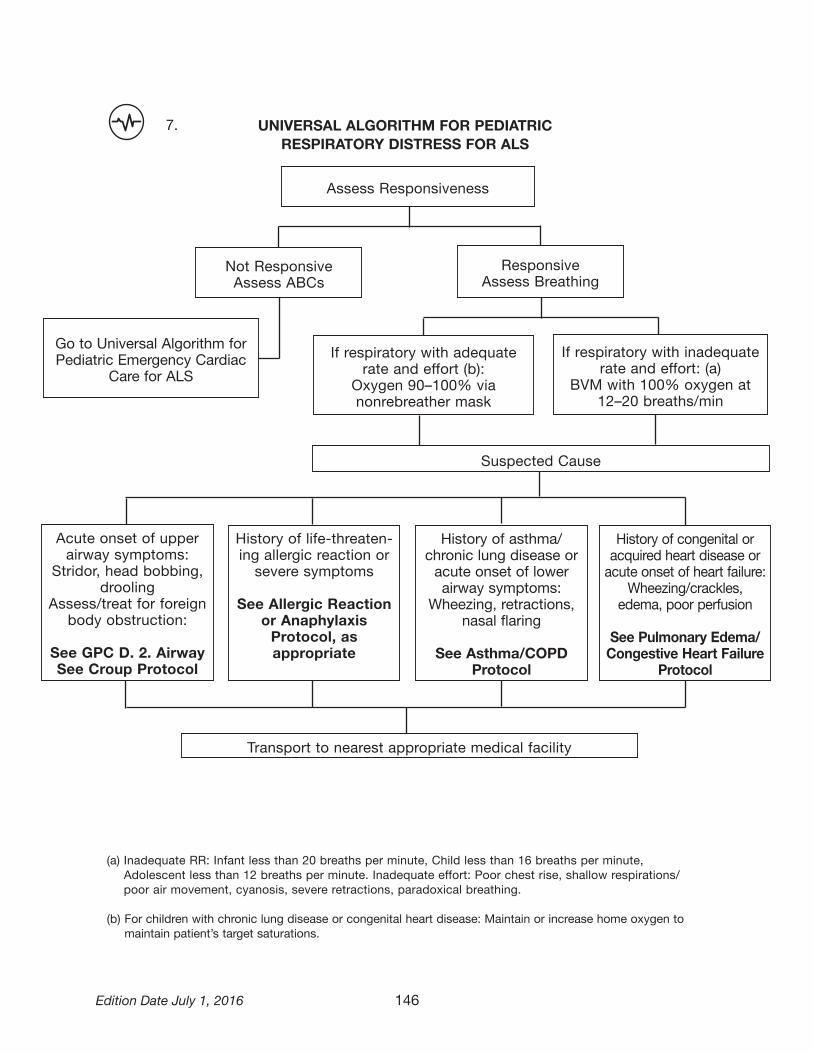
146Edition Date July 1, 2016
7. UNIVERSAL ALGORITHM FOR PEDIATRICRESPIRATORY DISTRESS FOR ALS
Assess Responsiveness
If respiratory with inadequate rate and effort: (a)
BVM with 100% oxygen at 12–20 breaths/min
If respiratory with adequate rate and effort (b):
Oxygen 90–100% via nonrebreather mask
Go to Universal Algorithm for Pediatric Emergency Cardiac
Care for ALS
Not ResponsiveAssess ABCs
ResponsiveAssess Breathing
Suspected Cause
Acute onset of upper airway symptoms:
Stridor, head bobbing, drooling
Assess/treat for foreign body obstruction:
See GPC D. 2. AirwaySee Croup Protocol
History of life-threaten-ing allergic reaction or severe symptoms
See Allergic Reaction or Anaphylaxis
Protocol, as appropriate
History of asthma/ chronic lung disease oracute onset of lower airway symptoms:
Wheezing, retractions, nasal flaring
See Asthma/COPD Protocol
History of congenital or acquired heart disease oracute onset of heart failure:
Wheezing/crackles, edema, poor perfusion
See Pulmonary Edema/Congestive Heart Failure
Protocol
Transport to nearest appropriate medical facility
(a) Inadequate RR: Infant less than 20 breaths per minute, Child less than 16 breaths per minute, Adolescent less than 12 breaths per minute. Inadequate effort: Poor chest rise, shallow respirations/poor air movement, cyanosis, severe retractions, paradoxical breathing.
(b) For children with chronic lung disease or congenital heart disease: Maintain or increase home oxygen to maintain patient’s target saturations.

147 Edition Date July 1, 2016
SS. SEPSIS: ADULT (NEW ’16)
1. Initiate General Patient Care
2. Presentationa) Infection can cause a systemic response resulting in fever, altered mental sta-
tus, shock including or excluding hypotension, and death. Early recognition and treatment with aggressive fluids, when not contraindicated, and early hospital notification may improve survival rates and patient outcomes.
b) The following patient populations are considered especially high risk for sepsis and should have their temperature measured: (1) Altered mental status(2) Patients in long term care facilities (nursing home)(3) Indwelling catheters(4) Oncology patients(5) Solid organ transplant(6) Bed ridden
c) For an adult patient, 18 years of age and older, to qualify for this protocol, he/she must have a suspected source of infection AND also present with at least two of the following criteria:(1) Temp greater than 100.4ºF (38ºC) or less than 95.9ºF (35.5ºC)(2) HR greater than 100 bpm (3) RR greater than 25 (or EtCO2 less than or equal to 32 mmHg)(4) Hypotension (systolic BP less than 90 mmHg) (5) Point of care lactate reading greater than or equal to 4 mmol/L (if available)
d) Patients with hypotension or altered mental status should be considered to have septic shock and treated and transported rapidly. Patients may be treated under this protocol if they do not meet the above criteria with medical consultation.
IF PATIENT MEETS ABOVE SEPSIS CRITERIA, THIS PATIENT IS A PRIORITY 1 OR 2 PATIENT AND REQUIRES NOTIFICATION OF THE NEAREST APPROPRIATE FACILITY AS SOON AS POSSIBLE TO ALLOW FOR HOSPITAL PREPARATION. DURING THE CONSULTATION WITH THE RECEIVING FACILITY, THE PROVIDER SHALL USE THE VERBIAGE, “SEPSIS ALERT” AS THE UNIVERSAL METHOD OF NOTIFYING THE FACILITY THAT THE PATIENT MEETS THE SEPSIS INCLUSION CRITERIA
3. Treatmenta) Place patient in position of comfort, or supine if hypotension is present. b) Carefully monitor airway and respiratory status, manage as required using the
appropriate respiratory distress protocol (especially for patients with suspected pneumonia).
c) Initiate large bore IV. If large bore IV not available, consider a second peripheral IV with the intention of not causing delay in transport and reserve the use of IO for priority 1 patient. If transport time is greater than 20 minutes and IV access is unsuccessful, consider placement of an IO (especially for septic shock). Con-sider performing a blood draw if time permits.

148Edition Date July 1, 2016
SS. SEPSIS: ADULT (Continued)
d) If lungs are clear, and patient does not have a history of CHF or end stage renal failure, provide 2 L of LR wide open. Reassess every 500 mL for shortness of breath, blood pressure, and SpO2 saturation changes.OR
e) If patient is fluid sensitive (i.e., has a history CHF, pulmonary edema, or end stage renal disease) infuse 250 mL and carefully monitor and reassess. Repeat 250 mL once if no worsening of respiratory status is noted to a max of 500 mL (consultation may be obtained to provide more fluid).
f) If available, perform point of care lactate testing (Jurisdictional Pilot Program only).
FLUID LIMITS OR DOSES MAY BE MODIFIED WITH CONSULTATION.
g) Place patient on cardiac monitor and perform 12-lead (do not delay IV therapy or fluid bolus).
h)
If hypotension persists after 2 L of LR are provided, consider an addition-al 2 L of LR (up to a maximum of 30 mL/kg total, including the first 2 L bolus) and/or dopamine 5–20 mcg/kg/min (paramedic only). Titrate to a Mean Arterial Pressure of 65 mmHg or systolic BP of 90 mmHg.

149 Edition Date July 1, 2016
TT. SEPSIS: PEDIATRIC (NEW ’16)
– – – – – – – – – – – – – – – – – – – – – – – – – – – – – – – – – – – – – – – – 1. Initiate General Patient Care
2. Presentationa) Infection can cause a systemic response resulting in fever, altered mental sta-
tus, shock including or excluding hypotension, and death. Early recognition and treatment with aggressive fluids, when not contraindicated, and early hospital notification may improve survival rates and patient outcomes.
b) The pediatric septic patient may be difficult to identify due to a poor history or providers may have difficulty identifying an obvious source of infection, as many pediatric sepsis patients are very young children or infants.
c) The following pediatric patients are at greater risk for sepsis and should have their temperature measured: (1) Altered mental status(2) Asplenia (spleen removed from treatment of trauma or illness) (3) Bone marrow or solid organ transplant (4) Cancer patients(5) Cerebral Palsy (6) Sickle Cell Disease(7) Central or indwelling catheters(8) Immunodeficiency or immunosuppression (9) Bed ridden(10) Severe mental delay
d) For a pediatric patient, who has not reached his/her 18th birthday, to qualify for this protocol, he/she must have a known or suspected infection AND also present with at least three of the Pediatric Sepsis Rule-In Criteria by Age.
e) A patient not meeting three or more Pediatric Sepsis Rule-In Criteria by Age may be treated under this protocol with Pediatric Base Station ap-proval if sepsis is suspected by the prehospital provider.
ALTERED MENTAL STATUS REQUIRES GLUCOSE CHECK.
f) Patients who meet the sepsis rule-in criteria and have at least one of the High risk Sepsis Rule-In Criteria by Age (shaded) should receive aggressive standing order fluid therapy. Other patients meeting the pediatric sepsis rule-in criteria but not having one of the high risk signs may be treated only after contacting a Pediatric Base Station for medical consultation.

150Edition Date July 1, 2016
TT. SEPSIS: PEDIATRIC (Continued)
Pediatric Sepsis Rule-In Criteria by AgeSuspected or known infection plus three criteria
Less than 28 days
1-12 months 1 year but less than 2 years
2-4 years 5-12 years 13-17 years
Heart Rate (sustained)
greater than 205 bpm
greater than 205 bpm
greater than 190 bpm
greater than 140 bpm
greater than 140 bpm
greater than 100 bpm
Respiratory Rate
greater than 60 rpm
greater than 60 rpm
greater than 40 rpm
greater than 40 rpm
greater than 34 rpm
greater than 25 rpm
Temp greater than 38.0 Cº or greater than 100.4 Fº
Cap Refill/Skin Delayed (greater than 3 seconds), mottled
Systolic BP (mmHg)
less than 60 less than 70 (less than 70+ (age x2))
(less than 70+ (age x2))
(less than 70+ (age x2))
less than 90
Mental Status Unresponsive, confused, inappropriate, lethargic
High Risk Condition
Cancer, Asplenia, Sickle Cell Disease, bone marrow or solid organ transplant, central or indwelling line/catheter, immunodeficiency or immunosuppression
Meeting any of these criteria indicates standing order initiation of a fluid bolus.
IF A PEDIATRIC PATIENT MEETS THE ABOVE PEDIATRIC SEPSIS RULE-IN CRITE-RIA BY AGE, THIS PATIENT IS A PRIORITY 1 OR 2 PATIENT AND REQUIRES NOTIFI-CATION AS “SEPSIS ALERT” TO THE NEAREST APPROPRIATE FACILITY PRIOR TO ARRIVAL.
IF A PEDIATRIC PATIENT MEETS ANY OF THE SEPSIS RULE-IN PLUS ONE OR MORE OF THE SHADED AREAS IN THE CHART, CONSULTATION WITH A DESIG-NATED PEDIATRIC BASE STATION IS REQUIRED AND SHOULD BE COMBINED WITH LOCAL BASE STATION CONSULTATION.
3. Treatmenta) Carefully monitor airway and respiratory status. Manage as required using the
appropriate respiratory distress protocol (especially for patients with suspected pneumonia).
b) Place patient on cardiac monitor. c) If patient meets the pediatric sepsis rule-in criteria and meets one of the high
risk criteria (shaded), initiate IV/IO access and provide a 20 mL/kg bolus of LR IV/IO over 5–20 min. Maximum single dose of 2L.
d) Monitor closely for signs of respiratory distress, rales or delayed capillary refill (greater than 2 seconds). If respiratory status deteriorates rapidly, stop bolus and obtain medical consultation.
e) For volume-sensitive children administer initial fluid bolus of 10 mL/kg LR IV/IO (max of 250 mL). (Volume-sensitive children are children who need smaller fluid bolus volumes due to special needs including neonates (birth to 28 days), con-genital heart diseases, chronic lung disease, or chronic renal failure.)

151 Edition Date July 1, 2016
TT. SEPSIS: PEDIATRIC (Continued)
f) If patient’s vital signs do not improve after 20 mL/kg fluid, consider additional 20 mL/kg LR boluses (up to a max of 60 mL/kg total, including first bolus, in one hour).
FLUID LIMITS OR DOSES MAY BE MODIFIED WITH CONSULTATION.
g) Dopamine 5–20 mcg/kg/min IV/IO. Titrate to age-specific vital signs.
h) Consider initiation of a second IV. Initiation of second IV shall not delay transport.i) Patients with fever or known or suspected infection and hypotension or altered
mental status should be considered to have septic shock and treated and trans-ported rapidly.
4. Continue General Patient Care.

152Edition Date July 1, 2016
EMS STROKE ALGORITHM (NEW ’16)
Support ABCs and provide any needed BLS/ALS
interventions
Transport to nearest Primary Stroke Center
Treat and transport per pt presentation
Determine presence of stroke severity using Cincinnati Prehospital
Stroke Scale
Determine timepatient last known well
Check GlucoseFibrinolytic Therapy
Checklist LAMS Assessment
NO
NO
New onset and positive
stroke assessment?
Signs and
symptoms consistent with
stroke AND onset less than 3.5 hrs.
Transport to nearest Stroke Center as Priority 1 and
Stroke Alert
UU. STROKE: NEUROLOGICAL EMERGENCIES

153 Edition Date July 1, 2016
UU. STROKE: NEUROLOGICAL EMERGENCIES (Continued)
1. Initiate General Patient Care.
2. PresentationPatient may present with numbness or weakness (often on one side only), dif-ficulty speaking, blurred vision, dizziness, or a severe, unexplained headache. May be accompanied by seizures or altered mental status.
3. Treatment a) Position patient with head elevated at 30 degrees.b) Complete the Fibrinolytic Therapy Checklist for Ischemic Stroke.c) If the patient is a candidate for fibrinolytic therapy AND can be delivered to the
hospital within 3.5 hours* of when patient was last known well, transport the patient to the closest Designated Stroke Center. If there is not one within 30 minutes, then go to the nearest hospital.
The Cincinnati Prehospital Stroke Scale(Kothari R, et al. Acad Emerg Med 1997; 4:9866-990.)
Facial Droop (have patient show teeth or smile): • Normal – both sides of face move equally • Abnormal – one side of face does not move as well as the other side
Arm Drift (patient closes eyes and holds both arms straight out for 10 seconds): • Normal – both arms move the same or both arms do not move at all (other findings, such as strength of
grip, may be helpful) • Abnormal – one arm does not move or one arm drifts down compared with the other
Abnormal Speech (have the patient say “you can’t teach an old dog new tricks”): • Normal – patient uses correct words with no slurring • Abnormal – patient slurs words, uses the wrong words, or is unable to speak
If Cincinnati Prehospital Stroke Scale is positive, perform the Los Angeles Motor Scale (LAMS). Relay LAMS score to the receiving hospital during Stroke Alert notification. (NEW ’16)

Edition Date July 1, 2016
UU. STROKE: NEUROLOGICAL EMERGENCIES (Continued)
IF PATIENT MEETS ABOVE STROKE CRITERIA, THIS PATIENT IS A PRIORITY 1 PATIENT AND REQUIRES NOTIFICATION OF THE NEAREST DESIGNATED STROKE CENTER AS SOON AS POSSIBLE TO ALLOW HOSPITAL PREPARATION. DURING THE CONSULTATION WITH THE RECEIVING FACILITY, THE PROVIDER SHALL USE THE VERBIAGE, “STROKE ALERT” AS THE UNIVERSAL METHOD OF NOTIFYING THE FACILITY THAT THE PATIENT MEETS THE STROKE INCLUSION CRITERIA. *STROKE TREATMENTS ARE TIME SENSITIVE. REDUCTION IN TIME OF SYMPTOM ONSET TO TREATMENT IMPROVES OUTCOMES WHILE STROKES DURING PREGNANCY OR SHORTLY AFTER GIVING BIRTH ARE RARE, THERE HAS BEEN A SIGNIFICANT RISE REPORTED IN THE LITERATURE. MOTHERS-TO-BE AND POSTPARTUM MOTHERS HAVE AN INCREASED RISK.
d) Use glucometer and treat if glucose less than 70 mg/dl.e) Establish IV access with LR.
f) If the patient is hypotensive, obtain medical consultation.
g) Consider obtaining blood sample using closed system.h) Do not treat hypertension in the field.
THE CAUSES OF STROKES IN CHILDREN ARE DIFFERENT FROM ADULTS. WHILE STROKES ARE UNCOMMON IN CHILDREN, THEY DO OCCUR AND ARE MOST OFTEN CAUSED BY ONE OF THE FOLLOWING CONDITIONS: CONGENITAL HEART DEFECTS, INFECTIONS (INCLUDING CHICKEN POX, MENINGITIS, OR ENCEPHALITIS), BRAIN INJURY, OR BLOOD DISORDERS (SUCH AS SICKLE CELL DISEASE). STROKES IN CHILDREN ARE MOST OFTEN SEEN IN INFANTS BUT DO OCCUR IN CHILDREN OF ANY AGE. CHILDREN WITH STROKE SYMPTOMS WHO HAVE NOT REACHED THEIR 18TH BIRTHDAY SHALL BE TREATED UNDER THE PEDIATRIC PROTOCOL. CONSULT WITH A LOCAL BASE STATION AND A PEDIATRIC BASE STATION TO ARRANGE TRANSPORT TO A MARYLAND PEDIATRIC TRAUMA CENTER – – – – – – – – – – – – – – – – – – – – – – – – – – – – – – – – – – – – – – – –
i) Administer oxygen at 2–6 liters via nasal cannula (unless hypoxic or in respira-
tory distress).
j) Position patient with head elevated at 30 degrees.
k) If a child presents with a SUSPECTED stroke (e.g., sickle cell patient), consult with the nearest Pediatric Base Station and local Base Station.
l) Use glucometer and treat accordingly.
(See Section IV, Glucometer Protocol.)
154

155 Edition Date July 1, 2016
UU. STROKE: NEUROLOGICAL EMERGENCIES (Continued)
m) Establish IV access with LR.
n) If the patient is hypotensive, obtain medical consultation.
o) Consider obtaining blood sample using closed system.
p) Do not treat hypertension in the field.
4. Continue General Patient Care.
Fibrinolytic Therapy Checklist for Ischemic Stroke
All of the “YES” boxes and all of the “NO” boxes must be checked before a patient should be transported to a “Designated Stroke Center.”
INCLUSION CRITERIA(All of the “YES” boxes must be checked)
YES ❑ 18 years of age or older ❑ Signs and symptoms of stroke with neurologic deficit (abnormal Cincinnati Stroke Scale)
❑ Patient can be delivered to a Stroke Center within 3.5 hours of when patient was last known well
EXCLUSION CRITERIA(All of the “NO” boxes must be checked)
NO ❑ Active internal bleeding (e.g., GI or urinary bleed-
ing within the last 21 days) ❑ Known bleeding disorder ❑ Within 3 months of intracranial surgery, serious
head trauma, or previous stroke
❑ Within 14 days of major surgery or serious trauma ❑ History of intracranial hemorrhage ❑ Witnessed seizure at stroke onset ❑ History of cancer of the brain
156Edition Date July 1, 2016
VV. TRAUMA PROTOCOL: BURNS
1. Initiate General Patient Care.2. Presentation
a) The primary objectives in burn care by EMS providers are to stop the burning process, establish IV access, avoid hypothermia, and transport patients quickly and safely to a burn center. While patients with large burns (greater than 20%), facial burns, and/or significant smoke inhalation often require endotracheal intubation and mechanical ventilation during their resuscitation and care, airway compromise in the first few hours following a burn is uncommon.(1) In adults, prehospital tracheal intubation following acute burns is generally
unnecessary unless signs of respiratory failure are present (symptomatic airway obstruction, shock, altered mental status, hypoxemia while receiving supplemental oxygen, or dyspnea, etc.).
(2) Pediatric airways are smaller than adult airways and require frequent and thorough assessment for signs of respiratory distress. Intubate if necessary.
b) Burns are the body’s response to injuries to the skin, muscles, bone, nerves, and blood vessels caused by thermal, chemical, electrical, radiation, or light source. Patients may exhibit any of the following: reddening of the skin, deep and intense pain, blisters, mottled appearance, and/or charred black or brown areas with severe or no pain.
c) Indications for Referral to a Burn Center(1) All third degree burns (full thickness)(2) Second degree burns (partial thickness) greater than 10% total body
surface area(3) Burns of the face, hands, feet, major joints, genitalia, or perineum(4) Electrical burns, including lightning or contact with high voltage
(greater than 120 volts)(5) Suspected inhalation injury of toxic smoke (Monitor the patients with sus-
pected inhalation injury for delayed airway obstruction, respiratory distress, or oxygen desaturation as the patient may need emergent airway manage-ment.)
(6) Circumferential burns involving the extremities or torso(7) Chemical burns should be transported to the closest appropriate hospital for
decontamination prior to referral to a burn center
PATIENTS WITH BURNS AND TRAUMA SHOULD BE REFERRED TO THE NEAREST APPROPRIATE TRAUMA CENTER FOR INITIAL CARE.
CHILDREN WHO MEET BURN INCLUSIVE CRITERIA WHO HAVE NOT REACHED THEIR 15TH BIRTHDAY SHOULD BE TRANSPORTED TO A PEDIATRIC BURN CENTER.
PATIENTS PRESENTING WITH ALTERED MENTAL STATUS OR NAUSEA WITH VOMITING, SEIZURES, LOSS OF CONSCIOUSNESS, OR MARKED DYSPNEA IN THE FACE OF SUSPECTED CARBON MONOXIDE OR TOXIC INHALATION WITH OR WITHOUT MINOR BURNS SHOULD BE CONSIDERED FOR TRANSPORT TO THE HYPERBARIC SPECIALTY CENTER. PATIENTS IN CLOSED SPACE INCIDENTS ARE MORE LIKELY TO MANIFEST THESE SYMPTOMS.
3. Treatmenta) Extract the patient from burning vehicles or buildings if safe to do so and move
patient to a place of relative safety.

157 Edition Date July 1, 2016
VV. TRAUMA PROTOCOL: BURNS (Continued)
b) Do what is necessary to stop the burning process. If water is used to extinguish the fire, remove wet clothing and dry the patient to prevent hypothermia.
c) Administer oxygen in as high a concentration of oxygen as possible (note: pulse oximetry is not reliable in the presence of carbon monoxide or cyanide expo-sure).
d) Determine percent of body surface area (BSA) burned and depth.e) Treat associated trauma.f) For burns greater than 10%, follow Hypothermia Protocol as well.g) Remove all rings, bracelets, and other jewelry.h) Cover wounds appropriately (with a clean sheet or Mylar blanket—sterile dress-
ings no longer recommended).i) For chemical burns, brush off dry chemical, remove clothing, flush with water.
DO NOT GIVE ANYTHING BY MOUTH. DO NOT PLACE ICE OR ICE PACKS ON ANY PATIENT WITH BURNS GREATER THAN 5% TOTAL BODY SURFACE AREA. CONSIDER UTILIZING AEROMEDICAL RESOURCE IF PATIENT IS MORE THAN 30 MINUTES FROM A BURN CENTER/HYPERBARIC MEDICINE SPECIALTY CENTER BY GROUND.
j) Establish IV access with LR, if appropriate.
(1) 10 mL/kg bolus.(2) For shock patients, administer a fluid bolus of 20 mL/kg LR followed by a sec-
ond 20 mL/kg LR if needed. Titrate to a systolic pressure of 100 mmHg.k) Administer opioid per Pain Management Protocol. l) Consider additional fluid administration. Maximum dose 2,000 mL without
medical consultation.
- – – – – – – – – – – – – – – – – – – – – – – – – – – – – – – – – – – – – – – – –
-
m) Establish IV access with LR, if appropriate.(1) 10 mL/kg bolus.(2) If age-related vital signs and patient’s condition indicate hypoperfusion,
administer initial fluid bolus of 20 mL/kg LR IV/IO. If patient’s condition does not improve, administer the second bolus of fluid at 20 mL/kg LR IV/IO.
n) Third and subsequent fluid boluses at 20 mL/kg LR IV/IO.
o) Administer opioid per Pain Management Protocol.
4. Continue General Patient Care.

158Edition Date July 1, 2016
RULE OF NINES
9 9
18
18 18
9
11
18
18
99
13.5 13.5
12
1
18
9 9
16.5 16.5ADOLESCENT/ ADULT
INFANT/TODDLER
CHILD
Note: The surface of the patient’s palm equals 1% of his/her body surface area.
Back: 18
Back: 18
Back: 18

159 Edition Date July 1, 2016
WW. TRAUMA PROTOCOL: EYE TRAUMA
1. Initiate General Patient Care.
2. PresentationThe patient may present with profuse bleeding, avulsions, lacerations, foreign objects, impaled objects, and/or soft tissue damage to the eye(s) and/or sur-rounding facial areas.
3. Treatment
NEVER APPLY PRESSURE TO THE EYEBALL OR GLOBE! IF THE PATIENT HAS OTHER ASSOCIATED TRAUMA OR BURNS, TRANSPORT THE PATIENT TO THE APPROPRIATE TRAUMA OR BURN CENTER; OTHERWISE, TRANSPORT THE PATIENT TO THE NEAREST EYE TRAUMA CENTER, IF APPROPRIATE. DO NOT USE CHEMICAL COLD PACKS ON THE FACE.
a) Foreign objects NOT embedded in the eye(s): Flush with copious amounts of water (preferably sterile), normal saline, or LR from the bridge of the nose out-ward.
b) Injury to orbits (area around the eye): Consider head stabilization and Spinal Protection Protocol.
c) Lacerations/injuries to the eyeball or globe: Shield affected eyeball and dress other eye to reduce movement and protect loss of fluids; consider head stabilization and spinal protection and elevate the head to decrease intraocular pressure.
d) Impaled objects: Stabilize object, shield affected eyeball, and dress other eye to reduce movement.
e) Establish IV access with LR, if appropriate.
f) Administer opioid per Pain Management Protocol.

160Edition Date July 1, 2016
WW. TRAUMA PROTOCOL: EYE TRAUMA (Continued)
- – – – – – – – – – – – – – – – – – – – – – – – – – – – – – – – – – – – – – – – –
g) Foreign objects NOT embedded in the eye(s): Flush with copious amounts of water (preferably sterile), normal saline, or LR from the bridge of the nose out-ward.
h) Injury to orbits (area around the eye): Consider head stabilization and Spinal Protection Protocol.
i) Lacerations/injuries to the eyeball or globe: Shield affected eyeball and dress other eye to reduce movement and protect loss of fluids; consider head stabilization and spinal protection and elevate the head to decrease intraocular pressure.
j) Impaled objects: Stabilize object, shield affected eyeball, and dress other eye to reduce movement.
k) Establish IV/IO access with LR, if appropriate.
l) Administer opioid per Pain Management Protocol.
4. Continue General Patient Care.

161 Edition Date July 1, 2016
XX. TRAUMA PROTOCOL: HAND/UPPER/LOWER EXTREMITY TRAUMA
1. Initiate General Patient Care.
2. Presentationa) Patient may exhibit injuries to skeletal or soft tissue components of the hand or
upper extremity at or below the level of the mid-humerus, including complete or incomplete amputations of the elements of the hand or upper extremity, crush or degloving injuries, and other trauma resulting in loss of perfusion or suspected nerve injury (e.g., compartment syndrome).
Upper Extremityb) Indications for:
Referral of adult patients to the Curtis National Hand Center at Union Memorial Hospital or Referral of pediatric patients to the nearest Pediatric Trauma Center (children who have not reached their 15th birthday) Stable patients with an isolated upper extremity injury at or below the mid-humerus
(Hand Center and/or nearest appropriate trauma center)
(1) Complete or incomplete hand or upper extremity amputation(2) Partial or complete finger or thumb amputation(3) Degloving, crushing, or devascularization injuries of hand or upper
extremity(4) High-pressure injection injuries to hand or upper extremity(5) Complicated nerve, vessel, or compartment syndrome (excessive swelling
and pain of extremity with possible evolving nerve deficit) injury of the fore-arm and hand
Lower Extremityc) Indications for Referral to Pediatric or Adult Trauma Center: Patient may exhibit
injuries to skeletal or soft tissue components with complete or incomplete am-putation of ankle/foot lower extremity, complicated nerve, vessel, or compart-ment syndrome (excessive swelling and pain of extremity with possible evolving nerve deficit injury).
LIFE BEFORE LIMB.
TOE INJURIES FROM LAWN MOWER ARE NOT CANDIDATES FOR REIMPLANTATION ANDPATIENTS SHOULD GO TO THEIR LOCAL MEDICAL FACILITY.
d) Contraindications for referral to a Hand Center(1) Patients with unstable or abnormal vital signs(2) Patients with major and/or multiple system trauma
e) Contraindication for referral to Pediatric or Adult Trauma CenterPatients with toe amputation (partial or complete)

162Edition Date July 1, 2016
XX. TRAUMA PROTOCOL: HAND/UPPER/LOWER EXTREMITY TRAUMA (Continued)
3. Treatment
a) Package amputated extremity in sealed plastic bag (keep dry) and place on top of ice to keep cool. DO NOT FREEZE.
DO NOT SUBMERGE IN WATER OR FREEZE AMPUTATED PART. USE TIME, DISTANCE, WEATHER, AND PROXIMITY TO DESIGNATED TRAUMA CENTER TO DETERMINE MODE OF TRANSPORT. IF ESTIMATED TRANSPORT TIME TO DESIGNATED HAND CENTER IS LESS THAN 30 MINUTES, USE GROUND TRANSPORT.
b) Establish IV access with LR, if appropriate.
c) Administer fluid bolus, if appropriate. 20 mL/kg of LR IV Titrate to a systolic pressure of 100 mmHg.
d) Administer opioid per Pain Management Protocol.
e) Consider additional fluid administration.Maximum dose 2,000 mL without medical consultation
– – – – – – – – – – – – – – – – – – – – – – – – – – – – – – – – – – – – – – – –
f) Establish IV/IO access with LR, if appropriate.
g) If age-related vital signs and patient’s condition indicate hypoperfusion, administer initial fluid bolus of 20 mL/kg LR IV/IO. If patient’s condition does not improve, administer the second bolus of fluid at 20 mL/kg LR IV/IO.
h) Third and subsequent fluid boluses at 20 mL/kg LR IV/IO.
i) Administer opioid per Pain Management Protocol.
4. Continue General Patient Care.

163 Edition Date July 1, 2016
YY. TRAUMA PROTOCOL: MULTIPLE/SEVERE TRAUMA
1. Initiate General Patient Care.
2. PresentationThe patient may present with hypovolemic or neurogenic shock, hypotension, hypertension, rapid or slow heart rate, unequal pupils, shallow or absent respira-tions, decreased distal pulses, decreased motor and sensory function in extrem-ities, internal or external bleeding, fractures, or lacerations.
WHILE TIME, DISTANCE, AND PROXIMITY ARE ALL FACTORS TO BE CONSIDERED IN THE TRIAGE DECISION, THE TRAUMA DECISION TREE SHOULD BE USED TO DETERMINE WHO SHOULD BE TRANSPORTED TO THE NEAREST APPROPRIATE TRAUMA CENTER AND WHEN THE TRANSPORT SHOULD OCCUR. CHILDREN WHO MEET INCLUSION BASED ON THE TRAUMA DECISION TREE AND WHO HAVE NOT REACHED THEIR 15TH BIRTHDAY SHOULD BE TRANSPORTED TO A PEDIATRIC TRAUMA CENTER.
3. Treatment
a) Apply Spinal Protection Protocol for blunt trauma patients. Patients with isolated penetrating trauma should not have spinal immobilization performed.
b) Control bleeding and immobilize patient, if blunt mechanism indicates. Spinal immobilization should not be performed on patients with isolated penetrating mechanism. If mechanism includes both blunt and penetrating trauma, apply Spinal Protection Protocol. Backboard may be used for patient transfer maneu-vers.
c) Hyperventilate the head-injured patient as follows (NEW ’16): Adult/Adolescent (greater than 13 years of age): 20 breaths per minute Child (1-12 years of age): 30 breaths per minute Infant (less than 1 year of age): 35 breaths per minute(1) Who has signs of herniation such as unequal pupils, posturing,
or paralysis or(2) Who is manifesting a rapidly decreasing GCS or(3) With on-line medical consultation
d) Consider pelvic stabilization technique if indicated.
e) Establish IV access with LR; administer 20 mL/kg bolus.
Titrate to a systolic pressure of 100 mmHg.
f) Consider additional fluid administration.Maximum dose 2,000 mL without medical consultation

164Edition Date July 1, 2016
YY. TRAUMA PROTOCOL: MULTIPLE/SEVERE TRAUMA (Continued)
- – – – – – – – – – – – – – – – – – – – – – – – – – – – – – – – – – – – – – – – –
g) Apply Spinal Protection Protocol for blunt trauma patients. Patients with isolated
penetrating trauma should not have spinal immobilization performed.
h) Control bleeding and immobilize patient, if blunt mechanism indicates. Spinal immobilization should not be performed on patients with isolated penetrating mechanism. If mechanism includes both blunt and penetrating trauma, apply Spinal Protection Protocol. Backboard may be used for patient transfer maneu-vers.
i) Hyperventilate the head-injured patient as follows (NEW ’16): Adult/Adolescent (greater than 13 years of age): 20 breaths per minute Child (1-12 years of age): 30 breaths per minute Infant (less than 1 year of age): 35 breaths per minute
j) Who has signs of herniation such as unequal pupils, posturing, or paralysis or(1) Who is manifesting a rapidly decreasing GCS or(2) With on-line medical consultation
k) Establish IV/IO access with LR.
l) If age-related vital signs and patient’s condition indicate hypoperfusion, administer initial fluid bolus of 20 mL/kg LR IV/IO. If patient’s condition does not improve, administer the second bolus of fluid at 20 mL/kg LR.
m) Third and subsequent fluid boluses at 20 mL/kg LR IV/IO.
4. Continue General Patient Care.

165 Edition Date July 1, 2016
GLASGOW COMA SCALE
Eye Opening
Spontaneously 4To Voice 3To Pain 2No Response 1
Motor Response
To Verbal Command - Obeys 6To Painful Stimulus - Localizes Pain 5
Flexion - Withdraw 4Flexion - Abnormal 3Extension 2No Response 1
Verbal Response
Less than 2 years old 2–5 years old Greater than 5 years old
5 SMILES/COOS/CRIES APPROPRIATE WORDS ORIENTED AND CONVERSES 5
4 CRIES INAPPROPRIATE WORDS DISORIENTED AND CONVERSES 4
3 INAPPROPRIATE CRIES/SCREAMS CRIES/SCREAMS INAPPROPRIATE WORDS 3
2 GRUNTS GRUNTS INCOMPREHENSIBLE SOUNDS 2
1 NO RESPONSE NO RESPONSE NO RESPONSE 1
Glasgow Coma Score Total (3–15)

166Edition Date July 1, 2016
ZZ. TRAUMA PROTOCOL: SEXUAL ASSAULT
1. Initiate General Patient Care.
2. Presentation Patient may present with no overt evidence of trauma, or may present with bruising, bleeding, or associated physical and/or emotional trauma.
ALL HEALTH CARE PROVIDERS ARE OBLIGATED BY LAW TO REPORT CASES OF SUSPECTED CHILD OR VULNERABLE ADULT ABUSE AND/OR NEGLECT TO EITHER THE LOCAL POLICE OR ADULT/CHILD PROTECTIVE SERVICE AGENCIES. DO NOT INITIATE REPORT IN FRONT OF THE PATIENT, PARENT, OR CAREGIVER.
3. Treatment
a) Patient may feel more comfortable talking to someone of the same gender.
b) Maintain non-judgmental, but caring attitude.
c) Preserve crime scene and clothing articles, if practical.
d) Maintain strict confidentiality.
e) Do not perform a genital examination.
f) Dress wounds (do not attempt to clean).
g) Discourage any self-treatment (shower, washing, changing clothes).
h) Treat injuries according to presentation.
4. Continue General Patient Care.

167 Edition Date July 1, 2016
AAA. TRAUMA PROTOCOL: SPINAL PROTECTION
1. Initiate General Patient Care.
2. PresentationIndications for initiating spinal protection: “Spinal protection” refers to the act of protecting the spinal cord from further injury. “Spinal immobilization” is the act of placing a patient on a backboard with cervical collar for the purpose of trying to prevent excessive movement of the spinal column.a) Patients who have a blunt trauma with a high-energy mechanism of injury that
has potential to cause spinal cord injury or vertebral instability and one or more the following should receive spinal protection.(1) Midline spinal pain, tenderness, or deformity(2) Signs and symptoms of new paraplegia or quadriplegia(3) Focal neurological deficit(4) Altered mental status or disorientation(5) Distracting injury: Any injury (e.g., fracture, chest, or abdominal trauma)
associated with significant discomfort that could potentially distract from a patient’s ability to accurately discern or define spinal column pain or tender-ness.
Indications for referral to an Adult Specialty Spinal Center.(6) 15 years of age or older AND(7) Signs and symptoms of new paraplegia or quadriplegia in the presence of
trauma AND(8) Patent airway AND(9) Hemodynamically stable
If considering referral to Adult Specialty Spinal Center, consult with both the nearest Trauma Center and the Adult Spinal Specialty Center, when possible.
3. Treatment
a) Initiate General Patient Care.
b) All patients meeting the Spinal Protection Protocol shall have manual in-line cer-vical spine stabilization and application of a correctly sized cervical collar.
c) Minimize flexion, extension, and rotation of the spinal column.
d) Patients meeting the Spinal Protection Protocol who are with neurological defi-cit, or not able to ambulate on their own accord, shall be immobilized with cervi-cal collar and a backboard.

168Edition Date July 1, 2016
AAA. TRAUMA PROTOCOL: SPINAL PROTECTION (Continued)
e) The following patients only need application of a cervical collar and do not need to be placed in full immobilization with a backboard:(1) Patients who are found by EMS providers to be standing or ambulatory,(2) Patients who have a GCS of 15 and are able to safely extricate themselves
from the environment (e.g., vehicle seat) without gross movement (flexion, extension, or rotation) of the spinal column, and
(3) Patients who do not have evidence of a neurological deficit.
f) Patients who are placed in a cervical collar without a need for immobilization on a backboard should be assisted in minimal movement to the EMS stretcher and allowed to lie down supine on their own accord.
g) Patients meeting Spinal Protection Protocol and not requiring immobilization with a backboard should be secured to the EMS stretcher in a supine position with the head elevated at 30 degrees.
h) Backboards may be used for patient extrication and patient transfer for patients not meeting Spinal Protection Protocol; however, other devices are preferred (e.g., sheet, Reeves sleeve, or scoop stretcher).
i) If the backboard is used for extrication from the scene to an ambulance, the pa-tient should be removed from the backboard as soon as possible. The stretcher mattress will provide support in place of the backboard.
j) Interfacility transport patients who have already been removed from a back-board should not be placed back on the backboard prior to transport.
k) Helmet Removal(1) If patient is wearing a helmet, the goals are assessment and management
of the airway, breathing, and circulation followed by protection of the spinal column by maintaining neutral alignment of the spinal column.
(2) If patient is wearing helmet and no shoulder pads, removal of the helmet is indicated.
(3) If patient is wearing helmet with shoulder pads, removal of the helmet is ac-ceptable only with concurrent removal of shoulder pads. Under these condi-tions, removal of the helmet is indicated for management of the airway or other facial trauma.
l) Patients found with backboard applied before EMS arrival(1) If EMS providers find patient immobilized on a backboard applied prior to
arrival, the principles of the Spinal Protection Protocol still apply.

169 Edition Date July 1, 2016
AAA. TRAUMA PROTOCOL: SPINAL PROTECTION (Continued)
m) Establish IV/IO access with LR, if appropriate.
n) Administer fluid bolus, if appropriate. 20 mL/kg of LR IV Titrate to a systolic blood pressure of 100 mmHg.
o) Consider dopamine. 2–20 mcg/kg/min IV/IOTitrate to a systolic blood pressure of 100 mmHg.
p) Consider additional fluid administration. Maximum dose 2,000 mL without medical consultation.
– – – – – – – – – – – – – – – – – – – – – – – – – – – – – – – – – – – – – – – In children who have not reached their 15th birthday:Indications for initiating spinal protection:q) Patients who have a blunt trauma with a high-energy mechanism of injury that
has potential to cause spinal cord injury or vertebral instability and the presence of or inability to assess one or more of the following should receive spinal pro-tection.
(1) Midline spinal pain, tenderness, or deformit(2) Signs and symptoms of new paraplegia or quadriplegia(3) Focal neurological deficit(4) Altered mental status or disorientation(5) Distracting injury(6) Neck pain or torticollis(7) High impact diving incident or high risk motor vehicle crash (i.e., head on col-
lision, rollover, ejected from the vehicle, death in the same crash, or speed greater than 55 mph)
(8) Substantial torso injury(9) Conditions predisposing to spine injury
Indications for referral to a Pediatric Trauma Center:(10) Patient is less than 15 years of age AND(11) Signs and symptoms of new paraplegia or quadriplegia in the presence of
trauma AND(12) Patent airway AND(13) Hemodynamically stable
Consult with nearest Trauma Center and, when possible, the nearest Pediatric Trauma Center.r) Initiate General Patient Care.
s) All patients meeting the Spinal Protection Protocol shall have manual in-line cer-vical spine stabilization and application of a correctly sized cervical collar.

170Edition Date July 1, 2016
AAA. TRAUMA PROTOCOL: SPINAL PROTECTION (Continued)
t) Minimize flexion, extension, and rotation of the spinal column.
u) Patients meeting the Spinal Protection Protocol who are with neurological deficit, not able to ambulate on their own accord, or who are unable to respond during assessment shall be immobilized with cervical collar and a backboard.
v) The following patients only need application of a cervical collar and do not need to be placed in full immobilization with a backboard: (1) Patients who are found by EMS providers to be standing or ambulatory(2) Patients who have a GCS of 15 and are able to safely extricate themselves
from the environment (e.g., vehicle seat) without gross movement (flexion, extension, or rotation) of the spinal column, and
(3) Patients who do not have evidence of a neurological deficit.
w) Patients who are placed in a cervical collar without a need for immobilization on a backboard should be assisted in minimal movement to the EMS stretcher and allowed to lie down supine on their own accord.
x) Patients meeting Spinal Protection Protocol and not requiring immobilization with a backboard should be secured to the EMS stretcher in a supine position with the head elevated at 30 degrees.
y) Backboards may be used for patient extrication and patient transfer for patients not meeting Spinal Protection Protocol; however, other devices are preferred (e.g., sheet, Reeves sleeve, or scoop stretcher).
z) If the backboard is used for extrication from the scene to an ambulance, the pa-tient should be removed from the backboard as soon as possible. The stretcher mattress will provide support in place of the backboard.
aa) Interfacility transport patients who have already been removed from a back-board should not be placed back on the backboard prior to transport.
bb) Helmet Removal(1) If patient is wearing a helmet, the goals are assessment and management
of the airway, breathing, and circulation followed by protection of the spinal column by maintaining neutral alignment of the spinal column.
(2) If patient is wearing helmet and no shoulder pads, removal of the helmet is indicated.
(3) If patient is wearing helmet with shoulder pads, removal of the helmet is ac-ceptable only with concurrent removal of shoulder pads. Under these condi-tions, removal of the helmet is indicated for management of the airway or other facial trauma.

171 Edition Date July 1, 2016
AAA. TRAUMA PROTOCOL: SPINAL PROTECTION (Continued)
cc) Patients found with backboard applied before EMS arrival(1) If EMS providers find patient immobilized on a backboard applied prior to
arrival, the principles of the Spinal Protection Protocol still apply.
dd) Establish IV/IO access with LR, if appropriate.
ee) Administer fluid bolus, if appropriate. 20 mL/kg of LR IV Titrate to a systolic blood pressure of 100 mmHg.
ff) Consider dopamine. 2–20 mcg/kg/min IV/IOTitrate to a systolic blood pressure of 100 mmHg.
gg) Consider additional fluid administration. Maximum dose 2,000 mL without medical consultation
4. Continue General Patient Care.
172Edition Date July 1, 2016
BBB. TRAUMA PROTOCOL: TRAUMA ARREST
1. Initiate General Patient Care.
2. PresentationEarly cardiac arrest secondary to trauma is usually due to severe hypoxia, neu-rologic injury, or massive hemorrhage. The patient is unresponsive, pulseless, and apneic.
3. Treatment
a) Rapid assessment and extrication
b) Determine if patient meets the criteria for termination of resuscitation for a patient in traumatic arrest. If patient meets criteria, discontinue resuscitation. If criteria are not met, continue resuscitation.
c) Perform spinal immobilization for blunt trauma patients only. Patients with isolated penetrating trauma should not have spinal immobilization performed. If mechanism includes both blunt and penetrating trauma, perform spinal immobi-lization.
d) CPR
e) Consider AED if arrest is believed to be medical in nature and the patient meets the criteria.
IF TRAUMATIC ARREST IS SECONDARY TO PENETRATING TRAUMA, PATIENT IS IN A RHYTHM OTHER THAN ASYSTOLE, AND THE TRAUMA CENTER IS WITHIN 15 MINUTES, TRANSPORT THE PATIENT. IF TRANSPORT TIME EXCEEDS 15 MINUTES, CONSULT.
f) Establish IV access with LR, if appropriate.
g) Administer fluid bolus, if appropriate. 20 mL/kg of LR IV Titrate to a systolic pressure of 100 mmHg.
h) Identify rhythm and refer to appropriate algorithm.
i) If traumatic arrest is suspected due to multi-system blunt trauma, or due to penetrating neck, chest, or abdominal trauma, bilateral needle decom-pressions should be performed. Once catheters are placed do not remove.

173 Edition Date July 1, 2016
BBB. TRAUMA PROTOCOL: TRAUMA ARREST (Continued)
- – – – – – – – – – – – – – – – – – – – – – – – – – – – – – – – – – – – – – – – –
j) Rapid assessment and extrication
k) Perform spinal immobilization for blunt trauma patients only. Patients with isolated penetrating trauma should not have spinal immobilization perfomed. If mechanism includes both blunt and penetrating trauma, perform spinal immobi-lization.
l) CPR
m) Consider AED if arrest is believed to be medical in nature. (See Section IV, AED.)
A PATIENT IN CARDIOPULMONARY ARREST SECONDARY TO TRAUMA SHOULD BE TAKEN TO THE NEAREST APPROPRIATE PEDIATRIC TRAUMA CENTER. CONSIDERATION SHOULD BE GIVEN TO TRANSPORTING THE PATIENT TO THE NEAREST EMERGENCY DEPARTMENT OR ADULT TRAUMA CENTER IF THE PEDIATRIC TRAUMA CENTER IS MORE THAN 10 MINUTES ADDITIONAL TRANSPORT TIME!
n) Establish IV/IO access with LR.
o) If age-related vital signs and patient’s condition indicate hypoperfusion, administer initial fluid bolus of 20 mL/kg LR IV/IO. If patient’s condition does not improve, administer the second bolus of fluid at 20 mL/kg LR IV/IO.
p) If traumatic arrest is suspected due to multi-system blunt trauma, or due to penetrating neck, chest, or abdominal trauma, bilateral needle decompressions should be performed. Once catheters are placed do not remove.
4. Continue General Patient Care.

174Edition Date July 1, 2016
q Olderadults•Riskofinjury/deathincreasesafterage55•SBPlessthan110mayindicateshockafterage65•Low-impactmechanisms(e.g.,ground-levelfalls)mayresultinsevereinjury
q Children(ShouldbetriagedtoPediatricTraumaCenter)
q 2ormoreproximallong-bonefractures
q Amputationproximaltowristorankle
q Chestwallinstabilityordeformity(e.g.,flailchest)
q Crushed,degloved,mangled,orpulselessextremity
q Openordepressedskullfracture
q Penetratinginjuriestohead,neck,torso,orextremitiesproximaltoelbowandknee
Measure vital signs and level of consciousness and assess for major injury
Category Alpha
Assess for other injuries.
Evaluate for evidence of mechanism of injury and high-energy impact.
Transport to trauma center or specialty center per protocol; alert trauma team; consider helicopter transport if quicker and of clini-cal benefit (refer to GPC Section I).
When in doubt, take patient to an appropriate Trauma Center
Transport to trauma center or specialty center per protocol; alert trauma team; consider helicopter transport if quicker and of clinical benefit (refer to GPC Section I).
Evaluateforotherconsiderations.TransporttoTraumaCenter;alerttraumateam.Patientswithina30-minute drive timeoftheclosestappropriatetrauma/specialtycentershallgobygroundunlessthereareextenuatingcircumstances.ReceivingTraumaCentermedicalconsultationrequiredwhenconsideringwhetherhelicoptertransportisofclinicalbenefit(refertoGPCSectionI).
q GCS less than or equal to 13
q Systolic BP less than 90 mmHg (Adult) less than 60 mmHg (Peds)
q Respiratory rate less than 10 or greater than 29 (less than 20 in infant age less than one year) or need for ventilatory support
q Pelvicfracture
q Paralysis(spine)
Category Bravo
Transport according to protocol.Consider medical direction and transport to trauma center. Patients within a 30-minute drive time of the closest appropriate trauma/specialty center shall go by ground unless there are extenuating circumstances. ReceivingTraumaCentermedicalconsultationrequiredwhenconsideringwhetherhelicoptertransportisofclinicalbenefit(refertoGPCSectionI).
Category Delta
Category Charlie
q HighRiskAutoCrash• Intrusion(includingroof)greaterthan12in.occupantsite; greaterthan18in.anysite• Ejection(partialorcomplete)fromvehicle• Deathinsamepassengercompartment• Vehicletelemetrydataconsistentwithhighriskofinjury
q Falls q Exposuretoblastorexplosion•Adult:greaterthan20feet(onestoryisequalto10feet)•Pediatric:greaterthan10feetor3timesthechild’sheight
• Rolloverwithoutrestraint• Autov.pedestrian/bicyclistthrown,runover,orwithsignificant(greaterthan20mph)impact
• Motorcyclecrashgreaterthan20mph
CCC. TRAUMA DECISION TREE
YES NO
NO
NO
NO
YES
YES
YES
q Burns•Withouttraumamechanism,triagetoBurnCenter•Withtraumamechanism,triagetoTraumaCenterq Pregnancygreaterthan20weeksq EMSproviderjudgmentq Anticoagulantsandbleedingdisorders(Patientswithheadinjuryareathighriskforrapiddeterioration)

250Edition Date July 1, 2016
2. AIRWAY MANAGEMENT: BAG-VALVE-MASK VENTILATION
a) PURPOSE
(1) Bag-valve-mask (BVM) ventilation is the technique of providing rescue breathing for patients with inadequate respiratory effort or cardiac arrest. Patients in respiratory failure may respond to BVM ventilation and not require endotracheal intubation.
(2) A BVM may also be used to administer inhaled medications for patients with severe respiratory failure.
b) INDICATIONS
(1) Inadequate respiratory rate (NEW ’16)(a) Adult less than 8(b) Adolescent (13–18 years of age) less than 12(c) Child (1–12 years of age) less than 16(d) Infant/Toddler (less than 1 year of age) less than 20
(2) Inadequate respiratory effort(a) Absent or diminished breath sounds(b) Paradoxical breathing (chest and abdomen moving in opposite direc-
tions)(c) Cyanosis or oxygen saturation less than 90% on 100% oxygen
by nonrebreather with the exception of patients with chronic hypoxemia
(3) Symptomatic Bradycardia (NEW ’16)(a) Adult/Adolescent Heart rate less than 60
(greater than 13 years of age)(b) Child (1–12 years of age) Heart rate less than 80(c) Infant (less than 1 year of age) Heart rate less than 100
(4) Cardiac arrest(5) Altered mental status
Glasgow Coma Scale of 8 or less
c) CONTRAINDICATIONS
None
d) POTENTIAL ADVERSE EFFECTS / COMPLICATIONS
(1) Gastric distension(2) Vomiting(3) Increased intracranial pressure as a result of increased vagal stimulation if
mask applied over the patient’s eyes

251 Edition Date July 1, 2016
Age/Weight
Infant to less than 1 year of age
Child 1-12 years
Adult/Adolescent
Bag Size
Infant 450–500 mL
Pediatric 750 mL
Adult 1,000–1,200 mL
e) PRECAUTIONS
(1) Have suction available since vomiting may occur.(2) Use an appropriate size airway adjunct with BVM.(3) Use an appropriate size mask to avoid pressure over the eyes
(pediatric patient), which may cause vagal stimulation.(4) For single provider BVM use the “E-C clamp” technique to achieve
an adequate seal and avoid pressure on the soft tissues of the face or neck: Place the third, fourth, and fifth fingers along the jaw to provide a chin lift (forming an E); use the thumb and index finger to hold the mask on the child’s face (forming a C).
(5) If the patient does not have adequate chest rise and breath sounds with BVM, consider the following interventions:(a) Use 2-hand jaw lift and oral airway to relieve tongue obstruction.(b) Use a larger bag to increase the volume of air delivered into the patient.(c) Evaluate and treat the patient for gastric distension.
Providers may manually decompress the stomach and/or open an existing gastric tube or button.
f) SUGGESTED SIZES FOR RESUSCITATION MASKS (NEW ’16)
g) SUGGESTED SIZES FOR RESUSCITATION BAGS (NEW ’16)
Age
Premature infants
Newborn to 1 year
1–4 years
5–12 years
Greater than 13 years of age
Adult
Mask Size
Neonatal
Infant
Toddler
Pediatric
Small adult
Adult

269 Edition Date July 1, 2016
14. ELECTRICAL THERAPY: AUTOMATED EXTERNAL DEFIBRILLATION (AED)
a) INDICATIONS
Sudden cardiac arrest (patients with no pulse and not breathing).Birth - less than 1 year of age Manual defibrillator preferred. (If unavailable, an AED with pediatric capability is preferred over an adult AED.) 1 year of age - 8 years of age AED with pediatric capability using the pediatric pad is preferred over an adult AED. Child 8 years of age or greater Adult AED
b) CONTRAINDICATIONS Patient exhibiting signs of life
USE OF THE AED IN THE MANUAL MODE IS RESERVED FOR ALS.
c) POTENTIAL ADVERSE EFFECTS/COMPLICATIONS
(1) Burns to skin(2) Deactivation of patient’s implanted pacemaker(3) Injury to patient, self, and/or bystanders
d) PRECAUTIONS
(1) Make sure the patient and the environment are dry.(2) Avoid placing pads over cardiac pacemakers/defibrillators or
nitroglycerin patches.(3) DO NOT touch the patient while the AED is analyzing the patient or discharg-
ing energy.(4) ENSURE that no one is touching the patient when the shock button
is pushed.(5) Never defibrillate while moving the patient or when in a moving
ambulance.
e) PROCEDURE
(1) Initiate analysis of rhythm.(2) If shock is indicated:
(a) Ensure all individuals are clear of the patient.(b) Initiate shock to the patient.(c) Immediately perform 5 cycles of CPR between shocks, then
initiate analysis of rhythm.(d) If patient remains pulseless, continue this cycle of CPR and shocks until
the patient regains a pulse, the AED prompt states “no shock advised,” or ALS arrives.
269

270Edition Date July 1, 2016
(3) No more than 3 stacked shocks (9) or 4 single new device shocks via AED without medical consultation.
(4) If shock is not indicated and the patient remains in cardiac arrest:(a) Perform 5 cycles of CPR.(b) Initiate analysis of rhythm.(c) If shock is indicated, see “If shock is indicated” section above.(d) If shock is not indicated, continue CPR and transport.
(5) If shock is not indicated and patient regains pulse, treat per Altered Mental Status Protocol.
f) SPECIFIC DOCUMENTATION
(1) Document the number of analyses and shocks delivered, times of assessments and treatments, and the patient’s response to shocks/CPR. Specify the type of AED, location of AED, bystander and pro-vider contact, and the triggering event.
(2) If using an AED with EKG strip recorder, generate 2 recordings.(3) Give one to the ALS provider or hospital and attach the other to your patient
care report.(4) Record the name of the contact for accessing AED data download summary.(5) Consider bringing the AED to the hospital for downloading.

276Edition Date July 1, 2016
18. GO-TEAM ACTIVATION
a) PURPOSE
The University of Maryland Medical System, R Adams Cowley Shock Trauma Center (STC) maintains a deployable advanced surgical team (Go-Team) that includes an attending physician with surgical skills and an anesthetist capable of assisting EMS providers with the care of seriously injured patients when extrica-tion times are anticipated to be more than 1 hour. On-scene incident command-ers may request the Go-Team by contacting SYSCOM.
b) INDICATIONSThe on-scene incident commander may contact SYSCOM and request the Go-Team for seriously injured patients with potentially life or limb threatening injuries when extrication times are anticipated to be more than 1 hour and who may require advanced resuscitative or surgical services that are beyond the scope of prehospital emergency services. Examples include:(1) During a prolonged extrication, assist rescue personnel with planning the
type and pace of the rescue by assessing the extent of injury and determine potential consequences that delays in time to definitive care might have on patient outcome.
(2) A patient trapped in heavy machinery requiring anesthesia/pain management to perform extrication
(3) A patient surviving a building collapse requiring an amputation to enable extrication
(4) A patient with a prolonged extrication requiring advanced fluid resuscitation including the administration of blood products
(5) Insertion of chest tubes or gastric and urinary catheters during the course of prolonged extrication
c) PROCEDURE(1) On-scene incident commander will request the Go-Team by
contacting SYSCOM. SYSCOM will coordinate the Go- Team’s transport to and from the scene with Maryland Express Care.
(2) If the Go-Team is dispatched by air, then SYSCOM will notify the Go-Team when the aircraft is landing on the STC helipad. If the Go-Team is dispatched by land, then Maryland Express Care will coordinate the Team’s response.
(3) Prior to the Go-Team’s departure to the scene, SYSCOM will notify the on-scene incident commander for the Go-Team’s ETA and reconfirm the need for the Go-Team.

277 Edition Date July 1, 2016
(4) If the Go-Team is dispatched, the EMS medical commander will contact them using the “Trauma Line” (or other radio) to update them about the circumstances of the entrapment and the patient’s condition.
(5) When the Go-Team arrives on the scene, they are to report to the on-scene incident commander and operate within the Incident Command System.
(6) Once the patient is extricated, the EMS system will transport the patient to the appropriate facility under established EMS guidelines with consultation by the Go-Team physician.
(7) The Go-Team will document the care they provide and file a patient care report with the State EMS Medical Director at MIEMSS.

281 Edition Date July 1, 2016
21. HIGH PERFORMANCE CPR (HPCPR) (NEW '16)
a) PURPOSETo improve the overall survival rate of sudden out-of-hospital cardiac arrest pa-tients within the State of Maryland. High Performance Cardio Pulmonary Resus-citation (HPCPR) employed with Code Resource Management (CRM) is a proven concept based on a team approach that ensures effective and efficient use of EMS resources. This systematic change in treatment and management of cardiac arrest patients is based on research and practices being used in many other high performance EMS systems across the country.
b) INDICATIONSPatients in cardiac arrest who have reached their 8th birthday.
c) CONTRAINDICATIONSPatients meeting the criteria for Pronouncement of Death in the Field ProtocolPatients who have not reached their 8th birthday (NEW ’16)
d) POTENTIAL ADVERSE EFFECTS/COMPLICATIONSNone
e) PRECAUTIONSNone
f) PROCEDURE FOR HIGH PERFORMANCE CPRThe first provider at the patient’s side will assess and initiate compressions. (1) Effective Compressions - Manual chest compressions should be initiated
immediately upon identification of cardiac arrest, as long as the scene is safe. When compressions are done manually, compressors should be rotat-ed every 2 minutes in order to maintain high-quality compressions. Ideally, one compressor is on each side of the patient’s chest; one person com-pressing the chest and the other person ready to start. Chest compressions will be performed at a depth of at least two inches allowing for complete recoil of the chest after each compression. Compressions should be accom-plished with equal time given for the down and up motion and achieve a rate of 100–120/min.
(2) Continuous Compressions - Chest compressions will be performed at a rate of 100–120 per minute and will NOT be interrupted during the two- min-ute cycle for any reason. Other treatments such as ventilations, IV access, or intubation attempts will be done while compressions are ongoing. After completion of a two-minute cycle, a phase to assess pulses and/or defibril-late will be limited to less than 10 seconds.

282Edition Date July 1, 2016
(3) Defibrillation – placement of the defibrillator pads will not interrupt chest compressions(a) Automatic External Defibrillation
The AED will be powered on as soon as the cardiac arrest is confirmed. Do not interrupt chest compressions to remove clothing or place defibril-lation pads. If the AED charges after analyzing, chest compressions will be performed while the device charges, then the patient will be “cleared” and defibrillated. Compressors will hover over the patient with hands ready during defibrillation so compressions can start immediately after a shock. Another two-minute cycle of compressions will be immediately performed. Pulse checks will not occur after a shock, but only after the AED prompts “no shock advised.” If no pulse is palpated, or if unsure, immediately perform another two minutes of CPR.
(b) Cardiac Monitor/Defibrillator When a manual defibrillator is in use, it will be charged to the appropriate energy level as the end of the compression cycle nears (approximately 1 minute and 45 seconds into a two-minute cycle). At the end of the two-minute cycle, the patient will be cleared, the rhythm will then be inter-preted rapidly, and the patient will either be defibrillated or the defibrilla-tor energy charge will be cancelled. This sequence must be performed within 10 seconds. During this sequence, the compressors will hover over the patient with hands ready. If a shock is delivered, the compres-sor will immediately resume CPR. Rhythm interpretation will not occur after a shock, but only after the two-minute cycle of CPR is performed. If a shock is not indicated, check for a pulse. If patient remains pulseless, immediately resume HPCPR.
(4) Ventilations - Ventilations will be performed without stopping chest com-pressions. One ventilation will be given every 10th compression during recoil (upstroke). Once an advanced airway is in place, ventilations will be asynchronous with compressions (1 ventilation every 6 to 8 seconds). High performance, continuous compressions remain the priority. Ensure ventila-tions are adequate with BVM attached to 100% oxygen. Providers will not interrupt compressions to obtain an advanced airway.
(5) Advanced Life Support - ALS providers will address defibrillation, IV/IO ac-cess, medication administration, and advanced airway placement, as indi-cated within these protocols; however, the placement of an advanced airway is no longer a focus of cardiac arrest management and will not interrupt chest compressions. Nasal capnography may be utilized to optimize CPR performance and evalu-ation of ROSC with use of bag-valve-mask ventilation.

283 Edition Date July 1, 2016
(6) Return of Spontaneous Circulation (ROSC) - Implement the Return of Spontaneous Circulation and Neuroprotective Induced Hypothermia Protocol as indicated and transport to the closest Cardiac Interventional Center. Following stabilization, post-ROSC, obtain a 12-lead EKG.
g) PROCEDURE: CODE RESOURCE MANAGEMENT (CRM)Crews should coordinate their duties keeping the call priorities in mind. Interven-tion priorities are (in order of highest to lowest):
The number of personnel on a given incident and the qualifications of those per-sonnel can vary; however, the priorities remain the same. Appropriate crew roles are outlined below:
2 provider crew:Provider 1 – Chest compressions Provider 2 – Ventilate, attach/operate AED/defibrillator, assume crew leader responsibilities (providers rotate positions every two minutes) Roles remain the same even if providers are ALS equipped
3 provider crew: Provider 1 – Chest compressions Provider 2 – Ventilate Provider 3 – Crew Leader, attach/operate AED/defibrillator (Providers 1 and 2 rotate every two minutes) Roles remain the same even if providers are ALS equipped

284Edition Date July 1, 2016
4 provider crew: Provider 1 – Chest compressions Provider 2 – Ventilate Provider 3 – Attach/operate AED/defibrillator Provider 4 – Crew leader (Providers 1, 2, and 3 rotate every two minutes)
** Once first two roles have begun treatment, ALS providers will establish IV/IO and administer medications.
Greater than 4 providers - Utilize the same initial assignments as the four provider crew. The crew leader will assign additional roles such as informing the family of patient status, gathering patient information, and documenting the medical interventions performed on the call. If resourc-es allow, rotate additional providers to do chest compressions to achieve optimal performance.
Crew leader - The crew leader will keep time, record interventions per-formed during the arrest, give compression feedback and ensure rotation of personnel doing compressions every two minutes. Verbal announce-ments of time should occur at one minute, 30 seconds before reassess-ment, 15 seconds left, and countdown to reassessment at 10 seconds.

287 Edition Date July 1, 2016
23. INTRAVENOUS MAINTENANCE THERAPY FOR EMT
a) Provider-controlled IV solutions
(1) The EMT is authorized to be the primary caregiver for patients with estab-lished intravenous (IV) therapy ONLY when the reason for transport is not related to complications associated with the IV line, and:
(a) The IV Solution DOES NOT contain:(i) MEDICATIONS,(ii) WHOLE BLOOD, or(iii) BLOOD PRODUCTS (such as plasma, platelets, or packed
red blood cells)(b) The IV catheter is placed in a PERIPHERAL LIMB VEIN, or(c) The IV catheter is a capped (e.g., heparin-locked) peripheral or central
line, and(d) No other ALS interventions are required.
(2) IV fluids The EMT is authorized to perform IV maintenance of NON-MEDICATED IV solutions that contain only:(a) LR solution(b) 2.5%–10.0% dextrose in water(c) 0.25%–0.9% saline solution(d) Potassium chloride (KCL) added to the solution. The amount of KCL in
solution shall not exceed 20 milli-equivilants (mEq)/ liter OR(e) Peripheral Parenteral Nutrition (PPN) or Total Parental Nutrition (TPN)
(NEW ’16)
IF IV FLUIDS OR PPN ARE BEING ADMINISTERED VIA INFUSION PUMP AND NOT PATIENT-CONTROLLED, THE PATIENT MUST BE ACCOMPANIED BY A NURSE OR APPROPRIATELY TRAINED ALS PROVIDER.
b) Patient-controlled medications or IV solutions
The EMT is authorized to be the primary caregiver for patients with established intravenous (IV) therapy ONLY when the reason for transport is not related to complications associated with the IV line or the medications being infused and the patient has been caring for the line, IV fluids, and/or IV medications at home without the assistance of a health care provider.
288Edition Date July 1, 2016
UNDER NO CIRCUMSTANCES SHALL THE EMT PROVIDER ATTEMPT TO MAKE ANY ADJUSTMENTS TO IV INFUSION PUMPS, NOR SHOULD THE EMT PROVIDER ADMINISTER ANY ADDITIONAL MEDICATIONS OR IV FLUIDS.
c) Provide patient care according to appropriate protocol.
d) Routine IV maintenance procedures
(1) Ensure IV solution and catheter placement meets criteria above.
(a) Request assistance of appropriate level health care provider if IV solution and/or IV catheter placement do not meet criteria above, or
(b) Request authorized personnel at health care facility to:
(i) Replace IV solution with an appropriate IV solution, or
(ii) Discontinue the IV prior to departing the scene.
(2) Confirm appropriate IV solution drip rate prior to transport.
(3) Ensure IV bag contains adequate volume of solution for duration of patient transport. If IV solution is not adequate, request authorized personnel at health care facility to:
(a) Replace IV solution with an adequate volume, or
(b) Discontinue the IV prior to departing the scene.
(4) Ensure IV solution is flowing at appropriate rate.
(5) Ensure patient has no signs or symptoms specifically related to complications of IV therapy prior to transport.
If patient has signs or symptoms related to complications of IV therapy: Request authorized personnel at health care facility to correct the com-plication.

289 Edition Date July 1, 2016
e) Complications of IV Therapy
(1) During patient transport, many possible complications of IV therapy may oc-cur that the EMT must be prepared to manage.
(a) Local complications may include: pain, hematoma, infiltration, infection, dislodged catheter, and tissue sloughing.
DO NOT ATTEMPT TO REINSERT DISLODGED IV CATHETER.
(b) Central complications may include: syncope, sepsis (infection), air embolism, pulmonary edema, pulmonary thromboembolism, conges-tive heart failure, overhydration, and catheter embolism.
(c) General complications may include: restricted flow (e.g., bent tubing, fluid-filled air chamber, inappropriate bag placement), and empty IV solution bag.
(2) Obtain medical direction and prepare to discontinue the IV if any of the complications described above are assessed and/or observed.
(3) If medical direction is genuinely not obtainable, the EMT shall discontinue the IV as soon as possible.
THE EMT IS AUTHORIZED TO DISCONTINUE PERIPHERAL LIMB VEIN IVs ONLY.
(4) Specific documentation includes:
(a) Type of provider-controlled IV solution
(b) Type of patient-controlled IV solution
(c) Type of patient-controlled IV medication
(d) Volume administered
(e) Complications encountered

296Edition Date July 1, 2016
25. PATIENT-INITIATED REFUSAL OF EMS
a) Initiate General Patient Care. For the purposes of this protocol, a patient is defined as any person encoun-tered by in-service rescue or emergency medical personnel with an actual or potential injury or medical problem. (The term “patient,” in this protocol only, refers both to patients and to persons who are potential patients. This protocol is not intended to determine the legal status of any person, the establishment of a provider-patient relationship, or a legal standard of care.)
– – – – – – – – – – – – – – – – – – – – – – – – – – – – – – – – – – – – – –– – – –A minor patient is defined as a patient who has not reached their 18th birthday and is not(1) Married, OR(2) Parent of a child, OR(3) Requesting:
(a) Treatment for drug abuse or for alcoholism,(b) Treatment for Sexual Transmitted Infection (STI) or for contraception,(c) Treatment of injuries from alleged rape or sexual offense, OR
(4) Living separate and apart from the minor’s parent, parents, or guardian, whether with or without consent of the minor’s parent, parents, or guardian, and is not self-supporting, regardless of the source of the minor’s income.
An authorized decision maker for minor patients is defined as an adult who identifies themselves as the parent or guardian, or has written authorization for medical decision making or states that they have written authorization for medi-cal decision making. Providers may request the parent or guardian to present identification and will document the name of the individual who identifies them-selves as the decision maker.
IN CASES OF ALLEGED RAPE OR SEXUAL OFFENSE, LAW ENFORCEMENT OR SOCIAL SERVICES SHALL BE NOTIFIED.
b) These persons may have requested an EMS response or may have had an EMS response requested for them. Because of the hidden nature of some illnesses or injuries, an assessment must be offered and performed, to the extent permitted, on all patients. For patients initially refusing care, attempt to ask them, “Would you allow us to check you out and evaluate whether you are OK?”
– – – – – – – – – – – – – – – – – – – – – – – – – – – – – – – – – – – – – – – – – – – IF THE AUTHORIZED DECISION MAKER REFUSES TO PERMIT THE EMS PRO-VIDER TO EXAMINE A MINOR PATIENT TO DETERMINE THE SEVERITY OF THE ILLNESS OR INJURY, THEN CONSIDER CONTACTING LAW ENFORCEMENT FOR ASSISTANCE. CONSIDER CONSULTATION WITH PEDIATRIC BASE STATION.

297 Edition Date July 1, 2016
c) Each patient’s assessment shall include:(1) Visual assessment - injuries, responsiveness, level of consciousness, orientation,
respiratory distress, gait, skin color, diaphoresis(2) Primary survey - airway, breathing, circulation, and disability(3) Vital signs - pulse, blood pressure, respiratory rate and effort, pulse oximeter when
available(4) Secondary survey - directed by the chief complaint
(a) Medical calls - exam of lungs, heart, abdomen, and extremities. Blood glucose testing for patients with Diabetes Mellitus. Neurological exam for altered con-sciousness, syncope, or possible stroke.
(b) Trauma calls - for patients meeting criteria in the Maryland Medical Protocols Trauma Decision Tree recommending transport to a Trauma Center: exam of neck and spine, neurological exam, palpation and auscultation of affected body regions (chest, abdomen, pelvis, extremities).
(5) Capability to make medical decisions (complete questions 1 through 4 on the Patient-Initiated Refusal of EMS form):(a) Disorientation to person, place, time, situation(b) Evidence of altered level of consciousness resulting from head trauma, medical
illness, intoxication, or other cause(c) Evidence of impaired judgment from alcohol or drug ingestion(d) Language communication barriers were removed by assuring “language line”
translation when indicated(e) The patient understands the nature of the illness
d) Following the assessment, complete items 5 through 9 on the Patient-Initiated Refusal of EMS Form, noting the presence of conditions that may place the patient at higher risk of hidden illness/injury or of worse potential outcome. Management
(1) Patients at the scene of an emergency who meet criteria to allow self-determination shall be allowed to make decisions regarding their medical care, including refusal of evaluation, treatment, or transport. These criteria include:(a) Medical capacity to make decisions - the ability to understand and discuss and
understanding of the nature and consequences of the medical care decision(b) Adult (18 years of age or greater)(c) Those patients who have not reached their 18th birthday and are:
(i) Married, OR(ii) Parent of a child, OR(iii) Requesting:
a. Treatment for drug abuse or for alcoholism,b. Treatment for STI or for contraception,c. Treatment of injuries from alleged rape or sexual offense, OR

298Edition Date July 1, 2016
(iv) Living separate and apart from the minor’s parent, parents, or guard-ian, whether with or without consent of the minor’s parent, parents, or guardian, and is self-supporting, regardless of the source of the minor’s income.
(d) A patient who has been evaluated by EMS providers as having ‘no’ answers to questions 1, 2, 3a, 3b, and 4 on the Patient-Initiated Refusal of EMS form shall be considered to be medically capable to make decisions regarding his/her care.
(e) Patients with ‘no’ answers to questions 1, 2, 3a, 3b, and 4 on the Patient-Initiated Refusal of EMS form but one or more ‘yes’ answers to questions 5 through 8 (medical conditions) have a higher risk of medical illness. The EMS provider should consider consulting medical direction if the patient does not wish transport. The purpose of the consultation is to obtain a “second opinion” with the goal of helping the patient realize the seriousness of his/her condition and accept transportation.
(f) If the EMS provider is unsure whether the patient has adequate ability to make medical decisions, he/she should seek medical consultation.
(g) At any time the EMS provider identifies patient conditions that indicate that the patient should be transported to a hospital, and the patient is refusing transport, then the provider should seek medical consultation.
(2) Any person at the scene of an emergency requesting an EMS response, or for whom an EMS response was requested, and who is evaluated to have any one of the following conditions, shall be considered incapable of making medical decisions regarding care and shall be transported, with law enforcement involve-ment, to the closest appropriate medical facility for further evaluation:(a) Continued altered mental status from any cause including altered vital signs,
influence of drugs and/or alcohol, metabolic causes (CNS or hypoglycemia), head trauma, or dementia
(b) Attempted suicide, danger to self or others, or verbalizing suicidal intent(c) Acting in an irrational manner, to the extent that a reasonable person would
believe that the medical capacity to make decisions is impaired(d) Severe illness or injury to the extent that a reasonable and medically capable
person would seek further medical care(e) On an Emergency Petition
(3) Further care should be provided according to Maryland Medical Protocols, “III E. Behavioral Emergencies” or other protocol sections as appropriate, based on patient’s condition.
e) Base Station Hospital Physician Consultation

299 Edition Date July 1, 2016
Patient refusals are one of the highest risk encounters in clinical EMS. Careful as-sessment, patient counseling, and appropriate base hospital physician consultation can decrease non-transport of high-risk refusals. Patients who meet any of the fol-lowing criteria require Base Station hospital physician consultation:(1) The provider is unsure if the patient is medically capable of refusing transport.(2) The provider disagrees with the patient’s decision to refuse transport due
to unstable vital signs, clinical factors uncovered by the assessment, or the provider’s judgment that the patient may have a poor outcome if not trans-ported.
(3) The patient was involved in any mechanism included in the Trauma Decision Tree of the Maryland Medical Protocols that would recommend transporta-tion to a Trauma Center.
(4) Minor patients: No parent, guardian, or authorized decision maker is avail-able or the provider disagrees with decision made by the parent, guardian, or authorized decision maker.
For patients with significant past medical history, consider consultation with the specialty center that follows the patient if possible.
Patients who do not meet the criteria above but have one or more positive answers to questions 6 through 10 on the Patient-Initiated Refusal of EMS form may have a higher risk of illness. In these situations, providers shall consult with the Base Station hospital physician.
f) Documentation(1) Complete Section One of the Patient-Initiated Refusal of EMS form, docu-
menting the patient’s medical decision-making capability and any “At-Risk” criteria.
(2) Complete Section Two, which documents provider assessment and actions.(3) Following patient counseling and Base Station hospital consultation, when
indicated, complete Section Three: Initial Disposition, Interventions, and Final Disposition.
(4) Have the patient and witness sign the refusal statement as determined by your jurisdiction.
(5) Document your assessment, the care provided, elements of the refusal, medical decision-making capability, and “At-Risk” criteria on the jurisdic-tion’s documentation (Medical Incident Report, MAIS form, or jurisdictional equivalent.)
(6) Submit copies of the Patient-Initiated Refusal of EMS form and the docu-mentation form to the EMS Supervisor.
(7) If the patient/authorized decision maker refuses to sign the refusal state-ment:(a) Contact a supervisor.(b) Explain the need for a signature and again attempt to have the patient
sign the refusal statement.(c) If not already done, have a witness sign the refusal statement.(d) Transmit the patient’s unwillingness to sign the refusal statement on a
recorded channel and document all steps taken to convince patient to sign.

300Edition Date July 1, 2016
Section One:When encountering a patient who is attempting to refuse EMS treatment or transport, assess his/her condition and record whether the patient screening reveals any lack of medical decision-making capability (1, 2, 3a, 3b, and 4) or high risk criteria (5–8):
1. Disoriented to: Person? ❑ yes ❑ no Place? ❑ yes ❑ no Time? ❑ yes ❑ no Situation? ❑ yes ❑ no 2. Altered level of consciousness? ❑ yes ❑ no 3. Alcohol or drug ingestion by history or exam with: a. Slurred speech? ❑ yes ❑ no b. Unsteady gait? ❑ yes ❑ no 4. Patient does not understand the nature of illness and potential for bad outcome? ❑ yes ❑ no
5. Abnormal vital signs For Adults
Pulse greater than 120 or less than 60? ❑ yes ❑ no Systolic BP less than 90? ❑ yes ❑ no Respirations greater than 30 or less than 10? ❑ yes ❑ no For minor/pediatric patients Age inappropriate HR or ❑ yes ❑ no Age inappropriate RR or ❑ yes ❑ no Age inappropriate BP ❑ yes ❑ no 6. Serious chief complaint (chest pain, SOB, syncope) ❑ yes ❑ no 7. Head Injury with history of loss of consciousness? ❑ yes ❑ no 8. Significant MOI or high suspicion of injury ❑ yes ❑ no 9. For minor/pediatric patients: ALTE, significant past
medical history, or suspected intentional injury ❑ yes ❑ no 10. Provider impression is that the patient requires hospital
evaluation ❑ yes ❑ no
Section Two:For providers: Following your evaluation, document information and care below:
1. Did you perform an assessment (including exam) on this patient? ❑ yes ❑ no If yes to #1, skip to #3 2. If unable to examine, did you attempt vital signs? ❑ yes ❑ no 3. Did you attempt to convince the patient or guardian to accept transport? ❑ yes ❑ no 4. Did you contact medical direction for patient still refusing service? ❑ yes ❑ no
Med
ical
Cap
acity
At Risk
Criteria
If yes, transport
If yes, consult

301 Edition Date July 1, 2016
Section Three: (CHECK ALL THAT APPLY)
Initial Disposition: ❑ Patient refused exam ❑ Patient refused treatment ❑ Patient refused transport❑ Patient accepted exam ❑ Patient accepted treatment ❑ Patient accepted transport❑ ADM refused exam ❑ ADM refused treatment ❑ ADM refused transport
Interventions:❑ Attempt to convince patient ❑ Attempt to convince family member/ADM ❑ Contact Medical Direction (Facility: ___________________________________)❑ Contact Law Enforcement ❑ None of the above available
Final Disposition:❑ Patient refused exam ❑ Patient refused treatment ❑ Patient refused transport❑ Patient accepted exam ❑ Patient accepted treatment ❑ Patient accepted transport❑ ADM refused exam ❑ ADM refused treatment ❑ ADM refused transport
Section Four: (MUST COMPLETE)Provide in the patient’s own words why he/she refused the above care/service:________________________________________________________________________________________________________________________________________________________________________________________________________________________________________________________________________________________________________________________________________________________________________________________________________________________________________________________________________________________________________________________________________________________________________________________________________
Patient Refusal of EMS
I, ________________________, have been offered the following by ___________________ (EMS Operational Program) but refuse (check all that apply):
❑ Examination ❑ Treatment ❑ Transport
Patient Name: _____________________________ Phone: ________________ Patient Address: __________________________________________________Signature: ________________________________Witness: ________________ ❑ Patient ❑ Parent ❑ Guardian ❑ Authorized Decision Maker (ADM)
If you experience new symptoms or return of symptoms after this encounter, we recommend that you seek medical attention promptly.
Jurisdiction ______________________ Incident: ______________________ Date: __________Unit #: _________________________ Provider Name/EID: _______________Time: __________

310Edition Date July 1, 2016
30. ACUPRESSURE FOR NAUSEA
a) PURPOSEAcupressure on the P6 point can be used to reduce the intensity of nausea for patients where ondansetron is not preferable or available. It may be help-ful as adjunct therapy for patients who have received ondansetron.
b) INDICATION(1) Patients with active nausea and vomiting(2) As adjunct therapy to patients receiving ondansetron(3) To prevent or reduce motion sickness
c) CONTRAINDICATIONNone
d) ADVERSE EFFECTSRedness, swelling, discomfort at site if commercial wrist bands are used
e) PRECAUTIONSPatients experiencing nausea should receive a complete assessment, espe-cially if cardiac risk factors are present.
f) PROCEDURE(1) Identify P6 point.
(a) Place three of the patient’s fingers on the patient’s opposite forearm at the wrist crease.
(b) Mark the space between the two tendons on the forearm as the P6 point.(2) Apply pressure at this point for several seconds and encourage the patient
to take over care, or apply a commercial device per manufacturer’s instruc-tions. Have patient or parent maintain firm pressure. Onset of relief is be-tween 30 seconds and 5 minutes.
(3) Reassess patient, rescore on BARF Scale at 5 minutes, and document re-sponse to therapy.

311 Edition Date July 1, 2016
31. MULTIPLE CASUALTY INCIDENT/UNUSUAL EVENT
A Multi-Casualty Incident (MCI) or Unusual Event is any event where the number of injured persons exceeds the normal capabilities of the EMS Operational Program in whose jurisdiction the event takes place. Due to the size of the incident, the re-sponding EMS Operational Program may require additional resources and/or must distribute patients to multiple hospitals.
Local EMS Operational programs should have a plan or operational procedures that address response to multiple patient incidents or unusual events. This protocol does not supersede those plans. There are some general practices and procedures that must be followed to ensure the EMS system can be prepared to respond ap-propriately to support a local response.
ALERT: THIS PROTOCOL IS SIMPLY A LIST OF REQUIRED TASKS IN THE EVENT OF AN UNUSUAL EVENT. IT IS NOT ALL-INCLUSIVE. ALL PROVIDERS ARE EN-COURAGED TO REVIEW LOCAL EMERGENCY RESPONSE PLANS, THE MARY-LAND TRIAGE SYSTEM TRAINING PROGRAM, START/JUMPSTART, AND NIMS PRACTICES AND PROCEDURES ON AT LEAST AN ANNUAL BASIS.
Procedure
a) Assess scene and recognize that the incident is an MCI or Unusual Event. The definition of MCI or Unusual Event for the purposes of this protocol is an inci-dent that causes more than 5 patient encounters or that involves unusual cir-cumstances that suggest it could place an extraordinary strain on EMS or health care resources. The following events are examples of an MCI or Unusual Event. (1) More than five patients from one or related incidents(2) Multi-patient events that require specialized rescue (3) Three or more immediate (Priority 1) patients(4) Multiple pediatric patients requiring specialty resources(5) More than one burn patient meeting burn center referral criteria (6) Use of more than two medevac helicopters(7) Use of Medical Ambulance Bus (MAB)(8) Multiple patients with unusual signs and symptoms(9) Unresolved WMD related activity that could result in multiple patients
(active shooter, bomb threat, intentional WMD agent release, etc.)(10) Decontamination of more than 5 patients resulting in at least one transport(11) Unresolved hazardous material incident that has the potential to affect
multiple patients(12) Evacuation of a licensed health care facility or housing complex for indi-
viduals requiring special assistance

312Edition Date July 1, 2016
b) Notify EMRC or the Regional EMRC as soon as the incident is recognized to be an MCI or Unusual Event. Use the specific terms “MCI” or ”Unusual Event” when communicating with EMRC to be clear this protocol is being enacted. This should be done as early in the incident as possible when there is a strong suspi-cion that such an event has occurred so that EMRC may begin to notify hospi-tals and response partners of the incident. Responding units can request their dispatchers notify EMRC before the scene is fully assessed if there is reasonable information to suggest that the incident meets the criteria above. As soon as available, the following information should be relayed to EMRC. (1) Type and general description of the incident(2) General location or address of the incident(3) Age range of patients(4) Estimated number of patients by priority(5) Approximate number of patients involved(6) Any hazardous agents involved
c) Initiate the incident command structure according to local SOPs and/or the Na-tional Incident Management System. Update EMRC with more details about the incident as they become available.
d) Consider utilization of the MCI Communications Protocol (Section II.G.6)
e) Triage patients using the START/JumpSTART methods (Section II.D.7.e).(1) Identify the patient’s triage category by utilizing the appropriately colored triage
ribbon and securely attach a MIEMSS-approved Triage Tag.
f) Do not delay transport of patients for extensive patient care procedures. Provide only the care required to sustain life and limb during transport to the hospital.
g) Track the care, movement, and disposition of EVERY patient utilizing the locally approved triage/treatment/transport logs and/or the state electronic patient tracking system (PTS). Patient information should be written on the triage tag and be entered directly into the PTS as it becomes available.
h) Consider the need for and request specialty resources through the local dis-patch center and/or emergency management as per local procedures. These may include,(1) Mass Casualty Support Units (MCSU) – (Medical Supply Caches)(2) Medical Ambulance Buses(3) CHEMPACK (Organophosphate antidotes - contact EMRC) (4) Ambulance Strike Teams or EMS Taskforces(5) Shock Trauma Go-Team

313 Edition Date July 1, 2016
i) The Transportation Group Supervisor and Medical Communications Coordina-tor responsibilities should be assigned as early as possible. They are the critical link to EMRC, hospitals, and the health care system. Their duties include: (1) Establish a final checkpoint through which all transport units MUST pass to
ensure accountability of all patients.(2) EMRC will have notified hospitals and acquired their bed availability based
on the information originally received and will transmit that information to the scene when requested.
(3) Coordinate through EMRC the patient destination, and communicate the number of patients, general illnesses, ages, and triage category on each transport unit as they leave the scene to the receiving facilities.
(4) If a central point of contact cannot be established, individual transport units MUST communicate the above information individually through EMRC to the receiving hospitals during transport. Those units must announce that they are associated with the MCI or Unusual Event.
j) Coordinate with law enforcement and, if requested, assist the Coroner or Medi-cal Examiner with identification and disposition of deceased casualties.
k) After the last patient has been transported, notify 9-1-1 dispatch center and EMRC that last patient has been transported. Demobilize scene, stand down or release resources dedicated to incident, and complete appropriate documenta-tion. Cooperate with local officials, EMRC, hospitals, and emergency manage-ment to complete a final accounting of the disposition of all the patients.

314Edition Date July 1, 2016
32. POTENTIALLY VOLATILE ENVIRONMENTS WITH LIFE-SUSTAINING INTERVENTIONS
a) BACKGROUND(1) A review of past active assailant incidents has shown that the conventional
prehospital practice of not entering the scene until it is deemed safe by law enforcement (LE) has been associated with additional loss of life.
(2) This protocol is designed to be all-hazards in nature. It is meant to provide a clinical concept of operations that empowers trained and equipped, but not necessarily tactical, EMS prehospital providers, to access casualties and ex-pedite life-sustaining interventions closer to the point and time of injury. For active assailant and other LE-related incidents, EMS providers shall be under LE escort. EMS providers shall use appropriate personal protective equip-ment as defined by local jurisdiction.(a) Examples of such potentially volatile environments include, but are not
limited to:(i) Active assailant (active shooter/IED) situations (ii) Post-blast detonations(iii) Intentional release of a chemical agent(iv) Industrial accident/explosion(v) Hazardous materials incident(vi) Structural collapse/urban search and rescue situations(vii) Transportation mishaps with limited scene access(viii) In the immediate aftermath of a natural disaster such as a
tornado
b) INTRODUCTION(1) This protocol provides guidelines for the type of intervention and care that
should be rendered at various proximities to a threat in a potentially volatile environment.
(2) By definition, potentially volatile environments are dynamic in nature. Scene conditions may change and emergent evacuation of responders and patients may interfere with the delivery of interventions described in this protocol.
c) INDICATIONS(1) This protocol does not replace or supersede the general patient care prac-
tices in The Maryland Medical Protocols for EMS Providers, which are still to be followed once the concern of active threat has been mitigated.
(2) Use of this protocol is an acknowledgement by the EMS provider that the situation is:(a) Unique, austere, and different than the conventional environment of care
in which EMS medicine is usually rendered AND
(b) The application of standard prehospital emergency practices could un-necessarily jeopardize the safety of the patient and/or medical provider.
(3) An active assailant incident or Potentially Volatile Environments with Life-
Sustaining Interventions (PVE/LSI) Protocol is declared.

315 Edition Date July 1, 2016
d) CONTRAINDICATIONS(1) Absent the presence of perceived or actual threat, standard general patient
care practices should be followed.
e) ZONES OF CARE/OPERATIONS(1) The zones described below are intended to standardize the terminology
used by responding emergency medical providers in Maryland and to es-tablish a common understanding of the interventions to be performed within each zone.
(2) Hot Zone (Direct Threat): (Integrated Tactical EMS) Operational area with a direct and immediate threat to personal safety or health(a) The overarching priority in the Hot Zone is mitigation of active threat.
Medical care is a secondary function to threat mitigation. (b) Medical providers must be an integrated tactical medic (i.e., TEMS) to
operate in this environment. Medical priorities are to prevent casualties and responders from sustaining additional injuries and include prompt evacuation to a more secure zone.(i) If at all possible, casualties should self-evacuate.(ii) Goals of care include keeping the response team engaged in neutral-
izing the threat, minimizing public harm, and controlling life-threaten-ing extremity hemorrhage.a. Control of severe hemorrhage in the direct threat environment is
best accomplished with commercially available tourniquets.b. Tourniquet should be placed as high up on the limb as possible
without taking the time to expose the area.c. For full or partial amputation, immediately place a tourniquet if
possible.d. Cardiopulmonary resuscitation (CPR) is not indicated in this envi-
ronment. (iii) In circumstances of chemical agent exposure, administration of
Nerve Agent Antidote Kits (NAAK/MARK-1) might be warranted if available.

316Edition Date July 1, 2016
(3) Warm Zone (Indirect Threat): (Limited LSI) Area with a potential threat to personal safety or health(a) Evacuation of patients to a completely safe area is the primary ob-
jective of care in this area. The following care guidance is dependent on the availability of equipment, supplies, and the appropriate level providers. Extrication should NOT be delayed to provide advanced or involved treatment measures. (i) The Warm Zone typically exists between the Hot Zone and Cold
Zone, but is not geographic and depends on the evolving situa-tion.
(ii) Responders must remain cognizant that scene security can change instantly.
(iii) A focused and deliberate approach to providing patient care should occur.
(iv) The potential benefits of providing medical care in these zones must outweigh the risks of the ongoing tactical operation and/or delaying opportunity to evacuate the patient.
(v) Care in the Warm Zone typically occurs at or near the point of injury once scene stabilizing measures have occurred. Care may also take place at a casualty collection point (CCP).
(vi) A CCP is a location concealed and covered from immediate threat where victims can be assembled for movement from areas of risk to the triage/treatment area. Multiple CCPs may be re-quired, which may be located in the Warm or Cold Zone. CCPs should be established and locations communicated as early as possible through operations to ALL responders.
(vii) If possible, an abbreviated triage system should be set up to identify the priority for the extrication of patients. The use of rib-bons or markers to clearly identify immediate and delayed (red and yellow, respectively) patients is highly recommended. De-ceased individuals should also be labeled/tagged appropriately to prevent repeat assessments by multiple providers.
(viii) Medical care in the Warm Zone should be limited to essential interventions only and is guided by the mnemonic “MARCHED”a. M – Massive Hemorrhage Control i. Massive hemorrhage remains the greatest threat to life in
most trauma patients. Attaining hemorrhage control is the top priority.
ii. Tourniquets remain the preferred means of hemorrhage control for life-threatening bleeding in this environment.

317 Edition Date July 1, 2016
1. If a tourniquet was applied in the Hot Zone, it should be reassessed.
2. Tourniquets applied over clothing are not as effective and may need to be adjusted.
3. Tourniquets should only be discontinued by an appro-priately trained ALS provider in consultation with medi-cal control.
4. Other methods of hemorrhage control include deep wound packing with either sterile gauze or hemostatic impregnated gauze.
5. Vascular injuries in the neck, groin, and axilla (i.e., junctional zones) are not amenable to traditional extremity tourniquets. In addition, effective pressure dressings are often extremely difficult to apply. He-mostatic impregnated dressings with direct pressure (minimum 5 minutes with continuous pressure is pre-ferred) have shown useful in such situations.
(b) A – Airway management(i) Patients in the Warm Zone with airway issues are high priority for
evacuation due to their often intense resource requirements.(ii) Consider applying oxygen if available and indicated.(iii) Unconscious casualty without airway obstruction:
a. Chin lift or jaw thrust maneuverb. Nasopharyngeal airwayc. Place casualty in the recovery position
(iv) Casualty with airway obstruction or impending airway obstruction:a. Chin lift or jaw thrust maneuverb. Nasopharyngeal airwayc. Allow casualty to assume position that best protects the airway,
including sitting up or leaning forwardd. Place unconscious casualty in the recovery position
(v) If previous measures unsuccessful, if time and resources permit, consider per protocol:a. Supraglottic Devices (e.g., King LT™,
EASYTube®, or CombiTube™).b. Oro/nasotracheal intubationc. Surgical cricothyroidotomy

318Edition Date July 1, 2016
(c) R – Respirations(i) The chest/upper abdomen should be assessed for any evidence of
an open chest wound and an occlusive dressing should be applied accordingly.
(ii) Tension pneumothorax remains a significant cause of preventable death in trauma patients.a. In suboptimal environments that interfere with complete physi-
cal assessment, any patient with significant blunt or penetrating chest trauma who displays dyspnea should be treated as a devel-oping tension pneumothorax and receive needle decompression, if appropriate.
b. To be effective, needle decompression needs to be performed using at least a 3.25 inch, 14g needle/catheter or needle decom-pression thoracostomy kit.
(d) C – Circulation(i) In general, healthy adult trauma patients with a radial pulse and nor-
mal mentation do not need IV therapy in the Warm Zone.(ii) Patients with evidence of hypotension:
a. If the patient displays signs of a closed head injury, IV fluid ther-apy is indicated to maintain at least a radial pulse or SBP of at least 90 mmHg.
b. Patients in hypovolemic shock should receive a one-time 500 mL bolus of IV fluid.
(iii) Patients in traumatic cardiac arrest should be considered deceased and no CPR should be performed in this zone.
(e) H – Hypothermia (i) Hypothermia in trauma patients has been associated with increased
mortality. Hypothermia is easier to prevent than treat.a. Patients should be moved to a warmed location if possible.b. Efforts should be made to minimize heat loss.
(f) E – Everything else (i) Consider Mark I/DuoDote for suspected organophosphate/nerve
agent exposure. (ii) Dependent upon resource availability, burns, eye injuries, and acute
pain should be managed per The Maryland Medical Protocols for EMS Providers.
(g) D – Documentation(i) Key findings and interventions should be conveyed to the next phase
of care.

319 Edition Date July 1, 2016
(4) Cold Zone: (Traditional Patient Care Protocols) Area surrounding the Warm Zone. Responders can operate without concern of danger or threat to per-sonal safety or health. (a) Casualties are moved from the Warm Zone to the Cold Zone by way of
an evacuation corridor(s). (i) Evacuation Corridor: An area transitioning between the Warm and
Cold Zone that is secured from immediate threat and allows for a mitigated risk in transporting victims from the CCP to the triage/treat-ment area beyond the outer perimeter.
(b) Once in the Cold Zone, casualties will require re-triage, particularly as-sessing for the development of a life-threatening condition and effects of Warm Zone therapy. (i) If massive hemorrhage has not been addressed or has been ineffec-
tively managed, it should be immediately readdressed with strategies mentioned above.
(c) Patients should be triaged and transported per standard practices.(d) Medical care in the Cold Zone should be dictated by resource availability
and, when possible, equate to the general patient care standards in The Maryland Medical Protocols for EMS Providers.
(e) CPR may have a larger role during the evacuation phase especially for patients with electrocution, hypothermia, non-traumatic arrest, or near drowning; however, it is still casualty count/resource dependent.

320Edition Date July 1, 2016
33. EMERGING INFECTIOUS DISEASE (EID) (NEW ’16)
1. Initiate General Patient Care.
2. Presentation An Emerging Infectious Disease is an infectious disease for which incidence in humans has increased in the past two decades or threatens to increase in the near future. These diseases, which respect no national boundaries, includea) New infections resulting from changes or evolution of existing organismsb) Known infections spreading to new geographic areas or populationsc) Previously unrecognized infections appearing in areas undergoing ecologic
transformationd) Old infections reemerging as a result of antimicrobial resistance in known agents
or breakdowns in public health measures.
The most recent example is Ebola Viral Disease (EVD). EIDs that meet this protocol will be posted on the MIEMSS website under the Infectious Disease Tab. Seasonal influenza is not considered an EID, but some of the same principles of infection control may apply to the more common infectious diseases.
e) Signs and Symptoms of an EID are based on specific case definitions for the disease:(1) EVD case definition includes:
Travel history or exposure and a set of signs and symptoms that are includ-ed in the case definition, which has evolved over time.
(2) Other future EID diseases may vary in their signs and symptoms, and could include:(a) Respiratory congestion (b) Sneezing/Coughing (c) Nausea/Vomiting(d) Skin rashes, hives, or “poxes”(e) Swollen lymph nodes(f) General malaise(g) Loss of appetite(h) Hemorrhage from mucosal membranes(i) Descending neurological deficits
f) Case Definition As EIDs become more prevalent, the Centers for Disease Control and Prevention (CDC) typically publish a description of each disease, which is utilized to deter-mine whether to include or exclude a Patient Under Investigation (PUI) for spe-cific testing or treatment and specific isolation or quarantine measures. These case definitions will be posted on the MIEMSS website and include specific guidance on the identification, treatment, and appropriate transport of these patients and the appropriate use of PPE.
g) Modes of transmission

321 Edition Date July 1, 2016
(1) In direct transmission, an infectious agent is transferred from a reservoir to a susceptible host by direct contact or droplet spread.(a) Direct contact occurs through skin-to-skin contact, kissing, and sexual
intercourse. Direct contact also refers to contact with soil or vegetation harboring infectious organisms.
(b) Droplet spread refers to spray with relatively large, short-range aerosols produced by sneezing, coughing, or even talking. Droplet spread is clas-sified as direct because transmission is by direct spray over a few feet, before the droplets fall to the ground.
(2) Indirect transmission refers to the transfer of an infectious agent from a res-ervoir to a host by suspended air particles, inanimate objects (vehicles), or animate intermediaries (vectors).(a) Airborne transmission occurs when infectious agents are carried by dust
or droplet nuclei suspended in air. Airborne dust includes material that has settled on surfaces and become re-suspended by air currents as well as infectious particles blown from the soil by the wind. In contrast to droplets that fall to the ground within a few feet, droplet nuclei may remain suspended in the air for long periods of time and may be blown over great distances
(b) Vehicles that may indirectly transmit an infectious agent include food, water, biologic products (blood), and fomites (inanimate objects such as handkerchiefs, bedding, or surgical scalpels).
(c) Vectors such as mosquitoes, fleas, and ticks may carry an infectious agent through purely mechanical means or may support growth or changes in the agent.
3. Treatment a) If the presence of an EID at a scene is known prior to entering, don the appro-
priate PPE and limit entry into the scene to essential personnel only. If an EID is discovered during assessment, immediately don the appropriate PPE, clear the scene of non-essential personnel and initiate the recommended decontamina-tion procedures.
b) Initiate General Patient Care.c) Treat the patient according to the signs and symptoms presented and according
to the MIEMSS guidance for the specific EID. Procedures that increase risk of distributing fluids or secretions should be limited to those absolutely necessary to maintain life and provide the patient with a reasonable level of comfort.
d) Contain any bodily fluids or respiratory excretions prior to transporting the patient. A SURGICAL mask may be placed on the patient to limit respiratory droplet aerosolization.
N-95 SHOULD NEVER BE PLACED ON A PATIENT AS THEY RESTRICT THE EXCHANGE OF RESPIRATORY GASES AND TYPICALLY HAVE A ONE-WAY EXPIRATORY VALVE THAT ALLOWS DROPLETS TO BE AEROSOLIZED UPON EXPIRATION DEFEATING THE PURPOSE OF PLACING A MASK ON THE PATIENT.

322Edition Date July 1, 2016
e) Transport the patient to the appropriate hospital. Hospitals have been categorized into three levels based on their capabilities to assess and treat PUIs for designated EIDs. A list of designated EIDs will be pub-lished on the MIEMSS website.(1) Frontline Hospitals (DHMH designated) – All hospitals with emergency de-
partments must have the capability to accept, identify, and isolate a PUI for a designated EID, then follow the approved procedures to notify the local health department to arrange for transfer to an Assessment Hospital. These patients will typically be transferred within 24 hours.
(2) Assessment Hospitals (DHMH designated) – A facility that has the capability to receive, isolate, and provide care for a patient while testing is completed to confirm or deny the diagnosis of the suspected EID. The patient will re-main at that hospital for 4 to 5 days until the patient is discharged or transfer to an designated Treatment Hospital
(3) Treatment Hospitals (DHMH designated) – A facility assessed by the CDC to have the capability to admit and provide comprehensive care for and man-age a patient with a confirmed designated EID, until the patient is no longer ill or has died.
f) Transport from the scene PUIs at a residence should be transported directly to an Assessment Hospital unless total transport time is no longer than 45 minutes greater than transport to the nearest Frontline Hospital ED. If transport time is longer than 45 minutes greater than transport to the nearest Frontline Hospital ED, the patient must be transported to the closest appropriate Frontline Hospital. Priority 1 and Prior-ity 2 patients with unresolved symptoms that cannot be managed outside the hospital should be taken to the closest Frontline Hospital. Receiving hospital notification of all suspected PUI patients should be done as early as possible to allow for hospital staff to prepare. Helicopter transport NOT indicated for the PUI patient (NEW ’16)
g) Transport of a health department monitored patient Individuals who were exposed and have some risk of contracting the disease may be monitored or even quarantined by the health department. MIEMSS will be notified by DHMH if these patients become ill and require transportation by EMS to hospitals and will contact the local jurisdictional or waivered commercial EMS Operational Program to arrange that transport. DHMH will determine the destination hospital.
h) Interfacility Transfer Transfers between hospitals will be completed by EMSOPs who have been granted a waiver from licensing to modify an ambulance specifically to transport an EID patient and have specific plans, training and quality assurance processes in place to do so. Public Safety EMSOPs may be called upon as a backup if the waivered commercial services are not available. DHMH will determine the desti-nation hospital in these cases.

323 Edition Date July 1, 2016
i) Communication EMS providers transporting PUIs for designated EIDs MUST contact the receiv-ing hospital via EMRC prior to beginning that transport and enter the hospital through the entrance designated by the receiving hospital. The term PUI must be used to ensure the hospital understands and is prepared to receive the patient. Obtaining medical direction from the closest Frontline and Assessment Hospitals is always an option to determine the appropriate destination.
j) Refusal of transport If a PUI for a designated EID refuses care or transport, the EMS provider should remove him/herself from the immediate presence of the patient and contact the local health department through their dispatch center or locally defined proce-dures and provide as much of the following information about the patient that is available. (1) Full name(2) Age(3) Gender(4) Home address(5) Contact phone numbers(6) Current location(7) Recent travel history(8) Signs and symptoms being displayed(9) Recent contact history with Ebola patientsThe EMS provider should expect to be involved in a discussion of the situation with health department and law enforcement officials, and if a quarantine/isola-tion order is issued, should be prepared to assist law enforcement in carrying out that order.
k) Treat the patient according to the signs and symptoms presented and according to the MIEMSS guidance for the specific EID. Limit invasive procedures and any that increase risk of distributing fluids or secretions to those absolutely neces-sary to maintain life and provide the patient with a reasonable level of comfort.
– – – – – – – – – – – – – – – – – – – – – – – – – – – – – – – – – – – – – – – – – –l) Pediatric patients under the age of 15 discovered at the home or in a non–health
care environment should be transported to a Treatment Hospital that is also a pediatric trauma center if transport times are not longer than 45 minutes greater than transport to the nearest Frontline Hospital ED. If transport times are longer than 45 minutes greater than transport to the nearest Frontline Hospital ED, the patient should be taken to an Assessment Hospital (if within 45 minute transport time) or the closest Frontline Hospital.

384Edition Date July 1, 2016
G. BLS GLUCOMETER PROTOCOL (EMT ONLY)
a) PURPOSE The glucometer should be utilized by BLS providers to determine the blood glu-cose level in an attempt to determine the etiology of the patient’s condition and provide treatment tailored to the needs of the patient before ALS intervention can be made.
b) INDICATIONS The glucometer should be utilized for any patient presenting with an altered mental status, seizure activity, unresponsiveness, stroke, combative, suspected cyanide poisoning, reported history of high or low blood sugar, and pediatric bradycardia or cardiac arrest.
c) TREATMENT Utilize the glucometer to determine the patient’s blood glucose level. If the glu-cose level is less than 70 mg/dl:(1) ADULT: Administer glucose paste (10–15 grams) between the gum and
cheek. Consider single additional dose of glucose paste if not improved after 10 minutes.
(2) PEDIATRIC: Administer glucose paste (10–15 grams) between the gum and cheek; this may be accomplished through several small administrations. Consider single additional dose of glucose paste if not improved after 10 minutes.
IF THE GLUCOSE LEVEL IS GREATER THAN 100 MG/DL, DO NOT ADMINISTER GLUCOSE PASTE.
OPTIONAL SUPPLEMENTAL PROGRAMBLS GLUCOMETER PROTOCOL
EMT ONLY


