43
DIGESTIVE SYSTEM Medical Therapeutics
| Date post: | 28-Dec-2015 |
| Category: |
Documents |
| Upload: | donna-hodge |
| View: | 213 times |
| Download: | 0 times |

DIGESTIVE SYSTEMMedical Therapeutics

What does the digestive system do?
Group of organs that changes food that has been eaten into a form that can be used by the body’s cells.
What is another name for the digestive system?
Also known as the gastrointestinal system or
GI Track The connecting chain of
organs is referred to as the alimentary canal.

What are the four phases of the digestive system?
1.Ingestion2.Digestion3.Absorption4.Elimination
What are the organs from the continuous
tube at the entrance to the exit of the body?
Mouth, pharynx, esophagus, stomach, small intestine, and large intestine

The Esophagus
Two layers of involuntary musclesWhen food enters the esophagus, the
muscles alternate contract and relax, squeezing the bolus.
Together they create the peristaltic movement which moves the bolus to the stomach
The whole process takes less than 5 seconds

The Stomach
The upper opening to the stomach is controller by a circular muscle called the cardiac sphincter
10 inches long, j shaped Constructed of 3 layers

Inner layer is thick and full of folds called rugae
Can hold ½ gal of foodCircular layer and
longitudinal muscles work together to churn the food thus breaking it down in to
small particles

Digestion is assisted by a chemical process.
Lining has about 35 million glands called gastric glands that secrete
hydrochloric acid and enzymes

Renin, curdles milkLipase , splits certain fats
Pepsin digests the milk curds from the renin
Hydrochloric acid unites with protein to form and other chemical , which in turn is split by the pepsin

To prevent holes in the stomach the gastric glands
also secrete ammonia which neutralizes HC
When the body does not produce enough ammonia then sores develop these are called peptic ulcers

Chyme-partially digested food that has been changed into a semiliquid state
When the chyme is ready the opening at the bottom of the stomach called the pyloric sphincter allows it to pass through in to the small intestine

Phases of digestion
1. Carbohydrates 2. Proteins 3. Fats
When you suffer from nausea and vomiting the abdominal muscles will
contract forcing the peristaltic waves to
reverse thus pushing the stomach contents upward
and out emesis occurs
Small intestine
A tube about 1 inch in diameter and about 20 feet long
Divided into 3 sections 1st is a c shaped section
about 9 inches long called the duodenum common area for ulcers known as duodenal ulcer

2nd segment jejunum about 8 feet long
3rd section about twelve feet long called the ileum
Reduced to about ½ inch in diameter at ileocecal valve

Ileocecal valve allows the chyme to enter the cecum which is the first section of
the large intestine

The liver
Largest gland in the body Lies below the diaphragm in the
upper right quadrant of the abdomen extending into the left upper quadrant
Secrets bile at a rate of over a pint a day
Gives the fecal material it brown color

Stores glycogen (glucose)
Burns protein and stores excess protein as fat
Produces fibrinogen (blood clotting)

Antibodies that counteract certain diseases are produced in the liver
Toxins are filtered through the liver and rendered
harmless
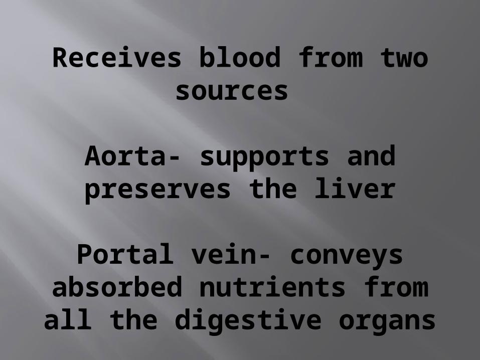
Receives blood from two sources
Aorta- supports and preserves the liver
Portal vein- conveys absorbed nutrients from all the digestive organs

The gallbladder
Small sac attached to the underside of the liver
Sole purpose is to store bile for use during digestion
Cystic duct empties the GB Hepatic duct from the liver connect to form
the common bile duct
The common bile duct empties into the duodenum to be added to the chyme during the digestion process

Stones from GB can empty thru the cystic duct and obstruct the duct thus
obstructing the excretion of bile forcing it into the
blood stream

The pancreas
Lies behind the stomach with the head in the bend of the duodenum
Empties pancreatic juices into the duodenum which aids in digestion
Secretes insulin directly into the blood stream

The large intestine
No digestion occurs in this area Colon also frames the abdomen Absorbs excess fluid from
chyme through capillaries in the lining
Only about five feet long

Consist of three sections and two flexures Found on the right side is the ascending colon connects to the small bowel at the ileocecal valve.

Transverse colon extends across the abdomen
Descending colon extends down the left side of the
abdomen and exit out the body at the rectum

Flexure found on the right side is names the
Hepatic flexure because it is located close to the hepatic
artery that connects to the liver

Splenic flexure is found on the left side
between the transverse and
descending colon over the spleen.

Diagnostic Examinations
Cholecystography-x-ray of the gallbladder after administration of contrast media
Colonoscopy- Examination using a fiber optic scope to examine the entire colon
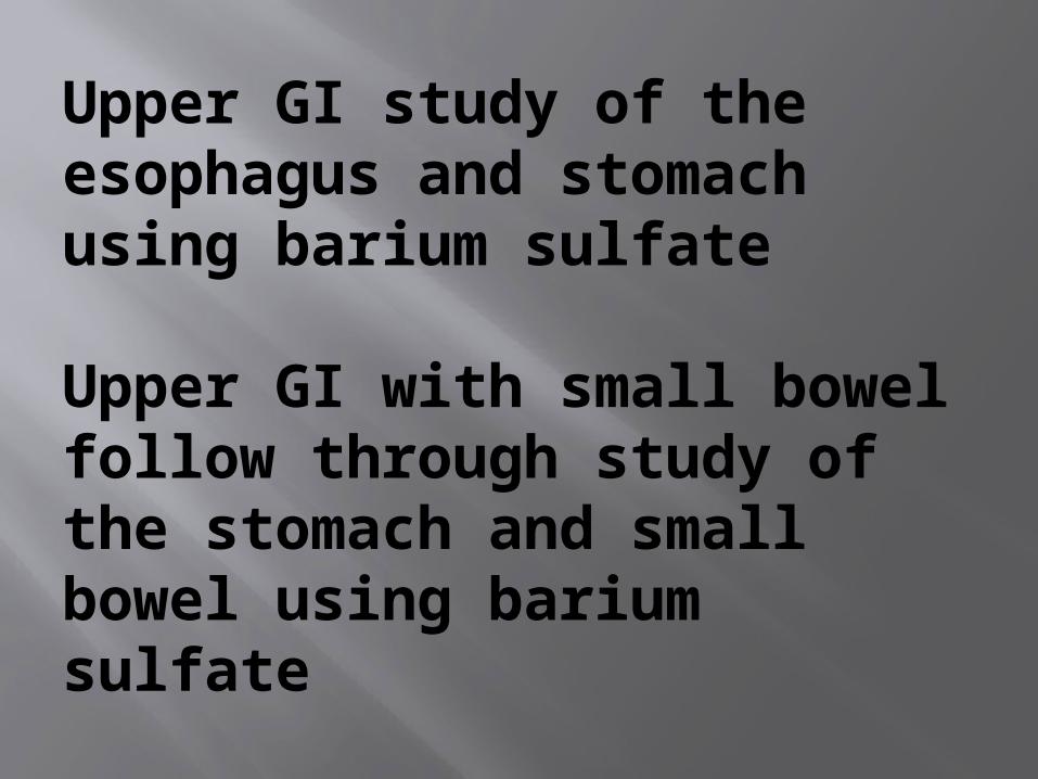
Upper GI study of the esophagus and stomach using barium sulfate
Upper GI with small bowel follow through study of the stomach and small bowel using barium sulfate

Lower GI study of the large bowel using barium sulfate
Barium Enema study of the large bowel by
administration of barium through the rectum

Gasteroscopy- using a lighted scope and examining the esop. Stomach and upper abdomen
Occult blood test collect stool and test for small amounts of blood

Blood from the rectum will be bright redBlood from the large bowel will be dark redBlood from the small bowel will be maroon Blood from the stomach will be black

Diseases and Disorders
Anorectal abscess and fistula- localized infection of the tissue adjacent to the rectum
Sings throbbing pain lump which makes sitting and coughing uncomfortable
Causes- sharp object in the feces such as fish bone or sea shell,
Treatment-surgery to drain the access
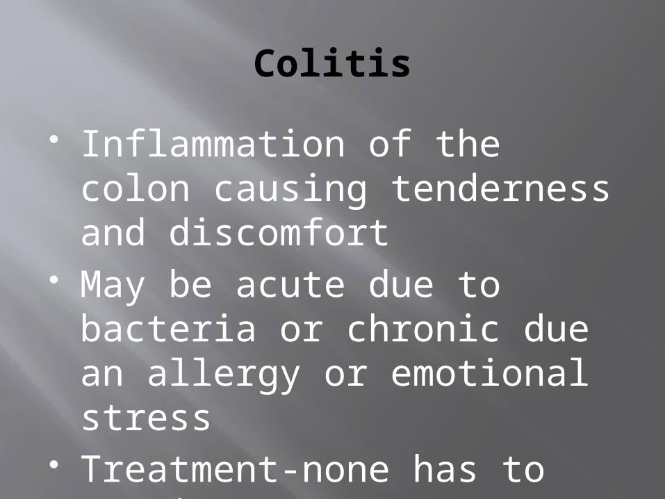
Colitis
Inflammation of the colon causing tenderness and discomfort
May be acute due to bacteria or chronic due an allergy or emotional stress
Treatment-none has to run its course
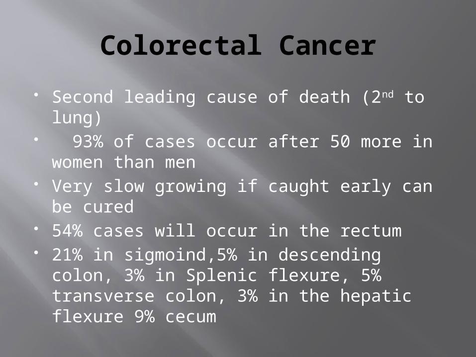
Colorectal Cancer
Second leading cause of death (2nd to lung)
93% of cases occur after 50 more in women than men
Very slow growing if caught early can be cured
54% cases will occur in the rectum 21% in sigmoind,5% in descending colon,
3% in Splenic flexure, 5% transverse colon, 3% in the hepatic flexure 9% cecum

Causes unknown but it is believed that increase intake of beef fat and low diet of fiber
Treatment Surgery, chemotherapy and radiation therapy

Colostomy-
An opening in the wall of the abdomen that allows fecal material to excrete from the body
Can be temporary or permanent
Indicated when an obstruction of the large bowel occurs near or at the sigmoid colon

Constipation
Sluggish bowel Signs- dry, hard, infrequent
bowel movement Causes- diet, meds,
dehydration, lack of exercise Treatment- stool softeners,
increase fluid intake Add fiber to the diet

Crohn’s Disease
Inflammation of any part of the colon
Most common at the end of the cecum Signs- appendicitis type pain, cramping, pain and tenderness, bloody stool, tenderness in right lower quadrant
Treatment- liquid diet, pain meds, steroids for inflammation

Diarrhea
Runny stools Signs- pain in abdomen followed
by urgency with watery loose stool
Treatment- antiditreatment of the underlying problem diarrhea meds, decrease fluid intake and fiber,

Gastroenteritis
Inflammation of the stomach Signs- pain the term may be applied to
such conditions as intestinal flu, diarrhea and food poisoning.
Cause- bacteria, food poisoning, drug reaction
Treatment- maalox, mylanna, anti gas type meds.


