Characterization of a novel insect-specific flavivirus from Brazil: potential for inhibition of infection of arthropod cells with medically important flaviviruses Joan L. Kenney 1 , Owen D. Solberg 2 , Stanley A. Langevin 2 , and Aaron C. Brault 1,* 1 Division of Vector-Borne Diseases, Centers for Disease Control and Prevention, Fort Collins, CO, 80521 2 Sandia National Labs, Livermore, California Abstract In the past decade there has been an upsurge in the number of newly described insect-specific flaviviruses isolated pan-globally. We recently described the isolation of a novel flavivirus (tentatively designated “Nhumirim virus”; NHUV) (Pauvolid-Correa et al., in review) that represents an example of a unique subset of apparently insect-specific viruses that phylogenetically affiliate with dual-host mosquito-borne flaviviruses despite appearing to be limited to replication in mosquito cells. We characterized the in vitro growth potential, 3’ untranslated region (UTR) sequence homology with alternative flaviviruses, and evaluated the virus’s capacity to suppress replication of representative Culex spp. vectored pathogenic flaviviruses in mosquito cells. Only mosquito cell lines were found to support NHUV replication, further reinforcing the insect-specific phenotype of this virus. Analysis of the sequence and predicted RNA secondary structures of the 3’ UTR indicate NHUV to be most similar to viruses within the yellow fever serogroup, Japanese encephalitis serogroup, and viruses in the tick-borne flavivirus clade. NHUV was found to share the fewest conserved sequence elements when compared to traditional insect-specific flaviviruses. This suggests that, despite being apparently insect-specific, this virus likely diverged from an ancestral mosquito-borne flavivirus. Co- infection experiments indicated that prior or concurrent infection of mosquito cells with NHUV resulted in significant reduction in viral production of West Nile virus (WNV), St. Louis * Address for Correspondence: Aaron C. Brault, Division of Vector-Borne Diseases, Centers for Disease Control and Prevention, Fort Collins, CO 80521; [email protected]; phone: (970) 266-3517. Sequences and accession numbers: Aedes Flavivirus NC012932, Alfuy virus AY898809, Alkhurma virus NC004355, Apoi virus NC003676, Aroa virus NC009026, Bagaza virus NC012534, Barkedji virus EU078325 , Bouboui virus DQ859057, Cell fusing agent virus NC001564, Chaoyang virus NC017086, Culex flavivirus NC008604, Deer tick virus AF311056, Dengue virus 1 NC001477, Dengue virus 2 NC001474, Dengue virus 3 NC001475, Dengue virus 4 NC002640, Donggang virus NC016997, Edge Hill virus DQ859060, Entebbe bat virus NC008718, Gadgets Gully virus DQ235145, Greek goat encephalitis virus DQ235153, Iguape virus AY632538, Ilheus BrMS-MQ10 KC481679, Japanese encephalitis virus NC001437, Kadam virus DQ235146, Kamiti River virus NC005064, Karshi virus DQ462443, Kedougou virus NC012533, Kokobera virus NC009029, Kyasanur forest virus HM055369, Lammi virus FJ606789, Langat virus NC003690, Meaban virus DQ235144, Modoc virus NC003635, Murray Valley encephalitis virus NC000943, Nakiwogo virus GQ165809, Nhumirim virus NC024017, Nounane virus FJ711167, Omsk hemorrhagic fever virus NC005062, Potiskum virus DQ859067, Powassan virus NC003687, Quang Binh virus NC012671, Rio Bravo virus NC003675, Rocio virus SPH34675, Royal Farm virus DQ235149, Saumarez Reef virus DQ235150, Sepik virus DQ837642, Spondweni virus DQ859064, St. Louis encephalitis virus NC007580, Tembusu Shandong1 JX965381, Tick-borne encephalitis virus NC001672, Tyuleniy virus DQ235148, Uganda S virus DQ859065, Usutu virus NC006551, Wesselsbron virus NC012735, West Nile virus NC009942, Yellow fever virus NC002031, Yokose virus NC005039, Zika virus NC012532 HHS Public Access Author manuscript J Gen Virol. Author manuscript; available in PMC 2015 December 01. Published in final edited form as: J Gen Virol. 2014 December ; 95(0 12): 2796–2808. doi:10.1099/vir.0.068031-0. Author Manuscript Author Manuscript Author Manuscript Author Manuscript
Transcript
Characterization of a novel insect-specific flavivirus from Brazil: potential for inhibition of infection of arthropod cells with medically important flaviviruses
Joan L. Kenney1, Owen D. Solberg2, Stanley A. Langevin2, and Aaron C. Brault1,*
1Division of Vector-Borne Diseases, Centers for Disease Control and Prevention, Fort Collins, CO, 80521
2Sandia National Labs, Livermore, California
Abstract
In the past decade there has been an upsurge in the number of newly described insect-specific
flaviviruses isolated pan-globally. We recently described the isolation of a novel flavivirus
(tentatively designated “Nhumirim virus”; NHUV) (Pauvolid-Correa et al., in review) that
represents an example of a unique subset of apparently insect-specific viruses that
phylogenetically affiliate with dual-host mosquito-borne flaviviruses despite appearing to be
limited to replication in mosquito cells. We characterized the in vitro growth potential, 3’
untranslated region (UTR) sequence homology with alternative flaviviruses, and evaluated the
virus’s capacity to suppress replication of representative Culex spp. vectored pathogenic
flaviviruses in mosquito cells. Only mosquito cell lines were found to support NHUV replication,
further reinforcing the insect-specific phenotype of this virus. Analysis of the sequence and
predicted RNA secondary structures of the 3’ UTR indicate NHUV to be most similar to viruses
within the yellow fever serogroup, Japanese encephalitis serogroup, and viruses in the tick-borne
flavivirus clade. NHUV was found to share the fewest conserved sequence elements when
compared to traditional insect-specific flaviviruses. This suggests that, despite being apparently
insect-specific, this virus likely diverged from an ancestral mosquito-borne flavivirus. Co-
infection experiments indicated that prior or concurrent infection of mosquito cells with NHUV
resulted in significant reduction in viral production of West Nile virus (WNV), St. Louis
*Address for Correspondence: Aaron C. Brault, Division of Vector-Borne Diseases, Centers for Disease Control and Prevention, Fort Collins, CO 80521; [email protected]; phone: (970) 266-3517.
(ISE6), African green monkey (Vero), and hamster (BHK21-clone 15), chicken (DF-1), and
Xenopus laevis (South African clawed toad) cells. Each cell monolayer was inoculated at a
multiplicity of infection of 10 TCID50 units from supernatant isolated from the original
passage of the triturated mosquito pool sample as determined by titration on C6/36 cells.
Cultures were observed for CPE for 7 days prior to harvest. Virus was serially blind
passaged three times each on Vero cells, ISE6 cells, and BHK21 clone15 cells as no initial
CPE was identified following a single passage. To confirm the presence or absence of viral
replication, RT-PCR was performed on supernatant taken from the third passage using pan-
flavivirus primers, FU1 and CFD3R, designed to amplify a ∼1085 nt portion of the NS5
gene region (Kuno et al., 1998). Negative RT-PCR samples were confirmed by IFA.
Inhibition of West Nile virus growth in vitro
West Nile virus utilized for co-infection studies was derived from an infectious clone of the
New York 1999 strain (Kinney et al., 2006). Twelve well plates of C6/36 cells all originally
Kenney et al. Page 9
J Gen Virol. Author manuscript; available in PMC 2015 December 01.
Author M
anuscriptA
uthor Manuscript
Author M
anuscriptA
uthor Manuscript
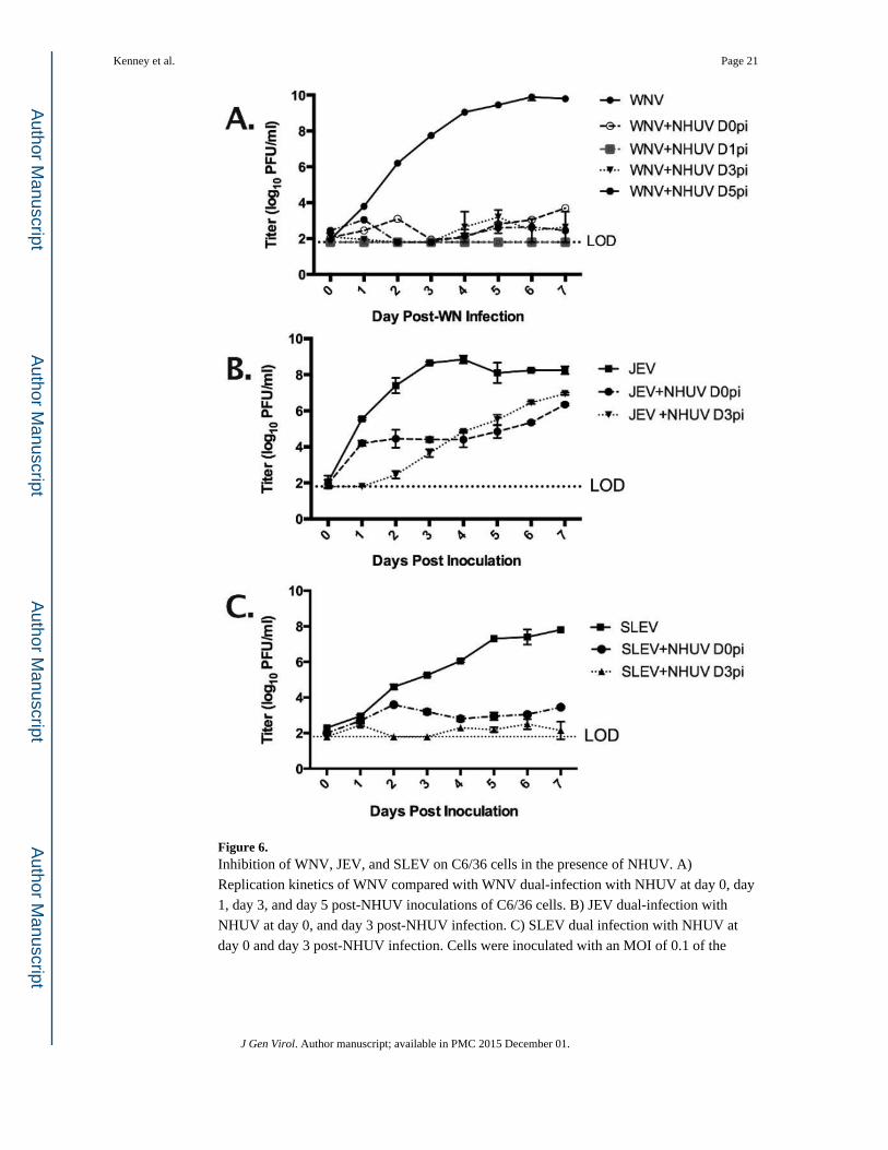
seeded at the same time and density, were inoculated at an MOI of 5 with NHUV. These
cultures were subsequently inoculated with WNV, JEV, or SLEV at an MOI of 0.1 on day 0
(simultaneous co-infection) and day 3 following initial NHUV infection. Additional pre-
inoculation of NHUV was performed at −1 and −5 dpi for WNV inhibition studies. All
infections were performed in duplicate with mock WNV, JEV, and SLEV infection controls
for each experimental time-point group. Additionally, a positive infection control for each
virus was inoculated at 0.1 MOI on C6/36 cells that were split at the same time as the
experimental dual infection replicate cultures. Supernatant samples were observed and
collected daily from triplicate cultures and subsequently titered by plaque assay. A two-way
ANOVA with an a posteriori Tukey’s multiple comparison was utilized to assess statistical
differences in viral titers between the control and dual-infection groups.
3’ UTR characterization
It has been previously proposed that an ancestral form of the flavivirus 3’ UTR has evolved
in such a way that divergence of the TBFV, MBFV, NKV, and ISF groups can be
distinguished by the presence and number of long repeated sequences (LRS) and shorter
direct repeats (DR), as well as the characterization of secondary structure RNA elements
that are found in the 3’ UTR (Grard et al., 2007; Gritsun & Gould, 2006a; b; c; 2007a; b;
Hahn et al., 1987). As such, the 3’ UTR of the NHUV isolate was compared to 3’ UTRs of
representative members from other flaviviruses representing the distinctive phylogenetic and
phenotypic grouping viruses in order to identify homologous secondary structures and repeat
elements that could associate with phylogenetic or phenotypic patterns. R-Coffee (Moretti et
al., 2008) was utilized to generate multiple alignments between available 3’ UTR regions of
flaviviruses for identification of conserved repeat regions and location of homologous
secondary structure RNA elements in concert with direct comparison to structural elements
and sequences identified from previous studies (Gritsun & Gould, 2006a; b; c; Markoff,
2003). Mfold web server was utilized to predict secondary structure formation with the
maximum distance between paired bases set to 80 as previously described by Gritsun et al.
2014 (Gritsun et al., 2014; Zuker, 2003).
Acknowledgements
We would like to thank Robert Tesh for providing the amphibian cell line, Nisha Duggal and Goro Kuno for reviewing the manuscript as well as Tamara Gritsun for advice on the 3’ UTR analysis. JLK was supported by an ASM/CDC postdoctoral fellowship. Sandia is a multi-program laboratory managed and operated by Sandia Corporation, a wholly owned subsidiary of Lockheed Martin Corporation, for the US Department of Energy’s National Nuclear Security Administration under contract DE-AC04-94AL85000.
References
Akashi H. Synonymous codon usage in Drosophila melanogaster: natural selection and translational accuracy. Genetics. 1994; 136:927–935. [PubMed: 8005445]
Aliota MT, Jones SA, Dupuis AP, Ciota AT 2nd, Hubalek Z, Kramer LD. Characterization of rabensburg virus, a flavivirus closely related to west nile virus of the Japanese encephalitis antigenic group. PLoS One. 2012; 7:e39387. [PubMed: 22724010]
Almiron WR, Brewer MM. [Host preference of Culicidae (Diptera) collected in central Argentina]. Revista de saude publica. 1995; 29:108–114. [PubMed: 8525319]
Aranda C, Sanchez-Seco MP, Caceres F, Escosa R, Galvez JC, Masia M, Marques E, Ruiz S, Alba A, Busquets N, Vazquez A, Castella J, Tenorio A. Detection and monitoring of mosquito flaviviruses
Kenney et al. Page 10
J Gen Virol. Author manuscript; available in PMC 2015 December 01.
Author M
anuscriptA
uthor Manuscript
Author M
anuscriptA
uthor Manuscript
in Spain between 2001 and 2005. Vector Borne Zoonotic Dis. 2009; 9:171–178. [PubMed: 18959502]
Biacchesi S, Skiadopoulos MH, Yang L, Murphy BR, Collins PL, Buchholz UJ. Rapid human metapneumovirus microneutralization assay based on green fluorescent protein expression. J Virol Methods. 2005; 128:192–197. [PubMed: 15955576]
Blitvich BJ, Lin M, Dorman KS, Soto V, Hovav E, Tucker BJ, Staley M, Platt KB, Bartholomay LC. Genomic sequence and phylogenetic analysis of Culex flavivirus, an insect-specific flavivirus, isolated from Culex pipiens (Diptera: Culicidae) in Iowa. J Med Entomol. 2009; 46:934–941. [PubMed: 19645300]
Bolling BG, Eisen L, Moore CG, Blair CD. Insect-specific flaviviruses from Culex mosquitoes in Colorado, with evidence of vertical transmission. Am J Trop Med Hyg. 2011; 85:169–177. [PubMed: 21734144]
Bolling BG, Olea-Popelka FJ, Eisen L, Moore CG, Blair CD. Transmission dynamics of an insect-specific flavivirus in a naturally infected Culex pipiens laboratory colony and effects of co-infection on vector competence for West Nile virus. Virology. 2012; 427:90–97. [PubMed: 22425062]
Calzolari M, Bonilauri P, Bellini R, Caimi M, Defilippo F, Maioli G, Albieri A, Medici A, Veronesi R, Pilani R, Gelati A, Angelini P, Parco V, Fabbi M, Barbieri I, Lelli D, Lavazza A, Cordioli P, Dottori M. Arboviral survey of mosquitoes in two northern Italian regions in 2007 and 2008. Vector Borne Zoonotic Dis. 2010; 10:875–884. [PubMed: 20370434]
Castle E, Leidner U, Nowak T, Wengler G, Wengler G. Primary structure of the West Nile flavivirus genome region coding for all nonstructural proteins. Virology. 1986; 149:10–26. [PubMed: 3753811]
Charlier N, Leyssen P, Pleij CW, Lemey P, Billoir F, Van Laethem K, Vandamme AM, De Clercq E, de Lamballerie X, Neyts J. Complete genome sequence of Montana Myotis leukoencephalitis virus, phylogenetic analysis and comparative study of the 3’ untranslated region of flaviviruses with no known vector. J Gen Virol. 2002; 83:1875–1885. [PubMed: 12124451]
Cirimotich CM, Ramirez JL, Dimopoulos G. Native microbiota shape insect vector competence for human pathogens. Cell host & microbe. 2011; 10:307–310. [PubMed: 22018231]
Clarke B. Darwinian evolution of proteins. Science (New York, NY). 1970; 168:1009–1011.
Cook S, Bennett SN, Holmes EC, De Chesse R, Moureau G, de Lamballerie X. Isolation of a new strain of the flavivirus cell fusing agent virus in a natural mosquito population from Puerto Rico. J Gen Virol. 2006; 87:735–748. [PubMed: 16528021]
Cook S, Moureau G, Harbach RE, Mukwaya L, Goodger K, Ssenfuka F, Gould E, Holmes EC, de Lamballerie X. Isolation of a novel species of flavivirus and a new strain of Culex flavivirus (Flaviviridae) from a natural mosquito population in Uganda. J Gen Virol. 2009; 90:2669–2678. [PubMed: 19656970]
Crabtree MB, Nga PT, Miller BR. Isolation and characterization of a new mosquito flavivirus, Quang Binh virus, from Vietnam. Arch Virol. 2009; 154:857–860. [PubMed: 19347244]
Crabtree MB, Sang RC, Stollar V, Dunster LM, Miller BR. Genetic and phenotypic characterization of the newly described insect flavivirus, Kamiti River virus. Arch Virol. 2003; 148:1095–1118. [PubMed: 12756617]
Crockett RK, Burkhalter K, Mead D, Kelly R, Brown J, Varnado W, Roy A, Horiuchi K, Biggerstaff BJ, Miller B, Nasci R. Culex flavivirus and West Nile virus in Culex quinquefasciatus populations in the southeastern United States. J Med Entomol. 2012; 49:165–174. [PubMed: 22308785]
Eaton BT. Heterologous interference in Aedes albopictus cells infected with alphaviruses. J Virol. 1979; 30:45–55. [PubMed: 480461]
Eaton BT. Viral interference and persistence in Sindbis virus infected Aedes albopictus cells. Canadian journal of microbiology. 1981; 27:563–567. [PubMed: 6266625]
Edgar RC. MUSCLE: multiple sequence alignment with high accuracy and high throughput. Nucleic acids research. 2004; 32:1792–1797. [PubMed: 15034147]
Evangelista J, Cruz C, Guevara C, Astete H, Carey C, Kochel TJ, Morrison AC, Williams M, Halsey ES, Forshey BM. Characterization of a novel flavivirus isolated from Culex (Melanoconion) ocossa mosquitoes from Iquitos, Peru. J Gen Virol. 2013; 94:1266–1272. [PubMed: 23515021]
Kenney et al. Page 11
J Gen Virol. Author manuscript; available in PMC 2015 December 01.
Author M
anuscriptA
uthor Manuscript
Author M
anuscriptA
uthor Manuscript
Farfan-Ale JA, Lorono-Pino MA, Garcia-Rejon JE, Hovav E, Powers AM, Lin M, Dorman KS, Platt KB, Bartholomay LC, Soto V, Beaty BJ, Lanciotti RS, Blitvich BJ. Detection of RNA from a novel West Nile-like virus and high prevalence of an insect-specific flavivirus in mosquitoes in the Yucatan Peninsula of Mexico. Am J Trop Med Hyg. 2009; 80:85–95. [PubMed: 19141845]
Farfan-Ale JA, Lorono-Pino MA, Garcia-Rejon JE, Soto V, Lin M, Staley M, Dorman KS, Bartholomay LC, Hovav E, Blitvich BJ. Detection of flaviviruses and orthobunyaviruses in mosquitoes in the Yucatan Peninsula of Mexico in 2008. Vector Borne Zoonotic Dis. 2010; 10:777–783. [PubMed: 20370430]
Gentry MK, Henchal EA, McCown JM, Brandt WE, Dalrymple JM. Identification of distinct antigenic determinants on dengue-2 virus using monoclonal antibodies. Am J Trop Med Hyg. 1982; 31:548–555. [PubMed: 6177259]
Gould EA, de Lamballerie X, Zanotto PM, Holmes EC. Origins, evolution, and vector/host coadaptations within the genus Flavivirus. Adv Virus Res. 2003; 59:277–314. [PubMed: 14696332]
Grard G, Moureau G, Charrel RN, Lemasson JJ, Gonzalez JP, Gallian P, Gritsun TS, Holmes EC, Gould EA, de Lamballerie X. Genetic characterization of tick-borne flaviviruses: new insights into evolution, pathogenetic determinants and taxonomy. Virology. 2007; 361:80–92. [PubMed: 17169393]
Gritsun DJ, Jones IM, Gould EA, Gritsun TS. Molecular Archaeology of Flaviviridae Untranslated Regions: Duplicated RNA Structures in the Replication Enhancer of Flaviviruses and Pestiviruses Emerged via Convergent Evolution. PLoS One. 2014; 9:e92056. [PubMed: 24647143]
Gritsun TS, Gould EA. The 3’ untranslated region of tick-borne flaviviruses originated by the duplication of long repeat sequences within the open reading frame. Virology. 2006a; 354:217–223. [PubMed: 17063566]
Gritsun TS, Gould EA. The 3’ untranslated regions of Kamiti River virus and Cell fusing agent virus originated by self-duplication. J Gen Virol. 2006b; 87:2615–2619. [PubMed: 16894200]
Gritsun TS, Gould EA. Direct repeats in the 3’ untranslated regions of mosquito-borne flaviviruses: possible implications for virus transmission. J Gen Virol. 2006c; 87:3297–3305. [PubMed: 17030864]
Gritsun TS, Gould EA. Direct repeats in the flavivirus 3’ untranslated region; a strategy for survival in the environment? Virology. 2007a; 358:258–265. [PubMed: 17067651]
Gritsun TS, Gould EA. Origin and evolution of 3’UTR of flaviviruses: long direct repeats as a basis for the formation of secondary structures and their significance for virus transmission. Adv Virus Res. 2007b; 69:203–248. [PubMed: 17222695]
Gritsun TS, Venugopal K, Zanotto PM, Mikhailov MV, Sall AA, Holmes EC, Polkinghorne I, Frolova TV, Pogodina VV, Lashkevich VA, Gould EA. Complete sequence of two tick-borne flaviviruses isolated from Siberia and the UK: analysis and significance of the 5’ and 3’-UTRs. Virus Res. 1997; 49:27–39. [PubMed: 9178494]
Gubler DJ. The global emergence/resurgence of arboviral diseases as public health problems. Archives of medical research. 2002; 33:330–342. [PubMed: 12234522]
Hahn CS, Hahn YS, Rice CM, Lee E, Dalgarno L, Strauss EG, Strauss JH. Conserved elements in the 3’ untranslated region of flavivirus RNAs and potential cyclization sequences. J Mol Biol. 1987; 198:33–41. [PubMed: 2828633]
Hobson-Peters J, Yam AW, Lu JW, Setoh YX, May FJ, Kurucz N, Walsh S, Prow NA, Davis SS, Weir R, Melville L, Hunt N, Webb RI, Blitvich BJ, Whelan P, Hall RA. A new insect-specific flavivirus from northern Australia suppresses replication of West Nile virus and Murray Valley encephalitis virus in co-infected mosquito cells. PLoS One. 2013; 8:e56534. [PubMed: 23460804]
Hoshino K, Isawa H, Tsuda Y, Sawabe K, Kobayashi M. Isolation and characterization of a new insect flavivirus from Aedes albopictus and Aedes flavopictus mosquitoes in Japan. Virology. 2009; 391:119–129. [PubMed: 19580982]
Huhtamo E, Putkuri N, Kurkela S, Manni T, Vaheri A, Vapalahti O, Uzcategui NY. Characterization of a novel flavivirus from mosquitoes in northern europe that is related to mosquito-borne flaviviruses of the tropics. J Virol. 2009; 83:9532–9540. [PubMed: 19570865]
Kenney et al. Page 12
J Gen Virol. Author manuscript; available in PMC 2015 December 01.
Author M
anuscriptA
uthor Manuscript
Author M
anuscriptA
uthor Manuscript
Ikemura T. Correlation between the abundance of Escherichia coli transfer RNAs and the occurrence of the respective codons in its protein genes: a proposal for a synonymous codon choice that is optimal for the E. coli translational system. J Mol Biol. 1981; 151:389–409. [PubMed: 6175758]
Johnston RE, Wan K, Bose HR. Homologous interference induced by Sindbis virus. J Virol. 1974; 14:1076–1082. [PubMed: 4473566]
Junglen S, Kopp A, Kurth A, Pauli G, Ellerbrok H, Leendertz FH. A new flavivirus and a new vector: characterization of a novel flavivirus isolated from uranotaenia mosquitoes from a tropical rain forest. J Virol. 2009; 83:4462–4468. [PubMed: 19224998]
Karpf AR, Lenches E, Strauss EG, Strauss JH, Brown DT. Superinfection exclusion of alphaviruses in three mosquito cell lines persistently infected with Sindbis virus. J Virol. 1997; 71:7119–7123. [PubMed: 9261447]
Kent RJ, Crabtree MB, Miller BR. Transmission of West Nile virus by Culex quinquefasciatus say infected with Culex Flavivirus Izabal. PLoS neglected tropical diseases. 2010a; 4:e671. [PubMed: 20454569]
Kent RJ, Deus S, Williams M, Savage HM. Development of a multiplexed polymerase chain reaction-restriction fragment length polymorphism (PCR-RFLP) assay to identify common members of the Subgenera Culex (Culex) and Culex (Phenacomyia) in Guatemala. Am J Trop Med Hyg. 2010b; 83:285–291. [PubMed: 20682869]
Kim DY, Guzman H, Bueno R Jr, Dennett JA, Auguste AJ, Carrington CV, Popov VL, Weaver SC, Beasley DW, Tesh RB. Characterization of Culex Flavivirus (Flaviviridae) strains isolated from mosquitoes in the United States and Trinidad. Virology. 2009; 386:154–159. [PubMed: 19193389]
Kinney RM, Huang CY, Whiteman MC, Bowen RA, Langevin SA, Miller BR, Brault AC. Avian virulence and thermostable replication of the North American strain of West Nile virus. J Gen Virol. 2006; 87:3611–3622. [PubMed: 17098976]
Kolodziejek J, Pachler K, Bin H, Mendelson E, Shulman L, Orshan L, Nowotny N. Barkedji virus, a novel mosquito-borne flavivirus identified in Culex perexiguus mosquitoes, Israel, 2011. J Gen Virol. 2013
Kuno G, Chang GJ, Tsuchiya KR, Karabatsos N, Cropp CB. Phylogeny of the genus Flavivirus. J Virol. 1998; 72:73–83. [PubMed: 9420202]
Langevin SA, Bent ZW, Solberg OD, Curtis DJ, Lane PD, Williams KP, Schoeniger JS, Sinha A, Lane TW, Branda SS. Peregrine: A rapid and unbiased method to produce strand-specific RNA-Seq libraries from small quantities of starting material. RNA biology. 2013; 10:502–515. [PubMed: 23558773]
Lauring AS, Acevedo A, Cooper SB, Andino R. Codon usage determines the mutational robustness, evolutionary capacity, and virulence of an RNA virus. Cell host & microbe. 2012; 12:623–632. [PubMed: 23159052]
Lee JS, Grubaugh ND, Kondig JP, Turell MJ, Kim HC, Klein TA, O’Guinn ML. Isolation and genomic characterization of Chaoyang virus strain ROK144 from Aedes vexans nipponii from the Republic of Korea. Virology. 2013a; 435:220–224. [PubMed: 23127596]
Lee RC, Hapuarachchi HC, Chen KC, Hussain KM, Chen H, Low SL, Ng LC, Lin R, Ng MM, Chu JJ. Mosquito cellular factors and functions in mediating the infectious entry of chikungunya virus. PLoS neglected tropical diseases. 2013b; 7:e2050. [PubMed: 23409203]
Lobo FP, Mota BE, Pena SD, Azevedo V, Macedo AM, Tauch A, Machado CR, Franco GR. Virus-host coevolution: common patterns of nucleotide motif usage in Flaviviridae and their hosts. PLoS One. 2009; 4:e6282. [PubMed: 19617912]
Markoff L. 5’- and 3’-noncoding regions in flavivirus RNA. Adv Virus Res. 2003; 59:177–228. [PubMed: 14696330]
Miller, MA.; Pfeiffer, W.; Schwartz, T. Proceedings of the Gateway Computing Environments Workshop (GCE). New Orleans, LA: 2010. Creating the CIPRES Science Gateway for inference of large phylogenetic trees.
Morales-Betoulle ME, Monzon Pineda ML, Sosa SM, Panella N, Lopez MR, Cordon-Rosales C, Komar N, Powers A, Johnson BW. Culex flavivirus isolates from mosquitoes in Guatemala. J Med Entomol. 2008; 45:1187–1190. [PubMed: 19058647]
Kenney et al. Page 13
J Gen Virol. Author manuscript; available in PMC 2015 December 01.
Author M
anuscriptA
uthor Manuscript
Author M
anuscriptA
uthor Manuscript
Moretti S, Wilm A, Higgins DG, Xenarios I, Notredame C. R-Coffee: a web server for accurately aligning noncoding RNA sequences. Nucleic acids research. 2008; 36:W10–W13. [PubMed: 18483080]
Pabbaraju K, Ho KC, Wong S, Fox JD, Kaplen B, Tyler S, Drebot M, Tilley PA. Surveillance of mosquito-borne viruses in Alberta using reverse transcription polymerase chain reaction with generic primers. J Med Entomol. 2009; 46:640–648. [PubMed: 19496438]
Parreira R, Cook S, Lopes A, de Matos AP, de Almeida AP, Piedade J, Esteves A. Genetic characterization of an insect-specific flavivirus isolated from Culex theileri mosquitoes collected in southern Portugal. Virus Res. 2012
Pauvolid-Correa A, Kenney JL, Couto-Lima D, Campos ZM, Schatzmayr HG, Nogueira RM, Brault AC, Komar N. Ilheus virus isolation in the pantanal, west-central Brazil. PLoS neglected tropical diseases. 2013; 7:e2318. [PubMed: 23875051]
Pauvolid-Correa A, Solberg OD, Couto-Lima D, Kenney JL, Serra-Freire NM, Brault AC, Nogueira JR, Langevin SA, Komar N. Nhumirim virus, a novel flavivirus isolated from mosquitoes from the Pantanal, Brazil. Arch Virol. in review
Pepin KM, Domsic J, McKenna R. Genomic evolution in a virus under specific selection for host recognition. Infection, genetics and evolution : journal of molecular epidemiology and evolutionary genetics in infectious diseases. 2008; 8:825–834.
Pesko K, Mores CN. Effect of sequential exposure on infection and dissemination rates for West Nile and St. Louis encephalitis viruses in Culex quinquefasciatus. Vector Borne Zoonotic Dis. 2009; 9:281–286. [PubMed: 19492941]
Proutski V, Gould EA, Holmes EC. Secondary structure of the 3’ untranslated region of flaviviruses: similarities and differences. Nucleic acids research. 1997; 25:1194–1202. [PubMed: 9092629]
Reed M, Muench H. A simple method of estimating fifty percent endpoints. Am J Hyg. 1938; 27:493–497.
Rice CM, Lenches EM, Eddy SR, Shin SJ, Sheets RL, Strauss JH. Nucleotide sequence of yellow fever virus: implications for flavivirus gene expression and evolution. Science. 1985; 229:726–733. [PubMed: 4023707]
Roiz D, Vazquez A, Seco MP, Tenorio A, Rizzoli A. Detection of novel insect flavivirus sequences integrated in Aedes albopictus (Diptera: Culicidae) in Northern Italy. Virology journal. 2009; 6:93. [PubMed: 19575816]
Sanchez-Seco MP, Vazquez A, Collao X, Hernandez L, Aranda C, Ruiz S, Escosa R, Marques E, Bustillo MA, Molero F, Tenorio A. Surveillance of arboviruses in Spanish wetlands: detection of new flavi- and phleboviruses. Vector Borne Zoonotic Dis. 2010; 10:203–206. [PubMed: 19485777]
Sang RC, Gichogo A, Gachoya J, Dunster MD, Ofula V, Hunt AR, Crabtree MB, Miller BR, Dunster LM. Isolation of a new flavivirus related to cell fusing agent virus (CFAV) from field-collected flood-water Aedes mosquitoes sampled from a dambo in central Kenya. Arch Virol. 2003; 148:1085–1093. [PubMed: 12756616]
Stamatakis A, Hoover P, Rougemont J. A rapid bootstrap algorithm for the RAxML Web servers. Systematic biology. 2008; 57:758–771. [PubMed: 18853362]
Vazquez A, Sanchez-Seco MP, Palacios G, Molero F, Reyes N, Ruiz S, Aranda C, Marques E, Escosa R, Moreno J, Figuerola J, Tenorio A. Novel flaviviruses detected in different species of mosquitoes in Spain. Vector Borne Zoonotic Dis. 2012; 12:223–229. [PubMed: 22022811]
Wang Z, An S, Wang Y, Han Y, Guo J. A new virus of flavivirus: Chaoyang virus isolated in Liaoning province. Chin Public Health. 2009; 25:769–772.
Weiss B, Aksoy S. Microbiome influences on insect host vector competence. Trends in parasitology. 2011; 27:514–522. [PubMed: 21697014]
Wengler G, Wengler G, Gross HJ. Studies on virus-specific nucleic acids synthesized in vertebrate and mosquito cells infected with flaviviruses. Virology. 1978; 89:423–437. [PubMed: 568848]
Zhao Z, Jiang C. Methylation-dependent transition rates are dependent on local sequence lengths and genomic regions. Molecular biology and evolution. 2007; 24:23–25. [PubMed: 17056644]
Zuker M. Mfold web server for nucleic acid folding and hybridization prediction. Nucleic acids research. 2003; 31:3406–3415. [PubMed: 12824337]
Kenney et al. Page 14
J Gen Virol. Author manuscript; available in PMC 2015 December 01.
Author M
anuscriptA
uthor Manuscript
Author M
anuscriptA
uthor Manuscript
Figure 1. Phase contrast image depicting NHUV cytopathology in C6/36 cells in vitro; A) negative
control mock infected, B) NHUV infected cells with syncytia.
Kenney et al. Page 15
J Gen Virol. Author manuscript; available in PMC 2015 December 01.
Author M
anuscriptA
uthor Manuscript
Author M
anuscriptA
uthor Manuscript
Figure 2. Epifluorescent images of IFA tests in the various cell types examined
Kenney et al. Page 16
J Gen Virol. Author manuscript; available in PMC 2015 December 01.
Author M
anuscriptA
uthor Manuscript
Author M
anuscriptA
uthor Manuscript
Figure 3. Phylogenetic analysis based on nucleotide sequences of complete polyprotein coding
sequences. Phylogenies were constructed using the maximum likelihood method with
labeled bootstrap percentages as support. Labels include taxon name and accession number.
NHUV is highlighted in gray and clades are labeled by host association designations on the
far right of the figure.
Kenney et al. Page 17
J Gen Virol. Author manuscript; available in PMC 2015 December 01.
Author M
anuscriptA
uthor Manuscript
Author M
anuscriptA
uthor Manuscript
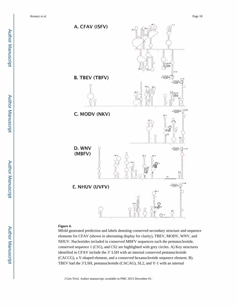
Figure 4. Mfold generated prediction and labels denoting conserved secondary structure and sequence
elements for CFAV (shown in alternating display for clarity), TBEV, MODV, WNV, and
NHUV. Nucleotides included in conserved MBFV sequences such the pentanucleotide,
conserved sequence 1 (CS1), and CS2 are highlighted with grey circles. A) Key structures
identified in CFAV include the 3’ LSH with an internal conserved pentanucleotide
(CACCG), a Y-shaped element, and a conserved hexanucleotide sequence element. B).
TBEV had the 3’LSH, pentanucleotide (CACAG), SL2, and Y-1 with an internal
Kenney et al. Page 18
J Gen Virol. Author manuscript; available in PMC 2015 December 01.
Author M
anuscriptA
uthor Manuscript
Author M
anuscriptA
uthor Manuscript
hexanucleotide sequence. C) MODV demonstrated the 3’ LSH, pentanucleotide (CUCAG),
and Y-1 with internal hexanucleotide sequence.multiple. D) WNV showed a 3’LSH, the
conserved pentanucleotide sequence (CACAG), SL2, conserved sequences CS1, CS2, and
CS3. E) NHUV was found to have a 3’ LSH, a conserved pentanucleotide (CACAG), SL2,
and only CS1 and CS2.
Kenney et al. Page 19
J Gen Virol. Author manuscript; available in PMC 2015 December 01.