31
Medium and deep chemical peeling Clinical science and practice Seaver Soon Division of Dermatology & Dermatologic Surgery Scripps Clinic La Jolla, CA

Medium and deep chemical peeling Clinical science and practice
Seaver Soon
Division of Dermatology & Dermatologic Surgery Scripps Clinic La Jolla, CA

Conflict of interest disclosure
Genentech Investigator, advisory board

Combination chemical peel Perioral Baker-Gordon phenol, unoccluded
Facial Jessner’s + TCA 35% solution
Pre-operative Post-operative week 8

Medium chemical peel

Medium depth peel • Wound to the level of the papillary or upper reticular
dermis – Actinic keratosis, dyschromia, fine wrinkling
• Historically
– TCA 50% solution – Penetration of solution unpredictable, risk of scarring
• Similar wounding and clinical results achieved
consistently and safely by first inducing epidermolysis with a superficial peeling or physical agent, followed by application of TCA 35% – Solid CO2 + TCA 35% (Brody) – Jessner’s solution + TCA 35% (Monheit) – Glycolic acid 70% + TCA 35% (Coleman)

Jessner’s solution characteristics • 14g resorcinol 14g lactic acid (85%) 14g salicylic acid q.s. 100mL ethanol • Induces corneocyte dyscohesion, intercellular
edema and cleavage of the stratum corneum above the stratum granulosum
• Frost: precipitation of salicylic acid crystals
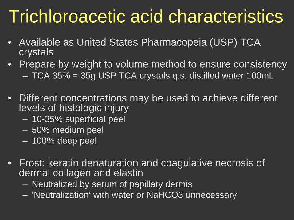
Trichloroacetic acid characteristics • Available as United States Pharmacopeia (USP) TCA
crystals • Prepare by weight to volume method to ensure consistency
– TCA 35% = 35g USP TCA crystals q.s. distilled water 100mL
• Different concentrations may be used to achieve different levels of histologic injury – 10-35% superficial peel – 50% medium peel – 100% deep peel
• Frost: keratin denaturation and coagulative necrosis of
dermal collagen and elastin – Neutralized by serum of papillary dermis – ‘Neutralization’ with water or NaHCO3 unnecessary
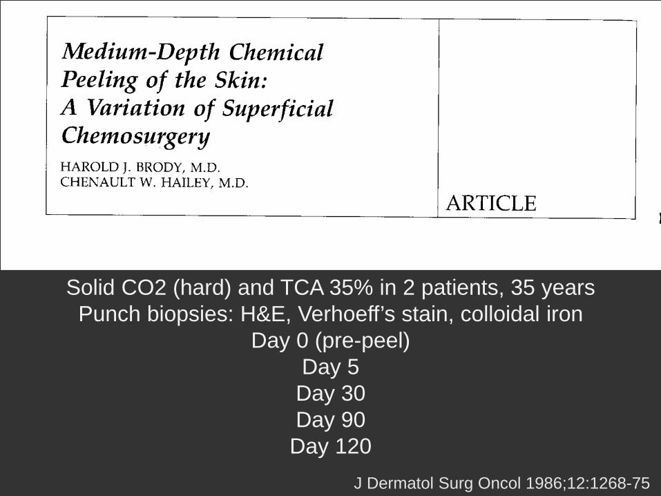
Solid CO2 (hard) and TCA 35% in 2 patients, 35 years Punch biopsies: H&E, Verhoeff’s stain, colloidal iron
Day 0 (pre-peel) Day 5 Day 30 Day 90
Day 120 J Dermatol Surg Oncol 1986;12:1268-75

Histologic effects common to medium depth chemical peeling: H&E
Pre-peel control: disorganized papillary dermis Day 5: epidermal necrosis with lymphocytic inflammation
to upper reticular dermis (depth of 0.62mm) Day 30: epidermal regeneration with
expansion and homogenization of dermal collagen
Day 120: papillary and upper reticular dermal collagen bundles organized in parallel array
J Dermatol Surg 1986;12:1268-75

Day 0
Medium depth chemical peels induce glycosaminoglycan synthesis in the papillary and
upper reticular dermis: colloidal iron stain
Day 90
J Dermatol Surg 1986;12:1268-75

Medium depth chemical peels induce formation of a Grenz zone of collagen above
pre-treatment regions of solar elastosis
Day 90
J Dermatol Surg 1986;12:1268-75
Day 0

Combination medium depth chemical peels (Solid CO2, 70% glycolic acid, or Jessner’s solution + TCA 35%) afford more safety than single-agent medium depth peels (TCA 50%) Histology: increased glycosaminoglycans and expansion of the papillary dermis above a band of pre-treatment solar elastosis (Grenz zone)

Deep chemical peel

Deep chemical peel • Wounds to the mid-reticular dermis
– Glogau photoaging type III, IV, atrophic scars
• Composition – Croton oil – Phenol – Surfactant or vegetable oils – Water
• Baker-Gordon phenol peel formula
– Phenol USP 88% 3 mL 50% – Croton oil 3 drops 2% – Hexachlorophene 8 drops 5% – Water 2 mL 43%
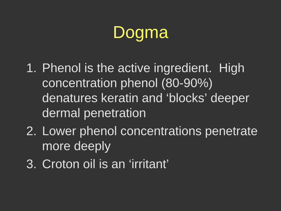
Dogma
1. Phenol is the active ingredient. High concentration phenol (80-90%) denatures keratin and ‘blocks’ deeper dermal penetration
2. Lower phenol concentrations penetrate more deeply
3. Croton oil is an ‘irritant’
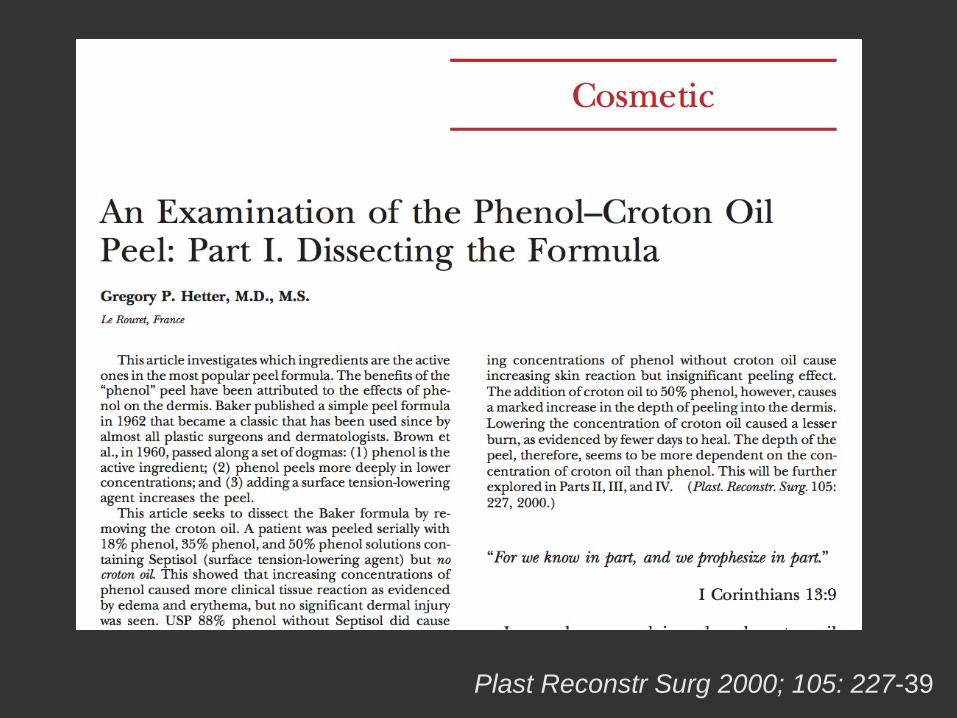
Plast Reconstr Surg 2000; 105: 227-39

Experiment 1 18% phenol to forehead 35% phenol to face 88% phenol to the glabellar rhytids
Experiment 2 50% phenol to forehead and upper cheek 50% phenol with croton oil to perioral area, lower cheek and glabella
Day 0
Day 5
Day 2
Day 8
Day 0 Day 2
Day 4 Day 7

The wounding effect of phenol increases with increasing concentration
Addition of croton oil results in deeper
wounding, prolongs healing time, and yields clinical results typical of a deep chemical peel

Characteristics 1. Croton oil
– Pressed from the seeds of croton tiglium – Hydroxyl radicals mediate epidermolysis and vesiculation at low
concentration – Increasing croton oil concentration deepens penetration, prolongs healing,
and improves clinical outcome
2. Phenol – 1-hydroxy-benzene or carbolic acid – Solvent in which croton oil is delivered to skin – Secondarily, a wounding agent: keratin disulfide bond disruption and
denaturation to the papillary dermis – Cardiotoxicity depends on individual myocardial sensitivity (reports of non-
toxic serum concentration are wide: 0.68-23mg/dL)
3. Surfactant or vegetable oils – Decreases surface tension to enable emulsification and even penetration
4. Water
– Diluent


Histology 1. Epidermal remodeling
– 2 days: Protein denaturation to the upper or mid-reticular dermis, marked inflammation
– 7-12 days: re-epitheliazation complete with normal epidermal polarity. Melanocytes present, but melanosomes small and sparsely distributed
– 60-90 days: dermal remodeling
1. Dermal remodeling – Quantity
• Increased dermal thickness due to increased collagen and glycosaminoglycans synthesis
– Quality • Collagen and elastin structural reorganization
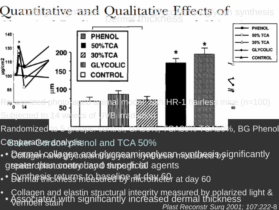
Baker-Gordon phenol and TCA 50% • Dermal collagen and glycosaminoglycan synthesis significantly greater than control and superficial agents • Synthesis returns to baseline at day 60
• Associated with significantly increased dermal thickness
Collagen synthesis Glycosaminoglycan synthesis Dermal thickness
Recognized photo-aged animal model: Skh:HR-1 hairless mice (n=100) Subjected to 14 weeks of UVB irradiation Randomized to 5 groups: control, GA50%, TCA30% TCA50%, BG Phenol Comparative analysis: • Collagen and glycosaminoglycan synthesis measured by
spectrophotometry day 0 through 60 • Dermal thickness measured by micrometer at day 60 • Collagen and elastin structural integrity measured by polarized light &
Verhoeff stain Plast Reconstr Surg 2001; 107:222-8

Photo-aged mouse model A: photo-aged control E: Baker-Gordon phenol Collagen A. Loss of collagen birefringence = collagen microfibril structural disarray
• most prominent in papillary dermis
E. Polarized light shows collagen fiber birefringence through all dermal layers
• horizontal compact bundles
Elastin A. Verhoeff stain demonstrates elastosis in papillary and reticular dermis
E. Replacement of elastosis with dense, horizontal network of fine elastic fibers

Summary
• Medium depth chemical peels (Solid CO2, 70% glycolic acid, or Jessner’s solution + TCA 35%) afford more safety than single-agent medium depth peels,TCA 50%
• Histologically: expanded papillary dermis (Grenz zone) separates pre-treatment band of solar elastosis and increased dermal glycosaminoglycans
Summary
• Croton oil is the principal wounding agent in phenol-croton oil peels
• Short-term histology: coagulative necrosis through mid-reticular dermis with re-epithelialization at day 7-12. Melanocytes present, but melanosomes sparse
• Long-term histology: Increased dermal thickness (Grenz zone) due to collagen, glycosaminoglycans synthesis, as well as structural reorganization of collagen, elastin, and normalized epidermal polarity

Clinical practice
Preparatory regimen • Topical retinoid
– Enables even and efficient peel penetration by compacting the stratum corneum
– Tretinoin 0.05% cream, apply a pea-sized amount to entire face qhs over moisturizer x 4 weeks
– May continue until procedure date for Fitzpatrick type I, II; consider stopping 1 week before peel for Fitzpatrick type III, IV
• Minimize sun exposure – Fitzpatrick type III, IV 4 weeks before procedure
• Prophylaxis begins day prior to procedure
– Cephalexin 500mg, 1 tab po QID x 7 days – Valacyclovir 1g, 1 tab po daily x 7 days

Procedure • No make up, no lotion, cleanse with chlorhexidine • Degrease
– Gauze dampened with acetone removes sebum and keratin • Mark cosmetic subunits
– Mandibular shadow and nasolabial fold • Anxious or low pain threshold
– Trigeminal nerve block or clonidine 0.1mg
• Apply peeling solution – 2 cotton tipped applicators to eyelids (2mm before ciliary line) – 4x4 gauze to remainder of face – Allow 3-4 minutes to achieve maximal frost before re-applying
Jessner’s or TCA 35% to prevent over-peeling – Feather into hairline – Immediate frost occurs with phenol-croton oil peel, but must rub to
achieve gray-white frost • Accidental eye exposure
– Flush with saline (TCA) or mineral oil (phenol-croton oil)
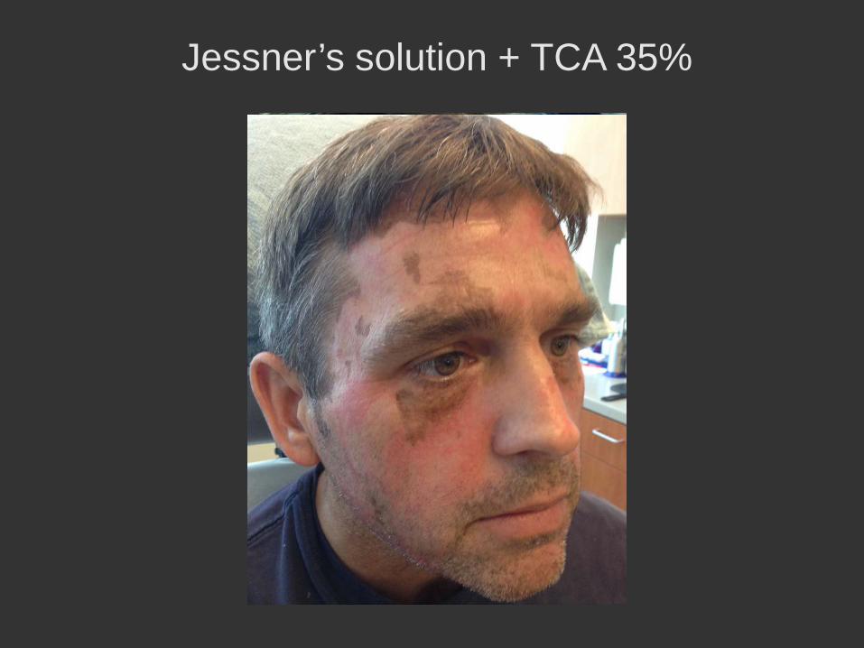
Jessner’s solution + TCA 35%

Aftercare • Medium peels
– Acetic acid soaks 4 times daily on day 1 • 1 tablespoon white vinegar in 1 pint of warm water
– Emollient 2-3 times daily • Ointment on day 1-3, transition to cream day 4-7
• Deep peels – Narcotic analgesia and acetic acid soaks day 1 – H2O2 debridement on day 2 – Emollients as above, may take 12 days to re-
epithelialize

Medium and deep chemical peeling Clinical science and practice
Seaver Soon
Division of Dermatology & Dermatologic Surgery Scripps Clinic La Jolla, CA


