Membrane-bound Cytochrome b5 Reductase (Methemoglobin Reductase) in Human Erythrocytes STUDY IN NORMAL AND METHEMOGLOBINEMIC SUBJECTS DANItLE CHOURY, ALENA LEROUX, and JEAN-CLAUDE KAPLAN, Institut de Pathologie Moleculaire, Institut National de la Sante et de la Recherche Medicale U 129, 75014 Paris, France A B S T R A C T In this study we present evidence that in human erythrocytes NADH-cytochrome b5 reduc- tase (methemoglobin reductase) is not only soluble but also tightly bound to the membrane. The membrane methemoglobin reductase-like activity is unmasked by Triton X-100 treatment, and represents about half of the total activity in the erythrocytes. Like the amphiphilic microsomal-bound cytochrome b5 reductase, the eryth- rocyte membrane-bound enzyme is solubilized by cathepsin D. Because this treatment is effective on unsealed ghosts but not on resealed (inside-in) ghosts, it is concluded that the enzyme is strongly bound to the inner face of the membrane. The erythrocyte mem- brane enzyme is antigenically similar to the soluble enzyme. The two forms of enzyme are specified by the same gene, in that both were found defective in six patients with recessive congenital methemoglobinemia. We suggest that the cytochrome b5 reductase of the erythrocyte membrane is the primary gene product. A posttranslational partial proteolysis probably gives rise to the soluble form of the enzyme, which serves as a methemoglobin reductase. INTRODUCTION The so-called methemoglobin-reductase or NADH- diaphorase has been described long ago as a soluble erythrocyte enzyme that has a major role in the enzy- matic reduction of methemoglobin (1). An inherited homozygous defect of this enzyme produces congenital recessive methemoglobinemia (2). The enzyme locus (DIA,) has been assigned to chromosome 22 (3, 4). It has been demonstrated that this erythrocyte enzyme is Preliminary data have been presented at the Twenty- second annual meeting ofthe American Society of Hematology in 1979. Blood. 541 (Suppl. 1): 342. (Abstr.) Received for publication 14 July 1980 and in revised form 15 September 1980. actually a soluble NADH-cytochrome b5 reductase (5-8) that reduces a soluble cytochrome b5. The re- duced cytochrome b5 interacts directly with methemo- globin (5, 9). In the other cells, the NADH-cytochrome b5 reductase is predominantly bound to the mem- branes: endoplasmic reticulum (10) and mitochondria (11). However, a soluble form of the enzyme has also been found in the cytosolic fraction of human placenta (8) and rabbit liver (12). In this paper, we report the finding of a NADH- cytochrome b5 reductase that is strongly attached to the inner face of the erythrocyte membrane and is re- leased either by detergent treatment or by partial digestion by cathepsin D. The immunologic and genetic characterization of the membrane-bound en- zyme suggests that it is produced by the same gene (DIA1) as the soluble erythrocyte diaphorase. METHODS Preparation of the erythrocyte membranes The erythrocyte membranes were prepared according to Marchesi et al. (13). Washed human erythrocytes were hemolysed by 30 vol of 5 mM Tris-HCl, pH 7.5, containing 1 mM EDTA, and the 22,000-g pellet was extensively washed in the same medium to obtain a completely white membrane preparation. For the preparation of inside-in resealed ghosts, we used the procedure of Steck and Kant (14) in which hemolysis is initiated by mixing 1 vol of washed erythrocytes with 40 vol of 5 mM sodium phosphate buffer, pH 8, containing 1 mM MgSO4. Extraction With detergent. The ghosts were suspended in a 10 mM Tris-HCl buffer, pH 7.4, containing 2% Triton X-100. After 30 min of incubation at 4°C, the suspension was frozen and thawed three times and centrifuged for 10 min at 105,000 g, at 4°C, in a Beckman Airfuge (Beckman Instruments Inc., Fullerton, Calif.) centrifuge to spin down small volumes (170 1l). The supematant was assayed for enzyme activity. J. Clin. Invest. (D The American Society for Clinical Investigation, Inc. - 0021-9738181/01/0149/07 $1.00 Volume 67 January 1981 149-155 149
Transcript
Membrane-bound Cytochrome b5 Reductase(Methemoglobin Reductase) in Human Erythrocytes
STUDYIN NORMALANDMETHEMOGLOBINEMICSUBJECTS
DANItLE CHOURY,ALENALEROUX, and JEAN-CLAUDEKAPLAN,Institut de Pathologie Moleculaire, Institut National de la Sante etde la Recherche Medicale U 129, 75014 Paris, France
A B S T RA C T In this study we present evidence thatin human erythrocytes NADH-cytochrome b5 reduc-tase (methemoglobin reductase) is not only soluble butalso tightly bound to the membrane. The membranemethemoglobin reductase-like activity is unmasked byTriton X-100 treatment, and represents about half of thetotal activity in the erythrocytes. Like the amphiphilicmicrosomal-bound cytochrome b5 reductase, the eryth-rocyte membrane-bound enzyme is solubilized bycathepsin D. Because this treatment is effective onunsealed ghosts but not on resealed (inside-in) ghosts,it is concluded that the enzyme is strongly bound to theinner face of the membrane. The erythrocyte mem-brane enzyme is antigenically similar to the solubleenzyme. The two forms of enzyme are specified by thesame gene, in that both were found defective in sixpatients with recessive congenital methemoglobinemia.We suggest that the cytochrome b5 reductase of theerythrocyte membrane is the primary gene product. Aposttranslational partial proteolysis probably gives riseto the soluble form of the enzyme, which serves as amethemoglobin reductase.
INTRODUCTION
The so-called methemoglobin-reductase or NADH-diaphorase has been described long ago as a solubleerythrocyte enzyme that has a major role in the enzy-matic reduction of methemoglobin (1). An inheritedhomozygous defect of this enzyme produces congenitalrecessive methemoglobinemia (2). The enzyme locus(DIA,) has been assigned to chromosome 22 (3, 4). Ithas been demonstrated that this erythrocyte enzyme is
Preliminary data have been presented at the Twenty-second annual meeting ofthe American Society of Hematologyin 1979. Blood. 541 (Suppl. 1): 342. (Abstr.)
Received for publication 14 July 1980 and in revised form15 September 1980.
actually a soluble NADH-cytochrome b5 reductase(5-8) that reduces a soluble cytochrome b5. The re-duced cytochrome b5 interacts directly with methemo-globin (5, 9). In the other cells, the NADH-cytochromeb5 reductase is predominantly bound to the mem-branes: endoplasmic reticulum (10) and mitochondria(11). However, a soluble form of the enzyme has alsobeen found in the cytosolic fraction of human placenta(8) and rabbit liver (12).
In this paper, we report the finding of a NADH-cytochrome b5 reductase that is strongly attached tothe inner face of the erythrocyte membrane and is re-leased either by detergent treatment or by partialdigestion by cathepsin D. The immunologic andgenetic characterization of the membrane-bound en-zyme suggests that it is produced by the same gene(DIA1) as the soluble erythrocyte diaphorase.
METHODS
Preparation of the erythrocyte membranesThe erythrocyte membranes were prepared according to
Marchesi et al. (13). Washed human erythrocytes werehemolysed by 30 vol of 5 mMTris-HCl, pH 7.5, containing 1mMEDTA, and the 22,000-g pellet was extensively washed inthe same medium to obtain a completely white membranepreparation. For the preparation of inside-in resealed ghosts,we used the procedure of Steck and Kant (14) in whichhemolysis is initiated by mixing 1 vol of washed erythrocyteswith 40 vol of 5 mMsodium phosphate buffer, pH 8, containing1 mMMgSO4.
ExtractionWith detergent. The ghosts were suspended in a 10 mM
Tris-HCl buffer, pH 7.4, containing 2% Triton X-100. After30 min of incubation at 4°C, the suspension was frozen andthawed three times and centrifuged for 10 min at 105,000 g,at 4°C, in a Beckman Airfuge (Beckman Instruments Inc.,Fullerton, Calif.) centrifuge to spin down small volumes(170 1l). The supematant was assayed for enzyme activity.
J. Clin. Invest. (D The American Society for Clinical Investigation, Inc. - 0021-9738181/01/0149/07 $1.00Volume 67 January 1981 149-155
Washed erythrocyte membranes,nmollminlrng protein 3.0+0.5 78+±10
In both cases the enzyme was assayed according to Hegeshet al. (15). The detergent-treated hemolysate and the detergent-treated membranes were incubated 30 min at 4°C with 2%Triton X-100 (final concentration) before the assay.
With cathepsin D. A 1:1 suspension of membranes in0.1 M Tris-maleate, pH 5.6, was incubated for 2 h at 37°Cwith cathepsin D (5 ,ug/10 mgmembrane protein). The prepa-ration was then centrifuged at 105,000 g as described above.The supernatant was assayed for enzyme activity.
Preparation of membrane-free hemolysateWashed erythrocytes were hemolysed with 4 vol of bidis-
tilled water and centrifuged at 105,000 g for 10 min at 4°C inthe Beckman Airfuge centrifuge. The supernatant containedthe soluble enzyme.
Preparation of semipurified solublecytochrome b5 reductase fromhuman erythrocytesThe membrane-free hemolysate was diluted with 2 vol of
a 5 mMpotassium phosphate buffer, pH 7.0, anid adjusted tothe same pH. Hemoglobin was removed from the dilutedhemolysate by a DEAE-cellulose 52 batchwise treatment inthe same buffer using 2 g of preswollen resin/ml of packederythrocytes. The resin was extensively washed in a Bucherfunnel with the buffer unitil the effluent was colorless. Theenzyme was eluted with 50 mMpotassium phosphate buffer,pH 5.8 that contained 0.1 mMEDTAand 0.3 NI KCI, and pre-cipitated by (NH4)2SO4 added to 60% saturationi. The precipi-tate was separated by centrifugation and dialyzed against aTris-HCl buffer 10 mM, pH 7.5, overnight at 4°C f'or enzymleassays. This procedure yielded a hemoglobin-free preparationof the soluble cytochrome b, reductase.
E)izymJ e assayjsAll assays were carried out at 25°C. The NADH-imiethenmo-
globin reductase activity was assayed with ferrocyanidemethemoglobin complex as an acceptor, according to Hegeshet al. (15).
The NADH-diaphorase activity was assayed with threedifferenit xeniobiotic electroni acceptors: (a) dichlorophenolindopheniol (1), (b) potassiumii ferricyaniide (16), (c) 3-(4,5-dimethyl thiazolyl-2)-2,5 diphenyl tetrazolium bromide(MTT)' (17).
Acetylcholiniesterase was assayed according to the method
'1Abbreviatiotn uised in this pa per: MTT, 3-(4,5-dimethyl-thiazolyl-2)-2,5 diphenyl tetrazoliumil bromide.
of Ellman et al. (18); glyceraldehyde-3-phosphate dehydroge-nase was assayed according to Beutler et al. (19).
Proteins were estimated by the method of Lowry et al. Whenlarge amount of Triton X-100 produced a precipitate in thefinal mixture, it was discarded by 5-min centrifugation at10,000 rpm.
ElectrophoresisHorizontal starch gel electrophoresis was performed as
described by Kaplan and Beutler (20) using a Tris-EDTAborate buffer, pH 8.6. Polyacrylamide gel isoelectrofocusingwas carried out in a pH 6-8 linear gradient (Ampholines;LKB Instruments, Inc., Orsay, France) according to themethod of Drysdale et al. (21). In all the electrophoreticstudies specific staining for NADH-diaphorase activity wasperformed with a mixture containing 1.2 mMNADH, 0.06mMdichlorophenol indophenol, and 1.2 mMMTTin a 0.25MTris-HCl buffer (pH 8.4) (20).
Imniunological studiesInlactivation by antiserum. The different preparations
were incubated with a chicken antiserum prepared againsthuman erythrocyte-soluble cytochrome b5 reductase (8) in thepresence of 5% (vol/vol) of polyethylene glycol. Increasingamounts of antiserum were added to constant amounts (ex-pressed as units of methemoglobin-ferrocyanide reducingactivity) of enzyme. After incubation for 1 h at 40C followedby centrifugation (20 min at 22,000 g) the residual activitywas measured in the supernatant.
Double irm munodiffusion. Double immunodiffusion wasperformed according to Ouchterlony (22). Antigens and anti-serum were incubated for 2-3 d and then the plate was ex-tensively washed with isotonic saline solution for 2 d to re-move the excess enzyme that had not reacted with the anti-serum. The precipitation lines were specifically visualizedusinig the NADH-diaphorase staining method (20).
X-100, potassium ferricyanide, cathepsin D, D-L glyceralde-hyde-3-phosphate, adenosine 5-triphosphate, 5,5 dithiobis-(2 nitrobenzoic acid), acetyl thiocholine iodide were fromSigma Chemical Co., St. Louis, Mo. DEAE-cellulose 52 wasfrom \Vhatman Inc., Clifton, N. J.
RESULTS
E rythtrocyte memtbran e methemiooglobin reductaseaictivity. Treating a noncentrifuged 1:4 hemolysateby 2% Triton X-100 (final concentration) doubles theactivity of the enzyme assayed by the method ofHegesh (15) (Table I).
The detergent had no effect on the soluble enzymepresenit in a rmemiibrane-free heinolysate (105,000 gsupernatant). Therefore, the presence of the enzymeas a component of the erythrocyte membrane wasinvestigated. The washed membranes were found tocontain a methemoglobin reductase-like activity thatwas increased 15-fold by detergent treatment (Table I).In contrast, extensive washing of the membranes by
150 D. Chouriy, A. Leroux, and J-C. Kaplani
1 MNaCl did not result in the appearance of this en-zyme activity in the 105,000 g supernatant.
Accessibility of various electron acceptors. Theaccessibility of each electron acceptor was determinedby measuring the enzyme activity with each of them inthe absence and presence of detergent. As shown inTable II, the membranes exhibit little accessibilityto the ferrocyanide methemoglobin complex (64,000mol wt) used in the Hegesh assay (15). In contrast,with smaller acceptors, such as ferricyanide, di-chlorophenol indophenol, or MTT the accessibilitywas dramatically increased.
Membrane solubilization by Triton X-100 and deter-gentlprotein ratio. The membranes were suspendedin 1 vol of 10 mMTris-HCl buffer, pH 7.4, and theprotein concentration adjusted to 4 mg/ml. Triton X-100was added to obtain a final concentration varying be-tween 0.05 and 10% (wt/vol). Each preparation wascentrifuged for 10 min at 105,000 g at 4°C. The super-natant was assumed to contain the solubilized fractionof the membrane. The precipitate contains the in-soluble part of the enzyme. The solubilization is de-pendent on the Triton X-100 concentration and maxi-mal solubilization is reached at 2% Triton (wt/vol).At this concentration 75% of the enzyme activity isrecovered in the supernatant, 25% remaining in theprecipitate.
Configuration of the erythrocyte membranepreparations. To check the configuration of the mem-branes prepared by Marchesi's method we haveassayed two enzyme markers, one of the inner face ofthe membrane (glyceraldehyde-3-phosphate-dehydroge-nase) and the second of the external face of the mem-brane (acetylcholinesterase). The membranes that weused were unsealed because both enzymes werefound to be equally accessible (94% accessibility). The
TABLE IIAccessibility to Various Electron Acceptors of the Erythrocyte
All enzyme activities determinations were carried out at25°C. The incubations in the presence of detergent wereperformed at +4°C with 2% Triton X-100. The percent ac-cessibility is determined by the without Triton X-100/withTriton X-100 ratio x 100.
TABLE IIISolubilization by Cathepsin D of the Erythrocyte
Membrane Methemoglobin Reductase
Methemoglobinreductase activity
105,000 gPellet supernatant Percent
+ Triton - Triton solubilized
nmollmin/mg protein
Unsealed ghosts- cathepsin D 57 0 0
Unsealed ghosts+ cathepsin D 21.3 12.3 36.6
Resealed ghosts (inside-in)+ cathepsin D 34 0 0
Ghosts prepared as described in text were incubated for 2 hat 37°C with 0.05% cathepsin Din 0.1 M, Tris-maleate, pH 5.6.Assays were performed on 105,000 g supematants and onpellets. The extent of solubilization of the enzyme is ex-pressed as the percentage of enzyme activity in the super-natant relative to the total values recovered in the supernatantand the pellet.
percentage of accessibility was derived from the fol-lowing ratio: activity minus detergent vs. activityplus detergent, times 100.
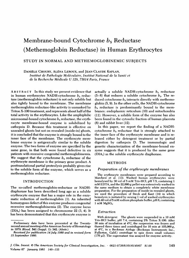
Solubilization of the membrane-bound cytochromeb5 reductase by cathepsin D. The ghosts were in-cubated 2 h at 37°C with cathepsin D at a concentra-tion of 0.05%. After incubation the preparation wascentrifuged at 105,000 g 10 min. The supernatant con-tained 36% of the total enzyme activity (Table III).Upon isoelectric electrofocusing in a pH 6-8 gradientthe cathepsin D solubilized enzyme migrated as asingle band with NADH-diaphorase activity (Fig. 1).The isoelectric point (pl) was 6.5. With the solubleNADH-diaphorase, present in a membrane-free(105,000 g supematant) hemolysate, an isoelectricpH of 6.7 was found (Fig. 1).
It was not possible to solubilize by cathepsin D themembrane-bound enzyme of sealed (inside-in) ghosts(Table III). This experiment shows that the erythro-cyte membrane methemoglobin reductase is bound tothe inner face of the membrane.
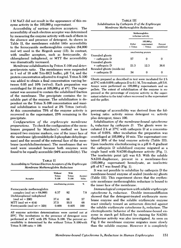
Immunological comparison with soluble erythrocytecytochrome b5 reductase. Double immunodiffusionshowed that the detergent-treated erythrocyte mem-brane enzyme and the soluble erythrocyte enzymereact similarly toward an antiserum directed againstthe soluble erythrocyte cytochrome b5 reductase. Theelectrophoretic behavior of the membrane-bound en-zyme in starch gel followed by staining for NADH-diaphorase activity was also investigated. As seen onFig. 2 the membrane enzyme migrates more slowlythan the soluble enzyme. However it is completely
Membrane-bound Cytochrome b5 Reductase in Human Erythrocytes 151
.:
** t.-- +
.. ..
... ..
_ w._ 6.5
F.::w.
1 2 3
a
S
+
FIGURE 1 Isoelectric focusing in a 6-8 pH gradient (21)followed by specific staining for NADH-diaphorase (20).1, Membrane free hemolysate; 2, Membrane free hemolysateplus 105,000g supematant of cathepsin D-treated erythro-cyte membrane; 3, 105,000g supernatant of cathepsin D-treated erythrocyte membrane.
abolished by pretreatment with the antiserum directedagainst the soluble cytochrome b5 reductase. Underthese conditions the soluble enzyme is also completelyinhibited (not shown) (8).
Inactivation test by increasing amounts of antiserumwas performed upon (a) the semipurified soluble cyto-chrome b5 reductase in the absence and presence ofcathepsin D, (b) the detergent-treated membraneenzyme, and (c) the cathepsin D-treated membraneenzyme. The inactivation curves obtained were similarin the four cases (Fig. 3).
Status of membrane cytochrome b5 reductasefrom patients with recessive congenital methemo-globinemia. We investigated six patients with re-
D gir .+
*~~~~~~~~~~As1J~~~~E
FIGURE 2 Starch gel electrophoresis of detergent-treatedmembrane erythrocyte enzyme and membrane-free hemoly-sate. The electrophoresis was performed in 12% starch gelin Tris-EDTA borate, pH 8.6. After migration for 18 h at+4°C, 200 volts the enzyme was stained as NADH-diaphorase(20). Before electrophoresis the membrane enzyme prepa-rations were incubated with identical amounts of saline,normal chicken serum or chicken antiserum directed againsthuman erythrocyte methemoglobin reductase. 1, detergent-treated membranes + NaCl 0.15 M; 2 and 4, detergent-treated membranes plus antiserum; 3, detergent-treatedmembranes plus normal serum; 5, detergent-treated mem-branes; 6, membrane-free hemolysate.
10 20pI antiseru m
FIGURE 3 Inactivation of human soluble erythrocyte methe-moglobin reductase and erythrocyte membrane enzymeby chicken antiserum directed against human solubleerythrocyte methemoglobin reductase. The incubation mix-ture (60 Al contained 10 ,ul of enzyme preparation, 10 Al ofdifferent dilutions of chicken antiserum, 6 ,ul of polyethyleneglycol (5% final) and 34 Al of saline. Base line enzymeactivities were made equal before incubation of antiserum.After incubation for 1 h at 4°C and centrifugation for 30 minat 22,000 g, the supernatant was assayed for residual methemo-globin reductase activity according to the method of Hegeshet al. (15). The results are expressed as percentage of theactivity measured in control experiments in which antiserumwas replaced by normal chicken serum. (O) Semipurifiedsoluble enzyme without cathepsin D; (U) semipurifiedsoluble enzyme plus cathepsin D, 0.05%; (0) detergent(2% Triton X-100) solubilized membrane enzyme; (0)cathepsin D solubilized membrane enzyme.
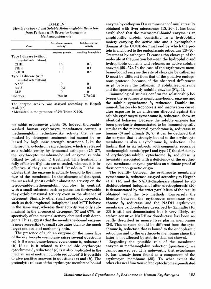
cessive congenital methemoglobinemia (Table IV).Three patients were without mental retardation (typeI disease) and three patients were severely mentallyretarded (type II disease) (23, 24 and unpublishedcases). In both types the soluble enzyme had beenfound to be defective (Table IV). Now we have foundthat the membrane-bound enzyme is also defectivein all the patients, but to a different extent. In twoof the three patients with the type I disease, the mem-brane-bound enzyme is not completely defective(Table IV).
DISCUSSION
It has been established that the erythrocyte NADH-methemoglobin reductase is a soluble form of cyto-chrome b5 reductase (5-8). In the present study weshow that the addition of a detergent to the total unspunhemolysate doubles the methemoglobin reductaseactivity. Inasmuch as the detergent has no activatingeffect on a membrane-free hemolysate, this phenome-non strongly suggests that the enzyme could be anintrinsic component of the human erythrocyte mem-brane, as previously proposed by Goto-Tamura et al.
152 D. Choury, A. Leroux, and J-C. Kaplan
TABLE IVMembrane-bound and Soluble Methemoglobin Reductase
from Patients with Recessive CongenitalMethemoglobinemia
The enzyme activity was assayed according to Hegeshet al. (15).* Measured in the presence of 2%Triton X-100.
for rabbit erythrocyte ghosts (6). Indeed, thoroughlywashed human erythrocyte membranes contain amethemoglobin reductase-like activity that is un-masked by detergent treatment, but cannot be re-leased by high ionic strength treatment. Like themicrosomal cytochrome b5 reductase, which is releasedas a soluble entity by lysosomal cathepsin (25-27),the erythrocyte membrane-bound enzyme is solu-bilized by cathepsin D treatment. This treatment isfully effective if ghosts are unsealed, whereas it is in-effective if they are resealed "inside-in." This in-dicates that the enzyme is actually bound to the innerface of the membrane. In the absence of detergent,the unsealed ghosts exhibit almost no activity on theferrocyanide-methemoglobin complex. In contrast,with a small substrate such as potassium ferricyanidethey exhibit maximal activity even in the absence ofdetergent. Similarly other small xenobiotic acceptors,such as dichlorophenol indophenol and MTTbehavein the same way, whereas their activity was only sub-maximal in the absence of detergent (57 and 67%, re-spectively of the maximal activity obtained with deter-gent). This suggests that the membrane-bound enzymeis more accessible to small substrates than to the muchlarger molecule of methemoglobin.
The presence of such an enzyme on the inner faceof the erythrocyte membrane raises several questions:(a) Is it a membrane-bound cytochrome b5 reductase?(b) If so, is it related to the soluble erythrocytecytochrome b5 reductase? (c) Is it also implicated in themechanism of methemoglobin reduction? It is possibleto give positive answers to questions (a) and (b). Theproteolytic release of the erythrocyte membrane bound
enzyme by cathepsin D is reminiscent of similar resultsobtained with liver microsomes (15, 26). It has beenestablished that the microsomal-bound enzyme is anamphiphilic protein consisting in a hydrophilicmoiety carrying the active site and a hydrophobicdomain at the COOH-terminal end by which the pro-tein is anchored to the endoplasmic reticulum (28-30).Treatment by cathepsin D causes tht cleavage of themolecule at the junction between the hydrophilic andhydrophobic domains and releases an active solubleenzyme (29-32). In the case of the erythrocyte mem-brane-bound enzyme the site of cleavage by cathepsinD must be different from that of the putative endoge-nous protease, because of the observed differencesin pI between the cathepsin D solubilized enzymeand the spontaneously soluble enzyme (Fig. 1).
Immunological studies confirm the relationship be-tween the erythrocyte membrane-bound enzyme andthe soluble cytochrome b5 reductase. Double im-munodiffusion electrophoresis and inactivation curve,after exposure to an antiserum directed against thesoluble erythrocyte cytochrome b5 reductase, show anidentical behavior. Because the soluble enzyme hasbeen previously demonstrated to be immunologicallysimilar to the microsomal cytochrome b5 reductase inhuman (8) and animals (6, 7), it can be deduced thatthe enzyme that is strongly bound to the erythrocytemembrane is also a cytochrome b5 reductase. Thefinding that in six subjects with congenital recessivemethemoglobinemia (type I and type II) the deficiencyof erythrocyte-soluble cytochrome b5 reductase isinvariably associated with a deficiency of the erythro-cyte membrane enzyme provides an ultimate proof oftheir common genetic origin.
The identity between the erythrocyte membranecytochrome b5 reductase assayed according to Hegeshet al. (15) and the NADH-diaphorase stained by 2,6dichlorophenol indophenol after electrophoresis (20)is demonstrated by the strict parallelism of the resultsobtained with the two methods. Conversely theidentity between the erythrocyte membrane cyto-chrome b5 reductase and the NADH erythrocytemembrane oxidoreductase described by Zamudio (16,33) is still not demonstrated but is very likely. Anatebrin-sensitive NADH-oxidoreductase has been re-cently described in mouse liver plasma membranes(34). This enzyme should be different from the cyto-chrome b5 reductase that is bound to the endoplasmicreticulum and to the erythrocyte membrane since thelatter is not affected by atebrin (data not shown).
Regarding the possible role of the membraneenzyme in methemoglobin reduction (question c), wecannot answer yet. It is noteworthy that cytochromeb5 has already been found as a component of theerythrocyte membrane (35). To what extent theother classical functions of the cytochrome b5-mediated
Membrane-bound Cytochrome b5 Reductase in HumanErythrocytes 153
electron transport system are operating in the erythro-cyte membrane is also still unexplored. Concerningmethemoglobin reduction, it should be noted that wehave found that, in spite of the absence of detectablesoluble methemoglobin-reductase in chicken erythro-cytes, the intact cells can promote methemoglobinreduction after exposure to nitrite (unpublished data).Board et al. (36) have found that in nucleated erythro-cytes from birds and reptiles the enzyme is only mem-brane-bound. It is therefore possible that the membrane-bound cytochrome b, reductase of the human anu-cleated erythrocytes does play a role in the reductionof methemoglobin.
Finally, whatever its* metabolic role, the membrane-bound erythrocyte cytochrome b5 reductase seems torepresent the primary gene product of the DIA1locus (3, 4). This is suggested by its deficiency in re-cessive congenital methemoglobinemia, which in-dicates that the erythrocyte membrane cytochromeb5 reductase is under the same genetic control as thesoluble erythrocyte enzyme. Their respective distribu-tion, about half and half, is probably a unique situa-tion proper to the mature circulating erythrocyte. Themembrane-bound enzyme could serve as a precursor ofthe soluble form that would be released by partialproteolysis of the former. Preliminary results, ob-tained by sucrose gradient centrifugation and highspeed gel filtration showed uis that the soluble en-zyme and the cathepsin D solubilized membraneenzyme display an identical molecular weight lowerthan that of the membrane bound detergent treatedenzyme (manuscript in preparation). A similar modelhas been suggested by Hultquist (37) for the produc-tion of the soluble cytochrome b5 from its reticulum-bound precursor during the erythroid maturation. Con-cerning the reductase it would be important to identifythe proteolytic enzyme involved in its maturation,and to determine at which stage the post-translationalprocessing of the membrane-bound enzyme occurs.These problems are under current investigation.
ACKNOWLEDGMENTS
We thank Doctors Chirouze, Seguin, Rousseau-Giral,Benabadji, and Vives-Corrons for referring blood specimensfrom methemoglobinemic subjects, and Mrs Brunner fortyping the manuscript. We also thank Miss M. Vibert andDr. Fischer for fruitful discussion.
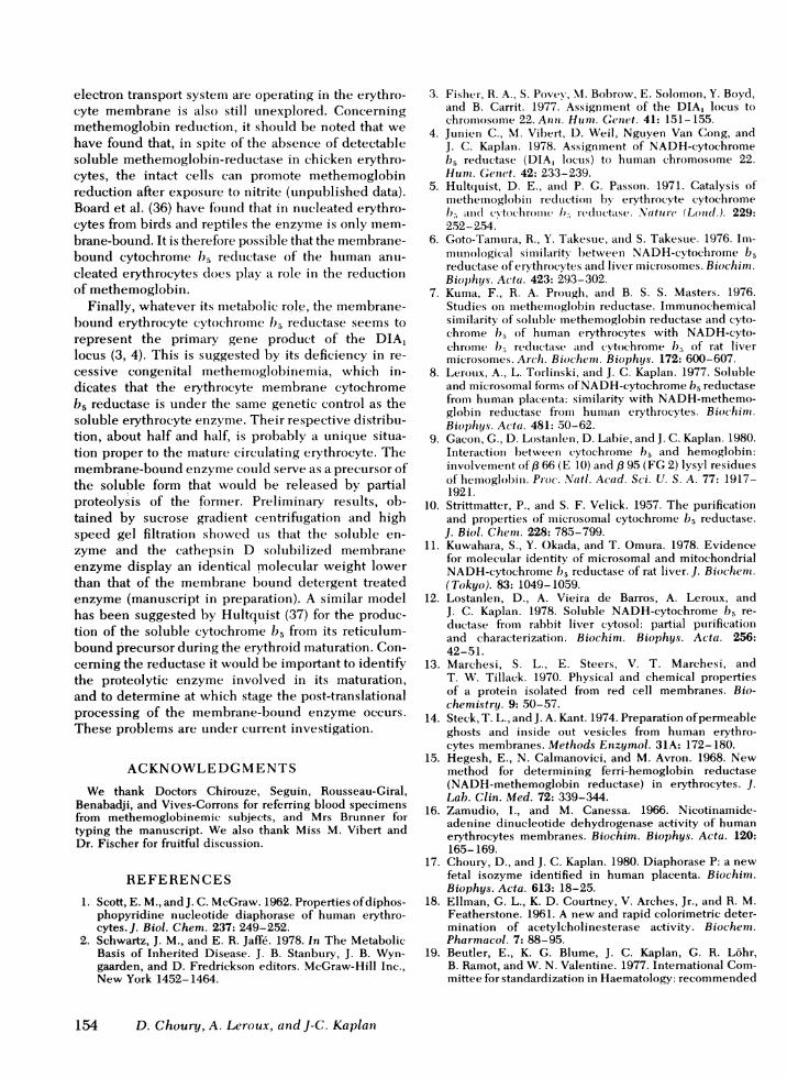
REFERENCES
1. Scott, E. M., and J. C. McGraw. 1962. Properties of diphos-phopyridine nucleotide diaphorase of human erythro-cytes.J. Biol. Chem. 237: 249-252.
2. Schwartz, J. M., and E. R. Jaffe. 1978. In The MetabolicBasis of Inherited Disease. J. B. Stanbury, J. B. Wyn-gaarden, and D. Fredrickson editors. McGraw-Hill Inc.,New York 1452-1464.
3. Fisher, R. A., S. Povey, MI. Bobrow, E. Solomon, Y. Boyd,and B. Carrit. 1977. Assignment of the DIAI locuis tochromosomiie 22. Anni. Hulm. Gete t. 41: 151-155.
4. Junien C., M. Vibert, D. Weil, Nguyen Van Cong, andJ. C. Kaplan. 1978. Assignment of NADH-cytochromeb5 reductase (DIA, locus) to human chromosome 22.Hum. Geuct. 42: 233-239.
5. Hultluist, D. E., and P. G. Passon. 1971. Catalysis ofmethemoglobin reductioni by erythrocyte cytochromeb5v amd cYtocbromle 1)5 redlztctase. .Natlre (Loud(l.). 229:252-254.
6. Goto-Tamura, R., Y. Takesue, anid S. Takestue. 1976. Im-munological si milarity betweeni NADH-cvtochrome b5reductase of erythrocvtes and liver microsomes. Biochim.Biophyjs. Acta. 423: 293-302.
7. Kuma, F., R. A. Prough, and B. S. S. Masters. 1976.Studies on metheinoglobin reductase. Immunochemicalsimilarity of soluble methemoglobin reductase and cyto-chrome bh5 of human erythrocytes with NADH-cyto-chrome b15 reductase and cytochrome b5 of rat livermicrosomes. Arch. Biochent. Biophys. 172: 600-607.
8. Leroux, A., L. Torlinski, and J. C. Kaplan. 1977. Solubleand microsomal forms of NADH-cytochrome b5 reductasefrom human placenita: similarity with NADH-methemo-globin reductase from human erythrocytes. B iochinm.Biophi s. Acta. 481: 50-62.
9. Gacon, G., D. Lostanlen, D. Labie, and J. C. Kaplan. 1980.Interaction between cytochrome b5 and hemoglobin:involvement of,8 66 (E 10) and , 95 (FG 2) lysyl residuesof hemoglobin. Proc. NVatl. Acad. Sci. U. S. A. 77: 1917-1921.
10. Strittmatter, P., and S. F. Velick. 1957. The purificationand properties of microsomal cytochrome b5 reductase.
J. Biol. Cheim. 228: 785-799.11. Kuwahara, S., Y. Okada, and T. Omura. 1978. Evidence
for molecular identity of microsomal and mitochondrialNADH-cytochrome b5 reductase of rat liver. J. Biochem.(Tokyo). 83: 1049-1059.
12. Lostanlen, D., A. Vieira de Barros, A. Leroux, andJ. C. Kaplan. 1978. Soluble NADH-cytochrome b5 re-ductase from rabbit liver cytosol: partial purificationand characterization. Biochim. Biophys. Acta. 256:42-51.
13. Marchesi, S. L., E. Steers, V. T. Marchesi, andT. W. Tillack. 1970. Physical and chemical propertiesof a protein isolated from red cell membranes. Bio-chemistry. 9: 50-57.
14. Steck, T. L., and J. A. Kant. 1974. Preparation of permeableghosts and inside out vesicles from human erythro-cytes membranes. Methods Enzymol. 31A: 172-180.
15. Hegesh, E., N. Calmanovici, and M. Avron. 1968. Newmethod for determining ferri-hemoglobin reductase(NADH-methemoglobin reductase) in erythrocytes. J.Lab. Clin. Med. 72: 339-344.
16. Zamudio, I., and M. Canessa. 1966. Nicotinamide-adenine dinucleotide dehydrogenase activity of humanerythrocytes membranes. Biochim. Biophys. Acta. 120:165-169.
17. Choury, D., and J. C. Kaplan. 1980. Diaphorase P: a newfetal isozyme identified in human placenta. Biochimn.Biophys. Acta. 613: 18-25.
18. Ellman, G. L., K. D. Courtney, V. Arches, Jr., and R. M.Featherstone. 1961. A new and rapid colorimetric deter-mination of acetylcholinesterase activity. Biochem.Pharmacol. 7: 88-95.
19. Beutler, E., K. G. Blume, J. C. Kaplan, G. R. Lohr,B. Ramot, and W. N. Valentine. 1977. International Com-mittee for standardization in Haematology: recommended
154 D. Choury, A. Leroux, and J-C. Kaplan
methods for red cell enzyme analysis. Br. J. Haematol.35: 331-340.
20. Kaplan, J. C., and E. Beutler. 1967. Electrophoresis ofred cell NADH and NADPHdiaphorases in normalsubjects and patients with congenital methemoglobin-emia. Biocheni. Biophys. Res. Commun. 29: 605-610.
21. Drysdale, J. W., P. Righetti, and H. F. Bunn. 1970. Theseparation of human and animal hemoglobins byisoelectric focusing in polyacrylamide gel. Biochim.Biophys. Acta. 229: 42-50.
22. Ouchter!ony, 0. 1953. Antigen antibody reaction in gels.Type of reaction in coordinated system of diffusion.Acta Pathol. Microbiol. Scand. 32: 231-240.
23. Kaplan, J. C., A. Leroux, S. Bakouri, J. P. Grangaud, andM. Benabadji. 1974. La 1esion enzymatique dans lamethemoglobinemie congenitale r6cessive avec encepha-lopathie. Nouv. Rev. Fr. HWmatol. 14: 755-770.
24. Kaplan, J. C., A. Leroux, and P. Beauvais. 1979. Formescliniques et biologiques du deficit en cytochrome b5reductase. C. R. Seances Soc. Biol. Fil. 173: 368-379.
25. Takesue, S., and T. Omura. 1970. Solubilization ofNADH-cytochrome b5 reductase from liver microsomesby lysosomal digestion. J. Biochem. (Tokyo). 67:259-266.
26. Sargent, J. R., P. J. St. Louis, and A. Blair. 1970. Iso-lation of NADH-cytochrome b5 oxidoreductase from ratliver microsomes. Biochim. Biophys. Acta. 223: 339-348.
27. Ito, A. 1974. Evidence obtained by cathepsin digestionof microsomes for the assembly of cytochrome b5 and itsreductase in the membrane. J. Biochem. (Tokyo). 75:787-793.
28. Spatz, L., and P. Strittmatter. 1973. A form of reducednicotinamide adenine dinucleotide cytochrome b5 re-ductase containing both the catalytic site and an additionalhydrophobic membrane-binding segment. J. Biol. Chem.248: 793-799.
29. Mihara, K., and R. Sato. 1975. Purification and propertiesof the intact form of NADH-cytochrome b5 reductasefrom rabbit liver microsomes. J. Biochem. (Tokyo). 78:1057-1073.
30. Mihara, K., R. Sato, R. Sakakibura, and H. Wada. 1978.Reduced nicotinamide adenine dinucleotide-cytochromeb5 reductase: location of the hydrophobic membrane-binding region at the carboxyl terminal end and themasked amino terminus. Biochemistry. 17: 2829-2834.
31. Ito, A., and R. Sato. 1968. Purification by means ofdetergents and properties of cytochrome b5 from livermicrosomes. J. Biol. Chemn. 243: 4922-4923.
32. Spatz, L., and P. Strittmatter. 1971. A form of cytochromeb5 that contains an additional hydrophobic sequence of40 amino acid residues. Proc. Natl. Acad. Sci. U. S. A.68: 1042-1046.
33. Zamudio, I., M. Cellino, and M. Canessa-Fischer. 1969.The relation between membrane structure and NADH(acceptor) oxydoreductase activity of erythrocytes ghosts.Arch. Biochem. Biophys. 129: 336-345.
34. Goldenberg, H., F. L. Crane, and D. J. Morr6. 1979.NADH-oxidoreductase of mouse liver plasma mem-branes.J. Biol. Chem. 254: 2491-2498.
35. Enomoto, K., and R. Sato. 1977. Asymetric binding ofcytochrome b5 to the membrane of human erythrocytesghosts. Biochemn. Biophys. Acta. 466: 136-147.
36. Board, P. G., N. S. Agar, M. Gruca, and R. Shine. 1977.Methemoglobin and its reduction in nucleated erythro-cytes from reptiles and birds. Comp. Biochem. Physiol.57: 265-267.
37. Hultquist, D. E., S. R. Slaughter, R. H. Douglas, L. S.Sannes, and G. C. Sahagran. 1978. Erythrocyte cyto-chrome b5 structure, role in methemoglobin reductionand solubilization from endoplasmic reticulum. Prog.ClitG. Biol. Res. The Red Cell, G. Brewer, Editor. AlanR. Liss Inc., New York. 21: 199-215.
Membratne-boun1d Cytochroniie b5 Rleductase inl Human E rythrocytes 155