Consejo Superior de Investigaciones Científicas (CSIC) y Universidad Autónoma de Madrid (UAM) Universidad Autónoma de Madrid Cantoblanco 28049 MADRID Tel.: (+34) 91 397 5070 Fax: (+34) 91 397 4799 http: //www.cbm.uam.es Biología celular Cell Biology Biología del desarrollo Developmental Biology Inmunología y virología Immunology and virology Neurobiología Neurobiology Regulación de la expresión génica Regulation of gene expresion
Transcript
Consejo Superior de Investigaciones Científicas (CSIC)y Universidad Autónoma de Madrid (UAM)
Universidad Autónoma de MadridCantoblanco28049 MADRIDTel.: (+34) 91 397 5070Fax: (+34) 91 397 4799http: //www.cbm.uam.es
Biología celularCell Biology
Biología del desarrolloDevelopmental Biology
Inmunología y virologíaImmunology and virology
NeurobiologíaNeurobiology
Regulación de la expresión génicaRegulation of gene expresion
Consejo Superior de Investigaciones Científicas (CSIC)y Universidad Autónoma de Madrid (UAM)
C. 8 Inmunología de los antígenos dehistocompatibilidad
Immunology of histocompatibilityantigens
José A. López de Castro
C. 9 Enzimas retrovirales: análisis estructuraly funcional de la retrotranscriptasa delvirus de la inmunodeficiencia humana
Retroviral enzymes: structural andfunctional analysis of humanimmunodeficiency virus reversetranscriptase
Luis Menéndez Arias
C.10 Regulación de la expresión génica enendotelio vascular
Regulation of gene expression invascular endothelium
Juan Miguel Redondo
C.11 Desarrollo del sistemalinfohematopoiético embrionario
Embryonic development of thelymphohematopoietic system
Miguel Angel Rodríguez Marcos
C.12 Virus de la peste porcina africana
African swine fever virus
María Luisa Salas
C.13 Desarrollo de nuevas estrategias parael control y prevención deenfermedades virales: el virus de lafiebre aftosa como modelo
Development of new strategies tocontrol and prevent viral diseases: thefoot-and-mouth virus as a model
Francisco Sobrino
C.14 Desarrollo del sistemalinfohematopoyético humano
Development of the humanlymphohematopoietic system
María Luisa Toribio
C.15 Replicación y transcripción del DNAdel bacteriófago ø29
Replication and transcription ofbacteriophage ø29
Margarita Salas
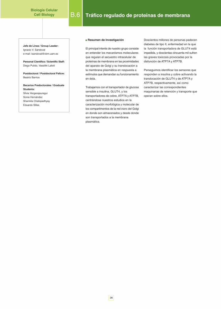
D. 5 Regulación de la expresión génica enenfermedades neurodegenerativas
Regulation of the gene expression inneurodegenerative diseases
Cecilio Giménez
D. 6 Bases moleculares de la adaptaciónal ejercicio y de la plasticidad muscular
Molecular bases of the adaptation toexercise and of skeletal muscleplasticity
Rafael Manso
D. 7 Biología de células troncales neuraleshumanas. Potencial para reposicióncelular y terapia génica enneurodegeneración
Biology of human neural stem cells.Potential for gene therapy inneurodegeneration
Alberto Martínez Serrano
D. 8 Mecanismos de señalización yregulación de receptores acoplados aproteínas G
G protein-coupled receptors signalingand regulatory mechanisms
Federico Mayor, jr.
D. 9 Neurotransmisión y desarrollo
Neurotransmission and development
Galo Ramírez.
D.10 Neurodegeneración y envejecimiento:señalización mitocondrial del calcio yseñalización de insulina/leptina enenvejecimiento
Neurodegeneration and ageing:calcium signalling in mitochondria andinsulin/leptin signalling during ageing
Jorgina Satrústegui.
D.11 Neuropatología molecular de laenfermedad de Alzheimer
Molecular neuropathology ofAlzheimer's disease
Fernando Valdivieso.
E. 6 Mantenimiento y variabilidad delgenoma: enzimología de la reparaciondel DNA
Genome maintenance and variability:enzymology of DNA repair
Luis Blanco
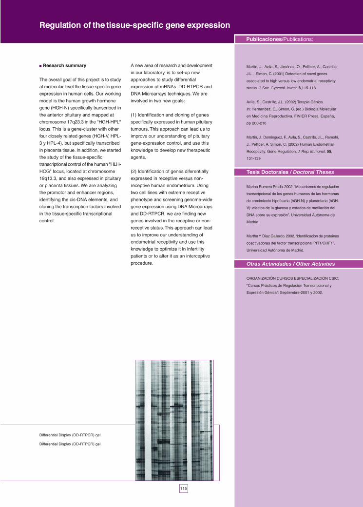
E. 7 Regulación de la expresión génicaespecífica de tejido
Regulation of the tissue-specific geneexpression
José Luis Castrillo Díez
E. 8 Estructura y función del ribosoma
Ribosome structure and function
Juan Pedro García Ballesta
E. 9 Síntesis de proteínas y su regulaciónen eucariontes
Protein synthesis and its regulation ineukaryotes
César de Haro, José M. Sierra
E.10 Expresión génica en levaduras yStreptomyces
Gene expression in yeast andStreptomyces
Antonio Jiménez
E.11 Iniciación interna de la traducción enmRNAs eucarióticos
Internal initiation of translation ineukaryotic mRNAs
Encarnación Martínez-Salas
E.12 Estructura cromatínica y transcripción
Chromatin structure and transcription
Enrique Palacián
E.13 Envolturas celulares
Cell envelopes
Miguel Angel de Pedro Montalban
E.14 Bases moleculares de lasenfermedades metabólicas
Molecular basis of metabolic diseases
Magdalena Ugarte
.......58
.......60
.......62
.......78
.......82
.......80
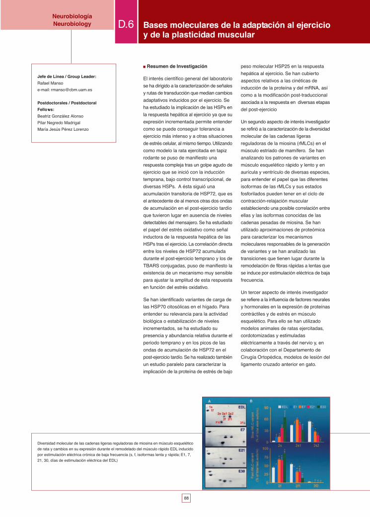
.......84
.......88
.......86
.......96
.......92
.......90
.......94
.......98
.......104
.......102
.......106
.......108
.......110
.......112
.......114
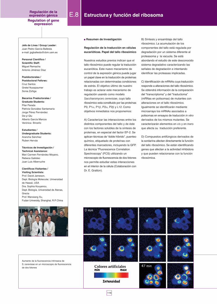
.......116
.......118
.......120
.......122
.......124
.......126
.......128
Responsables de LaboratorioPersons in charge of laboratories ....132Neurobiología
Neurobiology
D. 1 Bases moleculares de laneurotransmisión glicinérgica
Molecular basis of glycinergicneurotransmission
Carmen Aragón
D. 2 Función de las proteínasmicrotubulares en neuronas
Function of microtubular proteins inneurons
Jesús Avila de Grado
D. 3 Transducción de señales. Mecanismosde acción de neuromoduladores eimplicaciones farmacológicas
Signal transduction. Mechanisms ofaction of neuromodulators andpharmacological implications
Edgardo Catalán
D. 4 Bases moleculares de la plasticidadneuronal
Molecular basis of neuronal plasticity
F. Javier Díez Guerra
DRegulación de la expresión génicaRegulation of gene expression
E. 1 Parasitología molecular
Molecular parasitology
Carlos Alonso
E. 2 Expresión génica en linfocitos T
Gene expression in T lymphocytes
Miguel A. Alonso
E. 3 Biología molecular de extremófilos(microbiología aplicada)
Molecular biology of extremophylicmicroorganisms
Ricardo Amils
E. 4 División celular bacteriana y resistenciaa antibióticos
Bacterial cell division and antibioticsresistance
Juan Alfonso Ayala Serrano
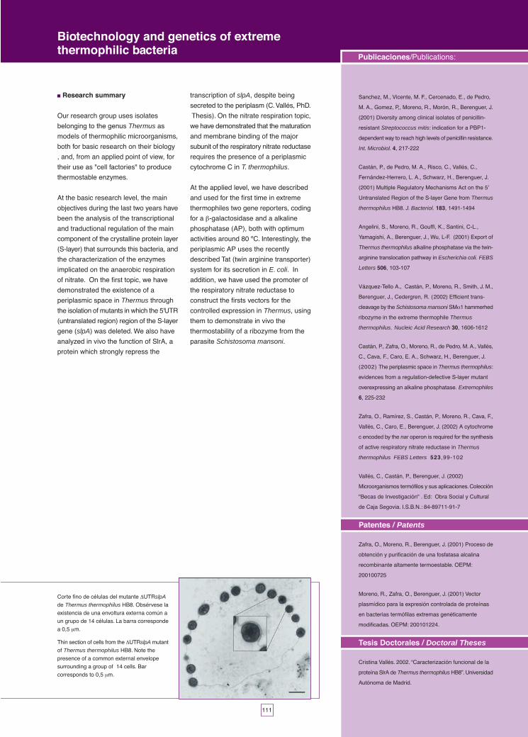
E. 5 Biotecnología y genética de bacteriastermófilas extremas
Biotechnology and genetics of extremethermophilic bacteria
José Berenguer
E
Próximos a celebrar el 50 aniversario del
descubrimiento de la doble hélice, podemos
observar lo mucho y bueno que se ha
realizado en estos años en el campo de la
Biología Molecular. En este último bienio,
tras haberse determinado la secuencia del
DNA de muchos organismos, incluyendo el
ser humano, se han comenzado los estudios
de genómica funcional en muchos de esos
organismos. Nuevos descubrimientos como
las moléculas de RNA que pueden prevenir
la expresión génica pueden ser valiosas
herramientas para estos estudios. Pero no
solo se han conseguido avances en el área
de la expresión génica, sino también en las
otras áreas de la investigación: Biología del
Desarrollo, Virología, Inmunología,
Microbiología, Biología Celular, Señalización
Celular o Neurobiología; todas estas
disciplinas se desarrollan en nuestro Centro.
Existe pues una gran rapidez de
acontecimientos científicos en el campo de
la Biología Molecular que requiere de
nosotros un gran esfuerzo para ser
competitivos a nivel internacional.
Adicionalmente, cada día aparecen nuevas
técnicas y equipamientos, lo cual nos hace
depender de recursos materiales para los
que necesitamos cada vez una mayor
subvención, para estar en las mismas (o por
lo menos no muy diferentes) condiciones
que nuestros colegas extranjeros.
El esfuerzo de nuestros investigadores
y personal de apoyo ha sido y es modélico
y es la razón fundamental por la que nuestro
Centro tiene un cierto reconocimiento a nivel
Internacional. El aumento de recursos es
muy mejorable. Afortunadamente, tenemos
la ayuda de nuestros patronos, el CSIC y la
UAM, así como el apoyo esencial y
constante de la Fundación Ramón Areces
(a través de una ayuda institucional a la
Ciencia que se realiza en nuestro Centro).
Igualmente, la valía de nuestros
investigadores nos ha permitido obtener
ayudas competitivas de organismos públicos
(fundamentalmente del Ministerio de Ciencia
y Tecnología, y del Ministerio de Sanidad y
Consumo), y también de instituciones
privadas. Sin embargo, requerimos de más
recursos.
Un problema fundamental del CBMSO
es la carencia de espacio. Por ello las
Direcciones anteriores (Federico Mayor
Menéndez y Miguel Angel de Pedro)
gestionaron la posible construcción de un
nuevo edificio. Gracias a su gestión, hoy en
día tenemos un proyecto para construir una
nueva sede en el Campus de Cantoblanco,
dentro del Parque Científico de Madrid.
Tenemos cedida por la UAM una parcela, y
actualmente existe un proyecto de
construcción de este nuevo edificio que está
llevando a cabo la empresa Euroespacios.
Con este nuevo edificio se pretende
reorganizar el CBMSO. En este reorganizado
Centro se piensa aunar las diferentes líneas
de trabajo, junto con algún otro nuevo grupo
externo, en cuatro áreas de trabajo que
hagan más eficiente su funcionamiento.
C O M E N TA R I O S D E L D I R E C T O RW O R D S F R O M T H E D I R E C T O R
4
En el bienio 2001-2002 la
producción de las diferentes líneas
del Centro ha dado lugar a 338
artículos de investigación con un
índice de impacto medio de
alrededor de 6. El número de
proyectos de investigación
financiados ha sido de 316
incluyendo aquellos
subvencionados por instituciones
públicas (del Gobierno Central o
de Comunidades Autónomas,
fundamentalmente la Comunidad
de Madrid, que adicionalmente ha
indicado que hará una valiosa
inversión en el desarrollo del nuevo
edificio), privadas, y de la Unión
Europea.
El CBMSO es un centro
universitario y por lo tanto cuida
la formación científica de sus
miembros más jóvenes de
acuerdo con las normas
académicas. Desde este punto de
vista, y en colaboración con el
Dpto. de Biología Molecular, ha
colaborado en algunos aspectos
docentes y se han leído tesis
doctorales. Adicionalmente, se ha
mantenido el Master de
Biotecnología. Además, se han
llevado a cabo 81 Seminarios, de
ellos 20 dentro del Ciclo Severo
Ochoa. Se han celebrado también
las Lecciones 8ª y 9ª en honor a
Severo Ochoa, y la 2ª y 3ª en
memoria de Eladio Viñuela,
nuestro pionero de la Biología
Molecular, en las que han
participado investigadores de talla
mundial. En recuerdo a Eladio
Viñuela, el Instituto de Biología
Molecular (CSIC) ha pasado a
denominarse Instituto de Biología
Molecular “Eladio Viñuela”. Otra
novedad relacionada con el
personal científico en formación
no en plantilla, ha sido la
instauración de dos premios para
una buena Tesis leída en el Dpto.
de Biología Molecular de nuestra
Facultad, y para un buen trabajo
publicado de investigación. Cuidar
la formación de nuestros
investigadores más jóvenes y
mejorar su modo de trabajar es
de vital importancia. En este
sentido se debe de buscar para
ellos unas mejores condiciones
contractuales que faciliten su labor.
No menos importante es la
formación de personal técnico
altamente capacitado. En este
sentido, el CBMSO ha mantenido
su colaboración con el INEM, a
través de la Fundación Severo
Ochoa, para favorecer la
formación de técnicos en la
Escuela-Taller.
Otra actividad novedosa ha
sido la iniciación de una Escuela
de Periodismo Científico,
subvencionada por la Fundación
Española para la Ciencia y la
Tecnología (FECYT), y que ha
buscado mejorar la capacidad de
comunicación de nuestros
científicos más jóvenes.
Uno de los pilares
fundamentales para el buen
funcionamiento del CBMSO es su
Departamento Técnico. Muchos
de los servicios de este
Departamento se han consolidado
durante este bienio y se ha
procedido a la instauración de uno
nuevo, el Servicio de Citometría
de Flujo.
De cara al futuro, la mayor
preocupación es la de tener a las
personas adecuadas y los medios
necesarios para llevar a cabo una
excelente labor investigadora; para
ello necesitaremos más
subvenciones para plazas y
contratos de trabajo, y para
infraestructuras.
Cerca del X aniversario de la
muerte de D. Severo Ochoa,
debemos de buscar que nuestro
Centro sea un lugar de excelencia
científica como él deseaba.
Tenemos un personal creativo y
con gran capacidad de trabajo.
Por ello debemos de encontrar
más recursos para poder realizar
el trabajo que deseamos. Estoy
convencido que con unas buenas
condiciones el CBMSO podrá
realizar un trabajo excelente y
competitivo que pueda dar frutos
a nuestra sociedad.
Jesús Avila
Director
5
Close to the celebration ofthe 50th anniversary of thediscovery of the double helix,we can look back on thebreakthroughs in the field ofMolecular Biology. In the lasttwo year period, after thedetermination of the DNAsequence of numerousorganisms — including that ofthe human — functionalgenomics research has beencarried out with many of theseorganisms. New discoveriessuch as RNA molecules thatcan prevent gene expressionmay become valuableinstruments for use in this areaof research. Advances havenot been restricted to the areaof gene expression but includeother research areas, such as:Developmental Biology,Virology, Immunology,Microbiology, Cell Biology,Cellular Signalling andNeurobiology; all thesedisciplines are followed at ourCentre.
There is rapid progressand change within the field ofMolecular Biology; in order toremain competitiveinternationally we will have towork hard. Additionally, newtechniques and systems aredeveloped daily, putting new
burdens on our resources. Ifwe do not keep up we will lagbehind our competitorsabroad.
The effort of ourresearchers and supportpersonnel has been and isexemplary. They are thefundamental reason why ourCentre has achieved a stronginternational recognition. Butmore resources are needed.Fortunately, we are backed byour sponsors — the CSIC(Spanish Board of ScientificResearch) and the UAM(Autonomous University ofMadrid) — and we enjoy theconstant and essential supportfrom the Ramón ArecesFoundation. Likewise, theprecious skills of ourresearchers have allowed usto obtain competitive grantsfrom public organisations —mainly from the Departmentof Science and Technologyand the Department of Healthand Consumer Affairs — aswell as from private institutions(acknowledged in the Lines ofWork section of theMemorandum Report).However, in spite of this, westill require more resources.
A fundamental problemfaced by the CBMSO is thelack of space. Therefore, theformers Directors, FedericoMayor and Miguel Angel dePedro, have planned theconstruction of a new building.Thanks to his work, today wehave a project for theconstruction of our new centralpremises at the CantoblancoCampus, located in theScientific Park of of Madrid .We have been given a plot ofland by the UAM, and currentlythere is a project for theconstruction of this newbuilding carried out by the firmEuroespacios. The aim of thisdevelopment is to reorganisethe CBMSO and gather in thisnew Centre groups in fourareas and to join them withsome groups from outside.This will make the running ofthe Centre much moreefficient.
In the 2001-2002 period,the production of the Centre’sdifferent teams has led to 338research papers, with anaverage impact value ofaround 6. Financed researchprojects amounted to 316,including all those projectssubsidised by publicinstitutions such as Central or
C O M E N T A R I O S D E L D I R E C T O R
W O R D S F R O M T H E D I R E C T O R
6
Autonomous Governments — mainly theCommunity of Madrid, which have expressedtheir interest in investing in the new buildingdevelopment — private institutions and theEuropean Union.
The CBMSO is a university centre and,therefore takes care of the scientific trainingof its youngest members in accordance withacademic ordinances. From this point ofview, and in collaboration with the Departmentof Molecular Biology, it has trained manygraduate students to PhD level. In addition,we have supported the Masters ofBiotechnology studies. Eighty-one seminarshave been given, of which twenty were partof the Severo Ochoa Cycle. The 8th and 9thlectures celebrated Severo Ochoa’s, and the 2nd and 3rd were a tribute to Eladio Viñuela,our pioneer in the field of MolecularBiology.These lectures were given byinternationally well-known researchers. Also,as a tribute to Eladio Viñuela, the CSIC(Institute of Molecular Biology) has beenrenamed Institute of Molecular Biology “EladioViñuela”. Other news in connection withscientific trainees has been the creation oftwo awards, one for the best Thesis read atour Faculty’s Department of MolecularBiology, and another for the best peer-reviewed piece of research. It is our policyto provide training and support to ouryoungest researchers, so we must achievethe best contractual terms for them and theirwork.
The training of highly qualified technicalpersonnel is equally important. The CBMSOis continuously collaborating with the INEM (Spanish Employment Office) through theSevero Ochoa Foundation in order topromote the training of technicians at theWorkshop School.
Another new activity was theestablishment of a Scientific JournalismSchool, subsidised by the FECYT (SpanishScience and Technology Foundation) whichseeks to improve the capacity forcommunication of our youngest scientists.
One of the foundations of the smoothoperation of the CBMSO is its TechnicalDepartment. Many of the services offeredby this Department have been consolidatedduring this two-year period. Furthermore, anew service, the Flux Cytometry Service hasalso been set up.
As for the future, our main concern is togather the appropriate people and thenecessary means to carry out excellentresearch work. To achieve this, we will needmore subsidies for labour positions andcontracts, as well as for infrastructures. Closeto the X anniversary of Severo Ochoa’spassing, we must build a Centre that is theplace of scientific excellence he envisaged.We have creative and hard workingpersonnel. Therefore, we must find moreresources so we can reach the goals set inour work. I am convinced that, under goodconditions, the CBMSO will be able to carryout outstanding and competitive work for thebenefit of our society.
Jesús Avila
Director
7
Personal Científico /Scientific Staff
CONSEJO SUPERIOR DEINVESTIGACIONES CIENTÍFICAS (CSIC)PROFESORES DE INVESTIGACIÓN /RESEARCH PROFESSORS
Alarcón Sánchez, BalbinoAlonso Bedate, CarlosAvila de Grado, JesúsDomingo Soláns, EstebanGarcía Ballesta, Juan PedroGarcía-Bellido y Gª de Diego, AntonioGutiérrez Armenta, CrisantoJimenez Martínez, AntonioLópez de Castro, José AntonioModolell Mainou, JuanMorata Pérez, GinésMoscat Guillén, JorgePalacián Gil, EnriqueRamírez Ortiz, GaloRipoll Quintas, Pedro M.Salas Falgueras, MargaritaVicente-Sandoval Rodríguez, Ignacio
INVESTIGADORES CIENTÍFICOS /RESEARCH SCIENTISTS
Alonso Lebrero, Miguel AngelBlanco Dávila, LuisCampuzano Corrales, SonsolesGuerrero Vega, IsabelHaro Castella, César Jesús, deMartínez Salas, EncarnaciónNieto López, AntonioPedro Montalbán, Miguel Angel, deRamírez Ortiz, AngelRedondo Moya, Juan MiguelSalas Falgueras, JoséSalas Falgueras, María LuisaSánchez-Herrero Arbide, ErnestoSobrino Castello, FranciscoToribio García, María Luisa
CIENTÍFICOS TITULARES /TENURED SCIENTISTS
Alcamí Pertejo, Antonio JavierAntón Canto, Luis CarlosAyala Serrano, Juan AlfonsoBusturia Jimeno, Ana MaríaCastrillo Díez, José LuisCelis Ibeas, José Felix deDíaz-Meco Conde, Mª TeresaEscarmís Homs, CristinaGómez Skarmeta, José LuisJiménez Díaz-Benjumea, FernandoLópez Carrascosa, Angel L.Lucas Lozano, José JavierMenéndez Arias, LuisPulido Vega, DiegoRevilla Novella, YolandaRodríguez Marcos, Miguel A.Ruiz Gómez, MarTalavera Díaz, AntonioVázquez Cobos, JesúsVillasante Atienza, AlfredoWandosell Jurado, Francisco
CENTRO DE BIOLOGIA MOLECULAR"SEVERO OCHOA"· Dirección /
Management Board
Director / Director
Jesús Avila de Grado
Vicedirector / Vicedirector
José Berenguer Carlos
Director del Instituto de BiologíaMolecular "Eladio Viñuela" del CSIC /Director of the Instituto de BiologíaMolecular "Eladio Viñuela" CSIC
Crisanto Gutiérrez Armenta
Director del Instituto Universitario deBiología Molecular de la UAM /Director of the Instituto Universitariode Biología Molecular UAM
José Manuel Cuezva Marcos
Gerente / Administrative Director
Salvador Fortes Alba
Oficina de Relaciones Institucionales/ External Relations andCommunication Department
Rosario Martín Rodríguez
A BPersonal Personnel
8
UNIVERSIDAD AUTÓNOMA DEMADRID (UAM)
CATEDRÁTICOS / PROFESSORS
Amils Pibernat, RicardoAragón Rueda, CarmenCarrasco Llamas, LuisCuezva Marcos, José ManuelFresno Escudero, ManuelGiménez Martín, CecilioHermoso Nuñez, José MiguelMayor Menéndez, FedericoMayor Zaragoza, FedericoMoreno Muñoz, FranciscoSatrústegui Gil-Delgado, JorginaSierra Pérez, José ManuelUgarte Pérez, MagdalenaValdivieso Amate, Fernando
PROFESORES TITULARES /ASSOCIATE PROFESSORS
Abad Lorenzo, José PascualAlmendral del Río, José MªArmas Portela, RosarioBerenguer Carlos, JoséBogónez Pelaez, ElenaBonay Miarons, PedroCarrascosa Baeza, Jose MªCatalán Tobar, EdgardoCorreas Hornero, IsabelDíaz Nido, JavierDíez-Guerra, Fco. JavierFernández Lobato, MaríaFernández Santarén, Juan AntonioGarcía Mateu, MauricioGarcía Ruiz, PredestinaciónHernández Sánchez, FranciscoIzquierdo Rojo, MartaJiménez Martínez, Juan SalvadorLópez Corcuera, BeatrizLópez Guerrero, José AntonioManso Martinez, RafaelMarín Palma, IrmaMartínez Serrano, AlbertoMontejo de Garcini Guedas, EstebanRemacha Moreno, MiguelRequena Rolania, José MªRuiz Gómez, AnaSantamaría Pérez, FranciscoSanz Martín, José LuisZafra Gómez, Francisco
PERSONAL CIENTÍFICOCONTRATADO /
Airaksinen, AnteroAzpiazu Torres, NataliaBarrio Olano, María RosaBravo García, AliciaBullido Gómez-Heras, Mª JesúsCalleja Requena, ManuelCamacho Pedrero, AnaCampillos González, MónicaCano López, Eva MªCastellano Moreno, Mª del MarDel Pozo Benito, Juan CarlosDesvoyes, Irene BenedicteFernández Malavé, EdgarGarcía García, Miguel AngelGarcía Muñoz, Mª JoséGironés Pujol, NuriaGorfinkiel Haim, NicoleHernández López de Munain, CristinaHernández Pérez, Félix
Iñiguez Peña, Miguel AngelIzquierdo Juárez, José MaríaLafuente Monasterio, Mª JoséLalioti, VassilikiLozano Salvatella, Juan JoséMartín Belmonte, FernandoMartín Fernández, PilarMartínez Martínez, SaraMeijer, WilfriedMendieta Gómez, JesúsMerinero Cortés, BegoñaMoreno Flores, Mª TeresaMurga Montesinos, CristinaNavarrete López de Soria, Rosa MaríaNúñez garrote, José IgnacioNusspaumer da Costa, GretelPardo Merino, BeatrizPenela Márquez, PetronilaPérez González, BelénPérez Martín, José ManuelPérez Martínez, Mª. del MarPérez-Cerdá, CeliaRibas Núñez, CatalinaRichartd Rodríguez, Eva MaríaRodríguez Márquez, AntonioRodríguez Martínez, Javier MaríaRojo Gozalo, SusanaRuiz Desviat, LourdesSan José Martínez, EstherSantos Tejedor, CruzSanz Alonso LauraSchamel, WolfgangSevilla Hidalgo, NoemíSoto Alvarez, ManuelSuzanne, Magali
Clones de células doble mutantes para patched 14 y dispatched en el
disco imaginal de ala teñidos con GFP y con anticuerpos contra lasproteinas Patched (azul) y beta-Gal (rojo). Las células mutantes parapatched se marcan por la falta de color rojo. Las células mutantes paradispatched por la falta de color verde. Las células doble mutantes tienenuna falta completa de color. Las distintas intensidades de verde y rojocorresponden a células silvestres y heterocigóticas para cada una de lasmutaciones.
12
ABiología del DesarrolloDevelopmental Biology
A.2 Mecanismos de señalización en el desarrolloA.2 Signalling mechanisms in development
Isabel Guerrero
A.1 Análisis genético de mecanismosmorfogenéticos en Drosophila
A.1 Genetic analysis of morphogeneticmechanisms in Drosophila
AntonioGarcía-Bellido
A.3 Comunicación intercelular en el desarrollodel Drosophila
A.3 Cell-cell signalling in Drosophiladevelopment
Fernando JiménezDíaz-Benjumea
A.4 Biología molecular del desarrolloA.4 Molecular biology of development
Juan Modolell
A.5. Control genético de la morfogénesisA.5 Genetic control of morphogenesis
Clones gemelos (rojo y amarillo) en las regiones dorsal (A) y ventral (B)
de ala ilustrando que su forma y tamaño depende de cada territorio del
disco.
Análisis genético de mecanismosmorfogenéticos en Drosophila
A.1Biología del� DesarrolloDevelopmental Biology
14
Extavour, C., García-Bellido, A. (2001) Germ cell selectionin genetic mosaics in Drosophila melanogaster. Proc.Natl. Acad. Sci. USA 98, 11341-11346
Mollerau B., Dominguez, M., Webel, R., Colley, N.J.,Keung, B., de Celis, J.F., Desplan, C. (2001) Two-stepprocess of photoreceptor formation in the Drosophilaeye. Nature 412, 911-913
Rusten, T. E., Cantera, R., Urban, J., Techanu, G.,Kafatos, F. C., Barrio, R. (2001) Spalt restricts EGFRmediated induction of chordotonal precursors in theembryonic PNS of Drosophila. Development 128, 711-722
López, P.P., Santarén, J.F., Ruiz, M.F., Esponda, P.,Sanchez, L. (2001) The Drosophila melanogaster X-linked mfs(1)6E locus is required for production of normalseminal fulid by the male accessory glands. Exp. CellRes. 267, 1-12
Rodríguez, J.M., Salas, M.L., Santarén, J.F. (2001)African swine fever virus-induced polypeptides in porcinemacrophages and in Vero cells: A high resolution twodimensional analysis. Proteomics 1, 1447-1456
Resino, J., Salama-Cohen, P., García-Bellido, A. (2002)Determining the role of patterned cell proliferation in theshape and size of the Drosophila wing. PNAS 99, 7502-7507
Rusten, T.E., Cantera, R., Kafatos, F.C., Barrio, R. (2002)The role of TGF- signaling in the formation of the dorsalnervous system is conserved between Drosophila andchordates. Development 129, 3575-3584
Cantera, R. Lüer, K., Rusten, T.E., Barrio, S., Kafatos,F.C., Technau, G.M. (2002) spalt mutations cause asevere but reversible neurodegenerative phenotype inthe embryonic central nervous system of Drosophilamelanogaster. Development 129, 5577-5586
Premios / Awards
- Doctor Honoris Causa por la Universidad MiguelHernández de Alicante, 2001.
- Miembro Electo del Neuroscience Institute. USA, 2001.
Tesis Doctorales / Doctoral Theses
Jaime Resino de Castro. 2002. “Tamaño y Proliferaciónen el Disco Imaginal de Drosophila melanogaster”.Universidad Autónoma de Madrid.
Genetic analysis of morphogeneticmechanisms in Drosophila
Publicaciones/Publications:
Twins clones (red-yellow pairs) in the dorsal (A) and ventral (B) wing imaginal disc. The size
and shape of the clones characteristically varies with the different regions.
Research summary
Size control. Lethal alleles in 5 loci
autonomously cause, in genetic mosaics
in the wing, smaller cells and clones
(l(3)Me10), larger cells but small clones
(gig y cdc2) and smaller cells but larger
territories (EP(3)622 y ft). Other
associated phenotypes reveal failures in
cell communication what suggests that
growth is determined by the local
generation of positional values.
Shape control. Twin-clon analysis
indicates the position of mother cells and
subsequent daughter cell allocation. Cell
proliferation in the wing is intercalary and
daughter cells allocate preferentially either
the proximo-distal or antero-posterior axis
depending on the region of the wing.
Disc eversion. The prospective border
of contact between discs is predefined
by the expression of puc (Jun K pathway)
a pathway related to the relaxation of the
cytoskeleton and loosening of cells. The
actual eversion takes place by
coalescence of holes in the stalk of the
disc and subsequent elimination of larval
epidermal cells.
The gene vestigial. Clones of cells over
expressing vg lead to ectopic
transformations to wing blade in territories
that already express wg. The
morphogenetic effects are complex,
indicating that vg acts in combination with
other genes in the distalization of the
wing.
Regulation of spalt expression in the
wing disc. We have identified the DNA-
regulatory region driving spalt expression
in the wing disc, and characterised the
binding sites of several transcription
factors.
Gain of function screening. We have
mapped 500 insertions of a PUAS
element affecting vein differentiation and
wing growth.
Proteome. We continue the catalogation
and characterization of wing proteins in
two-dimensional gels for comparisons
between wildtype and mutants.
15
Resumen de Investigación
El desarrollo de las estructuras adultas de
Drosophila melanogaster se ha utilizado
como sistema modelo para el estudio de la
inducción de patrones morfogenéticos. En
cada estructura adulta de la mosca, la
proteína Hedgehog (Hh) induce la activación
de nuevas señales morfogenéticas desde
el centro organizador anterior / posterior,
tales como los factores de secreción
Wingless (WG) y Decapentablegic (DPP),
pertenecientes a las familias proteicas Wnt
and TGF- respectivamente. Esta cascada
de inducción de señales es clave para el
desarrollo de todos los apéndices de la
mosca. Uno de los problemas mas
fascinantes actualmente en biología es
entender como este tipo de señales
morfogenéticas se secretan y difunden, y
como las células las reciben e interpretan.
En los últimos años nos hemos centrado
en el estudio de los mecanismos de
señalización de Hh debido a su importancia
en enfermedades humanas
(holoprosencefalia, polidactilia, carcinomas
de células básales, meduloblastomas, etc.).
Estudiando el receptor de Hh, Patched
(Ptc), hemos identificado dos dominios
funcionales en su proteína ya que ciertas
mutaciones afectan solo a la difusión de
Hh y otras solo a la interacción con
segundos mensajeros.
Hemos demostrado que la internalización
de Hh por Ptc no se requiere para la
señalización de Hh siendo necesaria esta
endocitosis de Hh solo para controlar su
degradación. Por otra parte, existen varias
líneas de evidencia que indican una relación
del colesterol y los lípidos con la vía de Hh.
Se ha demostrado que el colesterol se
requiere para la respuesta a la vía de Hh.
De acuerdo con este requerimiento, en Ptc
se ha identificado unas secuencias (dominio
sensible a esterol (SSD)), que presentan
homología con las proteínas que controlan
la homeostasis de colesterol. Nosotros
hemos encontrado que mutaciones en el
dominio SSD de Ptc abren constitutivamente
la vía de Hh y sin embargo, no impiden el
reconocimiento e internalización de Hh por
la proteína mutante de Ptc.
Paralelamente, estamos estudiando la
morfogénesis de la genitalia en Drosophila.
El disco genital, del cual deriva la terminalia,
es desde el punto de vista morfogenético
muy interesante ya que para su desarrollo
es necesaria la interacción entre los genes
de formación de patrón y los genes de
determinación sexual. Hasta ahora, hemos
establecido los parámetros que intervienen
en la organización del disco genital y como
las respuestas a las vías de señalización
de Hh, Wg y Dpp están controladas
diferencialmente en el macho y en la
hembra por los genes de determinación
sexual.
Clones de células doble mutantes para patched 14 y dispatched en el disco imaginal de ala teñidoscon GFP y con anticuerpos contra las proteinas Patched (azul) y beta-Gal (rojo). Las célulasmutantes para patched se marcan por la falta de color rojo. Las células mutantes para dispatchedpor la falta de color verde. Las células doble mutantes tienen una falta completa de color. Lasdistintas intensidades de verde y rojo corresponden a células silvestres y heterocigóticas paracada una de las mutaciones.
Mecanismos de señalización en el desarrolloA.2Biología del� DesarrolloDevelopmental Biology
Jefe de Línea / Group Leader:Isabel Guerreroe-mail: [email protected]
Personal Científico / Scientific Staff:Nicole GorfinkielIsabel Rodríguez
Patrones de expresión de wingless (rojo) y engrailed (verde) en un discode ala silvestre (izquierda) y mutante para omb (derecha). En mutantesomb, wingless se expresa ectópicamente en las células anteriores del bordede compartimento AP del ala.
Comunicación intercelular en el desarrollodel Drosophila
A.3Biología del� DesarrolloDevelopmental Biology
18
del Alamo Rodríguez, D., Terriente, J., Díaz-Benjumea,
F. J. (2002) Spitz-EGFr signalling via the Ras/MAPK
pathway mediates the induction of bract cells in
Drosophila legs. Development 129, 1975-19822
del Alamo Rodríguez, D., Terriente, J., Galindo, M. I.,
Couso, J. P., Díaz-Benjumea, F. J. (2002) Different
mechanisms initiate and maintain wingless expression
in the Drosophila wing hinge. Development 129, 3995-
4004
Cell-cell signalling in Drosophila development
Publicaciones/Publications:
Research Summary
The Drosophila wing is patterned by the
combined action of two different
organizers: the anterior-posterior (AP)
and the dorsal-ventral (DV). These two
organizers are placed in the disc at the
borders of expression of the selector
genes engrailed and apterous, which
specify the P and the D compartments of
the wing respectively. Cell interactions
mediate the activation of the genes
decapentaplegic (dpp) and wingless (wg)
at the AP and DV organizers respectively.
dpp and wg encode diffusible molecules
that pattern the disc through the activation
of different target genes.
The proximal-distal (PD) axis of the disc
is not patterned by a third organizer,
otherwise the combined action of Dpp
and Wg is required to activated a set of
genes that, it is considered, defines the
PD axis. Our work is focused in different
aspects of PD wing patterning:
The specification of the wing hinge
The earliest event in PD patterning in the
wing disc is the specification of the wing
fate by the gene vestigial (vg) in early
larval development. Later in development
cell interactions at the border of the vg-
expression domain drive the expression
of a set of genes that promotes the growth
of the wing hinge. Our goal is to identify
and to characterize both the genes
involved in the development of the wing
hinge and the components of this cell-
signalling pathway.
The function of the gene optomotor-
blind (omb) in wing development
The gene omb encodes a T-box protein.
omb expression in the wing disc is
activated by the combined action of Dpp
and Wg. The omb mutant phenotype
suggests that it plays an important role
in wing development but its real function
is not known. Our goal is to study the
function of omb and of other genes
involved in the development of the PD
axis of the wing.
Wingless (red) and engrailed (green) patterns of expression in wild-type (left) and omb mutant(right) wing discs. In omb wingless is ectopically expressed in the anterior side of the AP compartmentboundary of the wing blade.
19
Resumen de investigación
Especificación territorial. Un proceso
importante durante el desarrollo es la
subdivisión de un epitelio en distintos
territorios. La subdivisión que separa el ala
(apéndice) del mesotórax (tronco) de
Drosophila depende de los genes iroquois
(Iro-C) puesto que su ausencia del
mesotórax dorsal transforma a éste en ala.
Hemos demostrado que la señalización del
morfógeno Decapentaplegic, que proviene
del territorio de ala, confina la expresión
del Iro-C al territorio del tronco, y define por
tanto la subdivisión entre estos territorios.
Se está investigando en la actualidad la
función de genes adicionales (msh y
islet=tail-up) en estas especificaciones y/o
subdivisiones.
Inhibición lateral. La señalización entre
las células de un grupo proneural mediada
por el receptor Notch limita el número de
células que pueden devenir precursores
nerviosos (inhibición lateral). En
colaboración con el grupo de Jui-Chou Hsu
en Taiwan, hemos demostrado la función
de la proteína de membrana Echinoid de
potenciar la señalización de Notch.
Función de DaPKC. Los complejos
multiproteicos Par-3/Par-6/aPKC y
Crumbs/Discs lost/Stardust se requieren
para el establecimiento de la polaridad
apicobasal de las células epiteliales en
vertebrados e invertebrados. Hemos
demostrado que, en Drosophila, ambos
complejos interaccionan físicamente, que
aPKC fosforila in vitro a Crumbs y que esta
fosforilación se require para la actvidad in
vivo de Crumbs. En colaboración con el
grupo de J. Moscat.
Control de la expresión de los genes Iro.
Hemos identificado y estamos
caracterizando varias de las secuencias
reguladoras (enhancers) responsables de
la expresión espacial y temporalmente
regulada de los genes Iro-C.
Miogénesis. Los músculos de Drosophila
son sincitios formados por fusión de
mioblastos. Previamente a la fusión, los
mioblastos se subdivid en subtipos, siendo
esta segregación fundamental para que
progrese la miogénesis. Esto se debe a la
naturaleza asimétrica de la fusión, que
implica dos poblaciones de mioblastos:
fundadores y competentes en fusión.
Estamos utilizando una combinación de
técnicas genéticas, celulares y moleculares
para estudiar el control de la miogénesis,
centrándonos en el proceso de la
diversificación de los mioblastos. Para ello,
estamos caracterizando funcionalmente
genes específicos de las distintas
subpoblaciones.
Función de los genes iro en vertebrados.
Hemos comprobado que los genes iro en
Xenopus (Xiro) participan en múltiples
procesos del desarrollo. Así, los genes Xiro
se requieren para la formación del
organizador de Spemman y el
establecimiento del neuroectodermo. Estos
procesos tienen lugar a través de la
represión de Bmp4 por las proteínas Xiro.
Por otro lado, en estadios más tardios, los
genes Xiro se requieren para el
establecimiento y mantenimiento del
organizador secundario localizado entre el
cerebro medio y el cerebro posterior, y para
coordinar la salida del ciclo celular y la
diferenciación de las neuronas primarias.
Biología molecular del desarrolloA.4Biología del� DesarrolloDevelopmental Biology
La expresión forzada de chn provoca la activación del gen proneural scute(rojo) y la aparición de numerosos órganos sensoriales extra (amarillo).
20
Benos, P.V., Modolell, J., et al. (2001) From first base:the sequence of the tip of the X chromosome ofDrosophila melanogaster, a comparison of twosequencing strategies. Genome Res. 11, 710-730
Campuzano S. (2001) Emc, a negative HLH regulatorwith multiple functions in Drosophila development.Oncogene 20, 8299-8307
Cavodeassi, F., Modolell, J., Gomez-Skarmeta, J.L.(2001) The Iroquois family of genes: from body buildingto neural patterning. Development 128, 2847-2855
Culi, J., Martin-Blanco, E., Modolell, J. (2001) The EGFreceptor and N signalling pathways act antagonisticallyin Drosophila mesothorax bristle patterning. Development128, 299-308
Glavic, A., Gómez-Skarmeta, J.L., Mayor, R. (2001) Xiro-1 controls mesoderm patterning by repressing bmp-4expression in the Spemann organizer. Dev. Dyn. 222,368-376
Gomez-Skarmeta, J., de La Calle-Mustienes, E., Modolell,J. (2001) The Wnt-activated Xiro1 gene encodes arepressor that is essential for neural development anddownregulates Bmp4. Development 128, 551-560
Martin, B.S., Ruiz-Gomez, M., Landgraf, M., Bate, M.(2001) A distinct set of founders and fusion-competentmyoblasts make visceral muscles in the Drosophilaembryo. Development 128, 3331-3338
Cavodeassi, F., Rodríguez, I., Modolell, J.(2002) Dppsignalling is a key effector of the wing-body wallsubdivision of the Drosophila mesothorax. Development129, 3815-3823
Claveria, C., Caminero, E., Martinez-A , C., Campuzano,S., Torres, M. (2002) GH3, a novel proapoptotic domainin Drosophila Grim, promotes a mitochondrial deathpathway. EMBO J. 21, 3327-3336
de la Calle-Mustienes, E., Glavic, A., Modolell, J. Gómez-Skarmeta, J.L. (2002) Xiro homeoproteins coordinatecell cycle exit and primary neuron formation byupregulating neuronal-fate repressors and downregulatingthe cell-cycle inhibitor XGadd45- . Mech. Dev. 119, 69-80
de la Calle-Mustienes, E., Modolell, J., Gómez-Skarmeta,J.L. (2002) The Xiro-repressed gene CoREST isexpressed in Xenopus neural territories. Mech. Dev. 110,209-211
Dutta, D., Bloor, J.W., Ruiz-Gomez, M., VijayRaghavan,K., Kiehart, D.P. (2002) Real-time imaging ofmorphogenetic movements in Drosophila using Gal4-UAS-driven expression of GFP fused to the actin-bindingdomain of moesin. Genesis 34, 146-151
Glavic, A., Gomez-Skarmeta, J.L., Mayor, R. (2002) Thehomeoprotein Xiro1 is required for midbrain-hindbrainboundary formation. Development 129, 1609-1621
Gómez-Skarmeta, J,L., Modolell, J. (2002) Iroquoisgenes: genomic organization and function in vertebrateneural development. Curr. Opin. Genet. Dev. 12, 403-408
Pena-Rangel, M.T., Rodríguez, I., Riesgo-Escovar, J.R.(2002) A misexpression study examining dorsal thoraxformation in Drosophila melanogaster. Genetics 160,1035-1050
Peter, A., Modolell, J., et al. (2002) Mapping andidentification of essential gene functions on the Xchromosome of Drosophila. EMBO Rep. 3, 34-38
Ruiz-Gómez, M., Coutts, N., Suster, M.L., Landgraf, M.,Bate, M. (2002) myoblasts incompetent encodes a zincfinger transcription factor required to specify fusion-competent myoblasts in Drosophila. Development 129,133-141
Premios / Awards / Other Activities
Juan Modolell, Premio Rey Jaime I de Investigación,2002
Workshop on Neural Prepatterning and Specification.Fundación Juan March. Junio 2001
Molecular biology of development
Publicaciones/Publications:
Research Summary
Territorial specification. The subdivision
of an epithelium in distinct territories is an
important process during development.
The subdivision that separates a
Drosophila appendage (wing) of the trunk
(mesothorax) depends on the iroquois
(iro) genes, since their removal transforms
the dorsal mesothorax into wing hinge.
We have shown that signaling by the
Decapentaplegic morphogen, which
emanates from the wing territory, confines
iro expression to the trunk territory and
thereby defines the subdivision between
these territories. The function of msh and
islet=tail-up in these specifications and
subdivisions is being examined.
Lateral inhibition. Notch signaling
between cells of a proneural cluster limits
the number of its cells that can become
neural precursors (lateral inhibition). In
collaboration with Jui-Chou Hsu (Taiwan),
we have shown that the trans-membrane
protein Echinoid potentiates Notch
signaling.
Function of DaPKC. The multiprotein
complexes Par-3/Par-6/aPKC and
Crumbs/Discs lost/Stardust are requiered
to stablish apicobasal polarity in epithelial
cells, both in vertebrates and in
invertebrates. We have shown that both
complexes physically interact, that aPKC
phosphorylates Crumbs in vitro and that
this phosphorylation is required for the in
vivo activity of Crumbs. In collaboration
with J. Moscat group.
Control of Iro genes expression. We
have identified and are in the process of
characterizing several regulatory
sequences (enhancers) that govern the
spatially and temporally restricted
expression of the Iro-C genes.
Myogenesis. Muscles in Drosophila are
syncytial fibres formed by cell fusion. In
Drosophila the subdivision of the
mesoderm into different populations is
essential for normal myogenesis and
muscle patterning. This is so because
myoblast fusion is a polar process that
involves two kinds of myoblasts: founders
and fusion-competent myoblasts. We use
a combined genetic, cellular and molecular
approach to study how myogenesis is
regulated and the role of myoblast
diversification in this control. To this
purpose, we are functionally characterizing
genes specific of the different sub-
populations of myoblasts.
Function of vertebrate iro genes. We
have found that Xenopus iro genes (Xiro)
participate in multiple processes during
development. Thus, Xiro genes are
required to form the Spemman organizer
and the prospective neuroectoderm.
These processes take place through Xiro-
mediated Bmp4 downregulation. In
addition, at later stages, Xiro genes are
required for the formation and
maintenance of the midbrain-hindbrain
secondary organizer and for coordinating
cell cycle exit and differentiation of primary
neurons.
Forced expression of chn promotes activation of the proneural gene scute(red) and the emergence of many extra sensory organs (yellow).
21
Resumen de Investigación
En nuestro laboratorio co-existen dos gruposde trabajo, dirigidos por G. Morata y por E.Sánchez-Herrero respectivamente, quedesarrollan líneas de investigaciónindependientes dentro del área del controlgenético del desarrollo de Drosophila..
Durante el periodo 2001-2002 el grupodirigido por G. Morata ha desarrollado treslíneas de trabajo principales: 1) Identificacióny estudio de las subdivisiones genéticas enlos discos imaginales, 2) Estudio de losgenes que delimitan el eje dorsoventral delembrión, y 3) Análisis de los mecanismosde control de tamaño en el ala.
Con respecto al primer apartado se estánestudiando dos genes que codifican parafactores de transcripción con homeodominioy que muestran expresión restringida en eltórax adulto. El gen muscle specifichomeobox (msh) funciona en la regióntorácica lateral, mientras que eyegone (eyg)está activo en la región mas anterior ycentral del tórax. El estudio de ambosgenes se está realizando mediante lainducción de mutaciones y experimentosde expresión dirigida por el métodoGal4/UAS. Los resultados demuestran quejuegan un papel crítico en el proceso desubdivisión genética del notum. En el casode eyg se ha demostrado que está activadopor los genes iroquois and pannier (pnr) yque media la función torácica de estos.
El análisis de la especificación dorsoventral(D/V) del embrión se ha centrado en lafunción de pnr, buttonhead (btd) y LP1 tresgenes que se expresan en regionesespecíficas a lo largo del eje D/V. Losresultados con pnr indican que este gentiene una función selectora en el embriónsimilar a la que ejerce en el adulto,especificando el desarrollo de la regiónmediodorsal y regulando la actividad de losgenes que especifican el desarrollo dezonas dorsales adyacentes. La función debtd en la región ventral está restringida ala formación del Sistema Nervioso y de losprimordios de los discos imaginalesventrales, de antena, pata y genital. Enausencia de la función de btd y su gengemelo D-Sp1 no se forman las estructurasventrales del adulto. LP1 se expresa en laamnioserosa, la región mas dorsal delembrión y su papel parece estar relacionadocon la respuesta a la señal Dpp.
El estudio de los mecanismos de controlde tamaño es un tema reciente en ellaboratorio, pero relacionado conexperimentos realizados hace mas de 25años, cuando se encontró que en los discosimaginales las células de crecimiento lentoson eliminadas si co-existen con célulasque se dividen mas rápidamente. Estefenómeno se denominó “competicióncelular” e implicaba la existencia demecanismos de comunicación entre célulasque distinguen ritmos diferentes deproliferación. Recientemente se haencontrado en el laboratorio que laeliminación por competición celular se debea apoptosis originada por niveles diferentesde función de la vía Dpp. En la actualidadse está estudiando la relación funcionalentre la actividad de la vía Dpp, elcrecimiento del ala y la aparición deapoptosis. Los resultados indican que lafunción principal de Dpp es activarcrecimiento, pero esta función estáantagonizada por dos genes que a su vezson regulados por la vía Dpp, daughtersagainst dpp (dad) y brinker (brk). El tamañodel ala de Drosophila parece ser el resultadodel compromiso de estas dos funcionesantagónicas.
El grupo dirigido por E. Sánchez-Herreroestudia los mecanismos por los que losgenes Hox confieren especificidad en el ejeantero-posterior de Drosophilamelanogaster. El gen Hox Abdominal-B esnecesario para formar la genitalia y, en suausencia ésta se transforma en una patao una antena. Estos dos apéndicescomparten con la genitalia una informaciónposicional común, que es modificada porAbdominal-B para la formación de estaestructura. Para ello, Abdominal-B reprimegenes característicos de la pata o laantenna. Si bien éstos apéndices sonsimilares en varios aspectos de sudesarrollo, hemos caracterizado dos genesadyacentes y con gran similitud desecuencia que no se expresan en la patapero sí en la antena, además de en el ojo.La expresión ectópica de cualquiera deestos dos genes, que hemos llamadoHernández y Fernández, produce antenasu ojos ectópicos. El que se desarrolle unau otra estructura depende, al menos enparte, de la vía de señalización del genNotch. Nuestros resultados desarrollanestudios previos sobre la relación de genesHox y vías de señalización, determinanalgunos de los mecanismos por los que seespecifican diferentes estructuras en lamosca e identifican dos genes que medianla función de genes Hox para la formaciónde distintos apéndices.
Control genético de la morfogénesisA.5Biología del� DesarrolloDevelopmental Biology
22
Genetic control of morphogenesis
Research Summary
In the laboratory there two researchgroups, led by G. Morata and E. Sanchez-Herrero respectively, centered on thegenetic control of Drosophiladevelopment.
During the 2001-2002 period the researchwork by the Morata group has beenfocussed in three major lines 1)Identification and study of new geneticsubdivisions in the imaginal discs, 2)Study of the genes that establish thedorsal-ventral body axis in the embryoand 3) Analysis of the mechanismscontrolling the size of the wing.Regarding the first issue, there are twogenes encoding homeodomaintranscription factors and that areexpressed in restricted domains of thethorax. The muscle specific homeobox(msh) gene functions in the lateral regionwhereas eyegone (eyg) is active in theanterior central region. The function ofthe two genes is being studied by inducingmutations and by misexpressionexperiments with the Gal4/UAS method.The results so far indicate that the twogenes play critical roles in the subdivisionof the thorax. For eyg it has been shownthat it functions downstream the thoracicgenes iroquois and pannier (pnr) andmediates their patterning function.
In the analysis of the embryonic dorsal-ventral (D/V) specification, the workfocused on three genes, pnr, buttonhead(btd) and LP1, which are expressed indistinct zones along the D/V axis. In thecase of pnr the evidence is that it has aselector-like function similar to thatreported for the adult structures; itspecifies the development of themediodorsal region and interacts withother genes like iro and spalt responsiblefor the development of other dorsalregions. In contrast, btd function isrestricted to the Nervous System and theventral discs primordia, antennal, leg andgenital. The loss of btd function and ofthe related gene Dsp-1 the ventral adultstructures do not form. The LP1 gene isexpressed in the most dorsal zone of theembryo, the amnioserosa, and its functionappears to be related with the diffusionand response to the Dpp signal, which inthe embryonic development has adorsalising function.
The analysis of the size controlmechanisms is a new research topic inthe group but related to experimentsoriginated in the laboratory about 27 yearsago (Morata and Ripoll, Dev. Biol. 1975). It was found that in the wing disc cellsthat divide slowly are eliminated if theyco-exists in the same polyclone with fasterdividing cells. This phenomenon wascalled “cell competition” and implied theexistence of a cell communication processthat discriminates cells with differentproliferation rates. Recent work in thelaboratory has shown that the eliminationof slow dividing cells is due to apoptosistriggered by low levels of activity of theDpp signalling pathway. Experimentsunder way try to establish a functionalconnection between Dpp activity, growthof the wing and apoptosis. Preliminaryresults indicate that the Dpp pathwaypromotes growth but this activity isantagonised by those of two genes,daughters against dpp (dad) and brinker(brk), which in turn are regulated by Dpp. The final size of the Drosophila wingappears to be the result of these twoantagonistic forces.
The group headed by E. Sanchez-Herrerois studying the mechanisms responsiblefor the Hox genes specificity in thedetermination of the anteroposterior axisin Drosophila. The Hox gene Abdominal-B (Abd-B) is required to determine thegenitalia and, in its absence, this istransformed into leg or antenna. Thesetwo appendages share with the genitaliaa common positional information that ismodified by Abd-B to form this structure.This is achieved by Abd-B repressinggenes characteristic of legs or antennae.These two appendages are similar inmany respects, however, we haveidentified two adjacent genes with a similarsequence that are expressed in theantenna (and the eye) but not in the leg.Ectopic expression of either of these twogenes, that we have called Hernándezand Fernández, makes ectopic eyes orantenna. Whether one or another structureis made depends in part on the activityof the Notch gene. We have extendedprevious results as to the relationship ofHox genes and signalling pathways,determined some of the mechanisms tospecify fly structures and characterisedtwo genes that mediate Hox informationto make different appendages.
Morata, G. (2001) How Drosophila appendages develop.Nature Reviews Mol. Cell Biol. 2, 89-97
Morata, G. (2001) La Historia de los genes Homeóticos.Arbor 662, 229-246
Estrada, B., Sánchez-Herrero, E. (2001) The Abdominal-B Hox gene of Drosophila antagonizes appendagedevelopment. Development 128, 331-339
Morata, G. (2001) Compartmentalization. Encyclopediaof Genetics (Ed. Sydney Brenner and Jeffrey H. Miller)Academic Press, New York pp 426-427
Estrada, B., Sánchez-Herrero, E. (2001) To see or notto see. The ELSO Gazette: http://www.the-elso-gazette/magazines/issue5/ mreviews12.asp) Issue 5.
Morata, G. (2001) La revolución biológica y el futuro delhombre. En “Las incertidumbres de un mundo enmutación”. Vol. 1. pp 109-117 Forum Deusto, Universidadde Deusto Bilbao
Herranz, H., Morata, G. (2001) Different functions ofpannier during Drosophila embryogenesis. Development128, 4837-4846
Moreno, E., Basler, K., Morata, G. (2002) Cells competefor the Decapentaplegic survival factor to preventapoptosis in Drosophila wing development. Nature 416,755-759
Calleja, M., Renaud, O., Usui, K., Pistillo, D., Morata,G., Simpson, P. (2002) How to pattern an epithelium:lessons from achaete-scute regulation on the notum ofDrosophila” Gene 292, 1-12
Azpiazu, N., Morata, G. (2002) Distinct functions ofhomothorax in leg development in Drosophila.Mechanisms of Development 119, 55-67
Morata, G. (2002) The blueprints of animals revealed.In “Knowledge from Nature: Twenty-one discoveries thatchanged the world” Seminal Nature. Garwin, L. andLincoln, T. (Eds.) pp 189-197 Japanese edition
Estrada, B., Casares, F., Busturia, A., Sánchez-Herrero,E. (2002) Genetic and molecular characterization of anovel iab-8 regulatory domain in the Abdominal-B geneof Drosophila melanogaster. Develoment 129, 5195-5204
Tesis Doctorales / Doctoral Theses
Beatriz Estrada. 2001. “Estudio de la regulación y funcióndel gen Hox Abdominal-B de Drosophila melanogaster”.Universidad Autónoma de Madrid.Hector Herranz. 2002. “Estudio de la función del genpannier en el desarrollo embrionario de Drosophilamelanogaster”. Universidad Autónoma de Madrid.
Premios / Awards / Other Activities
Ginés Morata. Miembro del Comité Científico del Centrode Reuniones Internacionales de Biología de laFundación Juan March, desde 2001.
Ginés Morata. Miembro del Comité Evaluador delInstituto de Investigaciones Bioquímicas de la FundaciónCampomar. Buenos Aires (Argentina) Abril, 2001.
Ginés Morata. Premio Nacional Santiago Ramón y Cajalde Investigación en Biología 2002.
Ginés Morata. Miembro del Scientific Advisory Committeeof the European Molecular Biology Laboratory (EMBO),desde 2002.
Publicaciones/Publications:
23
Proliferación (A) y muerte (B) de una célula tumoral. A, Anafase:mitocondrias, verde; cromátidas, azul; microtúbulos del husoacromático, rojo. B, Apoptosis: DNA fragmentado (azul) y mitocondriashinchadas (verde).
Proliferation (A) and death (B) of a tumour cell. A, Anaphase:mitochondria, green; chromatids, blue; microtubules of the mitoticspindle, red. B, Apoptosis: fragmented DNA (blue) and swollenmitochondria (green).
24
B
Biología CelularCell Biology
B.5 Señalización celular por PKC atípicas eninmunidad y cáncer
B.5 Cell signaling through the atypical PKCpathways in immunity and cancer
Jorge Moscat
B.1 Citoesqueleto y nucleoesqueletoB.1 Cytoskeleton and nucleoskeleton
Isabel Correas
B.2 Mecanismos de regulación de la biogénesismitocondrial y alteración mitocondrial encáncer
B.2 Mechanisms that regulate the biogenesis ofmitochondria and mitochondrial alterationsin cancer
Agudo, M.Trejo, JL., Lim F., Avila, J., Torres-Aleman, I.,.Diaz-Nido, J., Wandosell, F. (2002) Highly efficient andSpecific gene transfer to Purkinje cells in vivo using aHerpes Simplex virus I amplicon. Human Gene Theraphy13, 665-674
Ávila. J., Lim, Moreno, FJ., Belmonte,. Claudio Cuello,A. (2002) Tau function and dysfunction in neurons. Itsrole in neurodegenerative disorders. Mol. Neurobiol. 25,213-231
Benítez, M.J., Cochet, C., Jiménez, J.S. (2001) A surfaceplasmon resonance study of the interactions betweenthe component subunits of protein kinase CK2 and twoprotein substrates, casein and calmodulin. Mol. Cell.Biochem. 227, 31-36
Benítez, M.J., Jiménez, J.S.(2002) A method of reversiblebiomolecular immobilization for the surface-plasmon-resonance quantitative analysis of interacting biologicalmacromolecules. Anal. Biochem. 302, 161-168
Diaz-Nido, J., Wandosell, F., Avila, J. (2002)Glycosaminoglycans and beta-Amyloid, Prion and TauPeptides in Neurodegenerative Diseases. Peptides 23,1321-1330
de Felipe, P., Izquierdo, M., Wandosell, F. Lim, F. (2001)Integration of a retroviral cassette extends gene deliveryof HSV-1amplicon vectors to diving cells. Biotechniques31, 394-405
González-Billault, C., Engelke, M., Jiménez-Mateos,EM.,
Wandosell, F., Cáceres, A., Avila
, J. (2002)
Participation of structural microtubule-associated proteins(MAPs) in the development of neuronal polarity. J.Neurosc. Res 67, 713-719
Hernández, F., Lim, F., Lucas, J.J., Pérez-Martín, C.,Moreno, FJ., Avila, J.(2002) Transgenic mouse modelsof Alzheimer’s disease with tau pathology to testtherapeutic agents. Med. Chem. 2, 51-57
Martínez, A., Alonso, M., Pérez-Martín, C., Moreno,FJ.(2002) First non-ATP competitive glycogen synthasekinase 3 beta (GSK 3b) inhibitors: thiadiazolidinones(TDZD) as potential drugs for the treatment of Alzheimer’sdisease. J. Med. Chem. 45, 1292-1299
Moreno-Flores, M.T., Martín-Aparicio, E., Salinero, O.,Wandosell, F. (2001) Fibronectin modulation by ßAamyloid peptide (25-35) in cultured astrocytes. Neurosci.Lett. 13;314(1-2), 87-91
Moreno-Flores, M.T., Martín-Aparicio, E., Avila, J., Diaz-Nido, D., Wandosell, F. (2002) Ephrin B1 a neuralpromotor in cerebellar neurons: A novel role in axonoand dendritogenesis. Mol. Cell. Neurosci. 20, 428-446
Moreno-Flores, M.T., Diaz-Nido, J., Wandosell, F., Avila,J. (2002) Olfactory ensheathing glia: drivers of axonalregeneration in the central nervous system?. J. Biomed.Biotech. 2, 1-37-43
Pérez-Martín, C., Vazquez, J., Avila, J., Moreno, F. (2002)p24, a glycogen synthase kinase (GSK 3) inhibitor.Biochim. Biophys. Acta 1586, 113-122
Sayas, CL., Lim, F.,.Diaz-Nido, J., Avila, J., Wandosell,F. (2001) The inhibition of phosphatidylinositil 3-kinaseinduce neurite retraction and activates GSK3J. Neurochem. 78, 468-481
Sayas, C.L., Avila, J., Wandosell, F. (2002) Regulationof Neuronal Cytoskeleton by Lysophosphatidic Acid:Roleof GSK3/Shaggy. BBA-Mol. and Cell Biol. Of Lipids 1582-1/3, 144-153
Sayas, CL., Avila, J., Wandosell, F (2002) GSK-3 isactivated by Galfa-12 and Galfa-13 by Rho-independentand Rho, dependent mechanism. J. Neurosci. 22, 6863-687
Tesis Doctorales / Doctoral Theses
Silvia Sánchez. 2001. “Análisis de los mecanismosmoleculares que controlan la elongación y retracciónaxonal: Papel de la PI3-Quinasa”. Univ. Autónoma deMadrid.
Química de proteínas y proteómicaB.8Biología Celular
Cell Biology
Caracterización de sitios de fosforilación mediante espectrometría de
masas en tándem de trampa iónica.
Characterization of phosphorylation sites by ion trap tandem mass
spectrometry.
40
Protein chemistry and proteomics
Research Summary
Our group is currently studying the
regulation of APOE gene promoter in the
context of the pathogenesis of Alzheimer’s
Disease (AD). Using mass spectrometry
and Proteomics, we have searched for
novel factors which regulate promoter
activity. These factors may provide new
clues to understand the mechanisms
underlying AD, as well as for the
identification of novel therapeutic targets.
We have demonstrated that the oncogene
DEK modulates the effect of AP-2, a brain-
specific activator of the promoter. We have
also found that the protein hnRNPA1
specifically modulates the activity of –219T
isoform of APOE promoter. Our results
demonstrate that hnRNPA1 plays a
physiological role in the differential activity
of the two –219 APOE promoter isoforms,
and provide a molecular mechanism to
explain the association of the –219T form
with an increased risk for AD.
Our laboratory is also working in the
emerging field of Proteomics. This
technology relies mainly in mass
spectrometry and bioinformatic
approaches, which are not fully developed
yet. We are therefore concentrating our
efforts in the implementation and
development of novel techniques. We are
working in methods for characterization
of postranslational modifications and
identification of interacting factors of
physiological relevance by tandem affinity
purification (TAP). Most of these
approaches have been successfully
applied in several works in the fields of
biomedicine and biotechnology.
One of the most promising techniques is
the so-called “shotgun Proteomics”, which
is based in the large-scale analysis of the
peptide pools generated from unseparated
proteomes. The vast amounts of peptide-
fragmentation information generated by
these approaches are only partially
deciphered by current bioinformatic tools.
We are working with a novel algorithm
developed in our laboratory (marketed by
Thermo Finnigan under the name of
“DeNovoX”) for automated “de novo”
sequencing of peptides. We are testing
and improving the performance of this
algorithm for increasing the confidence
of large-scale peptide identification, and
for the automated analysis of mutations,
postranslational modifications and
proteomes from species poorly
represented in databases.
Publicaciones/Publications:
Piñeiro, C., Vázquez, J., Marina, A., Barros-Velázquez, J.,Gallardo, J.M. (2001) Characterization and partial sequencingof species-specific sarcoplasmic polypeptides fromcommercial hake species by mass spectrometry followingtwo dimensional electrophoresis. Electrophoresis 22, 1545-1552
López, J.L., Mosquera, E., Fuentes, J., Marina, A., Vázquez,J., Alvarez, G. (2001) Two-dimensional gel electrophoresisof Mytilus galloprovincialis. Differences in protein expressionbetween intertidal and cultured mussels. Marine EcologyProgress Series 224, 149-156
Yagüe, J., Marina, A., Vázquez, J., López de Castro, J.A.(2001) Major histocompability complex class I moleculesbind natural peptide ligands lacking the amino-terminalbinding residue in vivo. J.Biol.Chem. 276, 43699-43707
Alvarez, I., Martí, M., Vázquez, J., Camafeita, E., Ogueta,S., López de Castro, J.A. (2001) The Cys67 residue of HLA-B27 influences cell surface stability, peptide specificity andT-cell antigen presentation. J. Biol. Chem. 276, 48740-48747
Pineda-Molina, E., Klatt, P., Vázquez, J., Marina, A., Garcíade Lacoba, M., Pérez-Sala, D., Lamas, S. (2002)Glutathionylation of the p50 subunit of NF-B: a mechanismfor redox-induced inhibition of DNA-binding. Biochemistry40, 14134-14142
Sesma, L., Montserrat, V., Lamas, J.R., Marina, A., Vázquez,J., López de Castro, J.A. (2002) The peptide repertoires ofHLA-B27 subtypes differentially associated tospondyloarthropathy (B*2704 and B*2706) differ by specificchanges at three anchor positions. J. Biol. Chem. 277,16744-16749
López, J.L., Marina, A., Vázquez, J. and Alvarez, J. (2002)A proteomic approach to the study of the marine mussels,Mytilus edulis and Mytilus galloprovincialis Marine Biology141, 217-223
García, M.A., Koonrugsa, N., Toda, T. (2002) Spindle-kinetochore attachment requires the combined action of KinI-like Klp5/6 and Alp14/Dis1-MAPs in fission yeast. EMBOJ. 21, 6015-6024
Ramos, M., Paradela, A., Vázquez, M., Marina, A., Vázquez,J. López de Castro, J.A. (2002) Differential association ofHLA-B*2705 and B*2709 to ankylosing spondylitis correlateswith limited peptide subsets but not with altered cell surfacestability. J. Biol. Chem. 277, 28749-28756
Fuentes, J., López, J.L., Mosquera, E., Vázquez, J., Villalba,A. Alvarez, G. (2002) Growth, mortality, pathologicalconditions and protein expresion of Mytilus edulis and Mytilusgalloprovincialis crosses cultured in the Ría de Arousa (NWof Spain). Aquaculture 213, 233-251
Enseñat, R., Martín, F., Barahona, F., Vázquez, J., Soria,B., Reig, J.A. (2002) Direct visualization by confocalfluorescent microscopy of the permeation of mirystoilatedpeptides through the cellular membrane. IUBMB Life 54,33-36
López, J.L., Marina, A., Alvarez, G., Vázquez, J. (2002)Application of proteomics for fast identification of species-specific peptides from marine species. Proteomics 2, 1658-1665
Alexa, A., Schmidt, G., Tompa, P., Ogueta, S., Vázquez, J.,Kulcsar, P., Kovacs, J., Dombradi, V., Friedrich, P. (2002)The phosphorylation state of threonine-220, a uniquelyphosphatase-sensitive protein kinase A site in microtubule-associated protein MAP2c, regulates microtubule bindingand stability. Biochemistry 41, 12427-12435
Urzainqui, A., Serrador, J.M., Viedma, F., Yáñez-Mó, M.,Corbí, A.L., Alonso-Lebrero, J.L., Luque, A., Vázquez, J.,Sánchez-Madrid, F. (2002) ITAM-based interaction of ERMproteins with Syk mediates signaling by the leukocyteadhesion receptor PSGL-1. Immunity 17, 401-412
Rey, M., Vicente-Manzanares, M., Viedma, F., Yáñez-Mó,M., Urzainqui, A., Barreiro, O., Vázquez, J., Sánchez-Madrid,F. (2002) Association of the Motor Protein Nonmuscle MyosinHeavy Chain-IIA with the C-terminus of the chemokine receptorCXCR4 in T Lymphocytes. J. Immunol. 169, 5410-5414
Esquema de la identificación de péptidos a gran escala mediante los motores actuales de búsqueda
en las bases de datos (Sequest), o mediante interpretación automática de secuencia utilizando el
programa DeNovoX. Este último permite la identificación de mutaciones y de modificaciones
posttraduccionales.
Scheme of large-scale peptide identification using current database searching engines (Sequest), or
by automated sequence intepretation using DeNovoX software. The latter allows the identification of
Variabilidad genética de virus RNAC.4Inmunología y Virología
Immunology and Virology
Tesis Doctorales / Doctoral Theses
Saleta Sierra Aragón. 2001. "Caracterización de la respuesta del virus de lafiebre aftosa a mutagénesis química". Universidad Autónoma de Madrid.
Carmen Martín Ruíz-Jarabo. 2002. "Coevolución de antigenicidad y tropismocelular del virus de la fiebre aftosa y descripción de memoria genética en
cuasiespecies víricas". Universidad Autónoma de Madrid.
Otras Actividades / Other Activities
Esteban Domingo, Director del CISA-INIA.Miembro del Consejo Editorial de Virology.
Vocal del Consejo Rector del Instituto Nacional de Investigación y TecnologíaAgraria y Alimentaria (INIA).
Colaborador Honorífico del Departamento de Patología Animal I (Sanidad Animal)de la Facultad de Veterinaria de la Universidad Complutense de Madrid.
50
Arias, A., Lázaro, E., Escarmís, C., Domingo, E. (2001)Molecular intermediates of fitness gain of an RNA virus:characterization of a mutant spectrum by biological andmolecular cloning. J. Gen. Virol. 82, 1049-1060
Baranowski, E., Ruiz-Jarabo, C. M., Domingo, E. (2001)Evolution of cell recognition by viruses. Science 292,1102-1105
Baranowski, E., Ruiz-Jarabo, C. M., Lim, M., Domingo,E. (2001) Foot-and-mouth disease virus lacking the G-H loop: the mutant spectrum uncovers interactions amongantigenic sites for fitness gain. Virology 288, 192-202
Briones, C., Mas, A., Pérez-Olmeda, M., Altisent, C.,Domingo, E., Soriano, V. (2001) Prevalence and geneticheterogeneity of the reverse transcriptase T69S-S-Xinsertion in preteatred HIV-infected patients. Intervirology44, 339-343
Domingo, E. (2001) Variabilidad genética en los virusRNA. En: Soriano, V., González-Lahoz, J. (eds.) "II Cursode Biologia Molecular para Clínicos", PublicacionesPermanyer S.L., Barcelona, pp. 141-148
Domingo, E., Biebricher, C., Eigen, M., Holland, J.J. (2001)Quasispecies and RNA Virus Evolution: Principles andConsequences. Landes Bioscience, Austin
Domingo, E., Mas, A., Yuste, E., Pariente, N., Sierra, S.,Gutiérrez-Rivas, M., Menéndez-Arias, L. (2001) Viruspopulation dynamics, fitness variations and the controlof viral disease: an update. Prog. Drug Res. 57, 79-115
Fares, M.A., Moya, A., Escarmís, C., Baranowski, E.,Domingo, E., Barrio, E. (2001) Evidence of positive selectionin the capsid-protein coding region of the foot-and-mouthdisease virus (FMDV) subjected to experimental passageregimens. Mol. Biol. Evol. 18, 10-21
Mas, A. (2001) Molecular mechanism in HIV-1 RTresistance to inhibitors. AISD Cyber Journal 4, 107-122
Mas, A., Yuste, E., Menéndez-Arias, L., Domingo, E.(2001) Retrovirus humanos. Estructura y ciclo dereplicación del VIH. En: Soriano, V. y González-Lahoz, J.(eds.) "Manual del SIDA", 4ª edición, PublicacionesPermanyer S.L., Barcelona, pp. 1-22
Menéndez-Arias, L., Abraha, A., Quiñones-Mateu, M.E.,Mas, A., Camarasa, J.-J., Arts, E. (2001) Functionalcharacterization of quimeric reverse transcriptases withpolypeptide subunits of highly divergent HIV-1 group Mand O strains. J. Biol. Chem. 276, 27470-27479
Núñez, J.I., Baranowski, E., Molina, N., Ruiz-Jarabo,C.M., Sánchez, C., Domingo, E., Sobrino, F. (2001) Asingle amino acid substitution in nonstructural protein 3Acan mediate adaptation of foot-and-mouth disease virusto guinea-pig. J. Virol. 75, 3977-3983
Pariente, N., Sierra, S., Loweinstein, P.R., Domingo, E.(2001) Efficient virus extinction by combinations of amutagen and antiviral inhibitors. J. Virol. 75, 9723-9730
Quer, J., Hershey, C.L., Domingo, E., Holland, J.J., Novella,I.S. (2001) Contingent neutrality in competing viralpopulations. J. Virol. 75, 7315-7320
Sobrino, F., Domingo, E. (2001) Foot-and-mouth diseasein Europe. EMBO Reports 2, 459-461
Thiry, E., Baranowski, E., Domingo, E. (2001)Epidemiologie moléculaire de la fievre aphteuse.Epidemiologie et Santé Animale 39, 59- 67
Villén, J., Borrás, E., Shaaper, W. M. M., Meloen, R. H.,Dávila, M., Domingo, E., Giralt, E., Andreu, D. (2001)Synthetic peptides as functional mimics of a viraldiscontinuous antigenic site. Biologicals 29, 265-269
Yuste, E., Mas, A., Domingo, E. (2001) Mecanismos genéticosde variabilidad vírica. Implicaciones en la terapia de VIH.En: Bueno, F y Nájera, R. (eds.) Salud Pública y SIDA.Ediciones Doyma, S.L., Madrid, pp. 32-41
Domingo, E. (2002) Quasispecies theory in virology. J.Virol. 76, 463-465
Domingo, E., Ruiz-Jarabo, C.M., Sierra, S., Arias, A.,Pariente, N., Baranowski, E., Escarmís, C. (2002)
Emergence and selection of RNA virus variants: memoryand extinction. Virus Res. 82, 39-44
Domingo, E., Baranowski, E., Escarmis, C., Sobrino, F.(2002) Foot-and-mouth disease virus. Comp. Immunol.Microbiol. and Infections Diseases 25, 297-308
Domingo, E., Baranoswki, E., Escarmís, C., Sobrino, F.,Holland, J.J. (2002) Error frequencies of picornavirusRNA polymerases: evolutionary implications for viruspopulations. In: Semler, B.L. and Wimmer, E. (eds.)Molecular Biology of Picornaviruses. ASM, Washington,DC, pp. 285-298
Escarmís, C., Gómez-Mariano, G., Dávila, M., Lázaro,E., Domingo, E. (2002) Resistance to extinction of lowfitness virus subjected to plaque-to-plaque transfers:diversification by mutation clustering. J. Mol. Biol. 315,647-661
Grande-Perez, A., Sierra, S., Castro, M.G., Domingo, E.,Lowenstein, P. (2002) Molecular indertermination in thetransition to error catastrophe. Systematic elimination oflymphocytic chriomeningitis virus through mutagenesisdoes not correlate linearly with large increases in mutantspectrum complexity. Proc. Natl. Acad. Sci. USA 99,12938-12943
Lázaro, E., Escarmis, C., Domingo, E., Manrubia, S.C.(2002) modeling viral genome fitness evolution associatedwith serial bottleneck events: evidence of stationary statesof fitness. J. Virol. 76, 8675-8681
Mas, A., Vázquez-Alvarez, B., Domingo, E., Menéndez-Arias, L. (2002) Multidrug-resistant HIV-1 reversetranscriptase: involvement of ribonucleotide-dependentphosphorolysis in cross-resistance to nucleoside analogueinhibitors. J. Mol. Biol. 18, 181-197
Menéndez-Arias, L., Domingo, E. (2002) Amino acidsubstitutions associated with resistance to HIV-1 reversetranscriptase, protease inhibitors, and other antiretroviraldrugs. In: Clotet, B., Menéndez-Arias, L. et al. (eds.) Guideto management of HIV drug resistance andpharmacokinetics of antiretroviral therapy, 2nd Edition,Editorial Taisa, S.L., Barcelona, España, pp. 37-112
Menéndez-Arias, L., Martínez-Picado, J., Domingo, E.(2002) Drug resitance-associated mutations and theireffects in viral fitness. In: Clotet, B., Menéndez-Arias, L.et al. (eds.) Guide to management of HIV drug resistanceand pharmacokinetics of antiretroviral therapy, 2nd Edition.Editorial Taisa, S.L., Barcelona, España, pp. 203-222
Pariente, N., Domingo, E. (2002) Mutagenesisincrementada como nueva estrategia antiviral: conceptosy perspectivas. Biopress, 4(www.biopress.net/artículos/artículo 0403.htm)
Quiñones-Mateu, M.E., Tadele, M., Mas, A., Weber, J.,Rangel, H.R.,Chakraborty, B., Clotet, B., Domingo, E.,Menendez-Arias, L., Martinez, M.A. (2002) Insertions inthe reverse transcriptase increase both drug resistanceand viral fitness in a human immunodefienciency virustype 1 harboring the multi-NRT I resistance 69 insertioncomplex mutation. J. Virol. 76, 10546-10552
Ruiz-Jarabo, C.M., Arias, A., Molina-París, C. Briones,C., Baranowski, E., Escarmís, C., Domingo, E. (2002)Duration and fitness dependence of quasispecies memory.J. Mol. Biol. 315, 285-296
Sevilla, N., Domingo, E., de la Torre, J. C. (2002)Contribution of LCMV towards deciphering biology ofquasispecies in vivo. Curr. Top. Microbiol. Immunol. 263,197-220
Villén, J., Borrás, E., Shaaper, W.M.M., Meloen, R.H.,Dávila, M., Domingo, E., Giralt, E., Andreu, D. (2002)Functional mimicry of a discontinuous antigenic site by adesigned synthetic peptide. Chembiochem. 3, 175-182
Woolhouse, M.E.J., Webster, J.P., Domingo, E.,Charlesworth, B., Levin, B.R. (2002) Biological andbiomedical implications of the coevolution of pathogensand their hosts. Nature Genetics 32, 569-577
Lázaro, E., Escarmís, C. (2002) Virus emergentes. Laamenaza oculta. Equipo Sirius, S. A.
translocation viaI KK or calcineurin activation respectively. We have found that full
activation of these factors requires the induction of a path way that involves the
Cot/PKCç axis leading to the phosphorylation of the transactivation domains.
Antinflammatory compounds as glucocorticoids and PGJ2 inhibit this path way.
52
González, E., Punzón, C., González, M., Fresno, M.(2001) HIV-1 tat inhibits IL-2 gene transcription throughqualitative and quantitative alterations of the cooperativeRel/AP1 complex bound to the CD28RE/AP1 compositeelement of the IL-2 promoter. J. Immunol. 166, 4560-4569
Gironès, N., Rodriguez, C.I., Carrasco-Marín, E., FloresHernáez, R., López de Rego, J., Fresno, M. (2001)Dominant T and B cell epitopes in a single autoantigencrossractive with Trypanosoma cruzi: implications forChagas disease pathology. J. Clin. Invest. 107, 985-993
Hernández, G.L., Volpert, O.V., Íñiguez, M.A., Lorenzo,E., Martínez-Martínez, S., Grau, R., Fresno, M., RedondoJ.M. (2001) Selective inhibition of Vascular EndothelialGrowth Factor–mediated Angiogenesis by CyclosporinA: Roles of the Nuclear Factor of Activated T Cells andCyclooxygenase 2. J. Exp. Med. 193, 607-620
Jiménez, J.L., González-Nicolás, J., Alvarez, S., Fresno,M., Muñoz-Fernández, M.A.. (2001) Regulation of HIV-1 replication in human T lymphocytes by nitric oxide. J.Virol. 75, 4655-4663.
de Gregorio, R, Iñiguez, M. A., Fresno, M., Alemany, S..(2001) Cot kinase induces cyclooxygenase-2 expressionin T cells through activation of the nuclear factor ofactivated T cells. J. Biol. Chem. 276, 27003-27009
Martin, A.G., San-Antonio, B., Fresno, M. (2001)Regulation of NF-kB transactivation. Implication ofphosphatidylinositol 3-kinase and protein kinase C in c-Rel activation by Tumor necrosis factor . J. Biol. Chem.276, 15840-15849
González-Nicolás, J., Resino, S., Jiménez, J.L., Alvarez,S., Fresno M., Muñoz-Fernández, M. A. (2001) Tumornecrosis factor-alpha and nitric oxide in vertically HIV-1- infected children: implications for pathogenesis. EurCytokine Netw 12, 437-444
Bonay, P. Molina, R., Fresno, M. (2001). Binding specificityof mannose-specific carbohydrate-binding protein fromthe cell surface of Trypanosoma cruzi. Glycobiology, 11,717-729.
Gironès, N., Rodríguez, C.I., Basso, B., Bellon, J.M.,Resino, S., Muñoz-Fernández, M.A., Gea, S., Moretti,E., Fresno, M. (2001) Antibodies to an epitope from theCha Human Autoantigen are markes of Chagas' Disease.Curr. Diag. Lab. Immunol. 8, 1039-1043
Jiménez, J.L., Punzón, C., Navarro, J., Muñoz-Fernández,M.A., Fresno, M. (2001) Phosphodiesterase 4 inhibitorsprevent cytokine secretion by T Lymphocytes by inhibitingnuclear factor kappa B (NF-B) and nuclaear factor ofactivated T cells (NFAT) activation. J. Pharmacol. Exp.Ther. 299, 753-759
Rodriguez, C. I., Nogal, M. L., Carrascosa, A. L., Salas,M. L., Fresno, M., Revilla, Y. (2002) African Swine FeverVirus IAP-Like Protein Induces the Activation of NuclearFactor Kappa B. J Virol. 76, 3836-3942
Punzón C., Resino S., Bellón J.M., Muñoz-Fernández,M.A., Fresno, M. (2002) Analysis of the systemic immuneresponse in HIV-1-infected patients suffering fromopportunistic Candida infection. Eur Cytokine Netw 13,215-23
San-Antonio, B., Iñiguez, M.A., Fresno, M. (2002) Proteinkinase C phoshorylates nuclear factor of activated T cellsand regulates its transactivating activity. J. Biol. Chem.277, 27073-27080
Goñi, O., Alcaide, P., Fresno, M. (2002)Immunosuppression during acute Trypanosoma cruziinfection: Involvement of Ly6G(Gr1+)CD11b+ immaturemyeloid suppressor cells. Int Immunol, 14, 1125-1134
Angulo, I, Fresno, M. (2002) Cytokines in the Pathogenesisof and Protection against Malaria. Curr. Diag. Lab.Immunol. 9, 1145-1152
Angulo, I., Jiménez-Díaz, M.B., García-Bustos, J.F.,Gargallo, D., Gómez de las Heras, F., Muñoz-Fernández,M.A., Fresno, M. (2002). Candida albicans infectionenhances immunosuppression induced bycyclophosphamide by selective priming of suppressivemyeloid progenitors for NO production. Cell. Immunol.218, 46-58
Kierszenbaum, F., Fresno, M., Sztein, M.B. (2002) TheTrypanosoma cruzi membrane glycoprotein AGC10inhibits human lymphocyte activation by a mechanismpreceding translation of both, interleukin-2 and its high-affinity receptor subunits. Mol. Biochem. Parasitol. 125,91-101
Lara-Pezzi, E., Gómez-Gaviro, Mª V., Gálvez, B.G., Mira,E., Iniguez, M.A., Fresno, M., Martínez-A, C., Arroyo,A.G., López-Cabrera, M. (2002) The hepatitis B virus Xprotein promotes tumor cell invasion by inducingmembrane-type matrix metalloproteinase-1 andcyclooxygenase-2 expression. J. Clin. Invest. 110, 1831-1838
Activation of the immune system
Publicaciones/Publications:
Research summary
T cell activation induces cyclooxygenase-
2 (COX-2) expression, involved in
protaglandin synthesis. COX-2 expression
and function had not been previously
described in T cells. Besides, COX-2 plays
a very important role in T cell activation.
COX-2 induction takes place through
activation of NFAT transcription factor
which in turn is activated by Cot kinase
and PCK which also regulates
transcriptional activation of NF-B and TNF
via PI3K (Fig1). COX-2 induction via NFAT
also plays an essential role in
angiogenesis and tumor metastasis.
HIV-1 infection of T lymphocytes induce
iNOS expression which synthesize NO
that activates NF-B and viral replication.
Besides, HIV-1 tat protein alters T cell
activation, being able to bind to c-rel and
AP-1 transcription factors, preventing their
association, leading to IL-2 transcription
inhibition.
The protozoan parasite, Trypanosoma
cruzi, causes Chagas’ disease
characterized by immunosupression in
the acute phase and autoimmunity in the
chronic phase. We have characterized
AgC10, a mucin, responsible for the
inhibition of IL-2 transcription in the acute
phase. Besides, we have identified 2
mechanism of immunosuppression: one
mediated by AgC10 and another that
involves the induction of Gr1+Mac1+
immature myeloid cells that secrete NO
that in turns inhibits T cell activation. Those
cells are also induced in other infections,
as Candida. Besides, we have clearly
demostrated the role of autoimmunity in
the pathology of the chronic phase. Thus,
we have identified a new autoantigen,
named Cha, that has crossreactivity with
2 T. cruzi proteins. One crossreactive
epitope is recognized by T cells and the
other by B cells. Adoptive transfer of T
cells to naive recipients causes a disease
similar to infected mice (Fig2).
Adoptive transfer of purified T cells from chronically
infected mice triggers myocarditis in syngeneic recipient
naive mice. Key: black arrow, subendocardial
lymphocytic cumulus; black star, limited myocarditis
zone; white star, general edema. Scale bar 1/4 25 mm.
Romaris, F., Escalante, M., Lorenzo, S., Bonay, P., Garate,T., Leiro, J., Ubeira, F.M. (2002). Monoclonal antibodiesraised in Btk(xid) mice reveal new antigenic relationshipsand molecular interactions among gp53 and otherTrichinella glycoproteins. Mol. Biochem. Parasitol. 125,173-183.
Bonay, P., Gonzalez, L.M., Benitez, L., Foster, M.,Harrison, L.J., Parkhouse, R.M., Garate, T. (2002).Genomic and functional characterisation of a secretedantigen of Taenia saginata oncospheres. Mol. Biochem.Parasitol.121, 269-273.
Bonay, M., Bancal, C., Crestani, B. (2002). Benefits andrisks of inhaled corticosteroids in chronic obstructivepulmonary disease. Drug Saf..25, 57-71.
Fernandez-Fernandez, M.R., Camafeita, E., Bonay, P.,Mendez, E., Albar, J.P., Garcia, J.A. (2002). The capsidprotein of a plant single-stranded RNA virus is modifiedby O-linked N-acetylglucosamine. J. Biol. Chem. 277,135-140.
Bonay, P., Avila, J. (2001). Apolipoprotein E4 stimulatessulfation of glycosaminoglycans in neural cells. Biochim.Biophys. Acta 1535, 217-220.
Tesis Doctorales / Doctoral Theses
Belén San Antonio Felipe. 2001. "Papel de la isoformaz de la proteína quinasa C en procesos desupervivencia y activación del linfocito T". UniversidadAutónoma de Madrid.
José Luis Jiménez Fuentes. 2002. "Efecto de lafosfodiesterasa tipo 4 en la activación del sistemainmune y en la replicación del virus de lainmunodeficiencia humana tipo 1": Universidad
Función de las proteínas microtubularesen neuronas
D.2NeurobiologíaNeurobiology
Tesis Doctorales / Doctoral Theses
Ester Martín Aparicio. 2002. “Caracterización del modelo
transgénico condicional de la enfermedad de Huntington:
estudios in vivo y en cultivos primarios”.
Premios / Awards
Premio de la Comunidad de Madrid, 2001. / Prize from
the Comunidad de Madrid, 2001.
Premio de la Fundación Pfizer, Envejecimiento y Calidad
de Vida, 2001. / Prize of Fundación Pfizer, 2001.
Premio de la Fundación Pfizer, Envejecimiento y Calidad
de Vida, 2002. / Prize of Fundación Pfizer, 2002.
Premio de la Fundación Lilly en Investigación Biomédica,
2002. / Prize of Fundación Lilly on Biomedical Research,
2002.
80
Function of microtubular proteins in neurons
Publicaciones/Publications:
Research summary
We have continued in our analysis of the
function of a microtubule associated
protein, MAP1B, in the development of
neuron morphology; in the study of the
role of other microtubule associated
protein, tau, in neurological disorders
(tauopathies) characterized by the
appearance of aberrant aggregates of
that protein; and in the analysis of axonal
regeneration in damaged neurons.
About the function of MAP1B in neuron
morphogenesis we have found that this
protein plays an important role on the
process of axonal elongation. Together
with other microtubule associated proteins
like MAP2 and tau, MAP1B could
determine the neuron shape. For these
analyses we have used a mouse lacking
MAP1B and neuron cultures where the
expression of MAP2 and tau has been
prevented by addition of antisense
oligonucleotides.
We have studied the aberrant tau
aggregation at three different levels; in
vitro, in cell culture and by using transgenic
mice. In vitro analysis has indicated the
region, in tau molecule, involved in self-
association. Additional in vitro experiments
indicated that tau protein modifies its
conformation when it polymerizes into
filaments.
By using cellular cultures, some conditions
were developed to allow the formation of
hyperphosphorylated tau aggregates.
Moreover, it was shown that the
modifications of tau protein by GSK3
could facilitate its aggregation.
Based in the previous results, a transgenic
mouse overexpressing GSK3, in a
conditional way, was isolated. In this
mouse, tau is in hyperphosphorylated
form, this modified tau shows a lower
microtubule binding capacity, but it does
not form aberrant aggregates. However,
some memory deficits were observed by
doing behavioural tests, and it presents
some pathological features similar to those
found in Alzheimer´s disease patients.
Additionally, we isolated another
transgenic mouse overexpressing human
tau containing those mutations that are
found in some patients with frontotemporal
dementia linked to chromosome 17
(FTDP-17).
The characterization of this mouse
showed the presence, in its brain, of
aberrant aggregates of tau. Thus, this
mouse and that one overexpressing GSK3
may be used as models for some
pathological features that take place in
Alzheimer´s disease.
About axonal regeneration we have
continued the characterization of
ensheating glia cells and we found that
during nerve regeneration is an increase
of MAP1B in phosphorylated form.
Perry, G., Nunomura, A., Avila, J., Pérez, Rottkamp, C.A., Atwood, C. S., Zhu, X., Aliev, G., Cash, A. D., Smith,M. A. (2001) Oxidative damage and antioxidant responsesin Alzheimer´s disease. In: K. Iqbal, S. S. Sisodia and B.Winblad. (eds.) Alzheimer´s Disease: Advances inEtiology, Pathogenesis and Therapeutics. John Wileyand Sons. Ltd. Ch. 34. 371-377
Avila, J., Lucas, J. J., Lim, F., Pérez, M., Hernández, F.,Arrasate, M., Armas, R., Perry, G., Smith, M.A., Díaz Nido,J. (2001) Phosphorylation, microtubule binding andaggregation of tau in Alzheimer’s disease. In: K. Iqbal, S.S. Sisodia and B. Winblad. (eds.) Alzheimer´s Disease:Advances in Etiology, Pathogenesis and Therapeutics.John Wiley and Sons. Ltd. Ch. 55. 601-607
Agudo, M., Trejo, J. L., Lim, F., Avila, J., Torres-Aleman,I., Diaz-Nido, J., Wandosell, F. (2002) Highly efficientand specific gene transfer to Purkinje cells in vivo usinga herpes simplex virus I amplicon. Hum. Gene Ther. 13,665-674
Alvarez, G., Munoz-Montano, J. R., Satrustegui, J., Avila,J., Bogonez, E., Diaz-Nido, J. (2002) Regulation of tauphosphorylation and protection against beta-amyloid-induced neurodegeneration by lithium. Possibleimplications for Alzheimer's disease. Bipolar Disord. 4,153-165
Avila, J., Lim, F., Moreno, F., Belmonte, C., Cuello, A. C.(2002) Tau function and dysfunction in neurons: its rolein neurodegenerative disorders. Mol. Neurobiol. 25, 213-231
Diaz-Nido, J., Wandosell, F., Avila, J. (2002)Glycosaminoglycans and beta-amyloid, prion and taupeptides in neurodegenerative diseases. Peptides 23,1323-1332
Gonzalez-Billault, C., Owen, R., Gordon-Weeks, P. R.,Avila, J. (2002) Microtubule-associated protein 1B isinvolved in the initial stages of axonogenesis in peripheralnervous system cultured neurons. Brain Res. 943, 56-67
Gonzalez-Billault, C., Engelke, M., Jimenez-Mateos, E.M., Wandosell, F., Caceres, A., Avila, J. (2002)Participation of structural microtubule-associated proteins(MAPs) in the development of neuronal polarity. J.Neurosci. Res. 67, 713-719
Guasch, A., Aloria, K., Perez, R., Avila, J., Zabala, J. C.,Coll, M. (2002) Three-dimensional structure of humantubulin chaperone cofactor A. J. Mol. Biol. 318, 1139-1149
Hartzler, A. W., Zhu, X., Siedlak, S. L., Castellani, R. J.,Avila, J., Perry, G., Smith, M. A. (2002) The p38 pathwayis activated in Pick disease and progressive supranuclearpalsy: a mechanistic link between mitogenic pathways,oxidative stress, and tau. Neurobiol. Aging 23, 855-859
Hernandez, F., Perez, M., Lucas, J. J., Avila, J. (2002)Sulfo-glycosaminoglycan content affects PHF-tausolubility and allows the identification of different typesof PHFs. Brain Res. 935, 65-72
Hernandez, F., Borrell, J., Guaza, C., Avila, J., Lucas, J.J. (2002) Spatial learning deficit in transgenic mice thatconditionally over-express GSK-3beta in the brain butdo not form tau filaments. J. Neurochem. 83, 1529-1533
Hernandez, F., Lim, F., Lucas, J. J., Perez-Martin, C.,Moreno, F., Avila, J. (2002) Transgenic mouse modelswith tau pathology to test therapeutic agents forAlzheimer's disease. Mini Rev. Med. Chem. 2, 51-58
Hoenicka, J., Arrasate, M., de Yebenes, J. G., Avila, J.(2002) A two-hybrid screening of human Tau protein:interactions with Alu-derived domain. Neuroreport 13,343-349
Martin, C. P., Vazquez, J., Avila, J., Moreno, F. J. (2002)P24, a glycogen synthase kinase 3 (GSK 3) inhibitor.Biochim. Biophys. Acta 1586, 113-122
Martin-Aparicio, E., Avila, J., Lucas, J. J. (2002) Nuclearlocalization of N-terminal mutant huntingtin is cell cycledependent. Eur. J. Neurosci. 16, 355-359
Moreno-Flores, M. T., Diaz-Nido, J., Wandosell, F., Avila,J. (2002) Olfactory Ensheathing Glia: Drivers of AxonalRegeneration in the Central Nervous System? J. Biomed.Biotechnol. 2, 37-43
Moreno-Flores, M. T., Martin-Aparicio, E., Avila, J., Diaz-Nido, J., Wandosell, F. (2002) Ephrin-B1 promotesdendrite outgrowth on cerebellar granule neurons. Mol.Cell. Neurosci. 20, 429-446
Perry, G., Taddeo, M. A., Nunomura, A., Zhu, X., Zenteno-Savin, T., Drew, K. L., Shimohama, S., Avila, J., Castellani,R. J., Smith, M. A. (2002) Comparative biology andpathology of oxidative stress in Alzheimer and otherneurodegenerative diseases: beyond damage andresponse. Comp. Biochem. Physiol. C Toxicol. Pharmacol.133, 507-513
Perry, G., Nunomura, A., Cash, A. D., Taddeo, M. A.,Hirai, K., Aliev, G., Avila, J., Wataya, T., Shimohama, S.,Atwood, C. S., Smith, M. A. (2002) Reactive oxygen: itssources and significance in Alzheimer disease. J. Neural.Transm. Suppl. 69-75
Perry, G., Nunomura, A., Hirai, K., Zhu, X., Prez, M.,Avila, J., Castellani, R. J., Atwood, C. S., Aliev, G., Sayre,L. M., Takeda, A., Smith, M. A. (2002) Is oxidative damagethe fundamental pathogenic mechanism of Alzheimer'sand other neurodegenerative diseases? Free Radic. Biol.Med. 33, 1475-1479
Sadqi, M., Hernandez, F., Pan, U., Perez, M., Schaeberle,M. D., Avila, J., Munoz, V. (2002) Alpha-helix structurein Alzheimer's disease aggregates of tau-protein.Biochemistry 41, 7150-7155
Perez, M., Hernandez, F., Gomez-Ramos, A., Smith, M.,Perry, G., Avila, J. (2002) Formation of aberrantphosphotau fibrillar polymers in neural cultured cells.Eur. J. Biochem. 269, 1484-1489
Sanchez, M. P., Gonzalo, I., Avila, J., De Yebenes, J. G.(2002) Progressive supranuclear palsy and tauhyperphosphorylation in a patient with a C212Y parkinmutation. J. Alzheimers Dis. 4, 399-404
Sayas, C. L., Avila, J., Wandosell, F. (2002) Glycogensynthase kinase-3 is activated in neuronal cells byGalpha12 and Galpha13 by Rho-independent and Rho-dependent mechanisms. J. Neurosci. 22, 6863-6875
Sayas, C. L., Avila, J., Wandosell, F. (2002). Regulationof neuronal cytoskeleton by lysophosphatidic acid: roleof GSK-3. Biochim. Biophys. Acta 1582, 144-153.
Wataya, T., Nunomura, A., Smith, M. A., Siedlak, S. L.,Harris, P. L., Shimohama, S., Szweda, L. I., Kaminski,M. A., Avila, J., Price, D. L., Cleveland, D. W., Sayre, L.M., Perry, G. (2002) High molecular weight neurofilamentproteins are physiological substrates of adduction by thelipid peroxidation product hydroxynonenal. J. Biol. Chem.277, 4644-4648
81
NeurobiologíaNeurobiology
Publicaciones/Publications:
Función de las proteínas microtubulares en neuronas
Agudo, M., Trejo, J. L., Lim, F., Avila, J., Torres-Aleman,I., Diaz-Nido, J., Wandosell, F. (2002) Highly efficientand specific gene transfer to Purkinje cells in vivo usinga herpes simplex virus I amplicon. Hum. Gene Ther. 13,665-674
Alvarez, G., Munoz-Montano, J. R., Satrustegui, J., Avila,J., Bogonez, E., Diaz-Nido, J. (2002) Regulation of tauphosphorylation and protection against beta-amyloid-induced neurodegeneration by lithium. Possibleimplications for Alzheimer's disease. Bipolar Disord. 4,153-165
Avila, J., Lim, F., Moreno, F., Belmonte, C., Cuello, A. C.(2002) Tau function and dysfunction in neurons: its rolein neurodegenerative disorders. Mol. Neurobiol. 25, 213-231
Avila, J., Lucas, J. J., Lim, F., Pérez, M., Hernández, F.,Arrasate, M., Armas, R., Perry, G., Smith, M.A., Díaz Nido,J. (2001) Phosphorylation, microtubule binding andaggregation of tau in Alzheimer’s disease. In: K. Iqbal, S.S. Sisodia and B. Winblad. (eds.) Alzheimer´s Disease:Advances in Etiology, Pathogenesis and Therapeutics.John Wiley and Sons. Ltd. Ch. 55. 601-607
Bonay, P., Avila, J. (2001). Apolipoprotein E4 stimulatessulfation of glycosaminoglycans in neural cells. Biochim.Biophys. Acta 1535, 217-220
Diaz-Nido, J., Wandosell, F., Avila, J. (2002)Glycosaminoglycans and beta-amyloid, prion and taupeptides in neurodegenerative diseases. Peptides 23,1323-1332
Gonzalez-Billault, C., Avila, J., Caceres, A. (2001).Evidence for the role of MAP1B in axon formation. Mol.Biol. Cell 12, 2087-2098
Gonzalez-Billault, C., Owen, R., Gordon-Weeks, P. R.,Avila, J. (2002) Microtubule-associated protein 1B isinvolved in the initial stages of axonogenesis in peripheralnervous system cultured neurons. Brain Res. 943, 56-67
Gonzalez-Billault, C., Engelke, M., Jimenez-Mateos, E.M., Wandosell, F., Caceres, A., Avila, J. (2002)Participation of structural microtubule-associated proteins(MAPs) in the development of neuronal polarity. J.Neurosci. Res. 67, 713-719
Guasch, A., Aloria, K., Perez, R., Avila, J., Zabala, J. C.,Coll, M. (2002) Three-dimensional structure of humantubulin chaperone cofactor A. J. Mol. Biol. 318, 1139-1149
Hartzler, A. W., Zhu, X., Siedlak, S. L., Castellani, R. J.,Avila, J., Perry, G., Smith, M. A. (2002) The p38 pathwayis activated in Pick disease and progressive supranuclearpalsy: a mechanistic link between mitogenic pathways,oxidative stress, and tau. Neurobiol. Aging 23, 855-859
Hernandez, F., Perez, M., Lucas, J. J., Avila, J. (2002)Sulfo-glycosaminoglycan content affects PHF-tausolubility and allows the identification of different typesof PHFs. Brain Res. 935, 65-72
Hernandez, F., Borrell, J., Guaza, C., Avila, J., Lucas, J.J. (2002) Spatial learning deficit in transgenic mice thatconditionally over-express GSK-3beta in the brain butdo not form tau filaments. J. Neurochem. 83, 1529-1533
Hernandez, F., Lim, F., Lucas, J. J., Perez-Martin, C., Moreno,F., Avila, J. (2002) Transgenic mouse models with tau pathologyto test therapeutic agents for Alzheimer's disease. Mini Rev.Med. Chem. 2, 51-58
Hoenicka, J., Arrasate, M., de Yebenes, J. G., Avila, J. (2002)A two-hybrid screening of human Tau protein: interactionswith Alu-derived domain. Neuroreport 13, 343-349
Lim, F., Hernandez, F., Lucas, J. J., Gomez-Ramos, P.,Moran, M. A., Avila, J. (2001). FTDP-17 mutations in tautransgenic mice provoke lysosomal abnormalities and Taufilaments in forebrain. Mol. Cell. Neurosci. 18, 702-714
Lopez-Fanarraga, M., Avila, J., Guasch, A., Coll, M., Zabala,J. C. (2001). Review: postchaperonin tubulin folding cofactorsand their role in microtubule dynamics. J. Struct. Biol. 135,219-229
Lorio, G., Avila, J., Diaz-Nido, J. (2001). Modifications of tauprotein during neuronal cell death. J. Alzheimers Dis. 3, 563-575
Lucas, J. J., Hernandez, F., Gomez-Ramos, P., Moran, M.A., Hen, R., Avila, J. (2001). Decreased nuclear beta-catenin,tau hyperphosphorylation and neurodegeneration in GSK-3beta conditional transgenic mice. EMBO J. 20, 27-39.
Martin, C. P., Vazquez, J., Avila, J., Moreno, F. J. (2002) P24,a glycogen synthase kinase 3 (GSK 3) inhibitor. Biochim.Biophys. Acta 1586, 113-122
Martin-Aparicio, E., Avila, J., Lucas, J. J. (2002) Nuclearlocalization of N-terminal mutant huntingtin is cell cycledependent. Eur. J. Neurosci. 16, 355-359
Martin-Aparicio, E., Yamamoto, A., Hernandez, F., Hen, R.,Avila, J., Lucas, J. J. (2001). Proteasomal-dependentaggregate reversal and absence of cell death in a conditionalmouse model of Huntington's disease. J. Neurosci. 21, 8772-8781.
Moreno-Flores, M. T., Diaz-Nido, J., Wandosell, F., Avila, J.(2002) Olfactory Ensheathing Glia: Drivers of AxonalRegeneration in the Central Nervous System? J. Biomed.Biotechnol. 2, 37-43
Moreno-Flores, M. T., Martin-Aparicio, E., Avila, J., Diaz-Nido,J., Wandosell, F. (2002) Ephrin-B1 promotes dendriteoutgrowth on cerebellar granule neurons. Mol. Cell. Neurosci.20, 429-446
Moreno-Herrero, F., Valpuesta, J. M., Perez, M., Colchero,J., Baro, A. M., Avila, J., Montejo De Garcini, E. (2001).Characterization by atomic force microscopy and cryoelectronmicroscopy of tau polymers assembled in Alzheimer's disease.J. Alzheimers Dis. 3, 443-451
Perry, G., Taddeo, M. A., Nunomura, A., Zhu, X., Zenteno-Savin, T., Drew, K. L., Shimohama, S., Avila, J., Castellani,R. J., Smith, M. A. (2002) Comparative biology and pathologyof oxidative stress in Alzheimer and other neurodegenerativediseases: beyond damage and response. Comp. Biochem.Physiol. C Toxicol. Pharmacol. 133, 507-513
Perry, G., Nunomura, A., Cash, A. D., Taddeo, M. A., Hirai,K., Aliev, G., Avila, J., Wataya, T., Shimohama, S., Atwood,C. S., Smith, M. A. (2002) Reactive oxygen: its sources andsignificance in Alzheimer disease. J. Neural. Transm. Suppl.69-75
Perry, G., Nunomura, A., Hirai, K., Zhu, X., Prez, M., Avila,J., Castellani, R. J., Atwood, C. S., Aliev, G., Sayre, L. M.,Takeda, A., Smith, M. A. (2002) Is oxidative damage thefundamental pathogenic mechanism of Alzheimer's andother neurodegenerative diseases? Free Radic. Biol. Med.33, 1475-1479
Perez, M., Arrasate, M., Montejo De Garcini, E., Munoz,V., Avila, J. (2001). In vitro assembly of tau protein:mapping the regions involved in filament formation.Biochemistry 40, 5983-5991
Perez, M., Hernandez, F., Gomez-Ramos, A., Smith, M.,Perry, G., Avila, J. (2002) Formation of aberrant phosphotaufibrillar polymers in neural cultured cells. Eur. J. Biochem.269, 1484-1489
Perry, G., Avila, J., Espey, M. G., Wink, D. A., Atwood, C.S., Smith, M. A. (2001). Biochemistry of neurodegeneration.Science 291, 595-597
Perry, G., Nunomura, A., Avila, J., Pérez, Rottkamp, C.A., Atwood, C. S., Zhu, X., Aliev, G., Cash, A. D., Smith,M. A. (2001) Oxidative damage and antioxidant responsesin Alzheimer´s disease. In: K. Iqbal, S. S. Sisodia and B.Winblad. (eds.) Alzheimer´s Disease: Advances in Etiology,Pathogenesis and Therapeutics. John Wiley and Sons.Ltd. Ch. 34. 371-377
Sadqi, M., Hernandez, F., Pan, U., Perez, M., Schaeberle,M. D., Avila, J., Munoz, V. (2002) Alpha-helix structure inAlzheimer's disease aggregates of tau-protein.Biochemistry 41, 7150-7155
Sanchez, M. P., Gonzalo, I., Avila, J., De Yebenes, J. G.(2002) Progressive supranuclear palsy and tauhyperphosphorylation in a patient with a C212Y parkinmutation. J. Alzheimers Dis. 4, 399-404
Sanchez S, Sayas CL, Lim F, Diaz-Nido J, Avila J,Wandosell F. (2001) The inhibition of phosphatidylinositol-3-kinase induces neurite retraction and activates GSK3.J Neurochem. 78, 468-481
Sayas, C. L., Avila, J., Wandosell, F. (2002) Glycogensynthase kinase-3 is activated in neuronal cells byGalpha12 and Galpha13 by Rho-independent and Rho-dependent mechanisms. J. Neurosci. 22, 6863-6875
Sayas, C. L., Avila, J., Wandosell, F. (2002). Regulationof neuronal cytoskeleton by lysophosphatidic acid: roleof GSK-3. Biochim. Biophys. Acta 1582, 144-153
Vecino, E., Avila, J. (2001). Distribution of thephosphorylated form of microtubule associated protein1B in the fish visual system during optic nerve regeneration.Brain Res. Bull. 56, 131-137
Wataya, T., Nunomura, A., Smith, M. A., Siedlak, S. L.,Harris, P. L., Shimohama, S., Szweda, L. I., Kaminski, M.A., Avila, J., Price, D. L., Cleveland, D. W., Sayre, L. M.,Perry, G. (2002) High molecular weight neurofilamentproteins are physiological substrates of adduction by thelipid peroxidation product hydroxynonenal. J. Biol. Chem.277, 4644-4648
(2002) Contribution of APOE promoter polymorphisms
to Alzheimer’s disease risk. Neurology 59, 59-66
Burgos, J.S., Ramírez, C., Tenorio, R., Sastre, I., Bullido,
M.J. (2002) Influence of reagents formulation on real-
time PCR parameters. Mol. Cel. Probes
16, 257-260
Frank García, A., Ramos, M.C., Bullido, M.J. Genética
y deterioro cognitivo severo.
(2002) Neurología 17 (supl.7) 56-62
Bullido, M.J., Fariñas, F., Sastre, I., Gil, P. (2002) Deterioro
cognitivo y funcional asociado a polimorfismos de ApoE
y A2M. Revista Española de Geriatría y Gerontología
37, 111-119
Martínez García, A., Bullido, M.J. (2002) Enfermedad
de Alzheimer. Genes y mecanismos moleculares.
BioPress.net 5
Burgos, J.S., Ramírez, C., Sastre, I., Bullido, M.J.,
Valdivieso, F. (2002) Involvement of Apoliprotein E in the
Hematogenous Route of Herpes Simplex Virus Type 1
to the Central Nervious Systems. J. Virol. 76, No.23,
12394-12398
Frank, A., Díez-Tejedor, E., Bullido, M.J., Valdivieso, F.,
Barreiro, P. (2002) APOE genotype in cerebrovascular
disease and vascular dementia. J. Neurol. Sci. 203-204,
173-176
Molecular neuropathology of alzheimer's disease
Publicaciones/Publications:
Research summary
Alzheimer's disease (AD) is a
neurodegenerative disorder produced by
the interactions among genes and
enviromental factors. Mutations in the
gene coding for the amyloid precursor
protein (APP) and in the presenilins cause
monogenic AD. The allele 4 of
apolipoprotein E (APOE4) is the main
genetic susceptibility factor for sporadic
AD. Moreover, the risk for
neurodegeneration associated to
enviromental factors such as major trauma
or certain infectious processes increase
in the presence of genetic factors as
APOE4.
To study the pathogenic mechanisms of
the AD, we have developed a strategy
based on the identification and functional
analysis of candidate genes using cell
and animal models. The candidate genes
are detected by expression microarray
analysis of cultured neuronal cells
subjected to different treatments inducing
stress; with each candidate: a) we perform
genetic association case-control studies,
as indicators of its actual implication in
pathogenesis, and b) we generate cell
models by silencing or overexpressing
the candidate, to analyze the mechanisms
mediating its participation in the proccess
of cell degeneration. Using this strategy
we have identified polymorphisms in two
genes involved in adrenergic signaling;
these polymorphisms change the
susceptibility for AD and the cAMP levels
in cultured neurons, suggesting that
adrenergic stress participates in AD
pathogenesis.
On the other hand, we are studying the
involvement of herpes simplex virus type
1 (HSV-1) in neurodegenerative
processes. Our studies in cell models
suggest that HSV-1 modifies tau
phosphorylation and initiates a neuronal
apoptosis process. In a murine model of
hematogenous infection we have found
that the allelic dose of APOE modulates
the neuroinvasivity of HSV-1, pointing to
the existence of a functional link between
these two factors, one genetic and the
other enviromental, of risk for AD. Thus,
all our results are in agreement with the
idea that AD is a multifactorial process,
with contribution of genes and non-genetic
injuries.
99
Representación del sitio activo de polimerización de la Pol ß humana, en la que se indican una seriede residuos implicados en la catálisis y fidelidad de inserción del nucleótido entrante (en amarillo),en base a la complementariedad con un nucleótido molde (en verde). Los residuos coloreados enmarrón son invariantes entre Pol ß y Pol l. La falta de conservación de Lys280, Asp276 y Ala185sugiere importantes diferencias entre la Pol ß y la Pol l. La figura ha sido realizada con los programasGRASP, MOLSCRIPT y RASTER3D, utilizando el fichero 1BPY del banco de datos PDB.
The active polymerization site of human Pol ß, indicating the sites involved in catalysis and incomingnucleotide insertion fidelity (yellow), based on the complementarity with a template nucleotide (green).Brown-colored residues are invariant in Pol ß and Pol l. The lack of conservation in Lys280, Asp276and Ala185 suggests important differences between Pol ß and Pol l. The figure has been preparedusing GRASP, MOLSCRIPT and RASTER3D software, using the 1BPY file and the PDB database.
100
101
E
Regulación dela expresióngénica
Regulationof geneexpresion
E.3 Biología molecular de extremófilos(microbiología aplicada)
E.3 Molecular biology of extremophylicmicroorganisms
Ricardo Amils
E.4 División celular bacteriana y resistencia aantibióticos
E.4 Bacterial cell division and antibioticsresistance
Juan AlfonsoAyala Serrano
E.5 Biotecnología y genética de bacteriastermófilas extremas
E.5 Biotechnology and genetics of extremethermophilic bacteria
José Berenguer
E.6 Mantenimiento y variabilidad del genoma:enzimología de la reparacion del DNA
E.6 Genome maintenance and variability:enzymology of DNA repair
Luis Blanco
E.7 Regulación de la expresión génicaespecífica de tejido
E.7 Regulation of the tissue-specific geneexpression
José LuisCastrillo Díez
E.9 Síntesis de proteínas y su regulación eneucariontes
E.9 Protein synthesis and its regulation ineukaryotes
César de Haro,José M. Sierra
E.10 Expresión génica en levaduras ystreptomyces
E.10 Gene expression in yeast andstreptomyces
Antonio Jiménez
E.11 Iniciación interna de la traducción enmRNAs eucarióticos
E.11 Internal initiation of translation ineukaryotic mRNAs
EncarnaciónMartínez-Salas
E.14 Bases Moleculares de lasEnfermedades Metabólicas
División celular bacteriana y resistencia aantibióticos
E.4
Topología de FtsW en la membrana interna de Escherichia coli, derivada de la actividad AP y la resistencia a -lactámicos de fusiones génicas. La secuencia aminoacídica se
muestra con el código de una letra y rodeadas por círculos, cuadrados o rombos. Se muestran las posiciones conservadas en al menos 60% (cuadrados) o en mas de 90%
(rombos) de los homólogos a FtsW. Las posiciones de las fusiones PhoA y Bla están colocadas en el modelo (caracteres blancos) de acuerdo con la actividad AP o resistencia
a Bla, con fondo oscuro (activas o resistentes) o fondo gris (inactivas o sensibles). Los residuos conservados entre las secuencias de FtsW de E. coli y S. pneumonia están
resaltados por una línea gruesa y fondo blanco. PER, periplasma. CM, membrana citoplásmica, CYT, citoplasma.
Membrane topology model of FtsW from Escherichia coli derived from AP activity and -lactam resistance of fusion proteins. The primary amino acid sequence is given in the
single-letter code and in black characters surrounded by a circle, a square or a diamond. Conserved position in at least 60% (squares) or in more than 90% (diamonds) of the
FtsW homologues are indicated. The positions of the PhoA and BlaM fusions are superimposed to the model (in white characters) according to the AP activity or Bla resistance
as black (active or resistant) or grey (inactive or sensitive) background. Conserved residues among the sequence of FtsW from E. coli and S. pneumonia are highlighted by a
bold line and white background. PER, periplasm. CM, cytoplasmic membrane, CYT, cytoplasm.
Regulación de laexpresión génica
Regulation of geneexpression
Resumen de Investigación
Los objetivos de nuestro grupo son elestudio desde un punto de vista moleculardel proceso de crecimiento y divisiónbacteriana y de los diversos mecanismosde resistencia a los antibióticos -lactámicos,que se han desarrollado por losmicroorganismos patógenos de origenclínico.
La mayor parte de los enzimas implicadosen el proceso de división bacteria seencuentran agrupados en el cluster dcw(division and cell wall), muy conservado enla evolución y donde se incluyen, entreotros, los genes pbpB, ftsW y ftsZ. El estudiode la funcionalidad de FtsW en Escherichiacoli ha sido uno de los aspectos que hemosdesarrollado en este periodo.
Los mecanismos de resistencia a losantibióticos desarrollados por las bacteriaspatógenas son muy complejos y incluyenentre otros, transmisión horizontal dedeterminantes de resistencia, modificaciónde la permeabilidad de la membrana yexpresión de bombas de eflujo. En esteperiodo hemos caracterizado una nueva-lactamasa (CTX-M-2) transmisible en un
nuevo integrón, así como la influencia dela longitud de cadena del antígeno-O en lainvasividad de Shigella flexneri. Además,estamos caracterizando las proteínas demembrana externa de Pseudomonasaeruginosa de origen clínico.
Tras la publicación de la secuencia completadel genoma de Salmonella Typhi se hahecho patente la presencia de genesparálogos para las PBPs no previamenteidentificados por estudios bioquímicos. Dosde estos genes codifican para parálogoscromosomales de pbpA y pbpB, que noestán presente en Escherichia coli. Estamosanalizando estas proteínas desde el puntode vista genético, enzimático y fisiológico,y encontrando evidencias de su implicaciónen los procesos de invasión y proliferaciónintracelular.
El peptidoglicano lateral durante laelongación bacteriana se sintetizaesencialmente por la proteína PBP1b. Estaproteína presenta una actividadtranspeptidasa (crosslinking) asociada aldominio C-terminal, y una actividadtransglicosilasa (elongación de cadena) enel dominio N-terminal. A pesar de losesfuerzos de varios grupos para lacristalización y determinación de laestructura de esta proteína, aun no se hanconseguido resultados positivos. Noobstante hemos avanzado en elconocimiento de la interacción con elantibiótico inhibidor de la transglicosilación,moenomicina A, y en la funcionalidad decada dominio de la proteína.
La proteína FtsZ es un componente esencialdel anillo septal para la división bacteriana.Este anillo, con estructura que presentaanalogías al citoesqueleto eucariota, pareceser un componente universal para elmecanismo de división. Dado queMycoplasma hominis tiene todas lascaracterísticas de la célula procariotamínima y algunos de los procesos de lasbacterias con envoltura, es un modeloprometedor para el estudio de los principiosbásicos del proceso de división. En esteperiodo hemos iniciado el estudio de estaproteína en este microorganismo y enmutantes de origen clínico.
Los logros esenciales en los dos últimosaños han sido: 1.-Estudio de la distribuciónde cadenas de antígeno O durante elcrecimiento exponencial e intracelular deShigella flexneri, 2.- La caraterizacióntopológica en la membrana para la proteínaesencial FtsW implicada en división celularen Escherichia coli, 3.- Descripción de unnuevo integrón de Clase 1 (InS21) portadordel gen de resistencia blaCTX-M-2 enSalmonella enterica Serovar Infantis. 4.-Análisis por Resonancia de PlasmaSuperficial (SPR) de la interacción entre elantibiótico Moenomicina A con la Penicillin-Binding Protein 1b de Escherichia coli, 5.-Caracterización del gen ftsZ de Mycoplasmahominis y la variabilidad de secuencia enmutantes clínicos de Mycoplasma.
108
Varela, G., Schelotto, F., diConza, J., Ayala, J. A. (2001)
Analysis of the O-antigen chain length distribution during
exponential and intracellular growth of Shigella flexneri.
Microbial Pathogenesis 31, 21-27
Lara, B., Ayala, J.A. (2002) Topological characterization
of the essential Escherichia coli cell division protein
FtsW. FEMS Microbiology Letters 216, 23-32
Stembera, K., Buchynskyy, A., Vogel, S., Knoll, D., Osman,
A. A., Ayala, J.A., Welzel, P. (2002) Moenomycin-mediated
affinity purification of penicillin-binding protein 1b.
ChemBioChem. 3, 332-340
Di Conza, J., Ayala, J.A., Power, P., Mollerach, M.,
Gutkind, G. (2002) Novel Class 1 integron (InS21) carrying
the blaCTX-M-2 in Salmonella enterica Serovar Infantis.
Antimicrobial Agents and Chemotherapy 46, 2257-2261
Stembera, K., Vogel, S., Buchynskyy, A., Ayala, J.A.,
Welzel, P. (2002) A Surface Plasmon Resonance Analysis
of the Interaction between the Antibiotic Moenomycin A
with Penicillin-Binding Protein 1b. ChemBioChem. 3,
559-565
Momynaliev, K. T., Smirnova, O. V., Lazyrev, V. N., Akopian,
T. A., Chelysheva, V. V., Ayala, J.A., Simankova, A. N.,
Borchsenius, S. N., Govorun, V. M. (2002) Characterization
of the Mycoplasma hominis ftsZ Gene and Its Sequence
Variability in the Mycoplasma Clinical Isolates..Biochem.
Biophys. Research Comm. 293, 155-162
Di Conza, J., Mollerach, M., Ayala, J.A., Gutkind, G.
(2002) Estudio de integrones clase 1 en Salmonella
aisladas en Santa Fe, Argentina. Revista FABICIB 6,
163-169
Bacterial cell division and antibiotics resistance
Biotecnología y genética de bacteriastermófilas extremas
E.5
Termoestabilidad in vivo de la ribozima SM 1 de Schistosoma mansoni. En regiones repetitivas del genoma de S. mansoni (A) se encuentra codificada una ribozima
(SM 1) capaz de producir la ruptura (en trans) de un RNA blanco (Syn) y la suya propia (en cis) (B). Ambos genes fueron clonados en T. thermophilus en direcciones
opuestas, bajo el control de un promotor inducible (Pnar) y otro constitutivo (Ps), respectivamente (C). Tras la inducción de Pnar se analizó la actividad en cis y
en trans de la ribozima a distintas temperaturas (50-80 ºC) mediante Northern blot (D) con sondas contra el RNA blanco (Osyn) y la ribozima (Orib1 y Orib2).
Nótese como ambas actividades máximas tienen lugar a 70 ºC.
In vivo thermostability of the ribozyme SM 1 from Schistosoma mansoni. Within the repetitive DNA sequences of Schistosoma mansoni (A) a ribozyme (SM 1) is
encoded, which is able to catalyze its self-cleavage (in cis) and the cleavage (in trans) of a target RNA (Syn) (B). Both genes were cloned divergently in T. thermophilus
HB8 under the control of inducible (Pnar) and constitutive (Ps) promoters, respectively (C). After induction of Pnar, cis and trans activities were analyzed by Northern
blot at different temperatures (50-80 ºC), with specific probes against the target RNA (Osyn) and the ribozyme (Orib1 y Orib2). Note how both activities reached
their maximum at 70 ºC.
Regulación de laexpresión génica
Regulation of geneexpression
110
Sanchez, M., Vicente, M. F., Cercenado, E., de Pedro,
M. A., Gomez, P,, Moreno, R., Morón, R., Berenguer, J.
(2001) Diversity among clinical isolates of penicillin-
resistant Streptococcus mitis: indication for a PBP1-
dependent way to reach high levels of penicillin resistance.
Int. Microbiol. 4, 217-222
Castán, P., de Pedro, M. A., Risco, C., Vallés, C.,
Fernández-Herrero, L. A., Schwarz, H., Berenguer, J.
(2001) Multiple Regulatory Mechanisms Act on the 5’
Untranslated Region of the S-layer Gene from Thermus
thermophilus HB8. J. Bacteriol. 183, 1491-1494
Angelini, S., Moreno, R., Gouffi, K., Santini, C-L.,
Yamagishi, A., Berenguer, J., Wu, L-F. (2001) Export of
Thermus thermophilus alkaline phosphatase via the twin-
arginine translocation pathway in Escherichia coli. FEBS
Letters 506, 103-107
Vázquez-Tello A., Castán, P., Moreno, R., Smith, J. M.,
Berenguer, J., Cedergren, R. (2002) Efficient trans-
cleavage by the Schistosoma mansoni SM 1 hammerhed
ribozyme in the extreme thermophile Thermus
thermophilus. Nucleic Acid Research 30, 1606-1612
Castán, P., Zafra, O., Moreno, R., de Pedro, M. A., Vallés,
C., Cava, F., Caro, E. A., Schwarz, H., Berenguer, J.
(2002) The periplasmic space in Thermus thermophilus:
evidences from a regulation-defective S-layer mutant
overexpressing an alkaline phosphatase. Extremophiles
6, 225-232
Zafra, O., Ramírez, S., Castán, P., Moreno, R., Cava, F.,
Vallés, C., Caro, E., Berenguer, J. (2002) A cytochrome
c encoded by the nar operon is required for the synthesis
of active respiratory nitrate reductase in Thermus
thermophilus FEBS Letters 523 ,99-102
Vallés, C., Castán, P., Berenguer, J. (2002)
Microorganismos termófilos y sus aplicaciones. Colección
"Becas de Investigación" . Ed: Obra Social y Cultural
de Caja Segovia. I.S.B.N.: 84-89711-91-7
Patentes / Patents
Zafra, O., Moreno, R., Berenguer, J. (2001) Proceso de
obtención y purificación de una fosfatasa alcalina
recombinante altamente termoestable. OEPM:
200100725
Moreno, R., Zafra, O., Berenguer, J. (2001) Vector
plasmídico para la expresión controlada de proteínas
en bacterias termófilas extremas genéticamente
modificadas. OEPM: 200101224.
Tesis Doctorales / Doctoral Theses
Cristina Vallés. 2002. “Caracterización funcional de la
proteína SlrA de Thermus thermophilus HB8”. Universidad
Autónoma de Madrid.
Biotechnology and genetics of extremethermophilic bacteria
Estructura de la fenilalanina hidroxilasa humana, localización y conservación
de residuos implicados en mutaciones expresadas, causantes de fenilcetonuria.
El modelo ha sido generado mediante un cálculo de variabilidad tras el
alineamiento de proteínas ortólogas de diferentes organismos. El grado de
variabilidad se representa por un código de colores.
Regulación de laexpresión génica
Regulation of geneexpression
128
Molecular basis of metabolic diseases
Publicaciones/Publications:
Research summary
The goal of our work is the diagnosis of
inborn errors of metabolism the
characterization and identification of the
genes involved, as well as the functional
and structural analysis of the different
mutant proteins.
In the last two years, we have included
more diagnostic techniques aimed at the
identification of patients with mitochondrial
ß-oxidation defects, peroxisomal diseases,
congenital glycosylation defects,
cholesterol biosynthesis defects and
purine and pyrimidine metabolism
disorders ().
We have studied the molecular basis of
phenylketonuria caused by defects in the
phenylalanine hydroxylase (PAH) gene
and of propionic acidemia and 3-
methylcrotonylglycinuria caused by
defects in the propionyl-CoA carboxylase
(PCC) and methylcrotonylCoA
carboxylase (MCC) genes, respectively.
Recently, we are analyzing the association
between SNPs in genes involved in folate
metabolism and cardiovascular disease
and with the risk of having a child with
Down syndrome. We have also started
a proteomic approach to study cobalamin
mitochondrial metabolism.
In 3-methylcrotonylglicinuria we
characterized structurally and functionally,
at genomic and cDNA levels, the nuclear
genes encoding both MCC subunits, using
bioinformatic tools and the EST and HTGS
databases. We have also identified
disease-causing mutations in both genes.
For the analysis of the structural and
functional effects of the different allelic
variants of the disorders studied, we have
developed different expression systems,
analyzing the effect of PKU, PCC and
MCC mutations on activity, oligomerization
and stability of the protein in its cellular
compartment. The results reveal that the
majority of the mutations affect the
structural properties of the enzyme, and
the activity can be modulated in vivo by
the quality control system, being the basis
to apply new therapies tailored to each
patient.
Gallardo, M.E., Desviat, L.R., Rodriguez, J.M., Esparza-Gordillo, J., Perez-Cerda, C., Perez, B., Rodriguez-Pombo, P., Criado, O., Sanz, R., Morton, D.H., Gibson,M.K., Le, T.P., Ribes, A., Rodriguez de Cordoba, S.,Ugarte, M., Peñalva, M.A. (2001) The molecular basisof 3-methylcrotonylglycinuria, a disorder of leucinecatabolism. Am. J. Hum. Genet. 68
Desviat, L.R., Perez, B., Gutierrez, E., Sánchez, A.,Barios, B., Ugarte, M. (2001) Molecular basis ofphenylketonuria in Cuba. Hum Mutat. Mutation in Brief#440 online.
Campeau, E., Desviat, L.R., Leclerc, D., Wu, X., Perez,B., Ugarte, M., Gravel, R. (2001) Structure of the PCCAgene and distribution of mutations causing propionicacidemia. Mol. Genet. and Metab. 74, 238-247
Muro, S., Perez, B., Desviat, L.R., Rodriguez-Pombo,P., Perez-Cerda, C., Clavero, S., Ugarte, M. (2001) Effectof PCCB gene mutations on the heteromeric andhomomeric assembly of Propionyl-CoA Carboxylase”.Mol. Genet. and Metab. 74, 476-483
Nakamura, K., Fukao, T., Pérez-Cerdá, C. LuqueHinojosa, C., Song, X-Q., Naiki, Y. Kohno, Y., Ugarte,M., Kondo, N. (2001) A novel single base substitution(380C>T) that activates a 5-bases downstream crypticsplice-aceptor site within exon 5 in almost all transcriptin the human mitochondrial acetoacetylCoA thiolasegene. Mol. Genet. and Metab. 72, 115-134
Fukao, T., Scriver, C., Kondo, N. and the T2 CollaborativeWorking Group (2001) The clinical phenotype andoutcome of mitochondrial acetoacetyl-Coa thiolasedeficiency (B-Ketothiolase or T2 deficiency) in 26enzymatically proved and mutation and mutation-definedpatients. Mol. Genet. and Metab. 72, 109-114
Pérez-Cerdá, C., Merinero, B. (2001) Acidemia isovaléricay otras alteraciones en el catabolismo de leucina y valina.Déficit múltiple de carboxilasas. En: Sanjurjo, P. y BaldellouErgon, A. (eds.) “Diagnóstico y tratamiento de lasenfermedades congénitas del metabolismo”, pp. 263-274
Desviat, L.R., Pérez, B., Ugarte, M. (2001) Basesmoleculares de las enfermedades metabólicashereditarias. En: Sanjurjo, P., Baldellou, A. (eds.)“Diagnóstico y tratamiento de las enfermedadesmetabólicas hereditarias”. Ediciones Ergón, Madrid, pp.1-13
Martínez-Bermejo, A., Arcas, J., Roche, C., López-Martín,V., Royo, A., Merinero, B., Garcia, M.J., Tendero, A.,Ugarte, M. (2001) Hipodensidad bilateral de gangliosbasales en neuroimagen. Correlato clinicoevolutivo enla edad pediátrica. Rev. Neurol. 33, 101-111
Perez-Cerda, C., Desviat, L.R., Perez, B., Ugarte, M.Rodríguez-Pombo, P. (2002) Transfection screening fordefects in the PCCA and PCCB genes encodingpropionyl-CoA carboxyñlase subunits. Molec. Genet. andMetab. 75, 276-279
Goicoechea de Jorge, E., Lorda, I., Esther, M., Gallardo,B. , Perez, C., Perez de Ferran, Mendoza, H., Rodríguezde Cordoba, S. (2002) Alkaptonuria in the DominicanRepublic. Identification of the founder AKU mutation andfurther evidence of mutation hot spots in the HGO gene.J. Med. Genet. 39 (7)
Fukao, T., Nakamura, H., Nakamura, K., Pérez-Cerdá,C., Sweetman, L., Baldellou, A., Girona, F., Kohno, Y.,Ugarte, M., Kondo, N. (2002) Characterization of 6mutations identified in 5 spanish patients withmitochondrial acetoacetyl-CoA thiolase deficiency: Theeffect of an aminoacid substitution on tertiary structure.Mol. Genet. and Metab. 75, 235-243
Castro, M., Pérez-Cerdá, C., Merinero, B., García, M.J.,Bernar, J., Gil Nagel, A., Torres, J., Bermúdea, M.,Garavito, P., Marie, S., Vincent, F., Van den Berg, G.,Ugarte, M. (2002) Screening for adenylosuccinate lyasedeficiency: clinical, biochemical and molecular findingsin four patients. Neuropediat. 33, 186-189
Arranz, J.A., Piñol, F., Kozak, L., Pérez-Cerdá, C.,Cormand, B., Ugarte, M., Riudor, E. (2002) Splicingmutations, mainly IV&-1g->t, account for 70% offumarylacetoacetate hydrolase gene alterations, including7 novel mutations, in a survey of 29 tyrosinemic type Ipatients. Human. Mutation 20, 180-188
Wolf, B., Jensen, K., Huner, G., Demirkol, M., Baykal,T., Divry, P., Rolland, M.-O., Pérez-Cerdá, C., Ugarte,M., Straussberg, R., Basel-Vanagaite, L., Baumgartner,E.R., Suormala, T., Scholl, S., Das, A.M., Schweitzer,S., Pronicka, E., Sykut-Cegielska, J. (2002) Seventeennovel mutations that cause profound biotinidasedeficiency. Mol. Genet. Metab. 77, 108-111
Urbón, A., Aldana, J., Reig, C., Nieto, C., Merinero, B.(2002) Aciduria metilmalónica con homocistinuria deinicio neonatal: mejoría bioquímica y clínica con betaína.An. Esp. Pediatr. 56, 337-341
Merinero, B. (2002) Caracterización clínica, bioquímicay molecular de la deficiencia de piruvato carboxilasa.An. Esp. Pediatr. 57, suplem 2, 9-11
Tesis Doctorales / Doctoral Theses
Alejandra Gámez Abascal. 2002. “Fenilcetonuria: Análisisgenético de la hiperfenilalaninemia benigna y efecto demutaciones en la proteína fenilalanina hidroxilasa”.
Publicaciones/Publications:
129
130
Responsables de LaboratorioPersons in charge of laboratories
131
Responsables de Laboratorio
Persons in charge of laboratories
132
Regulación del mantenimiento de la expresión de losgenes homeóticos de Drosophila melanogaster
Maintenance of homeotic gene expression in Drosophilamelanogaster
Ana de Busturia,
Publicaciones / Publications:
Busturia, D., Lloyd, A., Bejarano, F., Zavortnik, M., Xin, S., Sakonju, S. (2001) TheMCP silencer of the Drosophila Abd-B gene requires both Pleihomeotic and GAGAfactor for the maintenance of repression. Development 128, 2163-2173
Estrada, B., Casares, F., Busturia, A., Sanchez-Herrero, E. (2002) Genetic andmolecular characterization of a novel iab-8 regulatory domain in the Abdominal-Bgene of Drosophila melanogaster. Development 129, 5195-204
Estudio de la regeneración del sistema osteocondral apartir de células pluripotenciales humanas.
Bone and articular cartilage regeneration from humanpluripotent mesenchymal cells
Predestinación García Ruiz,
Publicaciones / Publications:
Ogueta, S., Muñoz, J., Obregón, E., Delgado-Baeza, E., García-Ruiz, J.P. (2002)Prolactin is a component of the human sinovial fluid and modulates the growth andchondrogenic differentiation of bone marrow-derived mesenchymal stem cells. MolCell Endocrinol 190, 51-63
Manso, M., Ogueta, S., Herrero-Férnandez, P., Vázquez, J., Langlet, M., García-Ruiz,J.P. (2002) Biological evaluation of aerosol-gel derived hydroxiapatite coatings withhuman mesenchymal stem cells. Biomaterials. 23, 3985-3990
Manso, M., Ogueta, S., Pérez-Regeiro, J.P., García, J.P., Martinez-Duart, J.M. (2002)Testing biomaterials by the in-situ evaluation of cell response. Biomolecular Eng 19,239-242
Manso, M., Martínez-Duart, J.M., Ogueta, S., García-Ruiz, J.P., Pérez-Rigueiro, J.(2002) Development of human mesenchymal stem cells on DC sputtered titaniumnitride thin films. Journal of Material Science:Materials in Medicine 13, 289-293
Manso, M., Ogueta, S., García-Ruiz, J.P., Perez-Rigueiro, J., Jiménez, C., Martínez-Duart, J.M., Langlet, M. (2002) Mechanical and in-vitro testing of aerosol-gel depositedtitania coatings for biocompatible applications. Biomaterials 23, 349-356
133
Unidad de Bioinformática
Bioinformatics Unit
Angel Ramírez Ortiz,
Publicaciones / Publications:
Fischer, D., Elofsson, A., Rychlewski, L., Pazos, F., Valencia, A., Rost, B., Ortiz, A.R.,Dunbrack, R.L. Jr. (2001) CAFASP2: the second critical assessment of fully automatedstructure prediction methods. Proteins Suppl 5, 171-183
Perez, C., Ortiz, A.R. (2001) Evaluation of docking functions for protein-ligand docking. J. Med. Chem. 44, 3768-3785
Kmunicek, J., Luengo, S., Gago, F., Ortiz, A.R., Wade, R.C., Damborsky, J. (2001)Comparative binding energy analysis of the substrate specificity of haloalkanedehalogenase from Xanthobacter autotrophicus GJ10. Biochemistry 40, 8905-8917
Fabrega, C., Farrow, M.A., Mukhopadhyay, B., de Crecy-Lagard, V., Ortiz, A.R.,Schimmel, P. (2001) An aminoacyl tRNA synthetase whose sequence fits into neitherof the two known classes. Nature 411, 110-114
Ortiz, A.R., Strauss, C.E., Olmea O. (2002) MAMMOTH (matching molecular modelsobtained from theory): an automated method for model comparison. Protein Sci. 11,2606-2621
De Las Rivas, J., Lozano, J.J., Ortiz, A.R. (2002) Comparative analysis of chloroplastgenomes: functional annotation, genome-based phylogeny, and deduced evolutionarypatterns. Genome Res. 4, 567-583
San Martín, B., Ruiz-Gómez, M., Landgraf, M., Bate, M., (2001) A distinct set offounders and fusion competent myoblasts make visceral muscles in the Drosophilaembryo. Development 128, 3331-3338
Ruiz-Gómez, M., Coutts, N., Suster, M.L., Landgraf M., Bate, M. (2002). Myoblastsincompetent encodes a zinc finger transcription factor required to specify fusioncompetent myoblasts in Drosophila. Development 129, 133-141
Dutta, D., Bloor, J.W., Ruiz-Gómez, M., VijayRaghavan K., Kiehart, D.P. (2002) Real-time imaging of morphogenetic movements in Drosophila using Gal4-UAS drivenexpression of GPF fused to the actin binding domain of moesin. Genesis 34, 146-151
Seminarios
ServiciosApoyo a la investigación
Lecciones Conmemorativas
Seminars
Research and SupportFacilities
Memorial Lectures
12 de Enero
Dr. Eugenio Santos (Centro deInvestigación del Cáncer, Salamanca)
“Activación de proteínas Ras porintercambio de nucleótidos”
25 de Enero
Dr. Francisco Sánchez - Madrid (Hospitalde la Princesa, Madrid)
“Estudio dinámico de la polaridad celulardel linfocito durante la migración y lasinteracciones inmunes”
2 de Marzo
Dr. Jim Smith (National Institute for MedicalResearch, Londres)
“Making mesoderm: upstream anddownstream of Brachyury”
16 de Marzo
Dr. Kim Nasmyth (Institute of MolecularPathology, Viena)
“Does a common mechanism trigger thesegregation of chromosomes during mitosisand meiosis?”
6 de Abril
Dr. Victor de Lorenzo (Centro Nacionalde Biotecnología, Madrid)
“Regulación transcripcional enPseudomonas: promotores en el tubo deensayo y en el medio ambiente”
20 de Abril
Dr. Richard Jackson (Department ofBiochemistry, University of Cambridge)
“Internal initiation of hepatitis C virus andpicornavirus RNA translation: contrasts withcapped mRNA translation”
4 de Mayo
Dr. Salvador Moncada (The WolfsonInstitute for Biomedical Research, Londres)
“NO and the physiology andpathophysiology of cell respiration”
25 de Mayo
Dr. Tony Kouzarides (Wellcome/CRCInstitute and Department of Pathology,Cambridge)
“Histone methylation in transcriptionalcontrol”
21 de Junio
Dr. Alain Fischer (Hôpital Necker Enfantsmalades, Paris)
“Severe combined immunodefiencies,models for the studies of T lymphocytedevelopment and gene therapy”
27 de Noviembre
Dr. Hans Clevers (University MedicalCenter, Utrecht)
“Wnt signaling establishes boundaries inthe gut”
11 de Enero
Juan Valcárcel (European MolecularBiology Laboratory, Heidelberg)
"Regulación del procesamiento alternativode ARN mensajeros"
1 de Febrero
Jorge Galán (Yale School of Medicine,New Haven)
"Mimetismo estructural y funcional durantela patogénesis por Salmonella"
8 de Marzo
José Ramón Naranjo (Centro Nacionalde Biotecnología, Madrid)
“Regulación transcripcional por DREAM:vías, dianas y dominantes negativos”
22 de Marzo
Michael Rossmann (Department ofBiological Sciences, Purdue University,West Lafayette, U.S.A.)
“Structure of Dengue Virus: Implicationsfor Flavivirus maturation and fusion”
“Análisis del error de replicación pordeslizamiento de hebra”
Villarreal, Luis (Director, Center forVirus Research, University ofCalifornia, Irvine, USA)
“The role of polyomavirus oncogenes indifferentiating tissue: evaluation in scidmice”
2002
Borlongan, César V. (Department ofNeurology and Institute of MolecularMedicine and Genetics, School ofMedicine, Medical College of Georgia,Augusta, USA)
"Immunosuppression and neuraltransplantation for Parkinson's disease"
Canelles, Matilde (Laboratory ofMolecular and Cellular Immunology,National Institute of Immunology andInfectious Diseases, NationalInstitutes of Health, Bethesda, USA)
"Señalización por TCR durante ladecisión CD4/CD8 en el timo"
Casal, José (Laboratory of MolecularBiology, Cambridge, UK)
"Polaridad planar en Drosophila: unaaproximación"
Cohen, Philip (University of Dundee,MRC Protein Phosphorylation Unit,School of Live Sciencies, Scotland,U.K.)
"How to cure diabetes without causingcancer?: a signal transduction approach"
Coll, Miquel (Instituto de BiologíaMolecular, CSIC, Barcelona)
"Procesamiento y transporte del ADNen la conjugación bacteriana"
Conway, Everly (Department ofBiomedical Sciences, School ofPublic Health, University at Albany,NY, USA)
"Transcription of stress genes in archaea:prokaryotes with eukaryotic factors"
Davis, Mark (Stanford University,California, USA)
"Visualizing T cell recognition"
Finley, Daniel (Department of CellBiology, Harvard Medical School,Boston, USA)
"The proteasome and its associatedproteins"
Franco, Rafael (Departamento deBioquímica y Biología Molecular,Universidad de Barcelona, Barcelona)
"Interacciones proteína-proteína yregulación de receptores acoplados aproteínas G"
Galbiati, Laura (International Centerfor Genetic, Engineering andBiotechnology, Triestre, Italy)
"p300/CBP promotes acetylation andubiquitination of transcription factorE2F-1"
García-García, María Jesús (MemorialSloan-Kettering Cancer Center, NewYork, USA)
"Estudio de la gastrulación mediantemutagénesis con ENU: caracterizaciónde la mutación Lazy Mesoderm, unnuevo componente de la vía FGF"
Goldberg, Alfred L. (Professor of CellBiology, Harvard Medical School,USA)
"The role of the proteasome in proteindegradation and antigen presentation"
Gotte, Matthias (McGill UniversityAIDs Centre Montreal, Canada)
"The translocation equilibrium of HIV-1reverse transcripatse and implicationsfor enzymatic functions"
Gubb, David (Department of Genetics,University of Cambridge)
"Drosophila Necrotic mutations activatethe innate immune response to fungalinfections and mirror human variantsassociated with serpin polymerdiseases"
Ingham, Philip (MRC IntercellularSignalling Group, Centre forDevelopmental Genetics, School ofMedicine and Biomedical Sciencie,University of Sheffield, U.K.)
"Mechanisms and roles of hedgehogsignalling in vertebrate development"
Inzé, Dirk (Department of PlantGenetics, VIB, University of Ghent,Belgium)
"How do plants coordinate cell divisionand development"
Seminarios Seminars
2002
CENTRO DE BIOLOGÍA MOLECULAR"SEVERO OCHOA"
138
Jiménez, Gerardo (Centro deInvestigación y Desarrollo, CSIC,Barcelona)
"Control materno del desarrollo deDrosophila mediante señalesextracelulares y represión génica"
Kappes, Dietmar (Fox Chase CancerCenter, Philadelphia, USA)
"Control of T cell development byCD3delta and HD genes"
Regan, John W. (Prof. ofPharmacology and Toxicology,University of Arizona, EEUU)
"Prostaglandin receptor signaling: novelinteractions of G protein-coupledreceptors with elements of the Wnt/beta-catenin signaling pathway"
Reiner, Orly (Weizmann Institute,Rehovot, Israel)
"Interaction of LIS-1 with microtubules"
Tercero, José Antonio (ImperialCancer Research Fund, UK)
"Regulación de la replicacióncromosómica en respuesta al daño enel DNA"
Thome, Margot (Universidad deLausanne, Suiza)
"CARMA 1, a lipid-associated proteininvolved in T-cell activation"
Van Den Heuvel, Marcel (MRCFunctional Genetics Unit, Departmentof Human Anatomy and Genetics,University of Oxford, U.K.)
"Hedgehog signalling and HDL receptorproteins: a possible link"
Villarreal, Luis P. (Department ofMolecular Biology and Biochemistry,Center for Virus Research 3232,Biological Science 2, University ofCalifornia, Irvine, USA)
"Study of human papillomavirus inhuman cervical tissues transplanted intoscid mice"
von Kobe, Cayetano (Laboratory ofMolecular Gerontology, NationalInstitutes on Aging, NIH, Baltimore,USA)
"La proteína del síndrome deenvejecimiento prematuro deWerner:localización, proteínas de interacción ypapel en respuesta a estrés oxidativo"
Yewdell, Jonathan W. (Laboratory ofViral Diseases, NIAID, HIH, Bethesda,MD, USA)
"Generation of MHC-Peptide complexesin vitro and in vivo"
139
Animalario
Producción y mantenimiento de ratay ratón.
Producción y mantenimiento deestirpes transgénicas ygenéticamente modificadas de ratón.
Manipulación animal con equipo deanestesia y campanas de gases.
Laboratorio para infecciónexperimental con patógenos nivelP-2.
Celdas para trabajo con diversasespecies animales.
Laboratorio de criopreservación deembriones y semen.
Fermentación
Equipo para cultivo y recolección demicroorganismos aerobios-anaerobios (fermentadores Chemupde 20 1 y Braun de 30 1).
Equipo para cultivo y recolección demicroorganismos extremófilos (Airliftde 100 1 y un reactor agitado STR).
Equipo para fraccionamiento celularde microorganismos.
Química de Proteínas
Síntesis de péptidos.
Control de calidad de péptidossintéticos madiante HPLC yespectrometría de masas.
Secuenciación de Edman depéptidos y proteínas.
Digestión de proteínas y aislamientode péptidos mediante HPLC.
Análisis de péptidos y proteínasmediante espectrometría de masastipo MALDI-TOF.
Análisis y secuenciación de péptidosmediante espectrometría de masastipo nanospray-trampa iónica.
Microscopía Electrónica
Preparación convencional demuestras para microscopíaelectrónica de transmisión: fijaciónquímica e inclusión en Epon.
Criofijación en propano o etanolíquidos.
Criosustitución e inclusión a bajatemperatura en Lowicryl (K4M,HM20).
Ultramicrotomía convencional.
Crioultramicrotomía.
Inmunomarcado post-inclusión conoro coloidal.
Hibridación “in situ” a nivelultraestructural.
Tinción negativa.
Microscopía de ácidos nucléicos.
Criosecado y criofractura.
Microscopía óptica y confocal
Microscopía de Luz Transmitida.
Microscopía de fluorescencia.
Microscopía confocal y espectral.
Microscopía de células vivas.
FRET (Fluorescence ResonanceEnergy Transfer).
FRAP (Fluorescence Recovery AfterPhotobleaching).
Procesamiento y deconvolución deimágenes.
Construcciones de proteínasfluorescentes.
Secuenciación de DNA
Equipo para secuenciación (AppliedBiosystems), PCR y análisis desecuencia.
Los servicios del CBMSO disponen de los siguientes equipamientos y técnicascomo apoyo directo a los grupos de investigación.
Servicios Técnicosde Apoyo a la Investigación
140
Research and SupportFacilities
The following facilities and techniques are regularly provided by the Central Servicesof the CBMSO in direct support of research teams.
Animal Facility
Breeding and maintenance of ratand mouse.
Breeding and maintenance oftransgenic and genetically modifiedmice.
Animal surgery and manipulationunder anesthesia.
Laboratory for experimental infectionwith P-2 level pathogens.
Embryo and sperm criopreservationfacility.
Fermentation
Facilities for medium-scale growthand harvesting of aerobicmicroorganisms (Fermentors 20 1Chemup and 30 1 Braun).
Facilities for large scale growth andand harvesting of extremophiles (1001 Airlift
and a STR stirred reactor)
Cellular fractionation ofmicroorganisms.
Protein Chemistry
Peptide synthesis.
Quality control of synthetic peptidesby HPLC and mass spectrometry.
Peptide and protein Edmansequencing.
Protein digestion and HPLC peptidepurification.
Peptide and protein analysis byMALDI-TOF mass spectrometry.
Peptide and protein analysis andsequencing by nano-spray ion trapmass spectrometry.
Electron Microscopy
Conventional processing of samplesfor “check spelling” of transmissionelectron microscopy.
Plunge cryofixation in liquid propaneor ethane.
Freeze-substitution andcryoembedding in Lowicryls.
Conventional ultramicrotomy.
Cryoultramicrotomy.
Postembedding immunogoldelectron microscopy.
Ultrastructural “in situ” hybridization.
Negative staining.
Electron microscopy of nucleic acids.
Freeze-drying and freeze fracture.
Optical and Confocal Microscopy
Transmitted light microscopy.
Fluorescence microscopy.
Confocal and spectral microscopy.
Microscopy with living cells.
FRET (Fluorescence Resonanceenergy Transfer).
FRAP (Fluorescence Recovery AfterPhotobleaching).
Image processing anddeconvolution.
Construction of fluorescent proteinsplasmids.
DNA sequencing
Equipment for DNA sequencing(Applied Biosystems) and PCRanalysis.
141
LeccionesConmemorativas
Memorial Lectures
2001-2002
VIII Lección Conmemorativa Severo Ochoa
Dr. Bruce Stillman
(Director of Cold Spring Harbor Laboratory)
“Duplication of the genome: Cell cycle and control of genomeduplication”
30 de Noviembre de 2001
IX Lección Conmemorativa Severo Ochoa
Dr. Nahum Sonenberg
(Department of Biochemistry, McGill University, Montreal, Canada)
“Signaling to translation initiation factors: effects on cell growthand proliferation”
8 de Noviembre de 2002
II Lección Conmemorativa Eladio Viñuela
Prof. Charles Weissmann
(Imperial College School of Medicine, London)
“Prions, mad cows and transgenic mice”
15 de Febrero de 2001
III Lección Conmemorativa Eladio Viñuela
Sir Aaron Klug
(MRC Laboratory of Molecular Biology, Cambridge, UK))
“The use of zinc finger peptides for the regulation of gene expression”