MERKEL CELL CARCINOMA Naren Venkatesan, MD Faculty Advisor: Susan McCammon, MD The University of Texas Medical Branch (UTMB Health) Department of Otolaryngology Grand Rounds Presentation February 2012
Transcript
MERKEL CELL CARCINOMA
Naren Venkatesan, MD
Faculty Advisor: Susan McCammon, MD
The University of Texas Medical Branch (UTMB Health)
Department of Otolaryngology
Grand Rounds Presentation
February 2012
Overview
History
Brief Facts
Typical Presentation
Cytopathology
Neuroendocrine Tumor
Staging
Treatment
History
1875 – Friedrich Merkel describes the Merkel cell
in the skin of ducks and geese
1972 – Cyril Toker writes of the first case of Merkel
Cell Carcinoma
Aliases
Trabecular carcinoma of the skin
Neuroendocrine cancer of the skin
Small Cell carcinoma of the skin
BRIEF FACTS OF
MERKEL CELL CARCINOMA
Why worry about Merkel Cell
Carcinoma?
Age-adjusted Incidence in the US:
0.15 per 100,000 in 1986
0.44 per 100,000 in 2001
US Incidence:
0.23 per 100000 among whites1
0.01 per 100000 among African Americans1
Why worry about Merkel Cell
Carcinoma?
Fatality Rates
Merkel Cell Carcinoma – 1 in 3
Melanoma – 1 in 6
Squamous Cell Carcinoma – 1 in 50
Basal Cell Carcinoma – < 1 in 10,000
Why worry about Merkel Cell
Carcinoma?
Most common site of primary MCC is the head and
neck region – nearly 47%3
In Head and Neck, most common sites are:
Perioral
Periocular
Mucosal presentations are rare – 4.5%4
Who gets Merkel Cell Carcinoma?
Presents in patients > 65 years of age
Significant Male Predilection
Studies place the ratio from 1.5 – 2.5 : 1
Risk Factors:
Fair Skin
Prolonged Sun Exposure
UVA therapy
Immunosuppression
Who gets Merkel Cell Carcinoma?
Immunosuppressed patients have a significantly
increased risk
HIV - 13.4-fold increase5
Organ Transplant – 10-fold increase6
Chronic Lymphocytic Leukemia
What are Merkel Cells?
Type I Mechanoreceptors – provide the sense of fine touch and hair movement
May operate independently or in conjunction with a tactile hair disc
Typically located in the basal layer of the epidermis, at the dermal–epidermal junction
Arise from neural crest cells which then form cells capable of Amine Precursor Uptake and Decarboxylation
Neural Crest Cells
Odontoblasts
Enterochromaffin
Parafollicular thyroid cells
Carotid body/Glomus cells
Adrenal medulla
Merkel cells
Satellite glial cells
Schwann cells
Melanocytes – Key differential of Merkel Cell Carcinoma
Iris pigment cells
TYPICAL PRESENTATION
Typical Presentation
HPI: A 70 yo male patient presents to the clinic with rapidly enlarging mass near the upper lip. He is a fair-skinned individual who lives on a farm. When asked about skin protection, he denies use of a hat or sunscreen. He notes that the mass has been increasing in size since first noted 2 months prior. It is not painful. It has not bled at any point.
PMH: None
PSH: Cholecystectomy
Social History: Smokes one pack per day x 30 yrs; No alcohol use
Typical Presentation
PE:
Firm, red non-tender papule in the upper lip
Measures 1-1.5 cm
No ulceration noted
No cervical lymphadenopathy palpable
No other lesions are present
Typical Presentation
Presentation
CYTOPATHOLOGY/HISTOLOGY
Cytopathology – Skin
Cytopathology – Lymph Node
Cytopathology
Predominantly single cells
Round, vesicular nuclei; scant cytoplasm
Scant to absent molding
Numerous mitoses
“Perinuclear button-like inclusions”
Positive cytokeratin, neurofilament, and neuron-
specific enolase
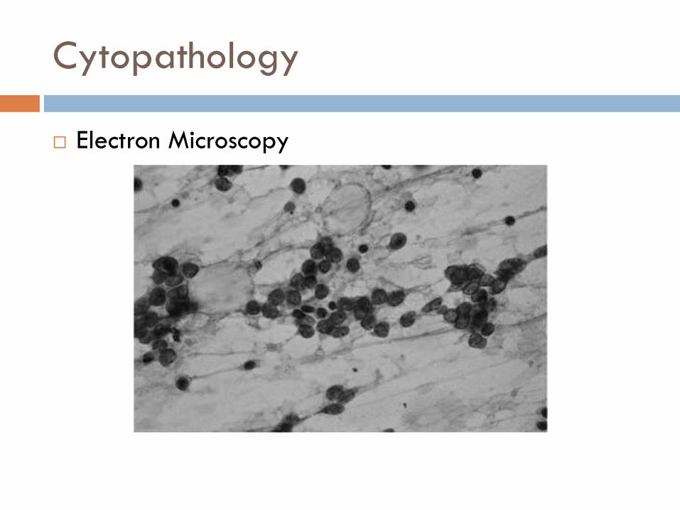
Cytopathology
Electron Microscopy
Cytopathology + Suspicion
In a series by Dr. Paul Nghiem of a 100 patients,
initial biopsy results are as follows:
Histologic Patterns
Three main histologic patterns seen:
Regardless of type, the prognosis remains the same
NEUROENDOCRINE ROLE
Role as Neuroendocrine Tumor
Rarely secretes a hormone
Case reports show an association with Adreno-
Cortico Tropic Hormone (ACTH) or Anti-diuretic
Hormone (ADH)
Postoperative hyponatremia in a patient with ACTH-
producing Merkel cell carcinoma by Anzai S et al.7
Paraneoplastic syndrome of inappropriate antidiuretic
hormone mimicking limbic encephalitis by Blondin NA et
al.8
Role as Neuroendocrine Tumor
Adreno-Cortico Tropic Hormone
Secreted by the anterior pituitary gland
Principal effects are increased production and release
of cortisol
Acts on the adrenal cortex
Often secreted in paraneoplastic syndromes, especially
Small Cell Lung Cancer
Role as Neuroendocrine Tumor
Excess Adreno-corticotropic Hormone (ACTH), aka
Cushing’s disease
Weight gain with central obesity and moon facies
Hirsutism
Purple Striae – thinning of skin
Polyuria
Hypertension
Hyperpigmentation
Role as Neuroendocrine Tumor
Anti-Diuretic Hormone
Typically made in the posterior pituitary gland
Acts on the distal renal tubule and collecting duct in the
nephron
Results in water retention without retaining solute
Often secreted in paraneoplastic syndromes, especially
Small Cell Lung Cancer
Role as Neuroendocrine Tumor
Excess Anti-diuretic Hormone (ADH)
Symptoms
Early findings: Headache + Nausea + Vomiting
Irritability
Confusion
Seizures
Coma
Signs/Findings
Hyponatremia – severe if <120
Concentrated Urine – urine osm >300
STAGING
Staging
Staging Updated
AJCC Staging Manual 2009
Staging Updated
AJCC Staging Manual 2009
Staging Updated
AJCC Staging Manual 2009
Differentiates
between pathologic
negative neck,
clinically negative
neck, and
extracutaneous
involvement
Staging Updated
AJCC Staging Manual 2009
Differentiates
between
microscopic vs
macroscopic/occult
lymph node
metastasis
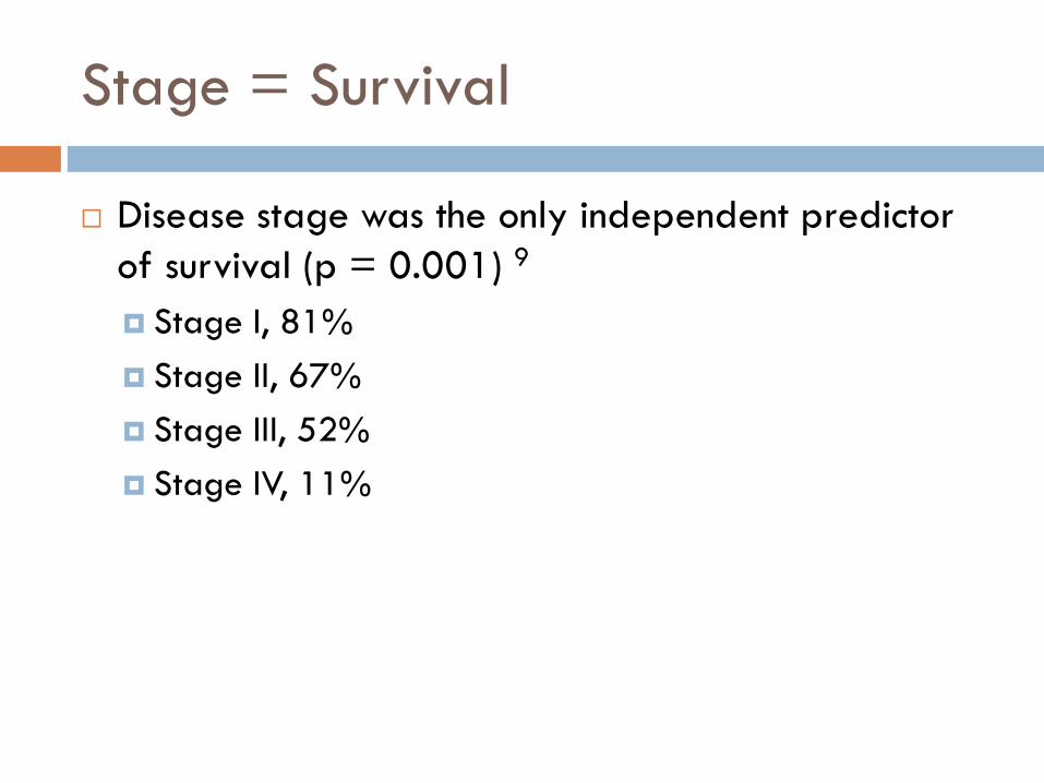
Stage = Survival
Disease stage was the only independent predictor
of survival (p = 0.001) 9
Stage I, 81%
Stage II, 67%
Stage III, 52%
Stage IV, 11%
Survival
Most common stage on Initial
Presentation
Average size of a Head and Neck tumor is 1.59
cm10
So often at onset is stage I by size with a negative
neck
However, survival rates are not that high
Role of CT for Nodal Disease and
Metastasis
CT is poor for evaluating neck disease but good for
distant mets – Gupta et al.15
What prevents better outcome?
Pathologic Diagnosis is tough
What prevents better outcome?
Pathologic Diagnosis is tough
Uncertainty regarding treatment of the clinically
negative neck
Nearly 50% of patients in one series had microscopic
evidence of disease in a clinically negative neck11
What prevents better outcomes?
Pathologic Diagnosis is tough
Uncertainty regarding treatment of the clinically
negative neck
Early Spread / Late Presentation
10-30 % present with nodal involvement12,13,14
6% present with metastasis12,13,14
TREATMENT
Treatment options
Wide Local Excision
Selective Lymph Node Biopsy
Regional Lymph Node Dissection
Radiation Therapy
Chemotherapy
Combined Modality
Treatment
Surgical Treatment is Wide Local Excision
Exact margins – Uncertain – Preferred is > 3 cm
Unlike other non-melanotic skin cancers, Merkel cell
has a high propensity for nodal metastasis
Merkel Cell Carcinoma tends to have nearly 30%
lymph node involvement – greatest among skin cancer
(Melanoma only has 5%)15
Selective/Sentinel Lymph Node Biopsy
Approximately 75% of patients present without
evidence of nodal metastasis.
Schmalbach et al.16 performed SLNB in 10 patients
with Stage I MCC showing 2 with nodal metastasis
and literature review showed 12% false-negative
rate when finding negative SLNB
Current MCC practice guidelines17 by the National
Comprehensive Cancer Network recommend SLNB
for untreated, localized, Stage I disease.
Need for Cytokeratin 20 Stain
Even tougher to ID Merkel cells in Lymph node
Radiation Therapy
Remains controversial
Currently, the National Comprehensive Cancer
Network recommends radiation therapy for:
Primary Tumor Site
In-Transit Lymphatics
Draining Nodal Basins in patients only undergoing a
WLE
Role of Post-Operative XRT
Arguments for Surgery
alone:
Allen et al.9 showed
that the combination of
wide local excision
with negative margins
and a selective neck
dissection results in a
8% local recurrence
rate.
Arguments for XRT:
Medina-Franco et al. –
Review of 11 case
series (1024 patients)
where local recurrence
rate was decreased
from 52.6 % to 10.5
% with use of XRT (p =
0.00001)14
Role of Post-Operative XRT
Local Recurrence is decreased nearly 4 fold by
post-op XRT18
Role of Primary XRT
Use 60 Gray to Primary Site
Arguments for XRT:
Pape et al.18 compared treatment of patients with
Stage I MCC with primary radiation therapy versus
wide local excision with radiation therapy with
comparable rates of regional recurrence
Mortier et al.19 treated a small patient group with
primary XRT with 0% recurrence in 3 years
Summary of Role of XRT
Best Summary:
- XRT as primary treatment may be considered in
elderly who cannot tolerate surgery or have disease in
an area that does not allow wide local excision
- In a clinically positive neck, XRT increases disease-free
survival but not overall survival20
- The role of XRT in a clinically negative neck is the
toughest area to assess but such patients should likely
undergo a selective lymph node biopsy and post-
operative XRT
Can Chemotherapy play a Role?
Currently, not often employed for the following
reasons:
Chemotherapy suppresses immune function
Decreased quality of life in elderly: fatigue, hair loss,
nausea/vomiting
Associated neutropenic fever and sepsis
Suggested Treatment Protocol
HIGHLIGHTS
Clinical Presentation
Summarized by the acronym ‘‘AEIOU’’:
Asymptomatic or nontender
Expanding rapidly
Immune suppressed
Older than 65 years
Ultraviolet exposure or Fair skin
Pathology - Bottomline
Peri-nuclear inclusions on H&E staining
Cytokeratin (20) staining differentiates from small cell lung cancer, melanoma, and lymphoma
Small Blue Cells are noted under H&E stain
First – Rule out Lymphoma and Rule in Carcinoma
Second – Rule out Basal Cell and Squamous Cell Carcinomas
Third – Rule out Metastatic neuroendocrine tumors – particularly small cell lung cancer
Finally – confirm Merkel cell with Cytokeratin 20 stain
Staging
Suggested Treatment Protocol
Bibliography
1. Pellitteri PK, Takes RP, Lewis JS, et al. Merkel Cell Carcinoma of the Head and Neck. Head Neck 2011 Jun; p. 1-10.
2. Nghiem P. Merkel Cell Carcinoma: Diagnosis, Management, and Controversies – Forum 542 in American Academy of Dermatology Annual Meeting 2008
3. Akhtar S, Oza KK, Wright J. Merkel cell carcinoma: report of 10 cases and review of the literature. J Am Acad Dermatol 2000; 43:755–767.
4. Agelli M, Clegg LX. Epidemiology of primary Merkel cell carcinoma in the United States. J Am Acad Dermatol 2003; 49:832–841.
5. Engels EA, Frisch M, Goedert JJ, Biggar RJ, Miller RW. Merkel Cell Carcinoma and HIV Infection. Lancet. 2002 Feb 9;359(9305):497-8.
6. Miller et al., Cancer Epidemiol Biomarkers Prev, 1999, using SEER.
7. Anzai S, Sato T, Takayasu S, Asada Y, Terashi H, Takasaki S. Post-operative Hyponatremia in a patient with ACTH-producing Merkel Cell Carcinoma. J Dermatol 2000 Jun; 27(6) : 397-400.
9. Allen PJ, Bowne WB, Jaques DP, Brennan MF, Busam K, Coit DG. Merkel cell carcinoma: prognosis and treatment of patients from a single institution. J Clin Oncol. 2005 Apr 1;23(10):2300-9.
10. Dancey AL, Rayatt SS, Soon C, Ilchshyn A, Brown I, Srivastava S. Merkell Cell Carcinoma: A report of 34 cases and literature review. J Plast Reconstr Aesthet Surg 2006; 59 : 1294-1299.
11. Goepfert H, Remmler D, Silva E, Wheeler B. Merkel cell carcinoma (endocrine carcinoma of the skin) of the head and neck. Arch Otolaryngol 1984; 110 : 707-712.
13. Reichgelt BA, Visser O. Epidemiology and survival of Merkel cell carcinoma in the Netherlands. A population-based study of 808 cases in 1993-2007. Eur J Cancer. 2011 Mar;47(4):579-85.
14. Medina-Franco H, Urist MM, Fiveash J, Heslin MJ, Bland KI, Beenken SW. Multimodality treatment of Merkel cell carcinoma: case series and literature review of 1024 cases. Ann Surg Oncol. 2001 Apr;8(3):204-8.
15. Gupta SG, Wang LC, Peñas PF, Gellenthin M, Lee SJ, Nghiem P. Sentinel lymph node biopsy for evaluation and treatment of patients with Merkel cell carcinoma: The Dana-Farber experience and meta-analysis of the
16. Schmalbach CE, Lowe L, Teknos TN, Johnson TM, Bradford CR. Reliability of sentinel lymph node biopsy for regional staging of head and neck Merkel cell carcinoma. Arch Otolaryngol Head Neck Surg. 2005
Jul;131(7):610-4.
17. National Comprehensive Cancer Network Inc. NCCN Clinical Practice Guidelines in Oncolocy, v.1.2004.
18. Pape E, Rezvoy N, Penel N, Salleron J, Martinot V, Guerreschi P, Dziwniel V, Darras S, Mirabel X, Mortier L. Radiotherapy alone for Merkel cell carcinoma: a comparative and retrospective study of 25 patients. J Am
20. Lawenda BD, Arnold MG, Tokarz VA, Silverstein JR, Busse PM, McIntyre JF, Deschler DG, Baldini EH, Kachnic LA. Analysis of radiation therapy for the control of Merkel cell carcinoma of the head and neck based on
36 cases and a literature review. Ear Nose Throat J. 2008 Nov;87(11):634-43.