Preface This is the 54th report of a series of workshops organised by the European Centre for the Validation of Alternative Methods (ECVAM). The main objective of ECVAM, as defined in 1993 by its Scientific Advisory Committee, is to promote the scientific and regulatory acceptance of alternative methods which are of importance to the biosciences, and which reduce, refine or replace the use of labo- ratory animals. One of the first priorities set by ECVAM was the implementation of procedures that would enable it to become well informed about the state-of-the-art of non-animal test development and validation, and of opportunities for the possible incorporation of alternative methods into regula- tory procedures. It was decided that this would be best achieved through a programme of ECVAM workshops, each addressing a specific topic, and at which selected groups of independent international experts would review the current status of various types of in vitro tests and their potential uses, and make recommendations about the best ways for- ward. A workshop on Metabolism: a bottleneck in in vitro toxicological test development, was held at ECVAM on 26–29 January 2004, with participants Metabolism: A Bottleneck in In Vitro Toxicological Test Development The Report and Recommendations of ECVAM Workshop 54 1 Sandra Coecke, 1 Hans Ahr, 2 Bas J. Blaauboer, 3 Susanne Bremer, 1 Silvia Casati, 1 Josè Castell, 4 Robert Combes, 5 Raffaella Corvi, 1 Charles L. Crespi, 6 Michael L. Cunningham, 7 Greetje Elaut, 8 Brighitta Eletti, 1 Andreas Freidig, 9 Alessandra Gennari, 1 Jean-François Ghersi-Egea, 10 Andre Guillouzo, 11 Thomas Hartung, 1 Peter Hoet, 12 Magnus Ingelman-Sundberg, 13 Sharon Munn, 14 Walter Janssens, 15 Bernhard Ladstetter, 16 David Leahy, 17 Anthony Long, 18 Annarita Meneguz, 19 Mario Monshouwer, 20 Siegfried Morath, 21 Fred Nagelkerke, 22 Olavi Pelkonen, 23 Jessica Ponti, 1 Pilar Prieto, 1 Lysianne Richert, 24 Enrico Sabbioni, 1 Beatrice Schaack, 25 Winfried Steiling, 26 Emanuela Testai, 27 Joan-Albert Vericat 28 and Andrew Worth 14 1 ECVAM, Institute for Health and Consumer Protection, European Commission Joint Research Centre, Ispra, Italy; 2 Bayer AG, PH-PD Toxicology, Wuppertal, Germany; 3 Institute for Risk Assessment Sciences, Division of Toxicology, Unit of Biochemical Toxicology, Utrecht University, Utrecht, The Netherlands; 4 Hospital Universitari La Fe, Valencia, Spain; 5 FRAME, Nottingham, UK; 6 Gentest, BD Biosciences, Woburn, MA, USA; 7 National Center for Toxicogenomics & National Toxicology Program, National Institutes of Health, National Institute of Environmental Health Sciences, NC, USA; 8 Vrije Universiteit Brussel, Department of Toxicology, Brussels, Belgium; 9 TNO Nutrition and Food Research, Department of Biomolecular Sciences, Zeist, The Netherlands; 10 INSERM U433, Faculté de Médecine Laënnec, Lyon, France; 11 INSERM U456, Rennes Cedex, France; 12 KU. Leuven, Laboratorium voor Pneumologie (Lung Toxicologie), Leuven, Belgium; 13 Karolinska Institute, Molecular Toxicology, Stockholm, Sweden; 14 ECB, Institute for Health and Consumer Protection, European Commission Joint Research Centre, Ispra, Italy; 15 Scientific Institute for Public Health, Division of Toxicology, Brussels, Belgium; 16 Merck KGaA, Institute of Drug Metabolism and Pharmacokinetics, Grafing, Germany; 17 Cyprotex PLC, Macclesfield, Cheshire, UK; 18 LHASA Limited, Department of Chemistry, University of Leeds, Leeds, UK; 19 Department of Drug Research and Evaluation, Biochemical Pharmacology and Technical Scientific Advice Coordination Unit, Rome, Italy; 20 Investigative ADME , Pharmacokinetics, Dynamics & Metabolism, Pharmacia, Gruppo Pfizer Inc., Nerviano, Italy; 21 University of Konstanz, Biochemical Pharmacology, Konstanz, Germany; 22 LACDR, Leiden University, Gorlaeus Laboratories, Leiden, The Netherlands; 23 Department of Pharmacology and Toxicology, University of Oulu, Finland; 24 University of Besancon, UFR Medicine, Pharmacie, Besacon, France; 25 CEA Grenoble-DRDC, Lboratoire BioPuces, Grenoble Cedex, France; 26 Product Safety, Toxicology, Dusseldorf, Germany; 27 Istituto Superiore di Sanita, Department of Environment and Primary Prevention- Mechanisms of Toxicity Unit, Rome, Italy, 28 NeuroPharma, Cantos (Madrid), Spain ATLA 34, 49–84, 2006 49 Address for reprints: ECVAM, TP 580, Institute for Health and Consumer Protection, European Commission Joint Research Centre, 21020 Ispra (VA), Italy. 1 This document represents the agreed report of the authors as individual scientists.
Transcript
Preface
This is the 54th report of a series of workshopsorganised by the European Centre for theValidation of Alternative Methods (ECVAM). Themain objective of ECVAM, as defined in 1993 by itsScientific Advisory Committee, is to promote thescientific and regulatory acceptance of alternativemethods which are of importance to the biosciences,and which reduce, refine or replace the use of labo-ratory animals. One of the first priorities set byECVAM was the implementation of procedures thatwould enable it to become well informed about the
state-of-the-art of non-animal test development andvalidation, and of opportunities for the possibleincorporation of alternative methods into regula-tory procedures. It was decided that this would bebest achieved through a programme of ECVAMworkshops, each addressing a specific topic, and atwhich selected groups of independent internationalexperts would review the current status of varioustypes of in vitro tests and their potential uses, andmake recommendations about the best ways for-ward.
A workshop on Metabolism: a bottleneck in invitro toxicological test development, was held atECVAM on 26–29 January 2004, with participants
Metabolism: A Bottleneck in In Vitro Toxicological TestDevelopment
The Report and Recommendations of ECVAM Workshop 541
Sandra Coecke,1 Hans Ahr,2 Bas J. Blaauboer,3 Susanne Bremer,1 Silvia Casati,1 Josè Castell,4Robert Combes,5 Raffaella Corvi,1 Charles L. Crespi,6 Michael L. Cunningham,7 Greetje Elaut,8Brighitta Eletti,1 Andreas Freidig,9 Alessandra Gennari,1 Jean-François Ghersi-Egea,10 AndreGuillouzo,11 Thomas Hartung,1 Peter Hoet,12 Magnus Ingelman-Sundberg,13 Sharon Munn,14
Walter Janssens,15 Bernhard Ladstetter,16 David Leahy,17 Anthony Long,18 Annarita Meneguz,19
Mario Monshouwer,20 Siegfried Morath,21 Fred Nagelkerke,22 Olavi Pelkonen,23 Jessica Ponti,1Pilar Prieto,1 Lysianne Richert,24 Enrico Sabbioni,1 Beatrice Schaack,25 Winfried Steiling,26
Emanuela Testai,27 Joan-Albert Vericat28 and Andrew Worth14
1ECVAM, Institute for Health and Consumer Protection, European Commission Joint Research Centre, Ispra,Italy; 2Bayer AG, PH-PD Toxicology, Wuppertal, Germany; 3Institute for Risk Assessment Sciences, Division ofToxicology, Unit of Biochemical Toxicology, Utrecht University, Utrecht, The Netherlands; 4Hospital UniversitariLa Fe, Valencia, Spain; 5FRAME, Nottingham, UK; 6Gentest, BD Biosciences, Woburn, MA, USA; 7NationalCenter for Toxicogenomics & National Toxicology Program, National Institutes of Health, National Institute ofEnvironmental Health Sciences, NC, USA; 8Vrije Universiteit Brussel, Department of Toxicology, Brussels,Belgium; 9TNO Nutrition and Food Research, Department of Biomolecular Sciences, Zeist, The Netherlands;10INSERM U433, Faculté de Médecine Laënnec, Lyon, France; 11INSERM U456, Rennes Cedex, France; 12KU.Leuven, Laboratorium voor Pneumologie (Lung Toxicologie), Leuven, Belgium; 13Karolinska Institute, MolecularToxicology, Stockholm, Sweden; 14ECB, Institute for Health and Consumer Protection, European CommissionJoint Research Centre, Ispra, Italy; 15Scientific Institute for Public Health, Division of Toxicology, Brussels,Belgium; 16Merck KGaA, Institute of Drug Metabolism and Pharmacokinetics, Grafing, Germany; 17CyprotexPLC, Macclesfield, Cheshire, UK; 18LHASA Limited, Department of Chemistry, University of Leeds, Leeds, UK;19Department of Drug Research and Evaluation, Biochemical Pharmacology and Technical Scientific AdviceCoordination Unit, Rome, Italy; 20Investigative ADME , Pharmacokinetics, Dynamics & Metabolism, Pharmacia,Gruppo Pfizer Inc., Nerviano, Italy; 21University of Konstanz, Biochemical Pharmacology, Konstanz, Germany;22LACDR, Leiden University, Gorlaeus Laboratories, Leiden, The Netherlands; 23Department of Pharmacologyand Toxicology, University of Oulu, Finland; 24University of Besancon, UFR Medicine, Pharmacie, Besacon,France; 25CEA Grenoble-DRDC, Lboratoire BioPuces, Grenoble Cedex, France; 26Product Safety, Toxicology,Dusseldorf, Germany; 27Istituto Superiore di Sanita, Department of Environment and Primary Prevention-Mechanisms of Toxicity Unit, Rome, Italy, 28NeuroPharma, Cantos (Madrid), Spain
ATLA 34, 49–84, 2006 49
Address for reprints: ECVAM, TP 580, Institute for Health and Consumer Protection, European Commission JointResearch Centre, 21020 Ispra (VA), Italy.1This document represents the agreed report of the authors as individual scientists.
from academia, national organisations, regulatorybodies and industry. The aim of the workshop was todiscuss the use of metabolising systems in in vitrotoxicity test methods (i.e. the need to take account ofbiotransformation, and the methods available), togive some examples of how chemicals can bescreened and tested for their susceptibilities tometabolic activation, and to consider how suchinformation can also be used, in conjunction withcomputer modelling approaches, for providing infor-mation for use in hazard identification and riskassessment. In addition to plenary sessions, theworkshop was organised into three working groups,to discuss: a) biokinetics and integration of tech-niques; b) metabolism and methodology; and c) target organ and target system toxicity. Somerecommendations are listed at the end of this report,and these should be considered for future imple-mentation, to encourage the incorporation of bio-transformation into in vitro toxicity test methods.
Introduction
The nature of biotransformation
Biotransformation is the process whereby a sub-stance, usually a foreign compound (xenobiotic), ischemically transformed in the body to form ametabolite or a variety of metabolites. This termwill be used throughout this report to distinguishthis process from other endogenous metabolic phe-nomena involved in controlling cellular functions,collectively known as metabolism.
Biotransformation is one of the main factorswhich influence the transport and partitioning of achemical into and within the body, its toxicity, andits rate and route of elimination. All chemicals arepotentially susceptible to biotransformation ofsome sort, and all cells and tissues are potentiallycapable of biotransforming xenobiotics, althoughthe major sites of such biotransformation are theliver and the entry portals of the body (1). Species,strain, sex, individual, organ, and age-specific dif-ferences, and even differences between cells in thesame organ, can have important effects on the bio-transformation of xenobiotics. Exposure to drugs,occupational and industrial chemicals, or environ-mental pollutants, can lead to either the inductionor the inhibition of biotransformation. Moreover,drug–drug, drug–toxicant and drug–food interac-tions, due to the induction and inhibition of specificbiotransformation enzymes, are one of the mostimportant factors which complicate drug therapyand cause adverse drug reactions.
The main phases of biotransformation
Biotransformation is usually divided into two mainphases, phase I and phase II. Phase I is usuallyoxidative (e.g. hydrolysis, although reductivemetabolism and non-redox reactions of functionali-sation can also occur), and has a major protectivefunction in rendering lipophilic molecules morepolar and more readily excretable. In phase II, oftenreferred to as detoxification, such oxidised moietiesare subsequently conjugated with highly polar mol-ecules, such as glucuronic acid, before they areexcreted (Table 1). Phase I oxidation reactions arecatalysed mainly by the many isoforms of thehaeme-thiolate cytochrome P450 (CYP) supergenefamily of enzymes (the so-called mixed function oxi-dases [MFOs]), which collectively have very widesubstrate specificity (2, 3). In humans and othermammalian species, the CYP1, CYP2 and CYP3isozyme families are primarily associated with thephase I metabolism of exogenous compounds.Flavin monoamine oxidase (FMO) can also oxidisexenobiotics (4), although to a lesser extent thanMFO enzymes. Phase II conjugation of the oxidisedmoiety with molecules such as glucuronic acid, sul-phate, methionine, cysteine or glutathione, is catal-ysed by a variety of isozymes of N-acetyltransferase(NAT), uridine diphosphoglucuronosyl transferase(UGT), sulphotransferases (SULTs), and glu-tathione S-transferase (GST; 5). These enzymes,listed in Table 2, occur variously in the cytosol orare bound to microsomal proteins, and their struc-tures and functions have been reviewed elsewhere(6).
Supergene families of isoforms of phase IIenzymes also exist, which show different affinitiesand therefore selectivity toward different sub-strates. For example, the SULTs are a supergenefamily of cytosolic enzymes, for which some elevendistinct isoforms of the enzyme are known inhumans, and these differ markedly in their catalyticactivities for the same substrates and in their tissuelocations (7; Table 2).
The practical aspects of studying CYP activitieshave been comprehensively reviewed by Phillipsand Shephard (8), and general techniques for incor-porating metabolism into in vitro toxicity assayshave been described by Castell and Gomez-Lechon(9), Plant (10), Ekins (11), and in reference 12.
At least 50 major enzymes catalyse the biotrans-formation of xenobiotic chemicals, and many moreare responsible for biotransforming at least a fewchemicals. The interactions of chemicals withenzymes are characterised by affinity, turnovernumber, intrinsic activity, and, at a more generallevel, whether or not the reaction is rate-limiting inthe metabolism of a given chemical, or a moreminor one.
More recently, it has been suggested that a further,phase III, stage of metabolism should be recognised,
50 S. Coecke et al.
ECVAM Workshop 54: metabolism 51
Table 1: A comparison of the main enzymes involved in biotransformation
Enzyme classes/enzymes Types of drugs metabolised Sources available/tissue distribution
Cytochrome P450 enzymes Practically 90% of all drug substances Hepatocytes most versatile and (about 13 “drug-metabolising” comprehensive; other tissues and cell types, enzyme forms) selectively
CYP1A1 Few pharmaceuticals, principally Recombinant enzyme; mainly extra-hepatic polycyclic aromatic hydrocarbons and expression (e.g. placenta from smokers)other carcinogens
CYP3A4 A majority (> 50%) of all clinically used Hepatocytes (genetically engineered)drugs
Flavin-monooxygenases Compounds with secondary and tertiary Hepatocytes most versatile and (FMO; 5 forms) amines or sulphydryl groups comprehensive
Alcohol/aldehyde dehydrogenases Various compounds with alcohol and Many tissues (cytosol location) and oxidases aldehyde functions (ethanol)
Monoamine oxidase Selegiline, moclobemide Many tissues (mitochondrial location)
Esterases/hydrolases/peptidases Compounds with cleavable ester/amide Many tissues, including blood and blood cellsbond (procaine, succinylcholine, lidocaine)
Reductases Many substances with azo, nitro and Present in hepatocytes (cytosol, some in carbonyl functions (chloramphenicol, microsomes)naloxone); importance not well characterised
UDP-glucuronosyl transferases; Most drugs with suitable O-, S- and Subcellular systems need UDPGA;(UGT) N-functional groups (morphine, diazepam, hepatocytes most versatile and glucuronide conjugation paracetamol) comprehensive (microsomes)
Sulphotransferases (SULT); Phenols, alcohols, aromatic amines Hepatocytes most versatile and sulphate conjugation (paracetamol, methyldopa) comprehensive (cytosol); subcellular systems
need 3-phosphoadenosine-5-phosphosulphate (PAPS)
GSH transferases (GST); Epoxides, arene oxides, nitro groups, Hepatocytes most versatile and glutathione conjugation hydroxylamines (ethacrynic acid) comprehensive (cytosol, microsomes)
Methyl transferases; Catecholamines, phenols, amines (L-dopa, Various tissues (cytosol)methylation thiouracil)
relating to the excretion of conjugates and involvingATP-dependent plasma membrane transporters (forexample, p-glycoprotein, MRP2, and organic aniontransporting polypeptides [OATPs; 13]), which areinvolved in the cellular uptake of drugs.
The role of biotransformation in the activation of xenobiotics
The biotransformation of xenobiotics can result inthe formation of highly reactive chemicals that are
known to play a role in toxicity, particularly inmutagenesis and carcinogenesis (a process knownas bioactivation). However, there are numerousexamples of a requirement for bioactivation forother toxicity endpoints and chemicals, includingendocrine disruptors such as methoxychlor and vin-clozolin, and neurotoxicants such as the organo-phosphorothionate (OPT) pesticides (14), andparacetamol, carbamazipine, tienylic acid andhalothane. Also, the resulting metabolites mightinteract with enzymes involved in the biosynthesisof endogenous compounds, with receptors, or with
Table 2: Examples of drug metabolising enzymes available for incorporation into in vitro assays
Isoenzymes Types of chemicals metabolised
Cytochrome P450 enzymes Practically 90% of all chemical substancesCYP1A1 PAHCYP1A2 Aromatic amines, amino acid pyrolysis productsCYP1B1 PAH, DMBA, oestrogensCYP2A6 Nitrosamines, aflatoxins, nicotine, cotinine, butadien
CYP2E1 Small molecular substances, solvents, benzene, styrene, nitosaminesCYP3A4 A majority of drugs, aflatoxin B1, 1-nitropyreneCYP3A5 Same as 3A4CYP3A7 Same as 3A4CYP4A9/11 Fatty acidsCYP4B
Flavin-monooxygenases (FMO) Oxidation of nucleophilic N, S and P heteroatoms of xenobioticsFMO1 ChlorpromazineFMO2 NicotineFMO3 NicotineFMO4FMO5
Glucuronide conjugation Most chemicals with suitable O-, S- and N-functional groupsUGT1A1 Bilirubin, catecholoestrogens, opioidsUGT1A3 Amines, flavonoids, 7-hydroxycoumarins, opioidsUGT1A4 Amines, plant steroids, androgens, progestinsUGT1A6 Small planar phenols, paracetamol, p-nitrophenol
UGT1A7 Phenols, hydroxylated PAHs, coumarinsUGT1A8 Opioids, oestrogens, propofol, primary and secondary aminesUGT1A9 Large bulky phenols, propofol, primary and secondary amines, benzodiazepins,
signal transduction mechanisms, so the generationof such metabolites may be important (e.g. in thecase of endocrine disruption caused by steroidogen-esis [14]).
During phase I reactions, in particular, highlyreactive and potentially toxic intermediates can begenerated. Much evidence exists for the bioactiva-tion of many chemicals and drugs, and much of thisreport is concerned with such effects (for recentreviews, see 15, 16). However, phase II metabolismis also capable of activating chemicals to producehighly reactive electrophilic intermediates. Forexample, it is well established that acyl glu-curonides, arising from the glucuronidation of awide range of carboxylic acid substrates, can cova-lently bind to macromolecules; this could well leadto toxicity in humans following the administrationof carboxylate drugs (16). Also, several 1,2-dihaloalkanes and 1,2-dihaloethanes, such as 1,2-dichloromethane, are susceptible to glutathionetransferase-dependent bioactivation as a result ofthe formation of S-(halomethyl)glutathione andglutathione episulphonium ions as electrophiles(17). In addition, sulphotransferases can bioactivateseveral chemicals via O-sulphonation (for example,hydroxymethyl polycyclic aromatic hydrocarbonsand N-hydroxy derivatives of carcinogenic ary-lamines and heterocyclic amines [18]). These strongelectrophiles arise because the sulphate group iselectron-withdrawing and is susceptible to cleavagefrom the rest of the molecule, leaving behind ahighly charged cation (7). Also, epoxide hydrolasescan activate certain molecules possessing epoxidegroups, either formed as a result of phase I oxida-tion (for example, during the biotransformation ofbenzo[a]pyrene) or present in the parent compound(19). Chemicals which have reactive and potentiallytoxic conjugates include morphine, safrole, and cer-tain nitro and chlorinated aromatic compounds.There is an important need to take account of thepossible biotransformation of chemicals in in vitroassays, since the absence of this crucial determi-nant of toxicity could give rise to false-positive data(due to lack of detoxification) or to false-negativedata (due to lack of activation).
Toxicity in whole animals can be due to the par-ent compound, to metabolites formed at the site ofentry into the body (such as lung, gut and skin)and/or in the liver, and which are sufficiently stableto reach other target organs (systemic toxicity), orto metabolites formed in the target organ itself.Substances that are metabolised in, for example,the liver, may be subjected to further metabolismonce they reach their target organs.
Many instances exist of metabolites generated atone site in the body and active at another targetsite. Several examples of this for carcinogenicitywill be given: benzo[a]pyrene, β-naphthylamine,heterocyclic amines, trichloroethylene and ecstasy.After hepatic metabolism, benzo[a]pyrene binds to
macromolecules in distal perfused lungs (20). Also,arylamines, like β-naphthylamine, are directlydetoxified by NAT isozymes NAT1 and NAT2 in theliver, leading to inactive amides. Alternatively,these arylamines can be bioactivated by CYP1A2,primarily to hydroxylated reactive products, whichare inactivated by UDPGT-mediated conjugation,mainly with glucuronic acid. The arylamines canalso be acetylated by NATs, although this is a lessprominent activation route. The resulting hydroxy-arylamines can enter the blood system, generally asglucuronides and undergo renal filtration, arrivingat the urinary bladder where they are deconjugatedby β-glucuronidases to re-form their respectivehydroxy-arylamines, the ultimate carcinogens. Inindividuals who are slow acetylators, much higheramounts of P450-mediated hydroxy-arylamines areformed, due to competition between NAT andCYP1A2 which is in favour of the latter. Thisincreases the risk of bladder cancer in such individ-uals (21).
The heterocyclic amines are potent mutagens,formed during the cooking of meat and fish. In theliver, the major metabolic reaction is N-hydroxyla-tion catalysed by hepatic CYPs, eventually leadingto N-glucuronidation by UDPGT. The resulting N-hydroxy-glucuronides are transported via the bileto the colonic lumen. The colonic mucosa containssignificant amounts of NATs, able to efficientlycatalyse the O-acetylation of N-hydroxy derivativesto reactive N-acetoxyarylamines, which are respon-sible for DNA adduct formation and colon cancer.This outcome occurs more frequently in individualswho are fast acetylators (22).
Trichloroethylene is also activated in the liverbefore exerting its carcinogenicity in another tissue,in this case, the kidney. OPT pesticides are acti-vated to toxic compounds in the liver and theninhibit AChE in the blood or brain, although a localcerebral metabolic activation step might also occur(23, 24). Lastly, ecstasy exerts its serotoninergicneurotoxicity as a result of its conjugated metabo-lites, the formation of which is initiated systemi-cally (25).
Biotransformation and toxicokinetics
Modulation of transport
The metabolic stability of a molecule and the rate ofits biotransformation can affect the potential for itsbioaccumulation, and whether it is likely to becomemore or less toxic, without it necessarily being con-verted into a more reactive metabolite. For exam-ple, topically applied substances can be renderedmore absorbable and therefore potentially moretoxic by being metabolised in the skin, so that theypass through it to exert their inherent toxicity
ECVAM Workshop 54: metabolism 53
locally and/or systemically (26, 27). These processescan be quantified during toxicokinetic investiga-tions.
However, the ability of the metabolites of a chem-ical that have been formed as a result of biotrans-formation at some site in the body, to inducetoxicity in a target organ elsewhere, is also depend-ent on the ability of the metabolites to be trans-ported to that target organ and to enter it. Thesephenomena are compound-dependent, and can alsodepend on the presence of the correct transporters,the permeability of membranes to passive diffusionof reactive intermediates, and/or the stereo-iso-meric form of the substrate, due to the stereoselec-tivity of biotransformation enzymes (28).
The blood–brain, placental and blood–testis bar-riers are usually efficient at excluding the metabo-lites of many chemicals, but nevertheless arerefractory to others. The passage of reactivemetabolites through cell membranes and across tis-sues can be impeded by their interactions withdiverse molecular targets. Also, metabolism canalter the structure of the molecule, resulting in themodification of its ability to undergo passive trans-port or transport mediated by an active system. Inview of their large total biomass and strategic posi-tions at the interface between the bloodstream andthe tissues, epithelia and endothelia have the poten-tial to act as important barriers with respect to themetabolism of substances before they enter thebloodstream.
There are no barriers that can exclude all reac-tive metabolites or parent compounds, if the phys-icochemical characteristics of the chemical and itsmetabolites are favourable for uptake. The mostrelevant physicochemical parameters of a moleculein this respect are: molecular size and shape; degreeof ionisation; and the relative lipid solubilities of itsionised and non-ionised forms. Other importantparameters include protein binding capacity, andrecognition by efflux transporters such as mul-tidrug resistance transporters. Clearly, these prop-erties will differ with respect to the nature of theparent compound, and for each of its metabolites.Moreover, biotransformation can generate mole-cules with a greatly enhanced ability to penetratebiological systems. A good example is the reductivemetabolism of azo dyes that are sulphonated onlyon one side of the azo bridge. This results in an aro-matic amine with reduced water solubility and amolecule of reduced size in comparison with theparent chemical (29).
Metabolism-mediated target organ toxicity
Differential metabolism between target organs cangive rise to specific target organ toxicity (metabo-lism-mediated toxicity), and can be due to varia-tions in: a) total biotransformation capacity; b)
expression levels of biotransformation enzymes andisoenzymes; and c) inducibilities of enzymes.Variations occur in the distribution of metabolisingenzymes in different tissues, and also between dif-ferent cells in the same organ, particularly in thecase of extra-hepatic tissues such as the lung, gas-tro-intestinal tract, the pancreas and the brain, andespecially with respect to their CYP and phase IIisozyme contents (30–37).
The target organ-specific effects of some carcino-gens are often attributed to differential metabolicactivation. However, this might not be true for sub-stances such as non-genotoxic carcinogens, whichcan exhibit high levels of tissue specificity andwhich often induce tumours at cytotoxic concentra-tions in conjunction with the induction of sustainedcellular proliferation (38). In such cases, differencesin receptor distribution between organs could bemore important than differential biotransform-ation.
A good example of metabolism-mediated species-specific toxicity concerns the carcinogenicity in thelungs of mice of trichloroethylene (TRE), due toTRE activation by biotransformation enzymes inthe Clara cells. However, this observation is not rel-evant to humans, in which there are smaller num-bers of Clara cells that are not metabolicallycompetent in this respect (39).
The role of biotransformation in determining tox-icity for other endpoints is less well understood, insituations where the site and/or mechanisminvolved is specific to a certain target tissue (forexample, neurotransmitter receptors in the brain).However, the distribution, activities and roles ofCYP isoenzymes and other biotransformationenzymes in the functioning of the central andperipheral nervous systems has begun to be investi-gated (40). There are recent indications thatCYP2D6, present in some specific brain areas andresponsible for many CNS-active drug biotransfor-mations (41–43), could be involved in the metabo-lism of endogenous substances in the brain (44).Also, CYP2 isoenzymes (including rat/humanCYP2D1/6, CYP2E1 and CYP2B1/6) have beenshown to be unevenly distributed between brainregions and brain cell types, and their regulationdiffers from that of the same isozymes in the liver.Therefore, while it is unlikely that CYPs in thebrain contribute significantly to the overall bio-transformation of xenobiotics, they might con-tribute to the identification of specific areas in thebrain which differ in drug and metabolite levels(36). It is noteworthy that the substrates for theseisoenzymes include psychotropic drugs, opioids andneurotoxins. The same reasoning could apply toother biotransformation pathways, such as glu-tathione-dependent activation/inactivation in thebrain (45, 46).
Different tissues show a hierarchy in relation totheir overall biotransformation potentials (liver >
54 S. Coecke et al.
kidney > lung > gastro-intestinal tract > nasalepithelium > placenta > brain). Nevertheless, cor-relation with route of exposure/administration andnature of the xenobiotic must be taken intoaccount, in order to assess the relevance of any bio-transforming hierarchy in determining organ sus-ceptibility to toxic effects. However, such ahierarchy would only be useful in a general sense,and its relevance would very much depend on theroute of administration or exposure, and the natureof the potentially toxic substance. For example,when a volatile chemical is inhaled, metabolism inthe nasal epithelium and in the lung is likely to beeven more important than biotransformation in theliver.
Differences in biotransformation between different organisms
Species differences
There are marked species differences in the expres-sion and catalytic activities of the CYPs that bio-transform xenobiotics. These differences can beboth quantitative and qualitative, although the for-mer are more frequently characterised by varyingenzyme kinetics, which can substantially differbetween rats and mice, as well as between rodentsand humans. This is especially notable between thecommonly-used experimental rodent models, andbetween these models and humans. This is a prob-lem, particularly when assessing the toxicity ofchemicals such as drugs, to which humans areintentionally exposed, so, ideally, human biotrans-formation should be investigated in the preclinicalphase. One solution has been to develop and usetransgenic mice expressing human CYPs, such asCYP2D6, and CYP3A4 (47, 48). These isozymes areexpressed in amounts comparable to, or higherthan, their corresponding levels in human tissues.However, a much more flexible approach is toexploit the great advantage of in vitro systems byvarying the source of metabolism when incorporat-ing biotransformation into in vitro toxicity tests,which is much more easily achieved than could everbe made possible by generating transgenic mice orby using a range of different species.
When qualitative differences in metabolism exist,there can be very far reaching consequences forobtaining correct toxicity predictions, if an inappro-priate species is used (49). An example of a qualita-tive difference in metabolism is the lack ofN-acetylation in the dog, a very important consid-eration when selecting a second species for deter-mining the toxicity of aromatic amines, for instance(50). Many other examples of species-specific bio-transformation were listed and discussed byBeasley (51).
All enzymes which metabolise xenobiotics displaysome variability (52, 53). For example, it is gener-ally found that CYP enzyme levels in the liver dis-play variabilities ranging from 5-fold to 100-fold.Much of this variability can be traced to geneticbackground, polymorphisms and single nucleotidepolymorphisms (SNIPs) at the level of structuraland/or regulatory sequences of xenobiotic metabo-lism (XME) genes. Environmental and host factorsalso contribute to the variability, sometimes to aneven greater extent. Thus, differential distributionand the presence of the various isozymes, as well asthe intracellular receptors for their induction, aremainly responsible for polymorphisms betweenindividuals and the specific metabolic activitiesmentioned above (54).
Particular isozymes are associated with themetabolism and activation of specific chemicalgroups. For example, CYP1A1 is very active inmetabolising polycyclic aromatic hydrocarbons,CYP1B1 and CYP1A2 preferentially metabolisearomatic amines, and CYP2E1 metabolises lowmolecular weight chemicals, whereas CYP3A4metabolises larger molecules. CYP2D6 is especiallyimportant in human drug metabolism. However,the CYP 2C, 2B and 2D subfamilies seem to con-tribute very little to the bioactivation of chemicalsor drugs (15).
An example often given for a CYP isozyme-spe-cific biotransformation in toxicity, is the bioactiva-tion by CYP2D6 of the tobacco-specific nitrosamine,NNK (55). However, this substance is also activatedby other CYPs, so this is unlikely to be the basis ofthe frequently-quoted possible explanation for theapparent association between CYP2D6 polymor-phism and lung cancer.
The existence of strain/species/gender/ethnic/agedifferences in susceptibility to toxicity is a wellestablished phenomenon, and can be influenced bydisease status and stress levels in the host animals(56, 57). For example, these parameters are knownto affect the carcinogenicity of chemicals, due tovariations in metabolic activation and detoxifica-tion, as well as differences in the formation andquenching of reactive oxygen species, and in theaccumulation of parent compounds and theirmetabolites in tissues. Interstrain differences inbiotransformation can give rise to discrepant toxic-ity data. Variation in the renal toxicity of chloro-form is another example of the effects of suchdifferences in biotransformation (58).
In addition, differential cytochrome expressionoccurs during embryonic differentiation, and thismight be of importance when trying to predictreproductive toxicity in vitro (59, 60). Moreover,general health status can modulate the levels ofCYPs, and such effects could be important whenperforming experiments for specific applications; anexample is the effect of inflammatory mediators onP450 levels (61).
ECVAM Workshop 54: metabolism 55
Conclusions
In conclusion, the following elements of biotrans-formation and biokinetics are all essential elementsof toxicological evaluation, hazard identificationand risk assessment: a) determining the suscepti-bility of a chemical to metabolism (its metabolic sta-bility); b) identifying the principal metaboliteslikely to be generated; c) identifying the targetorgans which could be involved; d) determiningother differences in metabolism (such as inter-species differences, mentioned above); e) ascertain-ing variations in the transport of parent chemicalsand their metabolites; and f) identifying the pres-ence of chemicals and/or their metabolites that canact as enzyme inducers or inhibitors, and their abil-ities to act as substrates for the different isozymesinvolved in biotransformation.
The Relative Importance of theBiotransformation ofPharmaceuticals and IndustrialChemicals
Safety testing in the pharmaceutical and chemicalindustries differs in a number of importantrespects. In the former, there has traditionally beena much greater emphasis on the use of rapid in vitroscreening techniques applied in a non-regulatorycontext at a very early stage of drug development.This stage coincides with the availability of manyclosely-related candidate molecules, and is whenrelatively high false-positive prediction rates, andconcomitant attrition rates, are not a major prob-lem. In addition, metabolism studies are particu-larly important in the development of new drugs,since the vast majority of drug molecules are bio-transformed. Therefore, drug metabolism is a majordeterminant of pharmacokinetics and the therapeu-tic index, and therefore particularly of the regula-tion of drug activity, especially in preclinical safetyassessment, in which increased attention is focusedon the role of new metabolites as potential media-tors of the toxicity of new drug products (62).
A crucial need during drug development is todevise new molecules with the desired therapeuticactivity that lack significant toxicity, and this canbe particularly difficult to achieve when the samestructural features of a compound are responsiblefor both types of biological activity (63). For thesereasons, there is much impetus for the pharmaceu-tical industry to devise in vitro screening methodsthat take account of the need for biotransformation.The main focus of research so far has been on theinvolvement of the major CYP isozymes, such asCYP3A4 and CYP2D6, which show propensities forinteracting with potentially toxic substrates, forinducibility, and for genetic polymorphisms.However, although other isozymes may be less-fre-
quently associated with troublesome characteris-tics, they are sometimes of crucial importance inthe metabolism of a single specific compound.
As far as can be ascertained, no data exist on theproportion of industrial chemicals, compared withpharmaceuticals, that are biotransformed to reac-tive compounds. However, there is no reason tothink that biotransformation is any more or anyless important with respect to determining the tox-icities of industrial chemicals or pharmaceuticals.Typically, but certainly not in all cases, biotrans-formation of pharmaceuticals leads to loss of effi-cacy, but is very important in determining thetoxicity of drugs. It is estimated that about 20% ofdrugs overall are susceptible to metabolic activa-tion, but it is difficult to be precise about this figure.Drug metabolism also contributes substantially tointer-individual differences in response, and is alsooften involved in drug interactions, resulting ineither therapeutic failure or adverse effects (52–54).Consequently, metabolic features are among themain characteristics to be determined in a moleculethat is being developed as a potential drug, particu-larly as many candidate drugs are capable of bind-ing to various target proteins to form toxic adductsas a result of bioactivation (16). Therefore, theabsorption, distribution, metabolism, excretion andtoxicity (ADMET) properties of a candidate drugstrongly influence its final clinical success (64–66).
Biotransformation and In Vitro Testing
One of the most frequently-cited limitations of invitro tests for assessing health effects such as sys-temic toxicity, reproductive toxicity and carcino-genicity, is qualitative and quantitative deficienciesin the biotransformation of test chemicals, in com-parison with the in vivo situation, i.e. the lack of aspecific metabolic component. Despite this, apartfrom genotoxicity testing and hepatotoxicity studies(usually by default when primary hepatocytes areused), there has been relatively little considerationof the potential importance of biotransformation asa component of the in vitro toxicity testing methodsthat have so far been developed and validated forregulatory use (67, 68).
When no biotransformation has been specificallyincorporated into an in vitro test, any good predic-tivity of such a method could have been due to oneor more of the following: a) the toxicity endpointbeing predicted has been induced by the parent sub-stance without the need for the generation of reac-tive metabolites; b) the residual biotransformationcapacity of the indicator cells has been sufficient;and c) biotransformation was not crucial for induc-ing the endpoint.
However, when using in vitro methods to assesshealth effects, such as endocrine disruption, acutetoxicity, neurotoxicity, embryotoxicity, skin sensiti-
56 S. Coecke et al.
sation and chronic toxicity, including carcinogenicityand hepatotoxicity in particular (69–73; Prieto, inpreparation), there is an important need to takeaccount of the contribution of metabolism to toxicity.The importance of using metabolising systems in invitro toxicity tests has been considered in some detailby Zucco et al. (74). In the case of sensitisation, it iswell-known that allergens can arise from parent mol-ecules (prohaptens) following their metabolism toelectrophiles that can covalently bind to immunolog-ically active proteins in the skin. Such reactions areoften catalysed by cytochrome P450 enzymes in thisorgan, as well as by monoamine oxidases and perox-idases following oxidative stress induced by the pres-ence of a xenobiotic.
A further example is the food flavouring substance,5-hydroxymethyl furfural, which can be bioactivatedto 5-[(sulphoxy)methyl] furfural, through sulphona-tion of its allylic hydroxyl functional group, catalysedby SULT. The resulting ester has been demonstratedto induce genotoxic effects, but only when the appro-priate cofactor (3-phospoadenosine-5-phosphosul-phate; PAPS) is added to the metabolic activationsystem in the Ames test (75).
The advantages of incorporating biotransformation into in vitro test systems
The ability to add metabolism to in vitro assays,unlike the situation with animals, has the addi-tional benefit that it is possible to use humansources of metabolism and to model individual dif-ferences in metabolism (pharmacogenetics) in vitro(76–78). An important principle concerning the useof in vitro systems that has clearly emerged over thelast 10 years, is that it is crucial to characterise thefunctionality of both primary models and cell linesbefore they are used for toxicity studies, because itcannot be assumed that tissue-specific functions aremaintained in vitro (79).
Problems with adding metabolism to in vitrotest systems
Several major problems are encountered in study-ing metabolism-related toxicity in vitro: a) model-ling human metabolism (80); b) maintainingtissue-specific functions in vitro; c) selecting anappropriate xenobiotic metabolising system; d)keeping enzyme activity stable over time; and e) theadverse effects to toxicity-indicator cells of subcel-lular metabolising fractions. The third problem (c)is crucial for improving target organ-specific invitro models, as is discussed below. It is due mainlyto the complexity of these issues that the develop-ment of relevant metabolic competents for in vitrotests for assessing different human health effectshas made relatively little progress.
Two further problems with respect to modellingbiotransformation in vitro concern: a) the testing ofmixtures of chemicals that might require differentenzyme systems (81); and b) the inactivation ofexogenous biotransformation systems, due to expo-sure to certain solvents and test substances (82).
Existing In Vitro Systems withMetabolic Competence
Introduction
There are two main ways to take account of bio-transformation when using in vitro systems for tox-icity testing: a) by testing the known principal andimportant metabolites of the parent substance ofinterest; and b) by providing indicator cells withmetabolic competence, by adding metabolising sys-tems either endogenously or exogenously. Curr-ently, the first strategy is much more rarelyadopted than the latter.
Testing metabolites
If metabolites can be identified, isolated and syn-thesised they can be tested in vitro, as was recentlydone by Freyberger and Sholz (83) in their studieson the endocrine disrupting potentials of severalchemicals. Examples of other studies in whichmetabolites have been tested in order to study theirtoxicity can be found in references 62 and 84.
However, such work depends on having knowl-edge of the likely metabolites of the substance ofinterest, and also having samples of each of its mainmetabolites. In addition, in some cases, the mostdangerous metabolites are the more reactive andmore unstable ones. Thus, this approach is severelylimited, although it is a way of avoiding some poten-tial technical problems.
Identifying metabolites is time-consuming, sinceit requires the use of radio-labelled chemicalsand/or sophisticated analytical techniques, such ashigh field nuclear magnetic resonance (NMR), highperformance liquid chromatography (HPLC) cou-pled with mass spectrometry (LC/MS) and/or NMR,tandem MS, hydrogen-deuterium exchange andderivatisation methods and antibody binding(85–87). However, recent advances in time-of-flight(TOF)-MS have made it feasible to tentatively iden-tify principal metabolites and to rapidly construct ametabolic route map (88). This tentative map canthen be refined and modified by using the above-mentioned, more definitive techniques. Sincemetabolites can be tentatively identified quickly onthe basis of in vitro studies, it has also become fea-sible to use more-comprehensive enzyme systems inmetabolite identification studies.
ECVAM Workshop 54: metabolism 57
Adding a metabolising system to indicator cells
This is by far the most-commonly adopted strategyfor taking account of the role of biotransformationin toxicity testing. The metabolising systems usedcan be either subcellular or cellular (Table 3).
Subcellular metabolising systems
Subcellular systems are added to in vitro test meth-ods as enzyme homogenates. Such homogenates cancomprise: a) purified enzymes; b) cytosolic solubleenzyme fractions (S100 supernatant); c) microso-mal particulate enzyme fractions (S100 pellet); andd) post-mitochondrial S9 supernatant (consisting ofboth cytosol and microsomes). All of these systemsare capable of activating and/or detoxifying xenobi-otics to differing extents and with varying results,depending on the chemical being studied and theco-factors added. The latter can be taken from com-mercially-available frozen stocks for which qualitycontrols have been established.
The above systems have been used mainly in geno-toxicity assays for regulatory applications. For rou-tine genotoxicity screening, male rat induced-liverS9 is the first choice for an exogenous metabolisingsystem, either from animals induced with aroclor, amixture of phenobarbitone and benzo-naph-thoflavone, or a variety of other enzyme inducers(89–91). Table 4 shows examples of the OECD testguidelines in which S9 is incorporated as a metabol-ically competent source. The Salmonella mutagenic-ity assay was one of the first tests that involved theroutine use of a metabolically competent source forregulatory application. A search on 3745 dossiers inthe European Chemicals Bureau new chemicalsdatabase shows clearly that 30% of chemicals whichare positive in the Ames test required the use of S9(Table 5). However, it is acknowledged that S9 is notan optimal source of biotransformation enzymes,since it represents an extracellular source of metab-olism; also, it does not accurately reflect the capacityof human cells for biotransformation, since it is usu-ally obtained from induced rat liver.
One approach to refining a genotoxicity assay hasbeen to change the metabolising system with regard
Table 3: Comparison of in vitro enzyme sources used in preclinical research
Microsomes Relatively good; from Relatively inexpensive; Contain only phase I enzymes and UDP-transplantation or easy storage glucuronosyl transferases (cofactor and, in commercial sources the latter case; detergent/alamethicin
addition is necessary; require strictly specific substrates and inhibitors or antibodies for individual enzymes
cDNA-expressed Good, commercially available Can be used with HTS The effects of only one or a few enzymes atindividual CYP substrates; the role of individual a time can be evaluatedforms (or CYPs in the metabolism of anmultiple CYPs) NCE can easily be studied
Immortalised Available on request, not many Unlimited source of enzymes; The expression of most enzymes is poorcell lines adequately characterised cell good cryopreservability or absent, if characterised at all; genotypic
lines exist and phenotypic instability
Primary Relatively difficult to obtain, Contain the whole complement; Require specific techniques and well-hepatocyte fresh tissue needed; commercially the induction effect of an NCE established procedures; the levels of many cultures available; cryopreservation can be studied; active transport enzymes decrease during culture
possible (but more difficult systems are presentthan with cell lines)
Liver slices Relatively difficult to obtain, Contain the whole complement Require specific techniques and well-fresh tissue mainly used; of DMEs and cell–cell established procedures; limited viabilitycryopreservation possible connections; the induction (< 24 hours), damaged cells at surface.
effect of a xenobiotic can be marginal aeration of inner cells, slowstudied transport through tissue
HTS = high throughput screening; NCE = new chemical entity.
58 S. Coecke et al.
Table 4: EC and OECD Test Guidelines for genotoxicity that require the addition of an exogenousmetabolising system
to the type of system, organ, sex, species, and, inparticular, to permit the use of human-derivedsources of enzymes (29). In the case of kidney-spe-cific toxicity of TRE discussed earlier, the bacterialmutagenicity of this chemical was only apparentwhen microsomes from the kidney were used (58).
Cellular metabolising systems
Cellular enzyme systems can also be used in in vitroassays (92), and these fall into one of three maincategories: a) residual metabolism of the indicatorcells; b) the use of co-culture systems comprisingthe indicator cells and a metabolically-competentcell source (for example, freshly isolated hepato-cytes or genetically-engineered cell lines); and c) theuse of a cell-based system that simultaneously actsas both an indicator of toxicity and a source ofmetabolism.
The currently available and most promisingmetabolic competent cell sources comprise a diver-sity of systems, ranging from subcellular fractions,precision-cut tissue slices, hepatocyte suspensions,long-term hepatocyte cultures (93) and stem cells,
to various cell lines, such as Hep G2 and Hepa RG(94), and cells expressing various cDNAs for bio-transformation enzymes.
Residual metabolism in indicator cells
The contribution of the indicator cells themselves tometabolism, with or without the addition of exoge-nous metabolism, can be a problem (which isunavoidable unless specific inhibitors are used).Although such residual metabolism is usually low,particularly with respect to phase I enzymes, itshould not be overlooked when interpreting toxicitydata (29). Often, the level and specificity of any suchresidual metabolism are unknown, although it is usu-ally low, and this can complicate the situation (as willbe discussed later, in more detail). However, theimportance of routinely characterising any inherentresidual biotransformation competence in any invitro test, before it is developed and recommended fordetecting toxicity, was recognised. This shouldinclude characterisation of phase I and II enzymeactivities, and the levels of other relevant cellularconstituents and activities, such as glutathione, per-
Table 5: Numbers of chemicals in the ECB new chemicals database that were positive in theSalmonella mutagenicity assay with respect to the use of S9 mix
Result with S9 Result without S9 Number of chemicals % positive
+ + 264 62+ – 128 30– + 33 8
Total number of positive chemicals 425a
Total number of negative chemicals 3320b
Total number of chemicals 3745
a11% of the total number of chemicals were positive with and without S9; b89% of the total number of chemicals werenegative with or without S9.
ECVAM Workshop 54: metabolism 59
oxidase and catalase levels, as undertaken by a num-ber of authors (29, 95–101; Table 6). Moreover, themetabolic competence of indicator cells should bechecked periodically, to ensure that basal activitieshave not altered significantly.
The use of co-culture systems
It is important to note that co-cultures can be estab-lished by using both cell types from primary cul-tures, or from cell lines, or by mixing a primary cell
culture with a cell line. Several physiologically rele-vant models have been established for liver cells,including co-cultures, by using primary cellsattached to an extracellular matrix, such as colla-gen, as in sandwich cultures (102, 103), or by usingliver spheroids. The immobilisation of hepatocyteson alginate gel (Liverbeads™) or alginate bios-pheres has been shown to be a useful technique forstudying the biotransforming capacity of hepato-cytes (104, 105). Choi et al. (106) described a dou-ble-layered co-culture system, comprising two celllines, Hep G2 and Caco-2, which showed a reduced
Table 6: The inherent xenobiotic metabolising potential of some commonly-used indicator celllines
Capacity for Phase II activation/
Cell type CYP enzymes Other enzymes detoxification
Chinese hamster None GST Catalase, superoxide dismutase, Low/?ovary (CHO) peroxidase; nitroreductase high
Mouse C3H10T1/2 Some evidence GST Catalase, superoxide dismutase, Medium/?cells for 2A1 and 1A2a peroxidase
Primary rat Various GST Catalase, superoxide dismutase, High/mediumhepatocytes peroxidase isozymes
Hep G2 cells Various isozymes ? Probably Medium/?present but notnecessarily expressed
Human hepatoma Various isozymes GST, ? Responded to inducers;BC2 present and UGT activation likely
expressed
Human pulmonary Some CYP activity ? ? Yes, shown for B[a]PA549 but different to
Hep G2
LLC-PK1 Very low ? ? ?activitiesa
HepaRG (patented A variety of phase GST ? CYPs 1A2 and 3A4 by INSERM) I reactions have been shown to be
demonstrated inducible
asee also Gonzalez & Tarloff (194).
60 S. Coecke et al.
level of toxicity of benzo[a]pyrene, compared withthe use of mono-cultures. This result agreed with invivo oral acute toxicity data.
All of these sophisticated culture models showpartially improved characteristics over regularhepatocyte monolayer cultures, whether comprisedof primary cells or cell lines. A further example isthe use of primary cultures of rat hepatocytes togenerate metabolites, the neurotoxicities of whichwere assayed in embryonic chicken brain primarycell cultures, as toxicity targets in the same co-cul-ture system (107). Also, Coecke et al. (108) haveused genetically-engineered cell lines with neuralcells to model metabolism-mediated neurotoxicity.Lastly, Bremer et al. (109) used the co-culture ofembryonic stem cells and metabolically competentcells to demonstrate the embryotoxicity ofcyclophosphamide, a chemical known to requiremetabolic activation to exert its genotoxic and ter-atogenic activities.
At present, however, no approach has producedthe desired total profile of maintained metabo-lism/tissue-specific functions. Also, none of thesesophisticated culture models are used regularly intoxicology studies, partly because they seem to beless sensitive than conventional hepatocyte mono-layer cultures, and because the standardised proce-dures have not been established (110).
This is a potential way to overcome situationswhere it is impossible to use a co-culture methoddue to incompatibility between the two cell typesinvolved. For example, such problems could arisebecause of the impossibility of finding a suitablemedium that will support both component celltypes. In such cases, it might be possible in certaincircumstances to expose the indicator cells tometabolites in pre-conditioned medium, free of thecells that produced them. However, this methodsuffers from the serious drawback that short-livedmetabolites might go undetected.
The use of precision-cut tissue slices in culturefrom various organs is another technique which iswidely-used in the pharmaceutical industry (111).Martignoni et al. (112) have demonstrated thatcryopreserved liver slices from a number of differ-ent species retained a series of marker enzymes forphase I and II metabolism. However, the relativelyshort life-span of tissue slices limits their applica-bility to many situations in toxicity assessment(113). Nevertheless, it might be possible to useslices for the rapid detection of toxicity in inte-grated reporter gene assays and in genomic andproteomic studies (114). For kidney cell culture, theuse of membranous culture inserts has led to more-physiological conditions (115).
The use of genetically engineered cell lines
Many different cell lines have been genetically engi-neered to express various phase I and phase II
enzymes, or a combination of both types, for use intoxicity testing (Table 2; 77, 116–126). Geneticallyengineered cell lines expressing various CYPs havebeen produced from mammalian cell lines such asV79, CHO-AS52, CHL, C3H10t1/2, NIH-3T3, PC-12, COS, AHH-1, AHH-1 TK+/–, MCL-5, THLE,BEAS-2B, Hep G2, NHBE (127, 128).
Some of these cell lines have a number of bene-fits, including a stable diploid karyotype and noCYP background activity, and they can be trans-fected with rodent and human CYPs. This permitsan investigation of the contributions of specificisozymes to metabolism, as well as studies onspecies specificity. The initial approach was totransfect stable cell lines with individual CYPs orother metabolising systems, genes, or combinationsof them. Such models can be used to answer specificmechanistic questions, but have so far not beenwidely used for toxicity studies (127, 129). It hasbeen recognised that, without the full complementof metabolic functions, these models lead to thegeneration of data which has to be interpreted withcaution. Also, the inducibility of the CYP system invivo is not reproduced fully in these in vitro models.Another approach in this area has been to transfectcells with various transcription factors, in the hopethat this will lead to a cascade effect that could up-regulate a large number of metabolism-relatedgenes (130). However, so far, only a limited successhas been reported with this approach.
The advantages and limitations of the different systems
Exogenous versus endogenous biotransformation
The main advantage of using exogenous biotrans-formation systems is that the test compound doesnot have to enter the cells before being metabolised,because it is in direct contact with the enzymes.However, unless the toxicity detection system issubcellular (for example, a receptor binding assay),the resulting metabolites have to enter the cells toexert their toxicity, a process that can be exacer-bated if they are rendered more polar, and thereforeless bioavailable, by phase I metabolism. In the caseof co-culture systems, xenobiotics have to enter themetabolically-competent cells, and then exit themto subsequently enter the indicator cells (131).However, in cells genetically engineered both tometabolise xenobiotics and to detect toxicity,metabolites are generated in close proximity to thetarget site for toxicity. Thus, intracellular activa-tion is more likely to lead to toxicity.
Metabolites that cause toxicity by interactingwith intracellular or membrane receptors at sitesremote from their formation (e.g. in the liver), willreach their target sites via the bloodstream. In
ECVAM Workshop 54: metabolism 61
such cases, biotransformation by adding enzymesexogenously, or by using a co-culture system (forexample, indicator cells with hepatocytes), mightconstitute a more realistic approach to assessingtoxicity in vitro. On the contrary, where reactivemetabolites are formed that cause toxicity com-mon to all cell types (such as DNA damage), theuse of target cells that have their own biotrans-formation capacity (either intrinsic or introducedby genetic modification) is likely to be the best sys-tem of choice.
The need for in vitro biokinetics
It should be remembered that the addition of meta-bolic competence to a cell system, especially ifexogenous metabolising systems are used, mightaffect the bioavailability of the chemical at the tar-get site.
Discrepant results in vitro can arise from inher-ent inadequacies of the test system or from arte-factual effects — for example, where a reactive,potentially toxic metabolite is quenched by anintracellular or extracellular component, as dis-cussed above, or where a metabolite or test mate-rial cannot enter the target indicator cells. It istherefore important to assess the bioavailability,biokinetics and biodynamics of substances testedin in vitro assays. Gülden and Seibert (132) haveshown that in vitro data can be corrected toaccount for compound bioavailability in in vitrotest systems. However, more information isrequired on how in vitro biokinetics and biody-namics should be quantified, expressed andapplied, and this should be the subject of a futureECVAM workshop. Figure 1 summarises the mainfactors involved.
Further considerations
Further considerations to be taken into accountwhen choosing a metabolising system, include: a) theneed to model in vitro entero-hepatic recirculation,which involves exogenous activation by gut epitheliaand bacteria; b) the fact that primary cells and celllines might be differentially susceptible to the samechemical, due to differences other than metaboliccompetence — for example, the presence of differentreceptors and transporters, and differences in cytol-ogy; and c) the well-known problem of preferentialactivation in vitro. Preferential activation occursbecause the use of enzyme homogenates under nor-mal conditions favours phase I metabolism, ratherthan conjugation reactions. This can be overcome, tosome extent by adding co-factors for phase II reac-tions, but is more usually accounted for by usingwhole cells such as hepatocytes (133) and/or othercells that express both phase I and II reactions.
Conclusions
It was agreed that the various methods available fortaking account of biotransformation have benefitsand disadvantages, and that no single method isideal. For example, there are still problems with thelimited number of different metabolising enzymesthat can be transfected into cell lines, and becauselow levels of expression are often obtained. Both ofthese problems need to be overcome, in order toincrease the usefulness of genetically engineered celllines.
To answer a specific question, the most appropriatesystem should be selected, and in particular, to per-mit the best extrapolation of the in vitro data pro-duced to the in vivo situation. The work of Boess et al.(134) is noteworthy, since they have used a genomicapproach to show that rat liver slices possessed thestrongest similarity to liver tissue with regard tomRNA expression, in comparison with the expressionprofiles of two rat cell lines and primary hepatocytes.
Existing Methods to IntroduceMetabolic Competence in In VitroToxicity Testing
Introduction
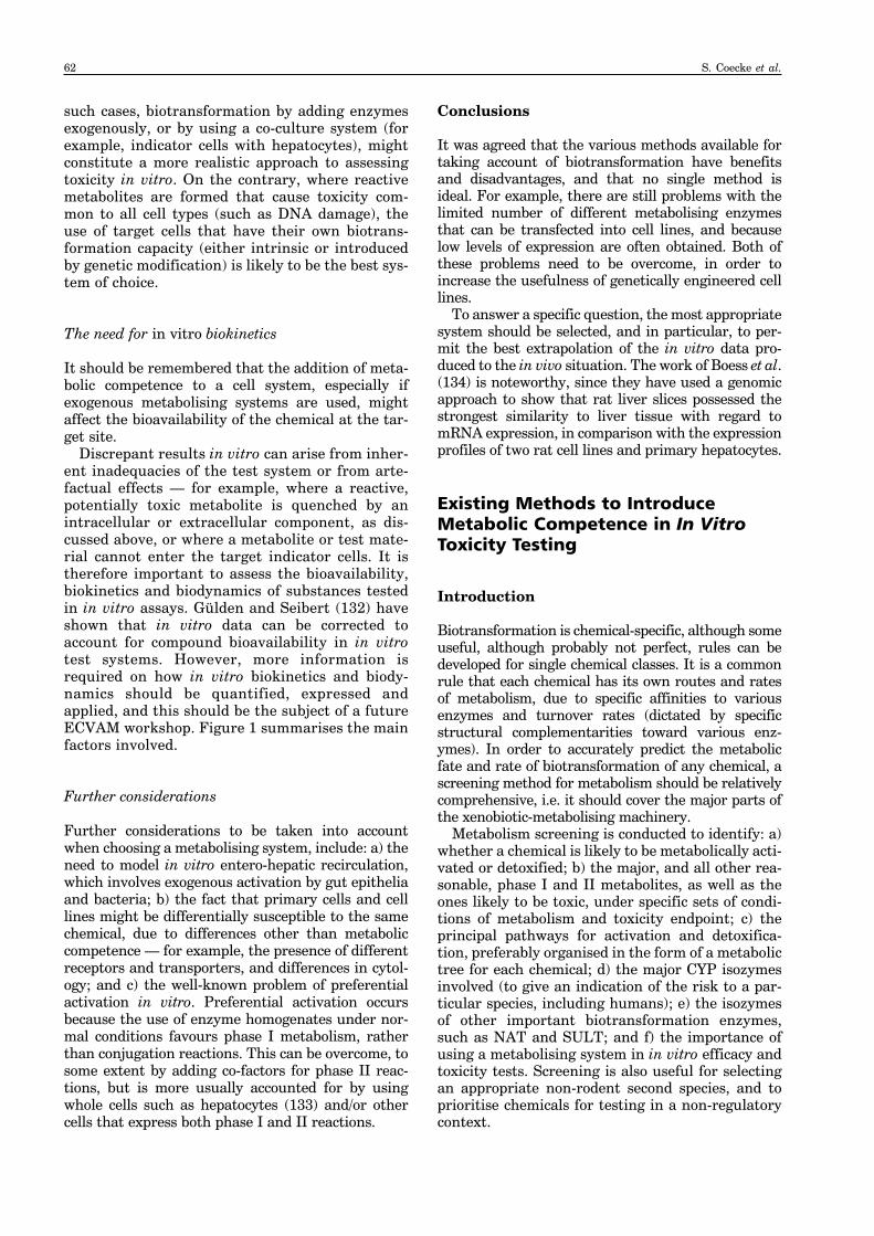
Biotransformation is chemical-specific, although someuseful, although probably not perfect, rules can bedeveloped for single chemical classes. It is a commonrule that each chemical has its own routes and ratesof metabolism, due to specific affinities to variousenzymes and turnover rates (dictated by specificstructural complementarities toward various enz-ymes). In order to accurately predict the metabolicfate and rate of biotransformation of any chemical, ascreening method for metabolism should be relativelycomprehensive, i.e. it should cover the major parts ofthe xenobiotic-metabolising machinery.
Metabolism screening is conducted to identify: a)whether a chemical is likely to be metabolically acti-vated or detoxified; b) the major, and all other rea-sonable, phase I and II metabolites, as well as theones likely to be toxic, under specific sets of condi-tions of metabolism and toxicity endpoint; c) theprincipal pathways for activation and detoxifica-tion, preferably organised in the form of a metabolictree for each chemical; d) the major CYP isozymesinvolved (to give an indication of the risk to a par-ticular species, including humans); e) the isozymesof other important biotransformation enzymes,such as NAT and SULT; and f) the importance ofusing a metabolising system in in vitro efficacy andtoxicity tests. Screening is also useful for selectingan appropriate non-rodent second species, and toprioritise chemicals for testing in a non-regulatorycontext.
62 S. Coecke et al.
Many methods (see Table 7) can be used to screenchemicals for their susceptibility to metabolism, andto establish the role of metabolism in their toxicity.The source of metabolism can also be varied accord-ing to sex, species and tissue, as well as enzyme, orby the use of enzyme inducers or inhibitors specificfor particular isozymes (see below). These systemsare also used in conjunction with a number of exten-sive databases on metabolism, such as the MDLmetabolite database, and extensive compendia onbiochemical transformation (see www.mdli.com/products/predictive/ metabolite/index.jsp).
The use of simple cell-based screens
Determining whether a chemical is likely to beactivated or detoxified is complicated by the fact
that it is necessary to assess the role of metabolismfor all the toxicity endpoints of interest, since thesame chemical might be toxic for a particular end-point without needing to be metabolised, as well asbeing activated or detoxified for another endpoint.However, a simple method for assessing the role ofmetabolism in the toxicity of a chemical would beto assess its basal cytotoxicity with and withoutmetabolism, either by using metabolically compe-tent and non-competent indicator cells, or by usingthe same cell in the presence of a metabolicinducer or inhibitor, or with and without the addi-tion of a metabolising system. This can be conve-niently done in industry by performing theSalmonella mutagenicity assay with and withoutliver S9, since this test involves genotoxicity, a keytoxicity endpoint in the development of chemicalsand drugs. However, the results of such a test will
Figure 1: In vitro biokinetics and metabolising systems
a = exogenous metabolising system; b = endogenous metabolising system; c = co-culture metabolising system. X = parent chemical; M = metabolites from various sources (upper/lower case depicts relative amounts); 1, 2 = mediacomponents (e.g. serum, other proteins); 3 = added metabolism (exogenous or endogenous); 4 = cytoplasmic membrane;5 = transporters/surface receptors; 6 = intracellular residual metabolism; T = target for toxicity. Physical properties ofX and M (e.g. molecular size, shape, lipophilicity, ionisability) determine bioavailability to the various cellular systemsfor metabolism/toxicity.
1X
a) 4,5
x,M x,M,m2 3 toxicity6 T
1X
b) 4,5
x,M,mx,m2 3 toxicity6 T
1Xx,M,m
c) 4,5
x,m2 3 toxicity6 1
4,5
2 T6
ECVAM Workshop 54: metabolism 63
not necessarily be relevant for predicting theimportance of metabolism for other forms of toxic-ity for the same chemical.
Yueh et al. (135) have described assays involvingthe use of the Hep G2 cell line for detecting chemi-cals that either induce CYP1A by binding to the Ahreceptor linked to a promoter region for theenzyme, luciferase, in a reporter gene, or thatinhibit the activity of the enzyme. The authors sug-gest that this assay could be adapted for highthroughput screening, so that a requirement formetabolism could be rapidly identified.
Another simple way to assess the susceptibility ofa chemical to metabolism (or, more accurately, theformation of an electrophilic reactive intermediate)is to measure its binding to protein in the presenceand absence of a source of metabolism. Metabolicpathways can also be monitored by exposing cells toradioactively labelled drugs.
The use of microorganisms
There has been much interest in the use of microor-ganisms, particularly certain fungi, as surrogatesfor mammalian metabolism, to investigate thelikely metabolites produced from a wide range ofxenobiotics (86, 136, 137). The principal assump-tion made is that fungi, being eukaryotic, sharemetabolic biotransformation pathways similar tothose in higher eukaryotes, including mammals,and this has been verified for a variety of differentsubstrates. Indeed, both phase I and II enzymeactivities have been demonstrated in organismssuch as Cunninghamella echinulata,
Saccharomyces cerevisiae, and species of the genus,Aspergillus. However, there are several cases wherethe expected mammalian metabolites are not gen-erated, or where metabolites are formed which arenot formed in mammals. It is well-known, for exam-ple, that the use of S. cerevisiae in genotoxicitystudies, with and without an added mammalianmetabolism system, and without optimising the cul-ture conditions for maximum residual biotrans-formation activity, can yield different results fromthose obtained with other test systems incorporat-ing a mammalian biotransformation system (138).However, yeast cells, genetically engineered toexpress human CYPs, have proved useful in drugmetabolism studies (139). It is therefore suggestedthat the use of fungi for metabolism screeningshould be approached with caution, particularlysince many mammalian-based methods are nowwidely available and can be easier to use.
The use of test batteries of cells
By using immortalised cells, or established cell linesgenetically engineered to express various phase Iand II enzymes, it is possible for one laboratory toscreen more than 200 chemicals per month(Johannes Doehmer, personal communication, seewww.genpharmtox.com/services/Metabolism). It istherefore recommended that the use of batteries ofcell lines with different and specific metabolisingcapacities for screening large numbers of chemicalsfor their susceptibility to metabolism, should beactively developed and promoted, especially as partof an integrated testing strategy for assessing the
Table 7: Systems for biotransformation screening
In silico prediction systems (see Table 9) Pure enzymesEnzyme fractionsPost-mitochondrial supernatant (S9)Microsomes
CytosolTissue slicesGut microflora metabolismProstaglandin H synthase system (see Table 1) Use of specific enzyme inhibitors
Use of specific co-factors for phase I and phase II (conjugating) enzymesMetabolically competent cells (e.g. hepatocytes) from different speciesGenetically engineered cell lines expressing single or multiple CYPs and phase II enzymesCo-cultures of metabolising cells and toxicity indicator cellsUse of metabolising cells as toxicity indicator cells
Host-mediated assaysMetabolite identification and testingTesting of known or suspected metabolites
64 S. Coecke et al.
toxicity of existing chemicals in compliance with theEU REACH System (140).
The use of in silico methods
Overview
There are three main ways of predicting metabo-lism by using computerised methods: a) modellingof whole molecules and the three-dimensional inter-actions between chemical substrates and biotrans-formation enzymes (3D-QSAR); b) (Q)SAR studieson the binding of chemicals with the active sites ofthese enzymes; and c) expert systems, that combineseveral of these approaches (for reviews, see141–144). The principles of these various physico-chemical methods are summarised in Tables 8 and9. They primarily involve calculating the quantummechanical properties of molecules in order toestablish their intrinsic reactivities, then predictingthe interactions between metabolising enzymes andother biological receptors, based on electronic and3D considerations. This information is then inte-grated, in order to predict susceptibility to metabo-lism, both qualitatively and quantitatively.
Molecular and QSAR modelling
Such metabolism modelling and (Q)SAR studieshave been greatly facilitated by the availability of
the crystal structures of some biotansformationenzymes, especially the CYPs. The majority of CYPsfor which such structures are available (thus far, 21in total) are microbial in origin, although the latestavailable information indicates that there are crys-tal structures for three human CYPs (2C8, 2C9 and3A4), and one rabbit CYP (2C5/3) (145). Vedani(146) has recently described the use of the crystalstructure of human CYP3A4 to develop a computa-tional model to accurately predict the dockingpotential of a number of diverse putative ligands(substrates for the isozyme). A further usefulresource for modelling metabolism is the availabil-ity of information on a wide range of protein struc-tures (see www.rcsb.org.pdb). The structures andmechanisms of substrate binding of various otherCYPs and other enzymes, such as epoxide hydro-lase, have been developed by homology modelling(147–149).
Lewis (150) has recently published the results ofa large study, involving the development of QSARmodels for a range of substrates for the humanCYP2 family of enzymes. He reported good correla-tions between binding affinities to, and molecularinteractions with, the active sites of the variousisozymes and a number of physicochemical parame-ters of the substrates. The model permitted the esti-mation of substrate binding energy for a givenisozyme from a combination of energy terms,including hydrogen bonding, desolvation, bondenergy changes and an electronic parameter.
Table 8: Molecular approaches to analysing xenobiotic metabolism
Method Principle
Determining intrinsic reactivity of a substrate Determination of electronic charge distribution, Elumo, Ehomo molecular orbital (MO) energies and related molecular parameters
Predicting rates of specific enzyme reactions Quantum mechanical and thermodynamic calculations of reactivity in relation to relative transition energies of reactions
Molecular modelling of enzymes Use of crystal structures (of available microbial CYPs) to model active sites followed by docking experiments with potential substrates
Pharmacophore modelling Determination of structural requirements for substrate binding to active sites
Molecular field analysis CoMFA as applied to congeneric series of enzyme substrates and inhibitors to generate QSARs based on 3D properties correlated with Km
Modelling specific enzyme-mediated Developing SARs for interactions between specific enzymes andbiotransformation reactions and substrate potential substratesrequirements
Application of QSARs without molecular Using physical parameters (e.g. log octanol/water coefficient) to predict modelling affinity constants for substrates and inhibitors
ECVAM Workshop 54: metabolism 65
The use of expert systems
Expert systems can be used either specifically orcoupled with rule bases for predicting general toxi-city (151, 152). Examples of this include CASE andMETACASE, HazardExpert and MetabolExpert,Derek for Windows and Meteor, as well as theTIMES metabolism simulator software (141, 153,154; Table 9). In this way, it is possible to identifypredicted metabolites in one program, and processthem for structural alerts by using the other relatedprogram.
Such programs can also be used to determine thelog P values of metabolites and parent compounds,and to predict important properties of compounds,such as skin permeability. In addition, Meteor (141,153), provides information on metabolites, meta-bolic trees, and metabolic pathways.
One of the problems of metabolism predictionprograms is that they are capable of generating verylarge numbers of metabolites from one parent mol-ecule, without necessarily giving any indication ofwhether these are likely to be major or minormetabolites under specific conditions. However,some models do provide such information (155),while others can be controlled to limit the numbersformed, or to set the species, target organ, sex andother parameters, such as whether only phase I orII reactions, or both, are involved.
Meteor controls the number of metabolites gen-erated at any one time from a query molecule, byusing absolute and relative reasoning modellingtechniques that are able to prioritise biotransfor-mations (156). Reasoning requires the combineduse of these two techniques to cope with the com-plexities of different biochemical pathways thatmight be competing for the same substrate.Meteor is also supplied with a knowledge base edi-tor, so that users can incorporate their own sets ofbiotransformation reactions and rules into the sys-tem.
Like Meteor, TIMES involves the use of a heuris-tic algorithm to produce plausible biotransform-ation pathways from a query molecule, by usingrules developed from a comprehensive library ofbiotransformations (153, 157). The generation ofmetabolites by TIMES can also be limited to themost likely ones or extended to include less likelyones. The developers of the software have also inte-grated reactivity models for various macromolecu-lar interactions (for example, for mutagenicity andsensitisation), to enable the software to simulatethe generation of reactive metabolites by specificmetabolising systems, such as S9.
COMPACT is a different system, which analysesthe ability of a molecule to fit into the active site ofthe CYP1A1 isozyme (and some other CYPisozymes), by modelling molecular shape (planarityor area/depth) and chemical reactivity (covalentbond formation). The use of COMPACT is therefore
limited to molecules that are activated by theseCYP isozymes (151).
Although in silico metabolism prediction systemsmay have great potential, they all require furtherimprovement before they can be considered accept-able for specific applications (158). Moreover, likeall in silico prediction systems, they need to be val-idated, and this is proving controversial (159). Noscreening system can be totally comprehensive, sothe coverage of any one system is a compromisebetween what is ideal and what is feasible.
The computational modelling of metabolic reac-tions is a highly complex task, and more use shouldbe made of available mathematical simulation soft-ware, such as DBsolve (160), to facilitate theprocess.
Biotransformation and ADME
Introduction
The prediction of ADME (absorption, distribution,metabolism and excretion) is an integral part ofdrug development and assessment of the safety ofthese and other chemicals. It is traditionally under-taken by using whole animal methods, in conjunc-tion with kinetic studies, by using metabolismcages, and according to standard test guidelines andprotocols. However, the use of computational meth-ods to model biokinetics is increasingly beingadopted, particularly in the pharmaceutical indus-try, as part of high throughput screening strategies(Table 10).
Biokinetic modelling
The use of biokinetic (or physiologically-based phar-macokinetic — PBPK) modelling outside the phar-maceutical industry is slowly beginning to increase(161, 162). This approach can provide informationthat is particularly useful in the design of in vivotoxicokinetic studies, and for interpreting in vitroand in vivo data. Biokinetic modelling is also usefulin addressing the fundamental problem in riskassessment of the need to relate the effects detectedat the dose level applied to the test system (theexternal dose) with the effects that might be causedby the dose that actually reaches the target in thewhole animal (the internal dose). The internal tar-get organ dose can be predicted by undertaking tox-icokinetic studies, taking account of ADME. Abetter, biologically-based, dose-response model of invivo toxicity can then be developed from externaldose data. Thus, the information from biokineticmodelling is very useful for identifying concentra-tions likely to be present at target sites in vivo fol-lowing the dosing of animals, so that objective
66 S. Coecke et al.
Table 9: Expert systems, QSARS and software for predicting toxicity and metabolism
Name Supplier Website Endpoints predicted
CASE/MCASE/ MultiCASE Inc www.multicase.com Carcinogenicity, mutagenicity, teratogenicity, CASETOX; mammalian acute and chronic toxicities and others; METACASE phase I and II metabolism
Derek for LHASA Ltd www.lhasalimited.org Carcinogenicity, genotoxicity, skin sensitisation, irritation, Windows; respiratory sensitisation and others; phase I and II Meteor metabolism
COMPACT Prof David Lewis, www.surrey.ac.uk/ Carcinogenicity via metabolic activation by CYP1A and University of Surrey SBMS CYP2E subfamilies
PASS Laboratory of structure www.ibmh.msk.su/ Mutagenicity, carcinogenicity, teratogenicity, embryotoxicity function based drug PASS/ and many other effectsdesign, V.N. Orechovich Institute of BiomedicalChemistry
EPI Suite of Syracuse Research www.syrres.com/esc/ Biodegradation (BioWin), dermal permeability (DermWin) and Programs Corporation est_soft.htm other environmental effects
Metasite Molecular Discovery www.moldiscovery. Phase I and II metabolismLtd com
BRENDA University of Koeln www.brenda.uni-koeln. Enzyme data and metabolic informationde
METALOG Imperial College www.beaconprojects. Metabolic pathway predictionDatabase London org.uk/metalog.htm
ECVAM Workshop 54: metabolism 67
concentrations can be applied to in vitro test sys-tems.
PBPK modelling involves combining experimen-tal results, in vitro data, data from the literature,and computational techniques, and extrapolatingdata from in vitro studies between species (163).Organs or groups of organs are considered as dis-crete interconnected compartments with physiolog-ical volumes and blood flows. These models canaccount for physiological influences and can be usedto estimate internal tissue dose, permitting extrap-olation between species, doses and routes. PBPKmodels simulate kinetic profiles from exposure dataand a number of physiological and chemical-specificparameters generated outside the model. The phys-iological parameters include, in particular, species-dependent tissue volumes and blood flows.Chemical-specific parameters, and often species-dependent parameters, include intrinsic clearance(Clint), enzyme affinity (Km) and rate of reaction(Vmax), plasma membrane permeabilities, renal andbiliary excretion clearances, plasma protein bind-ing, tissue-to-plasma partition coefficients (Kp), andabsorption parameters. In most cases, data on thesemetabolic parameters can be obtained from in vitrostudies (for example, by using hepatocytes and/orsubcellular fractions).
The use of human cells and tissues
In drug development, it is particularly useful to beable to use human hepatocytes and/or microsomes,and the level of success achieved with suchresources is encouraging. Ponsoda et al. (164)showed that the variability in human metabolism ofseveral drugs in the clinic was reflected in themetabolism of these substances by hepatocytes iso-lated from the respective patients. However, inter-individual variation must always be taken intoconsideration, and there can be problems in obtain-ing suitable samples. Also, preparations can be sus-ceptible to a wide range of factors, such as thesolvents used (165), and the ability of differentbatches to attach to extracellular matrices (166).
ECVAM has recently funded a project (ContractNo. 19471-2002-05F1ED ISP FR) to assess the possi-bility of obtaining high quality human hepatocytes inthe USA and in the EU. The work resulted in theproduction of SOPs for obtaining preparations suit-able for use as suspension or monolayer cultures.The problem of acquiring human liver cells is alsobeing addressed (167, 168), and should be facilitatedby the establishment of networks of human tissuebanks for ethically-approved research, as opposed toother medical purposes (169).
Table 10: Biokinetic PBPK and ADME prediction programs
Name Supplier Website Properties predicted
OraSpotter™ ZYX Bio www.zyxbio.com Absorption in GI tract
iDEA LION bioscience www.lionbioscience. Absorption in GI tract; systemic circulation or pkEXPRESS™ com bioavailability; plasma concentration; elimination
Megen100 Health & Safety Lab www.hsl.gov.uk/ Oral and i.v. absorption; concentration/time profiles for capabilities/pbpk.htm plasma and major organs and tissues; liver metabolism
PK-Sim® Bayer Technology www.bayertechnology. Oral absorption; concentration/time profiles for plasma and Services com major organs; bioavailability; renal and biliary excretion
GastroPlus™ Simulations Plus Inc www.simulations-plus. Absorption in GI tract; concentration/time profiles for com plasma; liver metabolism
Cloe PK® Cyprotex www.cyprotex.com Potential exposure; absorption in GI tract; plasma, tissue and organ concentrations; renal excretion; hepatic metabolism
COSMOfrag COSMO andreas.klamt@ Drugs and pesticides; ADME high throughput screening cosmologic.de based on quantum chemistry
68 S. Coecke et al.
In vitro–in vivo scaling
The above factors, in conjunction with other phar-macokinetic parameters, such as plasma half-life,volume of distribution and oral bioavailability,together with the possibility of drug–drug interac-tion (170), have also been considered for the esti-mation of in vivo kinetics on the basis of in vitrostudies in the process of in vitro–in vivo scaling. Inaddition, there are differences between the bioavail-ability of chemicals to target sites in in vitro sys-tems, due to non-specific binding to plasma andtissue proteins (in vivo), as opposed to binding tocomponents of tissue culture media (for example,serum protein) and microsomes in vitro (171, 172).The latter phenomenon has been suggested asbeing the reason why Km values with hepatocytesare consistently lower than those obtained withmicrosomes, despite the fact that microsomes andhepatocytes inherently have similar capacities tocarry out many CYP reactions (144).
Recent developments in applying biokineticmodelling
Significant advances are currently being made inbiokinetic modelling, including the development ofsoftware programs and databases for the rapid gen-eration of new models (George Loizou, personalcommunication; see http://www.hsl.gov.uk/capabili-ties/pbpk.htm). These advances should improve theusefulness of this approach for evaluating largenumbers of chemicals, and should assist in theinterpretation of in vitro hazard predictions for riskassessment purposes.
Modelling target organ and systemic toxicity
Hepatotoxicity
Since it is the principal site for biotransformationwithin the body, the liver is the most often-usedmetabolising system, and, as would be expected,most hepatotoxicity studies have been conductedwith freshly isolated hepatocytes. Many of the tech-nical issues involved in using hepatocytes (forexample, stability against dedifferentiation, includ-ing CYP inactivation during collagenase isolationtechniques, and the induction of apoptosis [173,174] and sensescence, isolation and storage meth-ods, criteria for viability, and methods for measur-ing enzyme activity and cell toxicity) are beginningto be overcome by using various techniques, such asthe use of gel sandwich cultures and liver spheroids,and, more recently, there have been attempts toderive hepatocytes from adult stem cells (175–177;Vera Rogiers, personal communication).
Advances are being made in the use of histonedeacetylase inhibitors to arrest the proliferation-related progressive loss of liver-specific functioning(dedifferentiation) and the spontaneous death ofprimary hepatocytes following their isolation fromthe liver, and to positively affect the maturationinto hepatocytes of stem cells derived from humanadult bone-marrow (178). Several potential sourcesof stem cells have been considered for generatingdifferentiated hepatocytes, including embryonicstem cells and intra-hepatic progenitors. Therehave been some reports that, under appropriate cul-ture conditions and with stimulation by cytokines,growth factors or chemical reagents, embryonicstem cells can differentiate into hepatocyte-likecells which express markers such as albumin andother plasma proteins, CK8/18, and glycogen, andpermit the xenobiotic-mediated induction of CYPgenes (179). Similarly, intra-hepatic progenitorcells have been shown to be able to generate hepa-tocyte-like cells expressing the same liver-specificmarkers. Ideally, the enzymes that are expressed bymetabolically competent cells should reflect theirrelative in vivo importance for biotransformation.However, despite these advances, it is still not pos-sible to routinely obtain differentiated hepatocytesfrom stem cells (179), although it is expected thatthis will be feasible in the near future.
Engl et al. (180) have shown that culturinghuman hepatocytes in three-dimensional collagengel sandwich cultures improves the maintenance ofspecific functions, including metabolic competence.This is thought to be because the receptors for hepa-tocyte and epidermal growth factors are relativelyeasily activated. Human hepatocytes obtained fromliver resections in this way have remained function-ally active for up to 150 days in culture (181).However, Yuan et al. (182) have shown that humanhepatocytes grown in monolayer two-dimensionalcultures in the presence of 1% rat tail collagen,maintained the activities of a number of CYPisozymes and UDP-glucuronyltransferase over atwo-week period, which raises questions about theneed to use more-complicated culture systems. Theability to avoid the use of collagen gels for main-taining human hepatocytes would eliminate theproblems of the variability in the attachment offreshly isolated hepatocytes from different donorsthat has been observed, in contrast to experiencewith rat hepatocytes, which attach readily (183).
There have been few efforts to validate the use ofhepatocytes for the specific purpose of predictingtoxicity. One study showed that primary rat hepa-tocytes could be used to predict the acute oral toxi-city of a number of chemicals (184). However, thiswas a small investigation, conducted in a single lab-oratory. The workshop participants agreed that it isstill not possible to recommend for formal prevali-dation and validation, a standard protocol for usinghepatocyte cultures for the specific purpose of pre-
ECVAM Workshop 54: metabolism 69
dicting hepatoxicity in vivo for regulatory purposes.The principal barrier to progress on this importantissue appears to be the lack of suitable in vivo dataon hepatoxicity for benchmarking purposes, despitethe recommendation in ECVAM Workshop 1 (heldin 1994) that such a database should be established.
Modelling toxicity to extra-hepatic organs
It is difficult to model metabolism-mediated toxicityto other target organs in vitro, although it is possi-ble to use subcellular metabolising fractions fromdifferent tissues with toxicity indicator cells and todemonstrate metabolic activation. The principalproblem is that the specific activities of most extra-hepatic metabolising enzymes are usually severalorders of magnitude lower than those of the corre-sponding enzymes found in the liver. Thus, thisaspect of target organ toxicity is often overlooked,despite the fact that extra-hepatic metabolismmight be extremely important when it is qualita-tively different from that which occurs in the liver.Examples of situations where extra-hepatic bio-transformation can be important are: a) oxidativemetabolism by the intestinal mucosal lining (185),and reductive metabolism by the gut microflora(particularly relevant for investigating the toxicityof foods and food additives); and b) theprostaglandin H synthase (PHS) system (alsoknown as the COX system). Metabolism by theintestinal mucosa, the gut microflora and the PHSsystem can all be taken into account in in vitro tox-icity assays (29).
PHS catalyses the first two steps in the synthesisof prostaglandin, thromboxane and prostacyclin.Two forms of the enzyme have been characterised(PHS 1 and PHS2), both of which are haeme pro-teins that exhibit both cyclooxygenase and peroxi-dase activity, the latter being responsible for theone electron oxidation of a wide range of xenobiotics(186). The peroxidase activity of PHS can beexpressed in vitro, if enzyme extracts are supple-mented with arachidonic acid, and several chemi-cals are activated by the PHS system to becomemutagens and carcinogens. In addition, extra-hepatic metabolism should be considered when it isnecessary to model toxicity arising from exposurevia other routes of entry, such as the skin (26) andthe nasal epithelium (187).
Case Studies on Existing In VitroTests which Require Metabolism
Embryotoxicity
Reproductive toxicity testing has advanced throughthe development and successful validation of three
in vitro embryotoxicity tests. The results of the pre-liminary phase of the validation study were pub-lished in 2000, and the final results of the validationstudy have now been published (188). The testsinvolved the use of: post-implantation wholeembryos; differentiating rat limb-bud cells in micro-mass cultures; and embryonic stem cells that retaintotipotency, and the ability to be programmed todevelop along a variety of specific differentiationpathways (189).
Embryotoxicity, particularly that which leads toteratogenicity, can be due to a variety of mecha-nisms, including the reactive binding of chemicalswith nucleic acids and proteins, leading to mutage-nesis and teratocarcinogenesis in the embryo, aswell as the disruption of tubulin, microfilamentsand the cytoskeleton, and many other subtle mech-anisms that can affect development. It would beexpected that the metabolites of certain chemicalswould be responsible for some of the effectsobserved, and, in vivo, such metabolism occurs inthe liver, the gonads, the placenta, and theembryo/fetus. The placenta has a relativelyrestricted complement of xenobiotic metabolisingenzymes, but inducible CYP1A1 has been detected(190). The literature suggests that reproductivetoxicity caused by metabolites is most likely to arisewhen exposure occurs during the adaptiveresponses caused by repeat dosing during thecourse of a reproductive toxicity study, a phenome-non due to the continued induction of the samemetabolism enzymes, as would occur during anytest involving chronic studies (15).
The inclusion of biotransformation systems inthese tests would therefore be expected to improvetheir predictivity, as has been shown previously forchemicals, such as cyclophosphamide in the micro-mass assay (191), and also by using co-cultures ofembryonic stem cells and metabolically competentcells (109). However, the ECVAM validation studywas focused entirely on test chemicals not requiringmetabolic activation, since the addition ofmetabolising systems might have complicated thestandardisation of the in vitro embryotoxicity tests(192). Thus, the further use of these in vitro embry-otoxicity tests will largely depend on how they canbe adapted for the routine inclusion of a metabolis-ing system for testing indirect-acting agents, andhow relevant and reliable these modified protocolsturn out to be. In fact, a group of experts recom-mended in 2003 that metabolic competence shouldbe added to the EST (193).
For the testing of large numbers of chemicals, itshould be noted that the EST is the only methodwhich: a) could provide a relatively high through-put; and b) does not involve the killing of largenumbers of pregnant animals. It is therefore rec-ommended that an improved protocol for this testshould be developed as a first priority for future val-idation, and there are plans in the recently initiated
70 S. Coecke et al.
EU ReProTect Project for further test developmentthat involves incorporating metabolism, followed bya validation study.
Endocrine disruptors
A further example of where biotransformation iserroneously ignored in in vitro testing concernsendocrine disruptors (EDs; 114, 194). This is surpris-ing, since: a) hormonally active chemicals, includingsome naturally occurring EDs, are known to bemetabolised in vivo, some being activated; b) manypotential EDs require metabolic activation to exertother toxicities; and c) the few studies available inwhich metabolising systems have been used, haveshown that biotransformation can substantially mod-ulate ED activity (195). One reason for the paucity ofpublished studies involving the use of exogenousmetabolism in in vitro assays for EDs is that potentialEDs often bind non-specifically to proteins in theenzyme fractions (another situation where the appli-cation of in vitro biokinetic analysis would be partic-ularly important, see earlier).
It is crucially important that in vitro methods fortesting EDs should take account of the need to pro-vide a biotransformation system. It would be prefer-able for this to involve cells that have been renderedmetabolically competent, instead of adding exoge-nous enzyme fractions. This is because the use of cellswould overcome the problem of binding to extracellu-lar microsomal proteins. Moreover, if the cellularmetabolising system expresses both phase I and IImetabolism, it would also alleviate the problem ofpreferential activation in vitro, particularly as phaseI ring hydroxylated products would be expected to behormonally active, based on evidence from (Q)SARstudies (196), but would act as substrates for conju-gation. It might even be feasible to generate geneti-cally engineered mammalian cell lines containingsteroid hormone nuclear receptors, their responseelements and the associated reporter genes, togetherwith genes expressing specific metabolising enzymes— all within the same cellular environment. Suchmultiple expressing cell lines could then be used toinvestigate the metabolic activation and detoxifica-tion of potential EDs in vitro. The possibility of devel-oping such cell lines should be seriously investigated.
Acute toxicity testing
Acute toxicity testing is undertaken to investigatethe effects of single large doses of a chemical. Theworkshop participants considered biotransform-ation to be likely to be less important as a mediatorof such chemical toxicity, as compared withrepeated dose toxicity. However, no consensus wasreached on the relative importance of the many rea-sons why this could be so.
The results of the Multicentre Evaluation of InVitro Cytotoxicity (MEIC) study showed good corre-lations between in vitro acute cytotoxicity data andhuman lethal blood concentrations for a diverserange of chemicals, and also with in vivo LD50 val-ues from rodent studies (197). The data supportedthe hypothesis that basal cell cytotoxicity is a usefulindicator of acute lethal systemic toxicity. It wasfurther hypothesised that, since this endpoint isdue to critical organ failure, particularly of thenervous system, such failure could arise from celldeath or loss of function in vivo. Interestingly, thegood correlation between basal cytotoxicity andacute toxicity was usually irrespective of cell typeand assay, although the use of human cells under-standably gave the best prediction of human lethalblood concentrations. However, there was no spe-cific control of metabolism or incorporation ofexogenous metabolising systems in the tests used,although these included human hepatoma cell linesand hepatocytes, which might have been expectedto have possessed some metabolic competence. It isnoteworthy that the level and nature of any suchcompetence was not specifically reported.
Several of the test chemicals used in the MEICstudy are known to be extensively metabolised toreactive forms, and in this respect it is notable thatHalle (198) deliberately excluded, as far as possible,any IC50 values obtained with liver cell cultures(hepatocytes, hepatoma cell lines) from his registryof cytotoxicity, to avoid complications resultingfrom the possible metabolic activation or detoxifica-tion of chemicals.
Since the initiation of the MEIC study, it hasbeen recognised that the predictivity of basal cyto-toxicity assays for acute toxicity could be improvedby various means, in particular, by including invitro methods for biotransformation, absorption inthe gut, passage across the blood–brain barrier, dis-tribution volumes, protein binding, and renal clear-ance/accumulation in a test battery, as well asmodels for target organ toxicity, in conjunctionwith tests for neurotoxic potential (199). Clearly,such extra parameters in the testing battery shouldalso take account of the use of human cells (200).
It is therefore to be welcomed that the recentlyinitiated EU AcuteTox project is aimed at develop-ing a testing strategy for using in vitro methods foracute toxicity prediction that will involve develop-ing a system of alerts based on accounting for suchknown outliers of the correlation between acutesystemic toxicity and basal cell cytotoxicity, prima-rily on the basis of biokinetics, metabolism and tar-get organ specific effects.
Cell transformation
Cell transformation is the induction of certain phe-notypic alterations in cells in tissue culture that are
ECVAM Workshop 54: metabolism 71
characteristic of tumourigenic cells (201). Severaldifferent cell transformation assays have beendevised, either involving rodent cell lines or immor-talised fibroblast cell lines of rodent or human ori-gin, and some systems comprise primary cells, suchas those used in the SHE cell assay (202, 203). Someof these systems are used to detect initiators of car-cinogenesis which act by a genotoxic mechanism viaelectrophilic interactions with cellular macromole-cules such as DNA, while others can be used in atwo-stage process to additionally detect tumourpromoters via non-genotoxic mechanisms. Also, anin vitro cell transformation assay for tumour pro-moters has been developed, involving Bhas 42 cellsderived from Balb/c 3T3 cells transfected with thev-Ha-ras oncogene (204).
In all the above cases, exogenous metabolisingsystems are not routinely used, on the assumptionthat any biotransformation necessary will be pro-vided by the indicator cells themselves. However,this might not be the case, and the assays are incon-sistently sensitive to carcinogens known to requireactivation. Moreover, when metabolising systemshave been added, there have been some problemswith the assays. This could be because: a) the test islengthier than other in vitro genotoxicity assays;and b) some of the mechanisms involved in celltransformation operate over a longer time thanthose involved in genotoxicity. These factors areexacerbated by the technical problems encounteredin attempting to maintain enzyme activity for a suf-ficiently long period of time.
However, it is known that SHE cells can expresssome metabolic competence (205), althoughCombes et al. (201) concluded that “a thoroughinvestigation is needed to ascertain the necessity ofusing exogenous metabolising systems in the vari-ous cell transformation assays.” Unfortunately,such an investigation has not been conducted, andit is suggested that it should be part of the valida-tion study of cell transformation assays beingplanned by ECVAM.
Conclusions
Prospects for using test systems incorporating metabolism
Apart from genotoxicity and hepatotoxicity studies,the incorporation of biotransformation into in vitrotest systems has been inconsistent, despite the factthat biotransformation is known to play a key rolein the toxicity of many chemicals. Since the residualmetabolising capacity of indicator cells is often verylow, inappropriate and variable, it is important torectify this unsatisfactory situation in order toimprove the predictivity of in vitro assays, particu-larly with respect to testing for sensitisation, cell
transformation, acute toxicity, target organ toxic-ity, endocrine disruption and embryotoxicity.
This will require further research to overcomesome practical and logistical problems, and to iden-tify the most appropriate ways of taking account ofbiotransformation. For example, a simple exoge-nous metabolising system, such as liver S9, cannotbe used to screen chemicals for embryotoxicity invitro, due to its own toxicity to the indicator cells.However, it might be possible to use metabolically-competent stem cells. Also, no consensus could bereached on whether, for mechanistic studies, theuse of genetically engineered cell lines expressingkey CYPs, and, in the case of embrytoxicity, otherenzymes unique to the fetus, should be investi-gated. In fact, in embryotoxicity testing it is oftenassumed that the potential contribution of placen-tal enzymes to metabolism is accounted for by theuse of hepatocytes alone. In addition, strategies forscreening chemicals for their susceptibility to bio-transformation via specific mechanisms and path-ways need to be developed and validated.
It is advisable that, as a matter of routine, theresidual basal biotransformation competence of anyin vitro test system should be characterised beforeit is developed and recommended for detecting tox-icity. Moreover, the metabolic competence of indi-cator cells should be checked periodically to ensurethat basal activities have not altered significantly.Lastly, the choice of metabolising system to use invitro should be based on the purpose of the test. Forexample, selection might vary according to whetherthe test is a screen, or is to be used to provide haz-ard or mechanistic information, according to thegeneralised decision-tree scheme presented in thisreport (Figure 2).
Validation
It is clear that the validation of toxicity tests thatincorporate metabolising systems will require thecareful selection of chemicals for which there isunequivocal information on whether they are meta-bolically activated or detoxified (a list of potentialchemicals for such a purpose is presented in Table11). Such information should include the effects ofhuman metabolism, since, ideally, where toxicitytesting is being undertaken for human risk assess-ment, human-based biotransformation systemsshould be used. Metabolism-mediated toxicity datawill be crucial, if such test systems are to be vali-dated for regulatory use. Unfortunately, there is alack of sufficient numbers of chemicals that meetthis criterion, due to lack of information in the pub-lic domain. This is the reason why the workshopparticipants concluded that the validation of pri-mary hepatocyte systems for detecting metabolism-mediated hepatotoxicity is not possible at thepresent time, although the systems can and should
72 S. Coecke et al.
be used for routine screening and research.Protocols for toxicity tests for metabolism-mediatedtoxicity will also need to include relevant positiveand negative control chemicals that have knownsusceptibilities to activation or detoxification.
In silico methods for predicting metabolismand biokinetics
Status
Several in silico systems for predicting metabolismare available, including (Q)SAR models and expertsystems, but none of these have been comparedextensively for their relative performances, andnone have been formally accepted for regulatoryuse, although some models can be used to providesupporting information in chemical risk assess-ments. There is currently no consensus on how in
silico models for predicting biotransformationshould be validated. Also, a variety of systems are indifferent stages of development, assessment andvalidation. If they are to be of more practical useoutside the pharmaceutical industry for regulatorytesting, then further research needs to be under-taken to make them more amenable for a widerrange of chemicals.
Problems with regard to the availability of goodquality data for benchmarking purposes, apply totechniques for using in silico prediction systemsand biokinetic models to assess the metabolic fate ofchemicals after uptake by different routes of expo-sure in different species. Before these systems canbe validated, more chemicals with good quality dataneed to be found, for use as test sets. Nevertheless,it was agreed that at least one biokinetic modellingsystem is, in principle, ready for more formal con-sideration for validation (206). Clearly, in silico sys-tems for predicting toxicity should take account ofthe possibility that biotransformation could modu-
Figure 2: Schemes for incorporating metabolising systems
a) incorporating metabolising systems into in vitro assays for mechanistic studies.
Test compound
What are the main routes of exposure?(intended usage, accidental, occupational)
OralIntestinalfractions
Gut microflora
InhalationNasal epithelium
Lung cells
Tracheal cells
Target organsLiver cells
Neural cells
Kidney cells
SkinKeratinocytes
Other cells?
Otherbarriers
Blood–brain
Blood–gonads
Are human CYPs involved in metabolism?
Importance of phase 2 metabolism?
Are there important genetic polymorphismsin metabolism?
Batteries of engineered cell linesUse of specific inhibitors of metabolismUse of specific inducersUse of specific antibodiesAddition of specific extra co-factors toexogenous systemsHuman dataIn silico methods
a)
ECVAM Workshop 54: metabolism 73
Figure 2: continued
b) incorporating metabolism into in vitro screening test systems.
b)
Test compound
1. Is the purpose of the test forscreening?
2. Do indicator cells havesufficient metabolic capacity?
No
Yes
3. Is test chemical susceptible toCYP metabolism?
No
4. Can S9 be used?
Yes
9. Is it important to considerspecies-specific effects?
Yes
11. Is it important to considergender effects?
No
12. Does aroclor induce relevantCYPs?
No
No
13. Is liver the appropriatetissue source?
Yes
Yes
Yes
Go to Figure 2a
Yes Select appropriate species
NoDetermine mostappropriate source ofmetabolism (including co-factors, enzyme fractions).Go to 9
No Select appropriate induceror consider not using aninducer and go to 13
Perform test
YesSelect appropriate sex andgo to 12, or perform test if
using whole cellmetabolising system
Perform test with aroclor-induced S9 from appropriateorgan from best species andsex
Perform test with aroclor-induced liver S9 from bestspecies and sex
No
Consider using co-cultures ofmetabolically-competentcells with indicator cells, ortissue slices, or a genetically-engineered cell line, orsamples of principalmetabolites
74 S. Coecke et al.
late toxicity. This could be achieved by modifyingthese systems, so that they can model the toxicity ofthe principal metabolites of chemicals, or by linkingthem with systems specifically designed to predictmetabolite formation.
Relevance to in vitro testing
The bioavailability of chemicals to target sites, suchas proteins, DNA or specific receptors in indicatorcells, is an important determinant of toxicity in invitro test systems. The bioavailability of test chem-icals in in vitro assays (referred to as “in vitro bio-kinetics”) is affected by the nature of: a) thechemical being tested; b) the cells and the cultureconditions; and c) the xenobiotic metabolising sys-tem (due both to the biotransformation of mole-cules and the non-specific binding to enzymeproteins). Such biokinetic analysis of in vitro testsystems, by using similar approaches to those usedto predict in vivo biokinetics, will enable target con-centrations of chemicals applied to them to be
determined, and will therefore permit a more real-istic assessment of the dose-response data obtainedby using different in vitro and in vivo test methodsfor the same chemicals.
Recommendations
Method development and validation
1. A study should be commissioned to establish adatabase of chemicals that are toxic to humans,either with or without metabolic activation, andwhich are detoxified as a consequence of metab-olism. It is suggested that information fromexisting databases, such as those maintained byIARC, ECETOC, RTEC, and IUCLID, should beassessed initially.
2. The above study should also include a compari-son of animal and human toxicity models, withrespect to the metabolite profiles and biokineticsof a range of chemicals.
3. An initiative should be undertaken to create apublicly available depository of reference chemi-cals that could be used in validation studies onbiotransformation systems. The inclusion of achemical in the depository should be based on arequirement for good quality animal and humantoxicity data, which clearly demonstrate the rolethat biotransformation plays in the toxicityobserved. Efforts should be made to balance thenumbers of chemicals requiring, and not requir-ing, metabolic activation.
Biokinetic modelling
4. A workshop should be organised on in vitro bio-kinetics: a) to identify and discuss key method-ological issues that need to be addressed, inorder to facilitate the use of biotransformationsystems in in vitro assays; b) to improve in vitrodata interpretation and extrapolation; and c) toidentify the analytical techniques required toelucidate the biokinetics of in vitro systems, withand without metabolism.
5. A project should be established with the objec-tive of defining the proper use of physiologically-based biokinetic (= pharmacokinetic [PBPK])modelling approaches for predicting ADMEparameters.
6. The status of in silico systems for predictingmetabolism, metabolites, metabolic pathways,and toxicity should be assessed by the ECVAMTask Force on Metabolism for their strengths
Table 11: List of chemicals requiring metabolicactivation, which are potentiallyuseful for method development andvalidation studiesa
and limitations, and for their suitability for vali-dation.
Incorporating biotransformation into toxicity tests
7. A study should be commissioned to develop atiered xenobiotic metabolism strategy, designedto screen chemicals for their susceptibility tomammalian metabolism.
8. Methods for using and incorporating extra-hepatic biotransformation systems into in vitrotoxicity assays should be developed and furtherimproved. Individual studies should be commis-sioned, to investigate: a) the metabolic compe-tence of rodent and human cell lines used in celltransformation assays and ways to improve it;and b) the role of metabolism in non-genotoxiccarcinogenesis.
9. Innovative, high throughput in vitro screeningmethods should be developed, in which severalcell types can be tested in parallel studies byusing multiplexed chemical, recombinant or bio-logical metabolites. A particular effort should bemade to characterise these in vitro cell-basedassays, in order to understand the reaction ofeach individual cell (high content analyses) ineach assay.
References
1. Klaassen, C.D., ed. (2001). Casarett & Doull’sToxicology: The Basic Science of Poisons, 6th edn,p.1236. New York, NY, USA: McGraw-Hill.
3. Crespi, C.L. (1995). Xenobiotic-metabolizing humancells as tools for pharmacological and toxicologicalresearch. Advances in Drug Research 26, 179–235.
4. Cashman, J.R. (2004). The implications of polymor-phisms in mammalian flavin-containing monooxy-genases in drug discovery and development. DrugDiscovery Today 9, 574–581.
17. Anders, M.W. (2004). Glutathione-dependent bioac-tivation of haloalkanes and haloalkenes. DrugMetabolism Reviews 36, 583–594.
18. Banoglu, E. (2000). Current status of the cytosolicsulfotransferases in the metabolic activation of pro-mutagens and procarcinogens. Current DrugMetabolism 1, 1–30.
19. Morisseau, C. & Hammock, B.D. (2004). Epoxidehydrolases: mechanisms, inhibitor designs and bio-logical roles. Annual Reviews of Pharmacology andToxicology 45, 311–333.
20. Deckers-Schmelzle, B., Klaus, E., Kahl, R. & Kahl,G.F. (1978). Binding of benzo(a)pyrene metabolitesto cellular DNA in perfused rat lungs. Naunyn-Schmiedeberg’s Archives of Pharmacology 303,303–307.
22. Turesky, R.J., Lang, N.P., Butler, M.A., Teitel, C.H.& Kadlubar, F.F. (1991). Metabolic activation of car-cinogenic heterocyclic aromatic amines by humanliver and colon. Carcinogenesis 12, 1839–1845.
23. Timchalk, C., Nolan, R.J., Mendrala, A.L., Sittenber,D.A., Brzak, K.A. & Mattsson, J.L. (2002). A physio-logically based pharmacokinetic and pharmacody-namic (PBPK/PD) model for the organophosphateinsecticide chlorpyrifos in rats and humans.Toxicological Sciences 66, 34–53.
24. Poet, T.S., Kousba, A.A., Dennison, S.L. &Timchalk, C. (2004). Physiologically based pharma-cokinetic/pharmacodynamic model for the organo-phosphorus pesticide diazinon. Neurotoxicology 25,1013–1030.
25. Jones, D.C., Duvauchelle, C., Ikegami, A., Olsen,C.M., Lau, S.S., de la Torre, R. & Monks, T.J.(2005). Serotonergic neurotoxic metabolites ofecstasy identified in rat brain. Journal of Pharm-acology and Experimental Therapeutics 313,422–431.
26. Hotchkiss, S.A.M. (1992). Skin as a xenobioticmetabolizing organ. In Progress in Drug Metab-olism, Vol. 13 (ed. G.G. Gibson), pp. 217–262.London, UK: Taylor & Francis.
27. Howes, D., Guy, R., Hadgraft, J., Heylings, J.,
76 S. Coecke et al.
Hoeck, U., Kemper, F., Maibach, H., Marty, J-P.,Merk, H., Parra, J., Rekkas, D., Rondelli, I.,Schaefer, H., Täuber, U. & Verbiese, N. (1996).Methods for assessing percutaneous absorption.The report and recommendations of ECVAM work-shop 13. ATLA 24, 81–106.
28. Skalova, L., Szotakova, B., Wsol, V., Kral, R.,Baliharova, V. & Lamka, J. (2000). Activity, stere-ospecificity, and stereoselectivity of microsomalenzymes in dependence on storage and freezing ofrat liver samples. Chirality 12, 649–653.
29. Combes, R.D. (1992). The in vivo relevance of invitro genotoxicity assays incorporating enzyme acti-vation systems. In Progress in Drug Metabolism,Vol. 13 (ed. G.G. Gibson), pp. 295–321. London, UK:Taylor & Francis.
30. Hall, P.M., Stupans, I., Burgess, W., Birkett, D.J. &McManus, M.E. (1989). Immunohistochemicallocalization of NADPH-cytochrome P450 reductasein human tissues. Carinogenesis 10, 521–530.
31. Windmill, K.F., Gaedigk, A., Hall, P.M., Samara-tunga, H., Grant, D.M. & McManus, M.E. (2000).Localization of N-acetyltransferases NAT1 andNAT2 in human tissues. Toxicological Sciences 54,19–29.
32. Ding, X. & KIaminsky, L.S. (2003). Human extra-hepatic cytochromes P450: function in xenobioticmetabolism and tissue toxicity in the respiratoryand gastrointestinal tracts. Annual Reviews ofPharmacology and Toxicology 43, 149–173.
33. Pelkonen, O. & Raunio, H. (1997). Metabolic activa-tion of toxins: tissue-specific expression and metab-olism in target organs. Environmental HealthPerspectives 105, 767–774.
34. Ullrich, A.B., Standop, J., Schmied, B.M.,Schneider, M.B., Lawson, T.A. & Pour, P.M. (2002).Species differences in the distribution of drug-metabolizing enzymes in the pancreas. ToxicologicPathology 30, 247–253.
35. Minn, A., Ghersi-Egea, J.F., Perrin, R., Leininger,B. & Siest, G. (1991). Drug metabolizing enzymes inthe brain and cerebral microvessels. Brain ResearchReviews 16, 65–82.
36. Strazielle, N. & Ghersi-Egea, J.F. (2000). Imp-lication of blood–brain interfaces in cerebral drugmetabolism and drug metabolite disposition. InMolecular Drug Metabolism and Toxicology (eds G.Williams & O.I. Aruoma), pp. 181–200. London,UK: OICA International.
37. Miksys, S. & Tyndale, R.F. (2004). The unique reg-ulation of brain cytochrome P450 2 (CYP2) familyenzymes by drugs and genetics. Drug MetabolismReviews 36, 313–333.
38. Gaylor, D.W. & Zheng, Q. (1996). Risk assessmentof nongenotoxic carcinogens based upon cell prolif-eration/death rates in rodents. Risk Analysis 16,221–225.
39. Goeptar, A.R., Commandeur, J.N.M., van Ommen,B., Van Bladeren, P.J. & Vermeulen, N.P.E. (1995).Metabolism and kinetics of trichloroethylene inrelation to toxicity and carcinogenicity. Relevanceof mercapturic acid pathway. Chemical Research inToxicology 8, 3–21.
40. Teissier, E., Fennrich, S., Strazielle, N., Daval, J-L.,Ray, D., Schlosshauer, B. & Ghersi-Egea, J-F.(1998). Drug metabolism in in vitro organotypic andcellular models of mammalian central nervous sys-tem: activities of membrane-bound epoxide hydro-lase and NADPH-cytochrome P450 (c) reductase.
able metabolism of antidepressants and neurolepticdrugs in man. Pharmacogenetics 3, 61–70.
42. Evans, W.E. & Relling, M.V. (1999). Pharmacogen-omics: translating functional genomics into rationaltherapeutics. Science, New York 286, 487–491.
43. Kalow, W. & Tyndale, R.F. (1992). Debrisoquine/sparteine monoxygenase and other P450s in thebrain. In Pharmacogenetics of Drug Metabolism (ed.W. Kalow), pp. 649–656. New York, NY, USA:Pergamon Press.
44. Gervasini, G., Carrillo, J.A. & Benitez, J. (2004).Potential role of cerebral cytochrome P450 in clini-cal pharmacokinetics. Clinical Pharmacokinetics43, 693–706.
45. Monks, T.J., Ghersi-Egea, J.F., Philbert, M., Cooper,A.J. & Lock, E.A. (1999). Symposium overview: therole of glutathione in neuroprotection and neurotoxi-city. Toxicological Sciences 51, 161–177.
46. Strazielle, N., Khuth, S.T. & Ghersi-Egea, J.F.(2004). Detoxification systems, passive and specifictransport for drugs at the blood-CSF barrier in nor-mal and pathological situations. Advanced DrugDelivery Reviews 56, 1717–1740.
47. Henderson, C.J. & Wolf, C.R. (2003). Transgenicanalysis of human drug-metabolizing enzymes: pre-clinical drug development and toxicology. MolecularInterventions 3, 331–343.
48. Gonzalez, F. (2004). Cytochrome P450 humanisedmice. Human Genomics 1, 300–306.
49. Bigger, C.A.H., Tomaszewski, J.E., Dipple, A. &Lake, R.S. (1980). Limitations of metabolic activa-tion systems used with in vitro tests for carcino-gens. Science, New York 209, 503–505.
50. Parkinson, C. & Grasso, P. (1993). The use of thedog in toxicity tests on pharmaceutical compounds.Human and Experimental Toxicology 2, 99–109.
51. Beasley, V. (1999). Absorption, distribution, metab-olism, and elimination: differences among species.In Veterinary Toxicology (ed. V. Beasley), 21pp.Ithaca, NY, USA: International VeterinaryInformation Service. Website http://www.ivis.org/advances/Beasley/AppC/chapter_frm.asp?LA=1#3(Accessed 30.09.05).
52. Pacifici, G.M. & Pelkonen, O., eds (2001). Inter-individual Variability in Human Drug Metabolism,pp. 358–394. London, UK: Taylor & Francis.
54. Ingelman-Sundberg, M. (2004). Human drugmetabolising cytochrome P450 enzymes: propertiesand polymorphisms. Naunyn-Schmiedeberg’s Arch-ives of Pharmacology 369, 89–104.
55. Rendic, S. (2002). Summary of information onhuman CYP enzymes: human P450 metabolismdata. Drug Metabolism Reviews 34, 83–448.
56. Rogiers, V., Coecke, S., Vandenberghe, Y., Morel, F.,Callaerts, A., Verleye, G., Van Bezooijen, C.F.,Guillouzo, A. & Vercruysse, A. (1991). Effect of theageing process on the gender and phenobarbitaldependent expression of glutathione S-transferasesubunits in brown Norway rat liver. BiochemicalPharmacology 42, 491–498.
57. Coecke, S., Vandenberghe, Y., Callaerts, A., Sonck,W., Verleye, G., Van Bezooijen, C.F., Vercruysse, A.& Rogiers, V. (1990). Hepatic cytosolic glutathione
ECVAM Workshop 54: metabolism 77
S-transferase activities in ageing brown Norwayrats — importance of sex differences and phenobar-bital treatment for studies of ageing. Mechanisms ofAgeing and Development 55, 189–198.
58. Ade, P., Guastadisegni, C., Testai, E. & Vittozzi, L.(1994). Multiple activation of CHCl3 in kidneymicrosomes from male and female DBA/2J mice.Journal of Biochemical Toxicology 9, 289–295.
59. Kimura, S., Donovan, J.C. & Nebert, D.W. (1987).Expression of the mouse P1450 gene during differ-entiation without foreign chemical stimulation.Journal of Experimental Pathology 3, 61–74.
63. Williams, D.P. & Park, B.K. (2003). Idiosyncratictoxicity: the role of toxicophores and bioactivation.Drug Discovery Today 8, 1044–1050.
64. Lin, J.H. & Lu, A.Y.H. (1997). Role of pharmacoki-netics and metabolism in drug discovery and devel-opment. Pharmacological Reviews 49, 403–449.
65. Leahy, D.E., Duncan, R., Ahr, H.J., Bayliss, M.K.,de Boer, A.G., Darvas, F., Fentem, J.H., Fry, J.R.,Hopkins, R., Houston, J.B., Karlsson, J., Kedderis,G.L., Pratten, M.K., Prieto, P., Smith, D.A. &Straughan, D.W. (1997). Pharmacokinetics in earlydrug research. The report and recommendations ofECVAM Workshop 22. ATLA 25, 17–31.
66. Van de Waterbeemd, H. & Gifford, E. (2003).ADMET in silico modelling: towards prediction par-adise? Nature Reviews — Drug Discovery 2,192–204.
67. Worth, A.P. & Balls, M., eds (2002). Alternative(non-animal) methods for chemicals testing: cur-rent status and future prospects. A report preparedby ECVAM and the ECVAM Working Group onChemicals. ATLA 30, Suppl. 1, 1–125.
68. Spielmann, H. & Liebsch, M. (2002). Validation suc-cesses: chemicals. ATLA 30, Suppl. 2, 33–40.
69. De Silva, O., Basketter, D.A., Barratt. M.D., Corsini,E., Cronin, M.T.D., Das, P.K., Degwert, J., Enk, A.,Garrigue, J.L., Hauser, C., Kimber, I., Lepoittevin,J-P., Peguet, J. & Ponec, M. (1995). Alternativemethods for skin sensitisation testing: the reportand recommendations of ECVAM workshop 19.ATLA 24, 683–705.
70. Kimber, I. (2000). Skin sensitisation: immunologi-cal mechanisms and novel approaches to predictivetesting. In Progress in the Reduction, Refinementand Replacement of Animal Experimentation (ed.M. Balls, A-M. van Zeller & M.E. Halder), pp.613–622. Amsterdam, The Netherlands: Elsevier.
71. Casati, S., Aeby, P., Basketter, D.A., Cavani, A.,Gennari, A., Gerberick, G.F., Griem., P., Hartung,T., Kimber, I., Lepoittevin, J-P., Meade, B.J.,Pallardy, M., Rougier, N., Rousset, F., Rubinstenn,G., Sallusto, F., Verheyen, G.R. & Zuang, V. (2005).Dendritic cells as a tool for the predictive identifica-tion of skin sensitisation hazard. The report andrecommendations of ECVAM Workshop 51. ATLA
A., van Vliet, E., Prieto, P., Boveri, M., Pellizzer, C.,Wendel, A. & Hartung, T. (2005). The value of alter-native testing for neurotoxicity in the context ofregulatory needs. Environmental Toxicology andPharmacology, in press.
73. Park, B.K., Kitteringham, N.R., Maggs, J.L.,Pirmohamed, M. & Williams, D.P. (2005). The roleof metabolic activation in drug-induced hepatotoxi-city. Annual Reviews of Pharmacology and Toxicol-ogy 45, 177–202.
74. Zucco, F., De Angelis, I., Testai, E. & Stammati, A.(2003). Toxicology investigations with cell culturesystems: 20 years after. Toxicology in Vitro 18,15–163.
75. Lee, Y.C., Shlyankevich, M., Jeong, H.K., Douglas,J.S. & Surh, Y.J., (1995). Bioactivation of 5-hydrox-ylmethyl-2-furaldehyde to an electrophilic andmutagenic allylic sulfuric acid ester. Biochemicaland Biophysical Research Communications 209,996–1002.
76. Bogni, A., Monshouwer, M., Moscone, A., Hidestrand,M., Ingelman-Sundberg, M., Hartung, T. & Coecke, S.(2005). Substrate specific metabolism by polymorphiccytochrome P450 2D6 alleles. Toxicology in Vitro 19,621–629.
77. Coecke, S., Bogni, A., Langezaal, I., Worth, A.,Hartung, T. & Monshouwer, M. (2001). The use ofgenetically engineered cells for assessing CYP2D6-related polymorphic effects. Toxicology in Vitro 15,553–556.
78. Glatt, H., Gemperlein, I., Turchi, G., Heinritz, H.,Doehmer, J. & Oesch, F. (1988). Search for cell cul-ture systems with diverse xenobiotic-metabolizingactivities and their use in toxicological studies.Molecular Toxicology 1, 313–334.
79. Freshney, I.R. (2000). Culture of Animal Cells: AManual of Basic Techniques, 4th edn, 608pp.Chichester, UK: John Wiley & Sons.
80. Clarke, S.E. (1998). In vitro assessment of humancytochrome P450. Xenobiotica 28, 1167–1202.
81. Guengerich, F.P. (2000). Metabolism of chemicalcarcinogens. Carcinogenesis 21, 345–351.
82. Easterbrook, J., Lu, C., Sakai, Y. & Li, A.P. (2001).Effects of organic solvents on the activities ofcytochrome P450 isoforms, UDP-dependent glu-curonyl transferase, and phenol sulfotransferase inhuman hepatocytes. Drug Metabolism and Dis-position 29, 141–144.
83. Freyberger, A. & Scholz, G. (2004). Endocrine toxi-cology — contributions of in vitro methods to the 3Rconcept. ALTEX 21, 20–27.
84. Anon. (2005). Guidance for Industry Safety Testingof Drug Metabolites. US Food and Drug Admin-istration. Rockville, MD, USA: FDA. Websitehttp://www.fda.gov/cder/guidance/6366dft.htm#_Toc101751578 (Accessed 30.09.05).
85. Miao, Z., Kamel, A. & Prakash, C. (2005).Characterization of a novel metabolite intermediateof ziprasidone in hepatic cytosolic fractions of rat,dog, and human by esi-ms/ms, hydrogen/deuteriumexchange, and chemical derivatization. Drug Metab-olism and Disposition 33, 879–883.
86. Clark, A.M. & Hufford, C.D. (1991). The use ofmicroorganisms for the study of drug metabolism: anupdate. Medicinal Research Reviews 11, 473–501.
87. Tolonen, A., Turpeinen, M., Uusitalo, J. &Pelkonen, O. (2005). A simple method for differen-
78 S. Coecke et al.
tiation of monoisotopic drug metabolites withhydrogen-deuterium exchange liquid chromatogra-phy/electrospray mass spectrometry. EuropeanJournal of Pharmaceutical Sciences 25, 155–162.
88. Pelkonen, O. & Raunio, H. (2005). In vitro screen-ing of drug metabolism during drug development:Can we trust the predictions? Expert Opinion onDrug Metabolism & Toxicology 1, 49–60.
89. Elliott, B.M., Combes, R.D., Elcombe, C., Gatehouse,D., Gibson, G.G. & Wolf, C.R. (1992). Alternatives toAroclor 1254 for genotoxicity assays — Report of theUKEMS Working Party on Enzyme Inducers.Mutagenesis 7, 175–177.
90. García Franco, S., Domínguez, G. & Pico, J.C.(1999). Alternatives in the induction and prepara-tion of Phenobarbital/naphthoflavone-induced S9and their activation profiles. Carcinogenesis 14,323–326.
91. Judson, P.N., Marchant, C.A. & Vessey, J.D. (2003).Using argumentation for absolute reasoning aboutthe potential toxicity of chemicals. Journal ofChemical Information and Computer Sciences 43,1364–1370.
92. Cervenková, K., Belejová, M., Veselý, J., Chmela, Z.,Rypka, M., Ulrichová, J., Modrianský, M. & Maurel,P. (2001). Cell suspensions, cell cultures, and tissueslices important metabolic in vitro systems.Biomedical Papers 145, 57–60.
93. Coecke, S., Rogiers, V., Bayliss, M., Castell, J., Doeh-mer, J., Fabre, G., Fry, J., Kern, A. & Westmoreland,C. (1999). The use of long-term hepatocyte culturesfor detecting induction of drug metabolising enzymes:the current status. ECVAM Hepatocytes andMetabolically Competent Systems Task Force report1. ATLA 27, 579–638.
94. Aninat, C., Piton, A., Glaise, D., Le Charpentier, T.,Langouët, S., Morel, F., Guguen-Guillouzo, C. &Guillouzo, A. (2006). Expression of cytochromesP450, conjugating enzymes and nuclear receptors inhuman hepatoma Hepa RG cells. Drug Metabolismand Disposition 34, 75–83.
95. Glatt, H., Gemperlein, I., Setiabudi, F., Platt, K.L.& Oesch, F. (1990). Expression of xenobiotic-metab-olizing enzymes in propagatable cell cultures andinduction of micronuclei by 13 compounds. Muta-genesis 5, 241–249.
97. Donato, M.T., Bassi, A.M., Gómez-Lechón, M.J.,Penco, S., Herrero, E., Adamo, D., Castell, J.V. &Ferro, M. (1994). Evaluation of the xenobiotic bio-transformation capability of six rodent hepatomacell lines in comparison with rat hepatocytes. InVitro Cellular and Developmental Biology —Animal 30, 574–580.
98. Urani, C., Doldi, M., Crippa, S. & Camatini, M.(1998). Human-derived cell lines to study xenobioticmetabolism. Chemosphere 37, 2785–2795.
99. Gómez-Lechón, M.J., Donato, T., Jover, R.,Rodriguez, C., Ponsoda, X., Glaise, D., Castell, J.V.& Gugen-Guillouzo, C. (2001). Expression andinduction of a large set of drug-metabolizingenzymes by the highly differentiated humanhepatoma cell line BC2. European Journal ofBiochemistry 268, 1448–1459.
100. Gripon, P., Rumin, S., Urban, S., Le Seyecd, J.,Glaise, D., Cannie, I., Guyomard, C., Lucas, J.,
Trepo, C. & Guguen-Guillouzo, C. (2002). Infectionof a human hepatoma cell line by hepatitis B virus.Proceedings of the National Academy of Sciences ofthe USA 99, 15655–15660.
101. Fabre, N., Arrivet, E., Trancard, J., Bichet, N.,Roome, N.O., Prenez, A. & Vericat, J-A. (2003). Anew hepatoma cell line for toxicity testing atrepeated doses. Cell Biology and Toxicology 19,71–82.
102. Treijtel, N., Barendregt, A., Freidig, A.P., Blaauboer,B.J. & van Eijkeren, J.C. (2004). Modeling the in vitrointrinsic clearance of the slowly metabolized com-pound tolbutamide determined in sandwich culturedrat hepatocytes. Drug Metabolism and Disposition32, 884–891.
103. Treijtel, N., van Helvoort, H., Barendregt, A.,Blaauboer, B.J. & van Eijkeren, J.C.H. (2005). Theuse of sandwich-cultured rat hepatocytes to deter-mine the intrinsic clearance of compounds with dif-ferent extraction ratios: 7-ethoxycoumarin andwarfarin. Drug Metabolism and Disposition 33,1325–1332.
104. Hammond, A.H., Garle, M.J. & Fry, J.R. (1999). Apreliminary comparison of LiverBeads™ with a con-ventional rat hepatocyte culture preparation: someaspects of xenobiotic metabolism and related toxic-ity. ATLA 27, 111–119.
105. Ringel, M., von Mach, M.A., Santos, R., Feilen, P.J.,Brulport, M., Hermes, M., Bauer, A.W., Schormann,W., Tanner, B., Schon, M.R., Oesch, F. & Hengstler,J.G. (2005). Hepatocytes cultured in alginatedmicrospheres: an optimized technique to studyenzyme induction. Toxicology 206, 153–167.
106. Choi, S.H., Nishikawa, M., Sakoda, A. & Sakai, Y.(2004). Feasibility of a simple double-layered cocul-ture system incorporating metabolic processes ofthe intestine and live tissue: application the analy-sis of benzo[a]pyrene toxicity. Toxicology in Vitro18, 393–402.
107. Bruinink, A., Yu, D. & Maier, P. (2002). Short-termassay for the identification of neurotoxic com-pounds and their liver derived stable metabolites.Toxicology in Vitro 16, 717–724.
108. Coecke, S., Eskes, C., Gartlon, J., Vliet, E.V.,Kinsner, A., Bogni, A., Raimondo, L., Parissis, N. &Langezaal, I. (2002). Metabolism and neurotoxicity:the significance of genetically engineered cell linesand new three-dimensional cell cultures. ATLA 30,Suppl. 2, 115–118.
109. Bremer, S., Pellizzer, C., Coecke, S., Paparella, M. &Catalani, P. (2002). Detection of the embryotoxicpotential of cyclophosphamide by using a combinedsystem of metabolic competent cells and embryonicstem cells. ATLA 30, 77–85.
110. Combes, R., Balls, M., Bansil, L., Barratt, M., Bell,D., Botham, P., Broadhead, C., Clothier, R., George,E., Fentem, J., Jackson, M., Indans, I., Laozou, G.,Navaratnam, V., Pentreath, V., Phillips, B.,Stemplewski, H. & Stewart, J. (2002). An assess-ment of progress in the use of alternatives in toxic-ity testing since the publication of the report of theSecond FRAME Toxicity Committee (1991). ATLA30, 365–406.
111. Bach, P.H., Vickers, A.E.M. & Fisher, R. (1996). Theuse of tissue slices for pharmacotoxicology studies.The report and recommendations of ECVAM work-shop 20. ATLA 24, 893–923.
112. Martignoni, M., Monshouwer, M., de Kanter, R.,Pezzetta, D., Moscone, A. & Grossi, P. (2004). Phase
ECVAM Workshop 54: metabolism 79
I and phase II metabolic activities are retained inliver slices from mouse, rat, dog, monkey andhuman after cryopreservation. Toxicology in Vitro18, 121–128.
113. Murdock, G.E., Aylott, J.M. & Westmoreland, C.(1999). Comparison of hepatocyte cultures and liverslices in in vitro toxicity testing. ATLA 27, 769–781.
114. Catania, J.M., Parrish, A.R., Kirkpatrick, D.S.,Chitkara, M., Bowden, G.T., Henderson, C.J., Wolf,C.R., Clark, A.J., Brendel, K., Fisher, R.L. &Gandolfi, A.J. (2003). Precision-cut tissue slicesfrom transgenic mice as an in vitro toxicology sys-tem. Toxicology in Vitro 17, 201–205.
115. Hawksworth, G.M., Bach, P.H., Nagelkerke, J.F.,Dekant, W., Diezi, J.E., Harpur, E., Lock, E.A.,MacDonald, C., Morin, J-P., Pfaller, W., Rutten,A.A.J.J.L., Ryan, M.P., Toutain, H.J. & Trevisan, A.(1995). Nephrotoxicity testing in vitro. The reportand recommendations of ECVAM workshop 10.ATLA 23, 713–727.
116. Gebhardt, R., Lippert, C., Schneider, A. & Doehmer,J. (1999). Improved determination of drug metabo-lism by perifusion of recombinant V79 cells carryinghuman CYP3A4. Toxicology in Vitro 13, 639–643.
117. Pfiefer, A.M.A., Cole, K.E., Smoot, D.T., Weston, A.,Groopman, J.D., Shields, P.G., Vignaud, J-M.,Juillerat, M., Lipsky, M.M., Trump, B., Lechner,J.F. & Harris, C.C. (1993). Simian virus 40 largetumor antigen-immortalized normal human liverepithelial cells express hepatocyte characteristicsand metabolise chemical carcinogens. Proceedingsof the National Academy of Sciences of the USA 90,5123–5127.
118. Wiebel, F.J., Andersson, T.B., Casciano, D.A.,Dickins, M., Fischer, V., Glatt, H., Horbach, J., Lang-enbach, R.J., Luyten, W., Turchi, G. & Vandewalle, A.(1997). Genetically engineered cell lines: characteri-sation and applications in toxicity testing. ATLA 25,625–639.
119. Doehmer, J., Krebsfänger, N., Schober, W., Luch,A., Buters, J.T.M., Jacob, J., Seidel, A., Mürdter, T.,Zanger, U.M. & Eichelbaum, M. (2000). Building abiotechnology platform for metabolism studies inpharmacology and toxicology. In Progress in theReduction, Refinement and Replacement of AnimalExperimentation (ed. M. Balls, A-M. van Zeller &M.E. Halder), pp. 139–146. Amsterdam, TheNetherlands: Elsevier.
120. Macé, K., Offord, E.A. & Pfeifer, A.M.A. (1997).Drug metabolism and carcinogen activation studieswith human genetically engineered cells. In In VitroMethods in Pharmaceutical Research (ed. J.V.Castell & M.J. Gómez-Lechón), pp. 433–456.London, UK: Academic Press.
121. Gasser, R., Funk, C., Matzinger, P., Klemisch, W. &Viger-Chougnet, A. (1999). Use of transgenic celllines in mechanistic studies of drug metabolism.Toxicology in Vitro 13, 625–632.
122. Crespi, C. (1995). Use of genetically engineered cellsfor genotoxicity testing. In Environmental Muta-genesis (ed. D.H. Phillips & S. Venitt), pp. 233–260.Oxford, UK: Bios Scientific Publishers.
123. Crespi, C. & Penman, B. (1997). Use of cDNA-expressed human cytochrome P450 enzymes tostudy potential drug–drug interactions. Advances inPharmacology 43, 171–188.
124. Crespi, C. & Penman, B. (1998). Use of human engi-neered cells in toxicology. Human and Experi-mental Toxicology 17, 485.
125. Crespi, C., Miller, V. & Penman, B. (1997). Micro-titer plate assays for inhibition of human, drug-metabolising cytochromes P450. AnalyticalBiochemistry 248, 188–190.
126. Coecke, S., Catalini, P., Bull, S., Langezaal, I.,Bogni, A., Bremer, S. & Balls, M. (2000). The inte-grated use of a genetically engineered cell line panelharbouring drug metabolizing enzymes to detectmetabolism related problem compounds. In Pro-gress in the Reduction, Refinement and Replace-ment of Animal Experimentation (ed. M. Balls, A-M.van Zeller & M.E. Halder), pp. 147–157.Amsterdam, The Netherlands: Elsevier.
127. Yoshitomi, S., Ikemoto, K., Takahashi, J., Miki, H.,Namba, M. & Asahi, S. (2001). Establishment of thetransformants expressing human cytochrome P450subtypes in HepG2, and their applications on drugmetabolism and toxicology. Toxicology in Vitro 15,245–256.
128. Bernauer, U., Glatt, H., Heinrich-Hirsh, B., Liu, Y.,Muckel, E., Vieth, B. & Gundert-Remy, U. (2003).Heterologous expression of mouse cytochrome P4502e1 in V79 cells: construction and characterisationof the cell line and comparison with V79 cell linesstably expressing rat P450 2E1 and human P4502E1. ATLA 31, 21–30.
129. Bogni, A., Simi, S., Hartung, T. & Coecke, S. (2006).Opportunities and limitations of using mammaliancell lines transfected with human cytochrome P450for metabolism and toxicity studies. CriticalReviews in Toxicology (in press).
130. Jover, R., Bort, R., Gómez-Lechón, M.J. & Castell,J.V. (1998). Re-expression of C/EBPα inducesCYP26, CYP2C9 and CYP2D6 genes in HepG2 cells.FEBS Letters 431, 227–230.
131. Bull, S., Langezaal, I., Clothier, R. & Coecke, S.(2001). A genetically engineered cell-based systemfor detecting metabolism-mediated toxicity. ATLA29, 703–716.
132. Gülden, M. & Seibert, H. (1997). Influence of pro-tein binding and lipophilicity on the distribution ofchemical compounds in in vitro systems. Toxicologyin Vitro 11, 479–483.
133. Gómez-Lechón, M.J., Donato, T., Ponsoda, X. &Castell, J.V. (2003). Human hepatic cell cultures: invitro and in vivo drug metabolism. ATLA 31,257–266.
134. Boess, F., Kamber, M., Romer, S., Gasser, R.,Muller, D., Albertini, S. & Suter, L. (2003). Geneexpression in two hepatic cell lines, cultured pri-mary hepatocytes, and liver slices compared to thein vivo liver gene expression in rats: Possible impli-cations for toxicogenomics use of in vitro systems.Toxicological Sciences 73, 386–402.
135. Yueh, M-F., Kawahara, M. & Raucy, J. (2005). Cell-based high-throughput bioassays to assess induc-tion and inhibition of CYP1A enzymes. Toxicologyin Vitro 19, 275–287.
136. Abourashad, E.A., Clark, A.M. & Hufford, C.D.(1999). Microbial models of mammalian metabolismof xenobiotics: an updated review. Current Med-icinal Chemistry 6, 359–374.
137. Cha, C-J., Doerge, D.R. & Cerniglia, C.E. (2001).Biotransformation of malachite green by the fungusCunninghamella elegans. Applied EnvironmentalMicrobiology 67, 4358–4360.
138. Parry, J. (1985). A summary of the effects of benzi-dine, 4,4´-diaminoterphenyl, 4-dimethylaminoa-zobenzene and 4-cyanodimethylaniline as observed
80 S. Coecke et al.
by the yeast group in the Second UKEMSCollaborative Study. In Comparative GeneticToxicology — the Second UKEMS CollaborativeTrial (ed. J.M. Parry & C.F. Arlett.), pp. 267–277.Basingstoke, UK: Macmillan.
139. Masimirembwa, C.M., Otter, C., Berg, M., Jonsson,M., Leidvik, B., Jonsson, E., Johansson, T.,Backman, A., Edlund, A. & Andersson, T.B. (1999).Heterologous expression and kinetic characteriza-tion of human cytochromes P-450: validation of apharmaceutical tool for drug metabolism research.Drug Metabolism Disposition 27, 1117–1122.
140. Combes, R., Barratt, M. & Balls, M. (2003). An over-all strategy for the testing of chemicals for humanhazard and risk assessment under the EU REACHsystem. ATLA 31, 7–19.
141. Payne, M.P. (2004). Computer-Based Methods forthe Prediction of Chemical Metabolism andBiotrnasformation Within Biological Organisms. InPredicting Toxicity and Fate (eds M.T.D. Cronin &D.J. Livingstone) pp. 205–227. Boca Raton, FL,USA: CRC Press.
142. Soffers, A.E.M.F., Boersma, M.G., Vaes, W.H.J.,Vervoort, J., Tyrakowska, B., Hermens, J.L.M. &Rietjens, I.M.C.M. (2001). Computer-modeling-based QSARs for analyzing experimental data onbiotransformation and toxicity. Toxicology in Vitro15, 539–551.
143. Pelkonen, O., Turpeinen., M., Uusitalo, J., Rautio,A. & Raunio, H. (2005). Prediction of drug metabo-lism and interactions on the basis of in vitro inves-tigations. Basic and Clinical Pharmacology &Toxicology 96, 167–175.
144. Raunio, H., Taavitsainen, P., Honkakoksi, P.,Juvonen, R. & Pelkonene, O. (2004). In vitro meth-ods in the prediction of kinetics of drugs: focus ondrug metabolism. ATLA 32, 425–430.
145. De Graaf, C., Vermeulen, N.P.E. & Feenstra, K.A.(2005). Cytochrome P450 in silico: an integrativemodeling approach. Journal of Medicinal Chemistry48, 2725–2755.
146. Vedani, A. (2005). Computer-based quantificationof (adverse) effects triggered by drugs and chemi-cals. 3R-Info-Bulletin 29, 2pp.
147. Lewis, D.F.V., Lake, B.G., Bird, M.G., Loizou, G.D.,Dickins, M. & Goldfarb, P.S. (2003). Homologymodelling of human CYP2E1 based on the CYP2C5crystal structure: investigation of enzyme-substrateand enzyme-inhibitor interactions. Toxicology inVitro 17, 93–105.
148. Lewis, D.F.V., Lake, B.G., Dickins, M. & Goldfarb,P.S. (2003). Homology modelling of CYP2A6 basedon the CYP2C5 crystallographic template: enzyme–substrate interactions and QSARs for binding affin-ity and inhibition. Toxicology in Vitro 17, 179–190.
149. Lewis, D.F.V., Lake, B.G. & Bird, M.G. (2005).Molecular modelling of human microsomal epoxidehydrolase (EH) by homology with a fungal(Aspergillus niger) EH crystal structure of 1.8 Åresolution: structure-activity relationships in epox-ides inhibiting EH activity. Toxicology in Vitro 18,517–522.
150. Lewis, D.F.V. (2004). Quantitative structure-activ-ity relationships (QSARs) for substrates of humancytochromes P450 CYP2 family enzymes. Toxi-cology in Vitro 18, 89–97.
& Yourick, J.J. (1997). The development and vali-dation of expert systems for predicting toxicity. Thereport and recommendations of an ECVAM/ECBworkshop (ECVAM workshop 24). ATLA 25,223–252.
152. Combes, R.D. & Rodford, R. (2003). The use ofexpert systems for toxicity prediction — illustratedwith reference to the DEREK program. InPredicting Chemical Toxicity and Fate (ed. M.T.D.Cronin & D. Livingstone), pp. 193–204. Boca Raton,FL, USA: CRC Press.
153. Testa, B., Balmat, A-L. & Long, A. (2004).Predicting drug metabolism: concepts and chal-lenges. Pure and Applied Chemistry 76, 907–914.
154. Mekenyan, O.G., Sabchop, D.D., Pavlov, T.S. &Veith, G.D. (2004). A systematic approach to simu-lating metabolism in computational toxicology. 1.The TIMES heuristic modelling framework.Current Pharmaceutical Design 10, 1273–1293.
155. Jaworska, J., Dimitrov, S., Nikolova, N. &Mekenyan, O. (2002). Probabilistic assessment ofbiodegradability based on metabolic pathways:CATABOL System. SAR and QSAR in Environ-mental Research 13, 307–323.
156. Button, W.G., Judson, P.N., Long, A. & Vessey, J.D.(2003). Using absolute and relative reasoning in theprediction of the potential metabolism of xenobi-otics. Journal of Chemical Information and Com-puter Sciences 43, 1371–1377.
157. Mekenyan, O., Dimitrov, S., Schmeider, P. & Veith,G. (2003). In silico modelling of hazard endpoints:current problems and perspectives. SAR and QSARin Environmental Research 14, 361–371.
158. Boobis, A., Gudert-Remy, U., Kremers, P., Mach-eras, P. & Pelkonen, O. (2002). In silico predictionof ADME and pharmacokinetics: report of an expertmeeting organised by COST B 15. Journal ofPharmaceutical Sciences 17, 183–193.
159. Combes, R.D. & Balls, M. (2005). Intelligent testingstrategies for chemicals testing — a case of morehaste, less speed? ATLA 33, 289–297.
160. Goryanin, I., Hodgman, T.C. & Selkov, E. (1999).Mathematical simulation and analysis of cellularmetabolism and regulation. Bioinformatics 15,749–758.
161. DeJongh, J., Forsby, A., Houston, J.B., Beckman,M., Combes, R. & Blaauboer, B.J. (1999). An inte-grated approach to the prediction of systemic toxic-ity using computer-based biokinetic models andbiological in vitro test methods: overview of aprevalidation study based on the ECITTS project.Toxicology in Vitro 13, 549–554.
162. Blaauboer, B.J., Barratt, M.D. & Houston, J.B.(1999). The integrated use of alternative methods intoxicological risk evaluation. ECVAM integratedtesting stategies task force report 1. ATLA 27,229–237.
163. Gubbels-van Hal, W.M.L.G., Blaauboer, B.J.,Barentsen, H.M., Hoitink, M.A., Meerts, I.A.T.M. &van der Hoeven, J.C.M. (2005). An alternativeapproach for the safety evaluation of new and exist-ing chemicals, an exercise in integrated testing.Regulatory Toxicology and Pharmacology 42,284–295.
164. Ponsoda, X., Donato, M.T., Perez-Cataldo, G.,Gomez-Lechon, M.J. & Castell, J.V. (2004). Drugmetabolism by cultured human hepatocytes: howfar are we from the in vivo reality? ATLA 32,101–110.
ECVAM Workshop 54: metabolism 81
165. Chauret, N., Gauthier, A. & Nicoll-Griffith, D.A.(1998). Effect of common organic solvents on invitro cytochrome P450-mediated metabolic activi-ties in human liver microsomes. Drug Metabolismand Disposition 26, 1–4.
166. Waring, J.F., Ciurlionis, R., Jolly, R.A., Heindel, M.,Gagne, G., Fagerland, J.A. & Ulrich, R.G. (2003).Isolated human hepatocytes in culture displaymarkedly different gene expression patternsdepending on attachment status. Toxicology inVitro 17, 693–701.
167. Combes, R.D. (2004). The use of human cells in bio-medical research and testing. ATLA 32, Suppl. 1,43–49.
168. Serralta, A., Donato, M.T., Orbis, F., Castell, J.V.,Mir, J. & Gómez-Lechón, M.J. (2003). Functionalityof cultured human hepatocytes from elective sam-ples, cadaveric grafts and hepatectomies. Toxicologyin Vitro 17, 769–774.
169. Lloyd, T.D.R., Orr, S. & Dennison, A.R. (2003). Asurvey of consumer attitudes to the supply and useof human hepatocytes in the United Kingdom.ATLA 31, 483–488.
170. Bjornsson, T.D., Callaghan, J.T., Einolf, H.J.,Volker, F., Gan, L., Grimm, S., Kao, J., King, S.P.,Miwa, G., Ni, L., Kumar, G., McLeod, J., Obach,R.S., Roberts, S., Roe, A., Shah, A., Snikeris, F.,Sullivan, J.T., Tweedie, D., Vega, J.M., Walsh, J. &Wrighton, S.A. (2003). The conduct of in vitro andin vivo drug–drug interaction studies: A pharma-ceutical research and manufacturers of America(PhRMA) perspective. Drug Metabolism and Dis-position 31, 815–832.
171. Benoit, J., Cormier, M. & Wepierre, J. (1987). Effectof proteins on the assessment of surfactant cytotox-icity by an in vitro test: possible correlations with invivo data. Toxicology in Vitro 1, 91–96.
172. Deglmann, C.J., Ebner, T., Ludwig, E., Happich, S.,Schildberg, F.W. & Koebe, H.G. (2004). Proteinbinding capacity in vitro changes metabolism ofsubstrates and influences the predictability of meta-bolic pathways in vivo. Toxicology in Vitro 18,835–840.
173. Binda, D., Lasserre-Bigot, D., Bonet, A., Thomassin,M., Come, M.P., Guinchard, C., Bars, R., Jacqueson,A. & Richert, L. (2003). Time course of cytochromesP450 decline during rat hepatocyte isolation andculture: effect of l-NAME. Toxicology in Vitro 17,59–67.
174. Vanhaecke, T., Henkens, T., Kass, G.E.N. &Rogiers, V. (2004). Effect of the histone deacetylaseinhibitor trichostatin A on spontaneous apoptosis invarious types of adult rat hepatocyte cultures.Biochemical Pharmacology 68, 753–760.
175. Richert, L., Binda, D., Hamilton, G., Viollon-Abadie,C., Alexandre, E., Bigot-Lasserre, D., Bars, R.,Coassolo, P. & LeCluyse, E. (2002). Evaluation ofthe effect of culture configuration on morphology,survival time, antioxidant status and metaboliccapacities of cultured rat hepatocytes. Toxicology inVitro 16, 89–99.
176. De Smet, K., Beken, S., Depreter, M., Roels, F.,Vercruysse, A. & Rogiers, V. (1999). Effect of epi-dermal growth factor in collagen gel cultures of rathepatocytes. Toxicology in Vitro 13, 579–585.
177. Walker, T.M. & Woodrooffe, A.J.M. (2001). Cyto-chrome P450 activity in control and induced long-term cultures of rat hepatocyte spheroids. Tox-icology in Vitro 15, 713–719.
178. Papeleu, P., Vanhaecke, T., Elaut, G., Vinken, M.,Henkens, T., Snykers, S. & Rogiers, V. (2005).Differential effects of histone deacetylase inhibitorsin tumor and normal cells — what is the toxicologi-cal relevance? Critical Reviews in Toxicology 35,363–378.
179. Maurel, P., Duret, C., Gerbal-Chaloin, S. & Daujat-Chavanieu, M. (2006). Differentiation of hepato-cyte-like cells from human embryonic stem cells andadult liver progenitors. Proceedings of the FifthWorld Congress on Alternatives and Animal Use inthe Life Science, Berlin, August, 2005. ALTEX (inpress).
180. Engl, T., Boost, K.A., Leckel, K., Beecken, W-D.,Jonas, D., Oppermann, E., Auth, M.K.H., Schaudt,A., Bechstein, W-O. & Blaheta, R.A. (2004).Phosphorylation of hepatocyte growth factor recep-tor and epidermal growth factor receptor of humanhepatocytes can be maintained in a (3D) collagengel sandwich culture system. Toxicology in Vitro 18,527–532.
181. Chen, H-L., Wu, H-L., Fon, C-C., Chen, P-J., Lai, M-Y. & Chen, D-S. (1998). Long-term culture of hepa-tocytes from human adults. Journal of BiomedicalSciences 5, 435–440.
182. Yuan, J., Liu, L., Shimada, M., Wand, A., Ruhnke,M., Heeckt, P., Muller, A.R., Nussler, N.C.,Neuhaus, P. & Nussler, A. (2004). Induction,expression and maintenance of cytochrome P450isoforms in long-term cultures of primary humanhepatocytes. ALTEX 21, 3–11.
183. Guillouzo, A. (1998). Liver cell models in in vitrotoxicology. Environmental Health Perspectives 106,511–532.
184. Paillard, F., Finot, F., Mouche, A., Prenez, A. &Vericat, J.A. (1999). Use of primary cultures of rathepatocytes to predict toxicity in the early develop-ment of new chemical entities. Toxicology in Vitro13, 693–700.
185. Combes, R.D. (1989). Cytochrome P-450, mixedfunction oxidases and formation of genotoxicmetabolites by the intestinal tract. Progress inPharmacology 7, 119–145.
186. Eling, T.E., Thompson, D.C., Foureman, G.L.,Curtis, J.F. & Hughes, M.F. (1990). Prostaglandinsynthase and xenobiotic oxidation. Annual Reviewsof Pharmacology and Toxicology 30, 1–45.
187. Dimova, S., Brewster, M.E., Noppe, M., Jorissen, M.& Augustijns, P. (2004). The use of human nasal invitro cell systems during drug discovery and devel-opment. Toxicology in Vitro 19, 107–122.
188. Genschow, E., Spielmann, H., Scholz, G., Seiler, A.,Brown, N., Piersma, A., Brady, M., Clemann, N.,Huuskonen, H., Paillard, F., Bremer, S. & Becker,K. (2002). The ECVAM International ValidationStudy on In Vitro Embryotoxicity Tests: results ofthe definitive phase and evaluation of predictionmodels. ATLA 30, 151–176.
189. Bremer, S. & Hartung, T. (2004). The use of embry-onic stem cells for regulatory developmental toxicitytesting in vitro — the current status of test devel-opment. Current Pharmaceutical Design 10,2733–2747.
190. Hakkola, J., Pelkonen, O., Pasanen, M. & Raunio,H. (1998). Xenobiotic-metabolizing cytochromeP450 enzymes in the human feto-placental unit:role in intrauterine toxicity. Critical Reviews inToxicology 28, 35–72.
191. Parsons, J.F., Rockley, J. & Richold, M. (1990). In
82 S. Coecke et al.
vitro micromass teratogen test: interpretation ofresults from a blind trial of 25 compounds usingthree separate criteria. Toxicology in Vitro 4,609–611.
192. Genschow, E., Spielmann, H., Scholz, G., Pohl, I.,Seiler, A., Clemann, N., Bremer, S. & Becker, K.(2004). Validation of the embryonic stem cell test inthe International ECVAM validation study on threein vitro embryotoxicity tests. ATLA 32, 209–244.
193. Pellizzer, C., Bremer, S. & Hartung, T. (2005).Developmental toxicity testing from animal towardsembryonic stem cells. ALTEX 22, 47–57.
194. Gonzalez, R.J. & Tarloff, J.B. (2004). Expressionand activities of several drug-metabolizing enzymesin LLC-PK1 cells. Toxicology in Vitro 18, 887–894.
195. Morohoshi, K., Yamamato, H., Kamata, R., Shiraishi,F., Koda, T. & Morita, M. (2005). Estrogenic activityof 37 components of commercial sunscreeen lotionsevaluated by in vitro assays. Toxicology in Vitro 19,457–469.
196. Combes, R.D. (2000). The use of structure-activityrelationships and markers of cell toxicity to detectnon-genotoxic carcinogens. Toxicology in Vitro 14,387–399.
197. Ekwall, B. (1999). Overview of the final MEICresults: II. The in vitro–in vivo evaluation of a prac-tical battery of cell tests for prediction of acutelethal block concentrations in humans. Toxicologyin Vitro 13, 665–673.
198. Halle, W. (2003). The registry of cytotoxicity: toxic-ity testing in cell cultures to predict acute toxicity(LD50) and to reduce testing in animals. ATLA 31,89–198.
199. Clemedson, C., Nordin-Andersson, M., Bjerregaard,H.F., Clausen, J., Forsby, A., Gustafsson, H.,Hansson, U., Isomaa, B., Jorgensen, C., Kolman, A.,Kotova, N., Krause, G., Kristen, U., Kurppa, K.,Romert, L. & Scheers, E. (2002). Development of anin vitro test battery for the estimation of acutehuman systemic toxicity: An outline of the EDITproject. Evaluation-guided Development of New InVitro Test Batteries. ATLA 30, 313–322.
Houston, J.B., Kadlubar, F.F., Kedderis, G.L.,Krishnan, D., Li, A.P., Parchment, R.E., Thummel,K., Tomaszewski, J.E., Ulrich, R., Vickers, A.E.M. &Wrighton, S.A. (2001). In vitro human tissue modelsin risk assessment: report of a consensus-buildingworkshop. Toxicological Sciences 59, 17–36.
201. Combes, R., Balls, M., Curren, R., Fischbach, M.,Fusenig, N., Kirkland, D., Lasne, C., Landolph, J.,LeBoeuf, R., Marquardt, H., McCormick, J., Müller,L., Rivedal, E., Sabbioni, E., Tanaka, N., Vasseur, P.& Yamasaki, H. (1999). Cell transformation assays aspredictors of human carcinogenicity. ATLA 27,745–767.
202. Mitchell, I. de G. & Combes, R.D. (1997). In vitrogenotoxicity and cell transformation assessment. InIn vitro Methods in Pharmaceutical Research, (edsJ.V. Castell & M.J. Gómez-Lechón), pp. 318–352.San Diego, CA, USA: Academic Press.
203. LeBoeuf, R.A., Kerckaert, K.A., Aardema, M.J. &Isfort, R.J. (1999). Use of Syrian hamster embryoand Balb/c 3T3 cell transformation for assessing thecarcinogenicity potential of chemicals. In The Use ofShort- and Medium-term tests for CarcinogenicHazard Evaluation. (ed. D.B. McGregor, J.M. Rice& S. Venitt) pp. 409–425. Lyon, France: IRACScientific Publications.
204. Sasaki, K., Mizusawa, H., Ishidate, M. & Tanaka, N.(1990). Transformation of ras transfected BALB3T3 clone (Bhas 42) by promoters: Application forscreening and specificity of promoters. Toxicologyin Vitro 4, 657–659.
205. Stuard, S.B., Kerckaert, G.A. & Lehman-McKeeman,L.D. (1999). Characterisation of the metabolic capac-ity of Syrian Hamster Embryo (SHE) Cells.Toxicological Sciences 48, 366.
206. Coecke, S., Blaauboer, B.J., Elaut, G., Freeman, S.,Freidig, A., Gensmantel, N., Hoet, P., Kapoulas, V.M.,Ladstetter, B., Langley, G., Leahy, D., Mannens, G.,Meneguz, A., Monshouwer, M., Nemery, B., Pelk-onen, O., Pfaller, W., Prieto, P., Proctor, N., Rogiers,V., Rostami-Hodjegan, A., Sabbioni, E., Steiling, A. &van de Sandt, J.J.M. (2005). Toxicokinetics andmetabolism. ATLA 33, Suppl. 1, 147–175.
ECVAM Workshop 54: metabolism 83
Appendix
Data collection for the ECVAM SIS databases on experimental systems for studies in the fields ofhepatotoxicity and metabolism mediated toxicity
Method Number of Number of summary test results different
Title sheeta data sheets test compounds
2001b
Preparation of precision-cut tissue slices Yes
Cold and cryopreservation of precision-cut liver slices Yes
Dynamic roller culture of precision-cut liver slices Yes 376 97
Shaken submersion culture of precision-cut liver slices Yes 231 80
Co-incubation of liver slices with other tissue slices Yes 2 2
The use of liver slices for hepatotoxicity and metabolism- Yesmediated toxicity studies
2002b
Primary monolayer culture of isolated hepatocytes Yes 1289 658
Three-dimensional hepatocyte culture as cell aggregates Yes 13 12(spheroids)
Co-culture of primary hepatocytes with another cell type Yes 15 13
Primary culture of isolated non-parenchymal liver cells Yes 15 13
Collagen-gel sandwich configuration culture of primary Yes 26 19hepatocytes
Monolayer culture of a liver cell line Yes 634 419
2004b
Primary monolayer culture of isolated hepatocyte couplets Yes 47 18
Suspension culture of isolated hepatocytes Yes 557 431
2005b
Isolated perfused liver Yes 645 101
Liver homogenate and subcellular fractions of liver tissue Yes 134 116
aMethod summary data sheet refers to the ECVAM SIS data sector, and does not necessarily mean that establishedmethods are described; byear that information was added to the database.