[CANCER RESEARCH 41, 1104-1109. March 1981 ]0008-5472/81/0041-OOOOS02.00
Metabolism of Benzo(a)pyrene by Murine Embryonal Carcinoma Cells1
Ron Filler2 and Sarah Garner-Shinpock3
Biology Division. Oak Ridge National Laboratory, Oak Ridge. Tennessee 37830
ABSTRACT
Murine embryonal carcinoma (EC) cells were characterizedwith respect to their ability to metabolize the polycyclic aromatic hydrocarbon, benzo(a)pyrene [B(a)P]. The extent of metabolic activation varied more than 100-fold among the terato-carcinoma-derived cell lines examined. This difference in met
abolic activity was correlated with an increase in the formationof specific metabolites that were identified by high-pressureliquid chromatography. Maximal in vitro formation of water-soluble products occurred 24 hr after the addition of [3H]B(a)Pto the EC cells. Long-term incubation of EC cells with [3H]B(a)P
indicated that, within the initial 24 hr, 2.3% of the input hadbeen taken up by the cells. Subcellular analysis of the distribution of radioactivity indicated that 70 to 80% of intracellularradioactivity was associated with isolated nuclei. Therefore,the intranuclear metabolites of B(a)P were also analyzed byhigh-pressure liquid chromatography.
Embryonal carcinoma cell lines OC15S1 and C86S1 showedsignificant in vitro toxic effects to B(a)P over a concentrationrange of 0.05 to 0.3 /¿g/ml, whereas F9 and PC13 wereresistant to concentrations of B(a)P up to 5 /ig/ml. Equallyresistant to B(a)P was the PYS cell line, a differentiated celltype derived from EC cells. Cytotoxicity was related to theextent of metabolic activation of parent compound.
INTRODUCTION
Previous investigations have used mammalian cell systemsin an effort to obtain insight into different aspects of themetabolic activation of polynuclear aromatic hydrocarbons andto elucidate the mechanism(s) involved in chemically inducedneoplasia (2, 7, 8, 11, 17, 20, 22, 26, 28, 31 ). Generally, thesein vitro studies have used established cell lines or low-passage
cell cultures derived from rodent or primate sources. To date,however, there has been a paucity in the application of earlyembryonic cells to complement these studies. This is due, inpart, to the limitations in the amount of embryonic material thatis available at early developmental stages.
EC4 cells offer a convenient alternative to the direct use of
mammalian embryos. EC cells, the undifferentiated pluripotentstem cells of teratocarcinomas, are developmentally bivalent inthat they have differentiational capabilities (14, 15) and aremalignant (24, 33). Consequently, EC cells offer a unique
' Research sponsored by Office of Health and Environmental Research. United
States Department of Energy, under Contract W-7405-eng-26 with the UnionCarbide Corporation.
2 To whom requests for reprints should be addressed.3 Present address: Lawrence Berkeley Laboratory, Berkeley, Calif. 94704.' The abbreviations used are: EC cells, embryonal carcinoma cells; B(a)P.
Received July 14. 1980: accepted December 8. 1980.
opportunity for studying mammalian regulatory mechanismsgoverning differentiation (30) as well as for probing the basisof cancer (21). In this report, we have characterized severalteratocarcinoma-derived cell lines with respect to their ability
to metabolize the polycyclic aromatic hydrocarbon, B(a)P.
MATERIALS AND METHODS
Chemicals. [G-3H]B(a)P(specific activity, 25 to 30 Ci/mmol),
obtained from New England Nuclear, Boston, Mass., was purified before use by thin-layer chromatography (silica gel) usinghexane:benzene (19:1). The purity of [3H]B(a)P was >98% as
determined by HPLC analysis. Unlabeled B(a)P (gold label)was obtained from Aldrich Chemical Co., Milwaukee, Wis., andwas determined to be >95% pure by HPLC.
Metabolism of B(a)P. [3H]BaP was redissolved in DMSOafter the hexane was evaporated under N2 (4°)and was added
to the cell culture medium at 1.3 /¿Ci/mland 0.5% DMSO. Thisconcentration of DMSO did not adversely affect cell growth.Twenty-four hr later, the tissue culture medium was removed
and extracted exhaustively with ethyl acetate (27). The extentof B(a)P metabolism was calculated from the amount of radioactive water-soluble metabolites formed after organic solvent
extraction (8) and from the amount of B(a)P metabolites presentin ethyl acetate as determined by HPLC analysis. The radioactivity recovered between the organic and water phases was>95% of the initial input. All experimental procedures wereperformed under yellow illumination.
Teratocarcinoma-derived Cells. EC and PYS cells used inthis study were kindly sent to us by Drs. M. McBurney (Department of Biology, University of Ottawa, Ottawa, Ontario,Canada), M. Hooper (University of Glasgow, Glasgow, Scotland), D. Solter (Wistar Institute, Philadelphia, Pa), and J.Lehman (University of Colorado Medical Center, Denver,Colo.). Teratocarcinomas were experimentally induced bytransferring 6- to 7.5-day embryos to an ectopie site (12, 32).
Stem and endodermal cells were isolated from the culturedoutgrowths derived from embryoid bodies, which are the asciticform of the solid tumor (3, 12, 16, 19). PYS-2, F9, PC13, and
OC15S1 were derived from the 129 teratocarcinoma,OTT6050; C86S1 was derived from the C3H teratocarcinoma,86. Embryonal carcinoma cells were grown as described previously (10, 19) either in Dulbecco's modified Eagle's medium
(Grand Island Biological Co.) and 10% fetal bovine serum(Grand Island Biological Co.) or in Dulbecco's modified Eagle's
Medium supplemented with nonessential amino acids, nucleo-
sides, sodium pyruvate, and 10% fetal bovine serum.Determination of Cellular Toxicity. EC cells, seeded at 3
x 104 cells/sq cm, were treated with various concentrations
of B(a)P for 24 hr. The cells were then harvested by mild trypsintreatment and counted in a hemocytometer. Trypan blue exclusion was used for the determination of the toxic effects of B(a)P(23). Cellular incubations were performed in triplicate and wererepeated 3 times. Percentage of survival was calculated as that
fraction of EC cells which were viable after exposure to PAH.Untreated EC cells represented 100% survival.
HPLC. For the Chromatographie analysis of B(a)P metabolites, the tissue culture medium was removed from EC cells thathad been previously incubated with [3H]B(a)P and subsequently
extracted with ethyl acetate. The organic solvent extract wasdried over anhydrous MgSO4 and evaporated to dryness underNj, and the metabolites were redissolved in methanol (Spectro-
grade and glass distilled; Burdick & Jackson Laboratories, Inc.,Muskegon, Mich.). The separation of metabolites was performed using a Spectra Physics 3500 high-pressure liquidChromatograph fitted with a DuPont 1-m ODS Permaphase
column. Gradient elution was by reverse phase using an initialsolvent composition of 30% methanol:70% water to a finalsolvent composition of 70% methanol:30% water (27). Fractions (0.2 ml) were collected, and 5 ml of Aquasol (New EnglandNuclear, Boston, Mass.) were added. Radioactivity was thenmeasured in a Beckman LS 8100 liquid scintillation counter.The elution positions of B(a)P and its metabolites were determined by using 14C-metabolite standards.
Isolation of Nuclei and Nuclear Metabolites. Nuclei wereisolated from EC cells using the hypotonie citric acid procedureas described by Birnie (4). After EC cells were incubated with[3H]B(a)P for 24 hr, the medium was removed, and the cells
were harvested by mild trypsin treatment. The cell pellet waswashed twice with phosphate-buffered saline and then resus-
pended in 20 volumes of 2 HIM citric acid. The cell suspensionwas transferred to a glass Potter-Elvehjem vessel, homogenized 10 times (4°),and then centrifuged at 200 x g for 10min (4°)in a Beckman TJ-6 centrifuge. The supernatant was
carefully removed, and the procedure was repeated twice.After each centrifugation, the isolated nuclei were microscopically monitored by Nomarski differential interference contrastfor gross cytoplasmic contamination, nuclear lysis, or distortion. A sample of the final nuclear pellet was also examined byhigh-resolution electron microscopy to verify the absence of
cytoplasmic contaminants and the removal of the outer nuclearmembrane. The final nuclear pellet was resuspended in 2 mlphosphate-buffered saline (4°)and centrifuged at 200 x g for
10 min, and 1.5 ml of 6 M guanidine•HCI (Matheson, Coleman& Bell, Norwood, Ohio) containing 10 HIM EDTA (pH 7.0) wereadded. Nuclei were sonicated (Bronwill; Biosonic III) for 10 sec(4°)and then extracted twice with 2.5 volumes of ethyl acetate.
The preparation of nuclear metabolites for HPLC analysis wasperformed as described previously for the tissue culture medium.
RESULTS
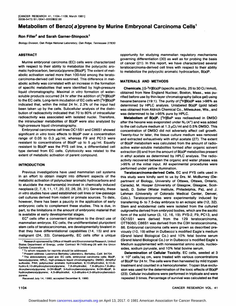
Cellular Toxicity to PAH. Teratocarcinoma-derived cell linesshowed variable susceptibility to the toxic effects of B(a)P(Chart 1). OC15S1 and C86S1 showed an initial toxicity toB(a)P at a concentration of 0.03 /ig/ml and a maximal effect at0.3 /ig/ml (55 and 83%, respectively). F9, PC13, and PYS,however, were resistant to B(a)P up to a concentration of 5fig/ml. To determine if a cytotoxic effect could be demonstrated, resistant cells were incubated in the presence of B(a)Pat 50 jug/ml, subcultured, and then repeatedly exposed toB(a)P (10 /ig/ml) for 48-hr intervals over a 2-week period. Notoxic effects were observed.5
Rate of Water-soluble Metabolite Formation. Since EC cellsshowed a gradational response to the toxic effects of B(a)P,the capacity of these cells to metabolize the parent PAH wasexamined. As indicated in Table 1, each EC cell line demonstrated a varying ability to metabolize B(a)P to water-soluble
derivatives. Consequently, cell cultures were divided into arbitrary groups based on their efficiency for metabolizing parentcompound (7). During a 24-hr incubation period, F9, PC13,
and PYS (Group 1) showed minimal activity (6% or less) andOC15S1 and C86S1 (Group 2) converted 30 and 60% of theinput B(a)P to water-soluble products. Overall water-solubleproduct and organic solvent-soluble metabolite formation varied 56- and 250-fold, respectively, among the cell lines examined. Long-term incubations (72 hr) of EC cells with B(a)Pindicated that the extent of water-soluble product formation
after the first 24 hr did not significantly increase either withGroup 1 or with Group 2 cells.5
Nuclear Uptake of B(a)P. Incubation of EC cells with |'H|-
B(a)P for 24 hr showed that about 2.3% of the input B(a)P hadbeen taken up by the cells; upon subcellular fractionation, 70to 80% of the cellular radioactivity was associated with isolatednuclei (Chart 2). As a result of the significant amount of radioactivity associated intracellularly with the nucleus, the rate ofnuclear accumulation was determined over 72 hr. Since the
0.3 0.5 5.0BENZO(a)PYRENE(^g/ml)
Chart 1. Cellular toxicity of teratocarcinoma-derived cell lines to B(a)P. Increasing concentrations of B(a)P were added in DMSO (final concentration.0.5%) to cells growing in 15 ml of medium. Cell cultures were exposed to PAHfor 24 hr at 37°. Toxicity was determined for each B(a)P concentration in 3
replicate cultures and was calculated as that fraction of viable cells survivingB(a)P treatment as compared to untreated cells (100% survival).
Table 1Extracellular B(a)P metabolite formation by teratocarcinoma-derived cell lines
EC cells were incubated for 24 hr with 750 pmol of [3H]B(a)P. Media was
removed and extracted with ethyl acetate, and the distribution of radioactivitywas determined between the aqueous and the organic solvent phases. Quantification of organic solvent metabolites was determined directly by HPLC analysis.
CelltypePYS-2
F9PC13OC15S1C86S1Water-solubleproducts
formed(pmol /106cells)<1.0(1)a
2.3 (3)4.5 (6)
22.5 (30)45.0 (60)Organic
solvent metabolites formed(pmol/106cells)ND*
0.1 (0.13)1.9(2.8)
20.5 (27.3)25.5 (33.6)Total
metabolism<1.0
3.28.8
57.393.6
5 R. Filler and S. Shinpock. unpublished data.
3 Numbers in parentheses, percentage of product formation with respect to
Chart 2. Nuclear uptake of ( 'H]B(a)P. EC cells were incubated in triplicate forvarious periods of time with | 'H]B(a)P and harvested, and an aliquot was assayed
by direct radioactive measurement for total cellular uptake. Nuclei were thenisolated, and an aliquot was assayed for nuclear accumulation (•).The remainingnuclei were subjected to organic solvent extraction as described in 'Materialsand Methods.' Nuclear uptake was normalized with respect to a constant cell
number. The percentage of nuclear extractable radioactivity (O) was determinedon the basis of that fraction of total nuclear radioactivity that was organic solventsoluble
2.5-
2.0-
1.5-
1.0-
0.5-
0Q.Q
¿ 2.0-
1.5-
10-
0.5-
MEDIUM F9
B(o)P—
Tetrols / TnolS
NUCLEI
Tetrols / Tnols
B(o)P 1
40 80 120FRACTIONNO.
160
Chart 3. Metabolism of B(a)P to ethyl acetate-soluble metabolites found in themedium (top) and in the nuclei (boftom) from F9 EC cells. An aliquot of themedium from EC cells incubated 24 hr with [!H]B(a)P was extracted with ethyl
acetate and subjected to HPLC analysis as described in Materials and Methods." Simultaneously, cells were harvested, and nuclei were isolated and then
extracted with organic solvent for subsequent HPLC fractionation. Although therewere slight variations in the elution positions of B(a)P metabolites, the identity ofindividual species was determined by running standard metabolites.
metabolic conversion of B(a)P appreciably increased after 8hr, this study was initiated at that time in order to correlateextracellular with nuclear metabolite accumulation.5
Nuclear uptake was significant within the first 8 hr (representing 100% of the cellular radioactivity), decreased slightlyat 24 hr, and then reached a constant minimal value (29% of
maximal) at 48 to 72 hr (Chart 2). Nuclei were also subjectedto ethyl acetate extraction to determine if the observed reduction in radioactivity associated with nuclei was accompaniedby a similar reduction in organic solvent extractable material.During the initial 24 hr, there was very little change in extract-
able radioactivity, but this amount did decrease over the following 48 hr at a rate similar to the rate of nuclear accumulation.
HPLC Analysis of Extracellular and Nuclear Ethyl Acetate-soluble Metabolites. Since the PYS cell line did not significantly metabolize B(a)P, the following comparison was limitedto the metabolite profiles from the EC cell lines (Charts 3 to 6).These stem cells showed both qualitative and quantitativedifferences in the activation of parent compound to B(a)Pderivatives which accumulated in the medium and in the nuclei.The overall metabolic activity of EC cells varied approximately30-fold with F9 and C86S1, converting 3 and 94%, respec
tively, of the input B(a)P to various metabolic species.Almost all of the extracellular ethyl acetate-soluble radioac
tivity from F9 and PC13 incubations consisted of unmetabo-
lized B(a)P (>90%). PC13 was able, nevertheless, to metabolize parent hydrocarbon at a very low level to several B(a)Pderivatives resolvable by HPLC. In comparison, OC15S1 andC86S1 showed significant increases in the conversion of B(a)Pto all metabolites. Analysis of the extracellular fraction by HPLCindicated that a large class of metabolites from the C86S1incubation (Chart 6, fop) appeared in the initial eluting peak.This region of the HPLC chromatogram, which represented26% of the total organic solvent-soluble radioactivity (Table 2),has been reported (5) to contain predominantly uncharacter-
5-
3-
1 -
3.0-
200 „i
1.5-
0.3-
PC13MEDIUM
B(o)P—
Tetrols/TrioIs
4,5-(Do)
7,8-did9.10-dioli J
Quiñones
9-OH
3-OH
NUCLEI
Tetrols/Trials
7 8-diol'
40 80 120FRACTION NO.
160 200
Chart 4. Metabolism of B(a)P to ethyl acetate-soluble metabolites found in themedium (top) and in the nuclei (bottom) from PC13 EC cells. 9.10-diol, 9,10-dihydrodiol; 4,5-diol, 4,5-dihydrodiol; 7.8-diol, 7.8-dihydrodiol; 9-OH, 9-OH-B(a)P; 3-OH, 3-OH-B(a)P.
ized tetrols and triols along with sulfate ester conjugates ofmonohydroxybenzo(a)pyrenes.
Of the vicinal glycols, C86S1 had the highest 9,10-dihydro-diol formation followed by OC15S1 and PC13. The K-region
diol was significantly higher in OC15S1 incubations than inPC13 or C86S1, and this diol represented 38% of the totalmetabolites (Table 2). The relative amount of the 7,8-dihydro-
diol appeared to increase with the increasing ability of EC cellsto activate B(a)P but actually reflected a quantitatively decreasing fraction of the total metabolites that were formed by a givenEC cell line (e.g., PC13 > OC15S1 ~ C86S1 ). The dihydrodiols
were the most abundant class of metabolites found in theorganic solvent-soluble fraction, representing 40 to 55% of the
total B(a)P metabolites.Quiñones were also present and represented 10 to 14% of
the total metabolites formed by the various EC cell lines.A striking observation was the ability of EC cells to metabo-
2.0-
1.6-
1.2-
0.8-
o 0.4lgX
o, 25
im2.0-
1.5-
1.0-
0.5-
MEDIUMOC15S1
B(o)P
Tetrols /Triols
9,10-diol4,5-dlol
I 7.8-diol
Quiñones
3-OH
NUCLEI
B(a)P
Quiñones
7,8-diol9-OH
Tetrols/TriolsI
4,5-diol
40 80 120FRACTION NO.
160 200
Chart 5. Metabolism of B(a)Pto ethyl acetate-soluble metabolites found in themedium (fop) and in the nuclei (bottom) from OC15S1 embryonal carcinomacells 9.10-diol, 9.10-dihydrodiol; 4.5-diol. 4,5-dihydrodiol; 7.8-diol. 7,8-dihydro-diol; 9-OH. 9-OH-B(a)P; 3-OH, 3-OH-B(a)P.
Metabolism of B(a)P by EC Cells
lize preferentially B(a)P to the 3-OH derivative relative to the 9-OH-B(a)P. While a relatively constant amount of 3-OH-B(a)P
(17 to 23%) accumulated in the extracellular fraction, theamount of the 9-OH-B(a)P derivative decreased from 11 %
(Table 2, PC13) to 2.9% (Table 2, C86S1). Consequently, theratio of 3-OH-B(a)P to 9-OH-B(a)P formation varied approximately 4-fold.
The presence of parent compound and ethyl acetate-soluble
metabolites within the nuclei of EC cells are also shown inCharts 3 to 6 (bottom). Although metabolite accumulation waslimited in the F9 nuclear fraction, as in the extracellular fraction,a distinct peak was resolved in the region eluting prior to the9,10-dihydrodiol (Chart 3, bottom). For the other EC cell linesexamined, it was apparent that tetrols, triols, 4,5- and 7,8-
dihydrodiols, quiñones,and phenol derivatives of B(a)P, as wellas unmetabolized parent compound, were present in the or
Ãnio
3.0-
2.4-
1.8-
1.2-
0.6-
Ê2.5Ho
2.0-
1.5-
1.0-
0.5-
MEDIUM C86S1
•Tetrols/Triols
9,10-diol
7,8-diol
1Quiñones
3-OH
B(a)P
NUCLEI
Tetrols/Triols
B(a)P
40 80 120FRACTION NO.
160 200
Chart 6. Metabolism of B(a)P to ethyl acetate-soluble metabolites found in themedium (fop) and in the nuclei (boffom) from C86S1 embryonal carcinoma cells.9,10-diol, 9,10-dihydrodiol; 4.5-diol, 4,5-dihydrodiol; 7,8-diol. 7,8-dihydrodiol;9-OH. 9-OH-B(a)P; 3-OH, 3-OH-B(a)P.
Table 2EC cell metabolism of fHJBfaiP: accumulation of ethyl acetate-soluble metabolites in media
EC cells were incubated with [3H]B(a)P for 24 hr; the medium was removed and extracted with ethylacetate, and then an aliquot was subjected to HPLC analysis as described in 'Materials and Methods." The
amount of each metabolite was determined from the HPLC chromatogram and then corrected for the totalpmol found in the original ethyl acetate-soluble fraction of the extracellular medium.
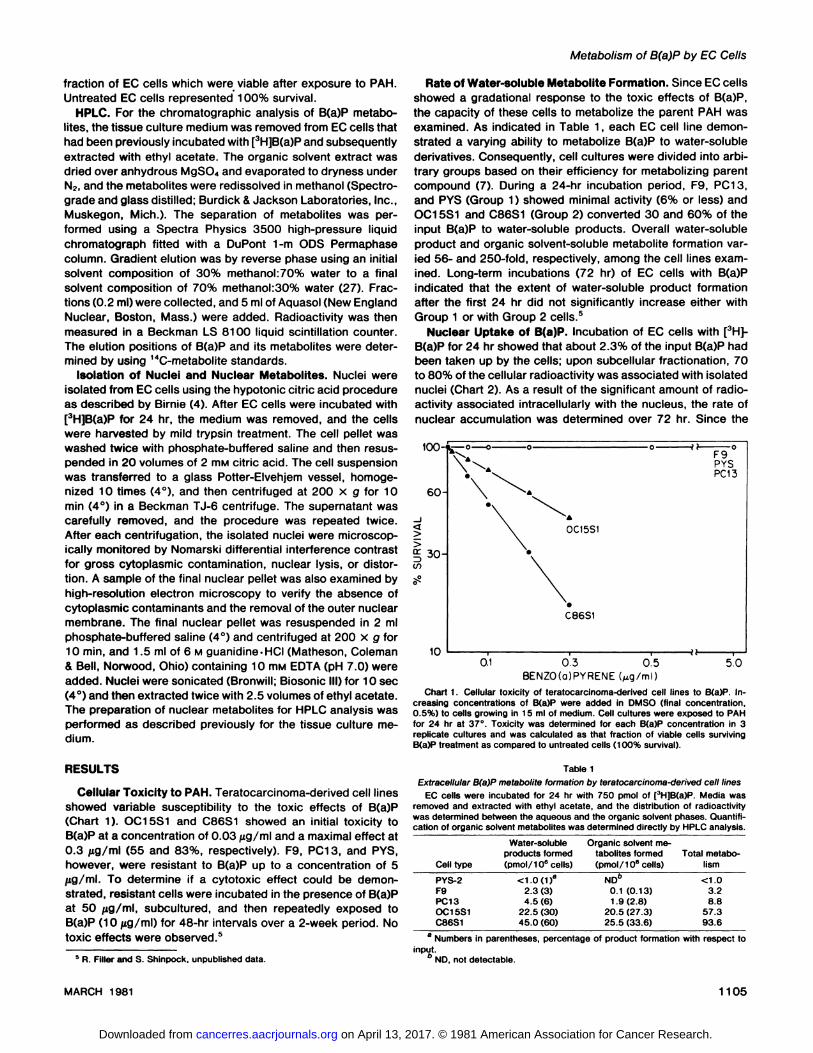
Table 3EC cell metabolism of [3H]B(a)P: nuclear accumulation of parent compound and ethyl acetate-soluble metabolites
EC cells were incubated with [3H]B(a)P as described in Table 2 except that nuclei were isolated from EC cells and extracted with ethyl
acetate, and the metabolites were analyzed by HPLC.pmol/30 X 106nucleiEC
cellsF9
PC13OC15S1C86S1Tetrols/
triols8.4
1.94.13.89,10-Dihy-
drodiol7.64,5-Dihydro-diol0.73.3
11.47,8-Dihydro-
diol2.25.210.8Quiñones7.6
24.425.29-OH-B(a)P3.49.118.33-OH-B(a)P3.0
11.823.8B(a)P121.6
111.172.129.1
ganic solvent fraction from EC cell nuclei. In contrast to themetabolite accumulation in the medium (Table 2), reducedamounts of tetrols and triols were extracted from the nuclei(Table 3). The amount of extractable dihydrodiols increased10-fold with their abundance from individual EC cells ranked in
the following decreasing order: C86S1 > OC15S1 > PC13. Ofthe 3 B(a)P-diols, no detectable quantities of the 9,10-dihydro-
diol were observed in the HPLC chromatograms of F9, PC13,or OC15S1 nuclear extracts. However, C86S1, the EC cell linewhich accumulated the largest amount of this metabolite in themedium (Chart 6, top), also had a significant amount of thisdihydrodiol in its nuclei. Lastly, the phenol derivatives of B(a)Pwere the most abundant class of nuclear metabolites andrepresented 32% of the extractable radioactivity. In contrast tothe extracellular accumulation of 3-OH-B(a)P to 9-OH-B(a)P
derivative formation, phenol accumulation in nuclei was maintained at a relatively constant ratio of 1.2.
DISCUSSION
Experiments performed with low-passage hamster or mouse
fetal cells have demonstrated significant cytotoxicity in response to increasing concentrations of PAH (6-8, 11 ). However, this effect on cell multiplication was observed after long-
term exposure (3 to 6 days). In contrast, OC15S1 and C86S1cells showed B(a)P-induced toxic effects within the first 24 hr.
Although no toxic effects were observed with F9, PC13, orPYS when incubated with B(a)P, organic solvent-soluble me
tabolites did accumulate, although at a very low level, in themedium from PC13 cells. This apparent lack of cellular toxicitymay be due to the presence of an efficient detoxificationsystem, the production of low levels of toxic intermediates, orthe inability to metabolize parent hydrocarbon.
HPLC analysis of the organic solvent-soluble metabolites
produced by the more active EC cell lines clearly demonstratesthe formation of both K- and non-K-region dihydrodiols, as well
as quiñones and phenolic derivatives. In comparison to theB(a)P metabolic profiles obtained from mouse and hamstercells (28), EC cells differ markedly in the production of significant quantities of the 4,5-dihydrodiol and show an apparent
reorientation in the enzymatic hydroxylation of B(a)P. Insteadof a 2- to 2.5-fold increase in the ratio of 3-OH-B(a)P to 9-OH-
B(a)P formation, as in the case of mouse or hamster cells (28),there is a 4-fold greater formation of the 3-hydroxy than the 9-
hydroxy species (Table 2). This difference in phenol accumulation may be due to a stereoselective specificity of the hy-
droxylating enzyme system or to a preferential conjugation ofthe 9-OH-B(a)P. However, this latter alternative appears unlikely since EC cells are unable, under the present incubation
conditions, to form glucuronic acid or sulfate ester conjugates.6
The metabolite pattern obtained from EC cells is more similarto microsomal preparations from rat, mouse, and hamster livers(28). The relative amount of water-soluble to organic solvent-soluble product formation indicated that there was a 10-fold
greater increase in nonconjugated product accumulation (Table 1). This suggests that detoxifying pathways of the moreactive EC cells appears to be limiting in its ability to handleoxygenated derivatives.
Initial experiments indicated that the kinetics of B(a)P uptakeinto each of the EC cell lines was very similar and that intra-
cellular saturation occurred within the first hr of exposure tothe hydrocarbon. Although others have shown that a very smallpercentage of the intracellular radioactivity was associated withthe nuclear fraction (17), we observed that nuclei, isolated fromEC cells incubated with high specific activity [3H]B(a)P, accu
mulated >70% of the cellular radioactivity. This large nuclear-
associated radioactivity may, in part, be due to the strikingmorphology of EC cells which have a high nucleocytoplasmicratio (25). The decrease in nuclear radioactivity during long-
term incubations probably reflects a metabolic equilibrium between intracellular and extracellular compartments and is supported by the concomitant decrease in ethyl acetate-extracta-
ble radioactivity from isolated nuclei. HPLC analysis of nuclearmetabolites indicates that unmetabolized parent hydrocarbon,as well as various reactive intermediates, were present. Ofnotable interest was the presence of the 9,10-dihydrodiol. It
has been reported that there appears to be a selectivity in thecellular distribution of the 9,10- and the 7,8-dihydrodiols with
respect to their preferential accumulation in either extracellularor intracellular compartments (13, 17). We have also observedthis with respect to their extracellular and nuclear distribution.Whether this difference is attributable to structural features orto their interaction with cellular macromolecules is currentlynot known.
There is an apparent correlation between the inherent differ-entiational capability of the various teratocarcinoma-derived
cell lines and their biotransformation of xenobiotics. One ofthese cell cultures (i.e., PYS-2) is an endodermal derivative
isolated from the in vitro differentiation of OTT6050 stem cells(16). Although EC cells have tumorigenic potential when injected into syngeneic mice, they can be further characterizedaccording to the cell type present in the tumor. EC cells whichgive rise to tumors containing only stem cells and have apparently lost their differentiational capacity are designated asnullipotent. F9 is such an EC cell type which almost exclusivelyundergoes stem cell renewal but can, under certain conditions,form a limited amount of primitive endoderm-like cells (29, 34).
1R. Filler and K. J. Lew, manuscript in preparation.
In contrast, PC13 and OC15S1 are pluripotent EC cells (19,30). Tumors derived from them contain derivatives of all 3embryonic germ layers. Although PC13 readily differentiates invivo, it has a restricted in vitro developmental capacity andrequires the presence of retinoic acid for limited differentiation(1). The C86S1 EC cell line has both in vivo and in vitrodevelopment capacity (19).
Although we are reporting the metabolic characterization of4 cell lines derived from the OTT6050 and one cell line derivedfrom the 86 teratocarcinomas, we have also examined another6050 pluripotent EC cell line, 247 DIP/DES (16), as well as asecond nullipotent EC cell, SCC1, derived from the spontaneous teratocarcinoma 402 (18). Analogous results were obtained with these 2 cell types as with OC15S1 and F9.7 It is
notable that the pluripotent state is associated with significantmetabolic activity and that either with restriction in developmental potential of these embryo-like cells (i.e., F9) or with
their developmental commitment to a differentiated phenotype(i.e., PYS), the presence of an active metabolic character isreduced. This would suggest that certain cell types of thedeveloping embryo may be primary sites of metabolite activation and may serve as a point of origin for producing reactiveintermediates that interact with adjacent cell types via metabolic cooperativity (9). This could be of importance in considering the ontological relationship between embryonic susceptibility to transforming species and transplacental carclnogen-
esis (35). Experiments are in progress to test this hypothesis.In conclusion, the results reported in this study indicate that
the inclusion of EC cells in the examination of biotransformationof PAH's to various reactive intermediates complements and
extends other investigations utilizing fetal and adult systems.The formation of toxic and transforming derivatives, which maybe involved in carcinogenesis, mutagenesis, or teratogenesis,is modulated by the metabolic balance between activating anddetoxifying enzyme systems. This complex biochemical relationship, which may vary with the developmental state of theorganism, determines overall susceptibility to chemically induced biological alterations. Thus, the EC cell system with itsdifferentiational characteristics would serve as a useful probefor the elucidation of regulatory parameters involved in theseprocesses.
ACKNOWLEDGMENTS
We especially thank Dr. Liane B. Russell for her encouragement and supportof our work and Dr. James K. Selkirk for his time and patience in unraveling thecomplexities of HPLC technology and PAH metabolism.
REFERENCES
1. Adamson. E. D.. Gaunt. S. J.. and Graham, C. F. The differentiation ofteratocarcinoma stem cells is marked by the types of collagen which aresynthesized. Cell, 17: 469-476, 1979.
2. Andrianov, L. N., Belitsky, G. A., Ivanova, D. J., Khesina, A. Y., Khitrovo, S.S., Shabad, S. M., and Vaseliev, J. M. Metabolic degradation of 3,4-benzopyrene in the cultures of normal and neoplastic fibroblasts. Br. J.Cancer, 2i: 566-575, 1967.
3. Bernstine. E. G., Hooper, M. L., Grandchamp, S., and Ephrussi. B. Alkalinephosphatase activity in mouse teratomas. Proc. Nati. Acad. Sei. U. S. A..,70: 3899-3903, 1973.
7 R. Filler, unpublished data.
4. Birnie. G. Isolation of nuclei from animal cells in culture. Methods Cell Biol.,17: 13-26, 1978.
5. Cohen, G. M., Marchok, A. C.. Nettesheim. P., Steele. V. E., Nelson, F.,Huang, S., and Selkirk, J. K. Comparative metabolism of benzo(a)pyrene inorgan and cell cultures derived from rat tracheas. Cancer Res., 39 1980-
1984, 1979.6. Diamond, L. The effect of carcinogenic hydrocarbons on rodent and primate
cells in vitro. J. Cell. Comp. Physiol., 66: 183-198, 1965.
7. Diamond, L. Metabolism of polycyclic hydrocarbons in mammalian cellcultures. Int. J. Cancer, 8. 451-467, 1971.
8. Diamond, L., Sardet, C., and Rothblat, G. H. The metabolism of 7,12-dimethylbenz(a)anthracene in cell cultures. Int. J. Cancer. 3. 838-849.1968.
9. Gaunt, S. J., and Papaioannou, V. Metabolic co-operation between embryonic and embryonal carcinoma cells of the mouse. J. Embryol. Exp. Mor-phol., 54. 263-275, 1979.
10. Hooper, M. L.. and Slack, C. Metabolic co-operation in HGPRT* andHGPRT" embryonal carcinoma cells. Dev. Biol., 55. 271 -284, 1977.
11. Huberman, E., Selkirk, J. K., and Heidelberger, C. Metabolism of polycyclicaromatic hydrocarbons in cell cultures. Cancer Res., 31: 2161-2167, 1971.
12. lies, S. A. Mouse teratomas and embryoid bodies: their induction anddifferentiation. J. Embryol. Exp. Morphol.. 38: 63-75, 1977.
13. Jones, C. A., Moore. B. P., Cohen, G. M., Fry, J. R., and Bridges. J. W.Studies on the metabolism and excretion of benzo(a)pyrene in isolated adultrat hepatocytes. Biochem. Pharmacol., 27: 693-702, 1978.
14. Kahan, B. W., and Ephrussi, B. Developmental potentialities of clonal in vitrocultures of mouse testicular teratomas. J. Nati. Cancer Inst., 44: 1015-1036, 1970.
15. Kleinsmith, L. J.. and Pierce, G. B. Multipotentiality of single embryonalcarcinoma cells. Cancer Res., 24: 1544-1552, 1964.
16. Lehman, J. M., Speers, W. C., Swartzendruber, D. E., and Pierce, G. B.Neoplastic differentiation: characteristics of cell lines derived from a murineteratocarcinoma. J. Cell. Physiol., 84: 13-28, 1974.
17. MacLeod, M. C., Cohen, G. M., and Selkirk, J. K. Metabolism and macro-molecular binding of the carcinogen benzo(a)pyrene and its relatively inertisomer benzo(e)pyrene by hamster embryo cells. Cancer Res., 39: 3463-3470, 1979.
18. Martin, G. R., and Evans, M. J. Differentiation of clonal lines of teratocarcinoma cells: formation of embryoid bodies in vitro. Proc. Nati. Acad. Sei., U.S. A., 72: 1441-1445, 1975.
19. McBurney, M. W. Clonal lines of teratocarcinoma cells in vitro: differentiationand cytogenetic characteristics. J. Cell. Physiol., 89: 441-456, 1976.
20. Miller, J. A. Carcinogenesis by chemicals: an overview. Cancer Res., 30:559-576, 1970.
21. Mintz, B., and Illmensee, K. Normal genetically mosaic mice produced frommalignant teratocarcinoma cells. Proc. Nati. Acad. Sei. U. S. A., 72: 3585-3589, 1975.
22. Nebert, D. W. Use of fetal cell culture as an experimental system forpredicting drug metabolism in the intact animal. Clin. Pharmacol. Ther., 14:693-699, 1973.
23. Patterson, M. K. Measurement of growth and viability of cells in culture.Methods Enzymol., 58: 141-152, 1979.
24. Pierce, G. B. Teratocarcinoma: model for a developmental concept ofcancer. Curr. Top. Dev Biol., 2: 223-246, 1967.
25. Pierce, G. B., and Beals, T. F. The ultrastructure of primordial germ cells ofthe fetal testes and of embryonal carcinoma cells of mice. Cancer Res., 24:1552-1567, 1964.
26. Rüdiger,H. W., Marxen, J., Kohl, F.-V., Melderis, H., and Wiehert. P. V.Metabolism and formation of DNA adducts of benzo(a)pyrene in humandiploid fibroblasts. Cancer Res., 39: 1083-1088, 1979.
27. Selkirk, J. K., Croy, R. G., and Gelboin, H. V. Benzo(a)pyrene metabolites:efficient and rapid separation by high-pressure liquid chromatography.Science (Wash. D.C.), 184: 169-171, 1974.
28. Selkirk, J. K., Croy, R. G., Wiebel, F. J., and Gelboin. H. V. Differences inbenzo(a)pyrene metabolism between rodent liver and embryonic cells. Cancer Res.. 36 4476-4479, 1976.
29. Sherman, M. I., and Miller, R. A. F9 embryonal carcinoma cells can differentiate into endoderm-like cells. Dev. Biol., 63: 27-34, 1978.
30. Sherman, M. I., and Solter, D. (eds.). Teratomas and Differentiation. NewYork: Academic Press, Inc., 1975.
31. Sims, P. The metabolism of some aromatic hydrocarbons by mouse embryocell cultures. Biochem. Pharmacol., 79: 285-297, 1970.
32. Stevens, L. The development of transplantable teratocarcinoma from intra-testicular grafts of pre- and postimplantation mouse embryos. Dev. Biol., 21:364-382, 1970.
33. Stevens, L. C. The biology of teratomas. Adv. Morphog., 6: 1-31, 1967.34. Strickland, S., and Mandavi, V. The induction of differentiation in teratocar
cinoma stem cells by retinoic acid. Cell, 15: 393-404, 1978.35. Tanaka, T. Transplacental induction of tumors and malformation in rats
treated with some chemical carcinogens. In: L. Tomatis and U. Mohr (eds.),Transplacental Carcinogenesis, pp. 100-111. Lyons, France: InternationalAgency for Research on Cancer, 1973.


![Benzo[a]pyrene in River Sediment](https://static.documents.pub/doc/80x56/613d64f9736caf36b75cd07b/benzoapyrene-in-river-sediment.jpg)


