METABOLISM OF THE THYROID HORMONE IN THE RAT AS SHOWN BY PHYSIOLOGICAL DOSES OF LABELED THYROXINE BY J. GROSS AND C. I’. LEBLOND (From the Department of Anatomy, McGill University, Montreal, Canada) (Received for publication, November 22, 1949) Since thyroxine, uncombined in peptide linkage, has been detected by isotope dilution and paper chromatography in thyroid tissue (1, 2) and in plasma (l-3), it is likely that this substance is the hormonal secretion of the thyroid gland. Previously, the fate of labeled thyroxine has been de- termined after the injection of large amounts (4-6), considerably in excess of the few micrograms which constitute the daily thyroxine secretion of the rat thyroid (7). In the present work, methods were devised by which it was possible to prepare 0.007 y of labeled thyroxine containing enough radioactivity to be easily traced in the body of the rat. The distribution of thyroxine thus obtained was then found to be comparable to that of radioiodide (NaI131)in rats sacrificed long enough after injection to insure the conversion of the radioiodide to thyroid hormone. Methods Chemical Synthesis of Labeled m-Thyrox&e-r&-Thyroxine labeled in the 3’ and 5’ positions was prepared from DL-diiodothyronine in the follow- ing manner. The radioactive iodine as iodide, mixed with 0.70 mg. of potassium iodide and a drop of strong ammonium hydroxide, was brought to a volume of about 0.1 cc. on a water bath under nitrogen. By the addition of 0.05 cc. of 1 N sulfuric acid and 0.05 cc. of potassium iodate solution containing 80 mg. per cc., elemental iodine was obtained.’ To this mixture 0.68 mg. of DL-diiodothyronine, dissolved in 0.1 cc. of strong ammonium hydroxide, was added rapidly. 10 per cent acetic acid was added dropwise until a precipitate appeared and the iodine color returned. The precipitate was removed by centrifugation, redissolved in strong NHdOH, and added to the supernatant containing free iodine for the io- dination of any remaining diiodothyronine. This procedure was repeated t,hree times. After the final acidification, 0.1 cc. of 0.1 N thiosulfate was 1 In some batches of iodine, the amounts of bisulfite present interfered with the formation of free iodine. This was overcome by oxidation with hydrogen peroxide in an acid medium. The excess peroxide was removed by adding ammonium hy- droxide and evaporating the mixture to dryness. Subsequent treatment was as described in the text. 489 by guest on August 22, 2018 http://www.jbc.org/ Downloaded from
Transcript
METABOLISM OF THE THYROID HORMONE IN THE RAT AS SHOWN BY PHYSIOLOGICAL DOSES OF LABELED
THYROXINE
BY J. GROSS AND C. I’. LEBLOND
(From the Department of Anatomy, McGill University, Montreal, Canada)
(Received for publication, November 22, 1949)
Since thyroxine, uncombined in peptide linkage, has been detected by isotope dilution and paper chromatography in thyroid tissue (1, 2) and in plasma (l-3), it is likely that this substance is the hormonal secretion of the thyroid gland. Previously, the fate of labeled thyroxine has been de- termined after the injection of large amounts (4-6), considerably in excess of the few micrograms which constitute the daily thyroxine secretion of the rat thyroid (7). In the present work, methods were devised by which it was possible to prepare 0.007 y of labeled thyroxine containing enough radioactivity to be easily traced in the body of the rat. The distribution of thyroxine thus obtained was then found to be comparable to that of radioiodide (NaI131) in rats sacrificed long enough after injection to insure the conversion of the radioiodide to thyroid hormone.
Methods
Chemical Synthesis of Labeled m-Thyrox&e-r&-Thyroxine labeled in the 3’ and 5’ positions was prepared from DL-diiodothyronine in the follow- ing manner. The radioactive iodine as iodide, mixed with 0.70 mg. of potassium iodide and a drop of strong ammonium hydroxide, was brought to a volume of about 0.1 cc. on a water bath under nitrogen. By the addition of 0.05 cc. of 1 N sulfuric acid and 0.05 cc. of potassium iodate solution containing 80 mg. per cc., elemental iodine was obtained.’ To this mixture 0.68 mg. of DL-diiodothyronine, dissolved in 0.1 cc. of strong ammonium hydroxide, was added rapidly. 10 per cent acetic acid was added dropwise until a precipitate appeared and the iodine color returned. The precipitate was removed by centrifugation, redissolved in strong NHdOH, and added to the supernatant containing free iodine for the io- dination of any remaining diiodothyronine. This procedure was repeated t,hree times. After the final acidification, 0.1 cc. of 0.1 N thiosulfate was
1 In some batches of iodine, the amounts of bisulfite present interfered with the formation of free iodine. This was overcome by oxidation with hydrogen peroxide in an acid medium. The excess peroxide was removed by adding ammonium hy- droxide and evaporating the mixture to dryness. Subsequent treatment was as described in the text.
added to remove the residual iodine. The precipitate was washed twice with 0.2 cc. of 1 per cent acetic acid, dissolved in 0.2 cc. of 2 N sodium hydroxide, and reprecipitated twice with a few drops of 10 per cent acetic acid. The final thyroxine precipitate was dissolved in a minimal amount of hot 0.1 N potassium carbonate; 0.05 cc. of 15 per cent sodium chloride was added, and, on cooling, the alkali salt precipitated. After centrifuga- tion, the residue was dissolved in 0.05 cc. of 2 N sodium hydroxide and diluted to 5 cc.
The iodine content, estimated by the method of Groak (8), was found to be equivalent to 655 y of thyroxine, while the radioactivity was equal to 32.6 per cent of the starting material, which had an activity of 5 mc. By means of the isotope dilution procedure detailed below, 95 per cent of the radioiodine was found to be in the form of thyroxine.
Extraction of Labeled L-Thyroxine from Plasma-Female albino rats, kept on Remington’s iodine-deficient Diet 342 (9), supplemented with 10 per cent brewers’ yeast, were injected subcutaneously with about 50 PC. of carrier-free radioactive iodide. The animals were exsanguinated 48 hours later, and the heparinized plasma was extracted with twice its volume of n-butanol, and three times again with an equal volume of butanol. The combined butanol fractions were then evaporated in vacua at room tem- perature. The dry residue was taken up in about 0.5 CC. of distilled water. An aliquot of this solution was removed for the determination of thyroxine content by means of isotope dilution (see below). If the radioactivity in this aliquot proved to consist of 83 per cent or more of radiothyroxine, the remainder was used for injection. On the average, the injection mixtures contained 92 per cent of their radioactivity in the form of thyroxine. Be- cause of the mild extraction method, it seems likely that little or no race- mization occurred, thus giving the L-amino acid labeled in any one of the four positions.
Isotope Dilution Technique-25 mg. of inactive m-thyroxine were added to the sample of radioactive material, dissolved with about 3 drops of 2 N
sodium hydroxide, and diluted to 5 cc. with water. An aliquot was re- moved for iodine estimation (8) and for measurement of radioactivity to determine the specific activity. 3 drops of 10 per cent acetic acid were added in order to precipitate thyroxine, which was then washed with two 0.5 cc. portions of distilled water and dissolved in a minimal amount of hot 0.1 N potassium carbonate (about 0.5 cc.). On cooling, the potassium salt of thyroxine separated and, after removal of the supernatant, was dissolved in 1 cc. of 0.2 N sodium hydroxide in 70 per cent ethanol. Thyroxine was precipitated with acetic acid, washed with water, redissolved in alkaline alcohol, and reprecipitated as the potassium salt four times. Aliquots were taken from the alcoholic solutions of the second, third, and fourth salts for the determination of specific activity, which became constant with
the formation of the second potassium salt. The percentage of the con- stant specific activity over the initial specific activity indicated the propor- tion of the radioactivity present as thyroxine in the solution injected.
Distribution of Radiothyroxine-Female albino rats, fed on an adequate iodine intake (Purina fox chow) and weighing 80 to 120 gm., were injected with radiothyroxine intravenously, via the femoral vein. After periods of 2, 24, or 72 hours, the animals were etherized and exsanguinated via the inferior vena cava. The organs and tissues were weighed wet and then put into solution in 2 N sodium hydroxide on a water bath for radio- activity measurement as previously described (5). In calculating the total plasma volume, the formula plasma volume = O.l22(body weight)0.778 was used (10).
Those samples containing a sufficient amount of radioactivity were frac- tionated with butanol into thyroxine-like and non-thyroxine iodine, with no further hydrolysis than had occurred in the course of the homogenization of the organs in sodium hydroxide. 50 y of inactive m-thyroxine were added to each sample. The material was then extracted with butanol (5). The radioactivity found in the butanol extract or thyroxine-like fraction was expressed as a percentage of the total activity; i.e., the sum of the activities of the butanol and alkali fractions. In control runs, 88 per cent of added radiothyroxine was recovered by this procedure.
Three series of distribution experiments (Fig. 1, Tables II and III) were carried out. (1) Three groups of two animals were injected with 20 y of m-thyroxine synthesized from nn-diiodothyronine, and sacrificed at 2, 24, and 72 hours after injection, respectively. (2) Three groups of two ani- mals were injected with about 0.07 y of L-thyroxine extracted from rat plasma, and sacrificed at 2, 24, and 72 hours after injection, respectively. (3) Three single animals were injected with about 0.007 y of L-thyroxine extracted from rat plasma, and sacrificed at 2, 24, and 72 hours after in- jection, respectively. The amounts of thyroxine injected in the latter two series were calculated from the iodine content of the butanol extract of the plasma of the iodine-deficient donor animals; i.e., about 1.2 y per 100 cc. of plasma.
Additional data on the excretion of thyroxine were obtained in an experi- ment in which three female albino rats, weighing about 170 gm., were in- jected intravenously with 0.001 y of thyroxine (Table I).
Distribution of Radioiodide-For purposes of comparison, six female albino rats, fed on Purina fox chow and weighing 80 to 120 gm., received an intravenous injection of carrier-free radioiodide, and were sacrificed in groups of two after 2, 24, and 72 hours. The radioactivity of the organs of these animals was estimated and fractionated with the same methods that were used on the organs of the thyroxine-treated animals (Table IV).
Finally an attempt was made to identify thyroxine in the peripheral or-
gans of radioiodide-treated rats by isotope dilution, with use of animals on a low iodine diet, which were sacrificed 48 hours after the injection of about 50 PC. of NaP. The organs of these animals were homogenized in a Waring blendor with 10 cc. of 0.9 per cent sodium chloride per gm. of tis- sue. The mixture was extracted three times with butanol containing 10 y
FIG. 1. Distribution of labeled thyroxine. The columns indicate the percentages of the injected doses present in the various organs at the three time intervals after injection. With the larger two doses, the values for the individual animals are indi- cated by the straight and notched lines, respectively, at the top of the column.
of carrier thyroxine per cc. The combined butanol fraction was evaporated under reduced pressure to about 5 cc. and washed twice with 5 cc. of 4 N
sodium hydroxide containing 5 per cent sodium carbonate. Isotope dilu- tion was then carried out on the dried butanol extract.
Results
Distribution of Radiothyrotine-The distribution of thyroxine on the basis of the percentage of the injected dose (Fig. 1) was qualitatively similar
for the three dose levels. Thus, as early as 2 hours after injection, over 70 per cent of the dose had left the circulation to find its way mainly into the liver, muscles, small intestine, and skin. At 24 and 72 hours, most of the injected radioactivity was found in feces and urine. With the ex- ception of the thyroid gland, in which there was a slow increase in radio- activity with time, radioactivity in the organs gradually decreased. How- ever, the large intestine showed a maximal value at 24 hours, while at the same time there was a marked decline in the radioactive content of the small intestine and, on the other hand, large amounts of radioactivity in the feces. Apparently, the material which entered the small intestine at 2 hours had passed into the large intestine and feces at the end of 24 hours.
A quantitative comparison of the three dose levels showed that with de- creasing dosage there was an increase in the percentage of the dose remain-
TABLE I Per Cent of Injected Dose Present in Feces and Urine at Various Time Intervals after
ing in the plasma, with a corresponding decrease in the proportion found in the liver. This phenomenon was most apparent 2 hours after injection (Fig. 1). At 24 hours and subsequently, the differences were most evi- dent in the amounts of radioactivity found in the excreta, a lower fecal and a higher urinary excretion occurring with a decreasing dose at either time interval. This was borne out by the experiment with 0.001 y of labeled thyroxine, in which about 40 per cent of the radioactivity passed into the urine and about 50 per cent into the feces (Table I).
The concentrations of the radioactivity in the various organs (Table II) showed the highest values in plasma, liver, and kidney at all time intervals, the highest plasma concentration being obtained with the smallest dose. Of the remaining organs, the lung, non-thyroid endocrine organs, cardiac muscle, pancreas, and salivary glands showed the higher concentrations. No remarkable concentration could be detected in the hypophysis, con- trary to results obtained in the rabbit (4, 11).
Butanob fractionation of some of the samples (Table III) revealed that the thyroxine-like fraction was high at 2 hours in most of the organs and,
TABLE III Fractionation of Organs and Excreta at Various Time Intervals after Injection of Vari-
ous Doses of Labeled Thyroxine The results are expressed as per cent total 11s’ present in the butanol fraction.
All blank spaces correspond to samples with insufficient radioactivity for fraction- ation.
Plasma
Stomach
Small intestine
Large “
Feces
Liver
Kidney
Urine
Spleen
Lungs
Skin
Skeletal muscle
Cardiac “
Adrenal
20 y m-thyroxine
- 1 hn. 4 hrs.
69 73 9
16 63 54 31 37
70 71 64 65
4 2
69 56 60 59 63 53 68 75 75 69 61 55
61 55 3 3
39 42 50 48 46 41 52 58 55 52
1
1 41 38 64 41 31 44 60 66 63 64 51 49
- 1 2 hrs.
_-
62 73 7 9
38 39 49 43 50 49 17 20 39 32
1
1 28 28 38 43 20 33 56 50 51 39
-
-
.-
-
0.07 y I-thyroxine i- 1 hrs.
79 82 27 19 25 67
85 86 83 90 6 7
82 90 86 72 61 64 60 64
!4 hn. 2 hrs.
77 61
5 8
31 39 14 42 12 40 74 70 69 71
1
10 61 54 69
83
54
18 10 20 82 61 85 54 78 44 32 64 34 4
20 46 59
64 37
62
13 70 59 63
2 hrs.
86
4 hrs.
80
40
27
50
‘2 hn.
i8
except for the plasma, decreased steadily with time., Two other exceptions to this pattern were the stomach and urine, in which very little of the radio- activity was present in the thyroxine-like form at any time interval. Note that the values in Table III are about 12 per cent too low (see “Methods”).
Distribution of Tracer Radioiodide-The distribution pattern of radio-
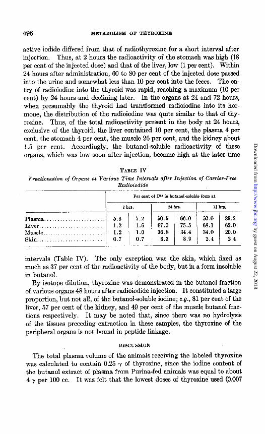
active iodide differed from that of radiothyroxine for a short interval after injection. Thus, at 2 hours the radioactivity of the stomach was high (18 per cent of the injected dose) and that of the liver, low (1 per cent). Within 24 hours after administration, 60 to 80 per cent of the injected dose passed into the urine and somewhat less than 10 per cent into the feces. The en- try of radioiodine into the thyroid was rapid, reaching a maximum (10 per cent) by 24 hours and declining later. In the organs at 24 and 72 hours, when presumably the thyroid had transformed radioiodine into its hor- mone, the distribution of the radioiodine was quite similar to that of thy- roxine. Thus, of the total radioactivity present in the body at 24 hours, exclusive of the thyroid, the liver contained 10 per cent, the plasma 4 per cent, the stomach 4 per cent, the muscle 26 per cent, and the kidney about 1.5 per cent. Accordingly, the butanol-soluble radioactivity ‘of these organs, which was low soon after injection, became high at the later time
TABLE IV
Fractionation of Organs at Various Time Intervals after Injection of Carrier-Free Radioiodide
intervals (Table IV). The only exception was the skin, which fixed as much as 37 per cent of the radioactivity of the body, but in a form insoluble in butanol.
By isotope dilution, thyroxine was demonstrated in the butanol fraction of various organs 48 hours after radioiodide injection. It constituted a large proportion, but not all, of the butanol-soluble iodine; e.g., 81 per cent of the liver, 57 per cent of the kidney, and 49 per cent of the muscle butanol frac- tions respectively. It may be noted that, since there was no hydrolysis of the tissues preceding extraction in these samples, the thyroxine of the peripheral organs is not bound in peptide linkage.
DISCUSSION
The total plasma volume of the animals receiving the labeled thyroxine was calculated to contain 0.25 y of thyroxine, since the iodine content of the butanol extract of plasma from Purina-fed animals was equal to about 4 7 per 100 cc. It was felt that the lowest doses of thyroxine used (0.007
and 0.001 -y) would not increase this amount significantly and, therefore should not interfere with normal physiological processes. Furthermore, since thyroxine combines easily with plasma proteins to form a complex which may be dissociated with butanol (2), as does the endogenous hor- mone (2, 3, 12), it was felt that the injected material would be similarly bound and would therefore behave like a tracer of the thyroid hormone. In favor of this assumption was the fact that the distribution of thyroxine was similar to that obtained 24 to 72 hours after the administration of radioiodide; that is, after a sufficient interval for the conversion of this iodide into thyroid hormone (see “Results” and Table IV). It was then concluded that the distribution of at least the smallest doses of labeled thyroxine reflected that of the endogenous hormone.
It is generally assumed that a hormone concentrates in its target organs. However, soon after injection, tracer doses of thyroxine were usually less concentrated in the organs than in plasma (Table II). There was a rapid fall of concentration with time in both, but the rate of fall was more rapid in the plasma than in the following organs: liver, kidney, skin, brain, spleen, muscle, fat, and perhaps adrenal and pancreas. This may indicate that the initial entry of thyroxine into the organs is dependent on the plasma level, but that the turnover of the thyroxine iodine is slower in the organs than in plasma.
The slower turnover of thyroxine in the organs was probably related to its conversion into butanol-insoluble metabolites, since most of the or- gans showed a steady decrease with time in the proportion of their butanol- soluble radioactivity, while in the plasma this fraction remained at about the same high level at all time intervals (Table III). The presence of thyroxine in the organs may be associated with biological activity, since this hormone was required by the thyroidectomized rat in order to main- tain the normal weight of liver, kidney, spleen, and adrenal (13) as well as the normal histology of skin and muscle (14). The high concentration of thyroxine in the liver may be also related to the biliary secretion of thyrox- ine, since it has been shown previously that the fecal radioactivity came from this organ through the bile (5, 6).
There is a rapid fall in the thyroxine content of the whole body, as well as in that of the individual organs. The retained thyroxine, calculated from the difference between the radioactivity injected and excreted (Ta- ble I), showed a roughly linear relation to time. 50 per cent of the dose was thus found to be excreted in 18 to 24 hours (that is, the biological half life). Therefore, the time for the complete renewal of the body thyroxine of these animals (1.44 X the ,half life (15)) ranged from 25 to 35 hours.
The elimination of radioactivity from the body takes place in urine and mostly in feces (Fig. 2). However, the excretion was less pronounced with
FIG. 2. A comparison of the excretion of radioactivity over the 24 hour period following the injection of various doses of radiothyroxine. The figure has been con- structed from results presented in this paper and in a previous publication (5). The stippled portions of the columns represent the urinary I131 excretion; the solid black, the fecal Ir*r excretion. The distance between the base-line and the white lines indi- cates the amount of fecal radioactivity present in a butanol-soluble form. As the dose increases, both the percentage of the dose excreted in the feces and the propor- tion present in the butanol-soluble fraction increase.
TABLE V
Comparison of AverageI’ Concentrations* of Radioactivity in Organs B HOUTS after Iniection of Various Doses of Radiothyroxine
the tracer than with the large doses (Fig. 2). The decrease in rate of ex- cretion with the dose could be related to a concomitant increase in the radioactivity of plasma and organs (Table V). This in turn was accom- panied by an increased utilization of thyroxine with the tracer doses, since it could be calculated from Fig. 1 and Table III that 75 per cent of such a dose became butanol-insoluble within 24 hours, in contrast to only 54 per cent of the large dose (800 7) previously used (5). Similarly, more butanol- insoluble radioactivity was present in the feces with small than with large doses (Fig. 2). From the excretion of tracer doses, it could be estimated that, of the thyroxine present in the body of the rat at any one time, less than a fifth is likely to be lost in the feces as thyroxine itself.
Finally, the rapid destruction and excretion of thyroxine contrasted with its well known ability to produce a sustained rise of oxygen consumption. The metabolic stimulation must, therefore, be the end result of a series of chain reactions initiated while thyroxine was present in the body.
SUMMARY
1. Female albino rats, weighing 80 to 120 gm. and fed on Purina fox chow, were injected intravenously with doses of 1131-labeled thyroxine vary- ing from 0.001 to 20 y. The 20 y dose was prepared from nn-diiodothy- ronine and was labeled in the 3’,5’ positions; the others were obtained from the plasma of radioiodide-treated rats and consisted of n-thyroxine randomly labeled in any of the four iodine positions. The purity of the radiothyroxine, estimated by isotope dilution, averaged 92 per cent.
The organs and excreta were analyzed for total radioactivity 2, 24, and 72 hours after injection (Fig. 1, Tables I and II), and, in some cases, were fractionated by means of butanol into thyroxine-like and non-thyroxine fractions (Table III).
2. Thyroxine enters all of the organs investigated without being stored in any one. The highest concentrations are found in plasma, liver, and kidney, but a large proportion of the body thyroxine is present in muscle. With time, the radioactivity decreases rapidly in all organs, although at a somewhat faster rate in the plasma than in liver, kidney, skin, brain, spleen, muscle, fat, and perhaps adrenals and pancreas. This difference could be accounted for by a transformation of thyroxine into butanol-insoluble metabolites in these organs (Table III). As an index of the over-all rate of metabolism, the turn-over time of thyroxine was calculated from Table I and found to be about 25 to 35 hours in the rat.
3. While thyroxine, administered in large doses, is to a great extent ex- creted in the feces as thyroxine-like material, tracer doses are excreted in both urine and feces, mostly in a non-thyroxine form (Fig. 2). Thus, it may be calculated from Fig. 1 and Table III that less than 20 per cent of
the body thyroxine is excreted as such, the rest being converted to its metabolites.
4. The distribution of thyroxine was compared to that of the endogenous thyroid hormone in experiments with tracer radioactive iodide. At 2 hours after iodide injection, the radioactivity is found mainly in the stomach and urine in a butanol-insoluble form (Table IV). After 24 to 72 hours, a suf- ficient interval for the thyroid to take up radioiodine and release it as thyroid hormone, the organ radioactivity, which is then largely butanol- soluble (Table V), has a distribution similar to that of radiothyroxine. Iso- tope dilution of the butanol fractions demonstrates the presence of some thyroxine, uncombined in peptide linkage.
From these facts, it is concluded that the results obtained with the small- est doses of labeled thyroxine indicate the pathways of metabolism of the endogenous thyroid hormone.
This work was supported by a grant from the National Research Council of Canada, from whose facilities at Chalk River, Ontario, the radioactive iodine was obtained.
The diiodothyronine and some of the thyroxine were supplied by Hoff- mann-La Roche, Ltd.
Considerable technical assistance was provided by Miss Jeanne Cambron and Mrs. Joan Mason Dougherty.
BIBLIOGRAPHY
1. Leblond, C. P., and Gross, J., J. Clin. Endocrinol., 9, 149 (1949). 2. Gross, J., Leblond, C. P., Franklin, A. E., and Quastel, J. H., Science, in press. 3. Taurog, A., and Chaikoff, I. L., J. Biol. Chem., 176,639 (1948). 4. Joliot, F., Courrier, R., Horeau, A., and Site, P., Compt. rend. Acad., 218, 769
(1944). 6, Gross, J., and Leblond, C. P., J. Biol. Chem., 171,309 (1947). 6. Clayton, J. C., Free, A. A., Page, J. E., Somers, G. F., and Woollett, E. A., Bio-
&em. J., 46, p. xx (1949). 7. Dempsey, E. W., and Astwood, E. B., Endocrinology, S2.509 (1943). 8. Salter, W. T., The endocrine function of iodine, Cambridge, 275 (1949). 9. Remington, R. E., J. N&r., 13,223 (1937).
10. Lippman, R. W., Proc. Sot. Exp. Biol. and Med., 66, 133 (1947). 11. Courrier, R., Horeau, A., Jacques, J., Marois, M., Morel, F., and Siie, P., Compt.
rend. Acad., 229, 275 (1949). 12. Riggs, D. S., Lavietes, P. H., and Man, E. B., J. Biol. Chem., 143,363 (1942). 13. Leblond, C. P., Rev. canad. biol., 3,357 (1944). 14. Grad, B., Dissertation, McGill University (1949). 15. Taurog, A., Chaikoff, I. L., and Entenman, C., Endocrinology, 40, 86 (1947).