Metabolomics Reveals Novel Pathways and DifferentialMechanistic and Elicitor-Specific Responses inPhenylpropanoid and Isoflavonoid Biosynthesis inMedicago truncatula Cell Cultures1[C][W][OA]
Mohamed A. Farag, David V. Huhman, Richard A. Dixon, and Lloyd W. Sumner*
Plant Biology Division, The Samuel Roberts Noble Foundation, Ardmore, Oklahoma 73401 (M.A.F., D.V.H., R.A.D.,L.W.S.); and Pharmacognosy Department, Faculty of Pharmacy, Cairo University, Cairo 11562, Egypt (M.A.F.)
High-performance liquid chromatography coupled to ultraviolet photodiode array detection and ion-trap mass spectrometrywas used to analyze the intra- and extracellular secondary product metabolome of Medicago truncatula cell suspension culturesresponding to yeast elicitor (YE) or methyl jasmonate (MeJA). Data analysis revealed three phases of intracellular response toYE: a transient response in mainly (iso)flavonoid metabolites such as formononetin and biochanin-A that peaked at 12 to 18 hfollowing elicitation and then declined; a sustained response through 48 h for compounds such as medicarpin and daidzin;and a lesser delayed and protracted response starting at 24 h postelicitation, e.g. genistein diglucoside. In contrast, mostcompounds excreted to the culture medium reached maximum levels at 6 to 12 h postelicitation and returned to basal levels by24 h. The response to MeJA differed significantly from that to YE. Although both resulted in accumulation of the phytoalexinmedicarpin, coordinated increases in isoflavonoid precursors were only observed for YE and not MeJA-treated cells. However,MeJA treatment resulted in a correlated decline in isoflavone glucosides, and did not induce the secretion of metabolites intothe culture medium. Three novel methylated isoflavones, 7-hydroxy-6,4#-dimethoxyisoflavone (afrormosin), 6-hydroxy-7,4#-dimethoxyisoflavone (alfalone), and 5,7-dihydroxy-4#,6-dimethoxy isoflavone (irisolidone), were induced by YE, and labelingstudies indicated that the first two were derived from formononetin. Our results highlight the metabolic flexibility within theisoflavonoid pathway, suggest new pathways for complex isoflavonoid metabolism, and indicate differential mechanisms formedicarpin biosynthesis depending on the nature of elicitation.
Medicago truncatula is a rapidly developing modelorganism for the study of legume biology and a closerelative of alfalfa (Medicago sativa), a premium andglobally grown forage legume. As a legume, M. trun-catula establishes symbiotic relationships with nitrogen-fixing rhizobia (Oldroyd, 2001) as well as beneficialarbuscular mycorrhizae (Harrison, 1999). Legumes pro-duce an array of natural products that have a substan-tial impact upon mutualism as well as plant disease/defense (Dixon and Sumner, 2003). Particularly impor-
tant are the phenylpropanoid-derived isoflavonoidsthat serve as key signaling molecules in plant-microbeinteractions and as primary defense compounds. Iso-flavonoids have also been ascribed a large number ofpharmacological and nutraceutical properties, includ-ing chemoprevention of osteoporosis (Alekel et al., 2000;Uesugi et al., 2001) and other postmenopausal disorders(MerzDemlow et al., 2000), antioxidants related toimproved cardiovascular health (Lichtenstein, 1998;Setchell and Cassidy, 1999; Heim et al., 2002), and re-duced risk of breast and prostate cancers in humans(Adlercreutz, 1998; Lamartiniere, 2000). However, a morecomprehensive understanding of isoflavonoid bio-synthesis is still needed for the efficient engineeringand exploitation of these compounds for plant, animal,and human benefit.
Flavanones are ubiquitous intermediates leading tothe biosynthesis of all other flavonoid subclasses (Fig. 1).Isoflavones are synthesized from the flavanonesnaringenin and liquiritigenin via migration of the B-ringfrom the 2- to the 3-position, followed by hydroxylationat the 2-position. This complex reaction is catalyzed byisoflavone synthase (IFS), a cytochrome P450 enzyme, andyields the immediate product 2-hydroxyisoflavanonethat is subsequently dehydrated, either spontaneouslyor enzymatically, to the corresponding isoflavone (Dixon,1999; Akashi et al., 2005). In this way, IFS convertsliquiritigenin to daidzein and naringenin to genis-
1 This work was supported by the National Science Foundation(Plant Genome Research Program Award no. DBI–0109732). Anyopinions, findings, and conclusions or recommendations expressedin this material are those of the author(s) and do not necessarilyreflect the views of the National Science Foundation. Additionalpersonnel and instrumentation support was provided by The Sam-uel Roberts Noble Foundation.
* Corresponding author; e-mail [email protected] author responsible for distribution of materials integral to the
findings presented in this article in accordance with the policydescribed in the Instructions for Authors (www.plantphysiol.org) is:Lloyd W. Sumner ([email protected]).
[C] Some figures in this article are displayed in color online but inblack and white in the print edition.
[W] The online version of this article contains Web-only data.[OA] Open Access articles can be viewed online without a sub-
Plant Physiology, February 2008, Vol. 146, pp. 387–402, www.plantphysiol.org � 2007 American Society of Plant Biologists 387 www.plantphysiol.orgon September 24, 2018 - Published by Downloaded from
tein. 4#-O-Methylation of the 2-hydroxyisoflavanoneformed by the action of IFS on liquiritigenin yields anintermediate that is dehydrated to yield formononetin,a precursor of the phytoalexin medicarpin. Figure 1 il-lustrates the core pathways related to isoflavonoidbiosynthesis, as well as the primary metabolic path-ways that provide precursors for isoflavonoid produc-tion. Although the basic features of these pathwayshave been known for more than a decade, several areasof isoflavonoid metabolism, including specific dehy-dration and substitution reactions (e.g. glycosylation,O-methylation, and prenylation), as well as the stepsinvolved in the biosynthesis of related compoundssuch as coumestans, have yet to be elucidated at the
molecular level. Furthermore, little is known concern-ing sites of flux control, cross talk between isoflavonoidbiosynthesis and competing pathways, the physicalbasis for association of biosynthetic enzymes in meta-bolic channels, or the regulatory mechanisms involvedin isoflavonoid biosynthesis in response to differentstress responses (Dakora and Phillips, 1996; Dixon andSumner, 2003).
M. truncatula is an ideal model for addressing le-gume natural product biosynthesis at the moleculargenetic level due to the availability of nearly 227,000Medicago ESTsequences (http://compbio.dfci.harvard.edu/tgi/cgi-bin/tgi/gimain.pl?gudb5medicago), a soon-to-be-completed genome sequence (Young et al., 2005;
Figure 1. Proposed biosynthetic pathways leading to the major classes of flavonoids in M. truncatula: chalcones, flavanones,isoflavones, and pterocarpans. Solid arrows indicate established biochemical reactions, whereas dashed arrows indicatepossible steps not yet described. Isoflavones highlighted in yellow are identified for the first time in M. truncatula. The carbonnumbering schema for flavone, flavanone, and isoflavone is marked. Enzymes are as follows: CA4H, cinnamate 4-hydroxylase;CHI, chalcone isomerase; CHR; CHS; CM, chorismate mutase; F2H, flavanone 2-hydroxylase; HI4#OMT, 2,7,4#-trihydroxyiso-flavanone 4#-O-methyltransferase; I2#H, isoflavone 2#-hydroxylase; IFS; PAL; PHEA, prephenate deyhdratase.
Farag et al.
388 Plant Physiol. Vol. 146, 2008 www.plantphysiol.orgon September 24, 2018 - Published by Downloaded from
Sato et al., 2007), and the availability of both a custom16,000 unigene oligonucleotide set (Suzuki et al., 2005)and a 61,200 probe set Affymetrix GeneChip for DNAmicroarray analyses (http://www.affymetrix.com/products/arrays/specific/medicago.affx). Unfortunately,the application of such molecular tools to the study ofplant secondary metabolism is currently limited bydeficient and/or inaccurate annotation of secondarymetabolic enzymes and by the incomplete knowledgeof the full secondary metabolic composition of plants.The union of global scale, nontargeted metabolite pro-filing, i.e. metabolomics (Fiehn, 2002; Sumner et al.,2003) with parallel analysis of the transcriptome andproteome, provides a powerful integrated platform forthe assessment of metabolic networks and the in vivofunctions of biosynthetic genes. We are currently usingsuch an integrated functional genomics approach tostudy stress responses and secondary metabolism inM. truncatula.
Cell suspension cultures of M. truncatula undergomassive genetic reprogramming in response to elicita-tion with yeast elicitor (YE) or the wound signal methyljasmonate (MeJA). Previous studies used targeted me-tabolite profiling to demonstrate that MeJA elicits theaccumulation of triterpene saponins (Suzuki et al.,2002, 2005; Achnine et al., 2005), whereas YE, a path-ogen mimic, induces the accumulation of both free andglycosylated isoflavonoids (Kessmann et al., 1990;Suzuki et al., 2005). The changes in primary metabo-lism in these elicited cell cultures have been describedearlier (Broeckling et al., 2005), and a detailed analysisof transcript profiles will appear in a parallel article
(Naoumkina et al., 2007). The goals of this work were toobtain a more holistic metabolomics data set for con-structing a global image of the Medicago phenylpro-panoid biosynthetic network. Here we describe thecharacterization of a number of previously unreportedisoflavonoids, including afrormosin, alfalone, irisolidone,and related gluco-conjugates, in elicited M. truncatulacell cultures. Use of stable isotope labeling providedevidence for the biosynthetic origins of medicarpin andseveral of the other isoflavonoids described here. Theresults also revealed differential mechanistic andelicitor-specific responses to YE and MeJA. YE inducedde novo biosynthesis of the isoflavonoids, whereasMeJA induced the accumulation of the phytoalexinmedicarpin through the remobilization of preformedstores of isoflavone glycosides.
RESULTS
Experimental Design and Analytical Parameters
Liquid suspension cell cultures generated from root(Broeckling et al., 2005) were exposed independentlyto two elicitors (YE and MeJA) and then harvested at21 time points following elicitation. Biological sampleswere harvested in triplicate from independent cultureflasks for both the control and elicited cell cultures.Cells and cell culture medium were extracted, andanalyzed using reverse-phase HPLC coupled to UVphotodiode array and electrospray ionization ion-trapmass spectrometry detection (HPLC-PDA-ESI-ITMS)
Figure 2. Negative-ion HPLC-ESI-MS chromatograms contrasting compositional differences between unelicited M. truncatulacellular extracts (A) and medium (B). Peak numbers correspond to identified compounds listed in Tables I and II. Unidentifiedcomponents are not labeled but listed in supplemental data.
Medicago truncatula Metabolomics
Plant Physiol. Vol. 146, 2008 389 www.plantphysiol.orgon September 24, 2018 - Published by Downloaded from
to examine the nature and extent of induced phenyl-propanoid biosynthesis.
The selected chromatographic parameters describedin ‘‘Materials and Methods’’ resulted in the separationand differentiation of a large number of secondarymetabolites within 60 min. The elution order of phe-nolic compounds correlated with decreasing polarity,whereby phenolic acids and flavonoid diglucosideseluted first, followed by monoglucosides, acylatedmonoglucosides, and finally free aglycones. To obtaina more comprehensive profile of the cell culture metab-olome, and to minimize competitive ionization effectscommonly observed in electrospray ionization (ESI),samples were analyzed in both positive- and negative-ion ESI modes. Approximately 178 cellular and 113 mediacomponents were detected in negative-ion HPLC-PDA-ESI-ITMS compared to 140 cellular and 75 media
components in positive-ion HPLC-PDA-ESI-ITMS mode.Positive-ion ESI mass spectra provided a greater num-ber of fragment ions for each component that aidedin structural identification, whereas negative-ion ESIyielded better sensitivity and higher signal to noise ra-tios, as previously noted (Huhman and Sumner, 2002). Atotal of 480 HPLC-PDA-ESI-MS chromatographic anal-yses were acquired with each profile containing approx-imately 180 components. Data tables containing relativequantitative values for all detected components, includ-ing both identified and unidentified components, areprovided as supplemental data.
Metabolites were identified based upon their UVabsorption spectra (200–600 nm), HPLC coupled tohigh-resolution quadrupole time-of-flight mass spec-trometry (HPLC-QTofMS) analysis for improved massaccuracy measurements, comprehensive analysis of
Table I. HPLC-PDA-MS peak identifications for (iso)flavonoids in extracts from cultured Medicago cells (peaks are listed in order of retention time)
fragmentation patterns obtained by tandem ion-trapmass spectrometry, and enzymatic hydrolysis followedby gas chromatography-mass spectrometry analysisfor the differentiation and determination of isomericsugar moieties. In addition, extensive custom-madeUV and mass spectral libraries of authentic flavonoidsallowed for the structural confirmation of compoundsidentified in both cells and medium without ambiguity.A detailed description of the cell culture peak identi-fications and strategy has been published elsewhere(Farag et al., 2007). Approximately 23% (40 of 178components) and 20% (23 of 113 components) of dif-ferentiated and quantified HPLC peaks were chemi-cally identified in cell culture and medium extracts,respectively (Fig. 2). Complete lists of identified peaksin cells and media, along with their characteristic UVand mass spectral data, are provided in Tables I and II,respectively.
Fundamental qualitative and quantitative differ-ences were observed between unelicited intracellularand medium phenolic profiles, with isoflavonoid con-jugates being the most abundant isoflavonoid deriva-tives in cells (i.e. afrormosin glucoside malonate; peak20 in Fig. 2A) and isoflavone aglycones being mostabundant in media (i.e. afrormosin; peak 20 in Fig. 2B).
Metabolic Responses to YE
Yeast elicitation induced the shikimate, phenylpro-panoid, and, most dramatically, the isoflavonoid bio-synthetic pathways based upon relative quantitative
changes in metabolite levels. Hierarchical cluster anal-ysis (HCA) and principal component analysis (PCA)of cellular and medium metabolites revealed severaltemporal induction trends in response to YE (Fig. 3).Cellular metabolic changes included an early transientresponse in metabolites that peaked at 12 to 18 h andthen declined, a sustained response through 48 h, anda lesser delayed and protracted response starting at24 h postelicitation. Induction kinetics for componentsin the culture medium were less complex, with mostcompounds reaching maximum levels at 6 to 12 h post-elicitation and then returning to basal levels by 24 h(Fig. 3, B and D). PCA analysis of the YE time coursedata showed that all the control samples and a few ofthe very early time points clustered together, whereaslater elicited cell samples segregated into two clusters,one representing cells at 6 to 30 h postelicitation andanother at 36 to 48 h postelicitation (Fig. 3C). PCA ofthe media extract components also showed segrega-tion of the controls for the 12- to 24-h postelicitationsamples (Fig. 3D). PCA loading plots, which define themost important components with respect to the clus-tering behavior, revealed that isoflavones made alarger contribution to the cluster segregation than didflavones. Many of the metabolites in the 6- to 30-hcluster had a transient accumulation pattern, whereasthose in the 36- to 48-h cluster exhibited a steady andincreasing accumulation pattern. ANOVA was usedto assess the statistical significance of the changesobserved in each metabolite. Figure 4 illustrates thetemporal induction profiles for numerous metabolites
Table II. HPLC-PDA-MS peak identifications for phenolics and (iso)flavonoids in the medium ofcultured Medicago cells (peaks are listed in order of retention time)
overlaid upon the phenylpropanoid and (iso)flavo-noid biosynthetic pathways.
YE treatment led to transient increases in the levelsof the flavanones naringenin and liquiritigenin thatserve as entry points into the flavone and isoflavonebiosynthetic pathways (Fig. 4). YE elicitation also ledto an increase in the chalcone isoliquiritigenin, whichis an immediate precursor of liquiritgenin. Increasedflavanone levels were correlated with significant in-creases in most isoflavones, but not with subsequentincreases in flavones. Marked increases in the levels ofseveral isoflavone aglycones were detected, whereasthe concentrations of the corresponding glycosidic con-jugates were less affected, with the exceptions of 2- to4-fold increases in daidzein, genistein, and iriloneglucosides (Fig. 4). In Lupin (Lupinus albus and Lupinusangustifolius) seedlings, YE and fungal infection bothinduced marked changes in the profiles of isoflavonoidaglycones, but not of glycosidic conjugates (Bednareket al., 2001, 2003). Medicarpin, a pterocarpan phyto-alexin known to accumulate in elicited cell cultures ofalfalfa (Tang and Smith, 2001), was induced up to 15-foldin response to YE. Increased levels of the medicarpinprecursors formononetin and vestitone were also ob-served (Fig. 4).
Three additional novel methylated isoflavoneswere also induced by YE; these were characterizedas 7-hydroxy-6,4#-dimethoxyisoflavone (afrormosin),6-hydroxy-7,4#-dimethoxyisoflavone (alfalone), and5,7-dihydroxy-4#,6-dimethoxy isoflavone (irisolidone).Spectral data (Fig. 5) including UV, full-scan massspectrometry (MS), and tandem MS, matched those ofauthentic standards for all three compounds. To thebest of our knowledge, this is the first report ofirisolidone in Medicago species. In the M. truncatulacell cultures, afrormosin was produced constitutivelyand its levels were further enhanced following expo-sure to YE, whereas alfalone and irisolidone were onlydetected following elicitation.
Metabolite accumulation in the medium was alsoevaluated and many previous studies on elicited cellcultures have overlooked this important source ofinformation. Metabolite profiles of the medium of YE-treated M. truncatula cells broadly mirrored those ob-served from the cells, but with different inductionkinetics for the various compounds. Significant in-creases in the media levels of most isoflavone aglyconeswere observed with maximum accumulations re-stricted to a period of 6 to 12 h, followed by rapiddisappearance from the medium (Fig. 3D). Generally,
Figure 3. Metabolite induction profiles in yeast-elicited M. truncatula cell cultures. A and B, HCA of extracts from cells (A) andmedium (B) from 0 to 48 h following exposure to YE. Three distinct HCA clusters were observed for cellular metabolites andare indicated by colored bars (blue, transient response that peaked at 12–18 h; red, sustained response through 48 h; green,delayed and protracted response starting at 24 h postelicitation). Metabolites included in the analyses had a minimum 2.0-foldchange for at least two time points. C and D, PCA of secondary metabolites detected in cells (C) and medium (D) over 48 hfollowing YE. The color coding for the PCA clusters is similar to that described for the HCA with the additional use of blackfor control and early time points containing minimal changes. Insets (C, D) depict frequent temporal trends typical of cellularand medium metabolites in each cluster. Specific metabolites implicated through the PCA loading plots as major contributorsto the cluster behavior and their respective fold changes are illustrated within the metabolic pathways in Figures 4 and 6.
Farag et al.
392 Plant Physiol. Vol. 146, 2008 www.plantphysiol.orgon September 24, 2018 - Published by Downloaded from
the levels of excreted glucoside conjugates did notincrease. Interestingly, four compounds were identi-fied that accumulated exclusively in the medium (Fig.6), namely, dihydroafrormosin, daidzein dimer, cho-rismic acid, and p-hydroxybenzoic acid. Peroxidase-catalyzed dimerization is an alternative to catabolicremoval of isoflavonoids and can also enhance theantimicrobial activity of phenolics (Sakasai et al., 2000).Thus, the media were analyzed for hydrogen peroxideand peroxidase activity and the resultant data revealedincreases in both (Fig. 7).
Metabolic Responses to MeJA
Elicitation of M. truncatula cell cultures with MeJAresulted in fewer changes in isoflavonoid profilesrelative to those observed in response to YE; however,large increases in medicarpin and afrormosin (35- and17-fold at 8 h postelicitation, respectively; Fig. 8), wereobserved. Interestingly, these massive increases werenot preceded by a comparable increase in any of theprecursors of medicarpin or afrormosin. Instead, markeddecreases were observed in the levels of formononetinglucoside and 2#-hydroxyformononetin glucoside con-jugates during 8 to 48 h postelicitation, and to a lesserextent in afrormosin glucoside starting at 18 h post-
elicitation (Fig. 8). Interestingly, no significant changesin extracellular metabolite levels were detected inresponse to MeJA.
Confirmation of Induction Profiles through ParallelElicitation with YE and MeJA
The YE and MeJA elicitation experiments describedabove were performed at high temporal resolution with21 sampling points between 0 and 48 h. These exper-iments were performed independently and sequen-tially over time using different passages of the sameculture lines because the logistics of harvesting suchlarge numbers of samples in a timely manner prohib-ited parallel analyses. To confirm the earlier findings,and to eliminate possible epigenetic effects related tothe growth stage and/or passage number of the cellcultures (Kombrink and Hahlbrock, 1985), an addi-tional experiment involving parallel YE and MeJAelicitations was performed, with sampling at a lowertemporal resolution (i.e. 11 sampling points at 0, 0.25,0.5, 1, 2, 4, 8, 12, 18, 24, and 36 h). Table III compares thefold increases in metabolites in the previous consecu-tive elicitations and in the additional parallel timecourse. For the majority of metabolites, a similar re-sponse was observed in both experiments. Importantly,
Figure 4. Mapping of cellular phenylpropanoid metabolite responses to YE onto biosynthetic pathways. Dashed arrows aftermetabolite names represent conjugating enzymes responsible for the attachment of a Glc and/or a malonate moiety to form thecorresponding conjugates, e.g. glucosyl transferases (GT) and malonyl transferases (MT). Metabolites that show significantdifferential accumulation in response to YE (P , 0.05) are followed by gray boxes illustrating the maximum fold increasedetected following elicitation.
Medicago truncatula Metabolomics
Plant Physiol. Vol. 146, 2008 393 www.plantphysiol.orgon September 24, 2018 - Published by Downloaded from
the differential mechanistic responses of YE and MeJAwere confirmed through the consistent observation ofincreased medicarpin and afrormosin accumulation atthe expense of decreased glycoconjugates.
Pulsed Labeling with Exogenous [3H1]Formononetin toProbe the Biosynthetic Origins of Afrormosin, Alfalone,and Irisolidone Isoflavones
Afrormosin is found in a wide range of legumespecies (Dewick, 1978; Al-Ani and Dewick, 1980);however, its biosynthetic origin has been debated.The kinetics of afrormosin formation in MeJA-treatedcells was characterized by a peak at 8 h, prior to adecrease in afrormosin glucoside levels (Fig. 8). Thissuggests that afrormosin is unlikely to be derivedsolely from afrormosin glucoside in response toMeJA. To further probe the biosynthetic origin of
afrormosin, unelicited M. truncatula cells were pulselabeled with exogenous [3H1]formononetin for 6 h.Isoflavonoids were then extracted, fractionated byHPLC, and the incorporation of 3H into the downstreammetabolites formononetin glucoside, afrormosin, afror-mosin glucoside, and afrormosin glucoside malonatewas measured by liquid scintillation counting (Fig.8B). 3H label was detected in afrormosin, afrormosinglucoside, and afrormosin glucoside malonate, withpercentage incorporations of 1.2%, 1.5%, and 2%, re-spectively (Fig. 8B, 3). Incorporation into formononetinglucoside was only 0.3% of the applied radioactivity.
Alfalone is a structural isomer of afrormosin,whereas irisolidone has an additional hydroxyl groupat the C5 position relative to afrormosin (Fig. 5). Toassess the biogenetic origins of these compounds,which only accumulate following YE elicitation,[3H1]formononetin was provided to YE-treated cells.The HPLC method used yielded baseline separation of
Figure 5. Identification of three novel isoflavonoids from YE-treated cultures. A, Identification of the HPLC peak at retention time(Rt) 40.26 min as alfalone was based upon UV, photodiode array, negative-ion HPLC-PDA-ITMS, and positive-ion HPLC-QTofMS/MS of the [M1H]1 ion observed at m/z 299. B, Identification of the HPLC peak at Rt 41.4 min as afrormosin was basedupon UV, photodiode array, negative-ion HPLC-PDA-ITMS, and positive-ion HPLC-QTofMS/MS of the [M1H]1 ion observed atm/z 299. C, Identification of the HPLC peak at Rt 50.6 min as irisolidone was based upon UV, photodiode array, negative-ionHPLC-PDA-ITMS, and positive-ion HPLC-QTofMS/MS of the [M1H]1 ion observed at m/z 315. All mass spectra are reported asrelative abundance with a scale to 100%. [See online article for color version of this figure.]
Farag et al.
394 Plant Physiol. Vol. 146, 2008 www.plantphysiol.orgon September 24, 2018 - Published by Downloaded from
alfalone, afrormosin, formononetin, and irisolidone(Fig. 8B, 1). In elicited cells, the majority of the radio-label accumulated in afrormosin (0.6%), and to a lesserextent in alfalone (0.35%), at 6 h postelicitation (Fig.8C). Incorporation into irisolidone was very weak andrepresented less than 0.06% of the applied radioactiv-ity (Fig. 8C). Incorporation of label into biochanin-Awas likewise low at 0.02% (Fig. 8C). Correlation anal-yses were performed and revealed that alfalone andafrormosin had similar induction kinetics (Fig. 8D)in response to YE with a correlation of r2 5 0.76,whereas irisolidone induction correlated weakly withthat of both alfalone and afrormosin (r2 5 0.2 and 0.3,respectively).
DISCUSSION
Deciphering the Isoflavonoid Biosynthetic Pathwayin M. truncatula
HPLC-PDA-ESI-MS analyses indicated that the cellcultures have the requisite machinery to synthesize arange of 5-deoxyflavones (e.g. 4#,7-dihydroxyflavone),5-hydroxyisoflavones (e.g. genistein), 5-deoxyisofla-vones (e.g. formononetin), pterocarpans (e.g. medicar-
pin), and isoflavans (e.g. vestitol). The biogeneticorigins of many of these compounds have been deter-mined from previous studies with chickpea (Cicerarietinum) and alfalfa (Edwards and Kessman, 1992;Dixon, 1999). However, three methylated isoflavones,afrormosin, alfalone, and irisolidone, were identifiedhere for the first time in M. truncatula, and the biosyn-thetic origins of these and other methylated isoflavo-noids is still a matter of some uncertainty (Edwardsand Dixon, 1991; Deavours et al., 2006).
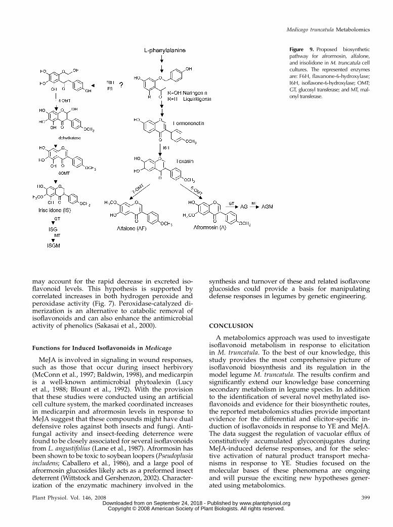
In unelicited cells, afrormosin was the major accu-mulated isoflavone, and alfalone was only formed intrace amounts. However, texasin, a potential interme-diate in the formation of both afrormosin and alfalone,was not detected. In unelicited cells, the key enzymedirecting flux into alfalone biosynthesis, presumablya texasin-7-O-methyltransferase (OMT), may be ratelimiting, and flow occurs instead through a 6-OMTthat favors afrormosin formation (Fig. 9). This portionof the pathway may operate as a ‘‘metabolic channel’’in which hydroxylases and O-methyltransferases co-localize, as suggested to occur at the entry point intoformononetin biosynthesis (Liu and Dixon, 2001).
Pulse labeling studies using exogenous [3H1]formo-nonetin revealed the accumulation of 3H label inafrormosin and alfalone, which supports a biosynthetic
Figure 6. Mapping of the extracellular phenylpropanoid metabolite responses to YE onto biosynthetic pathways. Dashed arrowsafter metabolite names represent conjugating enzymes responsible for the attachment of a Glc and/or a malonate moiety to formthe corresponding conjugates, e.g. glucosyl transferases (GT) and malonyl transferases (MT). Metabolites that show significantdifferential accumulation in response to YE (P , 0.05) are followed by gray boxes showing the maximum fold increase detectedover the time period at which elicitation occurs (in bold).
Medicago truncatula Metabolomics
Plant Physiol. Vol. 146, 2008 395 www.plantphysiol.orgon September 24, 2018 - Published by Downloaded from
link and evidence that formononetin is a precursor ofafrormosin and alfalone. Correlation analyses pro-vided further evidence for the close biogenetic originsof afrormosin and alfalone compared with irisolidone.Alfalone and afrormosin had very similar inductionkinetics (Fig. 8D) in response to YE with a correla-tion relationship of r2 5 0.76, whereas irisolidone in-duction correlated weakly with that of both alfaloneand afrormosin (r2 5 0.2 and 0.3, respectively). Corre-lation analyses have been used in a similar fashion toreveal relationships between primary metabolites inYE- and MeJA-elicited M. truncatula cells (Broecklinget al., 2005).
Label from exogenous [3H1]formononetin was notincorporated into irisolidone, a 5-hydroxyisoflavone.The presence of a hydroxyl group at the 5-position inirisolidone suggests that it is derived from naringenin,a precursor for 5-hydroxyisoflavones in legumes. Indeed,naringenin was detected in the cell cultures, alongwith several naringenin-derived isoflavones, includ-ing genistein and biochanin-A. A putative biosyntheticpathway from naringenin to irisolidone involving aflavanone-6-hydroxylase, IFS, 4#-OMT, dehydratase,and 6-OMT is shown in Figure 9. The relatively lowerincorporation of 3H label into formononetin gluco-side suggests that the conversion of formononetin toafrormosin is favored over the direct glucosylationof formononetin to form formononetin glucoside.
Irisolidone and other 5-hydroxyisoflavones were mi-nor components compared with the 5-deoxyisoflavonessuch as afrormosin. Pronounced differences in metabo-lism between 5-deoxy- and 5-hydroxyisoflavones havebeen observed in chickpea (Jaques et al., 1985; Barz andMackenbrock, 1994). 5-Hydroxyisoflavones are derivedfrom naringenin chalcone, the product of chalcone syn-thase (CHS) acting alone, whereas 5-deoxy isoflavonesare derived from isoliquiritgenin by the action of achalcone reductase (CHR), which coacts with CHS(Strack, 1997). In M. truncatula cell cultures, CHR islikely a key regulatory step for channeling of substratesinto the 5-deoxyisoflavone pathway.
Increased flavanone levels were observed in re-sponse to YE. However, this did not result in subse-quent changes in flavone levels, whereas significantincreases were observed in the levels of most isofla-vones. Inhibition of methylation reactions involved inisoflavonoid formation diverts alfalfa cells to accumu-late more flavones than isoflavones upon elicitation(Daniell et al., 1997), suggesting that this observationmay reflect channeling of flavanone to isoflavonerather than a specific suppression of flavone formation.
Differential and Elicitior-Specific Induction ofIsoflavonoids in Response to YE and MeJA
Increases in medicarpin and afrormosin levels wereobserved in response to both YE and MeJA with com-parable induction kinetics; however, relatively largerfold increases were observed in response to MeJA. YEtreatment resulted in marked increases in severalpathway components that precede isoflavonoid bio-synthesis, including chorismic acid from the shikimicacid pathway and both liquiritgenin and naringeninthat serve as entry points into (iso)flavonoid biosyn-thesis. Moreover, the pools of most constitutively ac-cumulated isoflavonoid glucosides remained relativelyunaltered after YE treatment.
Constitutively accumulated glucosides are gener-ally regarded as the stable, soluble storage forms ofisoflavonoids and typically localized within the cell’scentral vacuole (Mackenbrock and Barz, 1991). Theresponse to MeJA was in sharp contrast to that of YE,where many pathway precursors were unchanged andincreases in afrormosin and medicarpin were corre-lated with the consumption or significant decreases inisoflavonoid conjugate levels, particularly the gluco-sides of formononetin and 2#-hydroxyformononetin.
Importantly, the data document that M. truncatulacell cultures are responding to YE and MeJA with twofundamentally different and elicitor specific mecha-nisms. Based upon the relative abundance of pathwayprecursors, increases in isoflavonoids and the phyto-alexin end product medicarpin in response to YE areachieved from de novo biosynthesis; whereas, MeJA-induced accumulation of medicarpin occurs viahydrolysis and remobilization of vacuolar pools offormononetin glucoside. Formononetin then reentersthe isoflavonoid pathway and serves as the carbon
Figure 7. The oxidative burst observed in the medium of yeast-elicitedM. truncatula cell cultures. A, Accumulation of H2O2 in the medium ofyeast-elicited (black squares) M. truncatula cell cultures relative tocontrol (white diamonds). Results represent the average of duplicateexperiments. B, Peroxidase activity measured in the medium of yeast-elicited (black squares) and control (white diamonds) M. truncatula cellcultures. Error bars represent SE (n 5 3).
Farag et al.
396 Plant Physiol. Vol. 146, 2008 www.plantphysiol.orgon September 24, 2018 - Published by Downloaded from
source for the synthesis of medicarpin (Liu and Dixon,2001). The identification and characterization of glu-cosidase enzymes potentially involved in hydrolysisand remobilization of isoflavone glycosides are re-ported in a parallel publication (Naoumkina et al., 2007).
A marked decline was also observed in the levelsof several early phenylpropanoid-related compounds
such as p-hydroxybenzaldehyde and p-hydroxybenzoicacid in MeJA-treated cell cultures compared withcontrol cell cultures. Decreases in these compoundssuggest inhibition of flux through the early phenyl-propanoid pathway in response to MeJA. This responseis different from YE, which induces phenylpropanoidbiosynthesis and increases in pathway precursors,
Figure 8. Elucidating the biosynthetic origins of afrormosin, alfalone, and irisolidone. A, Two isoflavones (afrormosin andmedicarpin) accumulate following MeJA elicitation, whereas a decline is observed in formononetin and afrormosin glucosides. yaxis value represents relative peak areas after normalization to the mean peak area for that compound. Black squares representelicited sample means (with SE bars) and white diamonds represent control sample means. B, Incorporation of will be[3H1]formononetin into afrormosin, its glycosidic conjugates, and ononin in unelicited M. truncatula cell suspension cultures. B1,HPLC/UV trace showing separation of isoflavonoid standards. Chromatography was performed as described in ‘‘Materials andMethods’’ (labeling experiment). B2, HPLC/UV trace of extract from M. truncatula cell cultures that had been incubated with [3H1]-formononetin for 6 h. B3, Radioactivity associated with individual HPLC fractions (n 5 3) corresponding to individual isoflavonoidsderived from M. truncatula cell culture incubated with will be [3H1]formononetin for 6 h. Peaks, with retention times, include: FG,formononetin glucoside (ononin), 39.2 min; AF, alfalone, 67.3 min; F, formononetin, 68.3 min; A, afrormosin, 70.1 min; B,biochanin-A, 75.6 min; IS, irisolidone, 79.5 min; AG, afrormosin glucoside, 41.1 min; and AGM, afrormosin glucoside malonate,43.9 min. C, Accumulation of the radiolabeled isoflavonoids afrormosin (d), alfalone (:), irisolidone (¤), and biochanin-A (n)following treatment of yeast-elicited M. truncatula cells with will be [3H1]formononetin. Results represent average of duplicateexperiments. D, Induction kinetics for afrormosin and alfalone does not mimic that of irisolidone in response to YE. Black squaresrepresent elicited sample means with (SE bars) and white diamonds represent control sample means.
Medicago truncatula Metabolomics
Plant Physiol. Vol. 146, 2008 397 www.plantphysiol.orgon September 24, 2018 - Published by Downloaded from
but is consistent with the suggestion that endoge-nous MeJA suppresses the hypersensitive responseand Phe ammonia-lyase (PAL) expression during bac-terial elicitation (Andi et al., 2001). The differentialresponses observed here are also consistent with re-sults obtained in elicited chickpea cells treated witha PAL inhibitor. Chickpea cells treated with a fungalelicitor reportedly accumulate medicarpin via de novobiosynthesis, whereas cells elicited in the presence ofthe PAL inhibitor L-a-aminooxy-b-phenylpropionicacid utilized vacuolar pools of formononetin gluco-side conjugates as precursors for medicarpin biosyn-thesis (Mackenbrock and Barz, 1991). In M. truncatulacell culture, MeJA appears to be acting in a similarmanner and remobilizing vacuolar stores of phenyl-propanoid glycosides to serve as precursors for medicarpinand afrormosin biosynthesis. Thus, isoflavone conju-gates provide a rapidly available source of isoflavonoidphytoalexin precursors under conditions of rate-limitingphenylpropanoid biosynthesis such as might occurduring wounding or herbivory to mount a timely de-fense response.
Extracellular Secretion of Isoflavonoids
Intracellular isoflavonoid levels were induced byboth YE and MeJA; however, significant increases in
isoflavone levels in the culture medium were onlyobserved in response to YE. The selective release ofisoflavone aglycones as compared with isoflavone gly-cosides inresponse toYEsuggests carrier-mediatedtrans-port rather than a simple diffusion mechanism. It isknown that secondary metabolites are transportedacross membranes by specific carrier proteins (Walkeret al., 2003; Yazaki, 2005). For example, an ATP-bindingcassette-type transporter involved in antifungal ter-penpoid secretion from tobacco (Nicotiana tabacum) cellcultures has been identified (Jasinski et al., 2001). Weassume that similar transporters are present in Medi-cago for isoflavones; however, neither specific nor ge-neric isoflavonoid transporters have been identified inthis species to date.
Four identified compounds accumulated exclu-sively in the medium, including dihydroafrormosin,daidzein dimer, chorismic acid, and p-hydroxybenzoicacid. Increased chorismic acid levels in the mediumare consistent with YE inducing phenylpropanoidaccumulation from primary metabolism (shikimate/arogenate pathway), and gas chromatography-massspectrometry analyses of YE-treated M. truncatula cellslikewise showed an increase in shikimic acid (Broecklinget al., 2005). However, it is not clear why shikimatepools rise in the cells while chorismate is excreted.
The detection of daidzein dimers in the media sug-gests that oxidative oligomerization (Park et al., 1995)
Table III. Comparisons in the relative fold changes in peak areas for selected identified compoundsin two independent elicitation experiments
Metabolites are listed if the changes in their levels showed a similar response, at a significant value of P ,
0.05, in the two experiments (large-scale experiments in which YE and MeJA elicitations were applied toconsecutive passages of the culture and cultures sampled at 21 time points postelicitation, and theadditional smaller experiment in which YE and MeJA elicitation were carried out in parallel on a singlepassage culture). Values represent the fold change in peak area at the average of the 3-, 12-, and 24-h timepoints (values .1.0 represent increased levels in elicited samples, values ,1.0 represent decreased levelsdue to elicitation; empty cells, not significant in at least one of the two Student’s t tests).
may account for the rapid decrease in excreted iso-flavonoid levels. This hypothesis is supported bycorrelated increases in both hydrogen peroxide andperoxidase activity (Fig. 7). Peroxidase-catalyzed di-merization is an alternative to catabolic removal ofisoflavonoids and can also enhance the antimicrobialactivity of phenolics (Sakasai et al., 2000).
Functions for Induced Isoflavonoids in Medicago
MeJA is involved in signaling in wound responses,such as those that occur during insect herbivory(McConn et al., 1997; Baldwin, 1998), and medicarpinis a well-known antimicrobial phytoalexin (Lucyet al., 1988; Blount et al., 1992). With the provisionthat these studies were conducted using an artificialcell culture system, the marked coordinated increasesin medicarpin and afrormosin levels in response toMeJA suggest that these compounds might have dualdefensive roles against both insects and fungi. Anti-fungal activity and insect-feeding deterrence werefound to be closely associated for several isoflavonoidsfrom L. angustifolius (Lane et al., 1987). Afrormosin hasbeen shown to be toxic to soybean loopers (Pseudoplusiaincludens; Caballero et al., 1986), and a large pool ofafrormosin glucosides likely acts as a preformed insectdeterrent (Wittstock and Gershenzon, 2002). Character-ization of the enzymatic machinery involved in the
synthesis and turnover of these and related isoflavoneglucosides could provide a basis for manipulatingdefense responses in legumes by genetic engineering.
CONCLUSION
A metabolomics approach was used to investigateisoflavonoid metabolism in response to elicitationin M. truncatula. To the best of our knowledge, thisstudy provides the most comprehensive picture ofisoflavonoid biosynthesis and its regulation in themodel legume M. truncatula. The results confirm andsignificantly extend our knowledge base concerningsecondary metabolism in legume species. In additionto the identification of several novel methylated iso-flavonoids and evidence for their biosynthetic routes,the reported metabolomics studies provide importantevidence for the differential and elicitor-specific in-duction of isoflavonoids in response to YE and MeJA.The data suggest the regulation of vacuolar efflux ofconstitutively accumulated glycoconjugates duringMeJA-induced defense responses, and for the selec-tive activation of natural product transport mecha-nisms in response to YE. Studies focused on themolecular bases of these phenomena are ongoingand will pursue the exciting new hypotheses gener-ated using metabolomics.
Figure 9. Proposed biosyntheticpathway for afrormosin, alfalone,and irisolidone in M. truncatula cellcultures. The represented enzymesare: F6H, flavanone-6-hydroxylase;I6H, isoflavone-6-hydroxylase; OMT;GT, glucosyl transferase; and MT, mal-onyl transferase.
Medicago truncatula Metabolomics
Plant Physiol. Vol. 146, 2008 399 www.plantphysiol.orgon September 24, 2018 - Published by Downloaded from