Metadata of the chapter that will be visualized online Chapter Title Echocardiography in a Patient with a New Murmur Copyright Year 2018 Copyright Holder Springer International Publishing AG, part of Springer Nature Author Family Name Friedman Particle Given Name Julie Suffix Division Organization/University New York University Langone Medical Center Address New York, NY, USA Email [email protected]Corresponding Author Family Name Saric Particle Given Name Muhamed Suffix Division Organization/University New York University Langone Medical Center Address New York, NY, USA Email [email protected]Abstract Heart murmurs are common in cardiac care unit (CCU) patients. As many of CCU patients are intubated and unable to provide medical history, the physical exam becomes crucial in piecing together a patient’s medical history. Once a new murmur is identified, echocardiography can be a helpful tool in the critical care setting to further elucidate the cardiac abnormalities of a CCU patient. Whereas many patients have murmurs representing manifestations of chronic diseases, this chapter focuses on the diagnosis and etiology of murmurs that are presumably new at the time of CCU admission and represent an acute change in clinical status. Keywords (separated by “ - ”) Murmur - Aortic valve disease - Mitral valve disease
Transcript
Metadata of the chapter that will be visualized online
Chapter Title Echocardiography in a Patient with a New Murmur
Copyright Year 2018Copyright Holder Springer International Publishing AG, part of Springer NatureAuthor Family Name Friedman
Particle
Given Name Julie
Suffix
Division
Organization/University New York University Langone Medical Center
Organization/University New York University Langone Medical Center
Address New York, NY, USA
Email [email protected] Heart murmurs are common in cardiac care unit (CCU) patients. As many of
CCU patients are intubated and unable to provide medical history, the physical exam becomes crucial in piecing together a patient’s medical history. Once a new murmur is identified, echocardiography can be a helpful tool in the critical care setting to further elucidate the cardiac abnormalities of a CCU patient. Whereas many patients have murmurs representing manifestations of chronic diseases, this chapter focuses on the diagnosis and etiology of murmurs that are presumably new at the time of CCU admission and represent an acute change in clinical status.
Chapter 6Echocardiography in a Patient with a New Murmur
Julie Friedman and Muhamed Saric
Abstract Heart murmurs are common in cardiac care unit (CCU) patients. As many of CCU patients are intubated and unable to provide medical history, the physical exam becomes crucial in piecing together a patient’s medical history. Once a new murmur is identified, echocardiography can be a helpful tool in the critical care setting to further elucidate the cardiac abnormalities of a CCU patient. Whereas many patients have murmurs representing manifestations of chronic diseases, this chapter focuses on the diagnosis and etiology of murmurs that are presumably new at the time of CCU admission and represent an acute change in clinical status.
Heart murmurs are common in cardiac care unit (CCU) patients. As many of CCU patients are intubated and unable to provide medical history, the physical exam becomes crucial in piecing together a patient’s medical history. Once a new murmur is identified, echocardiography can be a helpful tool in the critical care setting to further elucidate the cardiac abnormalities of a CCU patient. Whereas many patients have murmurs representing manifestations of chronic diseases, this chapter focuses on the diagnosis and etiology of murmurs that are presumably new at the time of CCU admission and represent an acute change in clinical status.
The majority of new native valve murmurs represent nonstenotic lesions and they are related primarily to acute valvular regurgitation. On the contrary, new prosthetic valve murmurs may be either stenotic, such as in acute prosthetic valve stenosis, or
regurgitant, as in infective endocarditis. Other new murmurs that may present in a CCU patient include that of ventricular septal rupture and left ventricular outflow tract (LVOT) obstruction, both of which are described in this chapter as well.
Genesis and Types of Heart Murmurs
Murmurs are defined as sounds heard in addition to the sequence of two to three heart sounds during each heartbeat [1]. The two normal heart sounds in adults—the first heart sound (S1) and the second heart sound (S2)—are produced mainly by the closure of the atrioventricular (tricuspid and mitral) and semilunar (aortic and pul-monary) valves, respectively. Sometimes, an additional heart sound associated with the ventricles filling up with blood may be heard. It is referred to as S3 when it occurs in early diastole and S4 when heard in late diastole after atrial contraction. In chil-dren it can be normal to hear an S3 but in adults it often represents pathology. When an S3 or S4 heart sound is heard together with the two normal heart sounds, it is referred to as a “gallop.” A combination of S1 + S2 + S3 is a ventricular (“Kentucky”) gallop. In contrast, a combination of S4 + S1 + S2 is an atrial (“Tennessee”) gallop [2].
Murmurs are thought to be the result of abnormal movement of blood across valves and between cardiac chambers which result in turbulence of blood flow. The turbulence creates vibrations in the heart chambers and outflow tracts that are detected via auscultation with a stethoscope. The intensity of a murmur can vary greatly depending on the size of the orifice which blood is flowing through, the pres-sure gradient across the narrowing, and the volume of blood flowing across the site. Heart murmurs can be systolic, diastolic, or continuous (Table 6.1) and they can be reported with accompanying intensity grades (Table 6.2).
When clinically insignificant, murmurs are referred to as being “innocent”; they are caused by increased flow or turbulence across anatomically normal valves or other cardiovascular structures. Some systolic murmurs may be clinically innocent, while all diastolic and continuous murmurs are abnormal. Pathological murmurs occur as a result of either diseased cardiac valves or abnormal communications between cardiac chambers, blood vessels, or both. These lesions may be congenital or acquired, and have unique hemodynamic consequences.
Table 6.1 Types of heart murmurs
Start End
Systolic murmurs
Midsystolic After S1 Before S2Pansystolic (holosystolic) At or after S1 At S2Late systolic Middle or late systole At S2Diastolic murmurs
Early diastolic At S2 Before next S1Mid-diastolic Shortly after S2 Before next S1Late diastolic (presystolic) Late diastole At next S1Continuous murmurs Begin in systole and continue through the cardiac cycle
J. Friedman and M. Saric
26
27
28
29
30
31
32
33
34
35
36
37
38
39
40
41
42
43
44
45
46
47
48
49
50
51
52
53
54
55
t1.1
t1.2
t1.3
t1.4
t1.5
t1.6
t1.7
t1.8
t1.9
t1.10
t1.11
Detection of a Heart Murmur
Clinicians will often first detect murmurs via auscultation using a stethoscope. The invention of the stethoscope is often credited to René Laënnec who, in 1816, created the monaural stethoscope as a means of amplifying heart sounds without requiring physical contact. In 1851, Arthur Leared invented the binaural stethoscope and then in 1852 George Cammann, who was a practicing physician in New York City, per-fected the binaural stethoscope which is most reminiscent of the modern-day aus-cultation device used by medical professionals today [3].
The standard stethoscope often includes both a shallow bell (for low-frequency sounds) and a thin, stiff diaphragm (for high-frequency sounds). Clinicians rou-tinely assess at least four surface anatomical areas with the stethoscope when evalu-ating for murmurs as shown in Fig. 6.1. Murmurs are then further described based on their intensity (grade), pitch (frequency), configuration, timing, quality, and radiation (Table 6.3).
Table 6.2 Intensity and grades of heart murmurs
Grade Murmur intensity Comments
I Very faintII Quiet murmur As loud as S1, S2III Moderately loud murmur Louder than S1, S2IV Loud murmur With thrillV Very loud murmur Heard even with stethoscope barely on the
chestVI Extremely loud Heard even without stethoscope
AAORTIC AREA
2nd Right IntercostalP
PULMONIC AREA2nd Left Intercostal
TTRICUSPID AREA
4th/5th Left Intercostal
M
MITRAL AREA5th Left Intercostal at
Midclavicular Line
Fig. 6.1 Cardiac auscultation areas. The background thorax image with permission from BioDigital.com. Abbreviations: A aortic area, M mitral area, P pulmonic area, T tricuspid area
Role of Echocardiography After Detection of a Heart Murmur
Once detected, murmurs are typically further characterized via echocardiography which can help evaluate the primary lesion in terms of cause and severity, define the lesion’s hemodynamic significance, detect coexisting abnormalities, evaluate the size and function of cardiac chambers, and establish a reference point for future comparisons.
Modern ultrasound machines have become smaller and more portable allowing the exams to be performed at the patient’s bedside. In recent years, there has also been an increasing use of handheld pocket scanners as partial replacement tools for auscultation. While these tools cannot replace the detail obtained in a formal echo-cardiogram, there has been an increasing reliance on these tools to confirm pathol-ogy suspected based on auscultatory findings and these have become an extension of the physical exam in the critical care setting.
New murmurs in the CCU are most often of a regurgitant variety as a result of acute infective endocarditis, acute type A aortic dissection, or sequelae of an acute myocardial infarction. Acute valve stenosis is typically seen only with prosthetic valves and can occur in the setting of acute valve thrombosis which is a devastating and life-threatening condition. Other murmurs such as that of a new dynamic LVOT obstruction or ventricular septal rupture have distinctive murmurs which are impor-tant to recognize and are discussed in this chapter.
Table 6.3 Murmur types and conditions Murmur type Associated abnormality
Systolic
Early systolic Mitral regurgitationTricuspid regurgitationVentricular septal ruptureAortic stenosis
Myocardial infarction (MI) is a clinical or pathologic event caused by myocardial ischemia, resulting in myocardial necrosis or injury. The American College of Cardiology and the American Heart Association endorse the Third Universal Definition of Myocardial Infarction which defines an MI as a typical rise and/or fall of cardiac troponin with at least one value above the assay’s upper reference limit, accompanied by at least one other feature of ischemia (e.g., typical symptoms or ECG changes) [4]. Acute MIs—often in the form of ST-elevation MI (STEMI)—make up a high percentage of admission tons to cardiac ICUs.
The new murmurs that arise as a consequence of an acute MI are related to mechanical complications of the infarct. The most common lesions are acute mitral regurgitation (MR), a ventricular septal rupture (VSR), and LV wall rupture. MR and VSR but not LV rupture typically present as a new heart murmur. Papillary muscle rupture (and resulting mitral regurgitation) typically occurs 2–7 days after MI, ventricular septal defects typically occur 3–7 days after MI, and LV free wall rupture often occurs 5–14 days after MI (Table 6.4) [5].
Acute Mitral Regurgitation
Clinical Presentation
Acute MR in the setting of an acute MI (or endocarditis, which is discussed in sub-sequent section) often presents as a cardiac emergency with the sudden onset and rapid progression of hypotension, pulmonary edema, and signs and symptoms of cardiogenic shock. Acute MR due to MI is related to papillary muscle rupture.
The murmur of acute MR may be systolic, midsystolic, or holosystolic. It is often best heard along the left sternal border and may radiate to the back. However, since the pressure within the left atrium markedly increases during ventricular systole and the pressure gradient between the LA and LV diminishes or disappears by the end of systole, the systolic murmur is often soft, low pitched, and decrescendo. An important point to keep in mind while in the ICU is that the approximately 50% of
Table 6.4 Myocardial infarction related murmurs
Complication type Complication onset days after MI
patients with moderate-to-severe acute MR will have no audible murmur and there-fore the intensity of the murmur does not necessarily correlate with the severity of the valve lesion [6]. The presumed mechanism is thought to be secondary to a rela-tively low systolic pressure gradient between the left ventricle and left atrium due to the combination of low systemic blood pressure and elevated left atrial pressure. An S3 can commonly be heard but may be difficult to appreciate as these patients are often quite tachycardic.
Pathophysiology and Anatomic Considerations
The mitral valve apparatus consists of the mitral annulus, anterior and posterior leaflets, chordae tendineae, papillary muscles, and underlying ventricular wall (Fig. 6.2). Mitral regurgitation can occur as a complication of acute myocardial infarction by several mechanisms: (1) papillary muscle or chordal rupture, (2) papil-lary muscle displacement, and (3) LV dilatation/aneurysm.
Papillary muscle rupture is the primary mechanism of acute MR related to MI. The other two mechanisms (papillary muscle displacement and LV dilatation) typically lead to subacute or chronic MR murmur. Partial or complete rupture of a papillary muscle can have catastrophic effects. Additionally, there is a wide range of infarction size that can be associated with papillary muscle rupture. Many patients may have very limited overall left ventricular dysfunction from their MI and, in the setting of acute severe mitral regurgitation, may actually have a hyperdynamic LV which can further limit identification of wall motion abnormalities. It is for these reasons that echocardiography can be quite helpful in diagnosing this potentially life-threatening condition.
Fig. 6.2 Mitral valve apparatus. Based on the image of the mitral valve apparatus with permission from Geriatrics & Aging journal
J. Friedman and M. Saric
119
120
121
122
123
124
125
126
127
128
129
130
131
132
133
134
135
136
137
138
139
140
141
The posteromedial (PM) papillary muscle has solitary blood supply from the posterior descending artery whereas the anterolateral (AL) papillary muscle has a dual blood supply from the left anterior descending and left circumflex arteries (Fig. 6.3) [7]. It is for this reason that rupture of the PM papillary muscle occurs at a much higher frequency than that of the AL papillary muscle. It is rare that acute rupture of a papillary muscle involves the entire body of the papillary muscle; more commonly, only one of the subheads of the muscle is involved resulting in a partial leaflet flail. It is important to note, however, that chordae to both leaflets arise from each papillary muscle such that, in cases of complete rupture, both leaflets are often affected.
Echo Findings
The jet of acute MR from papillary muscle rupture is most often eccentric and directed opposite to that of the partial flail leaflet (Figs. 6.4 and 6.5). Given the eccentric nature, the severity of the regurgitant jet may often be underestimated. Associated findings include a small inferior wall motion abnormality, a mass (the ruptured papillary muscle) attached to flail segments of the leaflets often seen in the LA in systole and in the LV in diastole. Given the acuity of the regurgitant lesion, the LA is often nondilated. For this reason, the detection of an eccentric mitral regurgitation jet with a relatively normal-sized left atrium should raise suspicion for acute mitral regurgitation and, in the proper clinical context of myocardial ischemia or infarction, is concerning for papillary muscle rupture.
As discussed in the auscultatory findings, the murmur of acute MR is either soft or absent because of rapid pressure equilibration between LV and LA in systole. Echocardiographically, this is visualized on spectral Doppler tracings as a low- velocity triangular systolic flow velocity pattern (Fig. 6.6). This is often accompa-nied by a prominent, rapidly decelerating early diastolic (E) wave, which corresponds to auscultatory S3 sound.
ANTEROLATERALPAPILLARY MUSCLE
Dual SupplyLAD and LCx
POSTEROMEDIALPAPILLARY MUSCLE
Solitary SupplyPDA from either
RCA or LCx
Fig. 6.3 Papillary muscle blood supply. Short-axis TTE view at the level of papillary muscle; note the solitary blood supply of the posteromedial papillary muscle vs. dual blood supply of the anterolateral papillary muscle. Abbreviations: LAD left anterior descending artery, LCx left circumflex artery, PDA posterior descending artery, RCA right coronary artery
6 Echocardiography in a Patient with a New Murmur
142
143
144
145
146
147
148
149
150
151
152
153
154
155
156
157
158
159
160
161
162
163
164
165
166
167
168
Ventricular Septal Rupture
Clinical Presentation
The classic presentation of someone with ventricular septal rupture (VSR) is that of acute onset of hemodynamic compromise with hypotension, biventricular failure (right greater than left), and a new harsh murmur soon after an MI. It is yet another rare, but lethal complication of acute myocardial infarction and is often seen 3–5 days after acute MI. VSR is an utmost emergency and thus the prompt diagnosis is crucial for prompt surgical or percutaneous VSR closure.
c
LA
LVOT
b
LV
LA
RV
a
LV
LA
RV
EccentricPosterior
lyDirectedMR Jet
RupturedAnteriorPapillaryMuscle
RupturedAnteriorPapillaryMuscle
Fig. 6.4 Ruptured anterior papillary muscle on TEE. Panel (a): Ruptured anterior papillary mus-cle in the left ventricle during diastole. Panel (b): Ruptured anterior papillary muscle in the left atrium during systole. Panel (c): Color Doppler imaging showing very severe eccentric, posteriorly directed, mitral regurgitation. In this condition, a murmur of severe acute mitral regurgitation may be heard. Abbreviations: LA left atrium, LV left ventricle, LVOT left ventricular outflow tract, MR mitral regurgitation, RV right ventricle
J. Friedman and M. Saric
169
170
171
172
173
174
175
176
c
Centralcoagulative
necrosis(Infarction)
Myocytolysis
d
b
EccentricAnteriorlyDirectedMR Jet
LV
RV
LA
a
LV
LA
RupturedPosteriorPapillaryMuscle
RV
RupturedPosterior
Fig. 6.5 Ruptured posterior papillary muscle. Panel (a): Ruptured posterior papillary muscle in the left atrium during systole on TEE. Panel (b): Color Doppler imaging showing eccentric, ante-riorly directed, mitral regurgitation jet on TEE. Panel (c): Gross surgical specimen of a ruptured papillary muscle. Panel (d): Histology of a necrotic, ruptured papillary muscle. In this condition, a murmur of severe acute mitral regurgitation may be heard. Abbreviations: LA left atrium, LV left ventricle, MR mitral regurgitation, RV right ventricle
Severe ACUTE Mitral Regurgitation Severe CHRONIC Mitral Regurgitationa b
Low VelocityTriangular MR Jet
High VelocityParabolic MR Jet
ProminentE wave
Fig. 6.6 Acute vs. chronic MR on spectral Doppler. Panel (a): Continuous-wave Doppler across the mitral valve in severe acute mitral regurgitation. Note that the low-velocity, rapidly dissipating LV to LA systolic gradient may result in a soft or even absent mitral regurgitation murmur. Panel (b): Continuous-wave Doppler across the mitral valve in severe chronic mitral regurgitation. Abbreviations: E wave early diastolic flow, MR mitral regurgitation
6 Echocardiography in a Patient with a New Murmur
Murmur of Acute VSR
The murmur of VSR is very loud, harsh in character, holosystolic, and often heard throughout the precordium. Up to 50% of patients with acute VSR have a detectable thrill.
Pathophysiology
VSR develops after transmural infarction of the ventricular septum and can occur at any anatomic location along the septum. It can result from an MI of any of the coronary arteries as the septal blood supply comes from branches of the left ante-rior descending artery, the posterior descending branch of the right coronary artery, or the left circumflex artery when it is dominant (Fig. 6.7) [7]. However, septal rupture tends to occur most frequently with anterior MIs. The newly formed communication between the right and left ventricles results in left-to-right shunting of blood from the high-pressure LV to the lower pressure right ventricle (RV) [8].
Inferior SeptumLAD + PDA
Inferior SeptumPDA
Anterior SeptumLAD Alone
Inferior SeptumLAD + PDA
Anterior SeptumLAD Alone
Fig. 6.7 Coronary territories of septum. Note the difference in the blood supply between the ante-rior and the inferior interventricular septum. The image with permission from the Journal of American Society of Echocardiography. Abbreviations: LAD left anterior descending artery, PDA posterior descending artery
J. Friedman and M. Saric
177
178
179
180
181
182
183
184
185
186
187
188
189
190
Echo Findings
The key to identifying a VSR by echocardiography is noting a high-velocity, left-to- right systolic jet on continuous-wave Doppler and systolic turbulence on the RV side of the septum on color Doppler imaging. As this can be challenging to identify, it is often necessary to use nonconventional imaging planes, scanning up and down the septum with color Doppler imaging, to identify the pathologic left-to-right flow. Once the site of rupture is identified with color flow, color can then be turned off and anatomic imaging undertaken to help identify the site of dropout of the ventricular septum with 2D imaging. These defects may often be serpiginous through the myo-cardium and can range in size from millimeters to several centimeters making it challenging to visualize the chamber connection on echo (Fig. 6.8).
ba
LVRV
c
SystolicVSR Flow
d
DiastolicVSR Flow
Fig. 6.8 Ventricular septal rupture (VSR) on echo. Panel (a): Parasternal short-axis TTE view of VSR identifying dropout of the ventricular septum. Panel (b): Spectral Doppler confirms high- velocity systolic flow across VSR. Panel (c): Color Doppler imaging showing high-velocity sys-tolic flow across VSR. Panel (d): Color Doppler imaging showing low-velocity diastolic flow across VSR. In this condition, a harsh holosystolic murmur is typically heard. Abbreviations: LV left ventricle, RV right ventricle, VSR ventricular septal rupture
6 Echocardiography in a Patient with a New Murmur
191
192
193
194
195
196
197
198
199
200
201
New Murmur in a Febrile Patient
Infective Endocarditis
Clinical Presentation
Infective endocarditis is an infection of the endocardial surface of the heart, most commonly referring to an infection of one or more of the heart valves or of an intra-cardiac device (pacing wires, catheters, conduits, etc.). It is an uncommon infec-tious disease with an annual incidence ranging from 3 to 7 per 100,000 person-years in the most contemporary population [9]. The presentation can vary from a subacute or chronic disease with nonspecific symptoms to an acute, rapidly progressive infection with hemodynamic consequences resulting from valvular dysfunction. Cardiac complications occur in up to half of patients and can result in these patients requiring ICU-level care for heart failure from valvular dysfunction or conduction abnormalities from paravalvular abscesses.
Murmur in Infective Endocarditis
The presence of a new regurgitant murmur in a febrile patient should raise suspicion for infective endocarditis. Left-sided valvular lesions are more common than right- sided ones. Thus, one may expect to hear the murmur of aortic or mitral regurgita-tion. Right-sided lesions are typically associated with intravenous drug users (tricuspid valve involvement and less commonly pulmonic valve) but can also be found in those with right-sided indwelling catheters (such as hemodialysis cathe-ters) or intracardiac devices (pacemakers, defibrillators). It is important to keep in mind that even though most right-sided lesions are found in the setting of intrave-nous (IV) drug use, it is still much more common for an IV drug user to present with left-sided rather than right-sided valvular disease.
Echocardiographic Features of Vegetations
One of the major Duke criteria for infective endocarditis is the echocardiographic finding of evidence of endocardial involvement [10]. To that end, the findings of abscess, new partial dehiscence of prosthetic valve, or visualization of a vegetation support a diagnosis of infective endocarditis in the ICU in the setting of a new regur-gitant murmur.
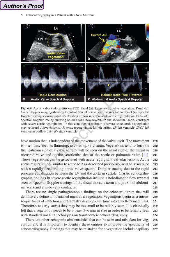
A vegetation is typically an irregularly shaped, echogenic, highly mobile mass that may be attached to any area of the valve leaflet, although lesions at the coapta-tion line are most common (Fig. 6.9). Since the vegetation is a discrete structure, it may often only be seen in certain tomographic planes and thus slow scanning between the standard image planes is crucial to help increase the probability of identifying a valvular vegetation. Most vegetations are highly mobile and tend to
J. Friedman and M. Saric
202
203
204
205
206
207
208
209
210
211
212
213
214
215
216
217
218
219
220
221
222
223
224
225
226
227
228
229
230
231
232
233
234
235
236
237
have motion that is independent of the movement of the valve itself. The movement is often described as fluttering, oscillating, or chaotic. Vegetations tend to form on the upstream side of a valve so they will be seen on the atrial side of the mitral or tricuspid valve and on the ventricular size of the aortic or pulmonic valve [11]. These vegetations can be associated with acute regurgitant valvular lesions. Acute aortic regurgitation, similar to acute MR as described previously, will be associated with a rapidly decelerating aortic valve spectral Doppler tracing due to the rapid pressure equalization between the LV and the aorta in systole. Classic echocardio-graphic findings in severe aortic regurgitation include a holodiastolic flow reversal seen on spectral Doppler tracings of the distal thoracic aorta and proximal abdomi-nal aorta and a wide vena contracta.
There are no single pathognomonic findings on the echocardiogram that will definitively define an identified mass as a vegetation. Vegetations begin as a micro-scopic focus of infection and gradually develop over time into a well-formed mass. Therefore, at early stages they may be too small to be reliably seen. It is classically felt that a vegetation needs to be at least 3–6 mm in size in order to be reliably seen with standard imaging techniques on transthoracic echocardiography.
There are other echogenic abnormalities that can be seen and mistaken for veg-etation and it is important to identify these entities to improve the specificity of echocardiography. Findings that may be mistaken for a vegetation include papillary
aAortic ValveVegetation
LVOT
LA
b
LA
RV
Aorta
Severe AR
Aortic Valve Spectral DopplerRapid Deceleration
*
Abdominal Aorta Spectral Doppler
*Holodiastolic Flow Reversal
dc
Fig. 6.9 Aortic valve endocarditis on TEE. Panel (a): Large aortic valve vegetation. Panel (b): Color Doppler imaging showing turbulent flow of severe aortic regurgitation. Panel (c): Spectral Doppler tracing showing rapid deceleration of flow in severe acute aortic regurgitation. Panel (d): Spectral Doppler tracing showing holodiastolic flow reversal in the abdominal aorta, consistent with severe aortic regurgitation. In this condition, a murmur of severe acute aortic regurgitation may be heard. Abbreviations: AR aortic regurgitation, LA left atrium, LV left ventricle, LVOT left ventricular outflow tract, RV right ventricle
Many of the patients who are critically ill in the ICU with a presentation of shock may have no murmur on physical exam. Others may have murmur from non- cardiogenic causes such as the flow murmur often appreciated in septic shock. The etiologies of murmurs with cardiogenic shock include the already discussed
a
LV
LABacterial
Endocarditis
b
LV
LAMaranticEndocarditis
c
LV
LAPapillaryFibroelas
toma
Fig. 6.10 Vegetation masqueraders on TEE involving the mitral valve. Panel (a): Bacterial endo-carditis. Note the shaggy appearance of the vegetation on the atrial side of the mitral valve. Panel (b): Marantic (nonbacterial, thrombotic) endocarditis. Note the so-called kissing lesions on either leaflet of the mitral valve. Panel (c): Papillary fibroelastoma. Note the well-circumscribed rounded appearance of the papillary fibroelastoma attached to the atrial surface of the mitral valve. In this condition, a murmur of severe acute mitral regurgitation may be heard. Abbreviations: LA left atrium, LV left ventricle
J. Friedman and M. Saric
258
259
260
261
262
263
264
265
murmur of acute mitral regurgitation (due to acute MI or mitral valve endocarditis) and the murmur of acute aortic regurgitation (due to aortic valve endocarditis). Additional etiologies of shock-associated murmurs include left ventricular outflow tract (LVOT) obstruction and type A aortic dissection.
Stress Cardiomyopathy with LVOT Obstruction
Clinical Presentation
LVOT obstruction which can be either a preexisting condition (such as in a patient with hypertrophic obstructive cardiomyopathy) or a newly developed such as in a patient presenting with a stress cardiomyopathy (also referred to as Takotsubo car-diomyopathy or apical ballooning syndrome) is a clinical syndrome characterized by transient focal LV dysfunction in the absence of obstructive coronary artery disease and is often on the differential of acute MI. The true incidence of this phe-nomenon is likely on the range of 1–2% of those presenting with troponin-positive acute coronary syndrome, but in the ICU studies have found that up to 28% of those patients admitted with a noncardiac diagnosis were found with stress cardiomyopathy [12].
A subset of this population will develop a dynamic LVOT obstruction felt to be induced by basal hyperkinesis in the setting of apical hypokinesis. One of the early signs of this may be the appreciation of a systolic murmur and the presence of the LVOT obstruction can contribute to the development of shock in an ICU patient. Echocardiographic findings can help support this diagnosis which can have major implications in medical management.
Murmur of LVOT Obstruction
The murmur of LVOT obstruction is typically a high-pitched, crescendo- decrescendo, midsystolic murmur that is best appreciated at the left lower sternal border. It can often be mistaken for the murmur of aortic stenosis (AS) given similar qualities; however, unlike the murmur of AS, this murmur does not radiate to the carotid arteries. Maneuvers that alter afterload and preload can also help differenti-ate the murmur of LVOT obstruction from others such as AS. The LVOT murmur will increase with Valsalva maneuver (decrease in preload) and will decrease with handgrip (increase in afterload).
Echocardiographic Features of LVOT Obstruction
In the transthoracic parasternal long-axis or apical three-chamber view, one will often see the classic finding of systolic anterior motion of the mitral valve (SAM) with apposition of the mitral leaflet and septum in mid to late systole (mitral-septal
6 Echocardiography in a Patient with a New Murmur
266
267
268
269
270
271
272
273
274
275
276
277
278
279
280
281
282
283
284
285
286
287
288
289
290
291
292
293
294
295
296
297
298
299
300
contact). Given its greater temporal resolution, M-mode recordings may be helpful to better identify the mitral-septal contact. M-mode can also be helpful in identify-ing mid-systolic abrupt partial closure of the aortic valve with coarse fluttering of the aortic valve leaflets in late systole that results from this late systolic dynamic outflow obstruction. The degree of SAM may not be uniform from medial to lateral across the mitral leaflets, so it is important to image in multiple planes with slight adjustments in transducer angulation to demonstrate the presence and extent of dynamic outflow obstruction.
While 2D and M-mode imaging can provide clues to the presence of an outflow obstruction, Doppler studies can be used to identify the site of obstruction and the severity of obstruction. Using pulsed-wave Doppler from the apical approach, the sample volume is moved slowly from the apex up to the base while recording the velocity at each step. At the site of obstruction, the velocity increases abruptly and this velocity reflects the degree of obstruction. Continuous-wave Doppler will reveal a late-peaking high-velocity systolic jet (Fig. 6.11).
Type A Aortic Dissection
Clinical Presentation
Aortic dissection is a rare, yet life-threatening condition for which the early and proper diagnosis is crucial for survival. The classic presentation of acute aortic dis-section is with severe chest pain and acute hemodynamic compromise. Progressive hemodynamic decline and death in patients with type A dissection can result from several mechanisms: (1) rupture of the dissection into the pericardium resulting in cardiac tamponade, (2) dissection into the aortic valve leading to severe aortic regurgitation, and (3) obstruction of the coronary artery ostia leading to acute myo-cardial infarction [13].
a
LVSAM
LA
AV
b
Late Peaking High-VelocitySystolic Gradient
Fig. 6.11 LVOT obstruction and SAM on TTE. Panel (a): Systolic anterior motion of the mitral valve with mitral-septal contact. Panel (b): Late-peaking, high-velocity systolic gradient consistent with LVOT obstruction. In this condition, the characteristic late-peaking systolic murmur is heard. Abbreviations: AV aortic valve, LA left atrium, LV left ventricle, SAM systolic anterior motion of the mitral valve
J. Friedman and M. Saric
301
302
303
304
305
306
307
308
309
310
311
312
313
314
315
316
317
318
319
320
321
322
323
324
325
Pathophysiology
The primary event is an intimal tear in the aortic wall, followed by degeneration of the aortic media (cystic and medial necrosis) and ultimately the passage of blood into the media via the tear further separating the intima from the surrounding media and/or adventitia, thus creating a false channel. This channel can remain localized or can propagate downstream.
Murmur of Type A Aortic Regurgitation
Aortic regurgitation is very frequently present in cases of type A aortic dissection as patients have either chronic AR due to aortic dilation over time or more acute AR due to retrograde extension of the dissection resulting in inadequate leaflet support. The murmur of acute AR is often low pitched and early diastolic whereas the mur-mur of chronic AR tends to be high pitched and holodiastolic. Given the increased blood volume crossing the aortic valve, a soft systolic murmur may be heard as well. This combination of a soft systolic and a low-pitched diastolic aortic murmur is often referred to as a “to-and-fro” murmur of acute AR. Additionally, similar to the murmur of acute MR, the murmur of acute AR may be faint or absent as a result of an equalization of the pressures between the aorta and the LV.
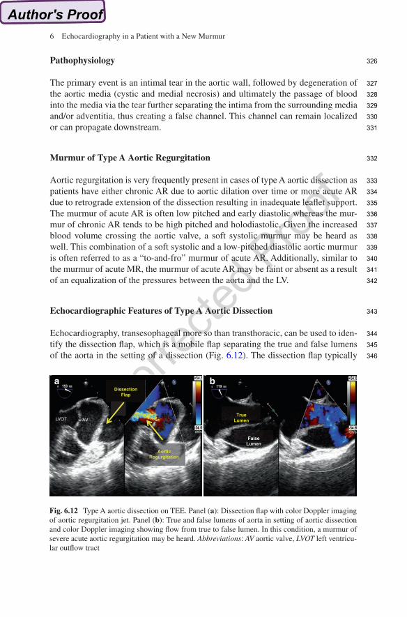
Echocardiographic Features of Type A Aortic Dissection
Echocardiography, transesophageal more so than transthoracic, can be used to iden-tify the dissection flap, which is a mobile flap separating the true and false lumens of the aorta in the setting of a dissection (Fig. 6.12). The dissection flap typically
a
AV
DissectionFlap
LVOT
AorticRegurgitation
b
TrueLumen
FalseLumen
Fig. 6.12 Type A aortic dissection on TEE. Panel (a): Dissection flap with color Doppler imaging of aortic regurgitation jet. Panel (b): True and false lumens of aorta in setting of aortic dissection and color Doppler imaging showing flow from true to false lumen. In this condition, a murmur of severe acute aortic regurgitation may be heard. Abbreviations: AV aortic valve, LVOT left ventricu-lar outflow tract
6 Echocardiography in a Patient with a New Murmur
326
327
328
329
330
331
332
333
334
335
336
337
338
339
340
341
342
343
344
345
346
moves towards the false lumen in systole due to systolic expansion of the true lumen. The flap’s motion is often independent of the aorta itself and should be visu-alized in more than one view to confirm its presence and to prevent mistaking beam- width artifacts and reverberations for dissection. In addition to helping determine the location of the true and false lumens, echocardiography can be useful to identify complications of type A aortic dissection such as acute AR, pericardial effusion (and resultant tamponade physiology), and LV function (in the case of coronary ostia dissection).
New Murmur in Patient with Known Prosthetic Valve
Valve Thrombosis
Whereas the majority of new murmurs in the CCU are regurgitant murmurs as described above, there is one important exception to this rule which is the stenotic murmur of prosthetic valve thrombosis.
Clinical Presentation
Valve thrombosis is more likely to occur with mechanical than bioprosthetic valves. Critical valve thrombosis, while rare, can be devastating, and is most often the result of inadequate anticoagulation. What is more commonly seen is prosthetic valve degeneration, pannus formation, and less critical thrombosis. Prosthetic valve thrombosis should be on the differential of a patient with a prosthetic valve who presents with a change in the quality of the mechanical valve closing clicks, new heart failure symptoms, a new stenotic murmur, or a stroke or other thromboem-bolic event.
Murmur of Prosthetic Valve Stenosis
The murmur of prosthetic valve stenosis is similar to the murmur of native valve stenosis. In addition, there may be absence of mechanical leaflet clicks.
Echocardiographic Features of Prosthetic Valve Stenosis
In general, the echocardiographic features used to identify native valve stenosis can be applied to the evaluation for prosthetic valve stenosis. Since prosthetic valves are inherently narrower than the native valves at baseline, the maximum velocity tends to be somewhat higher across a prosthetic valve than a healthy native valve. The
J. Friedman and M. Saric
347
348
349
350
351
352
353
354
355
356
357
358
359
360
361
362
363
364
365
366
367
368
369
370
371
372
373
374
375
376
function of the prosthetic valve is assessed via determination of the peak velocity, peak and mean gradient, effective orifice area (EOA), Doppler velocity index (DVI), as well as contour of the jet velocity (Fig. 6.13) [14]. In general, prosthetic valve obstruction should be suspected when there is a significant increase in mean trans-valvular gradient when compared with baseline (after exclusion of other causes such as a high output state).
References
1. Lessard E, Glick M, Ahmed S, Saric M. The patient with a heart murmur: evaluation, assess-ment and dental considerations. J Am Dent Assoc. 2005;136:347–56; quiz 80–1
2. Bickley LS, Szilagyi PG, Bates B. Bates’ guide to physical examination and history-taking. 11th ed. Philadelphia: Wolters Kluwer Health/Lippincott Williams & Wilkins; 2013.
3. David L, Dumitrascu DL. The bicentennial of the stethoscope: a reappraisal. Clujul Med. 2017;90:361–3.
4. Thygesen K, Alpert JS, Jaffe AS, et al. Third universal definition of myocardial infarction. J Am Coll Cardiol. 2012;60:1581–98.
5. Lavie CJ, Gersh BJ. Mechanical and electrical complications of acute myocardial infarction. Mayo Clin Proc. 1990;65:709–30.
6. Bursi F, Enriquez-Sarano M, Nkomo VT, et al. Heart failure and death after myocar-dial infarction in the community: the emerging role of mitral regurgitation. Circulation. 2005;111:295–301.
a
LV
NormalBioprosthesis
LA
Mean Gradient3 mm Hg
bLA
LV
ThrombosedBioprosthesis
Mean Gradient17 mm Hg
Fig. 6.13 Mitral prosthetic valve thrombosis on TEE. Panel (a): Normal mitral valve prosthetic function with corresponding low LA-LV gradient during diastole. Panel (b): Prosthetic mitral valve thrombosis with corresponding high LA-LV gradient during diastole consistent with marked prosthetic stenosis. In this condition, a murmur of severe mitral stenosis may be heard. Abbreviations: LA left atrium, LV left ventricle, mmHg millimeters of mercury
6 Echocardiography in a Patient with a New Murmur
377
378
379
380
381
382
383
384
385
386
387
388
389
390
391
392
393
394
395
396
7. Lang RM, Badano LP, Mor-Avi V, et al. Recommendations for cardiac chamber quantification by echocardiography in adults: an update from the American Society of Echocardiography and the European Association of Cardiovascular Imaging. Eur Heart J Cardiovasc Imaging. 2015;16:233–70.
8. Birnbaum Y, Fishbein MC, Blanche C, Siegel RJ. Ventricular septal rupture after acute myo-cardial infarction. N Engl J Med. 2002;347:1426–32.
9. Baddour LM, Wilson WR, Bayer AS, et al. Infective endocarditis in adults: diagnosis, anti-microbial therapy, and management of complications: a scientific statement for healthcare professionals from the American Heart Association. Circulation. 2015;132:1435–86.
10. Mylonakis E, Calderwood SB. Infective endocarditis in adults. N Engl J Med. 2001;345:1318–30.
11. Nishimura RA, Otto CM, Bonow RO, et al. AHA/ACC guideline for the management of patients with valvular heart disease: a report of the American College of Cardiology/American Heart Association Task Force on Practice Guidelines. J Am Coll Cardiol. 2014;63:e57–185.
12. Park JH, Kang SJ, Song JK, et al. Left ventricular apical ballooning due to severe physical stress in patients admitted to the medical ICU. Chest. 2005;128:296–302.
13. Goldstein SA, Evangelista A, Abbara S, et al. Multimodality imaging of diseases of the thoracic aorta in adults: from the American Society of Echocardiography and the European Association of Cardiovascular Imaging: endorsed by the Society of Cardiovascular Computed Tomography and Society for Cardiovascular Magnetic Resonance. J Am Soc Echocardiogr. 2015;28:119–82.
14. Zoghbi WA, Chambers JB, Dumesnil JG, et al. Recommendations for evaluation of prosthetic valves with echocardiography and doppler ultrasound: a report From the American Society of Echocardiography’s Guidelines and Standards Committee and the Task Force on Prosthetic Valves, developed in conjunction with the American College of Cardiology Cardiovascular Imaging Committee, Cardiac Imaging Committee of the American Heart Association, the European Association of Echocardiography, a registered branch of the European Society of Cardiology, the Japanese Society of Echocardiography and the Canadian Society of Echocardiography, endorsed by the American College of Cardiology Foundation, American Heart Association, European Association of Echocardiography, a registered branch of the European Society of Cardiology, the Japanese Society of Echocardiography, and Canadian Society of Echocardiography. J Am Soc Echocardiogr. 2009;22:975–1014; quiz 82–4
J. Friedman and M. Saric
397
398
399
400
401
402
403
404
405
406
407
408
409
410
411
412
413
414
415
416
417
418
419
420
421
422
423
424
425
426
427
428
Author QueryChapter No.: 6 0003613164
Queries Details Required Author’s Response
AU1 Please check the hierarchy of the section headings and confirm if correct.


![[Int. med] heart murmurs from SIMS Lahore](https://static.documents.pub/doc/80x56/55d2cd1fbb61eb744e8b4581/int-med-heart-murmurs-from-sims-lahore.jpg)


