Received: 6 November 2017 Revised: 28 December 2017 Accepted: 29 December 2017
FU L L PAP ER
DOI: 10.1002/aoc.4289
Metal complexes of novel Schiff base derived from ironsandwiched organometallic and 4‐nitro‐1,2‐phenylenediamine: Synthesis, characterization, DFTstudies, antimicrobial activities and molecular docking
Walaa H. Mahmoud1 | Reem G. Deghadi1 | Gehad G. Mohamed1,2
1Chemistry Department, Faculty ofScience, Cairo University, Giza 12613,Egypt2Egypt Nanotechnology Center, CairoUniversity, El‐Sheikh Zayed, 6th OctoberCity, 12588 Giza, Egypt
CorrespondenceWalaa H. Mahmoud, ChemistryDepartment, Faculty of Science, CairoUniversity, Giza, 12613, Egypt.Email: [email protected]
The condensation of 2‐acetylferrocene with 4‐nitro‐1,2‐phenylenediamine in a
1:1molar ratio, resulting in formation of a novel bi‐dentate organometallic Schiff
base ligand (L), (2‐(1‐((2‐amino‐5‐nitrophenyl)imino)ethyl)cyclopenta‐2,4‐dien‐
1‐yl)(cyclopenta‐2,4‐dien‐1‐yl)iron. Also, its Cr(III), Mn(II), Fe(III), Co(II),
Ni(II), Cu(II), Zn(II) and Cd(II) complexes have been synthesized. The stoichio-
metric ratios of the prepared compounds were estimated using elemental analy-
sis (C, H, N, M), molar conductivity, FT‐IR, UV‐Vis, 1H‐NMR, SEM and mass
spectral analysis. Furthermore, their TG and DTG properties were studied. The
geometrical structure of the complexes was found to be octahedral. From spectral
analysis, the Schiff base coordinated to metal ions through the azomethine and
amine groups. DFT‐based molecular orbital energy calculations of the synthe-
sized ligand have been studied, in which the ligand was theoretically optimized.
The Schiff base and its metal complexes have been screened for their antimicro-
bial activities against different bacterial and fungal species by using disc diffusion
method. The anticancer activities of the ligand and its metal complexes have also
been studied towards breast cancer (MCF‐7) and human normal melanocytes
(HFB‐4) cell lines. Molecular docking was also used to identify the interaction
between the Schiff base ligand and its Cd(II) complex with the active site of the
receptors of breast cancer mutant oxidoreductase (PDB ID: 3HB5), crystal struc-
ture of Staphylococcus aureus (PDB ID: 3Q8U) and yeast‐specific serine/threo-
nine protein phosphatase (PPZ1) of Candida albicans (PDB ID:5JPE).
KEYWORDS
antimicrobial activities, DFT, MCF‐7, molecular docking, organometallic Schiff base
1 | INTRODUCTION
Study of coordination chemistry of transition metalcomplexes of Schiff bases has been enhanced by thecurrent advancements in the fields of medicine andbioinorganic chemistry.[1] Schiff base ligands with iminegroups (–RC=N–) that coordinated with metal ions have
wileyonlinelibrary.com/journ
many advantages and they are very popular up to now.These ligands can be easily prepared by condensation ofan active amino group as nucleophile and carbonyl groupas electrophile.[2,3] These Schiff bases and their metalcomplexes have many applications in wide ranging areasuch as biology including antibacterial, anticancer,antifungal, antimalarial, antioxidant and antiviral
activities.[4,5] They can be used in reduction reaction ofketones and oxidation of organic compounds. Also Schiffbases are widely used as dyes, pigments, catalysts andpolymer stabilizers.[6] Organometallic compounds havebeen widely applied in organic synthesis in the past sev-eral decades. Large number of reports[7] on these com-pounds have been demonstrated their positive role in avariety of synthetic transformations or polymerizationsand acting as catalysts or reagents.[8] There are only afew groups of compounds that have possessed the atten-tion of chemists so intensively as ferrocenes. Since the dis-covery of this sandwich complex in 1951,[9] an excessive ofits derivatives have been synthesized and characterized.In the last two decades, ferrocene has shown greatundertaking in the area of medicinal organometallicchemistry.[10] Ferrocene based organometallics and metalcomplexes have unique properties like stability, lowtoxicity, aromaticity, redox activity, lipophilicity, differentmembrane permeation properties and anomalous metab-olism. Ferrocene containing metal‐chelate complexescan be regarded as multinuclear molecules possessingboth the features of coordination chemistry and of organ-ometallics. They have importance as antibacterial andantifungal activities.[11] Because the significant lipophilic-ity and the reversible redox properties of ferrocene, itcould be better in cell permeability and stability in biolog-ical aqueous medium. It also could be suitable for design-ing ferrocene‐based bioorganometallic species that can beused in therapeutic applications.[12] Also ferrocenyl com-pounds can be used as enzyme inhibitors, metabolic com-petitors, therapeutic agents, radiopharmaceutical andhistological agents. Their potential as antitumour agentsare well documented.[13] The anticancer activity offerrocene derivatives was firstly studied in 1970, whenscientists gave an account of the antitumor activity offerrocenyl compounds containing amine or amide groupsagainst lymphocytic leukemia. Firstly, the antitumoractivity of these synthesized compounds was low butsignificant enough to display that the incorporation ofthe ferrocenyl group into an appropriate carrier could pro-vide an agent with enhanced antitumor activity. So sincethen, different types of ferrocenyl compounds have beenprepared and evaluated in terms of anticancer proper-ties.[14] Schiff bases of o‐phenylenediamine derivativeshave different applications such as biological, analyticaland clinical applications. These Schiff bases can be usedas substrates in the synthesis of a number of biologicallyactive and industrial compounds.[15] The presentinvestigation was undertaken to prepare Schiff base bycondensation of 2‐acetylferrocene with 4‐nitro‐o‐phenylenediamine. Then its coordination behavior withdifferent transition metal ions was studied. The novelsynthesized ligand and its metal complexes were
characterized using different techniques. Also thermaldecomposition was studied. The biological and anticanceractivities of the Schiff base ligand and its complexes wereinvestigated. Molecular docking was studied to explainthe mode of binding between the organometallic Schiffbase ligand (L) and the receptors of breast cancer mutantoxidoreductase (PDB ID: 3HB5), crystal structure ofStaphylococcus aureus (PDB ID: 3Q8U)and yeast‐specificserine/threonine protein phosphatase (PPZ1) of Candidaalbicans (PDB ID:5JPE).
2 | EXPERIMENTAL
2.1 | Materials and reagents
All chemicals used were of the analytical reagent grade(AR) and of highest purity available. The chemicals usedincluded 2‐acetylferrocene which was supplied from StremChemicals Inc., 4‐nitro‐o‐phenylenediamine (Sigma‐Aldrich), CrCl3.6H2O, MnCl2.2H2O andFeCl3.6H2O(Sigma‐Aldrich, Germany), NiCl2.6H2O, CoCl2.6H2O,CuCl2.2H2O and ZnCl2 (BDH) and CdCl2 (Merck, Ger-many). Organic solvents used were ethyl alcohol (95 %),methyl alcohol and N,N‐dimethylformamide (DMF).Deionized water was usually used in all preparations.
2.2 | Solutions
Stock solutions of the ferrocene Schiff base ligand and itsmetal complexes of 1 X 10‐3 M were prepared by dissolvinganaccuratelyweighedamount inN,N‐dimethylformamide.The conductivity then measured for the metal complexessolutions. Dilute solutions of the Schiff base ligand and itsmetal complexes (1 X 10‐4 M) were prepared by accuratedilution from the previous prepared stock solutions formeasuring their UV–Vis spectra.
2.3 | Solution of anticancer study
A fresh stock solution (1 × 10‐3 M) of Schiff base ligand(0.12 X 10‐2 g l‐1) was prepared in the appropriate volumeof DMF (90%). DMSO was used in cryopreservation ofcells. RPMI‐1640 medium was used. The medium wasused for culturing and maintenance of the human tumorcell line. The medium was supplied in a powder form. Itwas prepared as follows: 10.4 g of medium was weighed,mixed with 2 g of sodium bicarbonate, completed to 1 lwith distilled water and shaken carefully until completedissolution. The medium was then sterilized by filtrationin a Millipore bacterial filter (0.22 ml). The preparedmedium was kept in a refrigerator (4 °C) and checked atregular intervals for contamination. Before use, themedium was warmed at 37 °C in a water bath and
MAHMOUD ET AL. 3 of 22
supplemented with penicillin–streptomycin and FBS.Sodium bicarbonate was used for the preparation ofRPMI‐1640 medium. Isotonic trypan blue solution(0.05%) was prepared in normal saline and was used forviability counting. FBS (10%, heat inactivated at 56 °Cfor 30 min), 100 units/mL penicillin and 2 mg/ml strepto-mycin were used for the supplementation of RPMI‐1640medium prior to use. Trypsin (0.25 X 10‐1% w/v) was usedfor the harvesting of cells. Acetic acid (1% v/v) was usedfor dissolving unbound SRB dye. SRB (0.40%) dissolvedin 1% acetic acid was used as a protein dye. A stock solu-tion of trichloroacetic acid (50%) was prepared and stored.An amount of 50 μL of the stock was added to 200 μL ofRPMI‐1640 medium per well to yield a final concentrationof 10% used for protein precipitation. Isopropanol (100%)and ethanol (70%) were used. Tris base (10 mM; pH =10.50) was used for SRB dye solubilization. Tris base(121.10 g) was dissolved in 1000 ml of distilled waterand the pH was adjusted using hydrochloric acid (2 M).
2.4 | Instrumentation
Microanalyses of carbon, hydrogen and nitrogen werecarried out at the Microanalytical Center, Cairo Univer-sity, Egypt, using a CHNS‐932 (LECO) Vario elementalanalyser. Analyses of the metals were conducted by dis-solving the solid complexes in concentrated HNO3, anddissolving the residue in deionized water. The metal con-tent was carried out using inductively coupled plasmaatomic absorption spectrometry (ICP‐AES), EgyptianPetroleum Research Institute. Fourier transform infrared(FT‐IR) spectra were recorded with a PerkinElmer 1650spectrometer (400‐4000 cm‐1) in KBr pellets. 1H‐NMRspectra, as solutions in DMSO‐d6, were recorded with a300 MHz Varian‐Oxford Mercury at room temperatureusing tetra‐methylsilane as an internal standard. Massspectra were recorded using the electron ionization tech-nique at 70 eV with an MS‐5988 GS‐MS Hewlett‐Packardinstrument at the Microanalytical Center, National Cen-ter for Research, Egypt. UV–visible spectra were obtainedwith a Shimadzu UVmini‐1240 spectrophotometer. Molarconductivities of 10‐3 M solutions of the solid complexesin DMF were measured using a Jenway 4010 conductivitymeter. Thermogravimetric (TG) and differential thermo-gravimetric (DTG) analyses of the solid complexes werecarried out from room temperature to 1000 °C using aShimadzu TG‐50H thermal analyser. Antimicrobial mea-surements were carried out at the Microanalytical Center,Cairo University, Egypt. Anticancer activity experimentswere performed at the National Cancer Institute, CancerBiology Department, Pharmacology Department, CairoUniversity. The optical density (OD) of each well wasmeasured spectrophotometrically at 564 nm with an
ELIZA microplate reader (Meter tech. R960, USA). Thescanning electron microscope (SEM) image of the com-plexes was recorded by using SEM Model Quanta 250FEG (Field Emission Gun) attached with EDX unit(Energy Dispersive X‐ray Analyses), with acceleratingvoltage 30 K.V., magnification 14X up to 1000000 and res-olution for Gun.1n, National Research Center, Egypt.
2.5 | Synthesis of organometallic Schiffbase ligand (L)
The organometallic Schiff base ligand (L) was prepared byrefluxing a mixture of 4‐nitro‐o‐phenylenediamine(0.03 mol, 4.59 g) dissolved in ethanol and 2‐acetylferrocene (0.03 mol, 7 g) dissolved in methanol.The resulting mixture was stirred under reflux for about2 h at100–150 °C, during which a dark brown solid com-pound was separated. It was filtered, recrystallized,washed with diethylether and dried in vacuum.
The Cr(III), Mn(II), Fe(III), Co(II), Ni(II), Cu(II), Zn(II)and Cd(II) complexes were prepared by a reaction of 1:1molar mixture of hot ethanolic solution (60 °C) of themetal chloride (1.10 X 10‐3 mol) and the DMF solutionof Schiff base ligand (L) (0.40 g, 1.10 X 10‐3 mol). Theresulting mixture was stirred under refluxing for 1 h, dur-ing which the complexes were precipitated. They werecollected by filtration and purified by washing severaltimes with diethyl ether. The solid complexes then driedin desiccator over anhydrous calcium chloride. The per-cent yield was ranged from 79 to 90%.
2.6.1 | [Cr(L)(H2O)2Cl2]Cl.4H2O
Yield 89%; m.p. >300 °C; black solid. Anal. Calcd forC18H29Cl3FeCrN3O8 (%): C, 34.31; H, 4.61; N, 6.67; Fe,8.90; Cr, 8.26. Found (%): C, 34.19; H, 4.57; N, 6.52; Fe,8.70; Cr, 9.29. FT‐IR (ν, cm‐1): azomethine (C=N)1647sh, H2O stretching of coordinated water 900w and826 s, amino group (NH2)bending 632w, (M─O coordinatedwater) 560 m, (M─N) 478w. UV‐vis (λmax, nm):263(π–π*).
4 of 22 MAHMOUD ET AL.
2.6.2 | [Mn(L)(H2O)2Cl2]2H2O
Yield 85%; m.p. >300 °C; black solid. Anal. Calcd forC18H25Cl2FeMnN3O6 (%): C, 38.50; H, 4.46; N, 7.49; Fe,9.98; Mn, 9.81. Found (%): C, 38.38; H, 4.34; N, 7.31; Fe,9.10; Mn, 10.41. FT‐IR (ν, cm‐1): azomethine (C=N)1647sh, H2O stretching of coordinated water 935 s and848 s, amino group (NH2)bending 613 s, (M─O coordinatedwater) 587w, (M─N) 478w. UV‐vis (λmax, nm): 277(π–π*).
2.6.3 | [Fe(L)(H2O)2Cl2]Cl.2H2O
Yield 83%; m.p. >300 °C; grey solid. Anal. Calcd forC18H25Cl3Fe2N3O6 (%): C, 36.15; H, 4.18; N, 7.03; Fe,18.75. Found (%): C, 36.04; H, 4.06; N, 6.70; Fe, 19.11.FT‐IR (ν, cm‐1): azomethine (C=N) 1647sh, H2Ostretching of coordinated water 903w and 824 m, aminogroup (NH2)bending 618 s, (M─O coordinated water)544w, (M─N) 489w. UV‐vis (λmax, nm): 315 (π –π*).
2.6.4 | [Co(L)(H2O)2Cl2]3H2O
Yield 90%; m.p. >300 °C; black solid. Anal. Calcd forC18H27Cl2FeCoN3O7 (%): C, 37.05; H, 4.63; N, 7.20; Fe,9.61; Co, 10.12. Found (%): C, 37.03; H, 4.54; N, 6.84; Fe,8.52; Co, 10.68. FT‐IR (ν, cm‐1): azomethine (C=N)1643sh, H2O stretching of coordinated water 913w and821 m, amino group (NH2)bending 676 s, (M─O coordi-nated water) 586 s, (M─N) 478w. UV‐vis (λmax, nm):264(π–π*), 307 (π –π*), 596 and 662 (d‐d splitting).
2.6.5 | [Ni(L)(H2O)3Cl]Cl.2H2O
Yield 88%; m.p. >300 °C; black solid. Anal. Calcd forC18H27Cl2FeNiN3O7 (%): C, 37.05; H, 4.63; N, 7.20; Fe,9.61; Ni, 10.12. Found (%): C, 36.82; H, 4.48; N, 6.82; Fe,8.37; Ni, 11.10. FT‐IR (ν, cm‐1): azomethine (C=N)1646sh, H2O stretching of coordinated water 935w and824 m, amino group (NH2)bending 676w, (M─O coordi-nated water) 578 s, (M─N) 479w. UV‐vis (λmax, nm): 266(π–π*).
2.6.6 | [Cu(L)(H2O)3Cl]Cl.H2O
Yield 79%; m.p. >300 °C; black solid. Anal. Calcd forC18H25Cl2FeCuN3O6 (%): C, 37.93; H, 4.39; N, 7.38; Fe,9.83; Cu, 11.15. Found (%): C, 37.54; H, 4.25; N, 7.13; Fe,10.20; Cu, 10.67. FT‐IR (ν, cm‐1): azomethine (C=N)1638sh, H2O stretching of coordinated water 935w and824 m, amino group (NH2)bending 601 s, (M─O coordi-nated water) 565w, (M─N) 489w. UV‐vis (λmax, nm): 266(π–π*).
2.6.7 | [Zn(L)(H2O)2Cl2]H2O
Yield 85%; m.p. >300 °C; black solid. Anal. Calcd forC18H23Cl2FeZnN3O5 (%): C, 39.06; H, 4.16; N, 7.60; Fe,10.13; Zn, 11.75. Found (%): C, 38.70; H, 3.88; N, 7.38;Fe, 10.74; Zn, 11.32. FT‐IR (ν, cm‐1): azomethine (C=N)1645sh, H2O stretching of coordinated water 935w and848w, amino group (NH2)bending 606 s, (M─O coordinatedwater) 544w, (M─N) 500w. UV‐vis (λmax, nm): 262(π–π*).
2.6.8 | [Cd(L)(H2O)2Cl2]
Yield 79%; m.p. 171 °C; brown solid. Anal. Calcd forC18H21Cl2FeCdN3O4 (%): C, 37.09; H, 3.61; N, 7.21; Fe,9.61; Cd, 19.30. Found (%): C, 37.04; H, 3.45; N, 6.88; Fe,10.20; Cd, 18.79. FT‐IR (ν, cm‐1): azomethine (C=N)1636sh, H2O stretching of coordinated water 883 sand812 s, amino group (NH2)bending 612 m, (M─O coordi-nated water) 544w, (M─N) 486 s. 1H NMR (300 MHz,DMSO‐d6, δ, ppm): 4.22–4.76 (m, 9H, ferrocene ring),6.00‐7.40 (m, 3H, aromatic ring), 5.08 (s, 2H, aminogroup). UV‐vis (λmax, nm): 278, 333 (π –π*) and 422(charge transfer).
2.7 | Spectrophotometric studies
The absorption spectra were recorded for 1 X 10‐4 M solu-tions of the free organometallic Schiff base ligand and itsmetal complexes that dissolved in DMF. The spectra werescanned within the wavelength range from 200 to 700 nm.
2.8 | Antimicrobial activity
The tests for in vitro antibacterial and antifungal activitieswere performed through the disc diffusion method.[16]
The bacterial organisms used were Gram (+) bacteria:[Streptococcus mutans, Staphylococcus aureus], Gram(−)bacteria: [Escherichia coli, Klebsiella pneumonia, Pseudo-monas aeruginosa] and fungal specie include [Candidaalbicans]. Stock solution (0.001 mol) was prepared by dis-solving the compounds in DMSO. The nutrient agarmedium for antibacterial was (0.50% Peptone, 0.10% Beefextract, 0.20% Yeast extract, 0.50% NaCl and 1.50% Agar‐Agar) was prepared, cooled to 47 °C and seeded withtested microorganisms. After solidification 5 mm diame-ter holes were punched by a sterile corkborer. The inves-tigated compounds, i.e. Schiff base ligand and theirmetal complexes, were introduced in Petri‐dishes (only0.1 m) after dissolving in DMSO at 1.0 x 10⁻³ M. Theseculture plates were then incubated at 37 °C for 20 h forbacteria. The activity was determined by measuring thediameter of the inhibition zone (in mm). The plates werekept for incubation at 37 °C for 24 h and then the plates
MAHMOUD ET AL. 5 of 22
were examined for the formation of zone of inhibition.The diameter of the inhibition zone was measured inmillimeters. Antimicrobial activities were performed intriplicate and the average was taken as the finalreading.[17]
2.9 | Anticancer activity
Potential cytotoxicity of the compounds was tested usingthe method of Skehan and Storeng.[18] Cells were platedin 96‐multiwell plate (104 cells/well) for 24 h before treat-ment with the compounds to allow attachment of cell tothe wall of the plate. Different concentrations of the com-pounds under investigation (0, 5, 12.5, 25, 50 and 100 μg/ml) were added to the cell monolayer and triplicate wellswere prepared for each individual dose. The monolayercells were incubated with the compounds for 48 h at37 °C and in 5% CO2 atmosphere. After 48 h, cells werefixed, washed and stained with SRB stain. Excess stainwas washed with acetic acid and attached stain was recov-ered with tris‐EDTA buffer. The optical density (O.D.) ofeach well was measured spectrophotometrically at564 nm with an ELIZA microplate reader, the mean back-ground absorbance was automatically subtracted andmean values of each drug concentration was calculated.The relation between drug concentration and survivingfraction is plotted to get the survival curve of breast tumorcell line for each compound.
Calculation:The percentage of cell survival was calculated as
follows:
Survival fraction = O.D. (treated cells) / O.D.(control cells). The IC50 values (theconcentrations of the Schiff base ligand (L) orits metal complexes required to produce 50%inhibition of cell growth). The experiment wasrepeated 3 times.
2.10 | Computational methodology
DFT and molecular modeling theoretical calculationsfor organometallic Schiff base ligand (L) were carriedout on Gaussian03 package[19] at density functional the-ory (DFT) level of theory. The molecular geometry forthe tested ligand was fully optimized using density func-tional theory based on B3LYP method along with theLANL2DZ basis set. The optimized structure of theligand (L) was visualized using Chemcraft version 1.6package[20] and Gauss View version 5.0.9.[21] Quantumchemical parameters such as the highest occupiedmolecular orbital energy(EHOMO), the lowest unoccupied
molecular orbital energy (ELUMO) and HOMO–LUMOenergy gap (ΔE) for the investigated molecule werecalculated.
2.11 | Molecular docking
Molecular docking studies were elaborated using MOE2008 software. These studies are very important forpredicting the possible binding modes of the most activecompounds with the receptors of breast cancer mutantoxidoreductase (PDB ID: 3HB5), crystal structure ofStaphylococcus aureus (PDB ID: 3Q8U) and yeast‐specificserine/threonine protein phosphatase (PPZ1) of Candidaalbicans (PDB ID:5JPE).[22] Docking is an interactivemolecular graphics program can be used to calculateand display feasible docking modes of a receptor, ligandand complex molecules. It necessitates the ligand andthe receptor as PDB format input. The water molecules,co‐crystallized ligands and other unsupported elements(e.g., Na, K, Hg, etc.,) were removed but the amino acidchain was kept.[23] The structure of the ligand in PDB fileformat was created by Gaussian03 software. The crystalstructures of breast cancer mutant oxidoreductase (PDBID: 3HB5), crystal structure of Staphylococcus aureus(PDB ID: 3Q8U) and yeast‐specific serine/threonine pro-tein phosphatase (PPZ1) of Candida albicans (PDBID:5JPE) were downloaded from the protein data bank(http://www.rcsb.org./pdb).
3 | RESULTS AND DISCUSSION
3.1 | Characterization of organometallicSchiff base ligand (L)
A new dark brown asymmetrical Schiff base ligand (L),(2‐(1‐((2‐amino‐5‐nitrophenyl)imino)ethyl)cyclopenta‐2,4‐dien‐1‐yl)(cyclopenta‐2,4‐dien‐1‐yl)iron was preparedfrom 2‐acetylferrocene and 4‐nitro‐o‐phenylenediamine.The results of elemental analysis for the Schiff baseligand were in good agreement with the calculatedvalues that confirm its molecular formula asC18H17FeN3O2. The infrared (IR) spectrum has animportant role for identifying the functional groupsand showing significant evidence for the interactionbetween the starting materials to get the Schiff baseligand (L).[24] IR spectrum of ferrocene showed peaksat 1500, 3000 and 475 cm‐1 which assigned to ν(C=C)double bond of cyclopentadienyl, ν(C‐H) stretching indi-cating the hydrogen involved in the cyclic ring and ν(Fe‐C) bond.[25] The IR spectrum of the Schiff base ligandshowed the absence of a band at ~1725 cm‐1 assignedto ν(C=O) of 2‐acetylferrocene with the appearance ofa new band at 1633 cm‐1 assigned to the azomethine
group (C=N). The presence of this azomethine groupsuggested that the condensation process has beenoccurred and the Schiff base was formed.[26] Also twosharp bands were appeared at 3432 and 3400 cm‐1 whichcorresponding to ν(NH2) stretching vibration.[27] In addi-tion, it was observed that the ν(NH2) bending band wasappeared at 633 cm‐1.[28] Also the Fe‐C bond of theSchiff base was observed at 462 cm‐1.[25] 1H‐NMR spec-trum of Schiff base ligand showed multiplet signals inthe region of 4.22‐4.76 ppm, which attributed to the fer-rocene protons.[29] Also proton signals were observed asmultiplet peak in aromatic region around 6.00‐7.39 ppm.In the 1H‐NMR spectrum of the free ligand, the NH2
protons signal was observed as a singlet signal at5.02 ppm.[30] Also, the spectrum showed the protons ofmethyl group at 1.20 ppm.[31] The mass spectrum ofthe synthesized Schiff base ligand (L) was recorded andthe obtained molecular ion (m/z) peak at 365 amu con-firmed the proposed formula in which the ligand moietywas C18H17FeN3O2 with atomic mass 363 g/mol.
3.1.1 | Geometrical optimization of theSchiff base ligand
The geometrical structure of the organometallic Schiffbase ligand (L) was shown in Figure 1. Also the bondlengths and bond angles were listed in (Table 1).TheHOMO–LUMO energy gap (ΔE), was used to develop the-oretical models for elucidating the structure and confor-mation barriers of the Schiff base ligand (L) in manymolecular systems.[32,33] Furthermore, the calculatedquantum chemical parameters of Schiff base ligand weregiven in Table 2. Additional parameters such as ΔE, abso-lute electro negativities, χ, absolute hardness, η, chemicalpotentials, Pi, absolute softness, σ, global softness, S,global electrophilicity, ω, and additional electroniccharge, ΔNmax were calculated.[34]
FIGURE 1 The optimized structure of organometallic Schiff base
ligand (L)
From these data, it was showed that the value of ΔEfor the organometallic Schiff base ligand (L) was foundto be 2.80 eV. The total energy gap of free ligand wassmall whichindicatedthe high stability of the preparedorganometallic Schiff base ligand (L).Also the positivevalue of electrophilicity index (χ) and the negative valueof chemical potential (Pi) showed that the ligand canaccept electrons from the environmentand its energyshould be decrease upon accepting electronic charge.[35]
3.1.2 | Molecular electrostatic potential(MEP)
The electrostatic potential is the energy which resultedfrom the interaction of positive charge (an electrophile)with the electrons of a molecule. Negative electrostaticpotentials indicated areas that were prone to electrophilicattack.[36] Molecular electrostatic potential (MEP) was avery useful description used for understanding sites forelectrophilicor nucleophilic attack and also it was verygreat for studying the biological recognition process.[37]
MEP surfaces were gained based on the optimized geom-etry, which was helpful for predicting reactive sites ofnucleophilic or electrophilic attack and hydrogen bondingor weak interaction for the investigated compounds. Inthis investigation, MEP of the organometallic Schiff baseligand (L) was showed in Figure 2. In MEP, electrostaticpotential at the surface can be represented by differentcolors: red colored surfaces corresponding to negativeMEP that represent high electron density, while lowestelectron density represented by blue colored surfaces.From MEP, the most negative regions were locatedaround oxygen atoms of the ligand so surrounded by redcolored but these atoms were present out of plane so notinterring in the coordination. The regions over the ben-zene and ferrocene rings were neutral and representedby green color.[38]
3.1.3 | Vibrational properties
From DFT, the optimized geometry can be also used incalculating the theoretical vibrational bands of the organ-ometallic Schiff base ligand (L). The vibrational frequen-cies computed at quantum chemical methods such asDFT levels that unfortunately contain systematic errors.To overcome these errors (which can be formed fromharmonicity), scaling factor of 0.9648 for LanL2DZ canbe used.[39,40] Figure 3 illustrated the calculated andexperimental infrared spectra of the Schiff base ligand(L) inthe frequency range from 450 to 4000 cm‐1. TheFT‐IR spectra of the prepared compound showedtwo characteristic bands at 1633 cm‐1 and 633 cm‐1
which corresponding to azomethine group ν(C=N) and
TABLE 1 The different optimized parameters (bond lengths and bond angles) of organometallic Schiff base ligand (L).
ν(NH2)bending, respectively. The DFT calculationsreproduced also two characteristic bands at 1645 cm‐1
and 621 cm‐1 which corresponded to ν(C=N) and ν(NH2)
bending groups, respectively. This confirmed the fact thatcondensation between 2‐acetylferrocene and 4‐nitro‐1,2‐phenylenediamine was occurred.
TABLE 2 The different quantum chemical parameters of
organometallic Schiff base ligand (L).
The calculated quantum chemical parameters
E (a.u) ‐1134.03
Dipole moment (Debye) 8.92
EHOMO (eV) ‐5.75
ELUMO (eV) ‐2.95
ΔE (eV) 2.80
χ (eV) 4.35
η (eV) 1.40
σ (eV)‐1 0.71
Pi (eV) ‐4.35
S (eV)‐1 0.36
ω (eV) 6.76
ΔNmax 3.11
FIGURE 2 Molecular electrostatic potential map of
organometallic Schiff base ligand (L). The electron density
isosurface is 0.004 a.u
MAHMOUD ET AL. 9 of 22
3.1.4 | UV‐visible analysis
The electronic absorption spectral data of Schiff base ligandwas recorded in 10−4 M DMF solution. This spectrumshowed a band at 276 nm which related to the π– π *transitions of the benzene and ferrocene rings.[41] The otherabsorption bands at λmax= 328 and 418 nmmay correspondto the π–π * transition of the azomethine group and chargetransfer, respectively.[42] The UV‐visible spectrum of theligand also has been calculated by using DFT. The elec-tronic excitation energies and electronic absorption wave-lengths (λ) have been computed using the DFT withoptimized structure. The theoretical UV‐Vis spectrumshowed three important absorption bands: 279, 360 and443 nm. The theoretical and experimental bands wereshown in Figure 4. The band appeared at 279 nm withan oscillator strength of f = 0.21 correspond to the transi-tion of HOMO‐5to LUMO+1. The peak computed at360 nm with an oscillator strength of f = 0.19 was arised
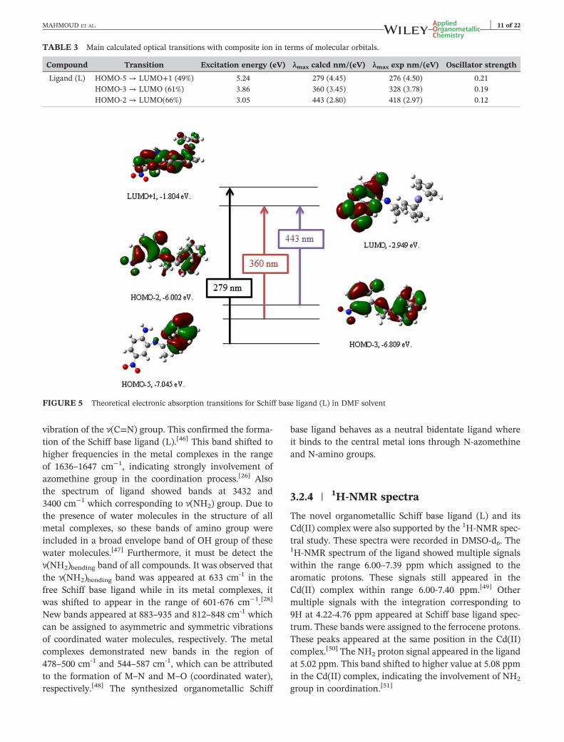
from the contribution of HOMO‐3 → LUMO. Finally, thepeak at 443 nm correspond to transition from HOMO‐2 toLUMO, was appeared with an oscillator strength off = 0.12.[43] These results were listed in Table 3 andshown in Figure 5.
3.2 | Characterization of metal complexes
A novel bidentate organometallic Schiff base ligand (L)was synthesized by condensation of 2‐acetylferrocenewith 4‐nitro‐o‐phenylenediamine. Among transitionmetal ions, Cr(III), Mn(II), Fe(III), Co(II), Ni(II), Cu(II),Zn(II) and Cd(II) complexes with the synthesized ligandwere prepared. These prepared compounds werecharacterized by using elemental analysis (C, H, N andmetal content), IR, 1H‐NMR, molar conductance,UV‐Vis, mass, SEM and thermal analyses (TG andDTG). The structures of the metal complexes were foundin Figure 6.
3.2.1 | Elemental analysis
From the data, all compounds elucidated a good agree-ment with the calculated values and suggested a 1:1metal‐to‐ligand stoichiometric ratio for all prepared com-plexes. The ligand and its metal complexes had high melt-ing points and they were stable in air. Also they weresoluble in DMF and DMSO but insoluble in methanol,ethanol and water.
3.2.2 | Molar conductivity measurements
The conductivity measurements play a serious role indetection the place of counter ions either outside or insidethe coordination sphere. This method was used todetermine the degree of ionization of the complexescomparing to the free ligands, with the higher molarconductance value corresponding to the presence ofcounter ions outside the coordination sphere andvice versa. The Cr(III), Fe(III), Ni(II) and Cu(II)complexes showed molar conductivity values of75, 54, 53and 50 Ω‐1 mol‐1 cm2 in DMF solution, respectively.These results proved the monomeric and electrolyticnatures of the complexes.[44] The Mn(II), Co(II), Zn(II)and Cd(II) complexes showed conductivity valuesbelow 50 Ω−1 mol −1 cm2 such that33, 18, 14 and2 Ω−1 mol−1 cm2, respectively, which indicated the non‐electrolytic characters of the complexes.[45]
3.2.3 | IR spectra
For detecting the coordination sites that may be involvedin chelation and get preliminary conformation of the
FIGURE 3 IR spectra of organometallic
Schiff base ligand (L) (a) theoretical
spectrum and (b) experimental spectrum
FIGURE 4 UV‐visible spectra of Schiff
base ligand (L) (a) theoretical spectrum
and(b) experimental spectrum
10 of 22 MAHMOUD ET AL.
structural aspects of new metal complexes, the IR spectraof the complexes were compared with that of the freeSchiff base ligand. The IR spectrum of the Schiff base
ligand showed disappearance of the ν(C=O) of 2‐acetylferrocene and illustrated a new intense band at1633 cm‐1, which can be assigned to the stretching
TABLE 3 Main calculated optical transitions with composite ion in terms of molecular orbitals.
FIGURE 5 Theoretical electronic absorption transitions for Schiff base ligand (L) in DMF solvent
MAHMOUD ET AL. 11 of 22
vibration of the ν(C=N) group. This confirmed the forma-tion of the Schiff base ligand (L).[46] This band shifted tohigher frequencies in the metal complexes in the rangeof 1636–1647 cm−1, indicating strongly involvement ofazomethine group in the coordination process.[26] Alsothe spectrum of ligand showed bands at 3432 and3400 cm−1 which corresponding to ν(NH2) group. Due tothe presence of water molecules in the structure of allmetal complexes, so these bands of amino group wereincluded in a broad envelope band of OH group of thesewater molecules.[47] Furthermore, it must be detect theν(NH2)bending band of all compounds. It was observed thatthe ν(NH2)bending band was appeared at 633 cm‐1 in thefree Schiff base ligand while in its metal complexes, itwas shifted to appear in the range of 601‐676 cm−1.[28]
New bands appeared at 883–935 and 812–848 cm‐1 whichcan be assigned to asymmetric and symmetric vibrationsof coordinated water molecules, respectively. The metalcomplexes demonstrated new bands in the region of478–500 cm‐1 and 544–587 cm‐1, which can be attributedto the formation of M–N and M–O (coordinated water),respectively.[48] The synthesized organometallic Schiff
base ligand behaves as a neutral bidentate ligand whereit binds to the central metal ions through N‐azomethineand N‐amino groups.
3.2.4 | 1H‐NMR spectra
The novel organometallic Schiff base ligand (L) and itsCd(II) complex were also supported by the 1H‐NMR spec-tral study. These spectra were recorded in DMSO‐d6. The1H‐NMR spectrum of the ligand showed multiple signalswithin the range 6.00–7.39 ppm which assigned to thearomatic protons. These signals still appeared in theCd(II) complex within range 6.00‐7.40 ppm.[49] Othermultiple signals with the integration corresponding to9H at 4.22‐4.76 ppm appeared at Schiff base ligand spec-trum. These bands were assigned to the ferrocene protons.These peaks appeared at the same position in the Cd(II)complex.[50] The NH2 proton signal appeared in the ligandat 5.02 ppm. This band shifted to higher value at 5.08 ppmin the Cd(II) complex, indicating the involvement of NH2
group in coordination.[51]
H2N
NC
H3CO2N
M
OH2
OH2Cl
Cl
.nCl.xH2O
M = Cr(III); n = 1, x = 4 Mn(II); n = 0, x = 2 Fe(III); n = 1, x = 2 Co(II); n = 0, x = 3 Zn(II); n = 0, x = 1 Cd(II); n = 0, x = 0
Fe
H2N
NC
H3CO2N
M
OH2
OH2OH2
Cl
.nCl.xH2O
M = Ni(II); n = 1, x = 2 Cu(II); n = 1, x = 1
Fe
FIGURE 6 The structure of organometallic Schiff base metal
The prepared complexes weren't single crystals, so it wasdifficult to get the XRD structure. As a result the powderdiffraction data were obtained for structural characteriza-tion. In order to get the degree of crystallinity of the pre-pared compounds, the powder X‐ray diffraction patternof the ligand, Cr(III), Mn(II), Fe(III), Cu(II), Ni(II), Co(II),Zn(II) and Cd(II) complexes were performed. The powderX‐ray diffraction pattern of all compounds was scanned inthe range 3‐80° (ϴ) at wavelength 1.54 Å. Also, thediffractogram and the associated data describe the inter‐planar spacing (d‐values), 2θ value for each peak and rel-ative intensity.[52]
Crystalline peaks are not seen in the powder XRD pat-tern of ligand (L), Cr(III), Fe(III), Ni(II), Cd(II) and Zn(II)complexes. This indicated amorphous nature of the
complexes. While Co(II), Mn(II) and Cu(II) complexeshad these crystalline peaks, indicating the crystallinenature of these complexes
The average crystallite size (ξ) can be calculated fromthe XRD pattern according to Debye‐Scherrerequation[53]:
ξ ¼ Kλβ1=2 cosθ
: (1)
The equation contains the reference peak width atangle (θ), where λ is wavelength of X‐ray radiation(1.542475 Å), K is constant taken as 0.95 for organic com-pounds and β1/2 is the width at half maximum of the ref-erence diffraction peak expressed in radians. Thedislocation density,δ, is the number of dislocation linesper unit area of the crystal. The value of δ is related tothe average particle diameter (ξ) by the relation[54,55]:
δ ¼ 1D2 (2)
The value of ξ was calculated and found to be 23.58,20.33 and 17.78 nm while the value of ξ was 1.80x10‐3,2.42x10‐3 and 3.16x10‐3 nm‐2 for Co(II), Mn(II) and Cu(II)complexes, respectively.
3.2.6 | Scanning electron microscope(SEM)
The morphology, size and structure of the nanomaterialswere conducted using field emission scanning electronmicroscopy (SEM).[40] The SEM micrographs of the Schiffbase ligand (L) and its [Cd(L)(H2O)2Cl2] complex werepresented in Figure 7. SEM images showed that the parti-cles were agglomerated with controlled morphologicalstructure.[56] It was evident from the SEM study that inthe synthesized Cd(II) complex, crystals were found togrow up from just a single molecule to several moleculesin an aggregate distribution with particle sizes that pres-ent in nanometers structures. As well, different character-istic shape of Cd(II) complex was identified and this SEMimage was quite different from that of the Schiff base. Thedifference in the shape of the Schiff base metal complexwas mainly dependent on the presence of metal ion.[57]
The micrograph of the ligand indicated collectedclouds shaped particles. The Cd(II) complex showednon‐uniform clusters structure. The average particle sizeof the ligand was 73 nm, but the average particle size ofthe [Cd(L)(H2O)2Cl2] complex was 61 nm. The synthe-sized nanopartical sized complex may help strongly in dif-ferent fields including biological applications.[58]
FIGURE 7 The SEM images of the nanoparticles produced a)
organometallic Schiff base ligand (L) and b) [Cd(L)(H2O)2Cl2]
complex
MAHMOUD ET AL. 13 of 22
3.2.7 | UV‐visible spectra of the Schiff baseligand and its metal complexes
The UV–Vis spectra of the ligand and its metal complexeswere measured at room temperature in the region of200–700 nm. The absorption spectrum of the Schiff baseligand (L) showed three absorption bands at 276, 328and 418 nm. The first high intensity band was appearedat λmax = 276 nm may be attributed to the π‐π* transitionof the aromatic rings. The second and third absorptionbands that were showed at λmax= 328 and 418 nm attrib-uted to the π‐π* transition of the azomethine group(C=N) and charge transfer, respectively.[44,47] Electronicspectra of the complexes showed bands which wereshifted from the free ligand to 262‐278 nm and
307‐333 nm for π‐π* of aromatic rings and π‐π* ofazomethine transitions, respectively, confirming the coor-dination of the azomethine nitrogen to the metal ions.These can be related to the binding of these coordinationcenters to the central metal ions. Also a band appeared at422 nm in the Cd(II) complex corresponding to chargetransfer. Furthermore, two absorption bands in the visibleregion of Co(II) complex were observed at 596 and662 nm. These bands were considered to arise from theforbidden d–d splitting, which was generally too weak.[10]
3.2.8 | Mass spectra
The stoichiometry of the novel organometallic Schiff baseligand and its [Cd(L)(H2O)2Cl2] complex were ascertainedfrom electron impact (EI)‐mass spectral analysis. The ES‐mass spectra of the compounds were elucidated theirmolar weight. It was found that mass spectra at 70 eV ofthe compounds were in good agreement with the pro-posed structures. The spectra of the ligand and Cd(II)complex exhibited a molecular ion peak at m/z 365 amuand 583 amu corresponding to their molecular weights363 and 582 g/mol, respectively. The other peaksappeared in the mass spectra of the ligand and its Cd(II)complex (abundance range 1–100%) were attributed tothe fragmentation of the compounds obtained from therupture of different bonds inside the molecule.
3.2.9 | Thermal analysis of Schiff baseligand and its metal complexes
In the present investigation, the thermal stability of theSchiff base ligand (L) and its metal complexes were inves-tigated using thermogravimetric technique (TG) and dif-ferential thermogravimetric (DTG) analyses at a heatingrate of 10 °C/min in nitrogen atmosphere over the rangefrom ambient temperature to 1000 °C. The data in(Table 4) provided information concerning thermaldecomposition of these complexes in solid state. Theorganometallic Schiff base ligand (L) with the molecularformula (C18H17FeN3O2) was thermally decomposed intwo successive decomposition steps. The first step withestimated mass loss of 32.28% (calculated mass loss =32.23%) within the temperature range 75–485 °C may beattributed to the loss of C7H5N2 molecule. The DTG curvegave maximum peak temperature at 219 °C. The secondstep occurred within the temperature range 485–1000 °Cwith an estimated mass loss 18.58% (calculated mass loss= 18.18%), which correspond to the loss of C2H12NOfragment. The DTG curve gave peak at 752 °C. FinallyFeO contaminated with carbon remain as residues. Theoverall weight loss amounted to 50.86% (calculated massloss = 50.41%).
TABLE 4 Thermoanalytical results (TG and DTG) of organometallic Schiff base ligand (L) and its metal complexes.
ComplexTG range
(°C) DTGmax (°C) n*
Mass lossTotal massloss Estim(Calcd) % Assignment Residues
Schiff base ligand (L) 75‐485 219 1 32.28 (32.23) ‐Loss of C7H5N2. FeO+9C485‐1000 752 1 18.58 (18.18)
The Cr(III) complex thermally decomposed in twostages. The first stage correspond to two steps with esti-mated mass loss of 22.23% (calculated mass loss =22.72%) within the range 50–340 °C and represented theloss of Cl2 gas and four molecules of water of hydration.The second stage correspond to estimated mass loss of54.44% (calculated mass loss =53.77%) within the range340–1000 °C and represented the loss of HCl andC18H20N3O1.5molecules, leaving FeO and ½ Cr2O3 as res-idues. The overall weight loss amounted to 76.67% (calcu-lated mass loss = 76.49%).
In the TG curve of Mn(II) complex, the first stepdisplayed a gradual mass loss of 7.27% (calculated mass
loss = 6.42%) within the temperature range of 30–100 °Cwhich may be attributed to the loss of two molecules ofwater of hydration. The DTG curve gave peak at 79 °C(the maximum peak temperature). The second and thirdsteps showed a mass loss of 28.37% (calculated mass loss=28.70%) which correspond to loss of 2H2O, Cl2 andC3H4N molecules within temperature range 100–570 °C.Two maximum peaks temperatures were found at186 °C and 260 °C. The final step was correspond to lossof C8H13N2 molecule with estimated mass loss = 24.08%(calculated mass loss = 24.42%) within temperature rangefrom 570 °C to 1000 °C. The maximum peak temperaturewas found at 760 °C. Finally FeO and MnO contaminated
MAHMOUD ET AL. 15 of 22
with carbon remained as residues. The overall weight lossamounted to 59.72% (calculated mass loss = 59.54%).
The thermogram of the Fe(III) complex showed fivedecomposition steps within the range 30–1000 °C. The firstand second decomposition steps were accompanied by lossof 2H2O molecules in the range 30–155 °C with an esti-mated mass loss of 5.79% (calculated mass loss = 6.02%).The DTG curve gave two peaks at 63 and 150 °C (the max-imum peaks temperature). The third step of decompositioncorrespond to loss of H2O and C4H8 molecules at 155–290 °C with an estimated mass loss of 12.85% (calculatedmass loss = 12.38%) with maximum peak temperature at254 °C. The final two decomposition steps within the range290–1000 °Cwere assigned to loss of Cl2 and C4H11N3O0.5Clmolecules with a mass loss of 36.13% (calculated mass loss= 36.07%). These peaks appeared at 707 and 816 °C. There-after the percentage of the residues correspond to ferric andferrous oxides contaminated with carbon were 45.22% (cal-culated mass loss = 45.52%). The overall weight lossamounted to 54.77% (calculated mass loss = 54.47%).
The [Co(L)(H2O)2Cl2]3H2O complex lost upon heating5H2O and CN in the first and second steps of decomposi-tion within the temperature range of 30–290 °C, at maxi-mum peaks temperatures 68 and 266 °C with estimatedmass loss of 19.24% (calculated mass loss = 19.90%). Thethird step accounted for the loss of Cl2 gas and C8H17N2
molecule within the temperature range of 290–1000 °C,at maximum peak temperature 730 °C, with estimatedmass loss of 36.70% (calculated mass loss = 36.36%), leav-ing FeO + CoO contaminated with carbon as residue ofdecomposition. The overall weight loss amounted to55.94% (calculated mass loss = 55.26%).
The Ni(II) complex gave decomposition pattern startedat 30 °C and finished at 1000 °C with three stages. The firststage was one step within the temperature range of 30–120 °C with maximum peak temperature at 80 °C and rep-resented the loss of 2H2O (hydrated) with a foundmass lossof 5.32% (calculated mass loss = 6.18%). The second stage
FIGURE 8 Biological activity of
organometallic Schiff base ligand (L) and
its metal complexes
was two steps and represented the loss of Cl2, 3H2O andC3H4molecules with a mass loss of 28.84% (calculated massloss = 28.30%) within the temperature range 120–640 °Candmaxima peaks temperature at 268 and 339 °C. The finalstage was one step representing the loss of C13H13N3 mole-cule with a mass loss of 36.34% (calculated mass loss =36.19%) within the temperature range 640–1000 °C. At theend of the thermogram, the metal oxide NiO and FeO con-taminated with carbon were the residues, which was ingood agreement with the calculatedmetal content obtainedand the results of elemental analyses. The overall weightloss amounted to 70.50% (calculated mass loss = 70.67%).
The TG curve of the Cu(II) complex showed that the firstand the second decomposition steps correspond to mass loss19.39% (calculated mass loss = 18.90%) which occurredwithin the temperature range from 50 to 315 °C with maxi-mum peaks temperatures at 94 and 264 °C and representedthe loss of ½Cl2 and 4H2O molecules. The third step withinthe temperature range 315–640 °Cwithmaximumpeak tem-perature at 577 °Cmay be attributed to the decomposition ofC3H4NCl molecule with found mass loss of 15.42% (calcu-lated mass loss = 15.72%). The final step started at 640 °Cand ended at 1000 °C with maximum peak temperature at845 °C and may be accounted to the loss of C6H13N2 mole-cule with mass loss of 19.87% (calculated mass loss =19.84%), leaving behind CuO and FeO oxides contaminatedwith carbon as residues of decomposition. The overallweightloss amounted to 54.68% (calculated mass loss = 54.46%).
For Zn(II) complex, the first step of decomposition corre-spond to mass loss of 3.43% (calculated mass loss = 3.26%)within the temperature range 30‐200 °C with maximumpeak temperature at 74 °C and represented the loss of onehydrated water molecule. The second step within the tem-perature range 200–490 °C with maximum peak tempera-ture at 241 °C may be attributed to the decomposition of2H2O and C3H6 molecules with found mass loss of 13.89%(calculated mass loss = 14.10%). The third step started at490 °C and ended at 745 °C with maximum peak
plexes.
Inhibitionzo
nediameter
(mm
/mgsample)
mnegative)
Candidaalbicans(fungu
s)erichia
coli
Klebsiellapneu
mon
iaPseudom
onasaeruginosa
00
0
±0.6
18.7±0.6
NA
13.3±1.5
NA
NA
NA
±0.6
NA
NA
NA
NA
NA
NA
±0.6
21.0±1.0
10.7±0.5
NA
NA
NA
NA
15.7±0.5
18.7±0.6
NA
±0.6
31±1.0
20.3±0.6
NA
±0.6
24.7±0.6
19.3±0.6
14.3±0.6
105
‐‐‐‐‐‐
‐‐‐‐‐‐
‐‐‐‐‐‐
9
16 of 22 MAHMOUD ET AL.
temperature at 666 °C and may be accounted to the loss ofCl2gas and C2H6molecule with mass losses of 18.29% (calcu-lated mass loss = 18.26%). The final step started at 745 °Cand ended at 1000 °C with maximum peak temperature at846 °C andmay be accounted to the loss of C3H5N3moleculewith mass losses of 15.02% (calculated mass loss = 15.01%),leaving behind ZnO and FeO oxides contaminated with car-bon as residues of decomposition. The overall weight lossamounted to 50.63% (calculated mass loss = 50.63%).
The [Cd(L)(H2O)2Cl2] complex upon heating lostC2H2Cl and 2H2O molecules in the first step of decompo-sition within the temperature range 75–245 °C, with amass loss = 16.06% (calculated mass loss = 16.74%). Thesecond step of decomposition occurred with maximumpeak temperature at 608 °C and correspond to loss ofC7H6N2Cl molecule with a mass loss = 26.88% (calculatedmass loss = 26.35%), in the temperature range of245‐630 °C. The final two steps started decomposition at630 °C and ended at 1000 °C with maximum peakstemperatures at 691 and 877 °C and may be correspondto the loss of C9H9N molecule with mass loss of 22.70%(calculated mass loss = 22.49%), leaving CdO and FeOoxides as residues of decomposition. The overall weightloss amounted to 65.64% (calculated mass loss = 65.58%).
As a result of these decompositions, the oxide residuesalways contaminated with carbon. This may be evidenceof the high stability of the synthesized compounds againsthigh temperature.
chiffbase
ligan
d(L)an
ditsmetal
com
(Gra
sStaphylococcu
saureus
Esch
00
NA
21.3
NA
NA
NA
10.7
NA
NA
14.7±0.5
19.7
NA
NA
NA
NA
17.0±1.0
25.7
14.3±0.5
25.7
96
‐‐‐‐‐‐
‐‐‐‐‐‐
4 | STRUCTURALINTERPRETATION
The structures of organometallic Schiff base ligand (L)and its metal complexes were characterized using variousphysico‐chemical and spectral data. Accordingly, thestructures of the complexes have been confirmed andthe proposed structural formulas of the complexes wereshown in (Figure 6).
TABLE5
Biologicalactivity
oforganom
etallicS
Sample
(Gra
mpositive)
Streptococcusmutan
Con
trol:DMSO
0
Schiffba
seliga
nd(L
)24.0±1.0
[Cr(L)(H
2O) 2Cl 2]C
l.4H
2ONA
[Mn(L
)(H
2O) 2Cl 2]2H
2ONA
[Fe(L)(H
2O) 2Cl 2]C
l.2H
2ONA
[Co(L)(H
2O) 2Cl 2]3H
2O15.7±0.6
[Ni(L)(H
2O) 3Cl]Cl.2H
2ONA
[Cu(L
)(H
2O) 3Cl]Cl.H
2O12.7±0.5
[Zn(L
)(H
2O) 2Cl 2]H
2O40.7±0.6
[Cd(L
)(H
2O) 2Cl 2]
22.0±1.0
Amikac
in10
Ketok
onaz
ole
‐‐‐‐‐‐
NA:Noactivity
4.1 | Antimicrobial activities
All the newly synthesized organometallic Schiff baseligand (L) and its metal complexes were screened for theirantifungaland antibacterial activities. The tested com-pounds showed variable antibacterial activity against bothGram(+) bacteria: [Streptococcus mutans, Staphylococcusaureus], Gram (−) bacteria: [Escherichia coli, Klebsiellapneumonia and Pseudomonas aeruginosa] and fungalspecies such as Candida albicans. The efficiencies of theSchiff base ligand (L) and its complexes have been testedagainst two Gram (+ve), three Gram (−ve) and one fungi(Figure 8). Also these activities were listed in Table 5. The
FIGURE 9 Activity index of
organometallic Schiff base ligand (L) and
its metal complexes against (a) different
Gram positive bacteria species (b) different
Gram negative bacteria species
TABLE 6 Anticancer activity of organometallic Schiff base ligand (L) and its metal complexes against breast cancer cell line.
Complex Concn.( μg/ mL)
Surviving fraction (MCF7)
IC50> (μg/mL)0.0 5.0 12.5 25.0 50.0
Schiff base ligand (L) 1.0 0.893 0.843 0.693 0.529 50.200
activities of the prepared Schiff base ligand and its metalcomplexes were confirmed by calculating the activityindex according to the following relation.[59,60]
Activity index Að Þ ¼ Inhibition Zone of compound½mmð Þ=Inhibition Zone of standard drug
mmð Þ�×100
From the data, it was concluded that Zn(II) complexhad the highest activity index, while Cr(III), Fe(III) andNi(II) complexes had no activity index, Figure 9.[61]
The results from the biological activities showed that theZn(II), Cd(II) and Co(II) complexes had higher activitiesagainst all different bacterial species than the other com-plexes. Zn(II) complex had the highest activity value againststreptococcus mutans specie. Also this value was higher thanthe Schiff base ligand and ampicillin standard. So, Zn(II)complex can be used as very active drug against streptococcus
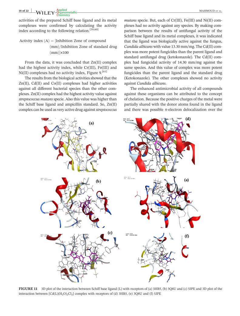
FIGURE 11 3D plot of the interaction between Schiff base ligand (L) w
interaction between [Cd(L)(H2O)2Cl2] complex with receptors of (d) 3HB
mutans specie. But, each of Cr(III), Fe(III) and Ni(II) com-plexes had no activity against any species. By making com-parison between the results of antifungal activity of theSchiff base ligand and its metal complexes, it was indicatedthat the ligand was biologically active against the fungus,Candida albicanswith value 13.30mm/mg. The Cd(II) com-plex was more potent fungicides than the parent ligand andstandard antifungal drug (ketokonazole). The Cd(II) com-plex had fungicidal activity of 14.30 mm/mg against thesame species. And this value of complex was more potentfungicides than the parent ligand and the standard drug(Ketokonazole). The other complexes showed no activityagainst Candida albicans.
The enhanced antimicrobial activity of all compoundsagainst these organisms can be attributed to the conceptof chelation. Because the positive charges of the metal werepartially shared with the donor atoms found in the ligandand there was possible π‐electron delocalization over the
ith receptors of (a) 3HB5, (b) 3Q8U and (c) 5JPE and 3D plot of the
5, (e) 3Q8U and (f) 5JPE
MAHMOUD ET AL. 19 of 22
metal complex formed. The lipophilic character of themetal complex increases and supports its permeation moreefficiently through the lipid layer of the micro‐organisms.This permits easy binding and penetration of thecomplexin the cellular structure of the pathogens.[62] The chelationleads to make the ligand act as more powerful and potentbactereostatic agents. This inhibits the growth of bacteriamore than the parent ligands. It was suspected that factorssuch as conductivity, solubility, dipole moment and cellpermeability mechanism may be influenced by the pres-ence of metal ion. This might be the possible reason forincreasing the activity after chelation.[63,64]
TABLE 7 Energy values obtained in docking calculations of Schiff ba
mutant oxidoreductase (PDB ID: 3HB5), crystal structure of Staphylococ
protein phosphatase (PPZ1) of Candida albicans (PDB ID: 5JPE).
5JPE N 36 O HIS 41O 44 OD2 ASPO 44 OD2 ASPO 44 OD2 ASPO 44 OD2 ASPO 45 OD2 ASP
5 | ANTICANCER ACTIVITIES
In various human diseases, cancer considered as the mostsevere disease to which humans were subjected and yetno effective drugs or any methods of control are availableto treat it. So, it is necessary to identify novel, selective,potent and less toxic anticancer agents which becameone of the most pressing problems. Cancer is a complexdisease that is normally correlated to a wide range of esca-lating effects both at the cellular and molecular levels.[65]
The cytotoxic ability of Schiff base ligand and its metalcomplexes were evaluated against breast carcinoma cells
se ligand (L) and its Cd(II) complex with receptors of breast cancer
cus aureus (PDB ID: 3Q8U) and yeast‐specific serine/threonine
FIGURE 12 The relation between the lowest binding energy of the
ligand and its Cd(II) complex with 3HB5, 3Q8U and 5JPE receptors
20 of 22 MAHMOUD ET AL.
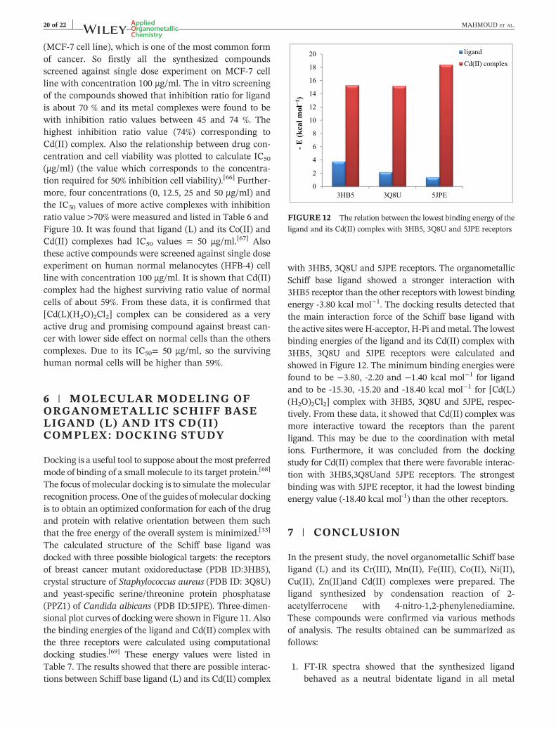
(MCF‐7 cell line), which is one of the most common formof cancer. So firstly all the synthesized compoundsscreened against single dose experiment on MCF‐7 cellline with concentration 100 μg/ml. The in vitro screeningof the compounds showed that inhibition ratio for ligandis about 70 % and its metal complexes were found to bewith inhibition ratio values between 45 and 74 %. Thehighest inhibition ratio value (74%) corresponding toCd(II) complex. Also the relationship between drug con-centration and cell viability was plotted to calculate IC50
(μg/ml) (the value which corresponds to the concentra-tion required for 50% inhibition cell viability).[66] Further-more, four concentrations (0, 12.5, 25 and 50 μg/ml) andthe IC50 values of more active complexes with inhibitionratio value >70% were measured and listed in Table 6 andFigure 10. It was found that ligand (L) and its Co(II) andCd(II) complexes had IC50 values = 50 μg/ml.[67] Alsothese active compounds were screened against single doseexperiment on human normal melanocytes (HFB‐4) cellline with concentration 100 μg/ml. It is shown that Cd(II)complex had the highest surviving ratio value of normalcells of about 59%. From these data, it is confirmed that[Cd(L)(H2O)2Cl2] complex can be considered as a veryactive drug and promising compound against breast can-cer with lower side effect on normal cells than the otherscomplexes. Due to its IC50= 50 μg/ml, so the survivinghuman normal cells will be higher than 59%.
6 | MOLECULAR MODELING OFORGANOMETALLIC SCHIFF BASELIGAND (L) AND ITS CD(II)COMPLEX: DOCKING STUDY
Docking is a useful tool to suppose about themost preferredmode of binding of a small molecule to its target protein.[68]
The focus of molecular docking is to simulate themolecularrecognition process. One of the guides of molecular dockingis to obtain an optimized conformation for each of the drugand protein with relative orientation between them suchthat the free energy of the overall system is minimized.[33]
The calculated structure of the Schiff base ligand wasdocked with three possible biological targets: the receptorsof breast cancer mutant oxidoreductase (PDB ID:3HB5),crystal structure of Staphylococcus aureus (PDB ID: 3Q8U)and yeast‐specific serine/threonine protein phosphatase(PPZ1) of Candida albicans (PDB ID:5JPE). Three‐dimen-sional plot curves of docking were shown in Figure 11. Alsothe binding energies of the ligand and Cd(II) complex withthe three receptors were calculated using computationaldocking studies.[69] These energy values were listed inTable 7. The results showed that there are possible interac-tions between Schiff base ligand (L) and its Cd(II) complex
with 3HB5, 3Q8U and 5JPE receptors. The organometallicSchiff base ligand showed a stronger interaction with3HB5 receptor than the other receptors with lowest bindingenergy ‐3.80 kcal mol−1. The docking results detected thatthe main interaction force of the Schiff base ligand withthe active sites were H‐acceptor, H‐Pi andmetal. The lowestbinding energies of the ligand and its Cd(II) complex with3HB5, 3Q8U and 5JPE receptors were calculated andshowed in Figure 12. The minimum binding energies werefound to be −3.80, ‐2.20 and −1.40 kcal mol−1 for ligandand to be ‐15.30, ‐15.20 and ‐18.40 kcal mol−1 for [Cd(L)(H2O)2Cl2] complex with 3HB5, 3Q8U and 5JPE, respec-tively. From these data, it showed that Cd(II) complex wasmore interactive toward the receptors than the parentligand. This may be due to the coordination with metalions. Furthermore, it was concluded from the dockingstudy for Cd(II) complex that there were favorable interac-tion with 3HB5,3Q8Uand 5JPE receptors. The strongestbinding was with 5JPE receptor, it had the lowest bindingenergy value (‐18.40 kcal mol‐1) than the other receptors.
7 | CONCLUSION
In the present study, the novel organometallic Schiff baseligand (L) and its Cr(III), Mn(II), Fe(III), Co(II), Ni(II),Cu(II), Zn(II)and Cd(II) complexes were prepared. Theligand synthesized by condensation reaction of 2‐acetylferrocene with 4‐nitro‐1,2‐phenylenediamine.These compounds were confirmed via various methodsof analysis. The results obtained can be summarized asfollows:
1. FT‐IR spectra showed that the synthesized ligandbehaved as a neutral bidentate ligand in all metal
MAHMOUD ET AL. 21 of 22
complexes, in which it can form a coordinate bondwith metal ions through azomethine nitrogen andNH2 group.
2. Elemental analysis showed that reaction betweenligand and metal ions occurred as 1:1molar ratio and the complexes had the formulae of[M(L)(H2O)2Cl2].nCl.xH2O if (M = Cr(III)) (n = 1)(x = 4); (M = Mn(II)) (n = 0) (x = 2); (M = Fe(III))(n = 1) (x = 2); (M = Co(II)) (n = 0) (x = 3); (M =Zn(II)) (n= 0) (x = 1)) and (M = Cd(II)) (n = 0) (x= 0), and formulae of [M(L)(H2O)3Cl].nCl.xH2O if(M = Ni(II)) (n = 1) (x = 2); (M = Cu(II)) (n = 1) (x= 1). Also all complexes had octahedral structures.
3. The data indicated that Cr(III), Fe(III), Ni(II) andCu(II) complexes weremonomeric and 1:1 electrolyteswhile other complexes had non‐electrolytic characters.
4. By screening some of the compounds against breastcancer cell line (MCF‐7) and human normal melano-cytes (HFB‐4) cell line, it showed that Cd(II) complexcan be considered as a very active drug against breastcancer with inhibition ratio (74%) with lower sideeffect on normal cells than the others.
5. The biological activity of the synthesized ligand andits complexes was studied against Gram (+) bacteria:[Streptococcus mutans, Staphylococcus aureus], Gram(−) bacteria: [Escherichia coli, Klebsiella pneumonia,Pseudomonas aeruginosa] and fungal specie include[Candida albicans]. It was found that the Zn(II),Cd(II) and Co(II) complexes had higher activitiesagainst all different bacterial species than the othercomplexes. Zn(II) complex had the highest activityvalue against streptococcus mutans specie. So, Zn(II)complex can be used as very active drug against thisspecie. But, each of Cr(III), Fe(III) and Ni(II) com-plexes had no activity against any species. Also, theSchiff base ligand and its Cd(II) complex were biolog-ically active against the fungus, Candida albicans.While, other complexes were biologically inactive.
6. The geometrical structure for organometallic Schiffbase ligand (L) was computed. From this, the bindingenergies, bond length and dipole moment were calcu-lated. Also the data of experimental IR and UV‐visspectra were compared with theoretical ones, whichshowed that ligand was prepared successfully.
7. Molecular docking studies of the free organometallicSchiff base ligand (L) and its Cd(II) complex withreceptors of (PDB ID: 3HB5, 3Q8U and 5JPE)detected that the ligand showed highest binding abil-ity with binding energy of ‐3.80 kcal mol−1 with thereceptor of breast cancer mutant oxidoreductase(PDB ID: 3HB5), while Cd(II) complex showed low-est binding energies ‐18.40 kcal mol−1 with thereceptor 5JPE.
ORCID
Walaa H. Mahmoud http://orcid.org/0000-0001-9187-4325
REFERENCES
[1] M. M. Omar, H. F. Abd El‐Halim, E. A. M. Khalil, Appl.Organometal. Chem. 2017, In press.
[2] L. S. Pogany, J. Moncol, M. Gal, I. Salitros, R. Boca, Inorg. Chim.Acta 2017, 462, 23.
[3] M. Habibi, S. A. Beyramabadi, S. Allameh, M. Khashi, A.Morsali, M. Pordel, M. Khorsandi‐Chenarboo, Mol. Str. 2017,1143, 424.
[4] M. Anar, E. H. Ozkan, H. Ogutcu, G. Agar, I. Sakiyan, N. Sari,Artificial Cells, Nanomedicine, and Biotechnology 2016, 44, 853.
[5] E. Logoglu, E. A. Koyuncu, M. N. S. Karaboga, N. Sari, GaziUniver. J. Sci. 2016, 29, 303.
[6] M. A. Diab, A. Z. El‐Sonbati, A. F. Shoair, A. M. Eldesoky, N. M.El‐Far, Mol. Str. 2017, 1141, 710.
[7] R. E. Kyne, M. C. Ryan, L. T. Kliman, J. P. Morken, Org. Lett.2010, 12, 3796.
[8] J. Luan, L. Zhang, Molecules 2011, 16, 4191.
[9] T. J. Kealy, P. L. Pauson, Nature 1951, 168, 1039.
[10] Y. Liu, G. Lian, D. Yin, B. Su, Spectrochim. Acta A 2013, 100, 131.
[11] M. Shabbir, Z. Akhter, I. Ahmad, S. Ahmed, M. Bolte, H.Ismail, B. Mirza, Inorg. Chim. Acta 2017, 463, 102.
[12] B. Maity, M. Roy, B. Banik, R. Majumdar, R. R. Dighe, A. R.Chakravarty, Organometallics 2010, 29, 3632.
[13] R. W. Mason, K. McGrouther, P. R. R. Ranatunge‐Bandarage, B.H.Robinson, J. Simpson,Appl.Organometal.Chem.1999,13, 163.
[14] C. Ornelas, New J. Chem. 2011, 35, 1973.
[15] A. A. Turki, J. kerbala univ. 2012, 10, 259.
[16] A. Albert, Selective Toxicity: Physico‐chemical Basis of Therapy(6th Edition), Wiley, New York 1979.
[17] S. Chandra, D. Jain, A. K. Sharma, P. Sharma, Molecules 2009,14, 174.
[18] P. Skehan, R. Storeng, D. Scudiero, A. Monks, J. McMahon, D.Vistica, J. T. Warren, H. Bokesch, S. Kenney, M. R. Boyd,J. National Cancer Instit. 1990, 82, 1107.
[19] M. J. Frisch, G. W. Trucks, H. B. Schlegel, G. E. Scuseria, M. A.Robb, J. R. Cheeseman, V. G. Zakrzewski, J. A. Montgomery, R.E. Stratmann, J. C. Burant, S. Dapprich, J. M. Millam, A. D. Dan-iels, K. N. Kudin,M. C. Strain, O. Farkas, J. Tomasi, V. Barone,M.Cossi, R. Cammi, B. Mennucci, C. Pomelli, C. Adamo, S. Clifford,J. Ochterski, G. A. Petersson, P. Y. Ayala, Q. Cui, K. Morokuma,D. K. Malick, A. D. Rabuck, K. Raghavachari, J. B. Foresman, J.Cioslowski, J. V. Ortiz, A. G. Baboul, B. B. Stefanov, G. Liu, A.Liashenko, P. Piskorz, I. Komaromi, R. Gomperts, R. L. Martin,D. J. Fox, T. Keith, M. A. Al‐Laham, C. Y. Peng, A. Nanayakkara,C. Gonzalez, M. Challacombe, P. M. W. Gill, B. G. Johnson, W.Chen,M.W.Wong, J. L. Andres, M. Head‐Gordon, E. S. Replogle,J. A. Pople, GAUSSIAN 03 (Revision A.9), Gaussian, Inc., Pitts-burgh 2003.
[20] G. G. Mohamed, Z. H. Abd El‐Wahab, Spectrochim. Acta A.2005, 61, 1059.
[21] R. Dennington, T. Keith, J. Millam (Eds), GaussView,Version 5.0.8, R, KS: Dennington, Semichem Inc., ShawneeMission 2009.
[22] C. J. Dhanaraj, I. U. Hassan, J. Johnson, J. Joseph, R. S.Joseyphus, J. Photochem. & Photobio. B 2016, 162, 115.
[23] C. Balakrishnan, L. Subha, M. A. Neelakantan, S. S. Mariappan,Spectrochim. Acta A 2015, 150, 671.
[24] L. H. Abdel‐Rahman, N. M. Ismail, M. Ismael, A. M. Abu‐Dief,E. Abdel‐Hameed, Mol. Str. 2017, 1134, 851.
[25] L. W. Jolly, The Synthesis and Characterization of InorganicCompounds, Prentice‐Hall, INC 1970 485.
[26] N. M. Hosny, M. A. Husien, F. M. Radwan, N. Nawar, Mol. Str.2017, 1143, 176.
[27] A. A. Maihub, A. M. Etorkil, S. M. Ben‐Saber, M. M. El‐ajaily,M. M. Abou‐Krisha, J. Chem. Eng. 2014, 8, 226.
[28] W. H. Mahmoud, N. F. Mahmoud, G. G. Mohamed, A. Z.El‐Sonbati, A. A. El‐Bindary, Mol. Str. 2015, 1095, 15.
[29] S. R. Gupta, P. Mourya, M. M. Singh, V. P. Singh, Organometal.Chem. 2014, 767, 136.
[30] S. Adhikari, W. Kaminsky, M. R. Kollipara, Organometal.Chem. 2017, 836, 8.
[31] M. Dehkhodaei, M. Khorshidifard, H. A. Rudbari, M. Sahihi, G.Azimi, N. Habibi, S. Taheri, G. Bruno, R. Azadbakht, Inorg.Chim. Acta 2017, 466, 48.
[32] A. Z. El‐Sonbati, M. A. Diab, S. M. Morgan, J. Mol. Liq. 2017,225, 195.
[33] G. G. Mohamed, A. A. El‐Sherif, M. A. Saad, S. E. A. El‐Sawy, S.M. Morgan, J. Mol. Liq. 2016, 223, 1311.
[34] S. M. Morgan, A. Z. El‐Sonbati, H. R. Eissa, J. Mol. Liq. 2017,240, 752.
[35] A. Y. Al‐Dawood, N. M. El‐Metwaly, H. A. El‐Ghamry, J. Mol.Liq. 2016, 220, 311.
[36] A. M. A. Alaghaz, M. E. Zayed, S. A. Alharbi, Mol. Str. 2015,1083, 430.
[37] N. Okulik, A. H. Jubert, Internet Electron. J. Mol. Des. 2005,4, 17.
[38] H. Wang, X. Zhang, Y. Zhao, D. Zhang, F. Jin, Y. Fan, Mol. Str.2017, 1148, 496.
[39] B. Annaraj, S. Pan, M. A. Neclakantan, P. K. Chattaraj, Comput.Theor. Chem. 2014, 1028, 19.
[40] Z. Parsaee, K. Mohammadi, Mol. Str. 2017, 1137, 512.
[41] S. Roy, K. Harms, S. Chattopadhyay, Polyhedron 2016, In Press.
[42] W. H. Mahmoud, N. F. Mahmoud, G. G. Mohamed, J. Therm.Anal. Calorim. 2017, In Press.
[43] H. Vural, M. Orbay, Mol. Str. 2017, 1146, 669.
[44] N. Mishra, K. Poonia, S. K. Soni, D. Kumar, Polyhedron 2016,120, 60.
[45] W. H. Mahmoud, N. F. Mahmoud, G. G. Mohamed, J. Coord.Chem. 2017, In press.
[46] Z. Beigi, A. H. Kianfar, G. Mohammadnezhad, H. Görls, W.Plass, Polyhedron 2017, 134, 65.
[47] N. Ribeiro, S. Roy, N. Butenko, I. Cavaco, T. Pinheiro, I. Alho,F. Marques, F. Avecilla, J. C. Pessoa, I. Correia, J. Inorg.Biochem. 2017, 174, 63.
[48] E. M. Zayed, G. G. Mohamed, A. M. M. Hindy, J. Therm. Anal.Calorim. 2015, 120, 893.
[49] A. A. Abdel Aziz, I. S. A. El‐Sayed, M. M. H. Khalil, Appl.Organometal. Chem. 2017, In Press.
[50] Y. Shi, H. Yang,W. Shen, C. Yan, X. Hu, Polyhedron 2004, 23, 15.
[51] S. Yadav, R. V. Singh, Spectrochim. Acta A 2011, 78, 298.
[52] G. Y. Nagesh, B. H. M. Mruthyunjayaswamy, J. Mol. Str. 2015,1085, 198.
[53] H. F. Abd El‐Halim, G. G. M. , M. N. Anwar, Appl OrganometalChem. 2017, In press.
[54] S. Velumania, X. Mathew, P. J. Sebastian, S. K. Narayandass, D.Mangalaraj, Sol. Energ. Mat. Sol. C. 2003, 76, 347.
[55] S. Basavaraja, D. S. Balaji, M. D. Bedre, D. Raghunandan, P. M.P. Swamy, A. Venkatarman, Bull. Mat. Sci. 2011, 34, 1313.
[56] N. K. Poonia, S. Siddiqui, M. D. Arshad, D. Kumar, Spectrochim.Acta A 2016, 155, 146.
[57] M. I. Khan, A. Khan, I. Hussain, M. A. Khan, S. Gul, M. Iqbal, I.R. F. Khuda, Inorg. Chem. Comm. 2013, 35, 104.
[58] A. M. A. Alaghaz, M. E. Zayed, S. A. Alharbi, Mol. Str. 2015,1084, 36.
[59] T. D. Thangadurai, K. Natarajan, Indian J. Chem. A 2001, 40, 573.
[60] Z. H. Chohan, H. Pervez, A. Rauf, K. M. Khan, C. T. Supuran,J. Enzyme Inhib. Med. Chem. 2004, 19, 417.
[62] G. More, D. Raut, K. Aruna, S. Bootwala, J. Saudi Chem. Soc2017, In Press.
[63] Z. H. Chohan, M. Praveen, Appl. Organometal. Chem. 2001,15, 617.
[64] Z. H. Chohan, C. T. Supran, A. Scozzafava, J. Enz. Inhib. Med.Chem. 2004, 19, 79.
[65] S. J. Kirubavathy, R. Velmurugan, R. Karvembu, N. S. P.Bhuvanesh, I. V. M. V. Enoch, P. M. Selvakumar, D. Premnath,S. Chitra, Mol. Str. 2017, 1127, 345.
[66] C. M. Sharaby, M. F. Amine, A. A. Hamed, Mol. Str. 2017,1134, 208.
[67] M. A. Arafath, F. Adam, M. R. Razali, L. E. A. Hassan, M. B. K.Ahamed, A. M. S. A. Majid, Mol. Str. 2017, 1130, 791.
[68] S. Mondal, S. M. Mandal, T. K. Mondal, C. Sinha,Mol. Str. 2017,1127, 557.
[69] J. Tang, J. Liu, Bioorg. Chem. 2016, 69, 29.
How to cite this article: Mahmoud WH, DeghadiRG, Mohamed GG. Metal complexes of novel Schiffbase derived from iron sandwiched organometallicand 4‐nitro‐1,2‐phenylenediamine: Synthesis,characterization, DFT studies, antimicrobialactivities and molecular docking. Appl OrganometalChem. 2018;e4289. https://doi.org/10.1002/aoc.4289