9 Metals in Medicine STEPHEN J. LIPPARD Department of Chemistry Massachusetts Institute of Technology I. INTRODUCTION AND OVERVIEW Metal ions are required for many critical functions in humans. Scarcity of some metal ions can lead to disease. Well-known examples include pernicious anemia resulting from iron deficiency, growth retardation arising from insufficient di- etary zinc, and heart disease in infants owing to copper deficiency. The ability to recognize, to understand at the molecular level, and to treat diseases caused by inadequate metal-ion function constitutes an important aspect of medicinal bioinorganic chemistry. Metal ions can also induce toxicity in humans, classic examples being heavy- metal poisons such as mercury and lead. Even essential metal ions can be toxic when present in excess; iron is a common household poison in the United States as a result of accidental ingestion, usually by children, of the dietary supplement ferrous sulfate. Understanding the biochemistry and molecular biology of natu- ral detoxification mechanisms, and designing and applying ion-specific chelating agents to treat metal overloads, are two components of a second major aspect of the new science that is evolving at the interface of bioinorganic chemistry and medicine. Less well known than the fact that metal ions are required in biology is their role as pharmaceuticals. Two major drugs based on metals that have no known natural biological function, Pt (cisplatin) and Au (auranofin), are widely used cis-diamminedichloroplatinum(II) (cisplatin, or cis-DDP) OAc OAc AcO OAc thioglucose(triethylphosphine)gold(I) (auranofin) 505
Transcript
9Metals in Medicine
STEPHEN J. LIPPARDDepartment of Chemistry
Massachusetts Institute of Technology
I. INTRODUCTION AND OVERVIEW
Metal ions are required for many critical functions in humans. Scarcity of somemetal ions can lead to disease. Well-known examples include pernicious anemiaresulting from iron deficiency, growth retardation arising from insufficient dietary zinc, and heart disease in infants owing to copper deficiency. The abilityto recognize, to understand at the molecular level, and to treat diseases causedby inadequate metal-ion function constitutes an important aspect of medicinalbioinorganic chemistry.
Metal ions can also induce toxicity in humans, classic examples being heavymetal poisons such as mercury and lead. Even essential metal ions can be toxicwhen present in excess; iron is a common household poison in the United Statesas a result of accidental ingestion, usually by children, of the dietary supplementferrous sulfate. Understanding the biochemistry and molecular biology of natural detoxification mechanisms, and designing and applying ion-specific chelatingagents to treat metal overloads, are two components of a second major aspectof the new science that is evolving at the interface of bioinorganic chemistryand medicine.
Less well known than the fact that metal ions are required in biology is theirrole as pharmaceuticals. Two major drugs based on metals that have no knownnatural biological function, Pt (cisplatin) and Au (auranofin), are widely used
cis-diamminedichloroplatinum(II)(cisplatin, or cis-DDP)
for the treatment of genitourinary and head and neck tumors and of rheumatoidarthritis, respectively. In addition, compounds of radioactive metal ions such as99mTc and complexes of paramagnetic metals such as Gd(III) are now in widespread use as imaging agents for the diagnosis of disease. Many patients admitted overnight to a hospital in the U.S. will receive an injection of a 99mTccompound for radiodiagnostic purposes. Yet, despite the obvious success ofmetal complexes as diagnostic and chemotherapeutic agents, few pharmaceuticalor chemical companies have serious in-house research programs that addressthese important bioinorganic aspects of medicine.
This chapter introduces three broad aspects of metals in medicine: nutritional requirements and diseases related thereto; the toxic effects of metals; andthe use of metals for diagnosis and chemotherapy. Each area is discussed insurvey form, with attention drawn to those problems for which substantial chemicalinformation exists. Since there is only a primitive understanding at the molecular level of the underlying biochemical mechanisms for most of the topics, thisfield is an important frontier area of bioinorganic chemistry. The major focus ofthis chapter is on the platinum anticancer drug cisplatin, which is presented asa case study exemplifying the scope of the problem, the array of methodologiesemployed, and the progress that can be made in understanding the molecularbasis of a single, if spectacular, metal complex used in medicine today.
II. METAL DEFICIENCY AND DISEASE1
A. Essential Metals
Four main group (Na, K, Mg, and Ca) and ten transition (V, Cr, Mn, Fe, Co,Ni, Cu, Zn, Mo, and Cd) metals are currently known or thought to be requiredfor normal biological functions in humans. Table 9.1 lists these elements, theirrelative abundances, and the medical consequences of insufficient quantities whereknown. The nutritional requirements for selected members of the essential metals are discussed in the following sections.
B. Anemia and Iron 2
Anemia results from insufficient oxygen supply, often because of a decrease inhemoglobin (Hb) blood levels. Approximately 65 to 70 percent of total bodyiron resides in Hb. In the U.S., many foods, especially those derived from flour,are enriched in iron. In third-world countries, however, scarcity of dietary ironis a major contributor to anemia. This information illustrates one important factabout disease that results from metal deficiency, namely, the need for an adequate supply of essential metals in food. A related aspect, one of greater interestfor bioinorganic chemistry, is the requirement that metals be adequately absorbed by cells, appropriately stored, and ultimately inserted into the properenvironment to carry out the requisite biological function. For iron, these tasks,
II. METAL DEFICIENCY AND DISEASE 507
Table 9.1Essential metals and medical consequences resulting from theirdeficiency.a
AbundanceDiseases resulting
Sea Water Earth's Crust fromMetal mg/l (ppm) mg/l (ppm) metal deficiency
Na 1.05 X 10⁴ 2.83 X 10⁴
K 380 2.59 X 10⁴
M- 1.35 x 10³ 2.09 X 10⁴
Ca 400 3.63 X 10⁴ bone deterioration
V 2 X 10 -3 135Cr 5 X 10 -5 100 glucose tolerance (?)
Mn 2 X 10 -3 950Fe 1 X 10 -2 5.00 X 10⁴ anemia
Co 1 x 10 -4 25 anemia
Ni 2 x 10 -3 75Cu 3 x 10 -3 55 brain disease, anemia, heart disease
Zn 1 x 10- 2 70 growth retardation, skin changes
Mo 1 x 10 -2 1.5Cd 1.1 x 10- 4 0.2
a Data taken from E.-i. Ochiai, Bioinorganic Chemistry, Allyn & Bacon, 1977, p. 6.
among others, are performed by specific iron-chelating agents, the storage protein ferritin and the transport protein transferrin, the bioinorganic chemistry ofwhich is extensively discussed in Chapter 1.
Another cause of anemia exists in individuals who have a mutant variety ofhemoglobin, HbS, in which valine has been substituted for glutamic acid in thesixth position of the f3 subunits. 3 Interestingly, extensive studies have shownthat this phenomenon, which leads to sickling of the red blood cells, does notresult from failure of the protein to bind heme or from changes in the O2 bindingconstant of the iron atom. Rather, deoxy HbS polymerizes into soluble, orderedfibrous structures that lower the ability of blood to carry oxygen effectively tothe tissues. These results illustrate the importance of structural features remotefrom the metal-binding domain in determining the functional characteristics ofa metalloprotein.
C. Causes and Consequences of Zinc Deficiency 4-6
The average adult contains ~ 2 g of zinc and requires a daily intake of 15 to20 mg, only half of which is absorbed, to maintain this level. Although food inmany technologically advanced societies contains sufficient zinc to afford thisbalance, zinc deficiencies occur in certain populations where there is either anunbalanced diet or food that inhibits zinc absorption. An especially interestingexample of the latter phenomenon is found in certain villages in the Middle East
508 9 I METALS IN MEDICINE
where phytates, organic phosphates present in unleavened bread, chelate zincion and render it inaccessible. Zinc deficiency produces growth retardation, testicular atrophy, skin lesions, poor appetite, and loss of body hair. Little is knownabout the biochemical events that give rise to these varied consequences, although the three most affected enzymes are alkaline phosphatase, carboxypeptidase, and thymidine kinase. About 30 percent of zinc in adults occurs in skinand bones, which are also likely to be affected by an insufficient supply of theelement. Zinc deficiency is readily reversed by dietary supplements such as ZnS04,but high doses (>200 mg) cannot be given without inducing secondary effectsof copper, iron, and calcium deficiency.
D. Copper Deficiency 7
More copper is found in the brain and heart than in any other tissue except forliver, where it is stored as copper thionein and released as ceruloplasmin or inthe form of a complex with serum albumin. The high metabolic rate of the heartand brain requires relatively large amounts of copper metalloenzymes includingtyrosinase, cytochrome c oxidase, dopamine-{3-hydroxylase, pyridoxal-requiringmonamine oxidases, and Cu-Zn superoxide dismutase. Copper deficiency, whichcan occur for reasons analogous to those discussed above for Fe and Zn, leadsto brain disease in infants, anemia (since cytochrome oxidase is required forblood formation), and heart disease. Few details are known about the molecularbasis for copper uptake from foods.
E. Summary
From the above anecdotal cases, for which similar examples may be found forthe other metals in Table 9. I, the biological consequences of metal deficiencyare seen to result from a breakdown in one or more of the following steps:adequate supply in ingestible form in foodstuffs; absorption and circulation inthe body; uptake into cells; insertion into critical proteins and enzymes requiringthe element; adequate storage to supply needed metal in case of stress; and anappropriate mechanism to trigger release of the needed element under such circumstances. Only for iron, and to a lesser extent copper and zinc, is there areasonably satisfying picture of the molecular processes involved in this chainof events. The elucidation of the detailed mechanisms of these phenomena, forexample, the insertion of iron into ferritin, remains an exciting challenge for thebioinorganic chemist (see Chapter I).
III. TOXIC EFFECTS OF METALS
A. Two Classes of Toxic Metal Compounds
As intimated in the previous section, the presence of excess quantities of anessential metal can be as deleterious as insufficient amounts. This situation can
III. TOXIC EFFECT OF METALS 509
arise from accidental ingestion of the element or from metabolic disorders leading to the incapacitation of normal biochemical mechanisms that control uptakeand distribution phenomena. These possibilities constitute one major class ofmetal toxicity. The other broad class results from entry of nonessential metalsinto the cell through food, skin absorption, or respiration. The toxicities associated with this latter class have received much recent attention because of thepublic health risks of chemical and radioisotopic environmental pollutants.
In this section, we survey examples of both categories, and discuss ways inwhich bioinorganic chemistry can contribute to the removal of toxic metals andrestoration of normal function. One way involves chelation therapy, in whichmetal-specific chelating agents are administered as drugs to complex and facilitate excretion of the unwanted excess element. The use of desferrioxamine totreat iron poisoning is one example of this approach. A second role of bioinorganic chemistry is to identify fundamental biological mechanisms that regulatemetal detoxification, and to apply the principles that emerge to help control thetoxic effects of metal ions in the environment. Recent studies of mercury resistance and detoxification in bacteria provide an elegant example of the way inwhich biochemistry and molecular biology can be used to elucidate events atthe molecular level. This work, which has uncovered the existence of metalloregulatory proteins, is described in some detail in Section III.F below. It represents a benchmark by which other investigations into the mechanisms of metaldetoxification phenomena may be evaluated.
B. Copper Overload and Wilson's Disease 8
Wilson's disease results from a genetically inherited metabolic defect in whichcopper can no longer be tolerated at normal levels. The clinical manifestationsare liver disease, neurological damage, and brown or green (Kayser-Fleischer)rings in the cornea of the eyes. Patients suffering from Wilson's disease havelow levels of the copper-storage protein ceruloplasmin; the gene and gene products responsible for the altered metabolism have not yet been identified. Chelation therapy, using K2Ca(EDTA), the Ca 2 + ion being added to replenish bodycalcium stores depleted by EDTA coordination, 2,3-dimercaptopropan-I-ol (BAL,British Anti-Lewisite), or d-penicillamine to remove excess copper, causes thesymptoms to disappear. The sulfhydryl groups of the latter two compoundspresumably effect removal of copper as Cu(I) thiolate complexes. Wilson's disease offers an excellent opportunity for modem methodologies to isolate andclone the gene responsible for this altered Cu metabolism, ultimately providinga rational basis for treatment.
C. Iron Toxicity9
Chelation therapy is also used to treat iron overload. Acute iron poisoning, suchas that resulting from accidental ingestion of FeS04 tablets, results in corrosionof the gastrointestinal tract. Chronic iron poisoning, or hemochromatosis, arises
510 9 / METALS IN MEDICINE
from digestion of excess iron usually supplied by vessels used for cooking. Aclassic case of the latter is siderosis induced in members of the Bantu tribe inSouth Africa, who consume large quantities of beer brewed in iron pots andwho suffer from deposits of iron in liver, kidney, and heart, causing failure ofthese organs. The chelating agent of choice for iron toxicity is the siderophoredesferrioxamine, a polypeptide having a very high affinity for Fe(III) but not forother metals. Ferrioxamine chelates occur naturally in bacteria as iron-transportagents. Attempts to mimic and improve upon the natural systems to providebetter ligands for chelation therapy constitutes an active area of bioinorganicresearch (see Chapter 1).
D. Toxic Effects of Other Essential Metals 10,11
When present in concentrations above their normal cellular levels, most of theother metals listed in Table 9.1 are toxic. Calcium levels in the body are controlled by vitamin D and parathyroid hormones. Failure to regulate Ca2+ leadsto calcification of tissue, the formation of stones and cataracts, a complex process about which little is understood (see Chapter 3). Chronic manganese poisoning, which can occur following ingestion of metal-oxide dust, e.g., amongminers in Chile, produces neurological symptoms similar to Parkinson's disease.Neuron damage has been demonstrated. Although Zn toxicity is rare, it can leadto deficiencies in other essential metals, notably calcium, iron, and copper. Cobalt poisoning leads to gastrointestinal distress and heart failure. Metal poisoning by those elements has been treated by chelating agents, most frequentlyCaNaiEDTA), but the selectivity offered by the ferrioxamine class of ligandsavailable for iron has not even been approached. Fortunately, there are fewcases involving these metals.
E. Plutonium: A Consequence of the Nuclear Age 12
Some of the chelating agents developed to treat iron toxicity have found application as therapeutics for plutonium poisoning. Diethylenetriaminepentaaceticacid (DTPA) salts and siderophores are especially effective. Some improvementover the naturally occurring chelates has been made by tailoring the ligand toencapsulate completely the eight-coordinate Pu(IV) center. Although few individuals have been affected, ingestion of 239pU, for example, as small particlesof PU02' at nuclear-reactor sites can have dire consequences. 239pU emits highenergy ex particles, leading to malignancies of bone, liver, lung and lymph nodes,to which tissues it is transported by transferrin. With a maximum tolerated doseof only 1.5 JLg, plutonium is among the most toxic metals known. We turn nowto other, more classic examples of such industrial pollutants.
F. Mercury Toxicity 13 and Bacterial Resistance 14-17
Mercury is released into the environment as Hg(II) ions through weathering ofits most common ore, HgS, red cinnabar. Organomercurials of general formula
III. TOXIC EFFECT OF METALS 511
RHgX used in agriculture have also entered the environment as toxic waste.Both RHgX and HgX2 compounds bind avidly to sulfhydryl groups in proteins,which can lead to neurological disease and kidney failure. Metallothionein is afavored protein target, which may help to limit mercury toxicity. A highly publicized case occurred in 1953 at Minimata, Japan, where 52 people died aftereating mercury-contaminated fish and crustaceans near a factory waste outlet.The volatile, elemental form of mercury, Hg(O) , is reportedly nontoxic, but itsconversion to alkylmercury compounds by anaerobic microorganisms utilizing avitamin B-12 biosynthetic pathway constitutes a serious health hazard.
Because of the high affinity of mercury for sulfur-donor ligands, mercurypoisoning is treated by BAL; N-acetylpenicillamine has also been proposed.Recently, a very interesting natural detoxification system has been discoveredin bacteria resistant to mercury; this system, when fully elucidated, might provide important strategies for treating heavy-metal poisoning in humans.
Presumably under environmental pressure, bacteria have developed mechanisms of resistance to HgX2 and RHgX compounds in which mercury is recycledback to Hg(O). At least five gene products are involved in the bacterial mercuryresistance mechanism. MerT and MerP mediate the specific uptake of mercurycompounds. MerB, organomercury lyase, and MerA, mercuric reductase, catalyze two of the reactions, given in Equations (9.1) and (9.2). Plasmids encodingthe genes for these two proteins have been isolated. A typical arrangement ofgenes in the mer operon
region of these plasmids is shown in Figure 9.1. The most thoroughly studiedgene product is MerR, a metalloregulatory protein that controls transcription ofthe mer genes. In the absence of Hg(II) the MerR protein binds to DNA as arepressor, preventing transcription of the merT, P, A, and B genes (Figure 9.1)and negatively autoregulating its own synthesis. When Hg(I1) is present, transcription of these genes is turned on. Interestingly, the MerR protein remainsbound to the same site on DNA whether acting as an activator in the presence
R A
~-~B
R, regulatory protein A, Hg(ll) - Hg(O)MerR reductase
T, transport protein B, R-HgX - RH + Hg(lI)lyase
P, periplasm-binding protein
Figure 9.1Arrangement of genes in mer operon of a gram negative bacterium(adapted from Figure 1, Reference 14).
512 9 / METALS IN MEDICINE
of Hg(II) or as a repressor in its absence. Random and site-specific mutagenesisstudies implicate several cysteine residues in the carboxyl terminal region of theprotein as candidates for the mercury-binding site.
Organomercury lyase, encoded by the merB gene, achieves the remarkableenzymatic step of breaking Hg-C bonds (Equation 9.1). It is a 22-kDa proteinwith no metals or cofactors. Two cysteine-sulfhydryl groups on the protein havebeen postulated to effect this chemistry, as depicted in Equation (9.3). Stereochemical studies of the Hg-C bond cleavage revealed retention of configuration,indicating that cleavage of the Hg-C bond probably does not proceed by a radical pathway. A novel concerted SE2 mechanism has been suggested. The enzyme turnover numbers, ranging from 1 min -1 for CH3HgCl to 240 min -1 forbutenylmercuric chloride, although slow, are ~105-108-fold faster than the nonenzymatic rate.
1l
~H RHgX
SH ~
SHe ~-
Hg(SR)2ll
1~2 RSH
ES\g ~ EtS\9---R: ~ ::B RH B----H
(9.3)
Mercuric ion reductase, the FAD-containing merA gene product, has severalpairs of conserved cysteines. From site-specific mutagenesis studies, cysteineresidues in the sequence 134-Thr-Cys-Val-Asn-Val-Gly-Cys-140 are known tocomprise a redox-active disulfide group; in addition, a redox-inactive pair ofcysteines near the carboxyl terminus is also required for the selective reductionof Hg(II). Exactly how the enzyme achieves the chemistry shown in Equation(9.2) is currently uncertain, but the redox activities of the flavin and disulfide/thiol centers are undoubtedly involved. This enzyme serves both to detoxifymercury supplied directly from the environment as Hg(II) salts and to completeclearance of Hg 2 + generated by the MerB protein from RHgX compounds.Clearly, Nature has invented a remarkable system to detoxify mercury in thisfascinating class of Hg-resistant bacteria.
G. Cadmium and Lead Toxicity18
Gastrointestinal, neurological, and kidney toxicity are among the symptoms experienced by acute or chronic exposure to these heavy metals. The use of unleaded gasoline and the removal of lead-containing pigments from paint havesubstantially diminished the quantity of this element released to the environmenteach year. Cadmium sources include alkaline batteries, pigments, and plating.
III. TOXIC EFFECT OF METALS 513
Lead poisoning can be treated by chelation therapy using CaNaz(EDTA) (acute)or penicillamine (chronic). Although both Cd(II) and Pb(II) bind to sulfhydrylgroups in thionein, we have little information at the molecular level on themechanisms by which these elements induce toxicity.
H. Metals as Carcinogens 19,20
Although most metal ions have been reported to be carcinogenic, the three mosteffective cancer-causing metals are Ni, Cr, and, to a lesser extent, Cd. Nickelsubsulfide, NizS3 , found in many nickel-containing ores, has been extensivelystudied and shown to be carcinogenic in humans and other animals. In shortterm bioassays including mutagenesis, enhanced infidelity of gene replication invitro and altered bacterial DNA repair were observed. Chromium is most carcinogenic as chromate ion (cr04
Z-), which enters cells by the sulfate uptake
pathway and is ultimately reduced to Cr(III) via a Cr(V)-glutathione intermediate species. The latter complex binds to DNA to produce a kinetically inert andpotentially damaging lesion. Despite the fact that much information is availableabout metal-DNA interactions, molecular mechanisms of metal-induced carcinogenesis have not been elucidated. Two aspects of the problem are tumor initiation and tumor development, which are likely to involve different pathways.As new methods become available for studying the molecular events responsiblefor cancer (oncogenesis), it should be possible for bioinorganic chemists to unravel details of how metals act as carcinogens and as mutagens. Since cancerhas genetic origins, metal/nucleic-acid chemistry is likely to be prominent insuch mechanisms. As discussed later, metal-DNA interactions are an importantaspect of the antitumor drug mechanism of cis-[Pt(NH3hClz].
I. Summary
Toxicity can arise from excessive quantities of either an essential metal, possibly the result of a metabolic deficiency, or a nonessential metal. Both acute andchronic exposure can be treated by chelation therapy, in which hard-soft acidbase relationships are useful in the choice of chelating agent. Since chelates canalso remove essential metals not present in toxic amounts, ligands with highspecificity are greatly desired. The design and synthesis of such ligands forchelation therapy remains an important objective for the medicinal bioinorganicchemist. Until recently, studies of the toxic effects of metals and their removal,sometimes categorized under "environmental chemistry," have been empirical,with little insight at the molecular level. Application of the new tools of molecular biology to these problems has the potential to change this situation, asillustrated by rapid progress made in cloning the genes and studying the geneproducts of the mercury-resistance phenotype in bacteria. The discovery of suchresistance phenomena in mammalian cells, and even the remote prospect oftransferring Hg-resistant genes from bacteria to humans, are exciting possibilities for the future.
514 9/ METALS IN MEDICINE
Radiodiagnostic r;.n,onl,,,,21,22
Metal complexes having radioactive nuclei find many applications in medicine,such as in organ, and tissue imaging, Early detection of cancer, forexample, by selective and imaging of the tumor using a radioactive metalcompound can facilitate surgical removal or chemotherapeutic treatment beforethe disease reaches an advanced stage. radioisotopes used for diagnosticpurposes be emit low-energy y and no a or f3particles. Table 9.2 lists the radionuclides most commonly employed forpurpose in nuclear medicine. Among these, 99mTc is perhaps the most desirable,23 for it gives off a 140-keV y ray that is detected scintillationcameras and produces clear images. radionuclide is prepared from an alumina column loaded with 99Mo042 -, which decays to form 99mTc04 -, whichin tum may be selectively eluted from the column with saline owingto its lower charge. treatment with a reducing agent in the presenceof the appropriate ligands produces radiopharmaceuticals with de-sired water stability, and properties. Such com-plexes may be injected at concentrations of 10 -6_10 -8 M. For example, isocyanide complexes such as [Tc(CNR)6] + = t-Bu, CH2C02But, etc.) havebeen found to be taken up selectively into heart tissue and thus have the potential to be used as heart-imaging agents. Figure 9.2 displays bone asimaged a 99mTc bone agent. The dark correspond to surface areas of
metabolic which can be used to diagnose or disease. One
Table 9.2Radionuclides most commonlyemployed in diagnostic nuclearmedicine.a
Radionuclide Half-Life Energy (keV)
57CO 271 d 83667Ga 78 h 1,00199mTc 6h 140lilln 67 h 172,247113mln 104 m 3921231 13 1,230169Yb 32 d 207197Hg 64h 159201TI 72h 135, 167
a Data are from Table of the Isotopes in D.R. Lide, ed., CRC Handbook of Chemistry andPhysics, CRC Press, 71 st ed., 1990-91, pp.11-33 ff.
IV. SURVEY OF METALS USED FOR DIAGNOSIS AND CHEMOTHERAPY 515
Figure 9.2Human skeleton (bone) imaged with 99mTc. Both anterior (left)and posterior (right) views are shown.
goal of research in this field is to images of myocardial infarcts or clogged arteries for physicians who can watch the patient's heart on avideo surgery. Although chemical details responsible for theselective tissue of Tc isocyanide, and other complexes arelargely synthetic modifications are and have manynew compounds for evaluation.
Among the few known to be absorbed selectively tumor cellsis the ,24,25 the structure of which is por-trayed in Figure 9.3. binds most radioactive metal ions, but the57Co(III) complex has the best tumor-to-blood ratio. Unfortunately,the long 57CO 9.2) has limited its clinical Attempts toprepare 99mTc complexes of BLM with selective uptake properties ap-proaching that of the cobalt have not yet been successful, although the
516
CrONH2H~ 0 R
N CONH 2 tripeptide ~CH N ~
N:?' N 3 0 ~I H S
~ 0 HO N NH
CH3 N CH3 HO CH3 S
deglyco
~\ ') tetrapeptide
HO HO0 0 ~ R = terminal amine
\--O~OH A2: R "N~S/CH3HO ~OH H "CH3
H~ 8 2: R = "N~~yNH2 O~R/ C)'" 'NH2 H NH+
decarbamoyl 2 N ~
H,N')<H,r~N N(8H \ i.N ;,YCO - Peptide ----lL)
~N __ '/ S
_~~e ---NN '
H2NOC N~ ~: H O-Sugar, 0
O2
CH3
Figure 9.3Structure of bleomycin and its proposed iron complex (reproduced by permissionfrom Reference 25).
target molecule would be a most valuable radiodiagnostic agent. One imaginative solution 26 to this problem was achieved by covalent attachment of an EDTAmoiety to the terminal thiazole ring of BLM (Figure 9.3). The resulting Co(III)BLM-EDTA molecule was radiolabeled with lIlIn3+ and found to be useful fordiagnosis of cancer in humans. Also used for tumor imaging are 99mTc and 67Gacitrate complexes, the latter being the agent of choice for many applications.Again, there is little known at the molecular level about the mechanism of tumor-cell specificity.
An alternative approach to radionuclide-based tumor-imaging agents for diagnosis of disease is to modify, with metal chelating agents, antibodies raisedagainst a biological substance, such as a tumor-cell antigen, hormone, or othertarget. Antibodies are proteins that are synthesized by specialized cells of theimmune system in response to an external stimulant, or antigen. The high specificity and affinity of antibodies for the antigen can be used to target the antibody to a particular biological site, such as a site on the membrane of a particular cell type. Chelating agents are now routinely attached to antibodies and
IV. SURVEY OF METALS USED FOR DIAGNOSIS AND CHEMOTHERAPY 517
used to bind radioactive metal ions. The resulting radionuclide-labeled productsare currently under extensive study in diagnostic medicine. 26
B. Magnetic Resonance Imaging (MRI)27
Nuclear magnetic resonance (NMR) spectroscopy can be used to image specifictissues of biological specimens because of differences in the relaxation times ofwater proton resonances, usually brought about by paramagnetic metal ions. Anearly, pioneering example was the demonstration that Mn(lI) salts localize innormal heart-muscle tissue in dogs rather than in regions affected by blockedcoronary arteries. Since the paramagnetism of the d 5 Mn(lI) ions alters the relaxation rate of nearby water protons, the normal and diseased tissue could bedistinguished. Of the various metal ions surveyed in attempts to provide clinically useful NMR images in humans, Gd(III) , Fe(III) , and Mn(lI) were foundto give the best proton-relaxation enhancements. The gadolinium complex[Gd(DTPA)(H20)f ,an agent currently used in the clinic, has been successfully employed to image brain tumors. Ferric chloride improves gastrointestinaltract images in humans and, as already mentioned, manganous salts can be usedfor heart imaging. NMR imaging methodologies have advanced to the stagewhere increases as small as 10 to 20 percent in T I -I, the inverse nuclear-spinrelaxation time, can be detected. As with radionuclide labeling, the complexesmust be soluble and stable in biological fluids and relatively nontoxic, and areof greatest value when able to target a specific tissue. Even more important thantargeting, however, is that proton relaxivity be maximally enhanced, an objective that depends not only upon the local binding constant but also upon largemagnetic moments, long electron-spin relaxation (Tie) values, access to and theresidence lifetime in the inner and outer coordination spheres by water molecules, and the rotational correlation time of the complex at its binding site. Anobvious advantage of paramagnetic NMR over radioisotopic imaging agents isthat there is no possibility of radiation damage; on the other hand, the need for10-100 pM concentrations at the site of imaging is a distinct drawback. Bothmethods are likely to continue to be used in the future, and both will benefitfrom the design of new stable chelates that are selectively absorbed by the tissueto be diagnosed.
C. Lithium and Mental Health 28- 31
One in every 1,000 people in the United States currently receives lithium, asLi2C03 , for the treatment and prophylaxis of manic-depressive behavior. Dosesof 250 mg to 2 g per day are administered in order to maintain a 0.5 to 2.0 mMconcentration window, outside of which the drug is either toxic or ineffective.The detailed molecular mechanism by which Li + ion brings about its remarkable chemotherapeutic effects is largely unknown, but there are various theories.One theory proposes that lithium binds to inositol phosphates, inhibiting theirbreakdown to inositol, and so reducing inositol-containing phospholipids. Aconsequence of this chain of events would be disruption of the neurotransmis-
518 9 / METALS IN MEDICINE
sion pathway based on inositol I ,4,5-triphosphate and 1,2-diacylglycerol, reducing neuronal communication, which is most likely hyperactivated in the manicstate. This theory does not account for the antidepressive action of the drug,however. An alternative explanation is that lithium inhibits cyclic adenosinemonophosphate (AMP) formation, again interfering with neurotransmission byintercepting this key intracellular signaling molecule. Recent experiments indicate that lithium affects the activation of G-proteins, a class of guanosine triphosphate (GTP)-binding proteins involved in information transduction. Possibly these effects result from displacement by Li + of Mg 2+ from GTP and/orfrom protein-binding sites normally required for activation. Use of 7Li NMRspectroscopy to study lithium transport in human erythrocytes suggests that itmight be possible to apply this method to unravel details of the bioinorganicchemistry of lithium associated with the management of manic depression.
D. Gold and Rheumatoid Arthritis 23,32,33
Gold compounds have been used in medicine for centuries, an application knownas chrysotherapy. Since 1940, however, complexes of gold have been used mostsuccessfully to treat arthritic disorders in humans and other animals. Au(I) compounds are currently the only class of pharmaceuticals known to halt the progression of rheumatoid arthritis.
Until recently, gold compounds used to treat arthritis were painfully administered as intramuscular injections. Included were colloidal gold metal, colloidalgold sulfides, Na3[Au(S203h] (Sanocrysin), gold thiomalate and its sodium andcalcium salts (Myochrisin), and polymeric gold thioglucose (Solganol, approvedby the FDA). It was discovered, however, that triethylphosphinegold(l) tetra-Oacetylthioglucose (auranofin, Figure 9.4, approved by the FDA) was equallyeffective against rheumatoid arthritis and could be orally administered. Theavailability of this compound has sparked many studies of its biodistribution,stability, and possible metabolism that lead to antiarthritic activity. The mode
cis-DDP (cisplatin) carboplatin
spirogermanium
Figure 9.4Structures and trivial names of metal-based antitumor drugs.
IV. SURVEY OF METALS USED FOR DIAGNOSIS AND CHEMOTHERAPY 519
of action of antiarthritic gold drugs is largely unknown, but it may involvebinding of Au(I) to protein thiol groups, a process that inhibits the formation ofdisulfide bonds, and could lead to denaturation and subsequent formation ofmacroglobulins .
E. Anticancer Drugs
1. Platinum ammine halides 34,35
The discovery that cis-diamminedichloroplatinum(Il), cis-DDP or cisplatin(Figure 9.4), has anticancer activity in mice, and its subsequent clinical successin the treatment of genitourinary and head and neck tumors in humans, constitutes the most impressive contribution to the use of metals in medicine. Givenin combination chemotherapy as an intravenous injection together with largeamounts of saline solution to limit kidney toxicity, cisplatin treatment results inlong-term (>5 yr) survival for more than 90 percent of testicular cancer patients. In a typical course, ~ 5 mg/kg body weight of the drug is administeredonce a week for four weeks. Extensive studies of platinum ammine halide analogues led to a series of empirical rules governing their chemotherapeutic potential. Specifically, it was concluded that active compounds should:
(1) be neutral, presumably to facilitate passive diffusion into cells;
(2) have two leaving groups in a cis configuration;
(3) contain nonleaving groups with poor trans-Iabilizing ability, similar tothat of NH3 or organic amines;
(4) have leaving groups with a "window of lability" centered on chloride.
These early structure-activity relationships have had to be modified somewhat,however, since chelating dicarboxylate ligands such as I, I-dicarboxylatocyclobutane can replace the two chloride ions, and since cationic complexes withonly one labile ligand, specifically, cis-[Pt(NH3hCI(4-X-py)] +, where X = H,Br, CH3 , etc., showed activity in some tumor screens. The two compoundsshown in Figure 9.4, cisplatin and carboplatin (Figure 9.4), were the first to beapproved for clinical use. Of particular interest to the bioinorganic chemist isthat complexes having a trans disposition of leaving groups are inactive in vivo.This difference suggests the presence of a specific cellular receptor that, whenidentified, should facilitate the design of new, metal-based anticancer drugs.Present evidence strongly points to DNA as being the relevant cellular targetmolecule. Section V of this chapter expands on this topic in considerable detail.
2. Metallocenes and their halides: Ti, V, Fe 36,37
Several compounds in this category, including [(CsHshTiX2] (X = CI, Br,02CCh), [(CsHshVCh]' [(CsHshNbCh], [(CsHshMoChJ, and [(CsHshFe] + salts,exhibit significant activity against experimental animal tumors. Higher quantities
520 9 I METALS IN MEDICINE
(200 mg/kg) of these compounds than of cis-DDP can be tolerated with fewertoxic side effects, but their failure in two mouse leukemia screens commonlyused to predict the success of platinum anticancer agents appears to have delayed their introduction into human clinical trials. Studies of Ehrlich ascitestumor cells treated with [(CsHshVCh] in v itro revealed selective inhibition ofincorporation of radiolabeled thymidine, versus uridine or leucine, indicatingthat the complex blocks DNA replication. Unlike cisplatin, however, metallocene halides undergo rapid hydrolysis reactions in aqueous media, forming oxobridged and aqua complexes that may have a higher affinity for phosphate oxygen atoms than the heterocyclic nitrogen atoms of the bases in DNA. 38 Exactlyhow the ferrocenium ion might bind to DNA is even more obscure, althoughpartial metallointercalation and groove binding are more likely than covalentattachment of the chemically unmodified cation. From the limited informationavailable, metallocenes and their halides appear to behave fundamentally differently from platinum antitumor compounds. As a class, they provide a promisingnew opportunity to expand the scope of metal complexes used in cancer chemotherapy.
3. Gold and other metal phosphines 39
Following the successful entry of the soluble gold-phosphine complex auranofin (Figure 9.4) into the metal-based pharmaceuticals industry, several goldphosphine complexes were examined for possible anticancer activity. Althoughauranofin itself was active in only a small fraction of the mouse tumor modelstested, biological activity approaching that of cisplatin was discovered for manyanalogues, most notably the diphosphine bridged complex [ClAu(PPh2CH2CH2
PPh2)AuCI]. Attempts to replace the phosphine with As or S donor ligands, toincrease or decrease the length of the 2-carbon bridge, or to replace the phenylwith alkyl groups all led to diminished activity. Most noteworthy is that thediphosphine ligands themselves have activity very similar to that of their goldcomplexes, and that Ag(I) and Cu(I) analogues are also effective. These resultsstrongly imply that the phosphine ligands are the chemical agents responsiblefor the anticancer properties of these compounds. Coordination to a metal presumably serves to protect phosphines against oxidation to the phosphine oxides,which independent investigations have proved to be ineffective. A possible rolefor the metal in the cytotoxicity of the compounds cannot be ruled out, however.
4. Other main group and transition-metal compounds 36,40,41
Several main group metal complexes exhibit anticancer activity. Gallium(III) nitrate is active against human lymphomas, but with dose-limiting sideeffects on the kidneys and gastrointestinal tract. Tin complexes of general formula R2L2SnX2, where R = alkyl or phenyl, L2 = PY2, bpy, or phen, andX2 = two cis-oriented halide or pseudohalide leaving groups, are active againstthe mouse P388 leukemia tumor. The cis disposition of the leaving groups suggests a possible mechanism analogous to that of cisplatin (see below). Organo-
IV. SURVEY OF METALS USED FOR DIAGNOSIS AND CHEMOTHERAPY 521
gennanium compounds are also active, notably the derivative spirogermaniumshown in Figure 9.4. Nothing is known about the mechanism of action of anyof these compounds.
Following the discovery of activity for cisplatin, several thousand platinumand nearly 100 other transition-metal complexes have been screened in varioustumor model systems in the hope of achieving better activity against a broaderrange of tumors. Among the classes of nonplatinum compounds showing someactivity are ruthenium complexes cis-[RuCh(DMSO)4], [Ru(NH3)s(Asc)](CF3S03),
where Asc is ascorbate dianion, and jac-[Ru(NH3hCh] , all of which are believed to bind to DNA; binuclear rhodium complexes [Rh2(02CR)4L2]; octahedral Pd(IV) complexes such as cis-[Pd(NH3hCI4]; and such miscellaneous molecules as the iron(II) complex of 2-formylpyridine thiosemicarbazone, the siteof action of which is thought to be ribonucleotide reductase. These examplesillustrate the broad scope encompassed by this field, which has a potential fordeveloping fundamental infonnation about metal-biomolecule interactions as wellas novel anticancer drugs. Much remains to be explored.
F. Miscellaneous Metals in Medicine
Numerous other anecdotal and some fairly elaborate studies have been reportedfor metal complexes as medicinal agents. The use of zinc applied topically topromote the healing of wounds dates back to around 1500 B.C., and silver isnow commonly applied to prevent infection in bum patients. 42 ,43 Osmium carbohydrate polymers have been reported to have antiarthritic activity.44 Transition-metal complexes have a long history of use as antibacterial and antiviralagents; for example, Zn2+ is used to treat herpes, possibly by inhibiting theviral DNA polymerase. 45 Early transition-metal (e.g., tungsten) polyoxoanionshave been employed to treat AIDS patients. 46 Numerous reports have appeareddetailing the anti-inflammatory, antiulcer, and analgesic activities of copper carboxylate complexes. 7 As in the previous section, these reports and others likethem require more serious attention from bioinorganic chemists to elucidate themolecular events responsible for such a fascinating menu of biologically activemetal complexes.
G. Summary and Prospectus
The clinical successes of platinum anticancer and gold antiarthritic drugs havechanged the attitudes of many who doubted that heavy-metal compounds, notorious for their deleterious effects on human health, would ever playa seriousrole in chemotherapy. Indeed, we have seen that Hg 2 +, Pb 2 +, and Cd 2+ aretoxic elements. Even essential metals can be highly toxic if present in excess,either because of chronic or acute poisoning or because of metabolic defectsthat deregulate their control in the cell. An important common theme runningthroughout this discussion is selectivity. For a drug to be effective, it must beselectively toxic to diseased tissue while leaving nonnal tissue alone; or it mustselectively kill harmful microorganisms at levels where it fails to deplete helpful
522 9 I METALS IN MEDICINE
ones. For a chelating agent to be useful in the toxic effects of llll:;ldl1>,
it must bind as selectively as possible to the deleterious ion while coordinatingonly weakly, if at to others. For a diagnostic metal complex to be itmust be taken up (or excluded) selectively from diseased cells relative to normalones, or to one tissue type versus another. Rarely has such selectivity beendesigned in advance of the discovery of a useful metal-based pharmaceutical,although spectacular advances in biology, such as monoclonal antibodies, maybe hastening the day when such an objective be common. Interestingly,the successes of such unlikely as cis-[Pt(NH3hCh] and[(Et3P)Au(OAc4-thioglucose)] in chemotherapy were driven by the personal involvement of individuals like B. Rosenberg for the former and B. Sutton forthe latter. Like Hollywood producers, these men mustered every conceivableresource to promote the compounds for testing, introduction into human clinicaltrials, and eventually approval by the FDA. Such zeal requires years, usuallymore than a decade, of sustained personal effort, and may be the reason whyother metal complexes, such as those mentioned above, have not hadthe impact of a cisplatin or an auranofin. On average, only one of 7,000 suchcompounds makes it from the laboratory bench to the patient, at an average costof 250 million dollars and a time interval of 13 years.
Another component of the evolving field of metals in medicine,however, is that, once a has proved its in the clinic, how doesit work? This question is deceptively for coordination chemistry in viva,and the of cells to respond to unnatural external stimuli such as metalcomplexes, are matters about which we are beginning tolearn. As progress is made in this latter area, it should become possible todesign drugs in a rational way to achieve the required selectivity.
The remainder of this chapter focuses on a case study where someprogress in unraveling the molecular mechanism of a metal-based drug, cispla-
is being made. If nothing else, this discussion will elucidate strategic guidelines that may be employed to attack similar questions about other chemotherapeutic metal compounds discussed earlier in this section. Unfortunately, there isvery little information available about the molecular mechanisms of these othercomplexes. At this transition in our discussion, we move from gen-eral considerations to a specific, analysis. The reader must here taketime to become familiar with the biological aspects of the new material.
A
the Discovery47
cis-[Pt(NH3hCh], a molecule known since the mid-19th century, has been asubject of considerable importance in the of inorganic stereochemistry and substitution reaction kinetics. 48 Its biological was discoveredby accident. In the mid-1960s, biophysicist Barnett Rosenberg, at MichiganState University, was studying the effects of electric fields on the growth of
v. PLATINUM ANTICANCER DRUGS: A CASE STUDY 523
I<~('hpn,..hln coli cells in culture. They had cell divi-SlOn, the orientation of the mitotic be affected by local electric
which they hoped to perturb. Instead, observed growth without cellthe being elongated, spaghetti-like bacterial approach-
ing I cm in After much detective work, realized that small amountsof from the electrodes used to apply the electric fields had reacted withNH4CI in their to produce various ammine halide compounds.Two of these, cis-[Pt(NH3hChl and cis-[Pt(NH3hCI4l, were capable of inducingfilamentous growth in the absence of any electric field. Since chemicals thatonJdllCe filamentation in bacteria had been known to exhibit activity,Rosenberg was eager to have his compounds tested. Unable to convince existing agencies like the National Cancer Institute to creditlater spearheaded the of that a heavy-metal complex couldactually be beneficial to the Michigan State group set up their ownanlm;dHlUrnor screens. The results were short of spectacular. Injectionof cis-DDP into the cavity of mice into which a Sar-coma-ISO tumor had been led within a few days to a blackening(necrosis), in size, and eventual of the tumor (Figure9.5). The cured mouse enjoyed a normal From these and other animal
Figure 9.5Photographic demonstration of the dramatic ability of cisplatin to cure a Sarcoma-ISO murinetumor (reproduced by permission from Reference 47).
524 9 I METALS IN MEDICINE
studies, physicians became convinced that administering compoundsto cancer patients might be and a new field involving bioinorganicchemistry and cancer chemotherapy was born. The drug, marketed as Platinolwith the generic name cisplatin, received FDA approval in 1979 and is todayone of the leading anticancer agents.
B.
1. Strategic considerations
There are two general routes to the development of inorganic complexes,and indeed most chemical compounds, as drugs. One, illustrated by cisplatin,arises from an observation of biological activity followed by attemptsto the efficacy through investigations of structure-activity re-lationships (SAR). The goals are to minimize toxicity, to develop cell cultureand animal screens for testing related compounds, and ultimately to elucidatethe mechanism. Knowledge of the molecular mechanism might even-
lead to a rational strategy for designing better drugs.The second general approach to drug design begins with known biochemis
try. For example, ribonucleotide reductase is required in the first stepin the biosynthesis of DNA, the conversion of ribo- to deoxyribonucleosidedir)hclsphat.es. The mammalian enzyme contains a binuclear non-heme iron center required for activity. Compounds that would selectively inhibit this enzymeby destroying this center are useful as antiviral or agents.Another example is the enzyme reverse Iranscriptase, encoded by the HIV (AIDS)virus and for its integration into the genome of the host cell. Compounds like 3' -azidothymidine (AZT) are accepted by the enzyme as substratesbut, when added to the growing DNA chain, cannot be linked to the next nu-cleotide. termination therefore occurs and the replication process becomespermanently Attempts to find organic molecules or inorganic com-plexes that are more effective chain terminators than AZT constitute a rationalstrategy for developing new anti-AIDS drugs.
In the remainder of this we describe research that has evolved fol-lowing the discovery of biological activity for cisplatin. Although the initialbreakthrough was serendipitous, subsequent studies have revealed many aspectsof the molecular mechanism. From this known biochemistry we may one daybe in a position to design more effective anticancer drugs and therapies basedupon the bioinorganic of cisplatin.
("I i r1 i(,,~::I1 trials 49
Predicting the chemotherapeutic of an inorganic compound such ascis-DDP prior to its introduction into human cancer patients is an ImpolrtaJlltobjective. Compounds are most easily tested for their cytotoxic effects on bacterial or mammalian cells in culture. Shown in Figure 9.6 are results for the
1.0
OJC.;;S:;(/) 0.1c0
"ugg
0.01
525
platinum bound to DNAJ.l moles/g
Figure 9.6Differential toxicity of cis- (II) and trans-DDP C,,) on HeLa cellsgrowing in culture (reproduced by permission from Reference 51).
survival of cultured Ll210 cells in the presence of increasing amounts of cisor trans-DDp. 50 ,51 The data reveal the markedly greater toxicity of the cis isomer, which is a much better anticancer agent than its stereoisomer. Unfortunately, no single assay has yet been found that can predict the chemotherapeuticpotential of compounds in humans. The best that can be obtained areresults relative to those for cis-DDP, in which case at low dose is usually scored positive.
The next level of testing, often employed directly without first examiningcell-culture results, involves animal (usually excludingscreens. 49 Among the most popular measures of the actiVItyof compounds has been their to prolong the survival of micebearing the LI210 or P388 leukemia. A suspension of cells is inoculated intraperitoneally (i.p., in the abdominal cavity), producing a leukemia eventu-ally progresses to the generalized disease. In one used protocol,inum compounds are dissolved in physiological saline (0.85 percent NaCl) orsterile H20 and injected 24 5, 9, and 13 days after inoculation of theleukemia cells. Several indices of antitumor and toxicity have been de-fined. The percent I.L.S., or increased measures the mean survival oftreated versus control animals that were given no drug. A related index
526 9 / METALS IN MEDICINE
is the median survival, percent T/C (Test/Control), which is 100 + percent I.L.S.The LOso value measures toxicity as mean lethal dose,the amount of drug (usually in mg/kg body weight) required to kill half the animals. Potency is definedby lOgo, the inhibiting dose at which 90 percent of the tumor cells are killed.From these values, a therapeutic index (TI) = LOso/IOgo is sometimes defined,which should be substantially greater than one. Typical values for cis-OOP are85 percent I.L.S. at 8 mglkg for the LI210 tumor, 13.0 mglkg LOso, and 1.6 mg/kg lOgo resulting in a TI of 8. 1.
In addition to being tested in mice, cisplatin and related compounds havebeen screened in other mammals, specifically dogs and monkeys, mainly to lookfor possible dose-limiting side effects. Severe vomiting, once thought to be aninsurmountable obstacle, was monitored by using ferrets. None of the animalscreens can substitute for the ultimate test, however, which is human clinicaltrials. In 1972, such trials were initiated using terminally ill cancer patients. Itwas determined that intravenous (i.v.) injection, rather than i.p. or oral administration, was the preferred method for giving the drug. Further details of theclinical development of cisplatin are discussed in a later section.
From the animal screens emerged the set of structure-activity relationshipsenumerated earlier (Section IV.E.l). Both cisplatin and carboplatin conform tothese rules, and to date no compounds with demonstrably better antitumor activity have been tested in humans. The decision to move an experimental drug intothe clinic is a difficult one, however, and it may be that molecules such as cis[Pt(NH3h(4-Br-py)Cl]Cl (see Section V.0.7.c) would be effective for tumorsthat are refractive to cisplatin chemotherapy. In any case, the foregoing chainof events, from studying the effects of a compound on cells in culture throughanimal screens and eventually to humans, constitutes the principal route forintroducing a new anticancer drug. The process can take more than a decade.
3. Mechanism of action studies
Once a class of compounds has been identified as biologically active, studies to elucidate the molecular mechanism of action can be undertaken. A firststep is to identify the major cellular target or targets responsible for the chemotherapeutic properties of the drug. These investigations must also focus onchemical transformations that might take place in the solutions being administered and in the biological fluids that transport the drug to its ultimate targetsite. The next major step is to characterize the adduct or family of adducts madewith the biological target molecule. The structure and kinetic lifetime of theseadducts need to be investigated. Once this information is in hand, the effect ofthe adducts on the structure, stability, and function of the biological target molecule must be studied. Here many powerful new methodologies of modern molecular biology, genetics, and immunology can be brought to bear on the problem. The ultimate goals are to translate the molecular events elucidated into arealistic mechanism for how the drug molecule brings about its toxic effectsselectively at the sites responsible for the disease and to use this information todesign even better drugs.
V. PLATINUM ANTICANCER DRUGS: A CASE STUDY 527
Having progressed this far, we next need to bridge the gap between fundamental knowledge gained in studies of the mechanism of action and the SARgleaned through pre-clinical and clinical trials. Whether such a happy situationcan be reached for cisplatin remains to be seen, but there are encouraging signs,as we hope to demonstrate in the following discussion.
C. Clinical Picture for Cisplatin and Carboplatin 49,52
1. Responsive tumors and combination chemotherapy
It was an early observation that the best responses to cisplatin occurred inpatients with genitourinary tumors. For testicular cancer, once a leading causeof death for males of age 20-40, cisplatin cures nearly all patients with stage A(testes alone) or B (metastasis or retroperitoneal lymph nodes) carcinomas. Platinum is usually given in combination with other drugs, commonly vinblastineand bleomycin for testicular cancer. This combination chemotherapy, as it isknown, has several objectives. Some tumors have a natural or acquired resistance to one class of drugs and, by applying several, it is hoped that an effectivereduction in tumor mass can be achieved. In addition, various drugs are knownto affect different phases of the cell cycle, so several are applied simultaneouslyto allow for this possibility. Finally, synergism, where the response is greaterthan expected from simple additive effects, can occur, although it is rare. Inaddition to testicular cancer, platinum chemotherapy has produced responses inpatients with ovarian carcinomas (>90 percent), head and neck cancers, nonsmall-cell lung cancer, and cervical cancer. Cisplatin is also effective whencombined with radiation therapy.
2. Dose-limiting problems; toxicology
An early and quite worrisome adverse side effect of cisplatin was kidneytoxicity. This problem, not commonly encountered with the older cancer drugs,nearly prevented its widespread use and eventual FDA approval. The majorbreakthrough here was made by E. Cvitkovic, who, while working at SloanKettering Memorial Hospital in New York, administered large quantities of waterby intravenous injection to patients, together with an osmotic diuretic agent suchas D-mannitol. The rationale was that such hydration could ameliorate kidneytoxicity by flushing out the heavy-metal complex. This simple idea worked, andthe dose of the platinum compound could be increased threefold without accompanying nephrotoxicity. Hydration therapy is now commonly employed whencisplatin is administered. Among the other toxic effects encountered in cisplatinchemotherapy are nausea and vomiting, but this problem has also been controlled by use of antiemetic agents. Patients have also been known to experiencebone-marrow suppression, a ringing in the ears, and occasional allergic reactions.
More recently, attempts have been made to extend cisplatin treatment toother broad classes of tumors by raising the dose above the ~ 5 mg/kg body
528 9 / METALS IN MEDICINE
weight levels given by i. v. injection every few weeks. Direct injection into theperitoneal cavity has been employed for refractory ovarian tumors. These moreaggressive therapeutic protocols have been frustrated by drug resistance, a phenomenon by which cells learn to tolerate a toxic agent and for which manymechanisms exist, and by the return of the usual cisplatin side effects, mostnotably kidney toxicity and neurotoxicity. In order to combat toxic effects tothe kidneys, chemoprotector drugs have been introduced. Based on the knownaffinity of platinum(II) complexes for sulfur-donor ligands, sodium diethyldithiocarbamate (DDTC) has been given both to experimental animals and to humans by i. v. infusion over about an hour following cisplatin administration. 53DDTC inhibits many of the toxic side effects, particularly to the kidneys andbone marrow, without itself producing long-term side effects or apparently inhibiting the antitumor properties of cis-DDP. Similar efforts have been made toreduce the toxic effects of cisplatin with other sulfur-containing compounds including thiosulfate and the naturally occurring biomolecules glutathione, cysteine, and methionine. The relative amounts of the latter three molecules can becontrolled by drugs that affect their normal cellular concentrations.
Another approach to reducing cisplatin toxicity is to develop new classes ofplatinum drugs or different routes of their administration. Carboplatin (Figure9.4) is one result of these efforts. The bidentate chelating dicarboxylate leavinggroup in carboplatin presumably retards the rates of reactions leading to toxicity,but does not adversely interfere with the chemistry required for antitumor activity. Recently, promising platinum compounds for oral administration have beendeveloped. 54 In Pt(IV) complexes of the kind cis, trans, cis-[Pt(NH3)(C6HlI NH2). (02CCH3hCh], where C6HllNH2 is cyclohexylamine, have been found to beeffective in preclinical screens. The greater kinetic inertness of these complexesapparently renders them sufficiently stable to the chemically harsh environmentof the gastrointestinal tract. Once absorbed into the bloodstream, these compounds are metabolized to the Pt(II) analogues, cis-[Pt(NH3)(C6HllNH2)Ch],which are presumed to be the active form of the drug. The Pt(IV) compoundhas recently entered clinical trials.
Although impressive inroads have been made in the management of humantumors by platinum chemotherapy, the fact remains that, apart from testicularand to a lesser extent ovarian cancer, the median survival times are measuredin months. Clearly, there is much room for improvement.
3. Pharmacology49,52
Solutions of cisplatin are usually given in physiological saline (NaCl), sincehydrolysis reactions occur that can modify the nature of the compound and itsreactions in vivo (see below). Cisplatin is rapidly cleared from the plasma afterinjection, 70-90 percent of the platinum being removed within the first 15 minutes. It has been found that more than half the platinum binds to serum proteinsand is excreted. Most of the platinum exits the body via the urine within a fewdays. These results account for the use of multiple-dose chemotherapy at inter-
V. PLATINUM ANTICANCER DRUGS: A CASE STUDY 529
vals of several weeks. Animal studies employing cis-DDP labeled with 195mpt,a 99 keV y-emitter with a 4. I-day half-life, reveal retention half-times in various tissues of 8.4 (kidney), 6.0 (ileum), 4.1 (liver), 2.8 (tumor), and 1.9 (serum)days following a single dose. Platinum distributes widely to all tissue, withkidney, uterus, liver, and skin having the most, and muscles, testes, and brainthe least amount of the compound. There is no evidence for selective uptakeinto normal versus tumor cells.
D. Bioinorganic Chemistry of Platinum Anticancer Drugs;How Might They Work?
The material in this section constitutes the major portion of this chapter. Oneimportant goal of the discussion is to illustrate, by means of an in-depth analysisof a single case history, the questions that must be addressed to elucidate themolecular mechanism of an inorganic pharmaceutical. Another is to introducethe techniques that are required to answer these questions, at least for the chosencase. The inorganic chemist reading this material with little or no biologicalbackground may find the experience challenging, although an attempt has beenmade to explain unfamiliar terms as much as possible. It is strongly advised thatmaterial in Chapter 8 be read before this section. Toward the end of this section,the results obtained are used to speculate about a molecular mechanism to account for the biological activity of the drug. Experiments directed toward evaluating the various hypotheses are delineated. Once the mechanism or mechanisms are known, it should be possible to design new and better antitumor drugswhich, if successful, would be the ultimate proof of the validity of the hypotheses. This topic is discussed in the next and final section of the chapter.Such an analysis could, in principle, be applied to probe the molecular mechanisms of the other metals used in medicine described previously. In fact, it ishoped that the approach will prove valuable to students and researchers in theseother areas, where much less information is currently available at the molecularlevel.
The material in this section has been organized in the following manner.First we discuss the relevant inorganic chemistry of platinum complexes in biological media. Next we summarize the evidence that DNA is a major target ofcisplatin in the cancer cell, responsible for its antitumor activity. The chemical,physical, and biological consequences of damaging DNA by the drug are thendescribed, followed by a presentation of the methodologies used to map itsbinding sites on DNA. The detailed structures of the DNA adducts of bothactive and inactive platinum complexes are then discussed, together with theway in which the tertiary structure of the double helix can modulate these structures. Finally, the response of cellular proteins to cisplatin-damaged DNA ispresented, leading eventually to hypotheses about how tumor cells are selectively destroyed by the drug. Together these events constitute our knowledge ofthe' 'molecular mechanism," at least as it is currently understood.
530 9 / METALS IN MEDICINE
1. Reactions of cis-DDP and related compounds in aqueous,biological, and other media
cis-Diamminedichloroplatinum(II) is a square-planar d8complex. As such,it belongs to a class of compounds extensively investigated by coordinationchemists. 55 Typically, such compounds are relatively inert kinetically, do notusually expand their coordination numbers, and undergo ligand substitution reactions by two independent pathways with the rate law as given by Equation(9.4). The rate constants k1 and k2 correspond to first-order (solvent-assisted) andsecond-order (bimolecular) pathways;
Rate = (k1 + k2[YD [complex] (9.4)
[Y] is the concentration of the incoming ligand. Usually, k1 < < k2 by severalorders of magnitude. In biological fluids, however, the concentration of a potential target molecule could be ~1O -6 M, in which case k1 2: k2[y]. Substitutionof ligands in cis-DDP, required for binding to a cellular target molecule, istherefore likely to proceed by the solvent-assisted pathway. Such a pathway isassumed in the ensuing discussion.
For the hydrolysis of the first chloride ion from cis- or trans-DDP,
(9.5)
the k1values at 25°C are 2.5 x 10 - 5 and 9.8 x 10 - 5S - 1, respectively. 55 Thesehydrolyzed complexes can undergo further equilibrium reactions, summarizedby Equations (9.6) to (9.9).
[Pt(NH3hCI(OH2)J +~ [Pt(NH3hCI(OH)1 + H +
[Pt(NH3hCI(OHdl + + H20~ [Pt(NH3h(OH2hj2+ + CI
[Pt(NH3h(OH2h1 2 + ~ [Pt(NH3h(OH2)(OH)] + + H +
[Pt(NH3h(OH2)(OH)J +~ [Pt(NH3h](OHh] + H +
(9.6)
(9.7)
(9.8)
(9.9)
The formation of dimers such as [Pt(NH3h(OH)h 2 + and higher oligomers canalso occur,56,57 but such reactions are unlikely to be important at the low platinum concentrations encountered in biological media, Reactions (9.5) to (9.9),which depend on pH and the chloride-ion concentrations, have been followedby 195pt (I = i, 34.4 percent abundance) and 15N (using enriched compounds)NMR spectroscopy. The latter method has revealed for the cis-diammine complexes pKa values of 6.70 ± 0.10 at 25°C for Reaction (9.6) and of 5.95 ± 0.1and 7.85 ± 0.1 at 5°C for Reactions (9.8) and (9.9), respectively.58
The effects of pH and Cl - ion concentration on the species distribution ofplatinum compounds have been used to fashion the following plausible argument for the chemistry of cis-DDP in vivo. 59 With the use of thermodynamicdata for the ethylenediamine (en) analogue [Pt(en)ChL the relative concentrations of hydrolyzed species at pH 7.4 were estimated (see Table 9.3) for bloodplasma and cytoplasm (Figure 9.7). The higher chloride ion concentration in
Table 9.3Distribution of various adducts formed between cis-DDP or [3H][Pt(en)Clda and DNA in vitro and in vivo.118-122
Adducts formed
Total Mono-incubation functional Remaining
DIN ratio time cis-[PtA2{d(pGpGl}lb cis-[PtA2{d(pApGl}lb cis-[PtA2{d(GMPlhlb adducts platinum C
In vitro0.055< 5 h (50°C) 47-50% 23-28% 8-10% 2-3% 10%0.022d 5 h (50°C) 60-65% 20% ~4% ~2% 9-14%O.Ol e 16 h (37°C) 62% 21% 7% 10%
ef 30 ill (37°C) 36% 3% 8% 40% 13%ef 2 h (37°C) 54% 9% 9% 14% 14%ef 3 h (37°C) 57% 15% 9% 4% 15%
In vivodg 1 h (37°C) 35.9 ± 4.7%h <34%' 3.1 ± 1.6%h 38.5%i ~22%
a A radiolabeled analogue of cis-DDP, [3Hldichloroethylenediamineplatinum(IIl.b A2 represents either (NH3h or ethylenediamine.< By difference.d Percentage of adducts based on total amount of platinum eluted from the separation column.e Percentage of adducts based on total amount of radioactivity eluted from the separation column.f Not given.g Chinese hamster ovary cells treated with 83 ILM cis-DDP.h Results from ELISA.i Results from AAS. Where the signal was too weak for reliable quantitation, the maximal amount possible is given. Adapted from Table
Figure 9.7Hydrolysis reactions of antitumor platinum complexes and an estimate of the speciespresent in plasma and cytoplasm (reproduced by permission from Reference 51).
plasma preserves the complex as the neutral molecule cis-DDP, which passivelydiffuses across cell membranes. The lower intracellular chloride ion concentration facilitates hydrolysis reactions such as Equations (9.5) to (9.9), therebyactivating the drug for binding to its biological target molecules. There is, ofcourse, a reasonable probability that cis-DDP and species derived from it willencounter small molecules and macromolecules in vivo that divert it from thisroute to the target. We have already seen such cases; cisplatin binds to serumproteins, and there is good evidence that intracellular thiols react with the drug. 60
Glutathione, for example, is present in millimolar concentrations in cells. How,one might ask, does cisplatin swim through such a sea of sulfur donors to findits target in the tumor cell? Is it possible that a modified form of the drug, inwhich a Pt-CI bond has been displaced by thiolate to fonn a Pt-S bond, is theactual species responsible for its activity? Although these questions have not yetbeen satisfactorily answered, there is reason to believe that such reactions arenot directly involved in the molecular mechanism of action. As evident fromstructure-activity relationship studies, the most active compounds have two labile ligands in cis positions. If Pt-S bonds were required, then compounds already having such linkages would be expected to exhibit activity and they donot. Rather, it seems most likely that the antitumor activity of cisplatin resultsfrom surviving species of the kind written in Equations (9.5) to (9.9) that findtheir way to the target molecule, and that the induced toxicity must arise froma significantly disruptive structural consequence of drug binding. Since only ciscomplexes are active, it is reasonable for the coordination chemist to infer thatthe stereochemistry of this interaction is of fundamental importance.
Reactions of platinum compounds with components in media used to dissolve them can give and undoubtedly have given rise to misleading results, bothin fundamental mechanistic work and in screening studies. A particularly noteworthy example is dimethylsulfoxide (DMSO), which even recently has been
V. PLATINUM ANTICANCER DRUGS: A CASE STUDY 533
used to dissolve platinum compounds, presumably owing to their greater solubility in DMSO compared to water. As demonstrated by 195pt NMR spectroscopy, both cis- and trans-DDP react rapidly (1112 = 60 and 8 min at 37°C, respectively) to form [Pt(NH3hCI(DMSO)] + complexes with chemical and biologicalreactivity different from those of the parent ammine halides. 61
2. Evidence that DNA is the target
Two early sets of experiments pointed to interactions of cisplatin with DNA,rather than the many other possible cellular receptors, as an essential targetresponsible for cytotoxicity and antitumor properties. 62,63 Monitoring the uptakeof radiolabeled precursors for synthesizing DNA, RNA, and proteins, showedthat [3H]thymidine incorporation was most affected by therapeutic levels of cisplatin for both cells in culture and Ehrlich ascites cells in mice. Since independent studies showed that cis-DDP binding to DNA polymerase does not alter itsability to synthesize DNA, it was concluded that platination of the template andnot the enzyme was responsible for the inhibition of replication.
In a second kind of experiment demonstrating that DNA is a target of cisplatin, hydrolyzed forms of the drug in low concentrations were added to astrain of E. coli K12 cells containing a sex-specific factor F. 64,65 After freeplatinum was removed, these F + cells were conjugated with a strain of E. coliK12 cells lacking this factor that had previously been infected with lambdabacteriophage. Addition of cis-DDP, but not trans-DDP, directly to the latterinfected F - cells had been shown in a separate study to accelerate cell lysis.Conjugation with the platinum-treated F + cells produced the same effect, stronglysuggesting that Pt had been transferred from the F + to the F cells. Since onlyDNA is passed between the F + and F - strains, it was concluded that Pt wasattached to the DNA and that this modification was essential for the observedlysis of the cell. Further studies showed a good correlation between cell lysisby platinum compounds and their antitumor properties.
Various other observations are consistent with the notion that platinum binding to DNA in the cell is an event of biological consequence. 66 The filamentousbacterial growth observed in the original Rosenberg experiment is one such pieceof evidence, since other known DNA-damaging agents, for example, alkylatingdrugs and x-irradiation, also elicit this response. Another is the greater sensitivity toward cis-DDP of cells deficient in their ability to repair DNA. Finally,quantitation of the amount of platinum bound to DNA, RNA, and proteins revealed that, although more Pt was bound to RNA per gram biomolecule, muchmore Pt was on the DNA when expressed as a per-molecule basis. In the absence of any selective interaction of Pt with a specific molecule, only one outof every 1,500 protein molecules (average M.W. ~ 60 kDa) in a cell will contain a single bound platinum atom, whereas hundreds or thousands of Pt atomsare coordinated to DNA (M.W. ~ 1011). If the replication apparatus cannotbypass these lesions, then cell division will not occur, and tumor growth isinhibited.
534 9 / METALS IN MEDICINE
Although these and other results all point to DNA as an important cellulartarget of cisplatin, most likely responsible for its anticancer activity, this information does not explain why tumor cells are more affected by cis-DDP thannon-tumor cells of the same tissue. Moreover, why is trans-DDP, which alsoenters cells, binds DNA, and inhibits replication, albeit at much higher doses(see discussion below), not an active anticancer drug? What causes cisplatin tokill cells and not merely to arrest tumor growth? The latter can be explained byDNA synthesis inhibition, but not necessarily the former. Very recent studieshave begun to address these questions using powerful new methodologies ofmolecular and cell biology, as described in subsequent sections of this chapter.The results, although preliminary, continue to point to DNA as the most important cellular target of cisplatin.
3. Aspects of platinum binding to DNA
Given that DNA is a major target of platinum binding in cells, it is incumbent upon the bioinorganic chemist to investigate the nature of these interactionsand their biological consequences. Of all the ligands studied in coordinationchemistry, DNA is surely among the most complex. In the ensuing discussion,we first present experiments that delineate the chemical steps involved in cisand trans-DDP binding to DNA as well as the chemical consequences of theadducts formed. We next describe the physical changes in the double helix thataccompany platinum binding, and then we discuss the biological consequencesthat attend the platination of DNA. Subsequent sections describe the major adducts formed, in other words the regiospecificity of the drug, the three-dimensional structures of the adducts, and the way in which different structures withinDNA can modulate platinum binding. Finally, we consider the response of thecell to Pt-DNA adducts, including studies with site-specifically modified DNA,and speculate about how this chemistry might relate to the antitumor drug mechanism.
a. Kinetics of Platinum Binding to DNA The binding of cis- and transDDP to DNA has been studied 67 by 195pt NMR spectroscopy with the use ofisotopically enriched 195Pt, which has a nuclear spin I = !. The DNA used inthis experiment was obtained from chicken red blood cell chromosomes that hadbeen enzymatically degraded to relatively small pieces ranging from 20 to 60base pairs in length (molecular-weight range 13 to 30 kDa). Since the 195Ptchemical shifts are very sensitive to chemical environment, this NMR studyprovided important details about the kinetics and mechanism of platinum binding to the biopolymer. The rate-determining step in platination of the DNA isloss of chloride ion (Equation 9.5) to form the monoaqua complex, which rapidly coordinates to a nitrogen donor on the nucleic acid. The identification ofthe coordinating atom as nitrogen was possible because the 195pt chemical shiftis characteristic of species having one chloride and three nitrogen ligands boundto Pt(II).67 The spectroscopic changes that accompany the formation of the family of monofunctional adducts are shown in Figure 9.8. Subsequent hydrolysis
cis-DDP
monofunctionaladducts
o(ppm)
bifunctionaladducts
Figure 9.8Time course of the reaction between double-stranded chicken erythrocyte DNA andcis-[Pt(NH3h<HzO)CI] + at a DIN = 0.07, in 3 mM NaCl and I mM NaHzP04 , 37°C, pH 6.5.Each spectrum consists of 200,000 transients. The inset shows the sum of the individual spectra(reproduc;ed by permission from Reference 67).
of the second chloride ion leads to the formation of a second bond with DNA.This sequence of events affords bifunctional adducts and is similarly accompanied by discrete 195pt spectral changes (Figure 9.8). From the 195pt chemicalshift range of the final products, it was apparent that the cis-{Pt(NH3hP+ moietyis bound primarily to two nitrogen donors on the nucleic acid. This chemistryis summarized in Equation (9.10), together with the half-lives for the mono-
-CI- + DNA[Pt(NH3lP2] = [Pt(NH3l2CI(Hpl] ------
cis- or trans-DDP
[Pt(NH3)pJ-DNA
monofunctional adducts
[Pt(NH3l2]=DNA
bifunctional adducts(9.10)
~ ~[Pt(NH3l2(H20l]-DNA
t l /2: cis-DDP, 1.9 h; trans-DDP, 2.0 h
functional adducts. The half-lives were calculated from a kinetic analysis of thetime-dependence of the 195pt spectral changes. As can be seen, the rates ofclosure of mono- to bifunctional adducts for the two isomers are quite similar,suggesting that their different biological properties are not a consequence of thekinetics of binding to DNA.
The next logical question to address is what donor atoms on DNA are coordinating to platinum in the mono- and bifunctional adducts. This importantissue is discussed in considerable detail in Sections V.DA and V.D.S. As willbe shown, the N7 atoms of the purine bases adenine and guanine are the principal binding sites. Alkylation of DNA at these positions facilitates depurination. Platinum binding to N7 atoms of purines (Figure 9.9), however, stabilizesthe glycosidic (N9-C1') linkage. 68
-7o Presumably the positive charge is better
distributed over the platinum atom and its ligands in the adduct than over apurine alkylated at N7. On the other hand, platinum binding to N7 of guaninedoes perturb the charge distribution in the purine ring, as evidenced by thelowering of the pKa of NI by = 2 units from its value in the unplatinated nucleotide (usually from pKa = 10 to pKa = 8).71,72 This effect has been used toassign platinum binding sites in DNA fragments, as discussed below.
What are the chemical changes at the platinum center when cis-DDP bindsto DNA? Both chloride ions are lost from the coordination sphere, as alreadyindicated. Platinum EXAFS studies of calf-thymus DNA modified with cis-DDPrevealed no chlorine backscattering features characteristic of Pt-CI bonds. 73 Thespectra were consistent with the presence of four Pt-N/O linkages, since thetechnique is unable to distinguish between the two low-Z elements oxygen andnitrogen. Various studies reveal that, under most circumstances, the NH3ligandsare not lost from DNA upon the binding of platinum ammine halides. For example, when 14C-Iabeled cis-[Pt(NH2CH3hChJ was allowed to bind to T7 (47percent GC content) or M. luteus (73 percent GC content) DNA, no loss ofradiolabel was found to accompany platinum binding. 74 In vivo, however, lossof amine ligands has been observed. Injection of 195mpt and l4C doubly labeled
adenine
~(i_I)Q)'(i-l)
-o-:~~ 05'
o
~HH~ I N~
C5' C2' N ~""" ~ / N NH2C4' ~04'----YC1'
C3'
C2'-endo "8"ANTI
N
H19 7
N/
8
H
"N-H---- ------- 0
~6"'" RCH3
LJ------H-N~: }-HN ~N
H 0 \8
thymine
5'
y
s~3'
P(i + I)
base
H" 0
H,N-<N;- N
N-()N H
C~' 5Z- /r':f-°4'~C1'C4 C2'
C3'-endo "N"8YN
8 = sugar
H
N O-----------H-N
hl
H
H (7 5 6 !j4 5J 'I 1N-H-------N3 "\9 d JJ 6 HN 3 2 \2 1
/ N"-;::::: ~N8 /N - H ------ 0 \
H 8
guanine cytosine
Figure 9,9Structural components of DNA. The left panel shows a schematic of the DNA backbone, including the deoxyribose ring numbering scheme and torsion angles. The center panel gives thetwo main classes of sugar pucker and base orientations. The right panel gives the bases in theirWatson-Crick, A-T, and G-C pairs and the base numbering scheme, viewed down the helixaxis, with the major grooves pointing toward the top of the figure and the minor grooves towardthe bottom.
[537]
538 9 I METALS IN MEDICINE
[Pt(en)CI2] into tumor-bearing mice resulted in unequal distribution of the twolabels in various biochemical fractions, but there is no reason to believe thatthis result is relevant to the antitumor mechanism. 75 Metabolic inactivation ofthe drug could occur in a variety of ways unrelated to anticancer activity. Thebest evidence that ammine loss does not occur at the critical biological target ofcisplatin is the finding, by using antibodies specific for cis-{Pt(NH3hP+ nucleotide complexes (see Section V.D.4.c), that DNA, extracted from cells in cultureor from human cancer patients treated with cis-DDP and subsequently degraded,contains intact Pt-NH3 Iinkages. 76
Once bonds are made between Pt and its targets on DNA, they are relativelyinert kinetically. Platinum-DNA complexes can be subjected to various physicalmethods of separation and purification, including gel electrophoresis, ethanolprecipitation, centrifugation, and chromatography, as well as to enzymatic andeven chemical degradation procedures that digest the DNA, without releasingthe platinum. Platinum can be removed, however, either by use of cyanide ion,to form the very stable (K ~ 10 41 ) [Pt(CN)4]2- complex, or by excess thiourea. 77,78 These properties have proved to be extremely valuable in facilitatinglocalization and characterization of the major cis- and trans-DDP binding siteson DNA.
Although Pt-DNA linkages are, generally speaking, kinetically inert, sometimes a particular adduct will rearrange into a more stable linkage isomer. Oneinteresting example is the product of the reaction of trans-DDP with the dodecanucleotide 5' -d(TCTACGCGTTCT). 79 Initially the platinum coordinates to thetwo guanosine residues, forming a trans-[Pt(NH3h{d(GCG)}] 1,3-intrastrandcrosslink. This complex rearranges to a more stable trans-[Pt(NH3h{d(CGCG)}]1,4-intrastrand crosslink with a half-life of 129 h at 30°C or 3.6 h at 62°C. Inthis rearrangement product the platinum is coordinated to a cytosine and a guanosine residue.
As just described, the binding of bifunctional platinum complexes to DNAproceeds in a stepwise fashion. The second step is sufficiently slow (a fewhours), however, that various reagents such as NH3 , nucleobases, and low concentrations of thiourea can coordinate to the fourth site and trap the monoadducts. Generally speaking, however, given sufficient time both cis- and transDDP will bind DNA in a bifunctional manner. As such, they bear some resemblance to organic alkylating agents, such as the nitrogen mustards, which havebeen employed as anticancer agents. 80
b. Crosslinking Reactions of Platinum Complexes There are three broadclasses of DNA adducts that can be made by bifunctional platinum complexes.As illustrated for cis-DDP in Figure 9.10, they are DNA-protein crosslinks,interstrand DNA-DNA crosslinks, and intrastrand crosslinks,81 A fourth possibility for platinum complexes is bidentate chelate ring formation utilizing twodonor atoms on a nucleotide. For many years, a favored such postulated modeof binding was chelation by the N7-06 positions of the guanine base (Figure9.9), since this structure could be formed only by cis- and not by trans-DDp.82,83
interstrand crosslink intrastrand crosslink
539
DNA-protein crosslink bifunctional binding to guanine
Figure 9.10Possible bifunctional binding modes of cis-DDP with DNA(reproduced by permission from Reference 81).
Such a structure has never been observed for cis-DDP binding to DNA, however. DNA-protein and interstrand crosslinks formed by platinum complexeshave been the focus of many attempts to explain cytotoxicity and antitumorbehavior. 84 The technique of alkaline elution, in which crosslinked DNA-DNAstrands or DNA-protein molecules bind to filter paper following denaturationunder basic conditions, sensitively and easily reveals such adducts. trans-DDPforms such adducts more rapidly than the cis isomer, perhaps because of itsfaster chloride-ion hydrolysis rates (see above) and a more favorable geometry,but they also seem to be repaired more rapidly in cells. As will be shown,interstrand and DNA-protein crosslinks are a small minority of adducts formedby cisplatin, and their contribution to the cytotoxic and anticancer properties of
540 9 / METALS IN MEDICINE
the drug remains to be established. In studies of SV40 replication in vivo, DNAprotein crosslinking by cis- and trans-DDP was shown not to be correlated withthe inhibition of DNA replication. 85
What proteins form crosslinks to DNA? One possibility is the histones thatmake up the spools around which DNA is wound when packaged into chromatinin the nucleus. Studies 86 of cis- and trans-DDP binding to nucleosome coreparticles (each particle made up of eight histone proteins; around each particleis wound a 146-bp piece of DNA in a shallow superhelix of 1.75 turns) revealedthe DNA binding to be little affected by the protein core. Both DNA-proteinand specific histone crosslinked species were observed; from the latter it wassuggested that DDP complexes might be useful crosslinking probes of biologicalstructures. Other proteins likely to form crosslinks to DNA in the presence ofplatinum complexes are DNA-processing enzymes, or enzymes requiring a DNAtemplate for normal function. In the in vivo SV40 study, for example, T antigenwas one of the proteins found to be crosslinked to SV40 DNA by cisplatin. 85
Other nuclear proteins such as the high-mobility group (HMG) class are alsocrosslinked to DNA in the presence of cis-DDP. In all cases so far, DNAprotein crosslinking has occurred when platinum was added to cells. There is asyet no evidence that transfection of platinated DNA into cells results in suchcrosslinking or that crosslinks form during in vitro enzymatic digestions of platinated DNAs.
c. Physical Effects of Platinum-DNA Binding(i). Unwinding, shortening, and bending of the double helix Early studies
of cis- and trans-DDP binding to DNA employed closed and nicked circularplasmids. 87 As was described in more detail in Chapter 8, closed circular DNAsare topologically constrained such that any change in the number of helical turnsmust result in an equal and opposite number of superhelical turns. Any reagentthat unwinds the double helix reduces the number of helical turns. Consider, forexample, a stretch of DNA that is 360 base pairs (bp) long. Normal B-DNAhas =10.5 bp per turn or a helical winding angle of = 34.3° per bp. Supposethe DNA is unwound, so that there are now 12 bp per turn or a winding angleof = 30°. Instead of 34.3 helical turns (360 -;- 10.5), the DNA now has only30 (360 -;- 12). If this DNA molecule were in the form of a covalently closedcircle, the helical unwinding of -4.3 turns would be accompanied by a superhelical winding of +4.3 turns.
Planar organic dyes such as ethidium bromide (EtdBr) and inorganic complexes such as [Pt(terpy)(HET)] + (Figure 9.11) bind to DNA by intercalation,inserting between the base pairs and unwinding the double helix by ~ 26° permolecule bound (Figure 9.12).88 This unwinding can be measured by monitoringchanges in the superhelicity of closed circular DNA. This kind of DNA is subjected to certain topological constraints that lead to the formation of supercoilsand superhelical winding that dramatically alter the hydrodynamic properties ofthe DNA. Either gel electrophoresis or analytical ultracentrifugation can be usedto measure this phenomenon. The platinum complexes cis- and trans-DDP also
!j
4
N+10 1 \
Ph Et
ethidium(Etd)
~
N~ I -
N-Pt-N /;
- I ~SCH2CHpH
[Pt(terpy)(HET)]+, PtTS
Figure 9,11Organic (top) and inorganic (bottom) intercalators.
1o
3.4A
T
Figure 9.12Schematic representation of double-stranded DNA without (left)and with (shaded area, right) a bound intercalator (reproducedwith permission from Reference 51).
1o
10.2A
T
541
542
Figure 9.13Electrophoresis in I percent agarose gels of nicked and closed circular PSMI DNA incubatedwith (a) cis- and (b) trans-[Pt(NH3hClz] as function of time. After electrophoresis, gels werestained with ethidium bromide. Reproduced with permission from Reference 51.
produce changes in the superhelix density when bound to closed circular DNA. 87
As shown in Figure 9.13, increasing concentrations of platinum bound per nucleotide on the DNA first retard its mobility and then increase its mobility throughthe gel. These interesting alterations in gel mobility occur because the negatively coiled superhelix unwinds first into an open, or untwisted, form and theninto a positively supercoiled form. The conformational changes, which are depicted in Figure 9.14, are directly proportional to the drug-per-nucleotide, or(DlNh, ratio. In addition to superhelical winding, both platinum complexes increase the mobility of nicked circular DNA in the gels (Figure 9.13). NickedDNA has one or more breaks in the sugar-phosphate backbone, which relievethe topological constraint and prohibit the DNA from twisting into superhelicalstructures.
What could be the cause of these physical changes in the DNA structureupon cis- or trans-DDP binding? Intercalation can be excluded, not only because the compounds do not have the aromatic character normally associatedwith intercalators (Figure 9.11), but also through studies of the manner by whichthese and other platinum complexes inhibit the intercalative binding of EtdBr toDNA. 89,90 Platinum metallointercalators such as [Pt(terpy)(HET)] + are competitive inhibitors of EtdBr binding, as measured by fluorescence Scatchard plots,whereas the non-intercalators cis- and trans-DDP are not. Moreover, intercalation tends to lengthen and stiffen the double helix, whereas the mobility changesof nicked circular DNAs upon binding of cis- or trans-DDP were shown byelectron microscopy experiments to arise from a pronounced shortening of theDNA with increased Pt binding.
One manner by which cis- or trans-DDP might produce these physical alterations in DNA structure is by kinking the double helix at or near the bindingsite. Such an effect could be produced by the bidentate attachment of platinum;the monofunctional [Pt(dien)Cl] + complex does not have these pronounced ef-
Negatively supercoiled DNA
j 1
Relaxed DNA
j 1
Positively supercoiled DNA
Figure 9.14Topological fonns of closed circular DNA.
fects on DNA secondary structure. 91 Recently, it has been demonstrated that cisDDP binding to DNA does indeed produce a pronounced bend in the helixaxis. 92,93 The proof employed a gel-electrophoretic method of analysis that hadpreviously been used to study DNA bending at naturally occurring specific sequences called A-tracts, consisting of five or six adenosine nucleosides in a rowfollowed by about the same number of thymidine residues. 94 When these d(AsTshsequences are positioned in the center of a DNA restriction fragment of, say,150 bp, the mobility of the DNA through polyacrylamide electrophoresis gels isgreatly retarded compared to that of a similar DNA fragment where the A-tractis at the end. For the former fragment, the bent molecules presumably cannotsnake their way through the pores of the polyacrylamide as well as the molecules whose bends are at the ends and have little effect on the linear structure.It was further shown that A-tracts bend the duplex toward the minor groove of
543
544 9 / METALS IN MEDICINE
the DNA. Moreover, in a DNA containing multiple A-tracts, the bends must beseparated by integral numbers of helical turns (~ 10.5 bp) or else the effect willcancel and the gel mobility will be that of normal DNA of similar length. Thislatter phenomenon has been referred to as phasing.
With this background information in mind, we can now discuss the experiments with cis-DDP that demonstrated bending. 92•93 By methods described inSection V.D.8, a 22-bp oligonucleotide (22-mer) containing self-complementaryoverhanging ends ("sticky ends") was synthesized with a single cis-diammineplatinum(II) moiety linking adjacent guanosine residues (Figure 9.15A). A22-mer was chosen since it has approximately two helical turns, accounting forsome platinum-induced unwinding, and will thus have phased bends when polymerized. This platinated DNA was then labeled with 32p and treated with theenzyme DNA ligase, which seals the ends, producing oligomers of the 22-merhaving lengths 22, 44, 66, 88, 110, etc., bp. In these oligomers, the platinumatoms are spaced apart approximately by integral numbers of helical turns. Asshown in Figure 9.15, studies of this family of oligomers by gel electrophoresisrevealed a pronounced retardation compared to the mobility of unplatinated DNAoligomers of comparable size (line P22 in Figure 9.15B). The plots in this figureshow the relative mobilities (Rd of the different length multimers, compared toa control in which the top strand is not platinated, as a function of the length inbase pairs. From the resulting curves may be extracted the extent of cooperativebending. When oligomers of a platinated DNA fragment in which the metalatoms were spaced apart by 27 bp were examined, their relative mobilities werefound to be nearly the same as unplatinated control molecules (line P27 in Figure 9.15B). These experiments unequivocally established that platinum kinksthe double helix. As with A-tract-induced bends, the platinum atoms must bephased in order to induce cooperative bending. Comparison of the magnitudesof the gel mobility changes made it evident that cis-{Pt(NH3h}2+ binding produces a bend comparable to that of two A-tracts, = 34°.
In a related series of experiments,93 the platinated 22-mer was copolymerized with various A-tract-containing Il-mers to produce ladders of oligomers inwhich the phasing of Pt with respect to the center of the A-tract was varied, butthe Pt atoms were always in phase. The results of these studies showed thatmaximum gel-mobility retardation occurred when the Pt and A-tract center werespaced apart by half-integral numbers of helical turns (Figure 9.15C). Since Atracts bend the DNA into the minor groove, this result implies that platinumbends the DNA into the major groove. Only when phased by nl2 (n = integer)turns will copolymers of species situated alternatively in the major and minorgrooves of DNA exhibit such cooperative bending. It will be shown later thathelix bending of cis-DDP-DNA adducts into the major groove is in accord withtheir known structures.
The ability to prepare site-specifically platinated oligonucleotides (see Section V.D.8) has provided a means for measuring the extent to which cis-DDPproduces local unwinding of the double helix. 95 When the platinum atoms arepositioned with respect to one another, or phased, by exactly integral numbers
1.2
2.0
1.6
18I
17f-- /e~e
~~::[/" \- -<>- -
13 f--
/O/" -"0,
/
,/
"/
"12t <)
l:>
-( I I13 14
I I15 16
I
relative phase (bp)17 18
(C)
length in base pairs
(B)
1.8
f I I I I j2.8 - ,IP222.6 .J'
-r,,
+,,,,,,,,,,,.,,,,-
.-/. P27~ ..
1.0 r~ ,.. ·p.tATi~~8I I I
40 70 100 130 160
1.4
ce-.J
(A)
PtA ~ cis - [Pt(NH3 )2{d(pGpG)}]
GG
~A
TCTCCTTCTTGGTTCTCTTCTCAGAGAGGAAGAACCAAGAGAAG
Figure 9.15Experiment to demonstrate that cis-lPt(NH3h{d(pGpG)}] intrastrand crosslinks bend duplex DNAby =34°. Panel A shows the platinated 22-mer sequence, panel B the effect of platination on thegel-electrophoresis mobility of the 22-mer (P22) and a control 27-mer (P27) oligomers, andpanel C the mobility of copolymerized DNAs containing cis-DDP and A-tract induced bends (e,128-bp Pt + A-tract DNA; 0, 96-bp Pt + A-tract DNA) that maximize at approximately halfintegral helical turns corresponding to their phasing. For more detail consult Reference 93.
[545]
546cis-GG
interplatinum distance (bp)
Figure 9.16Plot showing the relative mobility (RL ) versus actual length curvesfor the oligomers cis-GG-20, cis-GG-21, cis-GG-22, and cis-GG-23,denoted as 20,21,22, and 23, respectively.
of helical turns, the retardation of the DNA multimers in the gel is maximized.This phenomenon is illustrated in Figure 9.16, where the RL values are plottedas a function of the interplatinum spacing for oligonucleotides containing thecis-{Pt(NH3hd(GpG)} intrastrand crosslink. When the resulting curve was analyzed, the maximum was found to occur at 21.38 bp. Since normal B-DNA hasa helical repeat of 10.5 bp, one can compute the effect of platination from theexpression [(21.38 - 2(10.5)] bp = 0.38 bp. From the fact that one helicaltum of DNA comprises 360° and 10.5 bp, the unwinding of the DNA doublehelix due to the presence of a single cis-{Pt(NH3hd(GpG)} intrastrand crosslinkcan be calculated as
(0.38110.5) x 360 = 13°.
Similar studies of DNA platinated with trans-DDP have been carried out.In these, oligonucleotides containing the 1,3-trans-{Pt(NH3hd(GpNpG)} intrastrand crosslink were examined. The electrophoresis gels of polymerized 15mers and 22-mers containing this adduct showed cooperative bending. This result indicates that bends at the sites of platination by trans-DDP are not phasesensitive, and has been interpreted to imply the formation of a "hinge joint" atthese positions. 92•95 The directed bends and local unwinding of DNA producedby cisplatin could be an important structural element that triggers a response bycellular proteins. This subject is discussed in greater detail in Section V.D.7.d.
d. Biological Consequences of Platinum-DNA Binding(i). Inhibition of replication Binding of cis-DDP to DNA inhibits replica
tion both in vivo and in vitro, as shown by a variety of assays. Inhibition ofreplication of SV40 viral DNA in African green monkey cells as a function ofthe concentration of added cis-DDP is shown in Figure 9.17. When SV40 virusinfects monkey cells, it does not integrate its DNA into the genome of the host.
547
100
e"E 800(,)
~~
c 600 cis~.2Ci.~ 40«z0a 20,..>Cf)
0 25 50 75 100 125
platinum (flM)
o
125 100(B) (C)
100 80e"E0(,)
~
~ 75 ~ 60c
6 0
E ~:::J .2c Ci..~
50~
40Ci. «z0a,..>
25Cf)
20
DIN X 104 DIN X 104
Figure 9.17SV40 DNA replication in CV-l cells as a function of platinum concentration in the medium(panel A) or DIN (panel C). In panel B, DIN is plotted as a function of platinum concentrationin the medium. SV40-infected cells were treated with cis-DDP (e) or trans-DDP (0) at the indicated concentrations for 40 h. SV40 DNA replication relative to control (untreated) cells wasmeasured by incorporation of [3H]thymidine, added after the first 24 h of platinum treatment,and Pt in isolated SV40 chromosomes was measured by AAS. The data shown are from a representative experiment. Experiments were carried out in quadruplicate. Reproduced by permissionfrom Reference 85.
548 9 I METALS IN MEDICINE
Instead, it forms its own chromosomes in the cell nucleus. These so-called minichromosomes consist of = 20 nucleosomes, fundamental chromosome buildingblocks. SV40 has its own life cycle, using virally encoded and cellular proteinsto replicate and, ultimately, reassemble virus particles before lysing the cell anddeparting to infect neighboring cells.
In the experiment shown in Figure 9.17, the SV40-infected cells were treatedwith cisplatin. After 24 h, [3H]thymidine was added and, after 24 more hours,the cells were harvested, and SV40 DNA was isolated; the amount of DNAsynthesis was recorded by comparing incorporated radiolabel with results fromcontrol experiments where no platinum was present. The data show that, when25 pM platinum was present, SV40 DNA replication was reduced to about 5percent of control. Quantitation reveals that, at =2 platinum atoms bound perthousand nucleotides (drug-per-nucleotide, or (DlNh, = 0.002), synthesis is only10 percent that of control.
Recently, a related series of experiments has been carried out that can monitor DNA synthesis from templates platinated in vitro. 96 In this work, DNAplasmids containing the SV40 origin of replication are added to cellular extractsprepared from human kidney cells previously infected with adenovirus. In thepresence of large T antigen, a virally encoded protein required for replication,SV40 DNA is synthesized from the plasmid templates. Synthesis can be conveniently monitored by P2p]dATP incorporation. At a (D/Nh ratio of only1.7 X 10 -3, DNA synthesis is about 5 percent of control, in agreement withthe results of the in vivo study.
The binding of cis-DDP to DNA has also been measured for normal andtumor cells implanted in nude mice and in cells obtained from the ascites fluidof patients with ovarian carcinoma 24 h after their last dose. 97 The data formouse bone marrow and a human pancreatic tumor xenograft show that, at adose of 10 mg/kg, (D/Nh platinum binding levels of 3.3 x 10 -6 and1.82 x 10 -6 reduce survival to 20 and 10 percent of control, respectively. Theseratios are in good accord with platinum levels required to inhibit DNA synthesisin mammalian cells, as revealed by various studies, but substantially less thanthat needed for replication inhibition in the SV40 experiments described above.The difference can be readily explained, illustrating an important point. TheSV40 genome, like most other DNAs of viral or plasmid origin, consists ofonly 15,000 nucleotides whereas the nuclear DNA of mammals has about 10 9
nucleotides. Thus, (D/Nh levels of ~ 10 -6 would leave 99 out of 100 SV40DNA molecules with no platinum at all, and replication would hardly be affected. For the mammalian genome, (D/Nh values of 10 -6 place 103platinumatoms on each DNA genome, sufficient to inhibit replication and reduce cellsurvival. Platinum-DNA binding levels of this magnitude are found for ovarianascites cells taken from patients receiving cisplatin chemotherapy.97
(ii). Mutagenesis and repair Apart from inhibition of DNA synthesis, whatare the other biological consequences of cisplatin binding to DNA? One suchconsequence is mutagenesis, in which a normal base in the sequence is replaced
v. PLATINUM ANTICANCER DRUGS: A CASE STUDY 549
by a different base. This phenomenon has been demonstrated for cis-DDP-treatedcells in a variety of studies. What brings about such mutagenesis? There areseveral possibilities. One is that errors are introduced in DNA strands duringattempts of the replication apparatus to synthesize past a platinum lesion. Another is that the platinum-damaged DNA is recognized by cellular repair systemsthat, in attempting to eliminate the platinated stretch of DNA, incorporate oneor more incorrect nucleotides. Platinum-induced mutagenesis can lead to deleterious long-term health problems in patients treated with cisplatin. It is therefore important to understand the mechanism by which cellular DNA becomesmutated following platination, and to devise strategies for minimizing or eliminating this mutagenesis.
The foregoing considerations bring up another biological consequence ofcis-DDP binding, namely, DNA repair. Removal of platinum from DNA bycellular repair mechanisms has been demonstrated by several groups. For example, in studying cis-DDP-treated human fibroblast cells in culture, it wasfound that the amount of bound platinum per nucleotide decreased according tofirst-order kinetics, from (D/Nh of 2.3 x 10 -5 to 3.3 x 10 -6 over a six-dayperiod. Since Pt-DNA adducts are stable with respect to dissociation from DNAunder physiological conditions (see above discussion), loss of platinum was attributed to DNA repair. 98
How does the cell remove platinum from DNA? One mechanism is by aprocess known as excision repair, whereby the sugar-phosphate backbone on theplatinated strand is hydrolyzed (' 'nicked") on either side of the damage and theremaining, unplatinated strand is used as a template for new DNA synthesis.The platinated oligonucleotide is displaced and the resulting gap filled in. Insupport of this picture is the fact that, in xeroderma pigmentosum (XP) humanfibroblast cells, known to be deficient in excision repair, there is very littleremoval of platinum during post-treatment incubation. 99 Recent studies of invitro repair of cisplatin-DNA adducts by a defined enzyme system, the ABCexcision nuclease of E. coli, have provided some details at the molecular levelabout the process. lOO •101 [
32P]-Labeled double-stranded DNA fragments containing {Pt(NH3hP+ or {Pt(en)p+ adducts at random or defined sites were incubated with the enzyme. Cleavage of the platinated strand occurred at the 8thphosphodiester bond 5', and the 4th phosphodiester bond 3', to the GG or AGintrastrand crosslink. Further details about the identification and construction ofsuch specific crosslinks will be given later in this chapter.
(iii). Drug resistance Another biological consequence of DNA-platinuminteractions, probably related to the repair phenomenon, is resistance. Resistance of a cell to a chemotherapeutic agent, which can be inherent or acquired,is a phenotypical ability of the cell to tolerate doses of a drug that would betoxic to normal, or parent, cells. 102 Resistance is often acquired by prolongedexposure of cells in culture to the drug or, in patients, to repeated doses of drugtherapy. There is not yet any direct proof that platinum-DNA interactions areresponsible for acquired resistance to cisplatin. Studies of sensitive and resistant
550 9 / METALS IN MEDICINE
tumors in rats have shown, however, that after intravenous injection of 10 mg/kg of the drug, the platinum levels were the same after an hour, but after 24hours a larger proportion of adducts had been removed in the resistant cells. 103
Similar results have been found for studies of Pt-DNA adducts in cultured LI210cells of varying levels of resistance to cisplatin where, in the 18 h following a6-hour incubation with the drug, the resistant cells had up to fourfold moreplatinum removed than the sensitive cells. 104
Experiments have also been carried out showing that cis-DDP binding toDNA inhibits transcription, the formation of RNA from a gene, and that thisphenomenon is less efficiently reversed for parent versus resistant Ll210 cellsin culture. 105 The assay involves transfection (the process whereby free or viralDNA or RNA is taken into a cell) of pRSVcat plasmid DNA into LI210 cells.The plasmid contains the bacterial cat gene in a position that permits its expression in mammalian cells. The cat gene encodes the enzyme chloroamphenicolacyltransferase (CAT), an activity readily measured following lysis of the cells.Transfection of the cis-DDP-damaged plasmid into resistant LI210 cells showedthat up to eight times the amount of platinum was required in the resistant versussensitive cells to produce a mean lethal hit (63 percent reduction in activity).This result is consistent with greater repair of platinum-DNA adducts in theresistant cells.
These results should not be construed to mean that DNA repair is the onlymechanism of cisplatin resistance. There is evidence that relative amounts ofglutathione are increased in cisplatin-resistant cells. 106 Glutathione presumablyuses its thiol moiety to coordinate platinum and diminish the amount that canbind to DNA. Reduced influx or increased efflux of a drug constitutes additionalmechanisms by which cells become resistant. Further studies are required toascertain which of these possibilities is most important for the cisplatin resistance phenomenon.
The discovery that cells can become resistant to cisplatin by repairing DNAlesions suggests a way to explain the selectivity of the drug for certain tumortissue, and even the selective cytotoxicity of the drug for tumor versus normalcells of the same tissue. Tumor cells that cannot repair platinum-DNA adductswould be most affected by cis-DDP. This idea forms one of the central hypotheses about the molecular mechanism of action of cis-DDP, details of whichcan be probed by bioinorganic chemists. Specifically, it is important to inquirewhat DNA adducts formed by cis-DDP are both cytotoxic and repairable, whatenzymes are responsible for such repair in mammalian cells, by what mechanisms these enzymes operate, and how this knowledge can be used to designbetter metal-based antitumor drugs and chemotherapeutic protocols.
(iv). DNA-protein interactions Most of the phenomena discussed in thissection, inhibition of replication, DNA repair, drug resistance, and mutagenesis,probably involve interaction of a protein or group of proteins with platinatedDNA. These interactions are clearly important in determining the biologicalconsequences of DNA templates containing bound platinum. Very recent exper-
V. PLATINUM ANTICANCER DRUGS: A CASE STUDY 551
iments have uncovered the existence of proteins from a variety of mammaliansources that bind specifically to DNA platinated with cis- but not trans-DDp. 107
Identifying the nature and function of these factors may provide important cluesabout the mechanisms of antitumor activity, drug resistance, or repair. Study ofprotein-DNA-drug interactions is an essential feature of the bioinorganic chemistry of platinum chemotherapeutic agents.
4. Mapping the major adducts of cis- and trans-DDP on DNA;sequence specificity
As we have seen, the antitumor activity of cisplatin is most likely the resultof its DNA-binding properties. But what are the adducts? The human genomehas more than a billion nucleotides. Does platinum recognize any special regions of the DNA or any particular sequences? In other words, is binding simplyrandom or is there at least a regioselectivity? In this section, we discuss the beststrategies for answering these questions, strategies that evolved in pursuit oflearning how cis-DDP binds to DNA. We also illustrate their power in elucidating the DNA-binding properties of other metal complexes of interest to bioinorganic chemists.
a. Early Strategic Approaches The first experiments to imply the sequence preferences of cis-DDP binding to DNA employed synthetic polymers. 108,109 Specifically, the buoyant density of poly(dG)' poly(dC), poly(dG' dC),and their cis-DDP adducts was studied in the analytical ultracentrifuge. Thegreatest shift in buoyant density was seen for the platinum adducts ofpoly(dG) .poly(dC) , from which it was concluded that platinum forms an intrastrand crosslink between two neighboring guanosine nucleosides on the samestrand. This interpretation was suggested by the known preference of metal ions,and especially platinum, for binding at the N7 position on the guanine base(Figure 9.9), information available from model studies of metal-nucleobasechemistry. Although other interpretations of the buoyant-density shift were possible, especially since the amount of platinum bound was not quantitated, theconclusion proved to be correct, as confirmed by later investigations. Interestingly, trans-DDP did not selectively increase the buoyant density ofpoly(dG)' poly(dC).
Following these initial experiments, the regioselectivity of cis-DDP bindingwas investigated by studying the inhibition of enzymatic digestion of platinatedDNA. For example, the platinum complex inhibits the cleavage of DNA byrestriction enzymes that recognize specific sequences and cut both strands of thedouble helix. ItO The resulting fragments are readily identified on electrophoresisgels. One such restriction enzyme is Bam HI. As shown by the arrows in Scheme(9.11), Bam HI cleaves a six-bp palindromic sequence at the phosphodiesterbonds between two guanosine nucleosides. Formation of an intrastrand crosslinkbetween the two adjacent guanosine nucleosides inhibits digestion by the enzyme. Another method, termed exonuclease mapping, involves digestion of the
552 9 I METALS IN MEDICINE
strands of duplex DNA from its 3' _ends. 111 ,112 When the enzyme encounters abound platinum atom, it is unable to proceed further. Analysis of the digestionproducts by gel electrophoresis reveals the presence of discrete bands caused bythe inhibition of digestion by bound platinum at specific sequences. Resultsfrom experiments of this kind were the most definitive at this time in demonstrating the profound regioselectivity of cisplatin for adjacent guanosines, andstrongly supported the earlier conclusion that the drug was making an intrastrandd(GpG) crosslink.
5' t 3'-G GAT C C-
-C C TAG G-3' t 5'
(9.11)
A third enzymatic strategy for exploring the regioselectivity of cis- and transDDP binding to DNA is outlined in Figure 9.18. Platinum is first bound to asingle-stranded DNA template, in this example from bacteriophage M13mp18,to which is next annealed a short, complementary oligonucleotide termed a"primer" for DNA synthesis. Addition of the large (Klenow) fragment of E.coli DNA polymerase I and deoxynucleoside triphosphates, one of which bearsa 32p label, [a- 32P]dATP, initiates replication. When the enzyme encounters aplatinum adduct, the chain is terminated. By running out the newly synthesizedDNA strands on a sequencing gel, the sites of platinum binding can be detectedby comparing the positions of the radiolabeled fragments with those obtainedfrom sequencing ladders. The results of this procedure, which has been termed"replication mapping," confirmed that cis-DDP binds selectively to (dG)n (n ;:: 2)sequences. In addition, they showed that trans-DDP blocks replication, in amuch less regioselective manner, in the vicinity of sequences of the kindd(GpNpG), where N is an intervening nucleotide. These data afforded the firstclear insight into the sequence preferences for trans-DDP on DNA. A controlexperiment run with DNA platinated by the monofunctional complex [Pt(dien)CI] +
gave the interesting result that DNA synthesis was virtually unaffected.In yet another approach to the problem, DNA containing cis- or trans-DDP
adducts was electrostatically coupled to bovine serum albumin, to enhance its
replication mapping
~primerdenature
~
32p
large ~
fragment
DNA polymerase I32P-NTP
gel~
electrophoresis
Figure 9.18Diagram illustrating the replication mapping experiment. To a single-stranded, platinated template is annealed a short primer for DNA synthesis using DNA polymerase I (Klenow fragment)and radiolabeled nucleotides. Sites of platinum binding are revealed as bands on gel electrophoresis where chain termination occurs (see text for details).
V. PLATINUM ANTICANCER DRUGS: A CASE STUDY 553
antigenicity, and injected into rabbits. 113,114 The resulting antisera and antibodieswere then studied for their ability to recognize and bind specifically to platinatedDNAs having defined sequences, such as poly(dG)' poly(dC) andpoly[d(GC)]' poly[d(GC)].
From experiments of this kind, the major cis-DDP adduct recognized by theantibody was found to be cis-[Pt(NH3Md(GpG)}], in accord with the findingsof the enzymatic mapping experiments. Unplatinated DNA was not recognized,nor was DNA platinated with trans-DDP. On the other hand, the antibody recognized DNA platinated with antitumor-active compounds [Pt(en)Clz] and[Pt(DACH)(CP)], where DACH = 1,2-diaminocyclohexane and CP = 4-carboxyphthalate. This result revealed that the antibody recognized the structuralchange in DNA that accompanies formation of d(GpG) intrastrand crosslinks,irrespective of the diamine ligand in the coordination sphere of the platinumatom. The antibody is also capable of distinguishing adducts formed by activeversus inactive platinum complexes. Most importantly, DNA isolated from thecells of mice bearing the LI210 tumor five hours after cisplatin injection, wasrecognized. 113, 115 Subsequent studies 116 revealed that these antibodies could detect cisplatin-DNA adducts formed in the white blood cells of patients receivingplatinum chemotherapy. Thus, the antibody work linked the regiospecificity ofplatination chemistry in vitro with that occurring in vivo and in a clinicallyrelevant manner.
Additional studies with monoclonal antibodies generated using DNA platinated with cis- or trans-DDP further confirmed and extended these results. 117 Thislater work indicated that intrastrand crosslinked d(ApG) and d(GpG) sequencespossess a common structural determinant produced by cis-DDP platination, andthat carboplatin is also capable of inducing the same DNA structure. For transDDP-platinated DNA, a monoclonal antibody was obtained that appeared tohave the intrastrand d(GpTpG) adduct as its major recognition site. In all thesestudies, the primary structural determinant appears to be DNA duplex oppositethe site of platination, since fairly major stereochemical changes could be madein the amine ligands with no appreciable effect on antibody binding.
b. Degradation, Chromatographic Separation, and Quantitation of DNAAdducts Experiments in which DNA platinated with cis-DDP is degraded tochromatographically separable, well-defined adducts have been invaluable in revealing the spectrum of products formed. In a typical experiment, platinatedDNA is digested with DNAse I, nuclease PI, and alkaline phosphatase. Theseenzymatic digestions degrade DNA into nucleosides that can be readily separated by high-performance liquid chromatography (HPLC). Detection of the adducts can be accomplished by the UV absorption of the nucleoside bases at260 nm or, for platinum complexes containing a radioactively labeled ligandsuch as [14C]ethylenediamine, 118 by monitoring counts. In addition to peakscorresponding to dA, dC, dG, and dT, the chromatographic trace contains additional peaks corresponding to specific platinum nucleobase adducts such ascis-[Pt(NH3MdGh]. The precise nature of these adducts was established by
554 9 I METALS IN MEDICINE
comparison with chemically synthesized compounds structurally characterizedby NMR spectroscopy. 118-121 An alternative method for identifying the adductsemployed antibodies raised against specific platinum-nucleobase complexes. 122
This approach has revealed the relative amounts of various adducts formedby a variety of platinum complexes; selected results are summarized in Table9.4. Usually, for cisplatin, the relative amounts of the various adducts formedvaries according to the series cis-[Pt(NH3)z{d(pGpG)}]>cis-[Pt(NH3h{d(pApG)}]> cis-[Pt(NH3)2{d(GMP)}z] > monofunctional adducts. Only when the total incubation time was short, less than an hour, were the monofunctional adductsmore prevalent, as expected from the kinetic studies of cis-DDP binding toDNA discussed previously. It is noteworthy that no d(pGpA) adducts were detected. This result, which is consistent with information obtained by enzymaticmapping, can be understood on stereochemical grounds. 123 If the guanosine nucleoside N7 position is the most-preferred binding site on DNA, closure to makean N7,N7 intrastrand crosslink between two adjacent purine nucleotides is morefeasible in the 5' direction along the helix backbone (N7' .. N7 distance of = 3 A)than in the 3' direction (N7' .. N7 distance = 5 A). In addition, molecular-me-
Table 9.4Geometric features of the platinum coordination spheres of cis[Pt(NH3h{d(pGpG)}].
Bond distances and angles a
Molecule 1 Molecule 2 Molecule 3 Molecule 4
Pt-NI 2.03(2) 2.01(2) 2.08(2) 2.08(2)
Pt-N2 2.03(3) 2.09(2) 2.04(3) 2.06(3)
Pt-N7A 2.01(2) 2.02(2) 1.91(3) I.93(3)
Pt-N7B 2.05(2) I.95(3) 2.00(3) 2.06(3)
N7A-Pt-NI 88.6(9) 90.3(9) 91.0(1) 88.4(9)
N7A-Pt-N2 179(1 ) 173.3(8) 178(1) 177(1)
N7A-Pt-N7B 89.1(9) 90.0(1) 85(1) 89(1)
NI-Pt-N2 92(9) 90.8(9) 91( 1) 93(1)
qNI-Pt-N7B 176.5(9) 179.0(1) 173(1) 175(1)
N2-Pt-N7B 90.3(9) 89.0(1) 93(1) 89(1)
Dihedral anglesb
Molecule 3' -Gua/5' -Gua 5' -Gua/PtN4 3'-Gua/PtN4
1 76.2(5) 110.6(5) [3.30(3)] 86.1(5)
2 81.0(5) 110.8(5) [3.49(3)] 95.5(5)
3 86.8(6) 81.0(6) 58.0(6) [3.11(4)]
4 80.6(5) 76.6(6) 59.6(6) [3.18(4)]
a Bond distances are in Angstroms and angles are in degrees.b Conventions used for assigning Base/Base and Base/PtN4 dihedral angles can be
found in J. D. Orbell, L. G. Marzilli, and T. J. Kistenmacher, 1. Am. Chern. Soc.103 (l981), 5126. The numbers in square brackets refer to the correspondingN(ammine)'" 06 distance, in A (see text).
V. PLATINUM ANTICANCER DRUGS: A CASE STUDY 555
chanics modeling studies 124 indicate that a highly unfavorable steric clash occurs between the 6-amino group of the 3'-adenosine residue in a d(pGpA) crosslink and the platinum ammine ligand, whereas in the platinated d(pApG) sequence,the 6-oxo group forms a stabilizing hydrogen bond to this ligand. A 28 kJ mol - 1
preference of cis-DDP for binding d(pApG) over d(pGpA) was calculated.There are two likely sources of cis-[Pt(NH3h{d(GMP)}z] in the spectrum of
adducts. This species could arise from long-range intrastrand crosslinks, wherethe two coordinated guanosines are separated by one or more nucleotides. Insupport of this possibility is the fact that digestion of chemically synthesizedcis-[Pt(NH3h{d(GpNpG)}], where N = C or A, led to cis-[Pt(NH3h{d(Gua)}{d(GMP)}] and mononucleotides. 118 ,119,121 The other source of this product isinterstrand crosslinked DNA, known to occur from the alkaline elution studies.
As indicated in Table 9.4, in all the experiments there was platinum thatwas unaccounted for in the quantitation procedures, which employed either antibodies, platinum atomic absorption spectroscopy, or a radiolabeled ethylenediamine ligand. Some of this material was assigned to oligonucleotides havinghigh platinum content, resistant to enzymatic degradation.
Two important points emerge from the quantitation of adducts by this method.One is that intrastrand d(GpG) and d(ApG) crosslinks constitute the major adducts (>90 percent of total platination) made by cisplatin on DNA in vivo.Because they were identified by an antibody specific for their structures, nochemical change brought about by cellular metabolism has occurred. Secondly,the preponderance of these adducts far exceeds the frequency of adjacent guanosine or guanosine/adenosine nucleosides in DNA. This latter result implies akinetic preference for, or recognition of, d(pGpG)- and d(pApG)-containing sequences by cisplatin.
c. Postscript: A Comment on Methodologies With few exceptions, noneof the experimental studies described in this section could have been carried outin 1969, when Rosenberg first demonstrated the anticancer activity of cis-DDP.The techniques of DNA sequencing, monoclonal antibody formation, oligonucleotide synthesis, HPLC, FPLC, and many of the higher resolution gel electrophoresis methodologies employed were the result of later developments drivenby rapid advances in the fields of molecular biology and immunology. Futureprogress in elucidating the molecular mechanisms of action of cisplatin and otherinorganic pharmaceuticals will no doubt benefit from new technological discoveries and inventions of this kind yet to come.
5. Structure of platinum-DNA complexes
a. NMR Studies of Platinated Oligonucleotides Once the major spectrumof adducts formed by cis- and trans-DDP with DNA began to emerge, it wasof immediate interest to learn to what positions on the nucleobases the platinumatom was coordinated. Proton NMR spectroscopy soon proved to be an invalu-
556 9 I METALS IN MEDICINE
able tool for obtaining this information. 71 ,125,126 Several ribo- and deoxyribooligonucleotides containing GG, AG, or GNG sequences were synthesized, andallowed to react with cis-DDP or its hydrolysis products, and the resulting complexes were purified by chromatography. All GG-containing oligomers formedintrastrand crosslinks with the {Pt(NH3hP+ moiety coordinated to the N7 atoms.This structure was deduced from several criteria. Most frequently studied werethe nonexchangeable base protons H8 and H2 of adenine, H6 of thymine, H8of guanine, and H5 and H6 of cytosine (Figure 9.9). Coordination of platinumto N7 of guanine causes a downfield shift of the H8 proton resonance. Moreimportantly, however, it also lowers the pKa of the NI proton by ~ 2 units,because platination adds positive charge to the base. Thus, titration of the platinated oligonucleotide over a pH range, and comparison of the results to thoseobtained for the unplatinated oligomer, reveals a difference in the midpoint ofthe transition in chemical shift of the H8 proton by ~ 2 pH units if coordinationoccurs at N7. This effect is illustrated in Figure 9. 19 for the adduct cis[Pt(NH3h{d(ApGpGpCpCpT)}N7-G(2),N7-G(3)], where the pKa of Nl is seento shift from ~ 10 to ~ 8 upon platination. 71 The pH titration in this examplealso reveals the pH-dependent chemical shift of the cytosine 1H resonances at apH of ~ 4.5, corresponding to protonation of the N3 atoms. The protonation ofadenine N7 (pKa ~ 4) is also frequently observed in these studies. These resultsconclusively demonstrate platinum coordination at N7 of the two guanosine nucleosides.
Although several of the oligonucleotides studied have self-complementarysequences, such that they can form a double helix when unplatinated, in nosuch case was a duplex observed for their platinated forms. The presence of theplatinum-induced crosslink presumably decreases the stability of the doublestranded form of the oligonucleotide. Another interesting result is that all intrastrand {Pt(NH3hP+ adducts of d(GpG) or d(ApG) have an altered deoxyribosesugar ring conformation. In normal, unplatinated form, these single-stranded orduplex oligonucleotides have a C2'-endo sugar pucker (Figure 9.9). Upon platination, the 5' -nucleotide switches to C3' -endo. This change is readily monitored by the ring proton-coupling constants JHI '-H2' and JHI '-H2". These protonsconstitute an ABX spin system such that the sum, 2:,3J = 3J1'2' + 3J1'2'" ismost easily measured as the separation between the outermost peaks in the multiplet. For the C2'-endo conformation, a pseudotriplet occurs with 2:,3J = 13.6 Hz,and for C3'-endo, 2:,3J = 7.5 Hz. The 3'-guanosines in the adducts show greaterconformational flexibility, having ~ 70 to 80 percent C2' -endo sugar puckers,depending upon the temperature.
Another conformational feature that could be deduced from 1H NMR studiesof all cis-DDP-platinated oligonucleotides containing an embedded d(GpG) sequence is that both guanosine nucleosides retain the anti orientation of the basearound the Cl'-N9 glycosidic linkage (Figure 9.9). This result was deducedfrom the lack of a pronounced nuclear Overhauser effect (NOE) between H8and the HI' protons, such as would occur in the syn conformation. An NOEbetween H8 resonances on the two coordinated nucleosides was observed for
Figure 9.19Chemical shift (0) vs. pH* of thenonexchangeable base protons of D20solutions of [d(ApGpGpCpCpT)h(3.5 mM, 35°C) and its cis-DDPadduct (2.5 mM, 70°C). The pyrimidine resonances of the latter sampleshow no chemical shift changeswith temperature over the range35 < T < 70°C while the purineresonances show a slight temperature-dependent chemical shiftchange of up to 0.1 ppm. Tetramethylammonium chloride wasused as the internal standard (03.180). Reproduced by permissionfrom J. C. Caradonna, S. J. Lippard,M. J. Gait, and M. Singh, J. Am.Chern. Soc. 104 (1982), 5793.
98
d(ApGpGpCpCpT)+
cls-DDP
76543
CHS
CHS
AHBAH2
GHB
GHB
. .• •• •T • •HS
AHB
CHS
THS
1110
GHB----CHS
987
THS
AH2
AHB
CHS
GHB
CHS
GHB
6
d(ApGpGpCpCpT)
543
8.8
8.7
8.3
8.2
8.1
CHS-
8.0 'CHS(
/)
(ppm)
7.9
7.8
7.7
ITHS
7.6
7.5
7.4
pH'
[557]
558 9 I METALS IN MEDICINE
adducts of d(ApTpGpG) and d(CpGpG), indicating that the two bases are in ahead-to-head orientation with respect to the platinum coordination plane. In otherwords, both 06 atoms lie on the same side of that plane. Two oligonucleotidescontaining cis-[Pt(NH3MApG)] adducts have been examined; their structuralproperties closely resemble those of the (GpG) adducts, with platinum coordinated to N7 of both purine bases.
In order to study double-stranded DNAs platinated on one strand, it wasnecessary to adopt a special strategy. First, the desired oligonucleotide is synthesized. It is preferable that the DNA strands not be self-complementary, sincethe affinity of such an oligomer for itself is so much greater than that for itsplatinated form that the desired, singly platinated duplex will not form. Afterthe platinated single strand is synthesized and purified, the complementary strandis added. Several duplex oligonucleotide-containing cis-[Pt(NH3h{d(pGpG)}]embedded adducts prepared in this manner have been studied by 1H NMRspectroscopy. With the use of two-dimensional and temperature-dependent techniques, both the nonexchangeable base and sugar protons as well as the exchangeable (guanine Nl and thymine N3) N-H (imino) proton resonances wereexamined. The last are useful, since they give some measure of the extent towhich the double helix remains intact. When not base-paired to their complements in the other strand, these protons exchange more rapidly with solvent(water) protons, leading at moderate exchange rates to broadening of the resonances and, at high exchange rates (> 107 s -I), disappearance of the signals.Several interesting results were obtained in these studies. In all of them, platination of the d(GpG) sequence brought about the same C2'-endo ----i> C3'-endosugar-ring pucker switch for the 5' -guanosine as seen in the single-strandedadducts. Head-to-head, anti conformations were also observed. At low temperatures, below the melting transition temperature, above which the duplex becomes single-stranded, the imino proton resonances were observed. This resultwas interpreted to mean that normal, Watson-Crick base pairs can still existbetween the cis-DDP d(GpG) adduct and the d(CpC) sequence on thecomplementary strand. In the case of [d(TpCpTpCpG *pG *pTpCpTpC)]'[d(GpApGpApCpCpGpApGpA)], where the asterisks refer to the sites of platination, the imino proton resonances were assigned with the assistance of NOEexperiments. 125 Temperature-dependent studies showed that, in the range- 4° < T < 46°C, the imino resonances of the coordinated guanosine nucleosides broadened first with increasing temperature. Apparently the base pairs ofthe intrastrand crosslinked, platinated duplex DNA are disrupted, or "melted,"outward from the point of platination as well as from the ends. Since the aminohydrogen atoms involved in base pairing were not observed in this study, acompletely definitive structural analysis was not possible. Nevertheless, the authors proposed that the duplex would be kinked by an angle of ~ 60° at the cisDDP binding site in order to preserve full duplex character.
Another useful NMR nucleus for monitoring cis-DDP-DNA interactions is195Pt, which is 34 percent abundant with I = t. When used in conjunction with15N (I = "!) enriched NH3 ligands, 195pt NMR resonances provide a powerful
V. PLATINUM ANTICANCER DRUGS: A CASE STUDY 559
means for characterizing complexes in solution. The 19Spt and 15N chemicalshifts are both sensitive to the ligand trans to the NH3 group, as is the 195Pt_ 15Ncoupling constant. 127 195pt NMR studies of cis-DDP binding have been carriedout using nucleobases, small oligonucleotides, and even double-stranded fragments of 20 to 40 bp in length, as previously described (Section V.D.l). Themajor contribution of this method is to show whether platinum coordinates to anitrogen or an oxygen donor atom on the DNA, since the 195pt chemical shift issensitive to this difference in ligation.
b. X-ray Structural Studies In recent years several oligonucleotide duplexes have been crystallized and characterized by x-ray diffraction methods.The probability of forming suitable single crystals of DNA fragments is disappointingly low, however, with only I in 10 such attempts being successful.Correspondingly, it has been difficult to crystallize platinated oligonucleotides.An alternative approach has been to soak nucleic-acid crystals of known structure with the platinum reagent in the hope of forming an isomorphous derivative, the structure of which could be obtained by using the changes inphases from the native material. In attempts to characterize a cis-DDP nucleicacid adduct, crystals of tRNA Phe and the self-complementary dodecamerd(CpGpCpGpApApTpTpCpGpCpG) were soaked with cisplatin solutions in thehope of obtaining useful metric information. 123,128,129 These efforts have thus farfailed to produce a high-resolution structure, although they confirm the predilection for platinum to coordinate to the N7 position of purine rings. Addition ofcis-DDP tends to disorder the crystal, with platinum going to several sites ofpartial occupancy.
A more fruitful approach has been to crystallize a purified oligonucleotidecontaining the coordinated cis-{Pt(NH3hP+ moiety. The first x-ray structure tobe deciphered through such a strategy was that of cis-[Pt(NH3h{d(pGpG)}].13°This compound crystallizes with water solvent and glycine buffer molecules inthe lattice. The crystals were grown at pH 3.8, where the terminal phosphate ismonoprotonated in order to provide a neutral complex of diminished solubility.Two crystalline forms have been obtained, and both structures solved, one to0.94 A resolution. The latter contains four crystallographically independentmolecules, which, although complicating the structure solution, afforded fourindependent views of the major adduct formed by cis-DDP with DNA. The fourmolecules form an aggregate, held together by hydrogen bonding and intermolecular base-base stacking interactions (Figure 9.20). There are two conformationally distinct classes that comprise molecules I and 2, and molecules 3 and4; within each class, the molecules are related by an approximate C2 symmetryaXIS.
The molecular structure of molecule 1 is displayed in Figure 9.21; geometric information about all four molecules is contained in Table 9.4. As expectedfrom the NMR studies, platinum coordinates to N7 atoms of the guanine bases,which are completely destacked (dihedral angles range from 76.2 to 86.7°), toform a square-planar geometry. The bases have a head-to-head configuration
560
2
3
Figure 9.20Stereoview of aggregate of four cis-[Pt(NH3h{d(pGpG)}]molecules (reproduced by permission from Reference 130).
03" B
Figure 9.21Molecular structure of cis-[Pt(NH3Md(pGpG)}].
v. PLATINUM ANTICANCER DRUGS: A CASE STUDY 561
and conformational angles X (Table 9.4 and Figure 9.9) that fall in the antirange. The sugar puckers of the 5'-deoxyribose rings for all four molecules havea C3' -endo conformation, and some of the 3'-sugar carbon atoms exhibit largethermal parameters suggestive of a less well-ordered structure. These resultsfurther demonstrate the similarity of the structure as detected in the solid stateby x-ray diffraction and in the solution state by NMR spectroscopy.
An interesting additional feature of the cis-[Pt(NH3h{d(pGpG)}] crystalstructure is a hydrogen bonding interaction between an ammine ligand and theoxygen atom of the terminal phosphate group (OPIA'" Nl, Figure 9.21). Thisintramolecular hydrogen bond is prominent in three of the four molecules in theasymmetric unit. Although the relevance of this hydrogen bonding interactionto the solution structure and molecular mechanism of cisplatin is presently unknown, it is interesting to note that the antitumor activity of platinum aminehalide complexes is reduced when protons on coordinated NH3 are replaced byalkyl groups. 34
A second cis-DDP-oligonucleotide adduct characterized by x-ray crystallography is the neutral molecule cis-[Pt(NH3h{d(CpGpG)}].131 Here again, thereare several (three) molecules in the asymmetric unit. Although determined atlower resolution, the structure is similar in most respects to that of cis[Pt(NH3h{d(pGpG)}] except for the presence of some weak NH3·· '06(guanosine)intramolecular hydrogen bonding interactions and a few unusual sugar-phosphate backbone torsional angles. Also, no NH3(H)'" phosphate(O) hydrogenbonds were observed.
From the foregoing discussion, it is apparent that adequate x-ray structureinformation is available for the cis-{Pt(NH3h}2+ /d(pGpG) intrastrand crosslink.What is needed now are structures of the minor adducts and, most importantly,of adducts in double-stranded DNA. Very recently, dodecanucleotide duplexescontaining cis-{Pt(NH3hP+/d(pGpG) adducts have been crystallized, the structures of which are currently being investigated. 132
c. Molecular Mechanics Calculations on Platinated Duplexes As a supplement to x-ray structural information on double-stranded oligonucleotides containing an embedded cis-[Pt(NH3h{d(pGpG)}] adduct, several models have beenconstructed by using a molecular mechanics approach. 133 In this work, a set ofcoordinates was first obtained by amalgamation of structural information aboutstandard double-helical DNAs and the platinated d(pGpG) fragment. Variousstarting structures were assumed, both linear and bent. The models were thenrefined according to various charge and stereochemical constraints built into thecalculation. The results, which can reveal only what is feasible and not necessarily what actually happens, for both linear and bent structures are depicted inFigure 9.22 for two of the duplex sequences studied. In the linear model, the5' -coordinated guanosine is rotated out of the stack, and its hydrogen bondingto the cytosine on the complementary strand is seriously disrupted. The iminoN-H group is still involved in H-bonding, however; so this structure is not inconsistent with the NMR results. Two classes of kinked platinated duplex structures were encountered, with bending angles of 61 and 50°. In one of these, all
562
(A)
(8)
(C)
V. PLATINUM ANTICANCER DRUGS: A CASE STUDY 563
Watson-Crick hydrogen bonds remain intact. These kinked structures are supported most strongly by the gel-electrophoresis experiments discussed in SectionV.D.3.b.v.
Molecular mechanics and the related molecular dynamics calculations are apotentially valuable tool for the bioinorganic chemist interested in how metalcomplexes might perturb the structures of biopolymers. Analysis of the resultsfor cisplatin-DNA binding reveals that, compared with the sum of all contributions from the biopolymer, the Pt-DNA interactions constitute a small part ofthe overall energy. For the most accurate results, it is important to know thecharge distributions on the metal and its ligands as well as the effects of solventinteractions. Much work needs to be done in these areas before the results ofmolecular mechanics and dynamics calculations can be used reliably to predictor analyze structures. At present, however, they are far superior to examinationof space-filling molecular models, for example, and produce quantitatively revealing structural diagrams.
d. Platinum-Nucleobase Model Complexes Several studies have been carried out of the cis-diammineplatinum(II) moiety coordinated to nucleobases inwhich the N9 (purine) or N3 (pyrimidine) positions either have been alkylated,to simulate the glycosidic linkages, or in which the actual nucleotide (AMP,dGMP, etc.) is employed. 134 These investigations are in many respects analogous to the synthesis and characterization by bioinorganic chemists of modelcomplexes for the active site of a metalloenzyme. Their purpose is to simplifythe problem, revealing kinetic, thennodynamic, and structural preferences of theprimary building blocks involved in the metallodrug-biopolymer interaction,without the profound stereochemical constraints of the latter. Early studies ofcis- and trans-DDP adducts with nucleobases (i) revealed the kinetic preferencesfor platinum binding to GMP and AMP, (ii) mapped out the preferred sites ofplatination (N7 of A and G; NI of A; N3 of C; no N7-06 chelate; no ribose ordeoxyribose binding; only rare binding to phosphate oxygen atoms), (iii) demonstrated that Pt-N7 binding to G lowered the pKa of NI-H by ~ 2 units, and(iv) led to the discovery of interesting new classes of coordination complexessuch as the cis-diammineplatinum pyrimidine blues and metal-metal bonded diplatinum(III) complexes.
Figure 9.22 (facing page)(A) Stereoscopic view of the unkinked, platinated model of duplexd(TpCpTpCpG*pG*pTpCpTpC) from molecular-mechanics calculations. Counter ionsused to stabilize the negative charge of the phosphates are not shown. (8) Stereoscopic viewof the "high-salt" kinked, platinated model of duplex d(GpGpCpCpG*pG*pCpC) frommolecular-mechanics calculations. Counter-ions are not depicted, with the exception of thebridging ion. (C) Stereoscopic view of the "low-salt" kinked, platinated model of duplexd(GpGpCpCpG*pG*pCpC). Counter-ions are not depicted. Reproduced withpermission from Reference 133.
564 9 / METALS IN MEDICINE
Attempts to model the intrastrand d(GpG) crosslink with nucleobases havemet with only moderate success. Usually the 06 atoms of the two guanosinerings are on opposite sides of the platinum coordination plane ("head-to-tail"isomer). Only for cis-[Pt(NH3h<9-EtGh] 2+ was the correct isomer obtained.Nucleobase complexes of the cis-diammineplatinum(II) moiety have been valuable for testing the controversial proposal of N7,06 chelate formation, which todate has not been observed. Several interesting discoveries of metal-nucleobasechemistry are that metal binding can stabilize rare tautomers, for example, the4-imino, 2-oxo form of cytosine, through N4 binding, that coordination of platinum often produces unusual base pairing, and that metal migration from onedonor site to another on an isolated nucleobase can occur. These model studieswill continue to provide valuable insights into the possible chemistry of platinum antitumor drugs with DNA.
e. trans-DDP-DNA Adducts Because trans-DDP is biologically inactive,it has received less attention than the cis isomer. Nevertheless, knowledge of itsbinding to DNA is important to have as a reference point for mechanistic comparison with the active compounds. Shortly after replication mapping experiments established that trans-DDP binds preferentially to d(GpNpG) and d(ApNpG)sequences,135 several synthetic oligonucleotides containing such sequences wereprepared and used to investigate reactions with the trans isomer. 136-138 Kineticstudies of trans-DDP with d(GpCpG) and d(ApGpGpCpCpT) revealed the presence of, presumably monofunctional, intermediates that closed to form bothintra- and interstrand products. In the reaction with d(GpCpG), the 1,3-intrastrand G-G chelate accounted for 70 percent of the product, and 21 percent ofthe remaining material was unreacted oligonucleotide. Proton NMR studies ofpurified trans-[Pt(NH3h{d(GpCpG)}] as well as the d(GpTpG) analog established platinum binding to N7 positions of the two trans guanosine nucleosides.As with the cis-[Pt(NH3Md(pGpG)}] adducts, the 5'-guanosine residue no longerretained the normal B-DNA type conformation; instead, the sugar ring puckerswitched to C3' -endo. A fairly detailed lH NMR characterization of trans[Pt(NH3h{d(ApGpGpCpCpT)-N7-A(l),N7-G(3)}] revealed very similar features. This example nicely illustrates the different stereoselectivity of cis- andtrans-DDP binding to DNA. The cis isomer forms exclusively an intrastrandd(GpG) crosslink, whereas the trans isomer makes a 1,3-d(A*pGpG*) adduct.A schematic depiction of the trans-{Pt(NH3hP+ adduct is shown in Figure 9.23.As can be seen, the two purine rings enclose a large, 23-membered ring, thecentral guanosine residue is "bulged out," and the 5'-residue has a C3' -endosugar pucker. This structure may be compared with that of cis-[Pt(NH3h{d(pGpG)}](Figure 9.21), where the platinum is part of a smaller, 17-membered ring. Bothspace-filling model building studies and molecular mechanics calculations revealthat it would be stereochemically very unfavorable for the trans-{Pt(NH3hP+fragment to replace the cis analogue in an intrastrand crosslinked d(GpG) structure of the kind shown in Figure 9.21. Thus, for bidentate adducts, it seemsclear that the important difference between cis- and trans-DDP binding to sin-
o
H-<~NHNAN~NHo 2
H
H
Figure 9.23Structure of the intrastrand 1,3-d(A*pGpG*) crosslink formed in the reaction of trans-DDPwith d(ApGpGpCpCpT). Reproduced by permission from Reference 136.
gle-stranded DNA is revealed by the structures shown in Figures 9.21 and 9.23,respectively.
Information about trans-DDP binding to double-stranded DNA is scanty,but very recent studies indicate that the trans-[Pt(NH3Md(GpApG)-N7-G(1),N7G(3)}] intrastrand crosslinked fragment can be embedded in duplex dodecamers. 139 Interestingly, for one sequence the melting temperature (TM) of thisduplex is not reduced over that of the unplatinated DNA fragment, in contrastto results for cis-DDP intrastrand d(GpG) adducts. This intriguing result, whichagrees with earlier TM studies of DNA platinated by trans-DDP, does not yethave a structural rationale. It is possibly relevant to the processing of bifunctional trans-DDP-DNA adducts in vivo.
f Effects of Platination on DNA Structure It is valuable to summarize atthis stage all that has been learned concerning the changes in DNA structurethat occur upon cis- or trans-DDP binding. cis-DDP intrastrand crosslinks resultin unstacking of neighboring bases and a switch in the sugar pucker of the 5'nucleoside from C2'-endo, the standard B-DNA conformation, to C3'-endo, aconformation encountered in A-DNA. These various forms of DNA have already been introduced in the previous chapter. Watson-Crick base pairing, although weakened, is probably maintained. Evidence that base pairing is alteredcomes from studies with antinucleoside antibodies that bind appreciably betterto DNA platinated with cis-DDP than to unmodified DNA. These antibodies
565
566 9 I METALS IN MEDICINE
recognize the nucleobases much better in platinated than in unplatinated DNA,presumably because platination disrupts the double helix. Additional support forbase-pair disruption comes from gradient gel-denaturation experiments using sitespecifically platinated DNA (see Section V.D.8.b). Intrastrand crosslinking bycis-DDP also bends the helix by about 34° and unwinds the duplex by 13°.When trans-DDP forms 1,3-intrastrand crosslinks, the nucleotides situated between the platinated residues may be bulged out; consistent with this picture isthe fact that they present an especially good target for antinucleoside antibodies.In 1,3-intrastrand d(GpNpG) or d(ApNpG) adducts, the 5'-nucleoside sugar puckeris altered to C3 I -endo. Intrastrand crosslink formation by trans-DDP also leadsto DNA bending, but the platinum serves as the locus for a hinge joint and notfor cooperative bending. These different effects of platination on DNA structurebrought about by the two isomers are likely to be related to their different biological activities.
6. Effects of DNA structure on platinum binding
a. A-, B-, and Z-DNA 140 As discussed in more detail in Chapter 8, doublehelical DNA can adopt different polymorphic forms depending on the conditionsin solution or polycrystalline fiber. Even within a given DNA molecule, therecan be sequence-dependent local secondary and tertiary structural differencesthat constitute important signals for cellular DNA binding and processing molecules. An example already discussed is the recognition of palindromic sequences by type II restriction endonucleases. As shown in Figure 9.24, threesuch DNA polymorphs are the right-handed A- and B- and the left-handed Zforms. Most commonly encountered in solution is B-DNA, characterized bywell-classified major and minor grooves designated by arrows in Figure 9.24.The targets of platinum binding, guanine N7 atoms, are situated in the majorgroove.
To what extent do sequence-dependent local structural modulations affectplatinum binding? Although no general answer to this question can be given,there are several interesting anecdotal pieces of information worth mentioning.Z-DNA, a form favored by alternating purine-pyrimidine sequences such as inpoly d(GC), does not constitute a particularly good target for cis-DDP binding.For one thing, it lacks the preferred d(GpG) or d(ApG) sequences. The monofunctional [Pt(dien)CI] + complex, however, facilitates the B-DNA ---?> Z-DNAconformational transition, as demonstrated by circular dichroism and 31p NMRspectroscopic data. 141 In Z-DNA, the guanosine nucleoside adopts the syn conformation (Figure 9.9), which is presumably favored by placing a bulky{Pt(dien)p+ moiety on N7. Moreover, the local charge density on DNA is greaterin Z- than B-DNA, owing to the closer proximity of the phosphate groups, andthe former is presumably stabilized by the +2 charge on the platinum complex.
b. Effects of Local Sequence and of Free and Linked Intercalators on Platinum Binding Of more interest perhaps to anticancer drug-DNA interactions isthe fact that some d(GpG), d(ApG), and even d(GpA) targets for cis-DDP bind-
A- DNA B- DNA
567
z- DNA
Figure 9.24Representations of side (above) and top (below) views of three major classes of double-strandedDNA. For B-DNA, arrows near the top and bottom of the helix designate the minor and majorgrooves, respectively. Reproduced with permission from Reference 140.
568 9 / METALS IN MEDICINE
Figure 9.25Autoradiograph of Exo III mapping results for cis-DDPbinding to a l65-bp DNA restriction fragment. ThePtlnucleotide ratio is 0.05. Lanes 8-12 contain DNAplatinated in the presence of 0,0.012,0.057,0.12,and 0.23 Etd/nucleotide, respectively. For more details,see Reference 142.
ing are very sensitive to the sequences in which they are embedded. This phenomenon was first discovered during exonuclease III mapping studies of cisplatin binding to a 165-bp restriction fragment from pBR322 DNA. 142 Althoughcis-DDP binding stops the enzyme at G3 , Gs, and GAGGGAG sequences, at a(D/Nh ratio of 0.05 there is little evidence for coordination to an apparentlyfavored G6CG2 sequence (Figure. 9.25, lane 8). When platination was carriedout in the presence of the DNA intercalator EtdBr (Figure 9.11), however, theG6CG2 sequence became a Pt binding site (Figure 9.25, lanes 9-12). A moreextensive exonuclease III mapping study of this phenomenon suggested thatd(CGG)-containing sequences in general are less well platinated by cis_DDP. 14
3,144
Moreover, only EtdBr, and not other acridine or phenanthridinium type intercalators, was able to promote an enzyme-detectable cis-DDP binding to thesesequences. A suggested explanation for these results is that local d(purCGG)sequences might have an A-DNA-type structure (Figure 9.24) in which the major groove is narrow, inhibiting access of platinum to N7 of guanosine nucleosides. In the presence of the intercalator EtdBr, the local DNA structure mightbe altered in such a manner as to permit binding. 143,144
In accord with this interpretation, and further to delineate a possible reasonwhy acridines and deaminated ethidium cations do not promote cisplatin bindingto d(purCGG) sequences, NMR studies were performed that revealed the mean
NME2
Figure 9.26Structure of AO-Pt in which the {Pt(en)C12} moiety islinked by a hexamethylene chain to acridine orange.
569
CI..... "",NH
Pt JCI/'" 'NH
2
residence time of EtdBr on DNA to be 6 to 21 times longer than that of any ofthe other intercalators examined. 144 Thus, for these latter intercalators, the localDNA structure presumably can relax back to one unfavored for cisplatin bindingbefore it can diffuse to the site. Moreover, when acridine orange (AO), one ofthe five intercalators studied that does not promote cisplatin binding to excludedsites, was covalently attached to dichloroethylenediamineplatinum(ll) (Figure 9.26)via a hexamethylene linker chain, the resulting AO-Pt molecule was able to bindto all d(CpGpG) sites, as determined by exonuclease III mapping. In the tethered molecule, the high local concentration near the intercalator binding site facilitates attachment of the {Pt(en)}2+ moiety to DNA before the acridine orange fragment can diffuse away and the structure can relax to reform theexcluded site.
Subsequently, the excluded site phenomenon was found for cis-DDP binding, as assayed by the 3'-5' -exonuclease activity of T4 DNA polymerase. 145
Enzyme stopping sites were observed at all d(GpG) sequences, butonly weakly when at a d(GTGGTC) site. Similarly, d(ApG) was not modltledwhen embedded in pyGAGCpy and pyGAGCA sequences. Although most d(GpA)sequences were not platinated, as detected by T4 mapping, a few were. Theseresults further underscore the importance of local sequence modulation of cis-
binding to DNA.
c. DNA-Promoted Reaction Chemistry In the EtdBr-enhanced binding ofcis-DDP to DNA, a small fraction «5 percent) of the intercalator is stronglybound and can be dialyzed out only very slowly. 145 The detailed structure ofthis DNA-cisplatin-EtdBr ternary complex has been established, and involvescis-{Pt(NH3h}2+ binding to the exocyclic amino groups of ethidium as well asto donor sites on DNA. 146 This assignment was proved by synthesizing cis[Pt(NH3hCEtd)CI] 2+ complexes in dimethylformamide solution and then allowing them to react with DNA. The optical spectra of the resulting adducts wereidentical to that of the ternary complex. The reaction of cis-DDP, andDNA to form the ternary complex is promoted by the favorable orientation ofthe exocyclic amino group of intercalated Etd with respect to the coordinationplane of bound to the double helix. The N-8 exocyclic amino groupof ethidium, bound intercalatively at a site adjacent to a purine N-7 coordinatedcis-{Pt(NH3hCI} + moiety, is positioned above the platinum atom in a structure
570 9 / METALS IN MEDICINE
resembling the transition state for a square-planar substitution reaction. Thestructure of this transition state has been modeled in a molecular mechanicscalculation (Figure 9.27 See color plate section, page C_16.),147 and evidencehas been obtained that indicates selective binding of to the Etd N-8amino position. 148
Since cisplatin is usually administered in combination chemotherapy withother drugs, many of which contain intercalating functionalities, strong covalent, DNA-promoted interactions between drug molecules at a target site mustbe considered as possibly relevant to the molecular mechanism of action. Insuch a situation, there must be a strong binding preference for both drug molecules for the same target sequences, since on probability grounds alone it isunlikely that both would migrate to the same site by random diffusion at thelow concentrations found in vivo.
d. Effects of DNA Function on Platinum Binding Although there is yetlittle known about this topic for cisplatin, it is worth pointing out that otherDNA-targeted drugs, such as bifunctional alkylating agents, bind preferentiallyto actively transcribed genes. It is therefore possible that platinum exhibits suchpreferences, for example, to single-stranded DNA at or beyond the transcriptionfork, compared to DNA in chromatin structures. Or, perhaps, it toobinds selectively to actively transcribed DNA. Investigation of these possibilitiesseems worthwhile.
7. Speculations about the molecular mechanism
a. Is There a Single Mechanism? Most investigators now agree that DNAis the cytotoxic target of cisplatin. We have seen that the drug inhibits DNAreplication by binding to the template and halting the processive action of DNApolymerase. Less well-studied is the inhibition of transcription by pl::ltil1mnlDNA adducts, but recent evidence clearly indicates that can do so. Studiesof the effects of on cells growing in culture reveal that DNA replication and growth can continue without cell division in the presence of lowlevels (l ,ug/mL) of cells are arrested at the G2 phase, the stage ofcell growth just preceding division. lOS G2 arrest was reversible, but at highercisplatin levels (8 ,ug/mL), cell death occurred. These observations led to thespeculation perhaps, post-replication DNA repair can handle the toxicityassociated a platinum-damaged template, at least for DNA synthesis, butthat there is no known by can circumvent Pt-DNAlesions. Possibly, inhibition of transcription is a more lethal eventthan inhibition of replication. This idea is inconsistent with the well-establishedfact that incorporation into DNA is more affected by low levels ofcisplatin than is uridine incorporation into RNA. Might there be more than onebiochemical pathway by which cisplatin manifests its anticancer activity? Further work is necessary to address this intriguing question.
V. PLATINUM ANTICANCER DRUGS: A CASE STUDY 571
b. Is There a "Critical Lesion"? We now have an excellent understanding of the major DNA adducts made by cis-DDP, their structures, and the corresponding DNA distortions. Information about adducts made by the inactivetrans isomer, not as complete, is also substantial. During the periodwhen this knowledge was being it was of interest to learn whethera "critical lesion," a specific DNA with a unique molecular stnLlcturemight be responsible for the antitumor activity the drug. At present, it appears that bidentate adducts made by cis- and trans-DDP can replication, although they may not be equally efficient at doing SO.149 Even monofunctional adducts of the kind formed by cis-[Pt(NH3M4-Br-py)CI] + can blockreplication. 96 Thus, it be better to think about the concept of "criticallesion" in a functional sense, where the rates of adduct formation, removal, andenzyme inhibition together determine which of adducts will exhibit antitumor activity and which will not. Here the biochemistry of the host cell willalso be an Clearly, more are required to delineatethese possibilities.
c. and in the Tumor Cell 150 If the anticancer activityof cisplatin arises from damaged DNA then the drug could be selectively toxic to cancer versus normal cells of the same tissue if of DNAdamage occurred more efficiently in latter. The best way to study this phenomenon be to measure the platinum-DNA levels and list the sp(~ctrmn
of adducts formed in tumor versus normal biopsy tissue obtained from patientsundergoing cisplatin As described previously, methodologies arenow reaching the point where such experiments can be carried out in order totest the key hypotheses the mechanism of action of cisplatin. In addition,Dowe:rflll new methods have recently been to screen DNA bmdlnlgproteins. If one could proteins that bind selectively to cis-DDP-platin-ated DNA determine their insights into replicationand repair would be forthcoming. Such cellular factors that bindselectively to DNA adducts have, in fact, recently beendiscovered. 107 The experiments that led to this finding and their possible implications for the molecular mechanism of cisplatin are described in the next section.
d. (or Damage) Recognition Proteins 150 If selective re-of platinum-DNA adducts in cells of different origin is an integral part of
the mechanism of cis-DDP, then it is to identify thelar factors associated this phenomenon. In bacteria, cis-DDP adducts onDNA are removed by excision repair, a process in which the lesion is firstide~ntified and then excised the uvrABC excinuclease system. 151 In this pro-cess, the uvrA first binds to the adducted DNA. Subsequently, the uvrBand C excise the damaged strand, additional cellular proteinsrebuild by copying the genetic from the remaining strand.
572 9 / METALS IN MEDICINE
The repair of cis-DDP intrastrand crosslinks in mammalian cells is muchless well understood. Under the assumption that an analogue of the uvrA proteinmight exist in such cells, experiments were carried out to try to isolate and clonethe gene for such a protein. In the mobility of platinated DNA restric-tion fragments of defined length was to be substantially retarded in elec-trophoresis gels following incubation extracts from human HeLa cells. 107 Thisgel-mobility shift was attributed to the binding of factors termed "damage recognition proteins" (or DRPs). Subsequent studies with site-specifically platinated oligonucleotides (see V.D.8) revealed that the cisplatin DRP binds specifically to DNA containing the intrastrand cis-[Pt(NH3h{d(pGpG)}] or cis[Pt(NH3h{d(pApG)}] crosslink. In parallel work, the gene encoding for a DRPwas cloned 152 and used to demonstrate the occurrence of such a in nearlyall eukaryotic cells. Since binding of the DRP to platinated DNA is not specificfor the ammine ligands opposite the crosslinked nucleobases, the interaction isthougllt to involve recognition of local changes in the twist and bending of thedouble helix. Figure 9.28 depicts one possible structure for the complex formedbetween cis-DDP platinated DNA and a DRP. More recently, cloned proteins were found to contain a high mobility group (HMG) protein box, and evenHMG I itself binds to cisplatin-modified DNA. 152 The class of proteins was renamed "structure-specific recognition proteins" (SSRPs).
The discovery of SSRPs that bind specifically to cisplatin-modified DNAraises several questions that are the subjects of current study. The first is todetermine whether the proteins are an integral component in the mechanism ofaction of the drug. Although it has not yet been possible to induce the proteinsby treating cells with cisplatin, nor have elevated or suppressed levels beenfound in platinum-resistant cells, deletion of an SSRP gene in yeast has re(~enltlv
afforded a mutant strain less sensitive to cisplatin than cells. 153 Thisresult links a yeast SSRP with cellular sensitization to the drug. Such a proteincould contribute to the molecular mechanism in one of several ways (Figure9.28). It might be the analogue of uvrA, which, as mentioned above, recognizesdamage and signals the cell to perform excision repair. If so, one would
• Damage recognition as the first step inexcision repair of the Pt/DNA lesion
• Titration of a tumor regulatory protein;removal from its normal functional role
• Protection of the cisplatin/DNA lesionfrom repair, thus assuring its ability toblock replication and cell division
Figure 9.28Model depicting the binding of an SSRP to cisplatin-damaged DNAand several hypotheses for its role in the molecular mechanism.
V. PLATINUM ANTICANCER DRUGS: A CASE STUDY 573
like to depress the levels of the protein in cancer cells to make them moresensitive to the drug. A second possibility is that the true role of the SSRP is toserve as a tumor-cell activator, and that cisplatin lesions titrate it away from itsfunctionally active sites on the DNA. Alternatively, binding of the protein couldprotect cisplatin adducts from repair, preserving their lethality at the time of celldivision and leading to the arrest of tumor growth. This last hypothesis wouldrequire more of the SSRP in cells sensitive to the drug. Studies are currently inprogress to delineate these three and other hypotheses, and to learn whether thediscovery of the SSRPs has heralded the final chapter in the quest for the molecular mechanism of cisplatin or merely been an entertaining sidelight.
e. Drug Resistance: What Do We Know? 150 Perhaps the most seriousproblem for successful chemotherapy of tumors is drug resistance. 102 In mosttumors there exists a subpopulation of cells that are naturally resistant to a givendrug; as the sensitive cells are these refractory clones take over. In addition, resistance can be acquired by tumor cells following repeated apl)lJ(~at]lon
of the drug. Attempts to identify mechanisms responsible for cisplatin resistancehave therefore been the subjects of considerable research activity. Other DNA-damaging agents sometimes genes as a mechanism of drug resistance.An example is the resistance phenomenon, in which a gene encodedfor a P-glycoprotein is amplified in cells resistant to agents such as daunomycin.This protein is believed to increase efflux of the drug through the cell membraneby an ATP-dependent, energy-driven pump. There is currently an intensive searchunderway to see whether the cisplatin resistance phenomenon has a genetic origin. If a cisplatin resistance gene could be cloned and its phenotype identified,a powerful new avenue would be opened to overcome drug resistance.
8. Site-specifically platinated 154
a. The Problem Much of the information obtained about the mechanismof action of cisplatin has been derived from experiments where Pt-DNA bindinghas occurred in vivo or in vitro, with the use of random-sequence DNA havingall available targets for the drug. In these studies, platination is controlled bythe inorganic chemistry of cis-DDP in the medium and the accessibility of targetsites on the DNA, as already discussed in considerable detail. As such, thissituation best represents drug action as it actually occurs in the tumor cell. Onthe other the resultant spectrum of DNA adducts makes it if notimlpm;sit)!e, to the structural and functional consequences of any spe-cific adduct. In order to address this problem, a methodology has been developed in which a single adduct is built into a unique position in thegenome. This approach is powerful and has the potential to be extended to thestudy of many other metal-based drugs. In this section, we discuss the strategyused to construct such site-specifically platinated DNA molecules and the information obtained far from their study. Some uses have already been discussed.
Figure 9,29Map of the genome crcated by insertion of cis-DDP platinated or unplatinatedd(TpCpTpApGpGpCpCpTpTpCpT)-d(ApGpApApGpGpCpCpTpApGpA) intothe Hinc II restriction site of bacteriophage M13mp18 DNA.
b. Synthesis and Characterization Figure 9.29 displays the map of a genome constructed by insertion of or dodecanucleotide duplexes d(pTpCpTpApGpGpCpCpTpTpCpT)'d(pApGpApApGpGpCpCpTpApGpA)into DNA bacteriophage M13mp18. This genome was constructed in thefollowing manner. 154 Double-stranded DNA from M13mp18 was first digestedwith Hinc a restriction enzyme recognizes a six-base-pair sequence in the DNA and cleaves the double helix there, leaving a blunt-ended(no overhanging bases) cleavage site. The dodecamer wasnext into the Hinc site, and the DNA in vivo. The dodeca-mer can insert into the genome in two different orientations, the desired one of
termed M13-12A-Stu I, was identified by DNA sequencing. The presence of the insert in the new DNA was checked by its sensitivity to the restric-tion enzyme Stu I, which cleaves at the d(AGGCCT) sequence situatedin the dodecamer insert, and the absence of cleavage by Hinc the site forwhich was destroyed. Next, Hine II-linearized M13mpl8 replicative form
Hinc II
P------------------P
formamide tdenaturation,renaturation
(.) [,AGMGGCCTAGA']
[GMGGCCTAG](+) M13 - 12A
gappedheteroduplex
575
singly adductedgenome
1) T4 DNA ligase r3' - TCTTCCGGATCT - [32p] - 5'2) drop dialysis v3) sepharose 48
chromatography
AGAAGGCCTAGATCTTCC~GTACTJ
(+) (-) [32p]
M13 - 12A - Pt(-)
G N(l) N(l)G'\. /
Pt/ '\.
H3N NH3
Figure 9.30Scheme for constructing site-specifically platinated genomes via gappedheteroduplex synthesis (reproduced by permission from Reference 154).
DNA was allowed to form a heteroduplex with excess viral DNA hasonly the + in the presence of the denaturant which wasdialyzed away during the experiment. The resulting circular DNA has a gap inthe minus strand into which the d(TCTAG*G*CCTTCT) was ligated(Figure 9.30). The latter material was prepared by the methods described inSection V.D.5.a and characterized by IH NMR spectroscopy. The resulting sitespecifically DNA contains a single cis-[Pt(NH3h{d(pGpG)}] intrastrand crosslink built into a unique position. The methodology is general, andhas been used to create other known platinum-DNA adducts site-specifically inM13mp18.
The chemical properties of the platinated DNA, termed M13-12A-Pt(-)-StuI, were investigated by enzymatic, digestion and gel electrophoresis experiments. Platinum completely inhibits cleavage of the DNA by Stu I, as expectedfrom the earlier restriction enzyme mapping studies. In addition, the cis[Pt(NH3h{d(pGpG)}] and cis-[Pt(NH3h{d(pApG)}] intrastrand crosslinks were
576 9 / METALS IN MEDICINE
found to inhibit a variety of DNA polymerases, with only a small amount ofbypass of the platinum lesion. 149 These results indicate that the most abundantadducts of cisplatin on DNA are able to block replication efficiently.
c. Biological Properties When M13-12A-Pt(-)-Stu I DNA was introducedinto E. coli cells by transformation, DNA synthesis was uninterrupted, becausethe cell can both repair the damage and use the unmodified (+) strand forsynthesis. Consequently, a slightly different strategy was used to construct single-stranded M13-12A-Pt( + )-Stu I DNA, the details of which are availableelsewhere. 154 This platinated template, in which the damage can neither be repaired nor bypassed by known mechanisms in vivo, was then transformed intoE. coli cells co-plated with GW5100 cells. Under these conditions, viral DNAreplication is detected by the expression of the f3-galactosidase gene, which, inthe presence of appropriate reagents in the medium, leads to formation of blueplaques on a clear background. The results clearly indicate that many fewerplaques appear when M13-12A-Pt( +) is introduced into the cells than whenM13-12A-u( +) was employed, where u stands for unmodified DNA. In threerepeats of this experiment, survival of DNA containing only a single cis[Pt(NH3h{d(pGpG)}] crosslink was only 11 ± 1 percent.
These data provide unambiguous proof that the most frequent DNA adductformed by cisplatin is toxic, capable of inhibiting replication when only a singlesuch lesion is present on a natural DNA template of 7,167 nucleotides. The factthat as many as 10 percent of the transformed cells can bypass or repair thelesion is also of interest, and parallels the results found in vitro. In related work,it was found that the cis-[Pt(NH3Md(pGpG)}] intrastrand crosslink is not verymutagenic, but that cis-[Pt(NH3h{d(pApG)}] intrastrand adducts are considerably more so. This finding is important, since mutations could lead to long-termsecondary tumor production in patients treated with cisplatin. The methodologyaffords a way to screen new compounds that one would like to be equally effective at inhibiting replication but less mutagenic. In addition, by using repairdeficient mutant cell lines, as well as cisplatin resistant cells, one can study theeffects of varying the properties of the host cells. Incorporation of site-specifically platinated DNA sequences into appropriate shuttle vectors will also facilitate investigation of toxicity, repair, and resistance in mammalian cells.
d. Prospectus The foregoing discussion illustrates the power of site-specifically platinated DNAs as a probe of the molecular mechanism of the drug.We recall similar strategies were employed to obtain uniquely modifiedDNA in the bending 92
•93 and unwinding 95 experiments discussed previously. In
principle, technique can be applied to examine other aspects of the molecular mechanism of other metallochemotherapeutic agents. The requirements area synthetic route to the uniquely modified genome, for which both the inorganiccoordination chemistry and molecular biology must be amenable, an adduct stableto the biological conditions for DNA synthesis, and a method (usually genetic)for scoring the biological effects being investigated. Site-specifically platinated
V. PLATINUM ANTICANCER DRUGS: A CASE STUDY 577
DNAs allow the bioinorganic chemist to have maximal control over the geneticsand should continue to provide valuable information about the molecular mechanism of action of cisplatin.
Design of New Inorganic Anticancer Drugs
1. Objectives
Although chemotherapy has made significant contributions to cancer treatment, the effect of cisplatin on testicular cancer being a showcase example,early detection and surgical removal of all neoplastic tissue still remain the preferred means of combating most forms of the disease. What steps need to betaken to devise better chemotherapeutic agents? One answer is to understand thebiochemical mechanisms that underlie the transformation of normal into neoplastic cells and to attack the disease on the basis of that knowledge. The valueof this approach is indisputable, but it need not be the only one. We have seenthat cis-[Pt(NH3hClz], a simple third-row transition-metal complex containingno carbon atoms, can contribute significantly to cancer chemotherapy. This example alone should lead us to search for improved inorganic drugs based on theevolving knowledge of the mechanism of action of cis-DDP. What then shouldour objectives be? Three answers are immediately apparent. we need tofind compounds that are active against resistant cells. Such compounds are termed"second-generation" platinum drugs, and are the focus of much activity in thepharmaceutical industry. Their development will be facilitated by understandingthe fundamental biochemistry of cisplatin drug resistance, designing complexesto circumvent the cellular resistance mechanisms. Second, there needs to be animproved spectrum of activity, to be provided by the so-called "third-generation" compounds. The major cancers of the colon, breast, and lung are noteffectively diminished by cisplatin chemotherapy. Finally, cisplatin toxicity isoften dose-limiting, and there is a need for agents with a greater chemotherapeutic index-to-toxicity ratio. Some of these objectives may ultimately be metby modifying the mode of delivery of cisplatin, for example, by encapsulatingthe drug in a tumor-seeking liposome or attaching it to a tissue-specific monoclonal antibody. A major step in alternative delivery has recently been takenwith the development of a class of oral platinum complexes that have just entered clinical trials. 54 These complexes are platinum(IV) cycloalkylamine species of the kind cis, trans, cis-[Pt(NH3)(C6H5NH2)(02CCH3hClz]. The prospects are reasonably good that new platinum and other metal anticancer drugscan be designed in a bioinorganic chemical approach to the problem.
2. Strategies for drug development
a. Can We Build on Our Knowledge About Cisplatin? If we consider whatis known about the molecular mechanism of cisplatin, what properties are desirable in the design of new metal complexes for testing? The molecules should
578 9 / METALS IN MEDICINE
be reasonably stable and soluble in biological cross the cellmembrane, bind covalently to DNA, and inhibit gene function. As describedpreviously in this section of the chapter, powerful methods are now available toscreen compounds for these properties in a relatively short time. But there areadditional factors required for metallodrug anticancer activity, above and beyond these criteria; trans-DDP, after all, has all five of the above properties andis not active. Probably one should add to the list the requirement that the complex have two substitutionally labile cis sites for intrastrand crosslinking of adjacent DNA nucleotides; such a criterion would, of course, rule out moleculeslike trans-DDP. Recall, however, that cis-[Pt(NH3h(4-X-py)CI] + complexes(X = Br, Me) are active. These cations have the five properties listed above,but, as far as is currently known, monofunctionally to DNA. Thepyridine ring moiety of a covalently attached atom could possibly in-tercalate into a neighboring interbase pair site on the DNA, making a pseudointrastrand crosslinked adduct structurally similar to the cis-DDP-d(pGpG) structure. Further information is required about these active, cationsbefore any firm conclusions can be drawn. Nevertheless, it is useful to remember if the requirement of two substitutionally labile cis ligands had beenrigorously followed, this new class of monofunctional complexes wouldnot have been discovered.
Another rationale for designing new or other metal antitumor drugscould emerge with a better understanding of the SSRPs in the mechanism ofaction of cisplatin. For example, if they serve to protect cisplatin lesions onDNA from repair, one would want to design complexes that form adducts thatbind even more strongly to the protein. The strength of this bindinginteraction, having been a serendipitous discovery, surely cannot have beenmaximized. A tighter SSRP-platinated DNA complex would require the use ofless platinum, and thus afford lower toxicities.
b. Is Platinum Uniquely Suited? Given the above criteria, is theonly metal to be chosen for further drug development? The answer to this question is "probably " but a few need to be kept in mind. Given theassumption that the geometry of the cis-[Pt(NH3h{d(GpG)}] intrastrand crosslink was important for the antitumor activity of cisplatin, computer graphicsmethods were employed to probe the stereochemical consequences of modifying
structure. 155 Addition of axial chloride or water ligands in andcoordination positions to form pseudo-octahedral adducts, for intro-duces several steric clashes with the guanosine 06 atoms. An octahedral complex, for example cis,cis,cis-[Pt(NH3hCh(OHhL coordinated toDNA either would not form an intrastrand d(GpG) crosslink or would form anadduct different from that made by cis-DDP. This octahedralcomplex, known as "tetraplatin" in the pharmaceutical industry, is active, butis believed to be reduced in vivo to platinum(II) before coordinating to DNA. 156,157
These considerations might that the best strategy for inorganic drug development would be to employ square-planar d 8 complexes. Clearly there are as
V. PLATINUM ANTICANCER DRUGS: A CASE STUDY 579
yet no definitive answers. Nevertheless, the criteria derived from the mechanismof action studies of an excellent starting for designingnew antitumor me~taJlo(lrulgs.
c. How Are Amine Ligands? Here again, the answer is not un-equivocal, but amines (including NH3) are probably ideally suited ligands forcovalent DNA-binding metal complexes. Even completely inert complexes suchas [Co(NH3)6] 3+ show sequence and DNA binding preferences, 158suggesting that the N-H bonds orient toward the phosphate and heterocyclicnitrogen atoms groove, hydrogen-bonding interactions.This chemistry is analogous to the binding and recognition of organic aminesand polyamines, such as spermine and spermidine, by nucleic acids. Apart fromamines, hydrophobic groove-binding and/or intercalating ligands asO-I)he~nantl1follinle and its derivatives should be considered. Molecules such as[Rh(DIPh]3+ , where DIP = 4,7-diphenyl-l, bind to DNAand have proved to be useful structural probes (Chapter 8). Recent work hasshown that + binds preferentially to d(GpG) sequences, like cis
anltitulm()r properties have not yet been investigated. 159
3. Second-
Imprc)vements over have been most notably the mOlleculecar'boplalm (Figure 9.4), which is less nephrotoxic and has been reported to beeffective in some patients where chemotherapy has failed. These properties come solely from the dicarboxylate leaving group, which is kineticallymore inert to Studies with antibodies have shown theDNA adducts of to be identical with those formed cis-DDP. lI7
Other compounds that have undergone clinical trials are close ana-logues of cis-[PtA2X 2L or tetraplatin, cis,cis,cis-[PtA2X 2Y 2L that obeythe classic relationships. The activity of cationic triamines, cis-[Pt(NH3hLCI]CI, where L is pyridine, a substituted pyridine, orIJU''''''~, against Sl80 ascites and L1210 tumors in mice opens a new vista ofpossible structures to be tried. The intercalator-linked AO-Pt (Figure9.26) has also been found to show activity in the S180 ascites system, suggesting a further class of that could be studied. The oral compounds,cis, trans, cis-[Pt(NH3)(C6H5NH2)(02CCH3hChJ, survive the digestive tract,are taken across the gastrointestinal mucosa, and metabolize to cis-
"'UA,,, ''-'h'U~'' un "'-"J , a analogue. 54 As such they are effective pro-drugs that could become the agent in clinical use. Until theserecent advances, there was a general impression by chance, the best com-
discovered was the first one, cisplatin. There is now sufficient reason toexpect that innovative experimentation will to drugs, bearing inmind the comment made earlier (Section IV.G.) that sustained individual effortfor up to a decade can be required to move a compound from the laboratorybench into the clinic.
580 9 / METALS IN MEDICINE
4. metal complexes
a. Soft Metals As mentioned in Section IV.E., some compounds of Pd(II),Au(I) , and or have been screened for antitumor activity, butmuch more work needs to be done in this arena. The higher metal-ligand exchange rates of Pd(II), ~105 faster than those of Pt(Il), make these complexespotentially more toxic, as some preliminary animal studies have shown. By useof chelating or organometallic complexes, however, this problem might beavoided. The properties of Ru, and to a lesser extent Au amine and polypyridine complexes would seem to make them attractive candidates, and indeedthere appears to be renewed interest in these molecules. 160 Inorganic chemistsinterested in pursuing drug development these metals need to forge alliances with biological colleagues equipped to do the necessary animal screeningand to develop in-house expertise for cell culture and related biochemical work.The techniques are not all that and it is actually fun to undertake studies of the biological consequences of metallodrug chemistry.
b. Metallocenes and Metallocene Dihalides.~6:37 Although complexes suchas [(CsHshTiChJ are superficially analogous to cis-DDP, in being potentiallybifunctional DNA crosslinking agents, their hydrolytic reactions are sufficientlydifferent to cast doubt on the value of this comparison. The fact that antitumoractivity has been found for this very different class of inorganic compound,however, suggests that perhaps bioinorganic chemists have explored only a verysmall sample of possible metallodrugs.
The topics discussed in this chapter are helping to expand bioinorganic chemistry from a subject that arose chiefly from spectroscopic analysis of metal centersin proteins, because they were uniquely convenient functional groups, to a dis-
where knowledge about metal functions and applicationof metals as diagnostic and chemotherapeutic agents are making important contributions to medicine. As the case study of cisplatin is intended to demonstrate,progress in understanding how metals function in chemotherapy can be madeonly by the combined efforts of many disciplines, including synthetic and physical inorganic and organic chemistry, molecular and cell biology, immunology,pharmacology, toxicology, and clinical medicine. Although we have not yetreached the day where chemotherapeutic agents can be rationally designed fromknowledge of a molecular mechanism, such a concept does not seem that farfetched. If nothing else, knowledge of fundamental bioinorganic processes related to metal-macromolecule interactions will continue to grow enormouslythrough efforts to achieve this ultimate goal.
I.
1. H. Sigel, ed., Metal Ions in Biological Systems, Dekker, vol. 14, 1982.2. D. A. Brown, Metal Ions Bioi. Syst. 14 (1982), 125.3. A. D. Young and R. W. Noble, Methods Enzymol. 76 (1981), 792.4. D. A. Phipps, Metals and Metabolism, Oxford University Press, 1976, p. 63.5. G. J. Brewer, Metal Ions Bioi. Syst. 14 (1982), 57.6. A. S. Prasad, Metal Ions Bioi. Syst. 14 (1982),37.7. J. R. J. Sorenson, Metal Ions Bioi. Syst. 14 (1982), 77.8. D. R. Williams, An Introduction to Bioinorganic Chemistry, C. C. Thomas, 1976, p. 327.9. Reference 8, p. 371.
10. Reference 4, p. 56.11. Reference 8, p. 372.12. K. N. Raymond and W. L. Smith, Struct. Bonding 43 (1981),159.13. Reference 8, p. 366.14. C. T. Walsh et al., FASEB 2 (1988), 124.15. J. G. Wright et al., Prog. Inorg. Chem. 38 (1990),323.16. J. D. HeImann, L. M. Shewshuck, and C. T. Walsh, Adv. Inorg. Biochem. 8 (1990),331.17. M. J. Moore et al., Ace. Chem. Res. 23 (1990),301.18. Reference 8, p. 363.19. Reference I, vol. 10, 1980.20. K. E. Wetterhahn. J. Am. Coli. Toxicol. 8 (1989), 1275.21. C. J. Mathias et al., Nucl. Med. Bioi. 15 (1988), 69.22. A. Yokoyama and H. Saji, Metal Ions Bioi. Syst. 10 (1980), 313.23. R. C. Elder and M. K. Eidness, Chon. Rev. 87 (1987), 1027.24. J. C. Dabrowiak, Adv. lnorg. Chem. 4 (1982), 70.25. J. Stubbe and J. W. Kozarich, Chem. Rev. 87 (1987), 1107.26. C. F. Meares and T. G. Wensel, Acc. Chem. Res. 17 (1984), 202.27. R. B. Laufer, Chem. Rev. 87 (1987), 901.28. N. J. Birch, Metal Ions Bioi. Syst. 14 (1982),257.29. S. Avissar et al., Nature 331 (1988),440.30. M. C. Espanol and D. Mota de Freitas, Inorg. Chem. 26 (1987), 4356.31. P. F. Worley et af., Science 239 (1988), 1428.32. B. M. Sutton, in Reference 159, p. 355.33. K. C. Dash and H. Schmidbauer, Metal Ions Bioi. Syst. 14 (1982), 179.34. M. J. Cleare and J. D. Hoeschele, Bioinorg. Chem. 2 (1973), 187.35. B. Rosenberg et al., Nature 222 (1969),385.36. P. Kapf-Maier and H. Kapf, Chem. Rev. 87 (1987), 1137.37. P. Kapf-Maier and H. Kapf, Struct. Bonding 70 (1988), 105.38. J. H. Toney, C. P. Brock, and T. 1. Marks, J. Am. Chem. Soc. 108 (1986), 7263.39. S. J. Bemers-Price and P. J. Sadler, in Reference 41, p. 527.40. Reference I, vol. II, 1980.41. M. Nicolini, ed., Platinum and Other Metal Coordination Compounds in Cancer Chemotherapy, Nijhoff,
1988.42. Reference 8, p. 316.43. Reference 4, p. 60.44. C. C. Hinckley et aI., in Reference 159, p. 421.45. D. D. Perrin and H. Stiinzi, Metal Ions Bioi. Syst. 14 (1982),207.46. C. Hill, M. Weeks, and R. F. Sehinazi, 1. Med. Chem. 33 (1990), 2767.47. B. Rosenberg, Metal Ions in Bioi. 1 (1980), 1.48. S. J. Lippard, Science 218 (1982), 1075.49. B. Rosenberg, Metal Ions Bioi. Syst. n (1980), 127.50. J. M. Paseoe and J. J. Roberts, Biochem. Pharmacol. 23 (1974), 1345.51. J. K. Barton and S. J. Lippard, Metal Ions in Bioi. (1980), 31.52. P. J. Loehrer and L. H. Einhorn, Ann. Intern. Med. 100 (1984), 704.53. R. F. Boreh, in Reference 41, p. 216.
581
582 9 ! METALS IN MEDICINE
54. L. R. Kelland et al., Cancer Res. 52 (1992),822.55. M. E. Howe-Grant and S. J. Lippard, Metal Ions Bioi. Syst. H (1980), 63.56. R. Faggiani et al., J. Am. Chem. Soc. 99 (1977), 777.57. R. Faggiani et al., Inorg. Chem. 16 (1977), 1192.58. T. G. Appleton, 1. R. Hall, and S. F. Ralph, in Reference 41, p. 634.59. M. C. Lim and R. B. Martin, 1. Inorg. Nucl. Chem. 38 (1976), 1911.60. S. K. Mauldin et al., Cancer Res. 48 (1988),5136.61. W.1. Sundquist et al., Inorg. Chem. 26 (1987),1524.62. H. C. Harder and B. Rosenberg, Int. J. Cancer 6 (1970), 207.63. J. H. Howle and G. R. Gale, Biochem. Pharmacol. 19 (1970),2757.64. S. Reslova, Chem.-Biol. Interact. 4 (1971),66.65. S. Reslova-Vasilukova, in T. A. Connors and 1.1. Roberts, cds., Recent Results in Cancer Research,
Springer, 1974, p. 105.66. J. J. Roberts and A. 1. Thomson, Prog. Nuel. Acids Res. Mol. Bioi. 22 (1979),71.67. D. P. Bancroft, C. A. Lepre, and S. J. Lippard, J. Am. Chon. Soc. H2 (1990), 6860.68. J.-P. Macquet, 1.-L. Butour, and N. P. Johnson, in Reference 159, p. 75.69. N. P. Johnson et al., 1. Am. Chem. Soc. 107 (1985),6376.70. R. B. Ciccarelli, unpublished results.71. J. C. Caradonna and S. 1. Lippard, Inorg. Chem. 27 (1988), 1454.72. K. Inagaki and Y. Kidani, 1. Inorg. Biochem. n (1979), 39.73. B. K. Teo et al., 1. Am. Chem. Soc. 100 (1978), 3225.74. H. M. Ushay, Ph.D. Dissertation, Columbia University, 1984.75. A. B. Robins and M. O. Leach, Cancer Treat. Rep. 67 (1983), 245.76. P. Bedford et al., Cancer Res. 48 (1988), 3019.77. W. Bauer et al., Biochemistry 17 (1978), 1060.78 . .I. Filipski et al., Science 204 (1979), 181.79. K. M. Comess, C. E. Costello, and S. 1. Lippard, Biochemistry 29 (1990),2102.
80. W. A. Rembers, in Antineoplastic Agents, Wiley, 1984; p. 83.81. S. E. Sherman and S. J. Lippard, Chon. Rev. 87 (1987),1153.82. 1.-P. Macquet and T. Theophanides, Bioinorg. Chem. 5 (1975),59.83. D. M. L. Goodgame et al., Biochim. Biophys. Acta 378 (1975),153.84. L. A. Zwelling et al., Cancer Res. 38 (1978), 1762.85. R. B. Ciccarelli et al., Biochemistry 24 (1985),7533.86. S. 1. Lippard and J. D. Hoeschele, Proc. Natl. Acad. Sci. USA 76 (1979),6091.87. G. L. Cohen et al., Science 203 (1979), 1014.88. S . .I. Lippard, Ace. Chem. Res. II (1978), 211.89. M. Howe-Grant et al., Biochemistry 15 (1976), 4339.90. J.-L. Butour and .I.-P. Macquet, Eur. J. Biochemistry 78 (1977), 455.91. .I.-P Macquet and J.-L. Butour, Biochimie 60 (1978), 901.92. S. F. Bellon and S. J. Lippard, Biophys. Chem. 35 (1990), 179.93. J. A. Rice et aI., Proc. Natl. Acad. Sci. USA 85 (1988), 4158.94. H.-M. Wu and D. M. Crothers, Nature 308 (1984), 509.95. S. F. Bellon, J. H. Coleman, and S. J. Lippard, Biochemistry 30 (1991),8026.96. W. Heiger-Bernays, J. M. Essigmann, and S. J. Lippard, Biochemistry 29 (1990), 8461.97 . .I. J. Roberts et al., in Reference 41, p. 16.98. M. F. Pera, C. J. Rawlings, and J. J. Roberts, Chem.-Biol. Interact. 37 (1981), 245.99. A. M. J. Fichtinger-Schepman et al., in Reference 41, p. 32.
100. D. J. Beck et al., Nucleic Acids Res. 13 (1985), 7395.101. 1. D. Page et al., Biochemistry 29 (1990), 1016.102. G. A. Curt, N. J. Clendeninn, and B. A. Chabner, Cancer Treat. Rep. 68 (1984), 87.103. W. H. DeJong et al., Cancer Res. 43 (1983), 4927.104. A. Eastman and N. Schulte, Biochemistry 27 (1988), 4730.105. A. Eastman et al., in Reference 41, p. 178.106. V. M. Richon, N. Schulte, and A. Eastman, Cancer Res. 47 (1987), 2056.107. B. A. Donahue et al., Biochemistry 29 (1990), 5872.108. P. J. Stone, A. D. Kelman, and F. M. Sinex, 1. Mol. Bioi. 104 (1976), 793.109. P. J. Stone, A. D. Kelman, and F. M. Sinex, Nature 251 (1974), 736.110. H. M. Ushay, T. D. Tullius, and S. 1. Lippard, Biochemistry 20 (1981), 3744.Ill. T. D. Tullius and S. J. Lippard, 1. Am. Chem. Soc. 103 (1981), 4620.
VII. REFERENCES
112. B. Royer-Pokora, L. K. Gordon, and W. A. Haseltine, Nucleic Acids Res. ') (1981), 4595.1l3. M. C. Poirier et al., Proc. Natl. Acad. Sci USA 79 (1982), 6443.114. S. J. Lippard et al., Biochemistry 22 (1983),5165.115. E. Reed et al., J. Clin. Invest. 77 (1986), 545.116. M. C. Poirier et al., Environ. Health Persp. 62 (1985), 49.117. W. L Sundquist, S. J. Lippard, and B. D. Stollar, Proc. Natl. Acad. Sci. USA 84 (1987), 8225.118. A. Eastman, Biochemistry 25 (1986), 3912.119. A. M. J. Fichtinger-Schepman et al., Biochemistry 24 (1985), 707.120. A. M. J. Fichtinger-Schepman et al., Chem.-Biol. Interact. 55 (1975), 275.121. A. C. M. Plooyetal., Carcinogenesis 6 (1985), 561.122. A. M. J. Fichtinger-Schepman et al., Cancer Res. 47 (1987), 3000.123. 1. C. Dewan, 1. Am. Chem. Soc. 106 (1984), 7239.124. T. W. Hambly, J. Chan. Soc. Chem. Comm. (1988), 221.125. J. H. J. den Hartog et al., 1. Biomol. Struct. Dynam. 2 (1985), 1137.126. B. van Hemelryck et al., Biochem. Biophys. Res. Comm. 138 (1986), 758.127. L. S. Hollis et al., in Reference 41, p. 538.128. R. M. Wing et al., EMBO 3 (1984),1201.129. J. R. Rubin, M. Sabat, and M. Sundaralingam, Nucleic Acids Res. U (1983), 657 I.130. S. E. Sherman et al., Science 230 (1985), 412.131. G. Admiraal et al., 1. Am. Chem. Soc. 109 (1987),592.132. S. F. Bellon, T. Takahara, and S. J. Lippard, unpublished results.133. J. Kozelka et al., Biopolymers 26 (1987),1245.134. B. Lippert, Prog, Inorg. Chem. 37 (1989), 1.135. A. L. Pinto and S. J. Lippard, Proc. Natl. Acad. Sci. USA 82 (1985), 4616.136. C. A. Lepre, K. G. Strothkamp, and S. J. Lippard, Biochemistry 26 (1987),5651.137. D. Gibson and S. J. Lippard, Inorg. Chem. 26 (1987),2275.138. J. L. van del' Veer et al., J. Am. Chem. Soc. lO8 (1986),3860.139. C. A. Lepre et al., unpublished results.140. W. Saenger, Principles of Nucleic Acid Structure, Springer, 1984.141. B. Malfoy, B. Hartman, and M. Leng, Nucleic Acids Res. 9 (1981),5659.142. T. D. Tullius and S. J. Lippard, Proc. Natl. Acad. Sci. USA 79 (1982), 3489.143. B. E. Bowler and S. J. Lippard, Biochemistry 25 (1986),3031.144. B. E. Bowler, Ph.D. Dissertation, Massachusetts Institute of Technology, 1987.145. 1.-M Malinge, A. Schwartz, and M. Leng, Nucleic Acids Res. 15 (1987), 1779.146. W.1. Sundquist et al., 1. Am. Chem. Soc. H2 (1990), 1590.147. W.1. Sundquist et al., 1. Am. Chem. Soc. HO (1988),8559.148. D. P. Bancroft et al., J. Am. Chem. Soc., in press.149. K. M. Comess, Biochemistry 25 (1992),3975.150. S. L. Bruhn, J. H. Toney, and S. J. Lippard, Prog. Inorg. Chem. 38 (1990),477.151. A. Sancar and G. B. Sancar, Annu. Rev. Biochem. 57 (1988), 29.
152. J. H. Toney et al., Proc. Natl. Acad. Sci. USA 86 (1990),8328; S. L. Bruhn et al., Proc. Natl. Acad.Sci. USA 89 (1992),2307; P. M. Pil and S. J. Lippard, Science 256 (1992),234.
153. S. J. Brown, P. J. Kellett, and S. J. Lippard, Science 261 (1993), 603.154. L. J. Naser et al., Biochemistry 27 (1988), 4357.155. S. E. Shem1an et al., 1. Am. Chem. Soc. Hl7 (1988), 7368.156. E. Rotondo et al., Tumori 69 (1983),31.157. V. Brabec, O. Vrana, and V. Kleinwachter, Studia Biophys. U4 (1986), 199.158. G. E. Plum and V. A. Bloomfield, Biopolymer,l' 27 (1988), 1045.159. J. K. Barton, Chem. Eng. News 66 (Sept. 26, 1988), 30.160. S. J. Lippard, Platinum, Gold. and Other Metal Chemotherapeutic Agellts, American Chemical Socicty,
1983.161. W. 1. Sundquist and S. J. Lippard, Coord. Chem. Rev. 100 (1990), 293.162. I am gratcful to the Alexandcr von Humboldt Foundation for a U.S. Senior Scientist Award, Massachu
setts Institutc of Technology for sabbatical leavc time, and Prof. Drs. W. Herm1ann and K. Wicghardtfor their kind hospitality, all of which wcre essential for the preparation of thc first draft of this chapterduring the spring of 1988. I very much appreciate help from the following individuals: A. Davison, forproviding Figure 9.2, and D. L. Bancroft, S. F. Bellon, S. L. Bruhn, 1. N. Burstyn, K. M. Comess,G. B. Jameson, C. A. Lepre, and J. T. Toney for commenting critically on the manuscript. I also thankM. Mason for typing the first draft.
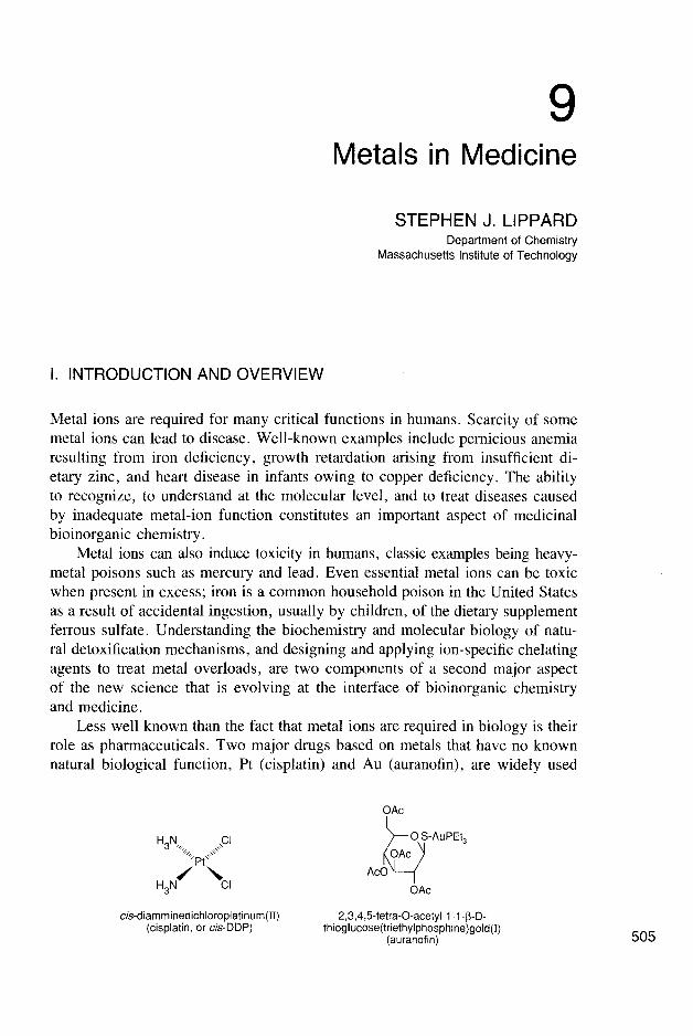

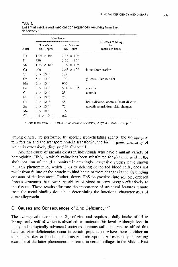

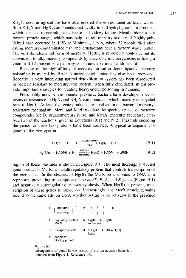

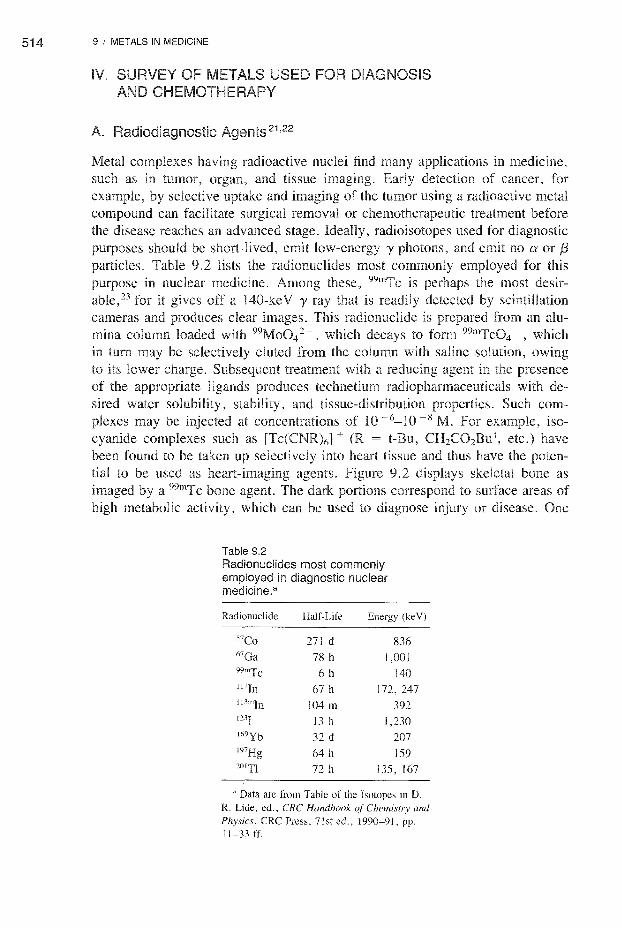
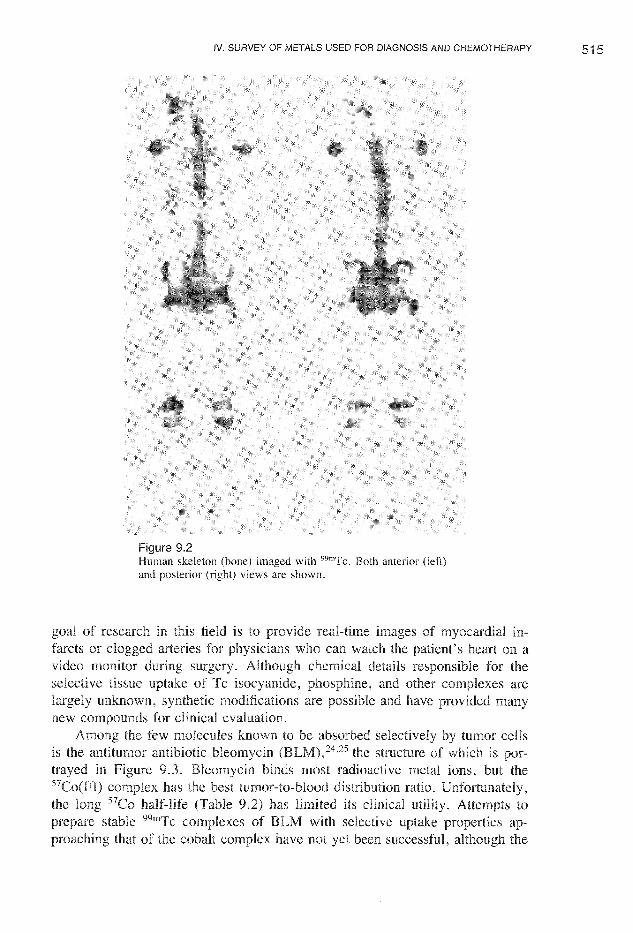


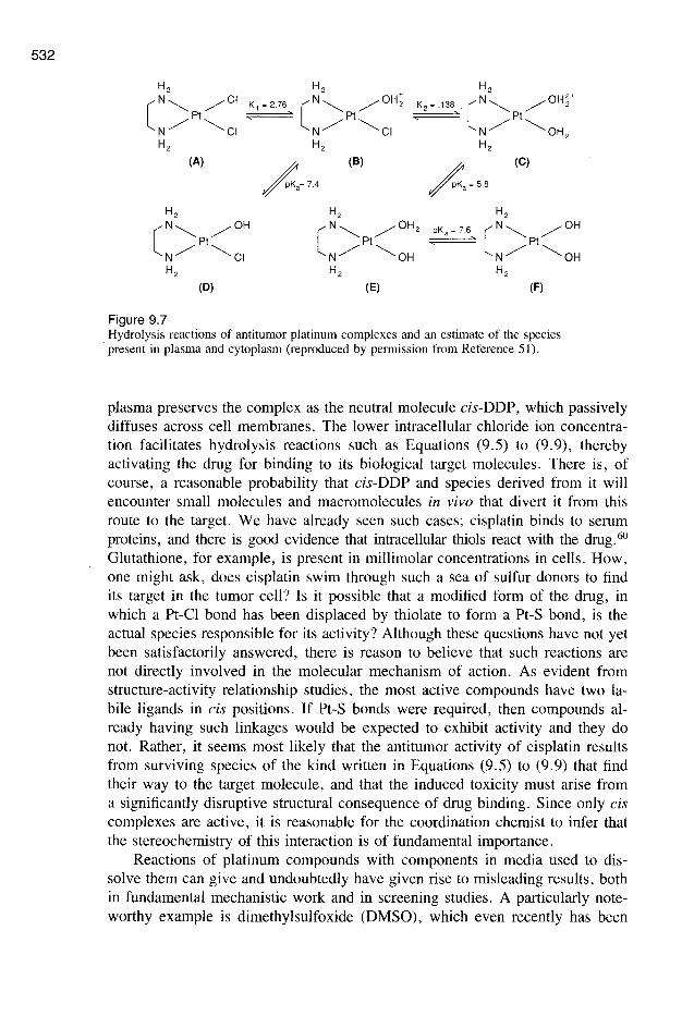


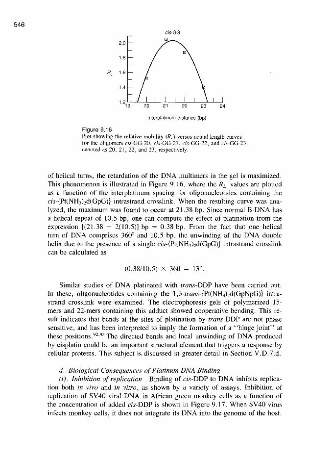


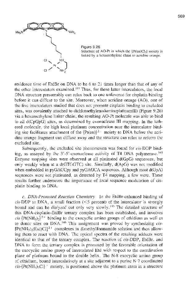


![[Lucy Lippard and John Chandler] Dematerialization](https://static.documents.pub/doc/80x56/563db86f550346aa9a93a929/lucy-lippard-and-john-chandler-dematerialization.jpg)


