Small molecule screening platform for assessmentof cardiovascular toxicity on adult zebrafish heartSatish Srinivas Kitambi*†, Erik S Nilsson†, Petra Sekyrova, Cristian Ibarra, Gilbert Nyah Tekeoh, Michael Andäng,Patrik Ernfors and Per Uhlén
Abstract
Background: Cardiovascular toxicity is a major limiting factor in drug development and requires multiple cost-effective models to perform toxicological evaluation. Zebrafish is an excellent model for many developmental,toxicological and regenerative studies. Using approaches like morpholino knockdown and electrocardiogram,researchers have demonstrated physiological and functional similarities between zebrafish heart and human heart.The close resemblance of the genetic cascade governing heart development in zebrafish to that of humans haspropelled the zebrafish system as a cost-effective model to conduct various genetic and pharmacological screenson developing embryos and larvae. The current report describes a methodology for rapid isolation of adultzebrafish heart, maintenance ex vivo, and a setup to perform quick small molecule throughput screening, includingan in-house implemented analysis script.
Results: Adult zebrafish were anesthetized and after rapid decapitation the hearts were isolated. The short timerequired for isolation of hearts allows dissection of multiple fishes, thereby obtaining a large sample size. Thesimple protocol for ex vivo culture allowed maintaining the beating heart for several days. The in-house developedscript and spectral analyses allowed the readouts to be presented either in time domain or in frequency domain.Taken together, the current report offers an efficient platform for performing cardiac drug testing andpharmacological screens.
Conclusion: The new methodology presents a fast, cost-effective, sensitive and reliable method for performingsmall molecule screening. The variety of readouts that can be obtained along with the in-house developedanalyses script offers a powerful setup for performing cardiac toxicity evaluation by researchers from bothacademics and industry.
Keywords: Heart, Screening, Zebrafish, Small molecule, Ex vivo, Ca2+ signaling
BackgroundThe drug discovery process is heavily impeded by cardio-vascular toxicity [1]. The sooner toxicity is discovered, thebetter for preclinical safety standards and cost of drugdevelopment. Various in vitro assays such as membranepotential dyes [2], rubidium efflux [3], radioligand binding[4] and patch clamp [5] have been used to identify com-pounds displaying cardiac toxicity. Since a compound canaffect more than one target, it is necessary to understandthe effects on the whole organ system. Therefore, various
ex vivo assays such as the purkinje fiber assay [6], isolatedheart assay [7] and in vivo assay using transgenic mice [8]have emerged. However, these assays are limited in specifi-city, reliability and throughput efficiency. Thus, moremodel systems are needed to evaluate cardiovasculartoxicity.Zebrafish has gained immense popularity as a model for
small molecule screening for factors influencing the cardi-ovascular system [9-11]. Various screens have been per-formed on zebrafish embryos and larvae to study factorsinfluencing development and homeostasis of the cardio-vascular system [11]. The availability of transgenic zebra-fish lines expressing fluorescent proteins in the vascularsystem, blood, endothelial cells and heart, facilitates
* Correspondence: [email protected]† Contributed equallyDepartment of Medical Biochemistry and Biophysics, Division of MolecularNeurobiology, Karolinska Institutet, Stockholm 17177, Sweden
Kitambi et al. BMC Physiology 2012, 12:3http://www.biomedcentral.com/1472-6793/12/3
precise and accurate evaluation of the effects of com-pounds on the cardiovascular system [12]. The small size,relative ease in handling, and the possibility of obtaining alarge number of samples (embryos or adult fish) for statis-tical evaluation, offers a reliable platform for performingsmall molecule screening.The current report describes a method for rapid isola-
tion of intact adult zebrafish hearts, maintenance ex vivoand presents a platform for small molecule screening onmultiple hearts. This setup offers a reasonable through-put-screening platform for generating rapid and statisti-cally significant readouts. Readouts can range fromrecording heartbeats or temporal changes in morphologyto measuring cellular calcium (Ca2+) signaling, rhythmicbeating and force of contraction. Taken together, this exvivo cardiac assay model offers a powerful tool for evaluat-ing several dimensions of potential cardiovascular toxicityof lead compounds.
MethodsPreparation of reagentsAdult wild type AB strain zebrafish to be dissected weretransferred to Egg Water containing “Instant Ocean” seasalts to a final concentration of 60 mg/ml, and anestheticsolution containing 0.1% Tricaine (Sigma) in Egg Waterwas prepared as described in The Zebrafish Book (Univer-sity of Oregon Press, 2000). Krebs-Ringer’s solution(onward referred to as Krebs solution) for dissection andscreening was prepared by mixing NaCl (119.0 mM), KCl(2.5 mM), NaH2PO4 (1.0 mM), CaCl2: 2H2O (2.5 mM),MgCl2: 6H2O (1.3 mM), HEPES (20 mM) and D-glucose(11.0 mM). The solution was adjusted to pH 7.4 and filtersterilized using a 0.2 μm filter (Sarstedt). If Ca2+ responseswere to be recorded, Fura-2/AM (Molecular Probes) wasused at a final concentration of 0.2 mM in Krebs solution.Culture medium for maintaining the hearts was preparedby adding fetal bovine serum (Invitrogen) to a final con-centration of 10% in DMEM (Invitrogen). The 0.5 to 1%low-melt agarose solution was prepared in filtered Krebssolution.
Animal husbandry and rapid dissection of the adultzebrafish heartAdult zebrafish of 4-6 months were anesthetized andimmediately transferred to a Petri dish under a microscopefor dissection. Using two forceps (Dumont Forcep No 5,Sigma), the zebrafish was rapidly decapitated. One forcepswas used to hold the body of the zebrafish and the otherforceps was then inserted into the body cavity below thepectoral fin and the overlying skin was pulled away toreveal the beating heart. The heart was carefully pulledaway from the rest of the body and all other tissues werecleared from the Petri dish (Figure 1A).
Ex-vivo cultureThe hearts were transferred to a 12 well plate, each wellhaving 1 ml of culture medium, and incubated at 37°C.We have successfully maintained beating hearts for upto 4 days without any contamination. Maintaining thehearts at temperature optimal for fish (28.3°C) can alsobe done without major impact on its survival and per-formance. For evaluating this assay, organotypic cultureof adult mice heart and in vitro culture of immortalizedHL-1 cardiomyocytes were done using previouslydescribed protocols [13,14]
Setup for performing small molecule screeningThe screening procedure can be performed with 3-5heart preparations, either embedded or un-embedded inagarose, in a glass bottom Petri dish (MatTek). First, theglass bottom of the dish was coated with laminin(Sigma), diluted 1:200 with Phosphate Buffered Saline(Invitrogen) from the stock concentration of 1 mg/mland incubated for at least 3 h at 37°C. Excess lamininsolution was removed and 3-5 hearts were transferredto the Petri dish with as little culture medium as possi-ble. If Ca2+ measurements were to be performed, thehearts were incubated for 10 min at 37°C with 5% CO2,immersed in 100 μl of 0.5 mM of Fura-2/AM. Next,150 μl of Krebs solution was added to the Petri dish andincubated for an additional 30 min. After transfer of theheart to the Petri dish, excess culture medium or Fura-2/AM was removed and 1-2 drops of low melt agarose(Lonza) was pipetted onto the heart (Figure 1B). Theagarose was allowed to solidify and 1 ml of pre-warmedKrebs solution was added into the Petri dish. The samesetup was also used for performing comparative screenson mouse heart and HL-1 cell cultures.
Performing screening on zebrafish heartThe Petri dish with embedded hearts was moved to thescreening station (Figure 1C) and mounted on a tempera-ture-controlled stage setup (Warner Instruments)clamped onto a Zeiss Axiovert 100 M microscope,equipped with a 25X/0.8NA water immersion objectiveand a 5x/0.15NA objective (all from Carl Zeiss), con-nected to a Lambda LS xenon-arc lamp, Lambda 10-3 fil-ter-wheel and a smartShutter (all from SutterInstruments). A container was filled with freshly pre-pared and pre-warmed Krebs solution for washing thesamples in between compound treatments. A peristalticpump was used to add and remove the washing buffer tohearts in the mounted Petri dish. Recording of heartbeats(Figure 1D) or spontaneous Ca2+ signals (Figure 1E-F)were acquired at 5 and 0.5 Hz respectively with anEMCCD camera Cascade II:512 (Photometrics) con-trolled by the acquisition software MetaFluor (Molecular
Kitambi et al. BMC Physiology 2012, 12:3http://www.biomedcentral.com/1472-6793/12/3
Page 2 of 7
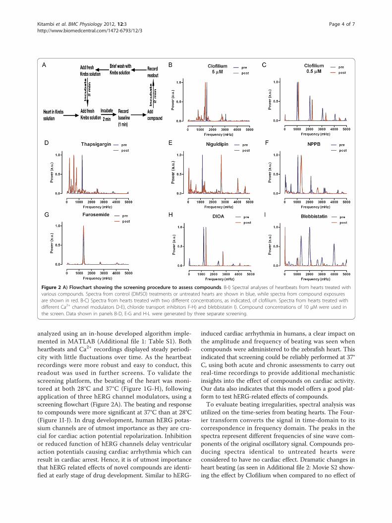
Devices). A control reading was acquired before startingscreening. Compounds were then added directly to thePetri dish using a pipette and changes in heart beating orCa2+ signaling were captured. Following this procedure,the compound was rinsed off by allowing Krebs solutionto flush the hearts. Thereafter, the recording continueduntil a signal was detected that resembled the initial con-trol reading whereupon a second compound was added(Figure 2A). This process could be repeated as long as astable control reading could be detected after eachtreatment.
Quick analyses of the heartbeatsBright field time-lapse movies of beating hearts wereimported to ImageJ and regions of interest (ROI) encom-passing the border of the hearts were marked. The meanintensities of each time point in the time-lapse recordingwere extracted, using the MultiMeasure plugin, andsaved in a .dat file. The values within this file were
thereafter imported to an in-house MATLAB softwaretool beating.m (Additional file 1: Table S1) or into a pre-viously described spectral analysis software tool, alsoimplemented in MATLAB, to characterize the temporalproperties of heart beating [15]. These algorithms nor-malize the time-series and remove any possible trend-lines, produced by e.g. focus shift, by fitting the trace to apolynomial (Figure 1D).Single or multiple heart preparations were used for cap-
turing dynamics of Ca2+ signaling (Figure 1E). For analyz-ing Ca2+ signals, the MetaFluor software was used toextract the Fura-2/AM intensities in different ROIs. Thedata was then exported to Excel and a graph was plotted(Figure 1F).
Results and discussionIn order to set up the screening platform for dissectedhearts, real-time recordings of heart beating (Figure 1D)and Ca2+ fluxes (Figure 1E-F) were performed and
Figure 1 A) Whole heart, isolated from wild type adult zebrafish showing Atrium (abbreviated: at) and Ventricle (abbreviated: v). B)Petri dish with embedded hearts used for screening. C) Diagrammatical representation of the screening setup. Petri dish with embedded heartsis placed on an inverted microscope having an in- and out-let to regulate the in- and out-flow of fresh Krebs buffer. D) Analyses of heartbeatingusing MATLAB. E) Three hearts mounted on a Petri dish to measure Ca2+ responses. The regions of interest (ROI1 and ROI2) that were used toanalyze Ca2+ responses are shown with blue and red circles. F) Graph showing spontaneous Ca2+ activity and compound-induced Ca2+ responsein ROI1 (in blue) and ROI2 (in red). Analyses of heartbeats at 28°C G) and 37°C H). Analyses of heartbeats obtained from screen of compounds asindicated carried out at 28°C I) and 37°C J). Compound concentrations of 10 μM were used in the screen. Scale bar: 200 μm in A.
Kitambi et al. BMC Physiology 2012, 12:3http://www.biomedcentral.com/1472-6793/12/3
Page 3 of 7
analyzed using an in-house developed algorithm imple-mented in MATLAB (Additional file 1: Table S1). Bothheartbeats and Ca2+ recordings displayed steady periodi-city with little fluctuations over time. As the heartbeatrecordings were more robust and easy to conduct, thisreadout was used in further screens. To validate thescreening platform, the beating of the heart was moni-tored at both 28°C and 37°C (Figure 1G-H), followingapplication of three hERG channel modulators, using ascreening flowchart (Figure 2A). The beating and responseto compounds were more significant at 37°C than at 28°C(Figure 1I-J). In drug development, human hERG potas-sium channels are of utmost importance as they are cru-cial for cardiac action potential repolarization. Inhibitionor reduced function of hERG channels delay ventricularaction potentials causing cardiac arrhythmia which canresult in cardiac arrest. Hence, it is of utmost importancethat hERG related effects of novel compounds are identi-fied at early stage of drug development. Similar to hERG-
induced cardiac arrhythmia in humans, a clear impact onthe amplitude and frequency of beating was seen whencompounds were administered to the zebrafish heart. Thisindicated that screening could be reliably performed at 37°C, using both acute and chronic assessments to carry outreal-time recordings to provide additional mechanisticinsights into the effect of compounds on cardiac activity.Our data also indicates that this model offers a good plat-form to test hERG-related effects of compounds.To evaluate beating irregularities, spectral analysis was
utilized on the time-series from beating hearts. The Four-ier transform converts the signal in time-domain to itscorrespondence in frequency domain. The peaks in thespectra represent different frequencies of sine wave com-ponents of the original oscillatory signal. Compounds pro-ducing spectra identical to untreated hearts wereconsidered to have no cardiac effect. Dramatic changes inheart beating (as seen in Additional file 2: Movie S2 show-ing the effect by Clofilium when compared to no effect of
Figure 2 A) Flowchart showing the screening procedure to assess compounds. B-I) Spectral analyses of heartbeats from hearts treated withvarious compounds. Spectra from control (DMSO) treatments or untreated hearts are shown in blue, while spectra from compound exposuresare shown in red. B-C) Spectra from hearts treated with two different concentrations, as indicated, of clofilium. Spectra from hearts treated withdifferent Ca2+ channel modulators D-E), chloride transport inhibitors F-H) and blebbistatin I). Compound concentrations of 10 μM were used inthe screen. Data shown in panels B-D, E-G and H-L were generated by three separate screening.
Kitambi et al. BMC Physiology 2012, 12:3http://www.biomedcentral.com/1472-6793/12/3
Page 4 of 7
DMSO control in Additional file 3: Movie S3) and shifts inspectra were clearly observed with clofilium (Figure 2B), apotent hERG channel inhibitor. Clofilium at concentra-tions below 5 μM produced spectra similar to untreatedhearts, indicating lack of readable effects (Figure 2C-D).As expected, modulators of Ca2+ signaling were producingstriking shifts in the spectra (Figure 2E-F). Compoundsmodulating potassium channels (Figure 2B-D) and inhibi-tors of chloride channels had less effects (Figure 2H-J).The inhibitor of myosin heavy chain ATPase activity, bleb-bistatin, had a dramatic effect on the spectrum (Figure2K). Galantamine, a previously used Alzheimer’s diseasedrug, with no documented cardiac effects, was withouteffect on the spectrum (data not shown), indicating nocardiac toxicity. Taken together, these results demon-strated that this setup offers a reliable platform for drugscreening.Similar experiments on organotypic cultures of mouse
heart sections and on cultured cardiomyocytes were car-ried out for comparison. Exposure of cardiomyocytes toclofilium or blebbistatin yielded similar spectrums as pro-duced by the zebrafish heart (Figure 3A-C). When clofi-lium was applied, the beating of the mouse heart ceased(data not shown). Our data shows that this assay can sen-sitively and precisely detect a range of effects that comple-ment existing models. Thus, this assay has the potential tobe used as a major screening model system for cardiactoxicity assessment or can be integrated as a part of multi-model cardiac toxicity risk assessment.Small molecule screening of compounds affecting car-
diovascular performance in zebrafish embryo or larvaehas been immensely successful thereby reflecting on therobustness of this model as a reliable screening platform.Accumulating data indicate a very high degree of devel-opmental and functional similarity between zebrafish andhuman heart, facilitating evaluation of cardiac-toxicity interms of human health risks. This platform allows usageof multiple hearts for testing/screening with generationof statistically significant data and little sample variation.The isolated hearts can be maintained in culture ex vivoand screening can be performed with multiple candidatemolecules, with alternating washing steps. The timerequired for assessment of one compound is around 3-5min, allowing scalability and automation. The number ofcompounds that can be used in one screen depends onwhether a stable baseline recording is established afterthe compound is washed away. The possibility of main-taining beating hearts ex vivo allows recording of acuteand chronic effects on the cardiac function and viability.Short-term or long-term readouts like heartbeat,mechanical contraction, viability, hERG activity, or Ca2+
Figure 3 Spectral analyses of the beating efficiency ofimmortalized HL-1 cardiomyocytes after exposure to DMSO(A), clofilium (B) and blebbistatin (C). Compound concentrationsof 1 μM were used in the screen.
Kitambi et al. BMC Physiology 2012, 12:3http://www.biomedcentral.com/1472-6793/12/3
Page 5 of 7
fluxes are easy and straightforward to measure, therebyoffering a sensitive model to assess cardiac toxicity.Screening with zebrafish heart maintained ex-vivo forfour days yield similar results as screening on freshly iso-lated heart (data not shown), however, the number ofcompounds that can be tested with a single heart mark-edly decreased (data not shown). Compounds primarilyaffecting the contractility properties of the heart (eg.Blebbistatin) may be missed by electrophysiologicalmethods, but can be evaluated here. Although this modelrequires extensive validation, it clearly demonstrates fea-tures such as sensitivity to generated readouts, reliabilityand adaptability with other available methods (such as invitro cardiomyocyte screens). The Matlab script and thespectral analyses allow display of the readout in timedomain or in frequency domain respectively, thereby pro-viding deeper understanding of the effects produced bythe compounds.Cardiac safety evaluation is an important part of the
drug discovery and development process; hence a wholeorgan assay model would be highly beneficial. Numerousdrug candidates are regularly evaluated using multiplecell lines based assays. Although the adult heart-screen-ing assay requires higher concentrations of test com-pounds than cell lines or zebrafish embryos/larvae, thisassay offers a possibility to test various aspects on wholeorgan from an adult fish. Since diverse cell types contri-bute to the function of the entire adult heart, this assayallows precise measurements of the effects produced onall type of cells, governing the function of the organ. Res-cue screens performed on hearts isolated from adultmutant zebrafish with heart defects (eg. Slow mo) will behighly beneficial for studies on heart development, regen-eration and homeostasis using chemical genetics, as wellas aiding in the process of drug discovery.
ConclusionFeatures such as rapid dissection and maintaining theheart ex vivo for several days offers an excellent possibi-lity for carrying out time series recording or acute andchronic studies in addition to compound screens. Thesmall size and relatively cheap housing of the fish pro-vides a cost effective setup in comparison to other invivo or ex vivo models. The hearts can be easily pro-cessed for immunohistochemistry or in situ hybridiza-tion with standard protocols thereby enabling testing ofthe expression of various biomarkers before and afterexposure to the test compounds. Experiments aiming atdetermining the structure activity relationship (SAR) orreverse medicinal chemistry approach to classify theeffect of various structurally diverse compounds on theheartbeat or the spectral profile it generates can beeasily adapted to this model. In conclusion the currentreport clearly demonstrates a platform for performing
cardiac toxicity evaluation either as a primary screen oras complement to existing methodologies.
Additional material
Additional file 1: Table S1. MATLAB script for analyzing heartbeats.
Additional file 2: Movie S2. Movie of beating zebrafish heart afteraddition of Krebs buffer containing clofilium to a final concentration of10 μM.
Additional file 3: Movie S3. Representative movie showing beating ofzebrafish heart in Krebs buffer and after addition of DMSO control.
AcknowledgementsThe authors thank Rebecca Frake, Daniel Gyllborg, Moritz Lübke and AnnaOmelyanenko for proofreading the manuscript, Drs. Gayathri Chandrasekarand Nicolas Fritz for input on Ca2+ signaling and zebrafish experiments,Susan Warner and the team at Zebrafish Core facility for wonderful supportduring the course of experiments and the CLICK Imaging Facility supportedby the Wallenberg Foundation. Dr. William Claycomb, Louisiana StateUniversity, kindly provided immortalized HL-1 cardiomyocytes.
Authors’ contributionsSK conceived the study, SK and EN designed the work, SK, EN, PS, CI, GNTcarried out the experiments, MA, PE, PU provided material support andconstructive analyses of the data. All authors contributed to writing themanuscript, analysis of the results and approved the manuscript.
Received: 13 October 2011 Accepted: 26 March 2012Published: 26 March 2012
References1. Guth BD: Preclinical cardiovascular risk assessment in modern drug
development. Toxicol Sci 2007, 97:4-20.2. Baxter DF, Kirk M, Garcia AF, Raimondi A, Holmqvist MH, Flint KK, Bojanic D,
Distefano PS, Curtis R, Xie Y: A novel membrane potential-sensitivefluorescent dye improves cell-based assays for ion channels. J BiomolScreen 2002, 7:79-85.
3. Terstappen GC: Functional analysis of native and recombinant ionchannels using a high-capacity nonradioactive rubidium efflux assay.Anal Biochem 1999, 272:149-155.
4. Finlayson K, Pennington AJ, Kelly JS: [3H]dofetilide binding in SHSY5Y andHEK293 cells expressing a HERG-like K + channel? Eur J Pharmacol 2001,412:203-212.
6. Gintant GA, Limberis JT, McDermott JS, Wegner CD, Cox BF: The caninePurkinje fiber: an in vitro model system for acquired long QT syndromeand drug-induced arrhythmogenesis. J Cardiovasc Pharmacol 2001,37:607-618.
7. Johna R, Mertens H, Haverkamp W, Eckardt L, Niederbroker T, Borggrefe M,Breithardt G: Clofilium in the isolated perfused rabbit heart: a new modelto study proarrhythmia induced by class III antiarrhythmic drugs. BasicRes Cardiol 1998, 93:127-135.
8. Fabritz L, Breithardt G, Kirchhof P: Preclinical testing of drug-inducedproarrhythmia: value of transgenic models. Cardiovasc Hematol AgentsMed Chem 2007, 5:289-294.
9. Zon LI, Peterson RT: In vivo drug discovery in the zebrafish. Nat Rev 2005,4:35-44.
10. Kitambi SS, McCulloch KJ, Peterson RT, Malicki JJ: Small molecule screenfor compounds that affect vascular development in the zebrafish retina.Mech Dev 2009, 126:464-477.
11. Tan JL, Zon LI: Chemical screening in zebrafish for novel biological andtherapeutic discovery. Methods Cell Biol 2011, 105:491-516.
12. Milan DJ, Macrae CA: Zebrafish genetic models for arrhythmia. ProgBiophys Mol Biol 2008, 98:301-308.
Kitambi et al. BMC Physiology 2012, 12:3http://www.biomedcentral.com/1472-6793/12/3
13. Habeler W, Pouillot S, Plancheron A, Puceat M, Peschanski M, Monville C:An in vitro beating heart model for long-term assessment ofexperimental therapeutics. Cardiovasc Res 2009, 81:253-259.
14. Claycomb WC, Lanson NA Jr, Stallworth BS, Egeland DB, Delcarpio JB,Bahinski A, Izzo NJ Jr: HL-1 cells: a cardiac muscle cell line that contractsand retains phenotypic characteristics of the adult cardiomyocyte. ProcNatl Acad Sci USA 1998, 95:2979-2984.
doi:10.1186/1472-6793-12-3Cite this article as: Kitambi et al.: Small molecule screening platform forassessment of cardiovascular toxicity on adult zebrafish heart. BMCPhysiology 2012 12:3.
Submit your next manuscript to BioMed Centraland take full advantage of:
• Convenient online submission
• Thorough peer review
• No space constraints or color figure charges
• Immediate publication on acceptance
• Inclusion in PubMed, CAS, Scopus and Google Scholar
• Research which is freely available for redistribution
Submit your manuscript at www.biomedcentral.com/submit
Kitambi et al. BMC Physiology 2012, 12:3http://www.biomedcentral.com/1472-6793/12/3