Microbial contamination levels of selected vegetable greens in the UAE American University of Sharjah, United Arab Emirates By: Dennis J. Russell, PhD., Sarah Abdul Majid, M.Sc., and Daniel Tobias, M.Sc.
Transcript
Microbial contamination levels of selected vegetable greens in the UAE
American University of Sharjah, United Arab Emirates
By: Dennis J. Russell, PhD., Sarah Abdul Majid, M.Sc., and Daniel Tobias, M.Sc.
Presentation by: Dr. Dennis J. Russell, PhD., American University of Sharjah, UAE
Microbial contamination levels of selected vegetable greens in the UAE
Food poisoning: a general term used to describe people who have become ill after eating.
It can be due to the presence of infectious or toxic materials in the food, prior to, during or after preparation.
Infectious organisms include viruses, bacteria (E. coli, Salmonella, tuberculosis, etc.), protozoa (Ghiardia., etc.) or worms.
Contaminated food poisoned children resulting in four deaths.
Symptoms of food poisoning:Vomiting, diarrhoea, dehydration, lowered blood pressure, death.
Children, pregnant women, diabetics and the elderly are the most susceptible to food poisoning
There is a concerted effort to assure the public is getting good clean food that is also not contaminated with undesirable bacteria
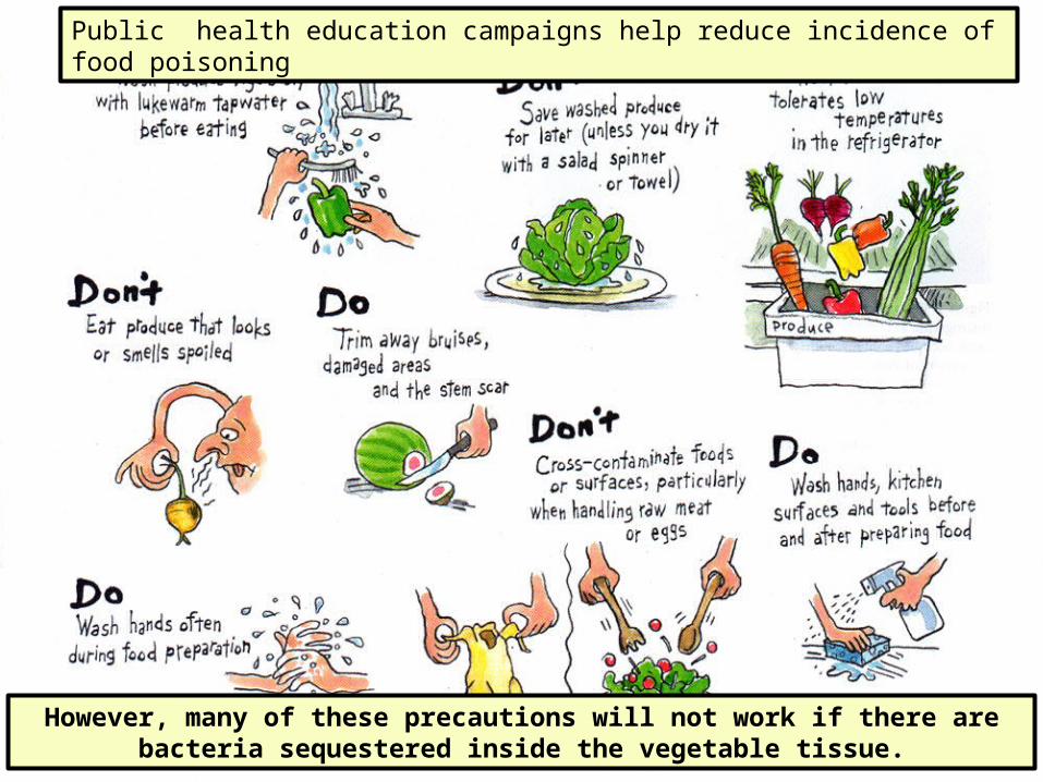
Public health education campaigns help reduce incidence of food poisoning
However, many of these precautions will not work if there are bacteria sequestered inside the vegetable tissue.
FIGURE 1: The mustard salad greens (Eruca sativa), which are grown on United Arab Emirate (UAE) produce farms, are called “jarjeer” or “jarjir” in Arabic and “rocket” in English or “roquette”in French, and are a favorite popular salad item at traditional Arab meals.
Hypothesis: Jarjeer retains fecal coliform bacteria in its leaves that are protected from repeated washings and disinfection.
Our experiment was originally designed to replicate what a person does in the kitchen when preparing salad greens for direct consumption without cooking.
We used much higher standards than in a kitchen and employed rigorous sterile technique.
The greens were so highly contaminated we found it was necessary to isolate the replicates 500 cm from each other to prevent cross contamination.
The greens were washed three times before macerating them in a blender. Our repetitive washings were supposed to reduce bacteria to zero.
In one experiment, we used a 5% Chlorox wash to get rid of as many surface bacteria as possible, before maceration. Diluted Chlorox is probably seldom if ever used by restaurants or households that wash their salad greens.
Finally, jarjeer greens were compared to lettuce and other salad greens as a control.
Testing the hypothesis:
• Jarjeer was washed three times and then macerated in a blender to release any additional sequestered bacteria.
• • A bacteria count was taken at each step to test the effectiveness of washing.
• The IDEXX system was used to test for coliforms and E. coli.
• Microscopic examination of fresh leaves was done to find the location of the sequestered bacteria.
• The presence of coliforms indicates the vegetables are contaminated with fecal matter from animals and if E. coli is also present it is a good indication that mammals or fowl are the sources of the feces, which could possibly include human waste.
During the experiment the repetitive series were kept 500 cm apart from each other to avoid cross-contamination.
10g of jarjeer was washed repeatedly in successive sterilized beakers containing 1 L sterile water .
This photo is for illustrative purposes only:During the actual tests the beakers were kept 500cm apart from each other.
How does the IDEXX system work?
• IDEXX tests for the presence of coliforms and E. coli using an indicator dye and an florescent antibody reaction.
• 100 ml of the diluted sample wash water is combined with a nutrient solution, indicator dye and florescent antibody.
• This 100ml mixture is then heat sealed into a tray that has 51 wells or compartments.
• If any compartment turns yellow it indicates the presence of a coliform bacteria unit.
• If any compartment glows under ultraviolet light it indicates the presence of E. coli bacteria. E. coli are present in human feces and it is one of many coliform species.
• This system has a proven 25 year track record and has performed at the highest standards required by various official water and food testing agencies.
IDEXX trays (yellow are coliforms) IDEXX trays (under UV light)
Wells with E. coli glow under UV light. The glowing E. coli wells were marked for easy counting in normal light
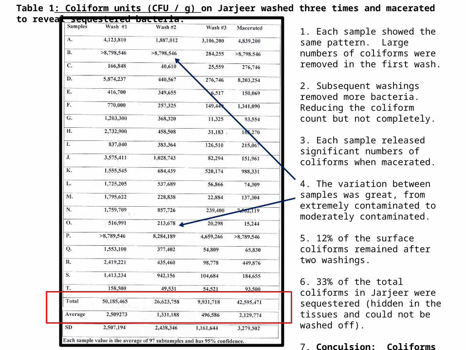
Table 1: Coliform units (CFU / g) on Jarjeer washed three times and macerated to reveal sequestered bacteria.
1. Each sample showed the same pattern. Large numbers of coliforms were removed in the first wash.
2. Subsequent washings removed more bacteria. Reducing the coliform count but not completely.
3. Each sample released significant numbers of coliforms when macerated.
4. The variation between samples was great, from extremely contaminated to moderately contaminated.
5. 12% of the surface coliforms remained after two washings.
6. 33% of the total coliforms in Jarjeer were sequestered (hidden in the tissues and could not be washed off).
7. Conculsion: Coliforms cannot be removed from Jarjeer by washing alone.
CFU / g0
10000000
20000000
30000000
40000000
50000000 49,278,749
26,623,758
492,141913,117
Wash #1Wash #2Wash #3Macerated
Coliforms on Jarjeer washed three times and then macerated to reveal sequestered bacteria. Numbers are the averages from 18 replicate tests.
After three washings of Jarjeer, an additional approximately 1 million coliforms / g were released when macerated.
49,278,74926,623,758 492,141 913,117
77,307,756 / g
E. coli / g0
20000
40000
60000
80000
100000
120000
140000
160000
180000
200000
167,171
49,314
10,541
51,578
Wash #1Wash #2Wash #3Macerated
FIGURE 3: E. coli / g on jarjeer washed three times and macerated to reveal sequestered bacteria.
167,171 49,314 10,541 51,578
278,604 / g
CFU / g0
100000
200000
300000
400000
500000
600000
700000
800000
900000
251,359
42,059
833,812
Wash #1Wash #3Macerated
FIGURE 4: Coliforms on jarjeer disinfected with mild bleach, rinsed, washed and then macerated.
E. coli / g0
500
1000
1500
2000
2500
3000
3500
4000
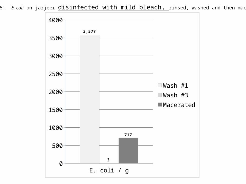
3,577
3
717
Wash #1Wash #3Macerated
FIGURE 5: E. coli on jarjeer disinfected with mild bleach, rinsed, washed and then macerated.
CFUs E. coli0
50000
100000
150000
200000
250000
300000
350000
400000
450000
200935
2037
97252
0
48549
0
404006
185
Wash #1Wash #2Wash #3Macerated
FIGURE 6: Coliforms and E. coli on parsley washed three times and then macerated to reveal sequestered
bacteria.
CFUs E. coli0
10000
20000
30000
40000
50000
60000
70000
80000
71048
41979
23436
2775
10240
948
59143
2556
Wash #1Wash #2Wash #3Macerated
FIGURE 7: Coliforms and E. coli on romaine lettuce washed three times and then macerated to reveal sequestered bacteria.
Conclusions:
• 100% of locally purchased fresh jarjeer greens were contaminated by coliforms and E. coli.
• One wash of jarjeer resulted in 2,509,273 CFU/g and 224,250 E. coli/g.
• Washing the greens three times reduced the CFU by 95% and E. coli by 83%.
• Maceration of thrice washed jarjeer released an additional 2,129,774 CFU/g and 56,292 E. coli/g.
• A statistically significant amount of coliforms and E. coli remain in the tissues of jarjeer even after being washed three times.
• Disinfection with diluted bleach reduced the coliform and E. coli counts, but upon maceration, both viable coliforms and E. coli were released.
• A statistically significant amount of coliforms and E. coli remain in the tissues of jarjeer even after being washed in disinfectant.
• There is clearly a persistent health threat to people who eat jarjeer.
• Further testing for the presence of Salmonella and other pathogens is required.
Avik, M, Speh, D., Jones, A., Busding, K., and Diez-Gonzales, F. 2006. Longitudinal microbiological survey of fresh produce grown by farmers in the upper Midwest. Midwest. J. Food Protection, 69:1928-1936.
CDSC.2005. Enumeration of coliforms and Escherichia coli by IDEXX (Colilert 18) Quanti-tray. Standards Unit, Evaluations and Standards Laboratory, Water Working Group CDSC, specialist and Reference Microbiology Division, Ref. No. W 18i2.3, p. 1-15.
Clesceri, L. S., Greenberg, A. E. and Eaton, A. D. (Eds). 1999. Standard Methods for the Examination of Water and Wastewater, 20th Ed. American Public Health Association. 1325pp.
Davis, J. G. and Kendall, P. 2007. Preventing E. coli from garden to plate. Colorado State University Extension, Nutrition Resources, No. 9.369
Fonseca, J. M. and Ravishankar, S. 2007. Safer Salads. Am. Sci. 95:494-501.
Froeder, H., Martins, C. G., DeSouza, K. L., Landraf, M., Franco, B. D. G. M., and Destro, M. T. 2007. Minimally processed vegetable salads: microbial quality evaluation. J. Food Prot. 70:1277-1280.
Ilic, S., Odomeru J. and LeJeune, J. T. 2008. Coliforms and prevalence of Esherichia coli and food borne pathogens on minimally processed spinach in two packing plants. J. Food Prot. 71:2398-2403.
Little, C. and Sagoo, S. 1996. LACORS/HPA Coordinated food liaison group studies: An evaluation of hygiene practices in mobile food vendors in the United Kingdom. Gastrointestinal, Emerging and Zoonotic Infections, Centre for Infections, Health Protection Agency, London. 29pp.
Lynch, M., Painter, J., Woodruff, R. and Braden, C. 2006. Surveillance for food borne disease outbreaks – United States, 1998-2002 (www.edc.gov).
Niemira, B. A. 2007. Relative efficacy of sodium hypochlorite wash versus irradiation to inactivate Escherichia coli O157:H7 internalized in leaves of romaine lettuce and baby spinach. J. Food Prot. 70:2526-2537.
Rao, P. V. 1998. Statistical Research Methods in the Life Sciences, Duxbury Press, Pacific Grove, i-xiv;889pp.
Simoes, M., Pisani, B., Marques, E. G. L., Prandi, M. A. G., Martini, M. H., Chiarini, P. F. T., Antunes, J. L. F. and Nogueira, A. P. 2001. Hygienic-sanitary conditions of vegetables and irrigation water from kitchen gardens in the municipality of Campias, SP. Braz. J. Microbiol. 32(4): 1-5
Vail, J. H., Morgan, R., Merino, C. R., Gonzales, F., Miller, R. and Ram, J. L. 2003. Enumeration of waterborne Escherichia coli with Perifim plates: comparison to standard methods. J. Environ. Qual. 32: 368-373.