Vol. 54, No. 10 Microbial Metabolism of Polycyclic Aromatic Hydrocarbons: Isolation and Characterization of a Pyrene-Degrading Bacterium MICHAEL A. HEITKAMP,t WIRT FRANKLIN, AND CARL E. CERNIGLIA* National Center for Toxicological Research, Food and Drug Administration, Jefferson, Arkansas 72079 Received 19 April 1988/Accepted 19 July 1988 Microbiological analyses of sediments located near a point source for petrogenic chemicals resulted in the isolation of a pyrene-mineralizing bacterium. This isolate was identified as a Mycobacterium sp. on the basis of its cellular and colony morphology, gram-positive and strong acid-fast reactions, diagnostic biochemical tests, 66.6% G+C content of the DNA, and high-molecular-weight mycolic acids (C58 to C64). The mycobacterium mineralized pyrene when grown in a mineral salts medium supplemented with nutrients but was unable to utilize pyrene as a sole source of carbon and energy. The mycobacterium grew well at 24 and 30°C and minimally at 35°C. No growth was observed at 5 or 42°C. The mycobacterium grew well at salt concentrations up to 4%. Pyrene-induced Mycobacterium cultures mineralized 5% of the pyrene after 6 h and reached a maximum of 48% mineralization within 72 h. Treatment of induced and noninduced cultures with chloram- phenicol showed that pyrene-degrading enzymes were inducible in this Mycobacterium sp. This bacterium could also mineralize other polycyclic aromatic hydrocarbons and alkyl- and nitro-substituted polycyclic aromatic hydrocarbons including naphthalene, phenanthrene, fluoranthene, 3-methylcholanthrene, 1-nitropyrene, and 6-nitrochrysene. This is the first report of a bacterium able to extensively mineralize pyrene and other polycyclic aromatic hydrocarbons containing four aromatic rings. Polycyclic aromatic hydrocarbons (PAHs) occur as natu- ral constituents and combustion products of fossil fuels (24, 39) and are widespread environmental contaminants (21, 26, 27). There is toxicological concern about the presence, persistence, and disposition of PAHs in the environment; some low-molecular-weight PAHs are acutely toxic (12, 44), most higher-molecular-weight PAHs are genotoxic (38), and there is a potential for their bioaccumulation into food chains (34, 37). Chronic exposure to PAHs has been associated with cancerous diseases in aquatic animals (36) and enhanced mutagenicity of sediments (41). Because of their toxicity, carcinogenicity, and ubiquitous distribution the U.S. Envi- ronmental Protection Agency has listed 16 PAHs as priority pollutants (28). PAHs may enter aquatic ecosystems by numerous routes and, because of their hydrophobicity (33), are usually bound and transported by fine particles which undergo sedimenta- tion (21, 34, 37). Although PAHs may undergo volatilization, chemical oxidation, photodecomposition, and microbial deg- radation while in open waters, most sediment deposition occurs below the photolytic zone, and the primary factor affecting the persistence of deposited PAHs is microbial degradation (14). For this reason, sediments serve as natural repositories for PAHs in aquatic ecosystems, where they may persist, undergo resuspension, or be degraded by com- plex natural communities of bacteria and fungi (6, 7, 15; C. E. Cerniglia and M. A. Heitkamp, in U. Varanasi, ed., Metabolism of Polycyclic Aromatic Hydrocarbons in the Aquatic Environment, in press). Chronic exposure to aromatic hydrocarbons has been reported to enhance PAH degradation in some soils and sediments, and several environmental isolates have been reported to degrade PAHs containing two or three aromatic rings (reviewed by Cerniglia and Heitkamp [in press]). The metabolic pathways, enzymatic reactions, and genetic con- * Corresponding author. t Present address: Monsanto Co., St. Louis, MO 63167. trol of the catabolism of lower-molecular-weight PAHs have been well documented (45). We recently confirmed the occurrence of known microbial metabolites of naphthalene in microcosms containing natural freshwater and estuarine sediments (19). To date, little is known about the ability of pure bacterial cultures to completely degrade PAHs contain- ing more than three fused benzene rings. In a previous study, we reported the effects of chronic chemical exposure and adaptation of sediment microbial populations on the mineralization rates of six PAHs in both estuarine and freshwater ecosystems (17). We found that relative differences in PAH mineralization among ecosys- tems were related to chemical structure, hexadecane miner- alization rates, the occurrence and concentration of aromatic hydrocarbon residues in sediments, and elevated popula- tions of hydrocarbon-degrading microorganisms. We are now presenting the results of subsequent microbiological investigation of sediments collected from sites chronically exposed to petrogenic chemicals within the watershed of Redfish Bay, Tex. (18). We isolated a pyrene-degrading bacterium from these sites and identified it as belonging to the genus Mycobacterium. To our knowledge, this investi- gation is the first report of the successful isolation and characterization of a bacterium able to completely degrade pyrene, a PAH containing four fused benzene rings, in pure culture. MATERIALS AND METHODS Chemicals. Radiolabeled PAHs, specific activities (milli- curies per millimole), and sources were as follows: [1,4, 5,8-'4C]naphthalene (5.10), Amersham Corp., Arlington Heights, Ill.; [9-'4C]phenanthrene (19.3), Amersham; [3-14C] fluoranthene (54.8), Chemsyn Science Laboratories, Le- nexa, Kans.; [4-'4C]pyrene (30.0), Midwest Research Insti- tute, Kansas City, Mo.; 1-nitro-[4,5,9,10-14C]pyrene (57.4), Chemsyn; 3-[6-'4C]methylcholanthrene (13.4), Dupont, NEN Research Products, Boston, Mass.; 6-nitro-[5,6,11,12- 14C]chrysene (57.4), Chemsyn. Nonlabeled pyrene was pur- 2549 APPLIED AND ENVIRONMENTAL MICROBIOLOGY, OCt. 1988, p. 2549-2555 0099-2240/88/102549-07$02.00/0 Copyright C) 1988, American Society for Microbiology on July 10, 2018 by guest http://aem.asm.org/ Downloaded from
Transcript
Vol. 54, No. 10
Microbial Metabolism of Polycyclic Aromatic Hydrocarbons:Isolation and Characterization of a Pyrene-Degrading Bacterium
MICHAEL A. HEITKAMP,t WIRT FRANKLIN, AND CARL E. CERNIGLIA*
National Center for Toxicological Research, Food and Drug Administration, Jefferson, Arkansas 72079
Received 19 April 1988/Accepted 19 July 1988
Microbiological analyses of sediments located near a point source for petrogenic chemicals resulted in theisolation of a pyrene-mineralizing bacterium. This isolate was identified as a Mycobacterium sp. on the basis ofits cellular and colony morphology, gram-positive and strong acid-fast reactions, diagnostic biochemical tests,66.6% G+C content of the DNA, and high-molecular-weight mycolic acids (C58 to C64). The mycobacteriummineralized pyrene when grown in a mineral salts medium supplemented with nutrients but was unable toutilize pyrene as a sole source of carbon and energy. The mycobacterium grew well at 24 and 30°C andminimally at 35°C. No growth was observed at 5 or 42°C. The mycobacterium grew well at salt concentrationsup to 4%. Pyrene-induced Mycobacterium cultures mineralized 5% of the pyrene after 6 h and reached a
maximum of 48% mineralization within 72 h. Treatment of induced and noninduced cultures with chloram-phenicol showed that pyrene-degrading enzymes were inducible in this Mycobacterium sp. This bacterium couldalso mineralize other polycyclic aromatic hydrocarbons and alkyl- and nitro-substituted polycyclic aromatichydrocarbons including naphthalene, phenanthrene, fluoranthene, 3-methylcholanthrene, 1-nitropyrene, and6-nitrochrysene. This is the first report of a bacterium able to extensively mineralize pyrene and otherpolycyclic aromatic hydrocarbons containing four aromatic rings.
Polycyclic aromatic hydrocarbons (PAHs) occur as natu-ral constituents and combustion products of fossil fuels (24,39) and are widespread environmental contaminants (21, 26,27). There is toxicological concern about the presence,
persistence, and disposition of PAHs in the environment;some low-molecular-weight PAHs are acutely toxic (12, 44),most higher-molecular-weight PAHs are genotoxic (38), andthere is a potential for their bioaccumulation into food chains(34, 37). Chronic exposure to PAHs has been associated withcancerous diseases in aquatic animals (36) and enhancedmutagenicity of sediments (41). Because of their toxicity,carcinogenicity, and ubiquitous distribution the U.S. Envi-ronmental Protection Agency has listed 16 PAHs as prioritypollutants (28).PAHs may enter aquatic ecosystems by numerous routes
and, because of their hydrophobicity (33), are usually boundand transported by fine particles which undergo sedimenta-tion (21, 34, 37). Although PAHs may undergo volatilization,chemical oxidation, photodecomposition, and microbial deg-radation while in open waters, most sediment depositionoccurs below the photolytic zone, and the primary factoraffecting the persistence of deposited PAHs is microbialdegradation (14). For this reason, sediments serve as naturalrepositories for PAHs in aquatic ecosystems, where theymay persist, undergo resuspension, or be degraded by com-
plex natural communities of bacteria and fungi (6, 7, 15;C. E. Cerniglia and M. A. Heitkamp, in U. Varanasi, ed.,Metabolism of Polycyclic Aromatic Hydrocarbons in theAquatic Environment, in press).
Chronic exposure to aromatic hydrocarbons has beenreported to enhance PAH degradation in some soils andsediments, and several environmental isolates have beenreported to degrade PAHs containing two or three aromaticrings (reviewed by Cerniglia and Heitkamp [in press]). Themetabolic pathways, enzymatic reactions, and genetic con-
* Corresponding author.t Present address: Monsanto Co., St. Louis, MO 63167.
trol of the catabolism of lower-molecular-weight PAHs havebeen well documented (45). We recently confirmed theoccurrence of known microbial metabolites of naphthalenein microcosms containing natural freshwater and estuarinesediments (19). To date, little is known about the ability ofpure bacterial cultures to completely degrade PAHs contain-ing more than three fused benzene rings.
In a previous study, we reported the effects of chronicchemical exposure and adaptation of sediment microbialpopulations on the mineralization rates of six PAHs in bothestuarine and freshwater ecosystems (17). We found thatrelative differences in PAH mineralization among ecosys-tems were related to chemical structure, hexadecane miner-alization rates, the occurrence and concentration of aromatichydrocarbon residues in sediments, and elevated popula-tions of hydrocarbon-degrading microorganisms. We are
now presenting the results of subsequent microbiologicalinvestigation of sediments collected from sites chronicallyexposed to petrogenic chemicals within the watershed ofRedfish Bay, Tex. (18). We isolated a pyrene-degradingbacterium from these sites and identified it as belonging tothe genus Mycobacterium. To our knowledge, this investi-gation is the first report of the successful isolation andcharacterization of a bacterium able to completely degradepyrene, a PAH containing four fused benzene rings, in pureculture.
MATERIALS AND METHODS
Chemicals. Radiolabeled PAHs, specific activities (milli-curies per millimole), and sources were as follows: [1,4,5,8-'4C]naphthalene (5.10), Amersham Corp., ArlingtonHeights, Ill.; [9-'4C]phenanthrene (19.3), Amersham; [3-14C]fluoranthene (54.8), Chemsyn Science Laboratories, Le-nexa, Kans.; [4-'4C]pyrene (30.0), Midwest Research Insti-tute, Kansas City, Mo.; 1-nitro-[4,5,9,10-14C]pyrene (57.4),Chemsyn; 3-[6-'4C]methylcholanthrene (13.4), Dupont,NEN Research Products, Boston, Mass.; 6-nitro-[5,6,11,12-14C]chrysene (57.4), Chemsyn. Nonlabeled pyrene was pur-
2549
APPLIED AND ENVIRONMENTAL MICROBIOLOGY, OCt. 1988, p. 2549-25550099-2240/88/102549-07$02.00/0Copyright C) 1988, American Society for Microbiology
chased from Chemical Service, Media, Pa. Chemical analy-ses by high-pressure liquid chromatography and gaschromatography-mass spectrometry showed that the purityof all PAH stocks exceeded 99%. Chloramphenicol waspurchased from P-L Biochemicals, Inc., Milwaukee, Wis.Bacterial media and reagents used to culture the Mycobac-terium sp. were purchased from Difco Laboratories, Detroit,Mich. All solvents and chemicals were of the highest purityavailable.Sample sites and monitoring mineralization of pyrene.
Composite sediment and water samples were collected inJuly 1986 from three sample sites (designated A, B, and C)located proximal to the Harbor Island oil tank farm in thewatershed of Redfish Bay near Port Aransas, Tex. Site Asamples were collected near a drainage pond located nearthe Harbor Island oil tank farm. Site B samples werecollected from a salt-water pool alongside the Aransas shipchannel, below the Harbor Island oil tank farm. Site Csamples were collected from a downstream site alongside theedge of Redfish Bay where an oil spill had occurred 9 yearsearlier. A detailed description of Redfish Bay has beenpreviously presented (17, 19). Mineralization of pyrene wasmonitored in a flowthrough microcosm test system thatallowed continuous measurement of 14Co2 evolution (23).Microcosms from each sample site contained 20 g of homog-enized moist sediment and 180 ml of collected water. Micro-cosms were exposed to 0.92 ,Ci of [4-'4C]pyrene and 100 ,ugof pyrene dissolved in 30 IlI of dimethylformamide. Miner-alization was monitored at 7-day intervals for 8 weeks at240C.
Isolation of pyrene-degrading bacteria. Sediment subsam-ples (1 g) were removed after 3.5 weeks from microcosmsfrom all three sample sites. This time point was chosen sincerapid pyrene mineralization occurred after an initial 2- to3-week lag phase observed in microcosms from all threesites. The sediment samples were serially diluted and as-sayed for the presence of pyrene-degrading microorganismsby a method modified from that of Kiyohara et al. (29).Growth medium consisted of a 2% agar mixture of minimalbasal salts (MBS) medium (43) containing low levels (250 ,ug/liter) of Bacto-Peptone (Difco), yeast extract, and solublestarch. The surfaces of the agar plates were sprayed with a2% pyrene solution in acetone-hexane (1:1, vol/vol) anddried overnight at 35°C. This treatment resulted in a visibleand uniform surface coat of pyrene on the agar plates.
Inocula (100 ,ul) from the 10-1, 10-2, 10-3, and 10-4dilutions of microcosm sediments were spread with sterileglass rods onto the agar surfaces of the petri dishes andincubated for 3 weeks at 24°C in clear plastic bags toconserve moisture. Although many heterotrophic bacteriawere capable of growth on this medium, pyrene-degradingbacteria were distinguished as colonies surrounded by clearzones due to pyrene uptake and utilization. These pyrene-degrading bacterial colonies were aseptically removed fromthe mixed-culture plates and grown in pure culture on MBSagar and broth containing low levels of organic nutrients andpyrene (0.5 Lg/ml).
Maintenance, identification, and characterization of a py-rene-degrading microorganism. The pyrene-degrading isolatewas routinely cultured on pyrene-supplemented MBS liquidmedium as described above and was inoculated into growthtubes containing the standard diagnostic substrates andreactants listed in Table 1. In addition, the cells were Gramand acid-fast stained. The isolate grew well on pyrene-supplemented MBS medium in the laboratory and, after
extensive subculturing, maintained its growth rate and py-rene-degrading activity.
Freshly grown pyrene-degrading bacterial cells werestrongly acid fast. Since acid-fast staining is indicative ofbacterial cell walls containing mycolic acids, thin-layer chro-matography (TLC) and mass spectral analyses of mycolicacids in whole-cell methanolysates were performed by themethods of Minnikin et al. (35) and Collins et al. (10),respectively. TLC was performed with 500 M silica gel GFplates (Analtech, Newark, Del.). Direct probe mass spectralanalyses of extracts from TLC spots were performed ona Finnigan MAT model 4023 (Finnigan MAT Corp., SanJose, Calif.) quadrupole mass spectrometer. Direct probeanalyses were conducted with a platinum wire probe and aVacumetrics model DCI current programmer. The massspectrometer was operated in 70-V electron impact andmethane chemical ionization modes, and the ion-sourcetemperature was maintained at 270°C. The mass spectrawere collected as the probe was inserted and heated to 3 Aover a ramp time of 120 s.The ability of the cells to utilize pyrene as a sole source of
carbon and energy was assayed by the inoculation of repli-cate tubes of MBS broth containing no supplemented or-ganic substrate and pyrene concentrations ranging from0.001 to 1.0 g/ml. These tubes were incubated at 30°C for 6weeks and visually examined for turbidity.The salt tolerance of the isolate was tested in brain heart
infusion broth and nutrient-supplemented MBS broth con-taining NaCl concentrations ranging from 0.1 to 10%. Eachtube was inoculated with a small sample of a pure culture ofthe pyrene-degrading isolate and incubated for 6 weeks at24°C. The tubes were mixed three times each week andvisually examined for turbidity.Agar plates containing individual pyrene-degrading colo-
nies and surrounding clear zones were examined by lightmicroscopy. Photomicrographs of the pyrene-MBS agarspread plates were taken through a green filter under lowmagnification (x 12.5) with an American Optical Series 20Advanced Microstar light microscope equipped with anExpostar automatic shutter control and a model 1053 35-mmcamera (American Optical Corp., Buffalo, N.Y.) by usingKodak Panatomic-X film (ASA 32; Kodak Chemical Co.,Rochester, N.Y.). Bacterial samples for transmission elec-tron microscopy were prepared by the method of Cole andPopkin (9). Transmission electron microscopy was per-formed on a Philips model EM301 electron microscope(Eindhoven, The Netherlands).PAH mineralization by induced and noninduced cells. Since
several aromatic hydrocarbons are known to be degraded byinducible catabolic enzymes encoded on plasmids containingboth structural and regulatory genes (45), the effects ofenzyme induction and protein synthesis on pyrene mineral-ization were examined. Induced starter cultures were grownin the presence of pyrene as described above, whereasnoninduced starter cultures were grown for 2 weeks in theabsence of pyrene. The purity of all starter cultures wasdetermined by Gram staining and visual examination after 48h of growth at 30°C. Experimental cell cultures were grownat 24°C with constant stirring in 500-ml microcosm chamberscontaining 200 ml of MBS broth supplemented with lowlevels of organic nutrients, 0.92 ,uCi of [4-'4C]pyrene, and0.5 ,ug of pyrene per ml. The optical density at 500 nm(OD500) of each starter culture was measured, and each ofthe experimental cultures was inoculated at a level to give anOD500 of 0.05 (1.49 x 10⁶ cells per ml). Mineralization ofpyrene was monitored for 72 h by measuring the evolution of
TABLE 1. Growth, biochemical, and morphological characteristics of a pyrene-degrading Mycobacterium sp. isolatedfrom microcosms exposed to pyrenea
Characteristic Results
Colony morphology
Cellular morphology
Gram stainAcid-fast stainG+C content of DNACarbon-chain length of mycolic acids
Utilization of carbohydratesbDextroseXyloseMannitolLactoseSucroseMaltose10% Dextrose10% LactoseNutrient brothSimmons citrateUreaNitrate reductionTriple sugar iron
H2S production
Litmus milk
0.5- to 1.0-mm circular, smooth with a convex surface, butyrous consistency, yellow pigmentedscotochromogens
Rods with a straight axis, length of 0.7 to 1.4 ,um, width of 0.4 to 0.7 p.m, multiple dark-staininggranules
Positive in fresh cultures, variable in older culturesStrongly acid fast66.6%C58 to C64
a The bacterium showed negative growth or reactions with adonitol, arabinose, cellobiose, dulcitol, galactose, inositol, malonate, melezitose, melibiose,raffinose, rhamnose, salicin, sorbitol, sorbose, salmonella-shigella agar, pseudosel agar, indole production (heart infusion broth), starch, trehalose, gelatin,motility, esculin, lysine decarboxylase, arginine dehydrolase, and omithine decarboxylase.
b Fermentative and oxidative-fermentative (within parentheses) utilization is indicated as follows: +, growth or positive reaction; -, no growth or negativereaction; NC, no change; Alk, alkaline; A, acid.
"CO2 as described above for sediment-water microcosms.Sterile control cultures were included to detect abioticdegradation of pyrene. Protein synthesis was inhibited inboth induced and noninduced cultures by the addition of 25p.g of chloramphenicol per ml at 0 h.The biodegradation of [1,4,5,8-"4C]naphthalene, [9-14C]
phenanthrene, [3-"4C]fluoranthene, 1-nitro-[4,5,9,10-_4C]py-rene, 3-[6-14C]methylcholanthrene, 6-nitro-[5,6,11,12-"4C]chrysene, and [7,10-"4C]benzo[a]pyrene was determined asdescribed above for pyrene mineralization. The pyrene-degrading bacterium was incubated in the flowthrough mi-crocosm test system and exposed in triplicate to 0.92 ,uCi of"4C-labeled PAH and 50 ,ug of unlabeled PAH. Total miner-alization was the sum of 14CO2 evolved from each cultureduring the incubation period.
RESULTS
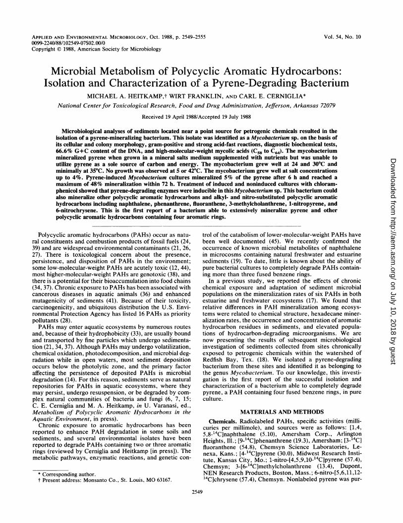
Pyrene mineralization. Pyrene was readily mineralized to14CO2 after initial 2- to 3-week lag phases in sediment-watermicrocosms from all three sample sites (Fig. 1). The highestinitial rate of pyrene mineralization was observed withsediment and water from site A, which is closest to theHarbor Island oil tank farm. In these samples, over 50% ofthe pyrene was mineralized to "4CO2 during weeks 3 through5 after an initial 2-week lag phase. Site B, located immedi-ately below the tank farm, showed the next highest rate ofpyrene mineralization with over 25% of the pyrene mineral-ized during weeks 3 through 5. However, total mineraliza-tion of pyrene in these samples reached a maximum of 36%after 8 weeks. Samples from site C, located further down-
stream alongside Redfish Bay, showed a 3-week lag phaseand the slowest initial rates of pyrene mineralization. How-ever, pyrene mineralization rates increased sharply after 4weeks in samples from site C, and over 50% of the pyrenewas mineralized during the last 4 weeks of the study,resulting in a total mineralization of 67.5%.
80 -
60
C 40-
20-j gI
A/ A
20
012 3 4 56 78Time (Weeks)
FIG. 1. Mineralization of pyrene in microcosms containing sed-iment and water from sites A (0), B (-), and C (A) proximal to theHarbor Island oil tank farm near Port Aransas, Tex.
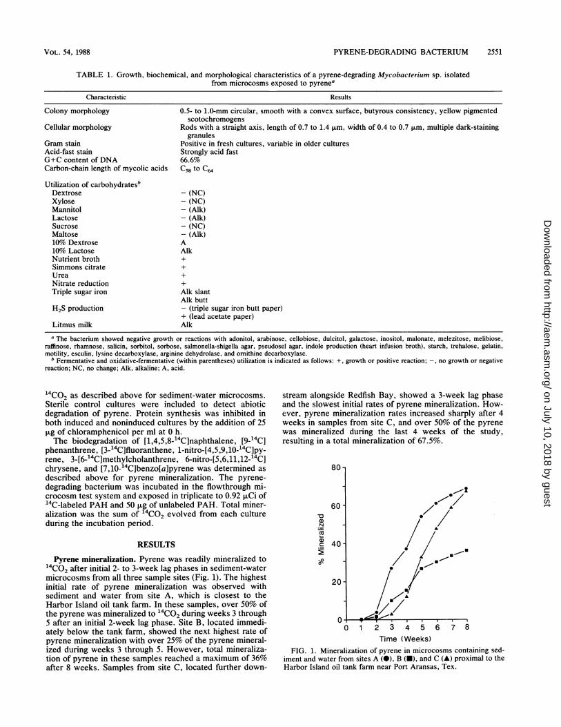
nFIG. 2. Photograph of individual pyrene-degrading bacterial col-
onies and clear zones of pyrene utilization on MBS agar containinglow-level nutrients and coated with pyrene (magnification, x12.5).
Isolation and characterization of a pyrene-mineralizing bac-terium. Since a rapid increase in pyrene mineralization was
observed in microcosms containing sediment and water fromsite A, sediment samples were taken at 3.5 weeks andassayed for the presence of pyrene-degrading bacteria. Nu-merous small colonies of pyrene-degrading bacteria wereobserved on pyrene-supplemented MBS agar plates from the10-3 and 10' dilutions of sediments from site A. Thesecolonies were all identical in colony and cellular morphologyand produced similar clear zones due to pyrene degradation.Individual pyrene-degrading colonies grown on MBS agarcontaining low-level nutrients appeared as circular coloniesand produced clear zones of pyrene utilization ranging from0.5 to 2.0 mm (Fig. 2). Although similar colonies probablyoccurred on plates from the 10-1 and 10-2 dilutions ofsediment from site A, they were not clearly observablebecause of heavy overgrowth of non-pyrene-degrading het-erotrophic bacteria. Pyrene-degrading bacteria were notisolated from sediments collected from sites B and C.
Pyrene-degrading colonies were aseptically removed witha sterile transfer loop and streaked via spread plating ontofresh pyrene-supplemented MBS agac- containing organicnutrients. The isolate was also subcultured in pyrene-sup-plemented MBS broth containing low levels of organicnutrients. Bacterial cell density was monitored by measuringthe OD500. Serial dilutions and plating of cultures at knownOD500 values determined a relationship of 2.97 x 10⁶ cells
A
N'-Y,S
7
6 12 18 24 48
Time (Hours)
Tr 1-
0 10 20 30 40 50 60 70 80Time (Hours)
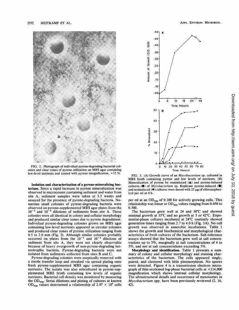
FIG. 3. (A) Growth curve of an Mycobacterium sp. cultured inMBS broth containing pyrene and low levels of nutrients. (B)Mineralization of pyrene by noninduced (A) and pyrene-inducedcultures (0) of Mycobacterium sp. Replicate pyrene-induced (a)and noninduced (*) cultures were dosed with 25 ,ug of chloramphen-icol per ml at 0 h.
per ml at an OD500 of 0.100 for actively growing cells. Thisrelationship was linear at OD5m0 values ranging from 0.050 to0.500.The bacterium grew well at 24 and 30°C and showed
minimal growth at 35°C and no growth at 5 or 42°C. Expo-nential-phase cultures incubated at 24°C routinely showedgeneration times ranging from 2.7 to 4.0 h (Fig. 3A). No cellgrowth was observed in anaerobic incubations. Table 1shows the growth and biochemical and morphological char-acteristics of fresh cultures of the bacterium. Salt-toleranceassays showed that the bacterium grew well at salt concen-trations up to 3%, marginally at salt concentrations of 4 to5%, and not at salt concentrations exceeding 5%.Morphology and identification. Table 1 presents a sum-
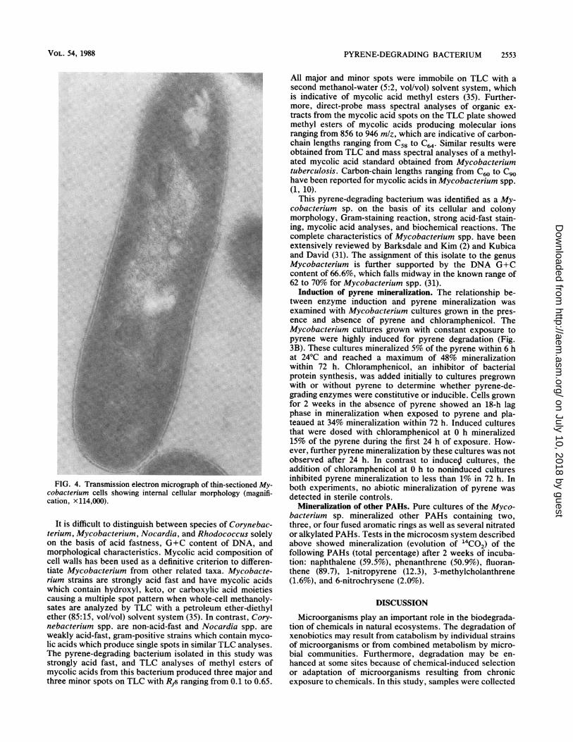
mary of colony and cellular morphology and staining char-acteristics of the bacterium. The cells appeared singly,paired, and clustered with little pleomorphism. No sporeswere detected. Figure 4 is a transmission electron micro-graph of thin-sectioned log-phase bacterial cells at x 114,000magnification which shows internal cellular morphology.The ultrastructural details and occurrence of mesosomes inMycobacterium spp. have been previously reviewed (2, 16,42).
FIG. 4. Transmission electron micrograph of thin-sectioned My-cobacterium cells showing internal cellular morphology (magnifi-cation, x114,000).
It is difficult to distinguish between species of Corynebac-terium, Mycobacterium, Nocardia, and Rhodococcus solelyon the basis of acid fastness, G+C content of DNA, andmorphological characteristics. Mycolic acid composition ofcell walls has been used as a definitive criterion to differen-tiate Mycobacterium from other related taxa. Mycobacte-rium strains are strongly acid fast and have mycolic acidswhich contain hydroxyl, keto, or carboxylic acid moietiescausing a multiple spot pattern when whole-cell methanoly-sates are analyzed by TLC with a petroleum ether-diethylether (85:15, vol/vol) solvent system (35). In contrast, Cory-nebacterium spp. are non-acid-fast and Nocardia spp. areweakly acid-fast, gram-positive strains which contain myco-lic acids which produce single spots in similar TLC analyses.The pyrene-degrading bacterium isolated in this study wasstrongly acid fast, and TLC analyses of methyl esters ofmycolic acids from this bacterium produced three major andthree minor spots on TLC with RAs ranging from 0.1 to 0.65.
All major and minor spots were immobile on TLC with asecond methanol-water (5:2, vol/vol) solvent system, whichis indicative of mycolic acid methyl esters (35). Further-more, direct-probe mass spectral analyses of organic ex-tracts from the mycolic acid spots on the TLC plate showedmethyl esters of mycolic acids producing molecular ionsranging from 856 to 946 mlz, which are indicative of carbon-chain lengths ranging from C58 to C64. Similar results wereobtained from TLC and mass spectral analyses of a methyl-ated mycolic acid standard obtained from Mycobacteriumtuberculosis. Carbon-chain lengths ranging from C60 to Cgohave been reported for mycolic acids in Mycobacterium spp.(1, 10).
This pyrene-degrading bacterium was identified as a My-cobacterium sp. on the basis of its cellular and colonymorphology, Gram-staining reaction, strong acid-fast stain-ing, mycolic acid analyses, and biochemical reactions. Thecomplete characteristics of Mycobacterium spp. have beenextensively reviewed by Barksdale and Kim (2) and Kubicaand David (31). The assignment of this isolate to the genusMycobacterium is further supported by the DNA G+Ccontent of 66.6%, which falls midway in the known range of62 to 70% for Mycobacterium spp. (31).
Induction of pyrene mineralization. The relationship be-tween enzyme induction and pyrene mineralization wasexamined with Mycobacterium cultures grown in the pres-ence and absence of pyrene and chloramphenicol. TheMycobacterium cultures grown with constant exposure topyrene were highly induced for pyrene degradation (Fig.3B). These cultures mineralized 5% of the pyrene within 6 hat 24°C and reached a maximum of 48% mineralizationwithin 72 h. Chloramphenicol, an inhibitor of bacterialprotein synthesis, was added initially to cultures pregrownwith or without pyrene to determine whether pyrene-de-grading enzymes were constitutive or inducible. Cells grownfor 2 weeks in the absence of pyrene showed an 18-h lagphase in mineralization when exposed to pyrene and pla-teaued at 34% mineralization within 72 h. Induced culturesthat were dosed with chloramphenicol at 0 h mineralized15% of the pyrene during the first 24 h of exposure. How-ever, further pyrene mineralization by these cultures was notobserved after 24 h. In contrast to induceSl cultures, theaddition of chloramphenicol at 0 h to noninduced culturesinhibited pyrene mineralization to less than 1% in 72 h. Inboth experiments, no abiotic mineralization of pyrene wasdetected in sterile controls.
Mineralization of other PAHs. Pure cultures of the Myco-bacterium sp. mineralized other PAHs containing two,three, or four fused aromatic rings as well as several nitratedor alkylated PAHs. Tests in the microcosm system describedabove showed mineralization (evolution of 14CO2) of thefollowing PAHs (total percentage) after 2 weeks of incuba-tion: naphthalene (59.5%), phenanthrene (50.9%), fluoran-thene (89.7), 1-nitropyrene (12.3), 3-methylcholanthrene(1.6%), and 6-nitrochrysene (2.0%).
DISCUSSION
Microorganisms play an important role in the biodegrada-tion of chemicals in natural ecosystems. The degradation ofxenobiotics may result from catabolism by individual strainsof microorganisms or from combined metabolism by micro-bial communities. Furthermore, degradation may be en-hanced at some sites because of chemical-induced selectionor adaptation of microorganisms resulting from chronicexposure to chemicals. In this study, samples were collected
from sites located proximal to Harbor Island oil tank farm, apoint source for petrogenic chemicals entering Redfish Bay,Tex. Since we had previously reported relatively high ratesof PAH degradation and elevated populations of hydrocar-bon-degrading microorganisms in sediment and water fromRedfish Bay (17-19), we speculated that the mild climate,high inorganic and organic nutrient levels, and long historyof exposure to high concentrations of petroleum hydrocar-bons would favor these sites as possible sources of bacteriaable to degrade higher-molecular-weight PAHs. In this pa-per, we report the isolation of a mycobacterium that readilydegraded pyrene as well as other PAHs. Although pyrenehas been shown to be degraded in soil (5), this study is thefirst report of a bacterial pure culture able to mineralizepyrene.
Pyrene is a highly symmetrical PAH containing four fusedaromatic rings and is one of 16 PAHs included in the list of129 priority pollutants compiled by the U.S. EnvironmentalProtection Agency (28). This PAH, encountered regularly inenvironmental samples (21), is not genotoxic but has achemical structure found in several carcinogenic PAHs suchas benzo[a]pyrene, indeno-(1,2,3-cd)-pyrene, and 1-nitropy-rene, and it has been used as a model compound in PAHmetabolism studies (8, 25).Pyrene mineralization rates in all three sample sites lo-
cated near the Harbor Island oil tank farm were significantlyhigher than those we previously reported for Redfish Bay(17). Although total amounts of pyrene mineralization inmicrocosms from sites A and C were similar after 8 weeks,the initial rates of pyrene mineralization observed in micro-cosms from all three sample sites during the first 3 weeks ofthe experiment were inversely proportional to the distancefrom the point source of aromatic hydrocarbon contamina-tion. These results support the hypothesis that a chemicalconcentration gradient exists below point sources of contam-ination, and optimal microbial adaptation may occur at apoint on this gradient where chemical concentrations aresubtoxic but are still above some threshold level necessaryfor enzyme induction and possible utilization as sources ofcarbon and energy. Several previous attempts in our labora-tory with similar enrichment methods to isolate a pyrene-degrading bacterium from Redfish Bay sediments were notsuccessful. The pyrene-degrading bacterium in this studywas isolated from site A, closest to the Harbor Island oiltank farm.The pyrene-degrading bacterium isolated in this study was
identified as a Mycobacterium sp. on the basis of its cellularand colony morphology, gram-positive and strong acid-faststaining, mycolic acids, diagnostic biochemical reactions,and nucleic acid contents. Mycobacterium spp. are commonin the environment (2) and have been reported to degraden-butane and 2-butanone (40), several gaseous unsaturatedhydrocarbons (13), cycloparaffinic hydrocarbons (4), n-alkyl-substituted cycloparaffins (3), and methyl ketone (32).The oxidation of aliphatic hydrocarbons and other hydrocar-bons by Mycobacterium spp. has been reviewed by Hou(22).
Inducible enzymes appear to be responsible for pyrenecatabolism in this Mycobacterium sp. since lag phases inpyrene mineralization were observed in cultures grown inthe absence of pyrene and no pyrene mineralization wasobserved in noninduced cultures dosed with chlorampheni-col at time zero. Inducible pyrene-degrading enzymes in thismycobacterium may be either chromosome or plasmid me-diated. Crawford and Bates (11) have isolated plasmid DNAfrom several strains of Mycobacterium avium-M. intracellu-
lare. The isolation and identification of a pyrene-degradingplasmid in this Mycobacterium sp. may have practical ap-plication for future cloning studies to construct geneticallyengineered bacteria able to degrade higher-molecular-weightPAHs and warrants further investigation. The inducibility ofpyrene-degrading enzymes in this mycobacterium is consis-tent with the observed inducibility of hydrocarbon-degradingenzymes encoded on plasmids containing both structural andregulatory genes in other bacteria (30, 45).There is a paucity of information on the microbial degra-
dation of pyrene. The chemical pathway for the degradationof pyrene by this Mycobacterium sp. is reported in theaccompanying paper (20). Since PAHs are widespread envi-ronmental contaminants and occur at relatively high concen-trations in some freshwater and marine sediments (24), thereis interest in the use of PAH-degrading microorganisms forthe bioremediation of PAHs in the environment. The Myco-bacterium sp. reported in this study readily degraded pyrenein pure culture under optimal conditions. The survival,performance, and overall ability of this Mycobacterium sp.to enhance the degradation of pyrene or other higher-molecular-weight PAHs in natural ecosystems warrant fur-ther investigation.
ACKNOWLEDGMENTS
We thank Warren Pulich for assistance in the collection ofenvironmental samples, Jerome Perry for G+C analyses of DNA,Shirley Hunter for mycolic acid standards, James P. Freeman formass spectral analyses of mycolic acids, Annette Andrews forelectron micrographs of the Mycobacterium sp., and RebeccaRobertson for providing excellent technical assistance in the labo-ratory.
LITERATURE CITED1. Apajalahti, J. H. A., P. Karpanoja, and M. S. Salkinoja-Salonen.
1986. Rhodococcus chlorophenolicus sp. nov., a chlorophenol-mineralizing actinomycete. Int. J. Syst. Bacteriol. 36:246-251.
2. Barksdale, L., and K. Kim. 1977. Mycobacterium. Bacteriol.Rev. 41:217-327.
3. Beam, H. W., and J. J. Perry. 1974. Microbial degradation andassimilation of n-alkyl-substituted cycloparaffins. J. Bacteriol.118:394-399.
4. Beam, H. W., and J. J. Perry. 1974. Microbial degradation ofcycloparaffinic hydrocarbons via co-metabolism and commen-salism. J. Gen. Microbiol. 82:163-169.
5. Bossert, I. D., and R. Bartha. 1986. Structure-biodegradabilityrelationships of polycyclic aromatic hydrocarbons in soil. Bull.Environ. Contam. Toxicol. 37:490495.
6. Cerniglia, C. E. 1984. Microbial metabolism of polycyclic aro-matic hydrocarbons. Adv. Appl. Microbiol. 30:31-71.
7. Cerniglia, C. E. 1984. Microbial transformation of aromatichydrocarbons, p. 100-128. In R. Atlas (ed.), Petroleum micro-biology. Macmillian Publishing Co., New York.
8. Chen, F. M. 1983. Binding of pyrene to DNA, base sequencespecificity and its implications. Nucleic Acids Res. 11:7231-7250.
9. Cole, R. M., and T. J. Popkin. 1981. Electron microscopy, p.34-51. In P. Gerhardt, R. G. E. Murray, R. N. Costilow, E. W.Nester, W. A. Wood, N. R. Krieg, and G. B. Phillips (ed.),Manual of methods for general microbiology. American Societyfor Microbiology, Washington, D.C.
10. Collins, M. D., M. Goodfellow, and D. E. Minnikin. 1982. Asurvey of the structures of mycolic acids in Corynebacteriumand related taxa. J. Gen. Microbiol. 128:129-149.
11. Crawford, J. T., and J. H. Bates. 1979. Isolation of plasmidsfrom Mycobacteria. Infect. Immun. 24:979-981.
12. Darville, R. G., and J. L. Wilhm. 1984. The effect of naphtha-lene on oxygen consumption and hemoglobin concentration inChironomus attenuatus and on oxygen consumption and lifecycle of Tanytarus dissimilis. Environ. Toxicol. Chem. 3:135-
141.13. DeBont, J. A. M., S. B. Primrose, M. D. Collins, and D. Jones.
1980. Chemical studies on some bacteria which utilize gaseousunsaturated hydrocarbons. J. Gen. Microbiol. 117:97-102.
14. Gibson, D. T., V. Mahadevan, D. M. Jerina, H. Yagi, andH. J. C. Yeh. 1975. Oxidation of the carcinogens benzo[a]py-rene and benz[a]anthracene to dihydrodiols by a bacterium.Science 189:295-297.
15. Gibson, D. T., and V. Subramanian. 1984. Microbial degrada-tion of aromatic hydrocarbons, p. 181-252. In D. T. Gibson(ed.), Microbial degradation of organic compounds. MarcelDekker, New York.
16. Greenawalt, J. W., and T. L. Whiteside. 1975. Mesosomes:membranous bacterial organelles. Bacteriol. Rev. 39:405-463.
17. Heitkamp, M. A., and C. E. Cerniglia. 1987. Effects of chemicalstructure and exposure on the microbial degradation of polycy-clic aromatic hydrocarbons in freshwater and estuarine ecosys-tems. Environ. Toxicol. Chem. 6:535-546.
18. Heitkamp, M. A., and C. E. Cerniglia. 1988. Mineralization ofpolycyclic aromatic hydrocarbons by a bacterium isolated fromsediment below an oil field. Appl. Environ. Microbiol. 54:1612-1614.
19. Heitkamp, M. A., J. P. Freeman, and C. E. Cerniglia. 1987.Naphthalene biodegradation in environmental microcosms: es-timates of degradation rates and characterization of metabolites.Appl. Environ. Microbiol. 53:129-136.
20. Heitkamp, M. A., J. P. Freeman, D. W. Miller, and C. E.Cerniglia. Pyrene degradation by a Mycobacterium sp.: identi-fication of ring oxidation and ring fission products. Appl.Environ. Microbiol. 54:2556-2565.
21. Hites, R. A., R. E. Laflamme, and J. G. Windsor. 1980.Polycyclic aromatic hydrocarbons in marine/aquatic sediments:their ubiquity, p. 289-311. In L. Petrakis and F. T. Weiss (ed.),Petroleum in the marine environment. Advances in chemistryseries. American Chemical Society, Washington, D.C.
22. Hou, C. T. 1982. Microbial transformation of important indus-trial hydrocarbons, p. 81-107. In J. P. Rosazza (ed.), Microbialtransformations of bioactive compounds, vol. 1. CRC Press,Boca Raton, Fla.
23. Huckins, J. N., J. D. Petty, and M. A. Heitkamp. 1984. Modularcontainers for microcosm and process model studies on the fateand effects of aquatic contaminants. Chemosphere 13:1329-1341.
24. International Agency for Research on Cancer. 1983. PolynuclearAromatic Compounds. Part 1, chemical, environmental andexperimental data, p. 95-451. In IARC Monographs on theevaluation of the carcinogenic risk of chemicals to humans.World Health Organization, Lyon, France.
25. Jacob, J., G. Grimmer, G. Raab, and A. Schmoldt. 1982. Themetabolism of pyrene by rat liver microsomes and the influenceof various mono-oxygenase inducers. Xenobiotica 12:45-53.
26. Jacob, J., W. Karcher, J. J. Befliado, and P. J. Wagstaffe. 1986.Polycyclic aromatic compounds of environmental and occupa-tional importance. Fresenius Z. Anal. Chem. 323:1-10.
27. Johnson, A. C., and D. Larsen. 1985. The distribution ofpolycyclic aromatic hydrocarbons in the surficial sediments ofPenobscot Bay (Maine, USA) in relation to possible sources andto other sites worldwide. Mar. Environ. Res. 15:1-16.
28. Keith, L. H., and W. A. Telliard. 1979. Priority pollutants I-aperspective view. Environ. Sci. Technol. 13:416-423.
29. Kiyohara, H., K. Nagao, and K. Yana. 1982. Rapid screen forbacteria degrading water-insoluble, solid hydrocarbons on agarplates. Appl. Environ. Microbiol. 43:454-457.
30. Kiyohara, H., M. Sugiyama, F. J. Mondeilo, D. T. Gibson, andK. Yano. 1983. Plasmid involvement in the degradation ofpolycyclic aromatic hydrocarbons by a Beijerinckia species.Biochem. Biophys. Res. Commun. 111:939-945.
31. Kubica, G. P., and H. L. David. 1980. The mycobacteria, p.1693-1730. In A. C. Sonnenwirth and L. Jarett (ed.), Grad-wohl's clinical laboratory methods and diagnosis, vol. 2. TheC. V. Mosby Co., St. Louis, Mo.
32. Lukins, H. B., and J. W. Foster. 1963. Methyl ketone metabo-lism in hydrocarbon-utilizing Mycobacteria. J. Bacteriol. 85:1074-1087.
33. Mackay, D., and W. Y. Shiu. 1977. Aqueous solubility ofpolynuclear aromatic hydrocarbons. J. Chem. Eng. Data 22:399-402.
34. Means, J. C., S. G. Ward, J. J. Hassett, and W. L. Banwart.1980. Sorption of polynuclear aromatic hydrocarbons by sedi-ments and soils. Environ. Sci. Technol. 14:1524-1528.
35. Minnikin, D. E., L. Alshamaony, and M. Goodfellow. 1975.Differentiation of Mycobacterium, Nocardia, and related taxaby thin-layer chromatographic analysis of whole-organism me-thanolysates. J. Gen. Microbiol. 88:200-204.
36. Mix, M. C. 1986. Cancerous diseases in aquatic animals andtheir association with environmental pollutants: a critical liter-ature review. Mar. Environ. Res. 20:1-141.
37. Morehead, N. R., B. J. Eadie, B. Lake, P. D. Landrum, and D.Berner. 1986. The sorption of PAH onto dissolved organicmatter in Lake Michigan waters. Chemosphere 15:403-412.
38. Mortelmans, K., S. Haworth, T. Lawlor, W. Speck, B. Tainer,and E. Zeiger. 1986. Salmonella mutagenicity tests. II. Resultsfrom the testing of 270 chemicals. Environ. Mutagen. 8(Suppl.7):1-119.
39. National Academy of Sciences. 1983. Polycyclic aromatic hydro-carbons: evaluation of sources and effects. National AcademyPress, Washington, D.C.
40. Phillips, W. E., and J. J. Perry. 1974. Metabolism of n-butaneand 2-butanone by Mycobacterium vaccae. J. Bacteriol. 120:987-989.
41. Sato, T., T. Momma, Y. Ose, T. Ishikawa, and K. Kato. 1983.Mutagenicity of Nagara River sediment. Mutat. Res. 118:257-267.
42. Shinohara, C., K. Fukushi, and I. Suzuki. 1957. Mitochondrium-like structures in ultrathin structures of Mycobacterium avium.J. Bacteriol. 74:413-415.
43. Skerman, V. B. D. 1967. A guide to the identification of thegenera of bacteria, 2nd ed. The Williams & Wilkins Co.,Baltimore.
44. Struble, V. G., and H. J. Harmon. 1983. Molecular basis forinhibition of mitochondrial respiration by naphthalene. Bull.Environ. Contam. Toxicol. 31:644-648.
45. Williams, P. A. 1981. Genetics of biodegradation, p. 97-130. InT. Leisinger, R. Hutter, A. M. Cook, and J. Nuesch (ed.),Microbial degradation of xenobiotics and recalcitrant com-pounds. Academic Press, Inc., New York.