Microbiology – Chapter 3 Culturing Microbes The Five “I’s Innoculation: Producing a pure culture Isolation: Colony on media, one kind of microbe, pure culture Incubation: growing microbes under proper conditions Inspection: Observation of characteristics (data) Identification: use of data, correaltion, to ID organism to exact species
Transcript
Microbiology – Chapter 3Culturing Microbes
The Five “I’s
Innoculation: Producing a pure culture
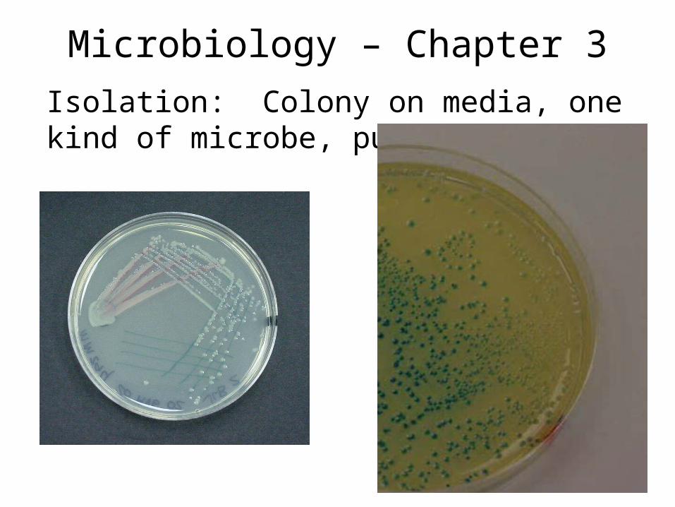
Isolation: Colony on media, one kind of microbe, pure culture
Incubation: growing microbes under proper conditions
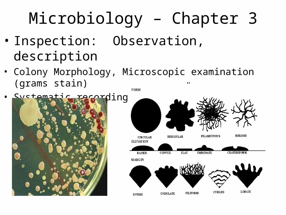
Inspection: Observation of characteristics (data)
Identification: use of data, correaltion, to ID organism to exact species
Microbiology – Chapter 3Culturing Microbes
The Five “I’s
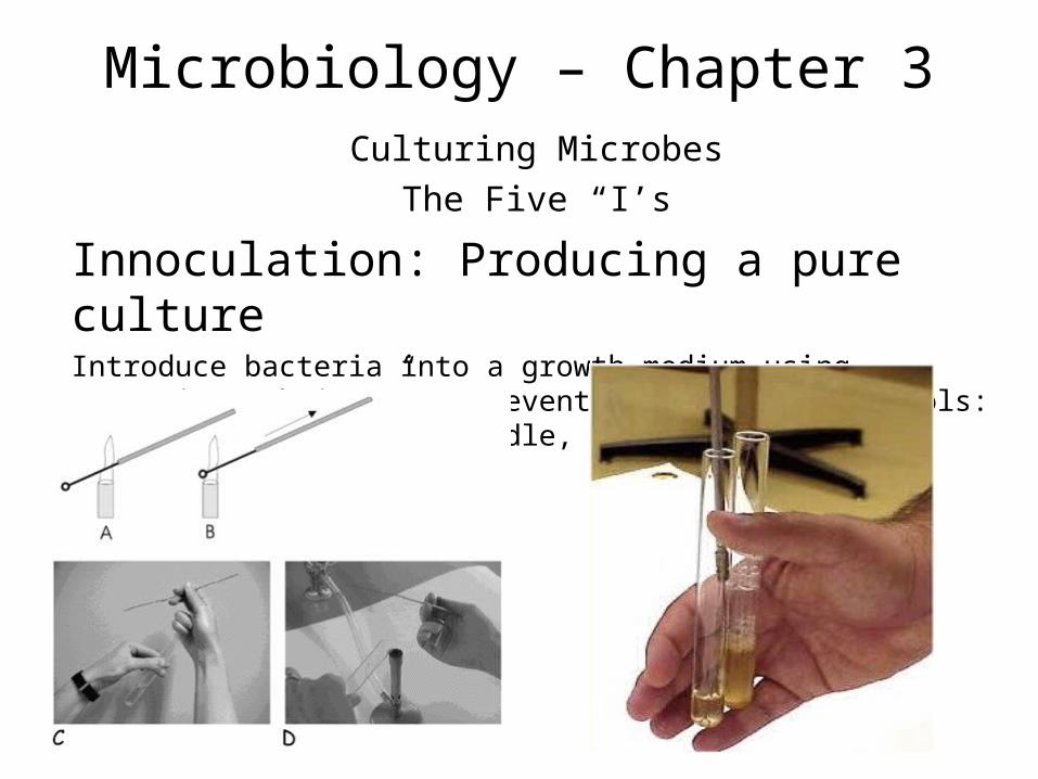
Innoculation: Producing a pure cultureIntroduce bacteria into a growth medium using “aseptic technique” to prevent contamination. Tools: Bunsen burner, loop. Needle, etc.
Microbiology – Chapter 3
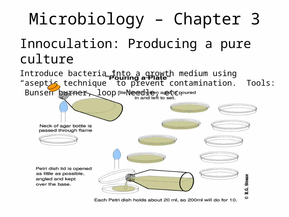
Innoculation: Producing a pure cultureIntroduce bacteria into a growth medium using “aseptic technique” to prevent contamination. Tools: Bunsen burner, loop. Needle, etc.
Microbiology – Chapter 3
Isolation: Colony on media, one kind of microbe, pure culture: isolation on general and special “differential media”
General growth media: NA, TSA
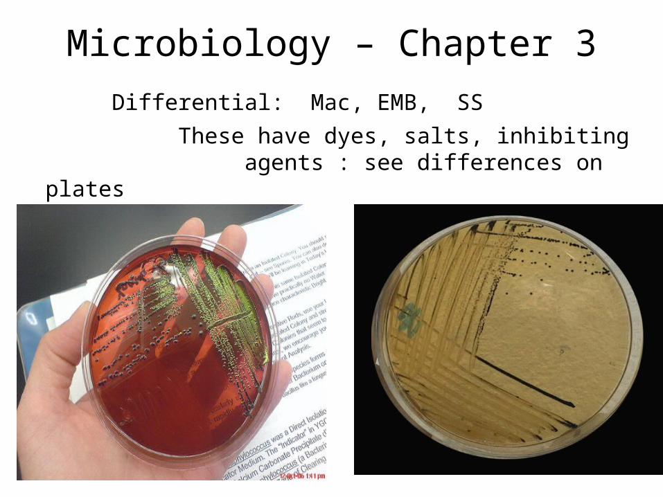
Differential: Mac, EMB, SS
These have dyes, salts, inhibiting agents : see differences on
plates
Microbiology – Chapter 3
Isolation: Colony on media, one kind of microbe, pure culture
Microbiology – Chapter 3
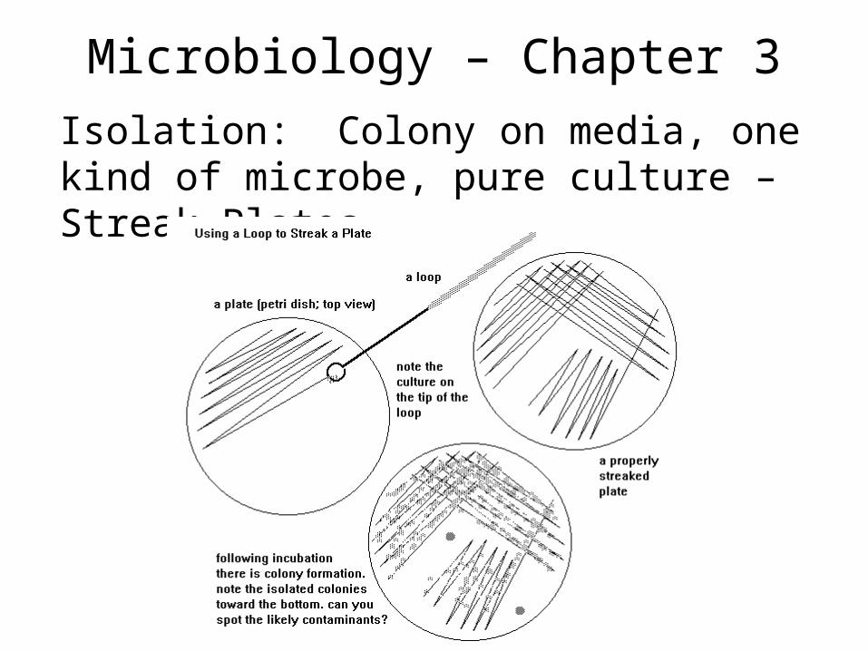
Isolation: Colony on media, one kind of microbe, pure culture – Streak Plates
Microbiology – Chapter 3
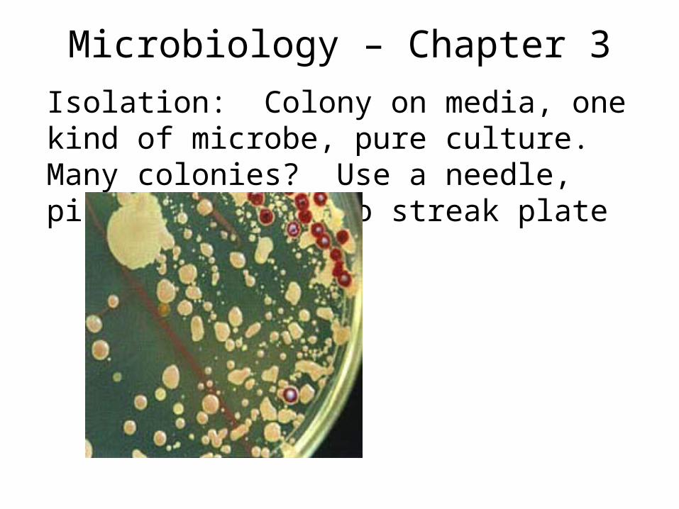
Isolation: Colony on media, one kind of microbe, pure culture. Many colonies? Use a needle, pick one, and redo streak plate
Microbiology – Chapter 3
Differential: Mac, EMB, SS
These have dyes, salts, inhibiting agents : see differences on plates
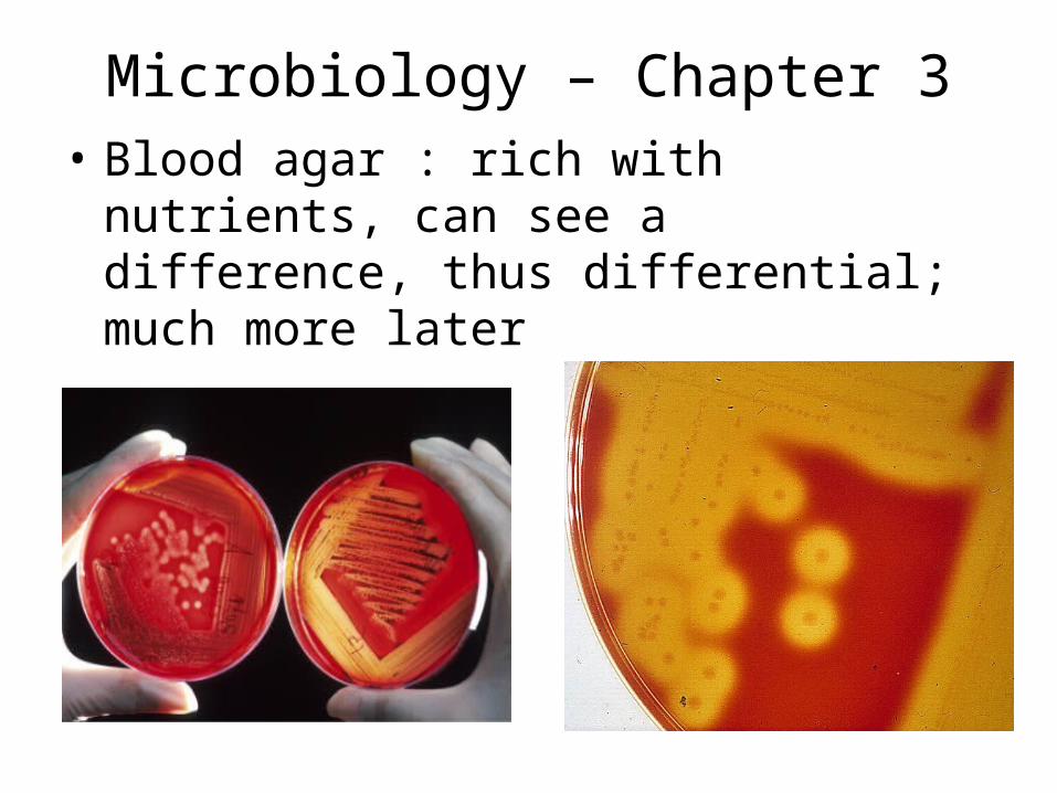
Microbiology – Chapter 3• Blood agar : rich with nutrients, can see a
difference, thus differential; much more later
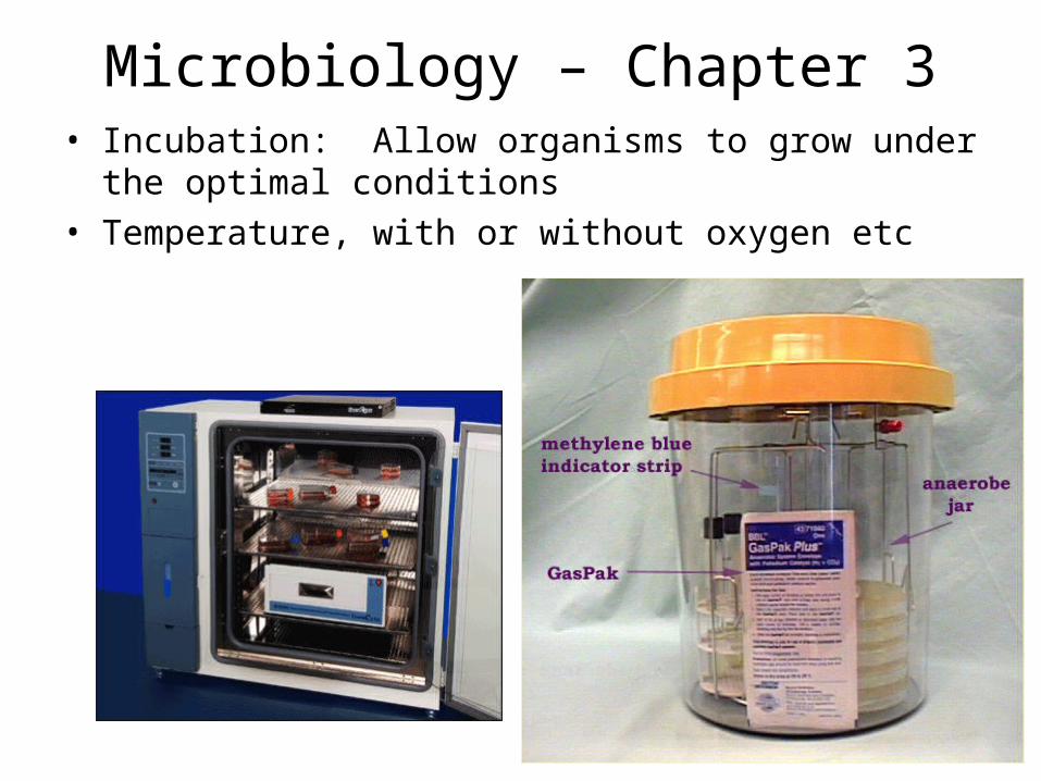
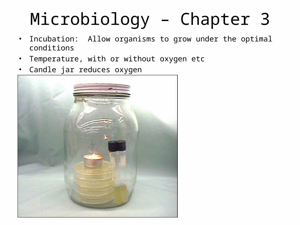
Microbiology – Chapter 3• Incubation: Allow organisms to grow under the optimal
conditions• Temperature, with or without oxygen etc
Microbiology – Chapter 3• Incubation: Allow organisms to grow under the optimal conditions
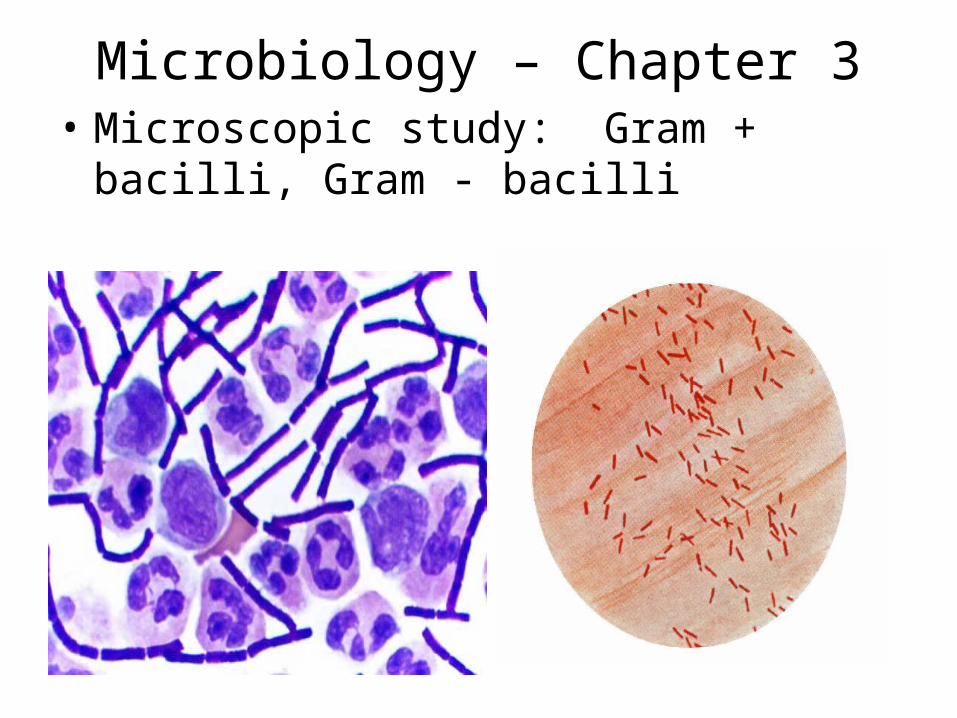
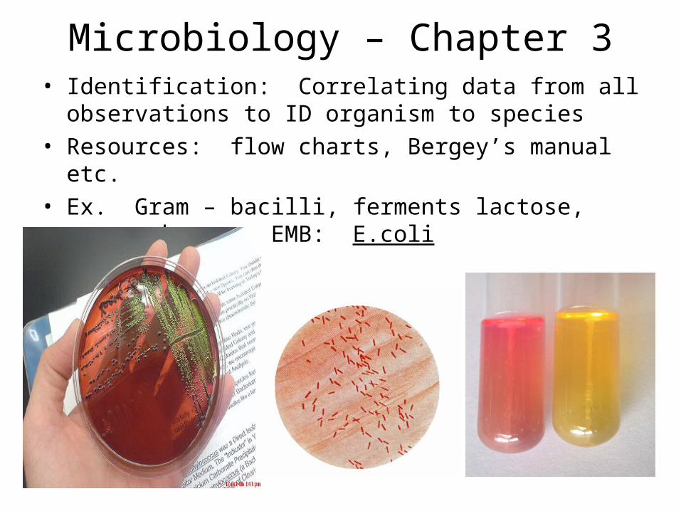
Microbiology – Chapter 3• Identification: Correlating data from all observations to
ID organism to species• Resources: flow charts, Bergey’s manual etc.• Ex. Gram – bacilli, ferments lactose, green sheen on
EMB: E.coli
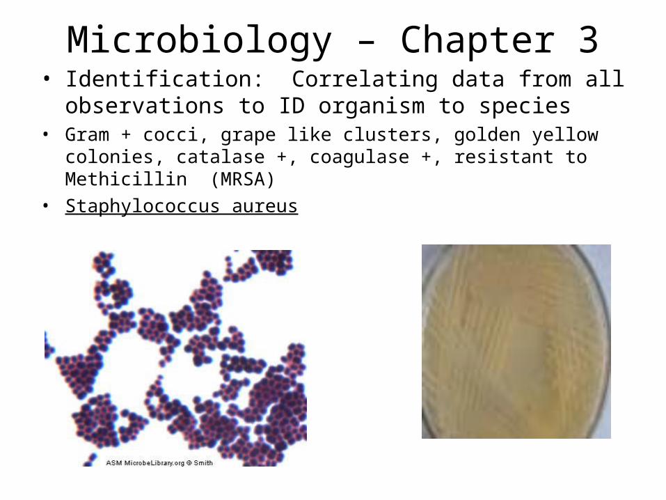
Microbiology – Chapter 3• Identification: Correlating data from all observations to
ID organism to species• Gram + cocci, grape like clusters, golden yellow colonies, catalase
+, coagulase +, resistant to Methicillin (MRSA)• Staphylococcus aureus
Microbiology Chapter 3, part 2
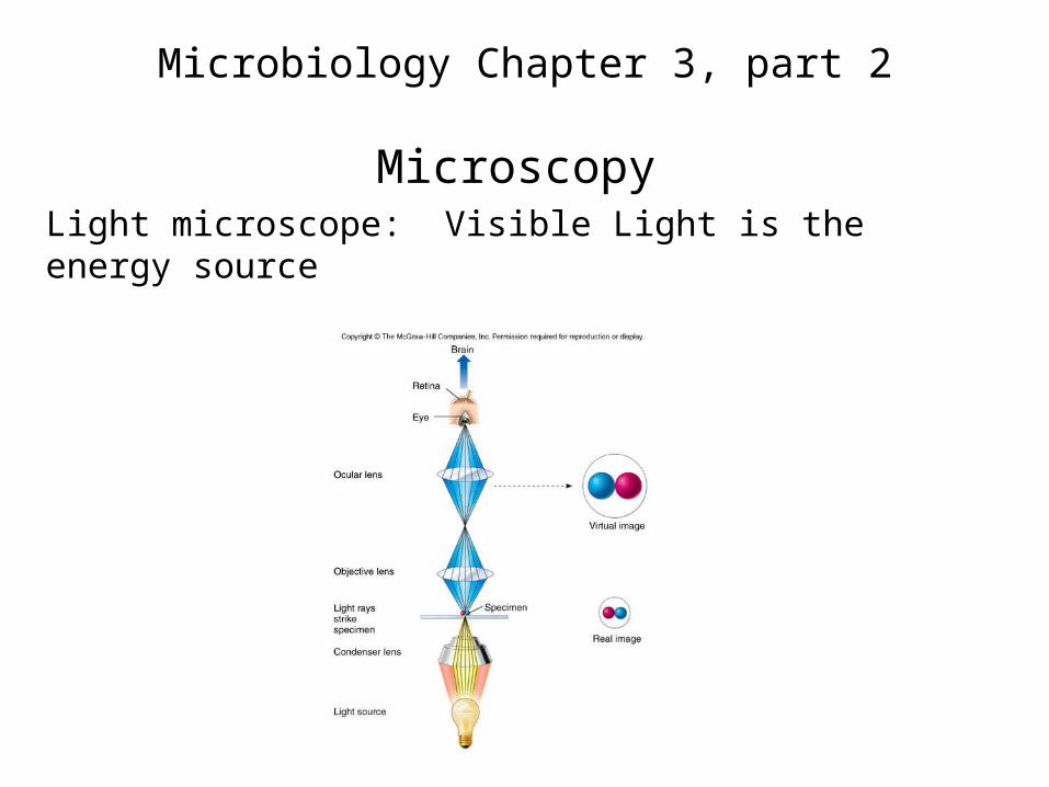
MicroscopyLight microscope: Visible Light is the energy source
Microbiology Chapter 3, part 2
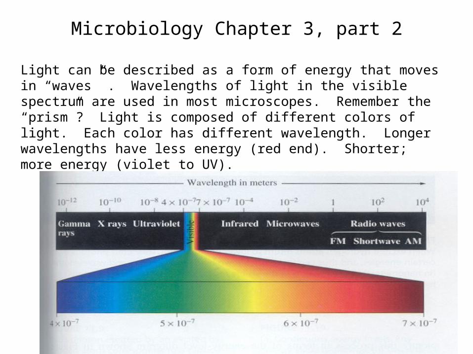
Light can be described as a form of energy that moves in “waves” . Wavelengths of light in the visible spectrum are used in most microscopes. Remember the “prism”? Light is composed of different colors of light. Each color has different wavelength. Longer wavelengths have less energy (red end). Shorter; more energy (violet to UV).
Microbiology Chapter 3, part 2
When light strikes an object the light can be:Reflected – Bounces off (Mirror)Transmitted – Passes through (GLASS)Absorbed – Soaked (black colored paper)Diffracted – Scattered as it passes through
(bugs on a dirty windshield)Refracted – Bent as it passes (objects seen
under water) Glass lensesRefractive index: degree of bending,
based on lens material and shape of lens
Microbiology Chapter 3, part 2
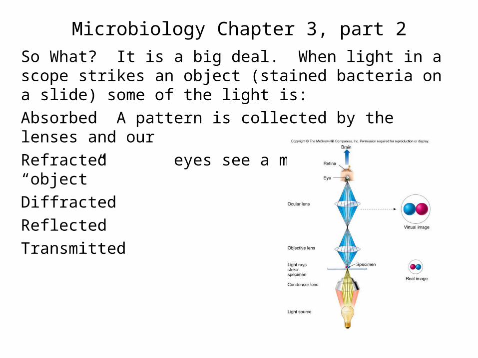
So What? It is a big deal. When light in a scope strikes an object (stained bacteria on a slide) some of the light is:
Absorbed A pattern is collected by the lenses and our
Refracted eyes see a magnified “object”
Diffracted
Reflected
Transmitted
Microbiology Chapter 3, part 2
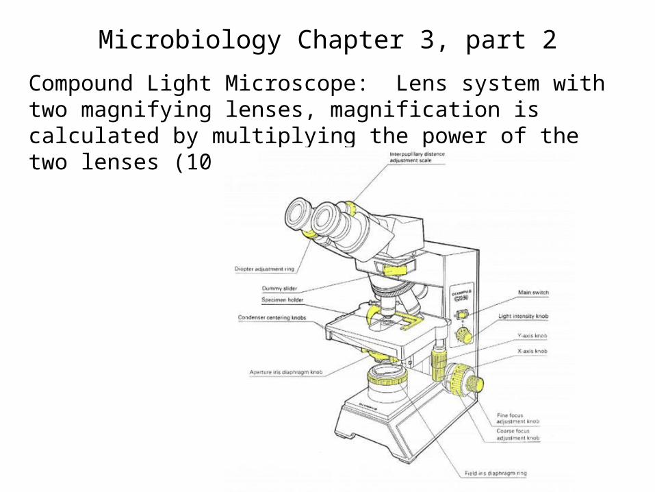
Compound Light Microscope: Lens system with two magnifying lenses, magnification is calculated by multiplying the power of the two lenses (10 X 10 = 100 power)
Microbiology Chapter 3, part 2
Technicality
Contrast: Bacteria have little contrast unstained. Light is only slightly refracted – diffracted – reflected etc. as it passes through the cells. To see them we usually stain them. Stains are colored dyes (chromophores) that increase contrast. Without stains, special expensive microscopes are needed.
Resolution: aka “resolving power” The ability of a lens system to allow an observer to see fine detail. Quality of lens systems (fine quality of glass and special lens coatings). The best lens systems allow one to see two points as distinct points eve when they are tiny and very close together.
Microbiology Chapter 3, part 2
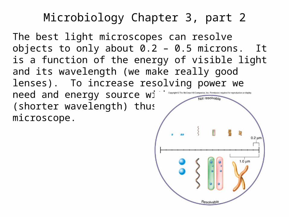
The best light microscopes can resolve objects to only about 0.2 – 0.5 microns. It is a function of the energy of visible light and its wavelength (we make really good lenses). To increase resolving power we need and energy source with more energy (shorter wavelength) thus the electron microscope.
Microbiology Chapter 3, part 2
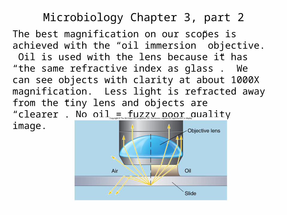
The best magnification on our scopes is achieved with the “oil immersion” objective. Oil is used with the lens because it has “the same refractive index as glass”. We can see objects with clarity at about 1000X magnification. Less light is refracted away from the tiny lens and objects are “clearer”. No oil = fuzzy poor quality image.
Microbiology Chapter 3, part 2
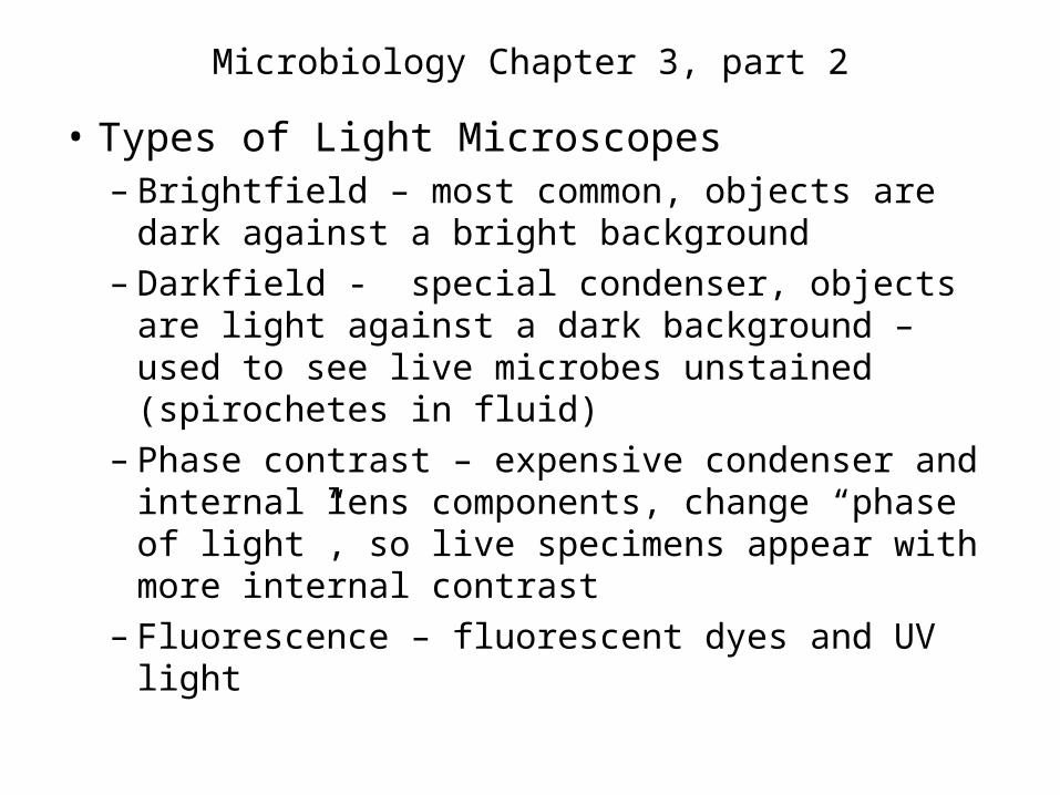
• Types of Light Microscopes– Brightfield – most common, objects are dark
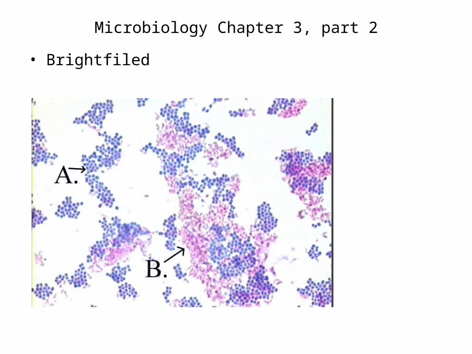
against a bright background– Darkfield - special condenser, objects are
light against a dark background – used to see live microbes unstained (spirochetes in fluid)
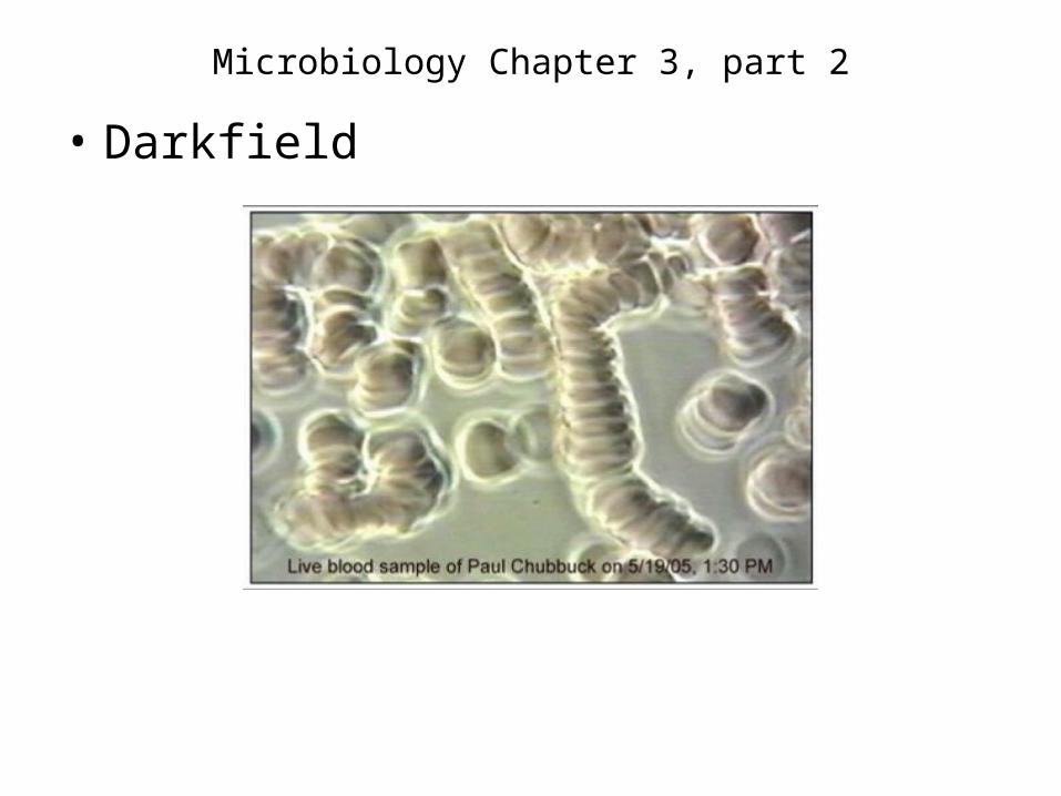
– Phase contrast – expensive condenser and internal lens components, change “phase of light”, so live specimens appear with more internal contrast
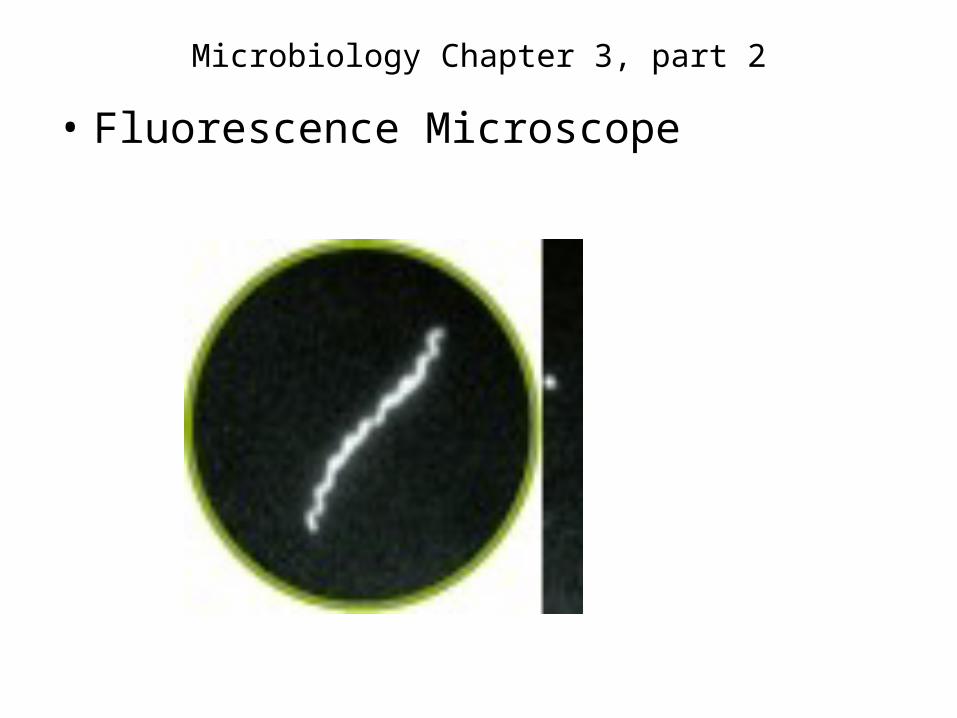
– Fluorescence – fluorescent dyes and UV light
Microbiology Chapter 3, part 2
• Brightfiled
Microbiology Chapter 3, part 2
• Darkfield
Microbiology Chapter 3, part 2
• Phase contrast
Microbiology Chapter 3, part 2
• Fluorescence Microscope
Microbiology Chapter 3, part 2
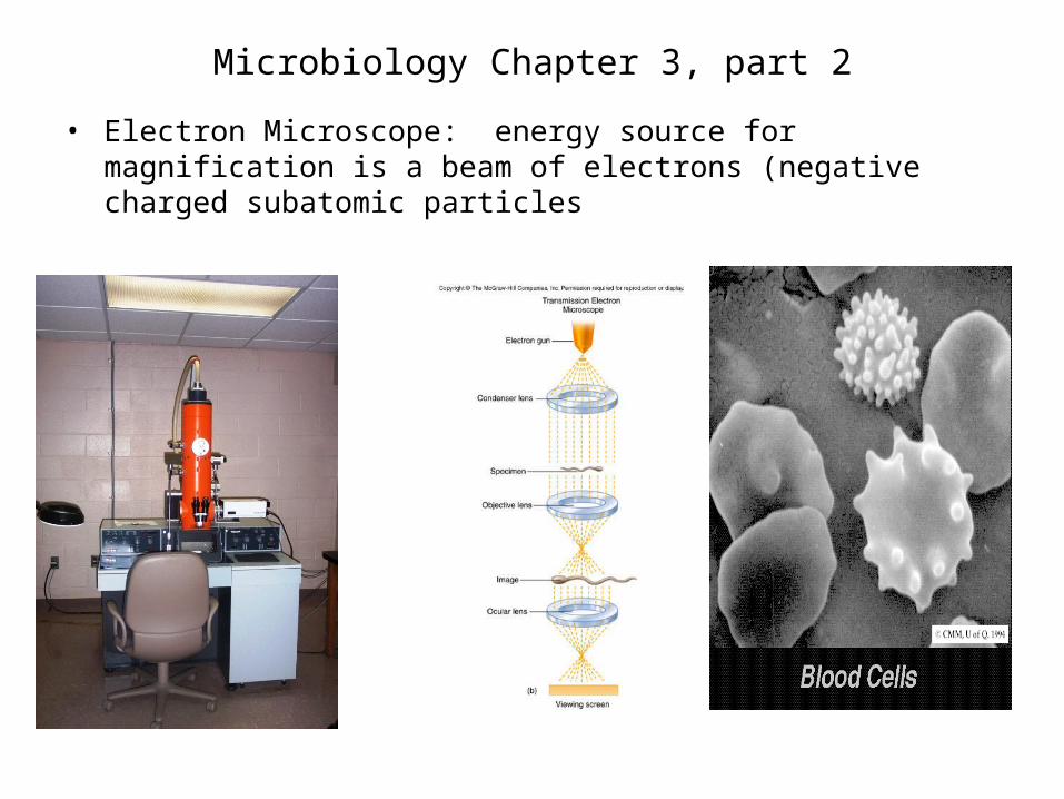
• Electron Microscope: energy source for magnification is a beam of electrons (negative charged subatomic particles
Microbiology Chapter 3, part 2
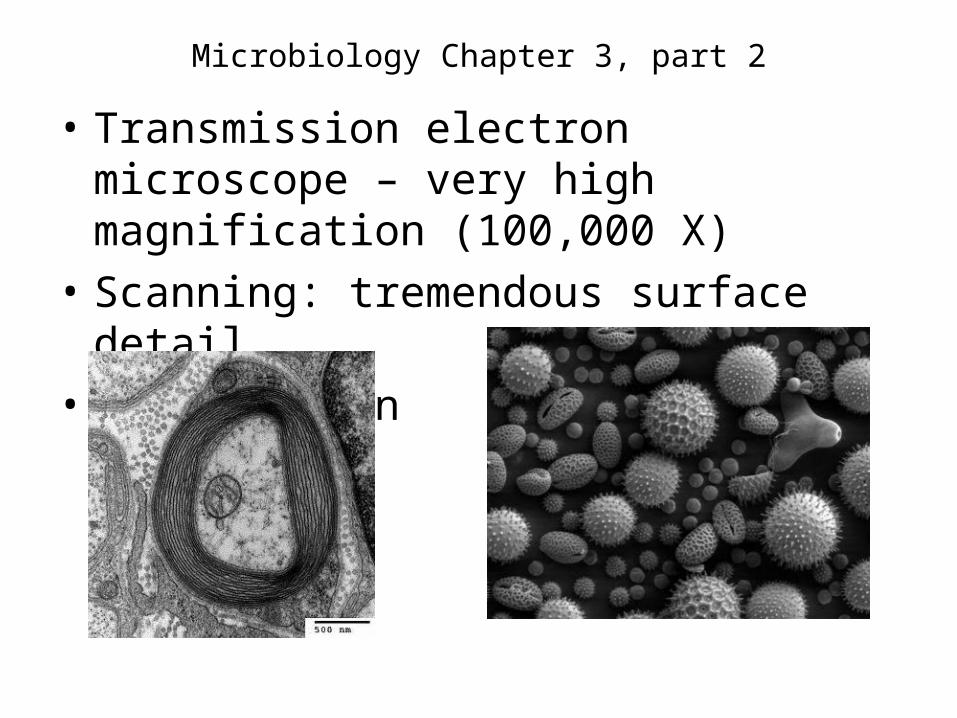
• Transmission electron microscope – very high magnification (100,000 X)
• Scanning: tremendous surface detail
• Transmission Scanning
Microbiology Chapter 3, part 2
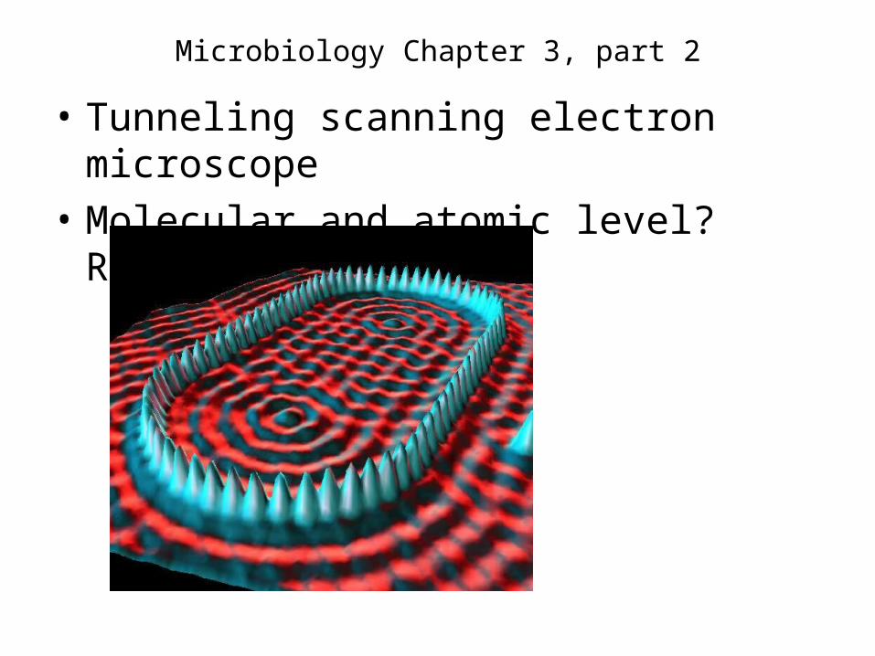
• Tunneling scanning electron microscope
• Molecular and atomic level? Research
Microbiology Chapter 3, part 2
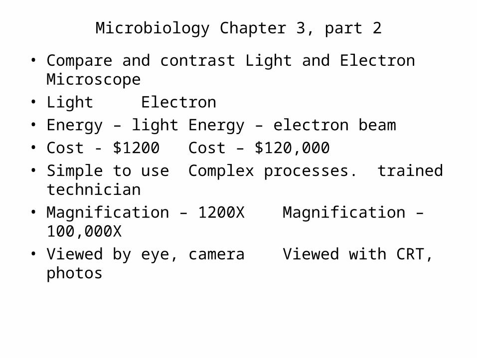
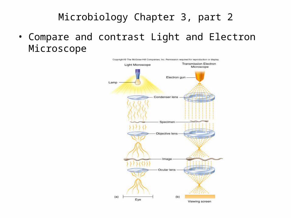
• Compare and contrast Light and Electron Microscope• Light Electron• Energy – light Energy – electron
beam• Cost - $1200 Cost – $120,000• Simple to use Complex processes.
• Compare and contrast Light and Electron Microscope
Microbiology Chapter 3, part 2
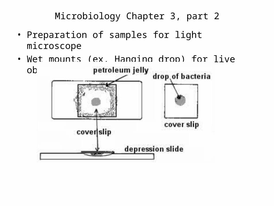
• Preparation of samples for light microscope• Wet mounts (ex. Hanging drop) for live observation
Microbiology Chapter 3, part 2
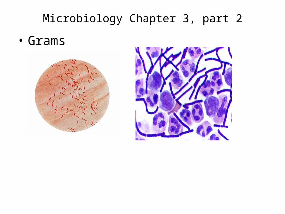
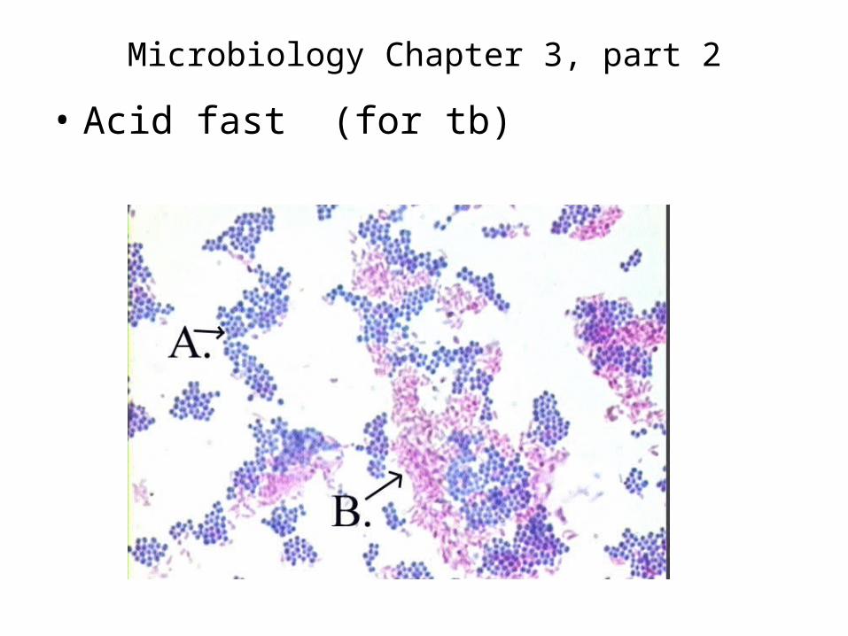
• Simple stain – one dye
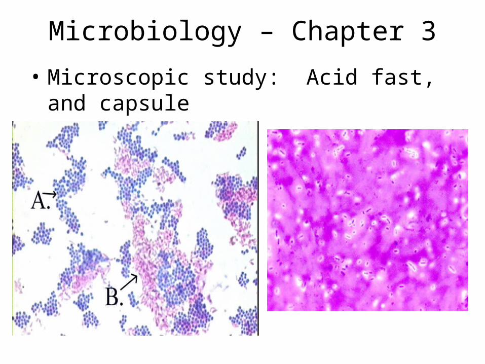
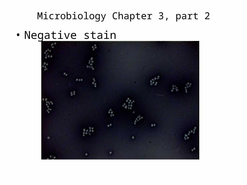
• Differential stain – complex procedure, see difference between cells– Grams + and (-)– Acid fast + and (-)– Negative – acid dye stains background and
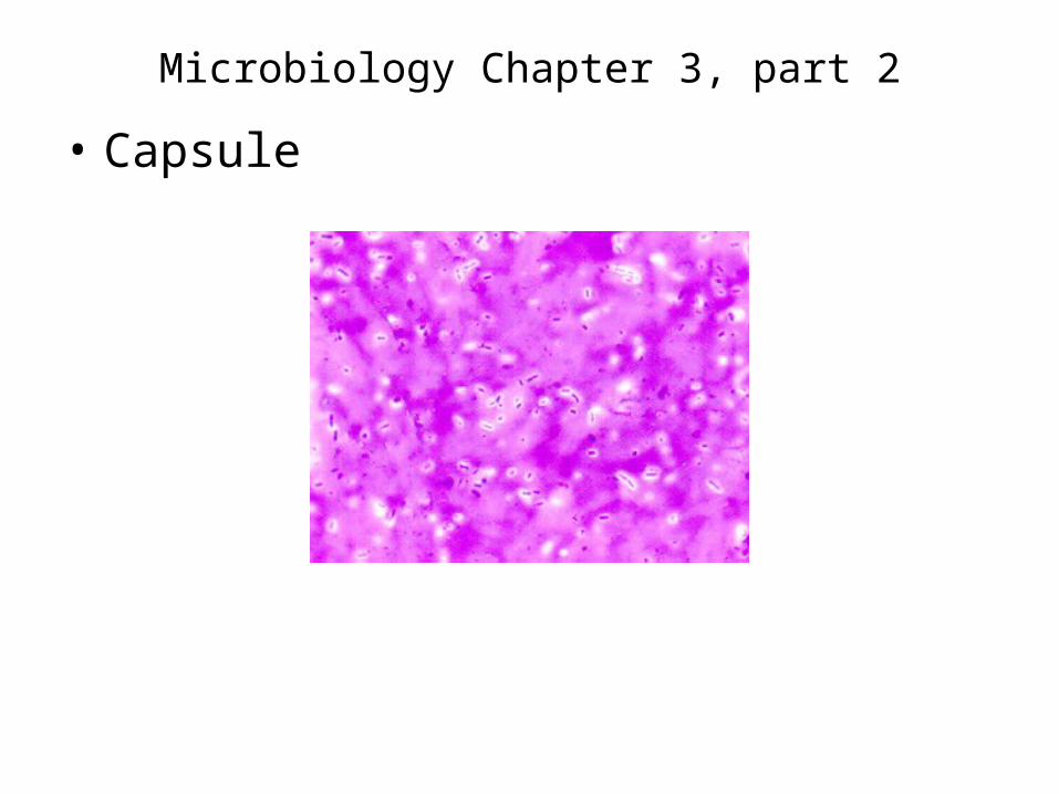
cells are white (cell wall repels stain)– Capsule – modified negative stain to show