Page 1
MICROBIOLOGY OF PUERPERAL SEPSIS AND ITS
CLINICAL IMPLICATIONS AMONG HIV-INFECTED AND
HIV-UNINFECTED WOMEN IN A HOSPITAL SAMPLE IN
ZIMBABWE
By
Dr Rumbidzai Majangara
Thesis submitted in partial fulfillment of the degree of Masters in
Medicine Obstetrics and Gynaecology, University of Zimbabwe
Supervised by Dr M. F. Gidiri and Professor Z. M. Chirenje
Department of Obstetrics and Gynaecology
College of Health Sciences, University of Zimbabwe
June 2016
Page 2
i
ABSTRACT
Title: Microbiology of puerperal sepsis and its clinical implications in a Hospital sample in
Zimbabwe
Introduction: Puerperal sepsis is infection of the female genital tract occurring at any time
between the rupture of membranes or labour, and the 42nd day postpartum. Puerperal sepsis has
become the leading cause of maternal death in Harare public health institutions accounting for
19% and 30% of maternal deaths for the years 2010 and 2014 respectively, from being the fourth
nationwide cause at 12.3% in the year 2007. The objectives of this study were to determine the
identity and antibacterial susceptibility profiles of bacteria colonizing the genital tract and blood
stream, and to assess clinical outcomes and association with HIV infection in women with
puerperal sepsis.
Methodology: A prospective cohort study was conducted at Parirenyatwa and Harare
Hospitals between 02 September 2014 and 01 July 2015. Endocervical swabs and blood were
collected for culture and susceptibility testing from 151 consecutive women who met the
World Health Organisation criteria for puerperal sepsis. HIV sero-status, immunological
status and antiretroviral therapy (ART) use were determined. Medical records were reviewed
for assessment of clinical outcomes. Proportions, categorical values and means were
compared using Z-test, χ² test and t- test along with 95% confidence interval (CI) and p-value
of <0.05.
Results: The mean age was 25.1 ±5.8years and most women were multiparous (53.6%). The
majority of women had delivered at a hospital (78.1%) and by caesarean section (57.6%). The
commonest bacterial isolates were Escherichia coli (30.6%) and Klebsiella pneumoniae
(15.3%). Multidrug resistant organisms (MDRO) accounted for 10.9% of the isolates. MDRO
were associated with prolonged mean hospital stay 23.0days (d) compared to 10.5d in women
without MDRO (p=0.009). The frequency of genital colonization with Enterobacter species
was significantly higher in HIV infected (9.1%) than uninfected women (1.7%) (p=0.04).
Among HIV infected women (21.9%), severe immunosuppression (CD4 <200/mm³) was
associated with a greater need for laparotomy 42.9% vs 4.5% (p=0.01) and prolonged mean
hospital stay 19.0d vs 10.2d (p=0.03) compared to mild-advanced (CD4 count 200-500/ mm³)
and insignificant immunosuppression (CD4 >500/mm³). There was a non-significant trend
towards, earlier onset of sepsis; and higher rates of pelvic abscess, septic shock, wound
dehiscence, peritonitis, death and need for admission into the intensive care unit (ICU) in
women with severe immunosuppression. Antiretroviral therapy use did not independently
influence outcomes. Puerperal sepsis case fatality rate was 7.3%.
Conclusion: Gram negative bacilli, particularly E. coli, are the commonest bacterial isolates in
puerperal sepsis. There is emergence of MDRO gram negative bacilli resistant to carbapenems,
especially K. pneumoniae. MDRO and HIV associated severe immunosuppression are risk factors
for prolonged hospital stay and need for surgery. Robust infection control strategies, emphasis on
rational drug use and clinical culture surveillance to identify MDRO and monitor epidemiologic
trends is recommended.
Page 3
ii
ACKNOWLEDGEMENTS
I am grateful to the following:
My supervisors Dr M F Gidiri and Professor Z M Chirenje for the professional assistance
in designing and overseeing the completion of this study
Professor V Robertson for professional advice on microbiological laboratory
considerations
NECTAR Mentored Research Scholarship Program for financial assistance and education
on principles of research
University of Zimbabwe (UZ) Department of Microbiology Laboratory Scientists Ms C
Berejena, Ms T Magombei and Mr R. Mudengezerwa for assistance in carrying out
microbiological laboratory procedures
Mr M Munjoma UZ Obstetrics and Gynaecology laboratory scientist for professional
advice
Staff at Harare Hospital and Parirenyatwa Hospital in the following departments-
Maternity, Gynaecology, Casualty, Intensive care and Outpatient units for assistance in
notification of patients; and the Public Health Laboratory for provision of results for tests
performed as standard of care
Women who agreed to participate in the study
Mr K Chimunda for assistance in statistical analysis of study results
Page 4
iii
Contents Page number
Abstract…………………………………………………………………...i
Acknowledgements…………………………………………………….....ii
Table of Contents…………………………………………………………iii
List of Tables, Figures and Appendices….……………………………….iv
Abbreviations, Glossary………………………………………………......vi
Chapter 1: Introduction 1.0 Introduction………………………………………………………….1
1.1 Justification…………………………………………………………..3
Chapter 2: Literature Review 2.1 Literature Review……………………………………………………5
2.2 Research Question…………………………………………………...9
2.3 Study Objectives………………………………………………….....9
Chapter 3: Research methodology
3.1 Study design…………………………………………………………11
3.2 Study setting…………………………………………………………11
3.3 Study population…………………………………………………….11
3.4 Inclusion criteria……………………………………………………..11
3.5 Exclusion criteria……………………………………………….........12
3.6 Sample size determination and sampling plan……………………....12
3.7 Methods of data collection …………………………………………13
3.7.1 History and examination…………………………………..13
3.7.2 Questionnaire administration……………………………...13
3.7.3 Laboratory procedures…………………………………….15
3.7.4 Review of medical records……………………………......21
3.7.5 Responsibility of care for women………………………...22
3.7.6 Outcome measures………………………………………..22
3.8 Pretesting the questionnaire………………………………………...23
3.9 Statistical analysis…………………………………………………..23
3.10 Ethical considerations……………………………………………..23
Chapter 4: Results
Results and Analysis…………………………………………………....25
Chapter 5: Discussion 5.1 Discussion…………………………………………………………..51
5.2 Study Limitations…………………………………………………..56
5.3 Conclusion and Recommendations………………………………...57
References……………………………………………………………..58
Appendices………………………………………………………….....64
Page 5
iv
LIST OF TABLES, FIGURES AND APPENDICES
Tables:
Table 1: Socio-demographic data
Table 2: Obstetric and medical risk factors
Table 3: Antibiotic susceptibility of blood culture isolates
Table 4: Association of blood culture isolates with HIV status.
Table 5: Association of endocervical swab isolates with HIV status.
Table 6: Association of presence of MDRO with clinical outcomes of puerperal sepsis
Table 7: Association of HIV status with clinical presentation of puerperal sepsis
Table 8: Association of immunological status with presentation of puerperal sepsis
Table 9: Association of immunological status with presentation of puerperal sepsis
Table 10: Association of use of antiretroviral therapy with presentation of puerperal sepsis
Table 11: Association of duration of use of antiretroviral therapy with presentation of puerperal
sepsis
Figures:
Figure 1: Endocervical swab isolates
Figure 2: Blood culture isolates
Figure 3: susceptibility to ceftriaxone
Figure 4: susceptibility to penicillin
Figure 5: susceptibility to chloramphenicol
Figure 6: susceptibility to gentamycin
Figure 7: susceptibility to clindamycin
Page 6
v
Figure 8: susceptibility to ciprofloxacin
Figure 9: susceptibility to erythromycin
Figure 10: Multidrug resistant organisms
Figure 11: HIV status and CD4 count
Figure 12: Presentation of puerperal sepsis
Figure 13: Complications of puerperal sepsis
Figure 14: Association of presence of MDRO with length of hospital stay
Figure 15: Association of method of removal of placenta with bacteremia
Appendices:
Appendix 1: Additional tables
Table a: Endocervical swab isolates
Table b: Blood culture isolates
Table c: Antimicrobial susceptibility profiles of endocervical swab isolates
Table d: Multidrug resistant isolates
Page 7
vi
LIST OF ABBREVIATIONS
AIDS Acquired immunodeficiency Syndrome
API Analytical profile index
ART Antiretroviral therapy
ATCC American Type Culture Collection
BA blood agar
bd twice daily
BMI body mass index
CA chocolate agar
CD4 cluster of differentiation 4
CI 95% Confidence Interval
CO₂ carbon dioxide
CoNS coagulase negative staphylococcus
dl deciliter
EDLIZ Essential Drugs List and Standard Treatment Guidelines for Zimbabwe
FBC Full blood count
GAS Group A streptococcus
g/L grams per liter
g/dl grams per deciliter
h hour
HIV Human Immunodeficiency Virus
ICU Intensive Care Unit
IM intramuscular
IQR interquartile range
IV intravenous
kg kilogram
Page 8
vii
MAC MacConkey
MDRO multidrug resistant organisms
Mcg micrograms
mg milligrams
ml milliliter
ml/kg/h milliliters per kilogram per hour
mm³ cubic millimeter
MU Mega Units
PMTCT Prevention of mother to child transmission of HIV
po orally
qid four times daily
RR Relative Risk
sd standard deviation
sp species
Stat immediately
tds three times daily
UK United Kingdom
USA United States of America
U&E urea and electrolytes
UZ University of Zimbabwe
WHO World Health Organisation
Page 9
1
CHAPTER 1
1.0 INTRODUCTION
Puerperal sepsis is infection of the female genital tract occurring at any time between the rupture
of membranes or labour, and the 42nd day postpartum in which 2 or more of the following are
present: pelvic pain, fever (i.e. oral temperature 38.5°C or higher on any occasion), abnormal
vaginal discharge (e.g. presence of pus), abnormal smell/foul odour of discharge, and delay in the
rate of reduction of the size of the uterus (<2cm/day during the first 8 days) (1,2).
Globally, the incidence of puerperal sepsis ranges from 2.7% to 5.7% (1). Puerperal sepsis
accounts for 2% of maternal deaths in the developed world and 10-12% of maternal deaths in
the developing world(3). Puerperal sepsis was the fourth leading cause of maternal death in
Zimbabwe accounting for 12.3% of maternal deaths in a national survey in the year 2007(4) .
An unpublished retrospective review comparing trends in maternal mortality for the public
maternity institutions of Harare reported that obstetric sepsis was now the leading cause of
maternal deaths accounting for 19% and 30% of maternal deaths for the years 2010 and 2014
respectively (5). In comparison, the contribution of genital tract sepsis to maternal death may
not be as significant in the developed world. Though direct and indirect sepsis combined were
the second commonest cause of maternal deaths in the latest United Kingdom Triennial
Report for 2011-2013, genital tract sepsis was the fourth leading cause of direct maternal
death accounting for 0.29 maternal deaths per 100 000 maternities, and was in fact showing a
downward trend from preceding years (6).
Page 10
2
If left untreated women may develop early complications such as wound dehiscence, peritonitis,
pelvic abscess, necrotizing fasciitis, toxic shock syndrome; and long-term maternal complications
such as chronic pelvic inflammatory disease, chronic pelvic pain and infertility(2,7). The
proportion of women developing infertility following puerperal sepsis ranges from 5% in
developed countries to 12% in developing countries(1).
Major risk factors for puerperal sepsis are caesarian section, prolonged and premature rupture of
membranes, prolonged labor (>24hours), frequent unsanitary vaginal examinations, retained
products of conception, hemorrhage, anemia, malnutrition and obesity (2,7,8). The incidence of
puerperal sepsis has been shown to be higher in HIV positive persons compared to HIV negative
persons (9,10).
Most pelvic infections are caused by overgrowth of bacteria indigenous to the female genital tract,
notably Groups A and B beta-hemolytic streptococci, Staphylococcus aureus, enteric bacteria such
as Escherichia Coli, Klebsiella species, pseudomonas species, clostridium species, Morganella
morganii and other anaerobes, mycoplasma, chlamydia and gonococci. Infection is frequently
polymicrobial(2,7,12). In the developed world, the most virulent infective organism causing life-
threatening infection is community acquired Group A beta-hemolytic streptococci (7,13). Studies
in developing countries have noted inter-country variance in the most frequent etiologic organisms
(14–18).
Page 11
3
Antibiotic resistance is a global public health problem worse in developing countries which have
a high burden of infectious disease, constraints on microbiological investigations to identify
resistant infections and unavailability of novel agents to treat them. There are accelerating rates of
antimicrobial resistance fueled by irrational drug use and shortfalls in infection control and public
health (19). Treatment of most infections in Zimbabwe is empiric/syndromic mainly due to lack
of microbiological services and diagnostic capacity from grassroots up to tertiary level(20). There
is therefore a need for periodic evaluation of antimicrobial effectiveness against infective bacteria.
This will allow identification of multi-drug resistant organisms (MDRO), and pave way for
concerted efforts at preventing their emergence and spread.
1.1 JUSTIFICATION FOR THE STUDY
Most data on the microbiology of puerperal sepsis in Zimbabwe was collected over 2 decades ago
before the advent of the HIV epidemic (21,22). Recent estimates of puerperal sepsis in our
hospitals come from retrospective studies of maternal deaths, without microbiological
investigation (4,5). Therefore these data reflect only clinically defined puerperal sepsis.
In our Zimbabwean institutions, puerperal sepsis is diagnosed clinically as infection of the female
genital tract occurring at any time between the rupture of membranes or labour, and the 42nd day
postpartum in which 2 or more of the following are present: pelvic pain or tenderness, fever (i.e.
oral temperature 38.5°C or higher on any occasion), abnormal vaginal discharge (e.g. presence of
pus, abnormal uterine bleeding), abnormal smell/foul odour of discharge, and delay in the rate of
reduction of the size of the uterus (<2cm/day during the first 8 days) (1,23).
Page 12
4
Empiric treatment is recommended using ampicillin 1-2g IV qid and metronidazole 500mg IV tds
and gentamycin 3-5mg/kg IV daily. Ceftriaxone 1-2g IV bd or benzyl penicillin 5MU IV qid are
alternatives to ampicillin. Recommended antibiotic prophylaxis before caesarian section is
ceftriaxone 1g IV stat or ampicillin 1g IV stat (23). The persistent use of select antibiotic classes
leads to development of resistance to those classes over time and may eventually lead to the
selection of multidrug-resistant organisms. During the economic crisis of the last decade, there has
been a lack of systematic, well-designed and funded research studies to determine the etiology of
puerperal sepsis and the emergence of drug resistance. This means that the recommended drug
regimens for prophylaxis and treatment of infections might be ineffective in our setting.
HIV infection has been associated with more severe clinical manifestations in most studies of
pelvic inflammatory disease, while other investigators found no increase in severity of clinical
outcomes (24–27). While evidence exists that HIV infection increases the risk of postpartum
infections (9), there is insufficient information on the association of HIV infection with clinical
outcomes.
This study aims to identify suspected infecting organisms, assess susceptibility of these organisms
to the current recommended empiric antibiotics, and assess the association of these organisms and
HIV infection with clinical outcomes in our population.
Page 13
5
CHAPTER 2
2.1 LITERATURE REVIEW
Puerperal sepsis is infection of the female genital tract occurring at any time between the rupture
of membranes or labour, and the 42nd day postpartum in which 2 or more of the following are
present: pelvic pain, fever (i.e. oral temperature 38.5°C or higher on any occasion), abnormal
vaginal discharge (e.g. presence of pus), abnormal smell/foul odour of discharge, and delay in the
rate of reduction of the size of the uterus (<2cm/day during the first 8 days).Clinical signs and
symptoms typically present more than 24 hours after delivery unless the patient had prolonged
rupture of membranes or prolonged labour without prophylactic antibiotics when the disease may
be noted earlier(1).
Puerperal sepsis can be caused by both exogenous and endogenous bacteria. Endogenous bacteria
are usually commensals in the vagina and rectum which may go up the uterus during prolonged
rupture of membranes, genital tract trauma or via examining fingers and instruments. Exogenous
bacteria may be introduced by unsterile examination, droplet infection, sexual activity and foreign
substances introduced into the vagina e.g.- herbs, oils and cloths (1,7) . Puerperal infection
following vaginal delivery primarily involves the placental implantation site and the decidua and
adjacent myometrium, while the pathogenesis of uterine infection following cesarean delivery is
that of an infected surgical incision (7).
Community acquired Group A Streptococcus (GAS) from the woman’s own respiratory tract or
hands, or hospital staffs’ hands, is increasingly causing invasive and life threatening puerperal
Page 14
6
infections worldwide (7,28). Puerperal sepsis mostly associated with GAS was the commonest
cause of direct maternal deaths in the United Kingdom (UK) during the triennium 2006-2008 (29).
The practice of endometrial aspirate and blood cultures in patients suspected of having puerperal
endometritis effectively contributes to the diagnosis and treatment of this infection (30). Studies
utilising endocervical swabs have shown an approximately 80% chance of identifying suspected
infecting organisms compared to blood cultures which were positive in 5% to 24% of samples
from women with puerperal sepsis (7,14,16,17). However genital tract swabs have the
disadvantage of picking up other non-infecting vaginal flora (7). In a Nigerian study in which
endocervical swabs were cultured in 139 women with puerperal sepsis, S. aureus, E. coli,
Streptococcus sp. and Pseudomonas sp. were the commonest isolated organisms. There was either
insignificant or no growth in 30/139 specimens (18). A study in India in which endocervical swabs
were analysed by microscopy, culture and sensitivity, the most commonly isolated organisms were
Klebsiellae, S. aureus, E. coli, Pseudomonas sp. and B-hemolytic streptococci. There was no
growth in 2/12 specimens (14). In a Bangladesh Study, there was significant growth in anaerobic
cultures of 42/50 endocervical swabs collected from women with puerperal sepsis (16).
Microscopy and culture of endocervical swabs and blood may identify infecting organisms and
assist in the choice of antimicrobials.
Page 15
7
Studies including vaginal and caesarian deliveries indicated that HIV-infected women had over
three times the risk of a puerperal sepsis compared with uninfected women; this figure increased
to nearly six amongst studies only including women who delivered by caesarian section (9). HIV-
positive women had a statistically significant increase in the incidence of post-operative fever (RR
1.3) and wound sepsis/sinus (10). In one study, post-partum endometritis was more prevalent in
HIV-infected women compared to HIV-uninfected controls (10.3% vs 4.2%), being inversely
related to CD4 percentage (31).
The empiric antibiotic regimen for treatment of puerperal sepsis in Zimbabwe is ampicillin 1-2g
IV qid, metronidazole 500mg IV tds and gentamycin 3-5mg/kg IV daily. Alternatives to ampicillin
are ceftriaxone 1-2g IV bd or benzyl penicillin 5MU qid IV. Alternative to gentamycin in case of
abnormal kidney function is chloramphenicol 50mg/kg in 4 divided doses(23). For the
management of severe sepsis, a combination of peperacillin/tazobactam or clindamycin and a
carbapenem may provide the broadest range.(12). Amoxicillin, ciprofloxacin and erythromycin
may be given to complete the antibiotic course on an outpatient basis. Recommended antibiotic
prophylaxis before caesarian section is ceftriaxone 1g IV stat or ampicillin 1g IV stat (23).
Persistent use of a select antibiotic class leads to development of resistance to that class over time.
Combination empiric treatment may eventually lead to the selection of multidrug resistant
organisms. Multidrug resistant organisms (MDRO) are defined as organisms with acquired non-
susceptibility to at least one agent in three or more antimicrobial categories (32). Emergence of
resistance to multiple antimicrobial agents in both gram negative and gram positive bacteria is a
significant threat to public health as there may be no effective antimicrobial agents to manage these
Page 16
8
infections, especially in resource limited settings which have limited access to novel
antimicrobials. MDRO associated infections are associated with poor patient outcomes (30).
Notable MDROs include multidrug-resistant carbapenemase-producing K. pneumoniae and
Acinetobacter sp which are resistant to all currently available antimicrobials or remain susceptible
only to older, toxic agents such as the polymyxins (32–35).
Earlier research conducted in Zimbabwe has shown emergence of anti-microbial resistance in
pathogens such as staphylococci, streptococci, gonococci and enteric bacteria. A study on
antimicrobial susceptibility of Neisseria gonorrhea showed >60% resistance to penicillin, >10%
resistance to cefuroxime, 50% resistance to erythromycin, 20% resistance to tetracycline and full
susceptibility to kanamycin and spectinomycin (36). A study on vaginal pathogens in which 130
isolates of Group B streptococci were isolated showed 5 isolates resistant to erythromycin, 7
isolates resistant to clindamycin, intermediate resistance to gentamycin, while all were susceptible
to beta lactams(22). A project investigating the role of S. aureus in local burn centers determined
a high prevalence of methicillin resistant S. aureus (MRSA) (64% and 49% prevalence at
Parirenyatwa and Harare Hospital, respectively) among randomly sampled wound cultures (37).
Page 17
9
2.2 Research Question
What is the microbiology of the female genital tract and blood stream in HIV infected and HIV-
uninfected women with puerperal sepsis, the in vitro antibacterial susceptibility of these
organisms, and the clinical response to empiric antibiotics in women admitted at Parirenyatwa and
Harare Hospitals?
2.3 Study Objectives
2.3.1 Broad Objective
To determine the microbiology of puerperal sepsis and its clinical implications among HIV-
infected and HIV-uninfected women admitted at Parirenyatwa and Harare Hospitals
2.3.2 Specific Primary Objectives
i. To identify the bacteria colonizing the female genital tract in women with puerperal sepsis
ii. To identify bacteria causing bacteremia in women with puerperal sepsis
iii. To determine the antibacterial susceptibility profiles of the cultured bacteria
iv. To determine if the spectrum of microbiological organisms is different among HIV-
infected and HIV-uninfected women
v. To compare women’s clinical response after empiric antibiotics to in vitro antibacterial
susceptibility profiles of colonizing bacteria
vi. To determine proportion of women who develop complications and those who require
surgery such as laparotomy for management of complications
Page 18
10
2.3.3 Secondary Objectives:
i. To determine risk factors for bacteremia
ii. To determine if immunological status (CD4 cell count), virological status (viral load) and
use of antiretroviral therapy are related to presentation of puerperal sepsis
Page 19
11
CHAPTER 3
3.0 RESEARCH METHODOLOGY
3.1 Study design
A Prospective Cohort Study
3.2 Study Setting
The study was conducted at Parirenyatwa Hospital and Harare Hospital maternity, gynecological
and intensive care units
3.3 Study Population
All women admitted with a clinical diagnosis of puerperal sepsis at Parirenyatwa and Harare
Hospitals.
3.4 Inclusion Criteria
Clinical diagnosis of puerperal sepsis defined as: Infection of the female genital tract
occurring at any time between the rupture of membranes or labor and the 42nd day
postpartum in which 2 or more of the following criteria are present:
Pelvic pain
Fever (i.e.- oral temperature 38.5⁰C/ axillary temperature 38⁰C or higher on any
occasion)
Abnormal vaginal discharge (e.g.- presence of pus)
Abnormal smell/foul odor of discharge
Page 20
12
Delay in rate of reduction of the size of the uterus(<2cm/day during the first 8 days)
Able and willing to give informed consent/ proxy available to give informed consent
3.5 Exclusion Criteria
Unwilling to give informed consent
Isolated extra-genital infection e.g.– isolated urinary tract infection, chest infection,
isolated wound infection
3.6 Sample Size Determination and Sampling Plan
3.6.1 Sampling:
Convenient sampling of all women presenting with puerperal sepsis who met the inclusion criteria
Sample size calculation:
Basing on a blood culture positive (bacteremia) rate of 10% [range 5-20% (8)] among people
diagnosed with puerperal sepsis and using the Dobson formula, the sample size needed to give an
80% power to determine the bacteriology of puerperal sepsis and its clinical implications in
patients admitted at Parirenyatwa and Harare Hospitals is 138.2976. Incorporating a 10% loss to
follow up rate, 151 participants were needed.
VIZ:
Page 21
13
n = Z2α/2 P(1-P)
Δ2
Where:
Z2α/2 = (1.96)2
P= proportion with positive blood culture = 0.1
Δ2= (0.05)2
3.7 METHOD OF DATA COLLECTION
3.7.1 Face to face interviews and clinical examinations were conducted to assess inclusion criteria
and acquire informed consent from each participant. Height and weight were measured on
admission.
3.7.2 An interviewer administered questionnaire was used to collect information on:
Demographic data such as age of patient and partner, physical address, level of education,
employment status, parity and marital status.
Page 22
14
Index pregnancy factors (factors associated with the last pregnancy) such as booking status,
number of fetuses, gestational age at delivery, place of delivery, mode of delivery,
induction of labor, pre-labor rupture of membranes, duration of labor, meconium staining
of liquor, method of placental delivery, perineal/genital tract trauma, postpartum
hemorrhage. Medical records from antenatal care and delivery were used to verify
information.
Definitions:
Booking was assessed according to the World Health Organization recommendation of 4
focused antenatal care visits. Women were classified into those who attended ≤3 antenatal
care visits and those with ≥4 antenatal care visits (38).
Prolonged labor was labor of duration greater than 24 hours (39)
Postpartum hemorrhage was blood loss >500ml at delivery (40)
Weight and height were measured on admission. The body mass index (BMI) was
calculated as the weight in kilograms divided by the square of the height in m². Women
were classified as underweight (BMI<18.50kg/m²), normal weight (BMI18.51-24.99
kg/m²), overweight (BMI 25.00-29.99 kg/m²), obese (BMI ≥ 30.00 kg/m²) (41)
Preterm delivery was birth after 28weeks 0days but before 37 weeks 0days of gestation
(23)
Page 23
15
3.7.3 Laboratory Procedures:
3.7.3.1 Blood for culture, full blood count (FBC), urea and electrolytes (U&E) and HIV testing
was collected at the time of admission. Where possible, the aim was to collect specimens before
administration of antibiotics if this would not delay antibiotic administration by greater than 45
minutes (42).
3.7.3.1.a
Blood culture specimen collection- Blood for culture was collected as below, at the time of
admission and tested at the University of Zimbabwe Medical Microbiology Laboratory.
Skin Preparation: A tourniquet was applied and the vein located, then the site of venepuncture
was cleansed with 70% alcohol for 30 seconds. This swab was discarded then the site was cleansed
with a second swab with iodine solution or alcohol again. The site was allowed to air dry for 1
minute before venepuncture. The vein was not re-palpated.
Bottle Preparation: The bottle surface, the media, and the sensor/autoclave tape were inspected
to ensure that the broth was clear and the sensor/autoclave tape was intact. The bottle was not used
if the sensor/autoclave tape was not intact, media was cloudy, or if the bottle was cracked or had
been dropped. The sterile sensor/autoclave tape was removed; the rubber stopper cleansed with
70% alcohol or iodine solution and allowed to dry for 1 minute before inoculation.
Fourteen milliliters of blood were collected [5ml for blood culture (minimum 5ml), and 4ml for
FBC, 4ml for U&E, 1ml for HIV]. The blood was inoculated first into the culture bottle containing
50ml of 30g/L tryptone soya broth before other blood collection tubes. The specimen and case
investigation forms were labeled and sent to the laboratory for immediate incubation.
Page 24
16
3.7.3.1.b Full blood count, urea and electrolytes were tested at the public health laboratories as per
standard of care and results collected from verifiable source documentation. Anemia was defined
as a hemoglobin <10.0g/dl (43). Renal failure was defined as an increase in serum creatinine by
≥26.5 micromoles/L within 48 hours; or ≥50% increase in serum creatinine within the prior seven
days; or urine volume <0.5 ml/kg/h for six hours(44)
3.7.3.1.c The HIV result was obtained from a primary source document if it was performed within
3 months of presentation. If there was no documented HIV test result, provider initiated counseling
and testing was offered as per the standard of care. The CD4 count result was also collected from
a primary source document if it was performed within 6 months of presentation; otherwise a new
specimen was collected and tested. Women were designated an immunological class based on their
CD4 count: Insignificant immunosuppression if CD4 count >500/mm³, mild immunosuppression
if CD4 count 350-499/mm³, advanced immunosuppression if CD4 count 200−349/mm³, and severe
immunosuppression if CD4 count <200/mm³ (45). HIV viral load was not tested for any of the
HIV infected women due to funding constraints.
3.7.3.2 Endocervical swabs for culture and sensitivity were collected as below, on admission and
tested at The University of Zimbabwe Medical Microbiology Laboratory.
Page 25
17
Endocervical specimen collection – An unlubricated sterile Cusco’s speculum was passed into
the vagina to visualize the cervix. A sterile dry cotton-tipped swab was then inserted into the
endocervix and rotated for 15-30seconds to collect lochia, avoiding contact with vaginal walls both
at entry and removal of the swab. The swab was immediately placed into a tube with Amies
transport media. The specimen and case investigation forms were labeled and transported to the
laboratory within 24 hours of collection at 4-8⁰𝐶 in a specimen carrier with frozen ice packs or at
room temperature if ice packs were unavailable.
3.7.3.3 Intra-operative pus swabs were collected for patients who had laparotomy for pelvic
abscess by the responsible teams, and tested at the public health laboratory as per standard of care.
Isolates were not identified to genus and species level at the public health laboratory, therefore the
results were not analyzed.
3.7.3.4 Laboratory handling of specimens- The specimens were processed within 30 minutes of
receipt in the laboratory.
3.7.3.4.a Blood culture processing
The information on the specimen and case investigation form was crosschecked to ensure
that it tallied.
A laboratory reference number was assigned to the specimen and entered into the record
book and onto the blood culture bottle.
The date of laboratory receipt of the specimen was entered into the record book.
The inoculated blood culture bottle was incubated at 37oC overnight.
Page 26
18
On day 1, blood was sub-cultured on blood agar (BA), chocolate agar (CA) and
MacConkey (MAC) plates using an isolation technique then incubated aerobically, but
with CA in CO2, for 24 hours.
If there was no growth, incubation was continued, and then subculture repeated on day 5.
If there was no growth on day 5, blood was re-incubated and the subculture repeated on
day 10. If there was no growth on day 10, the culture was regarded as negative.
The bacterial isolates were identified and antimicrobial susceptibility testing performed.
See procedure in sections 3.7.3.4.c and 3.7.3.4.d below.
The results were documented in the record book and on the case investigation form.
3.7.3.4.b Endocervical swab processing in the laboratory
Information on the specimen and case investigation form was crosschecked to ensure that
it tallied.
A laboratory reference number was assigned to the specimen and entered into the record
book and on culture plates.
The date of laboratory receipt of the specimen was recorded onto culture plates and record
book
The swab was inoculated sequentially onto 2 BA plates, 1 CA plate and 1 MAC plate, using
the isolation technique. 1 BA plate was for aerobic culture and 1 for anaerobic culture.
A smear was prepared from the swab, gram stained and examined under the microscope to
check for organisms in case growth was inhibited on the culture plates.
The CA plate was placed in a jar of 5-10% CO2, and the anaerobic BA plate was placed in
an anaerobic jar.
Page 27
19
All 4 plates were incubated at 37oC aerobically and anaerobically.
The aerobic BA, CA and MAC plates were examined for bacterial growth at 24 hours. If
there was no growth in aerobic BA and MAC plates, the result was reported as ‘no growth
obtained’. If there was no growth in CA plate, it was re-incubated for a further 24 hours
only.
The anaerobic plate was examined after 48 hours of incubation. If no growth was obtained,
the result was recorded as ‘no growth obtained’.
The bacterial isolates were identified and antimicrobial susceptibility testing performed.
See procedure in sections 3.7.3.4.c and 3.7.3.4.d below
The results were documented in the record book and on the case investigation form
3.7.3.4.c Bacterial Identification: A gram stain was performed on each isolates. Conventional
phenotypic bacterial identification tests were set up and interpreted as per District Laboratory
Practice in Tropical Countries guidelines(46). Bacteria that were difficult to identifying using
conventional methods were identified using the analytical profile index (API= Biomerieux)
system.
3.7.3.4.d Antimicrobial susceptibility testing:
Susceptibility testing was done using the Kirby Bauer technique on all bacterial isolates according
to the Clinical and Laboratory Standards Institute (CLSI) guidelines(47). The Essential Guide to
Management of Common Obstetrics and Gynecology conditions in Zimbabwe (2012) and
Essential Medicines List and Standard Treatment Guidelines for Zimbabwe (2011) were used to
determine the choice of antibiotic discs to use for testing (20,23). The following antibiotic discs
were used: ceftriaxone 30 micrograms (mcg), penicillin G 10mcg, chloramphenicol 30mcg,
Page 28
20
gentamycin 10mcg, clindamycin 2mcg, ciprofloxacin 5mcg and erythromycin 5mcg. The
antibiotics discs were controlled using S. aureus ATCC 25923 and E. coli ATCC 25922.
Isolates were inoculated onto Mast Muller Hinton Agar, following which drug impregnated discs
were placed and incubated for 24 hours at 37⁰C. The inhibition diameter/zone was measured to
the nearest millimeter. Organisms with acquired non-susceptibility to at least one agent in three or
more antimicrobial categories above were designated as multidrug resistant organisms (MDRO)
(32). MDROs were tested for susceptibility to carbapenems (meropenem 10mcg and imipenem
10mcg discs). Meropenem is the carbapenem readily available in Zimbabwean public and private
health institutions, therefore it was tested first. The first 3 MDRO (E. coli, Klebsiella oxytoca,
Enterobacter sp) were susceptible to meropenem therefore imipenem susceptibility was not tested.
However, when the fourth MDRO was noted to be resistant to meropenem, susceptibility to both
imipenem and meropenem were subsequently tested for the remainder of MDRO. There was no
reference for susceptibility tests using the Kirby Bauer technique for the following isolates and
respective antibiotics: Group D streptococcus (ceftriaxone, gentamycin, clindamycin);
Streptococcus viridans (penicillin, gentamycin, ciprofloxacin); Moraxella (all antibiotics).
Bacillus sp, Coagulase negative staphylococci and fungal growth were considered blood culture
contaminants in this protocol and antibiotic susceptibility tests were not performed. Bacillus sp
and Coagulase negative staphylococci are part of the indigenous flora of the skin, and their
isolation in a blood culture may simply represent contamination. Though both may represent
significant growth in special circumstances such as immunosuppression, intravenous drug abuse
and indwelling foreign devices; their presence as pathogens should be confirmed by sustained
Page 29
21
growth on repeat blood cultures which was not possible in this study (48–50) Bacillus sp are
female genital tract commensals therefore antibiotic susceptibility testing was not done if
bacillus was isolated from the endocervical swab (7).
3.7.4 Perusal of medical records from local clinics, hospitals and laboratories. These
included antenatal care and delivery books/cards, nurses and doctors charts, drug charts
and laboratory result slips, to assess:
Index pregnancy factors
Clinical examination findings such as blood pressure, pulse, respiratory rate, axillary
temperature, abdominal and pelvic examination– to assess clinical response and
complications
Laboratory test results
Management offered to patient such as type and duration of antibiotic therapy,
laparotomy, intensive care unit admission, length of hospital stay, type and duration of
antiretroviral therapy
Early retrieval of medical records for extraction into electronic storage was done to
minimize loss of data
3.7.5 Responsibility of care for women
Page 30
22
Women were admitted and managed as per routine standard of care by the on call physicians.
Results of culture and sensitivity testing were availed by the researcher to the attending physicians
who utilized them to their discretion. Women were followed up by the researcher until discharge
from hospital or death.
3.7.6 Outcome measures:
1. Identity of organisms colonizing the female genital tract in puerperal sepsis
2. Identity of organisms causing bacteremia in puerperal sepsis
3. Association of HIV status with microbiology of puerperal sepsis
4. Antibacterial susceptibility profiles of colonizing organism
5. Proportion of women who developed early complications such as wound dehiscence,
peritonitis, pelvic abscess, septic shock, renal failure and death
6. Association of antibacterial susceptibility patterns with clinical outcomes such as length of
hospital stay, need for ICU admission and proportion of women who developed
complications
7. Proportion of women who required surgery e.g.-laparotomy
8. Association of HIV status, CD4 count and use of antiretroviral therapy with time to onset
of puerperal sepsis, development of complications, need for ICU admission and length of
hospital stay
9. Demographic, obstetric and medical risk factors for bacteremia
Page 31
23
3.8.1 Pretesting the Questionnaire
Pre-testing of the questionnaire was done at Parirenyatwa Hospital. Amendments and adjustments
were made to the questionnaire where necessary before the final data collection process was done.
3.8.2 Data capture, processing and quality control
Entryware software was used for data entry. Consistency and logic checks were done. Data was
cleaned and coded for analysis.
3.9 STATISTICAL ANALYSIS
Data was analysed using Statistical package for Social Scientist (SPSS) version 16. 0. Descriptive
statistical analysis was carried out using Z test for proportions, for testing the differences in
proportions between the groups. Student‘s t-test for independent groups were used to check
relationships on continuous variables while categorical variables were expressed as percentages
and frequencies , and compared using the Chi-square test to compute a p-value.. Descriptive
statistics were expressed as mean (sd) or median (inter-quartile range [IQR]) for quantitative
variables that are normally distributed or skewed respectively. All statistical tests were considered
statistically significant if p< 0.05.
3.10 ETHICAL CONSIDERATIONS
The study was approved by the Harare Hospital Ethics Committee, Joint Research Ethics
Committee and the Medical Research Council of Zimbabwe. The purpose of the study was
explained to all potential participants and written informed consent was obtained.
Page 32
24
Participants invited to participate in the study were informed that they were free not to participate
and that they were free not to answer any questions on the questionnaire and to drop out at any
stage of the study and still continue to receive the standard of care equal to research participants.
Strict confidentiality was maintained. Documents with participants’ personal information were
reviewed by the Principal Investigator and assistants only and kept in a locked cupboard and
password locked computer. Personal identifiers were coded with a participant identity number for
sensitive tests such as the HIV test. No personal identifiers were used during data analysis and the
same will apply for publication.
Page 33
25
CHAPTER 4
RESULTS AND ANALYSIS
A total of 151 women were enrolled between 02 September 2014 and 19 June 2015, 90(59.6%)
from Parirenyatwa Hospital and 61(40.4%) from Harare Hospital. All women admitted with
puerperal sepsis who met the inclusion criteria (see section 3.4) for the study agreed to participate.
Follow up was completed on 01 July 2015.
Socio-demographic data (table 1)
The mean age was 25.1 years with standard deviation of 5.8 years (range 16-44years). Table 1
below shows the social demographic characteristics of the study participants. The majority of
women were married (85.4%), had at least 2 children (53.6%), had attained secondary education
(76.2%), were unemployed (86.8%), and lived in urban areas (74.8%).
Table 1: Socio-demographic data
Characteristic n=151 %
Marital status Married 129 85.4
Single 22 14.6
Parity 1 70 46.4
≥1 81 53.6
Educational status None 1 0.7
Primary 24 15.9
Secondary 115 76.2
Tertiary 11 7.3
Employment status Employed 20 13.2
Unemployed 131 86.8
Residence Rural 38 25.2
Urban 113 74.8
Page 34
26
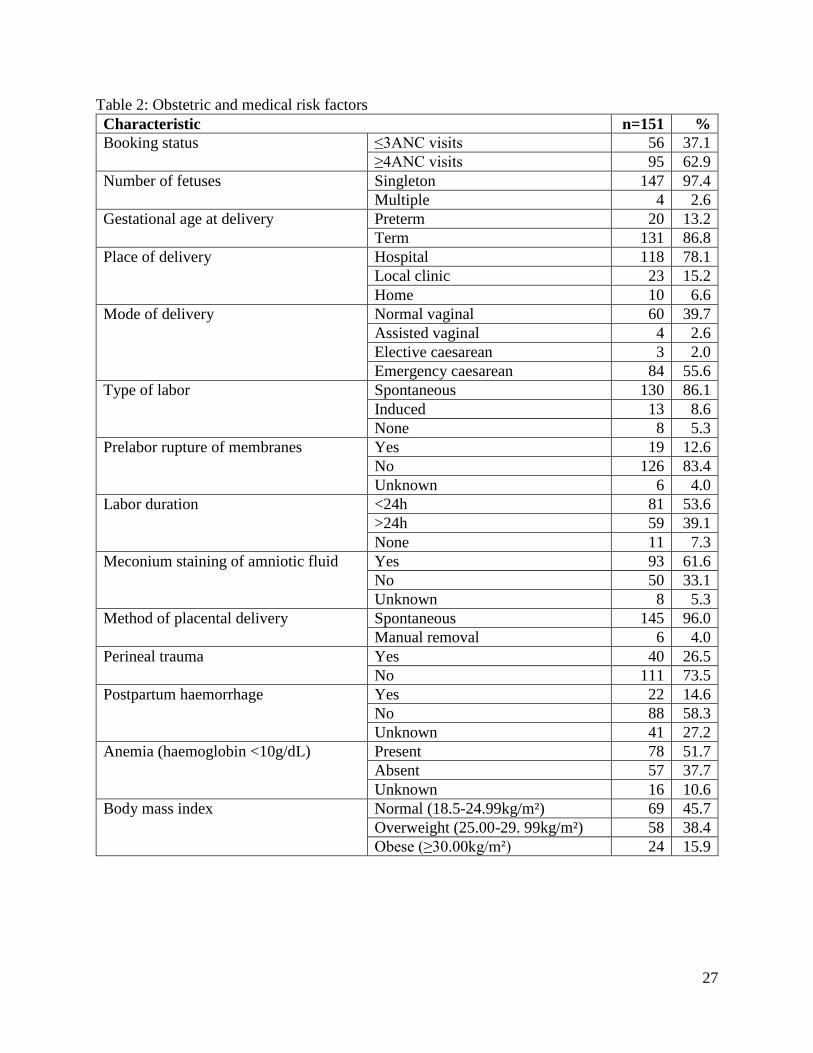
Obstetric and Medical Risk Factors (table 2)
Most women (62.9%) had attended ≥4 antenatal care visits in line with the WHO recommended
minimum of 4 focused antenatal care visits.
The majority of deliveries were of singleton term fetuses at a hospital. Of the 118 hospital
deliveries, 58 had delivered at Parirenyatwa Hospital, 33 at Harare Hospital, 5 at private hospitals
and 22 at district hospitals. Parirenyatwa Hospital and Harare Hospitals were the places of delivery
for 64% (58/90) and 54% (33/61) of cases of puerperal sepsis they managed respectively. The
commonest mode of delivery was Caesarean section (57.6%), mostly as emergency following
spontaneous labor of duration <24hours. Only 4 (2.6%) women had assisted vaginal deliveries, all
vacuum extractions. Oral misoprostol was the method of induction in all 13 labors that were
induced. Few pregnancies had been complicated by pre-labor rupture of membranes (12.6%).
Meconium staining of amniotic fluid was noted in 61.6% of pregnancies. Most placentas had been
delivered by controlled cord traction or spontaneously (96.0%). Perineal trauma at delivery had
occurred in 26.5% of women.
Postpartum hemorrhage was objectively documented to have occurred in 14.6% of women, while
27.2% of women did not report excessive blood loss, but had no documentation. Anemia
(hemoglobin <10.0g/dl) was present in just over half of women (51.7%) while hemoglobin status
was unknown in 10.6%. Most women were of normal weight (BMI 18.5-24.99kg/m² ) 45.7%, or
overweight (BMI 25-29.99kg/m²) 38.4% (41) . No woman admitted to symptoms or a history of
diabetes mellitus or gestational diabetes.
Page 35
27
Table 2: Obstetric and medical risk factors
Characteristic n=151 %
Booking status ≤3ANC visits 56 37.1
≥4ANC visits 95 62.9
Number of fetuses Singleton 147 97.4
Multiple 4 2.6
Gestational age at delivery Preterm 20 13.2
Term 131 86.8
Place of delivery Hospital 118 78.1
Local clinic 23 15.2
Home 10 6.6
Mode of delivery Normal vaginal 60 39.7
Assisted vaginal 4 2.6
Elective caesarean 3 2.0
Emergency caesarean 84 55.6
Type of labor Spontaneous 130 86.1
Induced 13 8.6
None 8 5.3
Prelabor rupture of membranes Yes 19 12.6
No 126 83.4
Unknown 6 4.0
Labor duration <24h 81 53.6
>24h 59 39.1
None 11 7.3
Meconium staining of amniotic fluid Yes 93 61.6
No 50 33.1
Unknown 8 5.3
Method of placental delivery Spontaneous 145 96.0
Manual removal 6 4.0
Perineal trauma Yes 40 26.5
No 111 73.5
Postpartum haemorrhage Yes 22 14.6
No 88 58.3
Unknown 41 27.2
Anemia (haemoglobin <10g/dL) Present 78 51.7
Absent 57 37.7
Unknown 16 10.6
Body mass index Normal (18.5-24.99kg/m²) 69 45.7
Overweight (25.00-29. 99kg/m²) 58 38.4
Obese (≥30.00kg/m²) 24 15.9
Page 36
28
MICROBIOLOGY
Endocervical swabs isolates (Figure 1 and Appendix 1, table a)
Endocervical swabs were collected for 151 women. Bacteria were isolated from 103 of 151
(68.2%) endocervical swabs, while 48 (31.8%) swabs were negative for growth. One isolate grew
on 83(55.0%) swabs, two isolates on 19(12.6%) swabs, while three isolates grew on 1(0.7%) swab.
The total number of endocervical swab isolates was 124. Gram negative rods were commonly
isolated, most commonly Escherichia coli 38(30.6% of total endocervical swab isolates),
Klebsiella pneumoniae 19(15.3%), 5(4.0%) isolates each of Pseudomonas aeruginosa and
Enterobacter species; Citrobacter freundii 4(3.2%); Alcaligenes species 3(2.4%), 2(1.6%) isolates
each of Providencia species, Morganella morganii, Shigella species and Yersinia species; 1(0.8%)
isolate each of Salmonella species, Klebsiella oxytoca and Kluyvvera species. Gram positive cocci
isolated were: Coagulase negative staphylococci (CoNS) 9(7.3%), Staphylococcus aureus
8(6.5%), Group D streptococcus 8(6.5%), Streptococcus pyogenes 4(3.2%), Streptococcus
viridans 2(1.6%) and Streptococcus agalactae 1(0.8%). Gram positive rods isolated were:
Corynebacterium species 5(4.0%) and Bacillus species 2(1.6%). No strict anaerobic organisms
were isolated.
Page 37
29
Figure 1
Blood culture isolates (Figure 2 and Appendix 1, table b)
Blood for culture was collected from 150 women. One woman declined further venipuncture after
an initial failed attempt. Bacteria were isolated from 14 of 150 bottles and each bottle grew one
organism. Overall, blood culture positive rate was 9.3%. This decreased to 3.3% after excluding
Bacillus sp, CoNS and fungus as possible contaminants. Isolates obtained were Bacillus sp
5(35.7% of blood culture isolates), CoNS 3(21.4%), E. coli 2(14.3%), S. aureus 1(7.1%),
Alcaligenes sp 1(7.1%), Moraxella sp 1(7.1%) and fungus 1(7.1%). Both E. coli grew on day 1.
Moraxella sp and S. aureus grew on day 5, while Alcaligenes sp was noted on day 10. Only one
0
5
10
15
20
25
30
35
40
Num
ber
of
iso
late
sEndocervical swab isolates
Page 38
30
woman had the same organism, E. coli, isolated from both the blood culture and endocervical
swab, with similar antibiotic susceptibility profile.
Figure 2
Antibiotic use prior to specimen collection
Specimen collection before administration of antibiotics was achieved in only 23/151(15.2%)
women as the other women had already received antibiotics at the primary care center or during
screening in casualty or as prophylaxis prior to and post caesarian section. Ceftriaxone and
metronidazole had been administered in 74.8% and 78.1% respectively. Other antibiotics given
were benzyl penicillin(7.3%), ampicillin(1.3%), cloxacillin(0.7%), amoxicillin(4.0%),
chloramphenicol(5.6%), gentamycin(2.6%), ciprofloxacin(0.7%), clindamycin(0.7%),
erythromycin(0.7%).
0
1
2
3
4
5
6
E. Coli S. aureus Alcaligenes sp Moraxella sp CoNS Bacillus sp Fungus
Nu
mb
er o
f is
ola
tes
Blood culture isolates
growth on culture day 1 growth on culture day 5 growth on culture day 10
Page 39
31
Antibiotic Susceptibility Profiles of Endocervical swab isolates (Figures 3-9 and Appendix 1,
table c)
High level resistance to the 7 empiric antibiotics tested was noted for most gram negative isolates.
Levels of antibiotic resistance for E. coli, the most prevalent isolate were: ceftriaxone (60.5%),
penicillin (94.7%), gentamycin (50%), clindamycin (92.1%), erythromycin (86.8%),
chloramphenicol (39.5%) and ciprofloxacin (34.2%). Antibiotic resistance levels for K.
pneumonia, the second most prevalent isolate, were: ceftriaxone (68.4%), penicillin (89.5%),
chloramphenicol (63.2%), gentamycin (63.2%), clindamycin (100%), ciprofloxacin (42.1%), and
erythromycin (94.7%). Enterobacter sp resistance was 100% to gentamycin, clindamycin and
erythromycin; 80% to ceftriaxone and penicillin; and 40% to chloramphenicol and ciprofloxacin.
Gram positive organisms were generally susceptible to the empiric antibiotics. S. pyogenes isolates
were fully susceptible to penicillin, chloramphenicol, clindamycin and erythromycin; while
resistance was 50% to ceftriaxone and 25% to gentamycin and ciprofloxacin. S. aureus isolates
exhibited 50% resistance to ceftriaxone, gentamycin, clindamycin and erythromycin; 37.5%
resistance to penicillin and 25% resistance to chloramphenicol and ciprofloxacin. CoNS were fully
susceptible to clindamycin; while resistance was 55.6% to penicillin, 44.4% to ceftriaxone,
chloramphenicol and erythromycin, and 33.3% to gentamycin and ciprofloxacin. S. agalactae was
susceptible to all the antibiotics except gentamycin. S. viridans isolates were fully susceptible to
ceftriaxone, clindamycin and erythromycin, while resistance to chloramphenicol was 50%.
Blank spaces in graphs mean the organism was not tested for susceptibility to the antibiotic.
Antimicrobial susceptibility of Bacillus sp was not done, see section 3.7.3.4.d.
Page 40
32
Figure 3
E.
coli
K.
pneu
mon
iae
CoN
S
S.
aure
us
Gro
up
D s
trep
toco
ccu
s
P.
aeru
gin
osa
En
tero
bac
ter
sp
Cory
neb
acte
riu
m s
p
S.
pyo
gen
es
C. fr
eun
dii
Alc
alig
enes
sp
M.
morg
anii
Pro
vid
enci
a sp
S.
vir
idan
s
Sh
igel
la s
p
Yer
sinia
sp
S.
agal
acta
e
Sal
mo
nel
la s
p
Klu
yver
a sp
K.
oxy
toca
0
5
10
15
20
25
30
35
40
Fre
qu
ency
Susceptibility to Ceftriaxone
Intermediate
Resistant
Sensitive
Page 41
33
Figure 4
Figure 5
0
5
10
15
20
25
30
35
40
E.
coli
K.
pneu
mon
iae
CoN
S
S.
aure
us
Gro
up
D s
trep
toco
ccu
s
P.
aeru
gin
osa
En
tero
bac
ter
sp
Cory
neb
acte
riu
m s
p
S.
pyo
gen
es
C. fr
eun
dii
Alc
alig
enes
sp
M.
morg
anii
Pro
vid
enci
a sp
S.
vir
idan
s
Sh
igel
la s
p
Yer
sinia
sp
S.
agal
acta
e
Sal
mo
nel
la s
p
Klu
yver
a sp
K.
oxy
toca
Fre
qu
ency
Susceptibility to penicillin
Resistant
Intermediate sensitivity
Sensitive
0
5
10
15
20
25
30
35
40
E.
coli
K.
pneu
mon
iae
CoN
S
S.
aure
us
Gro
up
D s
trep
toco
ccu
s
P.
aeru
gin
osa
En
tero
bac
ter
sp
Cory
neb
acte
riu
m s
p
S.
pyo
gen
es
C. fr
eun
dii
Alc
alig
enes
sp
M.
morg
anii
Pro
vid
enci
a sp
S.
vir
idan
s
Sh
igel
la s
p
Yer
sinia
sp
S.
agal
acta
e
Sal
mo
nel
la s
p
Klu
yver
a sp
K.
oxy
toca
Fre
qu
ency
Susceptibility to Chloramphenicol
Resistant
Intermediate sensitivity
Sensitive
Page 42
34
Figure 6
Figure 7
0
5
10
15
20
25
30
35
40
E.
coli
K.
pneu
mon
iae
CoN
S
S.
aure
us
Gro
up
D s
trep
toco
ccu
s
P.
aeru
gin
osa
En
tero
bac
ter
sp
Cory
neb
acte
riu
m s
p
S.
pyo
gen
es
C. fr
eun
dii
Alc
alig
enes
sp
M.
morg
anii
Pro
vid
enci
a sp
S.
vir
idan
s
Sh
igel
la s
p
Yer
sinia
sp
S.
agal
acta
e
Sal
mo
nel
la s
p
Klu
yver
a sp
K.
oxy
toca
Fre
qu
ency
Susceptibility to Gentamycin
Resistant
Intermediate sensitivity
Sensitive
0
5
10
15
20
25
30
35
40
E.
coli
K.
pneu
mon
iae
CoN
S
S.
aure
us
Gro
up
D s
trep
toco
ccu
s
P.
aeru
gin
osa
En
tero
bac
ter
sp
Cory
neb
acte
riu
m s
p
S.
pyo
gen
es
C. fr
eun
dii
Alc
alig
enes
sp
M.
morg
anii
Pro
vid
enci
a sp
S.
vir
idan
s
Sh
igel
la s
p
Yer
sinia
sp
S.
agal
acta
e
Sal
mo
nel
la s
p
Klu
yver
a sp
K.
oxy
toca
Fre
qu
ency
Susceptibility to Clindamycin
Resistant
Intermediate sensitivity
Sensitive
Page 43
35
Figure 8
Figure 9
0
5
10
15
20
25
30
35
40
E.
coli
K.
pneu
mon
iae
CoN
S
S.
aure
us
Gro
up
D s
trep
toco
ccu
s
P.
aeru
gin
osa
En
tero
bac
ter
sp
Cory
neb
acte
riu
m s
p
S.
pyo
gen
es
C. fr
eun
dii
Alc
alig
enes
sp
M.
morg
anii
Pro
vid
enci
a sp
S.
vir
idan
s
Sh
igel
la s
p
Yer
sinia
sp
S.
agal
acta
e
Sal
mo
nel
la s
p
Klu
yver
a sp
K.
oxy
toca
Fre
qu
ency
Susceptibility to Ciprofloxacin
Resistant
Intermediate sensitivity
Sensitive
0
5
10
15
20
25
30
35
40
E.
coli
K.
pneu
mon
iae
CoN
S
S.
aure
us
Gro
up
D s
trep
toco
ccu
s
P.
aeru
gin
osa
En
tero
bac
ter
sp
Cory
neb
acte
riu
m s
p
S.
pyo
gen
es
C. fr
eun
dii
Alc
alig
enes
sp
M.
morg
anii
Pro
vid
enci
a sp
S.
vir
idan
s
Sh
igel
la s
p
Yer
sinia
sp
S.
agal
acta
e
Sal
mo
nel
la s
p
Klu
yver
a sp
K.
oxy
toca
Fre
qu
ency
Susceptibility to Erythromycin
Resistant
Intermediate sensitivity
Sensitive
Page 44
36
Blood culture antibiotic susceptibility profiles (table 3)
E. coli isolates were fully susceptible to ceftriaxone and ciprofloxacin, while resistance was 50%
to clindamycin and erythromycin; and 100% to gentamycin, chloramphenicol and penicillin G.
S. aureus was resistant to all 7 antibiotics. Alcaligenes sp was susceptible to ceftriaxone,
chloramphenicol, gentamycin and ciprofloxacin.
Table 3: Antibiotic susceptibility of blood culture isolates
CRO P C Gn Clin CIP E
Isolate Susce
ptibili
ty
No % No % No % No % No % No % No %
E. Coli S 2 100 0 0 0 0 0 0 1 50 2 100 1 50
R 0 0 2 100 2 100 2 100 1 50 0 0 1 50
S. aureus S 0 0 0 0 0 0 0
R 1 1 1 1 1 1 1
Alcaligenes
sp
S 1 0 1 1 0 1 0
R 0 1 0 0 1 0 1
Key:
CRO- ceftriaxone, P- penicillin, C- chloramphenicol, Gn- gentamycin, Clin- clindamycin,
CIP- ciprofloxacin, E- erythromycin, No- number
S- Susceptible
I- Intermediate susceptibility
R- Resistant
Susceptibility not tested for Bacillus sp, CoNS, Moraxella sp, and fungus. See section 3.7.3.4.d.
Multi-drug resistant organisms (Figure 10 and appendix 1, table d)
Organisms with acquired non-susceptibility to at least one agent in three or more antimicrobial
categories above were designated as multidrug resistant organisms (MDRO) (32). MDROs were
tested for susceptibility to carbapenems (imipenem and meropenem). There were fifteen MDROs.
All 14 MDRO isolated from endocervical swabs were gram negative bacilli, most commonly K.
pneumoniae. The one MDRO isolated from the blood stream was S. aureus. MDRO were common,
Page 45
37
representing 32% of K. pneumoniae, 5% of E. coli, 50% of M. morganii, 20% of Enterobacter sp,
20% of P.aeruginosa, 33% of Alcaligenes sp, 25% of C. freundii and 11% of S. aureus.
Figure 10
Of the six MDRO K. pneumoniae, 3 isolates were susceptible to both meropenem and imipenem,
2 isolates were resistant to meropenem but susceptible to imipenem, while 1 isolate was resistant
to both carbapenems. Both E. coli were susceptible to meropenem. Imipenem susceptibility was
confirmed for 1 isolate, but not tested for the other, see section 3.7.3.4.d . S. aureus was resistant
to meropenem but susceptible to imipenem. M. Morganii, Alcaligenes sp, P. aeruginosa, C.
freundii, were susceptible to both carbapenems. Imipenem susceptibility was not tested for K.
xytoca and Enterobacter sp, see section 3.7.3.4.d.
05
10152025303540
Fre
qu
ency
Multidrug Resistant Organisms
non-MDRO
MDRO
Page 46
38
HIV Status
Valid HIV status results were obtained for all 151 women. Thirty-three women (21.9%) were HIV
positive (Figure 11). Valid CD4 counts were available for 29 of 33 women. The majority of
women, 15 (45.5%), had mild (CD4 350-499/mm³) to advanced immunosuppression (CD4 200-
349/mm³). There were 7(21.2%) women in each of the insignificant (CD4 > 500/mm³) and severe
immunosuppression (CD4 <200/mm³) categories.
Twenty-three (69.7%) women were on treatment with antiretroviral therapy (ART). HIV was
diagnosed for the first time at admission for puerperal sepsis in the 10 (30.3%) women who were
not on ART. Eleven women had been on ART for >6months, while 12 had been on ART for <6
months
Figure 11: HIV status and CD4 Count
Page 47
39
Association of HIV status with microbiology of puerperal sepsis (table 4 and table5)
Enterobacter sp were more prevalent in the genital tract of HIV-infected than HIV-uninfected
women 9.1% vs 1.7% (p=0.04). There was no statistically significant difference between the two
groups with regard to the other organisms isolated from the blood stream and the endocervix. There
was no difference between number of isolates per swab with 1 isolate growing in 86.4% vs 87.9%
(p= 0.55); 2 isolates in 12.7% vs 12.1% (0.93) ; 3 isolates in 0.8% vs 0% (p=0.596) of HIV-
uninfected and HIV-infected women respectively. The proportion of MDRO was no different
between the 2 groups, 9.1% compared to 11.9% (p=0.78) of HIV-infected women and of HIV-
uninfected respectively.
Table 4: Association of blood culture isolates with HIV status.
HIV status
p-value
Blood culture isolate
Positive(n=32) Negative(n=118)
Number % Number %
E. coli 0 0 2 1.7 0.46
Bacillus sp 1 3.0 4 3.4 0.94
S. aureus 0 0 1 0.8 0.60
Moraxella sp 0 0 1 0.8 0.60
Alcaligenes sp 0 0 1 0.8 0.60
CoNS 2 6.3 1 0.8 0.05
Fungal contamination 0 0 1 0.8 0.60
No growth 29 90.6 107 90.7 1.00
Page 48
40
Table 5: Association of endocervical swab isolates with HIV status.
Endocervical swab isolate
HIV status
p-value
Positive(n=33) Negative(n=118)
Number % Number %
E. coli 11 33.3 27 22.3 0.22
K. pneumonia 6 18.2 13 11.0 0.27
K. oxytoca 0 0 1 0.8 0.60
M. morganii 0 0 2 1.7 0.45
Providencia sp 1 3.0 1 0.8 0.33
P. aeruginosa 0 0 5 4.2 0.23
Alcaligenes sp 0 0 3 2.5 0.35
Bacillus sp 0 0 2 1.7 0.45
C. freundii 1 3 3 2.5 0.88
S. aureus 1 3 7 5.9 0.51
CoNS 1 3 8 6.8 0.42
S. pyogenes 1 3 3 2.5 0.88
Corynebacterium sp 0 0 5 4.2 0.76
Enterobacter sp 3 9.1 2 1.7 0.04
Salmonella sp 0 0 1 0.8 0.60
Kluyvera sp 1 3 0 0 0.06
Yersinia sp 0 0 2 1.7 0.45
Group D streptococcus 2 6.1 6 5.1 0.83
S. viridans 0 0 2 1.7 0.45
S. agalactae 0 0 1 0.8 0.60
Shigella sp 0 0 2 1.7 0.45
No growth 9 27.3 39 33.1 0.53
Presentation of puerperal sepsis and clinical outcomes
Median time from delivery to onset of puerperal sepsis was 6 days, IQR 3-11days (range 0-
42days). The commonest presenting symptoms were abnormal vaginal discharge and pelvic pain,
see Figure 12.
Page 49
41
Figure 12
Eighty-one women (53.6%) experienced at least one complication. Two or more complications
occurred in 13.9% of women. Surgical wound dehiscence was most prevalent occurring in 39.1%
of women, see Figure 13. Eleven women died, a case fatality rate of 7.3%.
77.5%
87.4% 88.1%83.4%
60.3%
0.0%
10.0%
20.0%
30.0%
40.0%
50.0%
60.0%
70.0%
80.0%
90.0%
100.0%
Fever pelvic pain Abnormal
vaginal discharge
Abnormal smell
of vaginal
discharge
Delayed in
reduction of
uterine size
Fre
qu
ency
Presentation of Puerperal Sepsis
Page 50
42
Figure 13
Laparotomy was performed in 25 (16.6%) women. A pelvic abscess was confirmed by the
presence of pus in the pelvis in 23 women. There was no obvious pus in the pelvis in two women;
one had a grossly necrotic uterus and anterior abdominal wall tissue planes, while the other had
peritonitis. Laparotomy was performed twice in four women (2 for re-accumulation of pus, 2 for
colostomy complications), while the fifth woman died whilst awaiting re-laparotomy for burst
abdomen. Hysterectomy for gross pelvic sepsis was performed in 1 woman.
Admission into the intensive care unit (ICU) was required for eighteen women (11.9%). The
median length of ICU stay was 3 days, IQR 2.75-5.25 days (range 1 to 12days)). Median length of
hospital stay was 8days, IQR 4-16 days (range1-51days).
39.1%
15.2%
7.3% 6.0% 5.3%2.6%
0.0%
5.0%
10.0%
15.0%
20.0%
25.0%
30.0%
35.0%
40.0%
45.0%
wound
dehiscence
pelvic abscess death renal failure septic shock peritonitis
Fre
qu
ency
%
Complication
Complications of Puerperal sepsis
Page 51
43
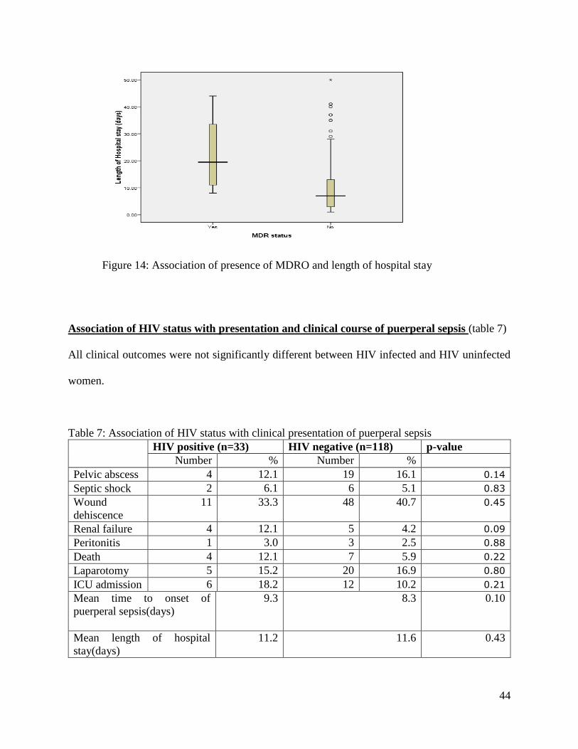
Association of antibiotic susceptibility with clinical outcomes (table 6 and Figure 14)
MDRO were found in 12 women, (one woman had 3 MDRO, and another had 2 MDRO, while ten
women had one each). Mean length of hospital stay was significantly longer in women with
MDRO compared to those without MDRO, 23.0 days vs 10.5 days respectively (p=0.009). There
was a trend towards higher complication rates (pelvic abscess, septic shock, wound dehiscence,
death), need for laparotomy and ICU admission in women with MDRO than those without MDRO,
but this did not reach statistical significance.
Table 6: Association of presence of MDRO with clinical outcomes of puerperal sepsis
MDRO (n=12) Non-MDRO
(n=139)
p-value
Number % number %
Pelvic abscess 4 33.3 19 13.7 0.07
Septic shock 1 8.3 7 5.0 0.42
Wound dehiscence 5 41.7 54 38.8 0.85
Renal failure 0 0 9 6.5 0.36
Peritonitis 0 0 4 2.9 0.55
Death 2 16.7 9 6.5 0.19
Laparotomy 4 33.3 21 15.1 0.10
ICU admission 3 25.0 15 10.8 0.14
Length of hospital stay (mean) 23.0 10.5 0.009
Page 52
44
Figure 14: Association of presence of MDRO and length of hospital stay
Association of HIV status with presentation and clinical course of puerperal sepsis (table 7)
All clinical outcomes were not significantly different between HIV infected and HIV uninfected
women.
Table 7: Association of HIV status with clinical presentation of puerperal sepsis
HIV positive (n=33) HIV negative (n=118) p-value
Number % Number %
Pelvic abscess 4 12.1 19 16.1 0.14
Septic shock 2 6.1 6 5.1 0.83
Wound
dehiscence
11 33.3 48 40.7 0.45
Renal failure 4 12.1 5 4.2 0.09
Peritonitis 1 3.0 3 2.5 0.88
Death 4 12.1 7 5.9 0.22
Laparotomy 5 15.2 20 16.9 0.80
ICU admission 6 18.2 12 10.2 0.21
Mean time to onset of
puerperal sepsis(days)
9.3 8.3 0.10
Mean length of hospital
stay(days)
11.2 11.6 0.43
Page 53
45
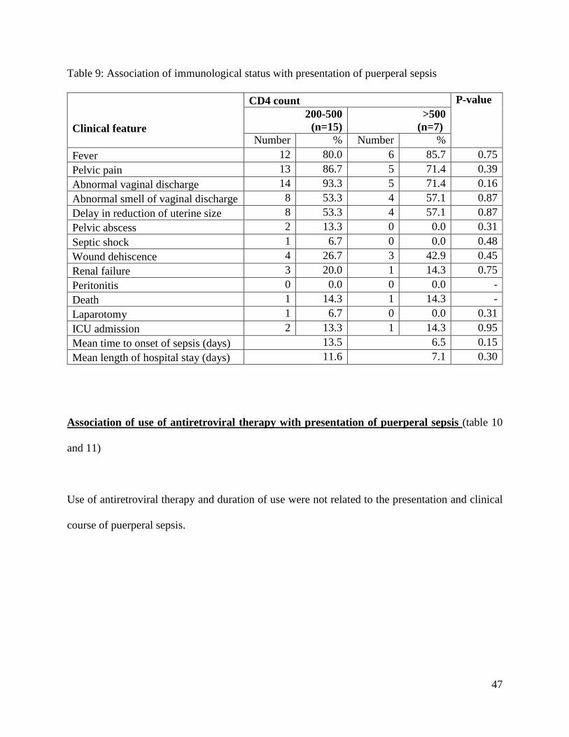
Association of immunological status (CD4 cell count) with clinical presentation of puerperal
sepsis (tables 8 and 9)
The frequency of fever, pelvic pain, abnormal vaginal discharge, foul-smelling vaginal discharge,
and delay in rate of reduction of size of the uterus was no different between the immunological
groups. Severe immunosuppression (CD4 <200/mm³, n=7) resulted in greater need for laparotomy,
42.9% vs 4.5% (p=0.01), and prolonged mean hospital stay, 19.0days vs 10.2days (p=0.03),
compared to mild-advanced or insignificant immunosuppression (CD4 >200/mm³, n=22). There
was a trend towards earlier onset of sepsis; and higher rates of pelvic abscess, septic shock, wound
dehiscence, peritonitis, death and need for ICU admission in women with severe
immunosuppression though this did not reach statistical significance. There were no significant
differences in clinical presentation and clinical course between women with mild-advanced and
those with insignificant immunosuppression.
Page 54
46
Table 8: Association of immunological status with presentation of puerperal sepsis
Clinical Feature
CD4 count
P-value <200
(n=7)
≥200
(n=22)
Number % Number %
Fever 7 100.0 18 81.8 0.22
Pelvic pain 7 100.0 18 81.8 0.22
Abnormal vaginal discharge 5 71.4 19 86.4 0.36
Abnormal smell of vaginal discharge 5 71.4 18 81.8 0.36
Delay in reduction of uterine size 3 42.9 12 54.5 0.59
Pelvic abscess 2 28.6 2 9.1 0.19
Septic shock 1 14.3 1 4.5 0.37
Wound dehiscence 4 57.1 7 31.8 0.23
Renal failure 0 0.0 4 18.2 0.22
Peritonitis 1 14.3 0 0.0 0.07
Death 2 28.6 2 9.1 0.19
Laparotomy 3 42.9 1 4.5 0.01
ICU admission 3 42.9 3 13.6 0.10
Mean time to onset of sepsis(days) 2.5 11.8 0.07
Mean length of hospital stay(days) 19.0 10.2 0.03
Page 55
47
Table 9: Association of immunological status with presentation of puerperal sepsis
Clinical feature
CD4 count P-value
200-500
(n=15)
>500
(n=7)
Number % Number %
Fever 12 80.0 6 85.7 0.75
Pelvic pain 13 86.7 5 71.4 0.39
Abnormal vaginal discharge 14 93.3 5 71.4 0.16
Abnormal smell of vaginal discharge 8 53.3 4 57.1 0.87
Delay in reduction of uterine size 8 53.3 4 57.1 0.87
Pelvic abscess 2 13.3 0 0.0 0.31
Septic shock 1 6.7 0 0.0 0.48
Wound dehiscence 4 26.7 3 42.9 0.45
Renal failure 3 20.0 1 14.3 0.75
Peritonitis 0 0.0 0 0.0 -
Death 1 14.3 1 14.3 -
Laparotomy 1 6.7 0 0.0 0.31
ICU admission 2 13.3 1 14.3 0.95
Mean time to onset of sepsis (days) 13.5 6.5 0.15
Mean length of hospital stay (days) 11.6 7.1 0.30
Association of use of antiretroviral therapy with presentation of puerperal sepsis (table 10
and 11)
Use of antiretroviral therapy and duration of use were not related to the presentation and clinical
course of puerperal sepsis.
Page 56
48
Table 10: Association of use of antiretroviral therapy with presentation of puerperal sepsis
Clinical feature
Use of ART
P-
value
Yes (n=23)
No (n=10)
Number % Number %
Fever 19 82.6 9 90.0 0.59
Pelvic pain 19 82.6 9 90.0 0.59
Abnormal vaginal discharge 19 82.6 9 90.0 0.59
Abnormal smell of vaginal discharge 18 78.3 9 90.0 0.42
Delay in rate of reduction of uterine size 11 47.8 6 60.0 0.52
Pelvic abscess 3 13.0 1 10.0 0.81
Septic shock 1 4.3 1 10.0 0.53
Wound dehiscence 8 34.8 3 30.0 0.79
Renal failure 3 13.0 1 10.0 0.81
Peritonitis 1 4.3 0 0.0 0.50
Death 3 13.0 1 10.0 0.81
Laparotomy 4 17.4 1 10.0 0.59
ICU admission 5 21.7 1 10.0 0.64
Mean Time to onset of sepsis (days) 10.2 7.8 0.72
Mean length of hospital stay (days) 9.8 14.5 0.32
Page 57
49
Table 11: Association of duration of use of antiretroviral therapy with presentation of puerperal
sepsis
Clinical feature
Duration of use of ART
P-value
<6 months
(n=12)
> 6 months
(n=11)
Number % Number %
Fever 9 75.0 10 90.9 0.32
Pelvic pain 11 91.7 8 72.7 0.23
Abnormal vaginal discharge 11 91.7 8 72.7 0.23
Abnormal smell of vaginal discharge 9 75.0 9 81.8 0.69
Delay in rate of reduction of uterine size 7 16.7 1 9.1 0.59
Pelvic abscess 2 16.7 1 9.1 0.59
Septic shock 0 0.0 1 9.1 0.48
Wound dehiscence 4 33.3 4 36.4 0.88
Renal failure 1 8.3 2 18.2 0.59
Peritonitis 1 8.3 0 0.0 0.33
Death 1 8.3 2 18.2 0.59
Laparotomy 3 25.0 1 9.1 0.59
ICU admission 3 25.0 2 18.2 0.69
Mean time to onset of sepsis (days) 12.1 7.9 0.33
Mean length of hospital stay (days) 11.7 7.7 0.40
Page 58
50
Risk factors for bacteremia
Manual removal of placenta was associated with a non-significant trend towards a higher risk for
bacteremia [RR 4.893, p=0.02 (CI= 0.98-24.38)] (Figure 15). Other demographic, obstetric and
medical factors were not associated with development of bacteremia.
Figure 15
8.3%
33.3%
0.0%
5.0%
10.0%
15.0%
20.0%
25.0%
30.0%
35.0%
Spontaneous (n=145 manual removal (n=6)
Fre
qu
ency
Method of removal of placenta
Association of method of removal of
placenta and bacteremia
Page 59
51
CHAPTER 5
5.1 DISCUSSION
The majority of women managed for puerperal sepsis at Parirenyatwa and Harare Hospitals had
delivered at a hospital (78.1%), by caesarean section (57.6%), raising concerns about nosocomial
infections. Most cases of postpartum endometritis follow operative delivery. In a Cochrane review,
the rate of puerperal infections was 2.5% in women who had a vaginal delivery while the average
rate of endometritis was 9.2% in those women undergoing elective caesarean section and 28% in
women undergoing non-elective caesarean section(51).
Bacteria were isolated in 68.2% of endocervical swabs, a rate consistent with other studies in which
laboratory-confirmed infection was reported for 63.8% of cases of severe maternal sepsis in a
United Kingdom case control study (52), and in up to 80% of cases in other studies from resource
limited settings (7,14,16,17). Bacteria were isolated in 9.3% of blood culture specimens, but
significant isolates were obtained in 3.3% of specimens. Bacillus sp and Coagulase negative
staphylococci are part of the indigenous flora of the skin, and their isolation in a blood culture may
simply represent contamination. However, both may represent significant growth in special
circumstances such as immunosuppression, intravenous drug abuse, indwelling foreign devices
(e.g. - intravascular catheters, pacemakers, central nervous system shunts) and sickle cell disease
which should be confirmed by sustained growth on repeat blood cultures. Although 12-25% of
CoNS and 5-10% of Bacillus sp isolated from blood cultures are due to significant blood stream
infections, CoNS and Bacillus sp were considered contaminants because repeat cultures to confirm
infection could not be done(48–50,53).
Page 60
52
Blood culture positive rate was not significantly different from findings of a review by Williams
and Pastorek in which rate of bacteremia was 5-20% in studies investigating postpartum
endomyometritis(8). Failure to identify a causative organism is expected. Manual removal of the
placenta was the single risk factor that was associated with development of bacteremia, though it
did not reach statistical significance. This may be associated with introduction of nosocomial
pathogens to the raw placental surface in the uterus via infected hands.
Gram negative enterobacteriacae were commonly isolated from the endocervix, E. coli and K.
pneumoniae representing 45.9% of the total isolates. E. coli were also the commonest significant
cause of bacteremia, being isolated in 2 specimens in this study. E. coli has been reported as the
commonest cause of severe maternal sepsis originating from the genital tract (15,52). Furthermore,
genes associated with high virulence such as hly-, cnf-, pap- and iroN-genes have been found
significantly more frequently in E. coli strains isolated from vaginal and/or endocervical samples
of pregnant women. These predispose to severe maternal sepsis(54). Group A streptococci, an
important cause of severe maternal sepsis leading to septic shock and death in the developed
world(52), were identified in only 3.2% of genital specimens in this study implying that it may not
be a significant pathogen in our setting or it is highly susceptible to empiric antimicrobials.
High level resistance by most gram negative isolates form the endocervix to the first line regimens
for the treatment of puerperal sepsis was demonstrated in this study. There is emerging resistance
by both gram negative and gram positive bacteria to multiple antimicrobial agents including to
meropenem and imipenem, the most readily available carbapenems used for salvage therapy for
resistant infections in both public and private hospitals in Zimbabwe. These multidrug resistant
Page 61
53
organisms (MDRO), defined as organisms with acquired non-susceptibility to at least one agent in
three or more antimicrobial classes (32), are a significant threat to public health. Furthermore,
extremely drug-resistant enterobacteriacae are increasingly isolated worldwide (55,56) placing
additional strain on limited resources.
Puerperal sepsis is associated with high rates of maternal morbidity and mortality. Laparotomy
was needed in 16.6% of women with puerperal sepsis, of which 20% of them had a repeat
laparotomy. Case fatality rate in this prospective cohort of hospitalized women was 7.3%. A
review of the global burden of maternal sepsis in the year 2000 reported case fatality rates of 4-
50% in Sub-Saharan African countries, the wide differences being probably due to different
definitions of puerperal sepsis and whether the study was hospital based or population based (1).
MDRO significantly increased length of hospital stay, and there was a clinically important trend
towards higher rates of pelvic abscess, septic shock, death, need for laparotomy and ICU
admission. Several investigators have noted increased length and cost of hospital stay and
mortality associated with infections due to multidrug-resistant S. aureus and gram-negative bacilli
such as E. coli, K. pneumoniae, Enterobacter sp, P. aeruginosa, and Acinetobacter sp (57–62).
Page 62
54
The prevalence of HIV infection was higher in this cohort of women with puerperal sepsis (21.9%)
than in the general Zimbabwean population (15%) (63). HIV infection predisposes to the
development of puerperal sepsis. In a systematic review, Calvert found that HIV-infection was
associated with a three-fold increased risk of a puerperal sepsis in women after a vaginal or
caesarian delivery; a figure that increased to nearly six amongst studies only including women
who delivered by caesarian section(9).
A significant number of women are still going through pregnancy (30.3%) without HIV counseling
and testing, placing themselves at risk of HIV/AIDS related illness and their babies at risk of
maternal to child transmission of HIV. A trend towards universal access to ART for HIV infected
pregnant women was confirmed in this cohort where all women who were known to be HIV
infected had commenced antiretroviral therapy.
Enterobacter sp were more prevalent in HIV-infected women (p=0.04). In a retrospective review
by Manifredi, Enterobacter sp were more frequently isolated in severely immunosuppressed HIV
infected patients, demonstrating a possible increased pathogenicity of Enterobacter sp in HIV
infection (64). However, HIV infection did not influence colonization with the rest of the
organisms, nor did it increase the rate of polymicrobial colonization of the genital tract or blood
stream as well as the proportion of MDRO in this study. The microbiology of organisms causing
upper genital tract infections has been found to be similar among HIV infected and uninfected
women by other investigators (25,65). In one case-control study, HIV infected women exhibited
more severe clinical manifestations of pelvic inflammatory disease with no effect on microbial
cause or response to therapy (25).
Page 63
55
Overall, being infected with HIV did not appear to significantly alter clinical course of puerperal
sepsis. Since only a minority of HIV-positive women with puerperal sepsis in this study had severe
immunosuppression of CD4 count <200/mm³ (21.2%), the impact of HIV on clinical disease was
not apparent. After sub-group analysis, HIV- associated severe immunosuppression was evidently
a risk factor for prolonged hospital stay and need for laparotomy. A clinically significant trend
towards earlier onset of puerperal sepsis and higher complication rates was observed with severe
immunosuppression. This is consistent with findings by Cohen who showed that a CD4 <14%
was associated with an increased likelihood of a tubo-ovarian abscess and a longer hospital stay in
women with acute salpingitis (65). Use of antiretroviral therapy did not independently affect
clinical manifestations of puerperal sepsis in this cohort.
Page 64
56
5.2 Study Limitations
Bacterial growth rate from blood cultures and endocervical swabs were lower because
84.5% of the women had received broad spectrum antibiotics prior to specimen collection.
However, this demonstrates efficient implementation of the policy of early administration of
antibiotics upon recognition of severe sepsis. Media with resins, lytic agents, or other neutralizing
substances would have increased blood culture growth rates in women with prior exposure to
antibiotics. Due to funding constraints, one 5ml specimen was collected instead of the
recommended minimum of two sets (aerobic + anaerobic) of blood cultures, each of ≥10ml, taken
from separate venipuncture sites (42). I was unable to repeat blood cultures in women when
Bacillus sp and Coagulase negative staphylococci were isolated to confirm if they were significant
infections or contaminants. No strict anaerobes were isolated from both endocervical swabs and
blood cultures because Amies transport media for endocervical swab is aerobic and may have
inhibited anaerobic growth and no anaerobic blood culture bottle was collected in this study.
Neisseria gonorrhea and Chlamydia trachomatis were not detected. This is attributed to the
fastidious nature of the organisms which would have been better identified using nucleic acid
amplification tests. Microbial susceptibility to ampicillin was not tested. Penicillin G, which has a
narrower antimicrobial spectrum compared to ampicillin, was the only penicillin available and
thus tested. Therefore antimicrobial resistance to penicillins may have been falsely elevated.
Despite the funding constraints, this study provides important information on the microbiology of
puerperal sepsis and the need for clinical culture surveillance.
The sample size may have been too small to detect significant differences during comparisons of
microbiology with clinical outcomes among HIV infected and uninfected women.
Page 65
57
5.3 Conclusion and Recommendations
The majority of women with puerperal sepsis have delivered by caesarian section. E. coli is the
commonest isolate from the genital tract and blood stream, reflecting ascend of infection from the
genitourinary tract and gastrointestinal system. There is emergence of MDRO gram negative
bacilli resistant to carbapenems, especially K. pneumoniae. HIV infection is a risk factor for
puerperal sepsis. MDRO and HIV-associated severe immunosuppression are independent risk
factors for a higher disease severity score, need for surgery and prolonged hospital stay.
Enterobacter sp. may be more pathogenic in HIV infection. Manual removal of the placenta may
increase risk of bacteremia.
In the United Kingdom, the mortality rate from genital tract sepsis reduced by more than half
between the trienniums 2006–08 and 2010–12 (1.13 per 100 000 maternities to 0.50 per 100 000)
maternities) (13,29). A downward trend was maintained through to 2013 (0.29 per 100 000
maternities) (6). Initiatives such as the ‘Surviving sepsis campaign and development of guidelines
for recognition and management of severe sepsis have been largely responsible for this decrease
(13). Prevention of community acquired and nosocomial multidrug resistant infections can be
achieved through implementation of robust infection control strategies, emphasis on rational drug
use, clinical culture surveillance to identify MDRO and monitoring of epidemiologic trends (66).
More funding should be availed regularly for continuous education on infection control and to
allow clinical culture surveillance and further research.
Page 66
58
REFERENCES
1. Dolea C, Stein C. Global burden of maternal sepsis in the year 2000. 2003 [cited 2016
Jan 20]; Available from: http://www.who.int/entity/healthinfo/statistics/bod_maternalsepsis.pdf
2. World Health Organization, International Confederation of Midwives, editors. Education
material for teachers of midwifery: midwifery education modules. 2nd ed. Geneva [Switzerland]:
World Health Organization : International Confederation of Midwives; 2008. 6 p.
3. 3. Khalid S Khan, Daniel Woojdyla, Lale Say, A Metin Gulmezoglu, Paul F A Van Look.
WHO Analysis of the Causes of Maternal Death: A Systematic Review. The Lancet. 2006 Apr
1;367(9516):1066-74. - Google Search [Internet]. [cited 2016 Jan 20].
4. Munjanja Steve P. Zimbabwe Maternal and Perinatal Mortality Study. Minist Health Child
Welf Zimb. 2007;
5. Manjeya Farikai. Trends in Maternal Mortality for the Public Maternity Institutions of
Harare, Zimbabwe, 2010 to 2014. University of Zimbabwe, Masters of Medicine in Obstetrics
and Gynaecology; 2015.
6. Knight M, Kenyon S, Brocklehurst P, Neilson J, Shakespeare J, Kurinczuk JJ. on behalf
of MBRRACE-UK. Saving Lives, Improving Mothers’ Care-Lessons learned to inform future
maternity care from the UK and Ireland Confidential Enquiries into Maternal Deaths and
Morbidity 2009–12. Oxford: National Perinatal Epidemiology Unit, University of Oxford; 2014.
7. Cunningham FG, Leveno KJ, Bloom SL, Hauth JC, Gilstrap L, Wenstrom KD. Williams
Obstetrics. 22nd ed. Vol. chapter 31. 2005.
8. Williams KL, Pastorek II JG. Postpartum endomyometritis. Infect Dis Obstet Gynecol.
1995;3(5):210–216.
9. Calvert C, Ronsmans C. HIV and the Risk of Direct Obstetric Complications: A
Systematic Review and Meta-Analysis. PLoS ONE. 2013 Oct 4;8(10):e74848.
10. Zvandasara P, Saungweme G, Mlambo JT, Moyo J. Post Caesarean section infective
morbidity in HIV-positive women at a tertiary training hospital in Zimbabwe. Cent Afr J Med.
2007;53(9–12):43–6.
11. Zvandasara P, Saungweme G, Mlambo JT, Moyo J. Post Caesarean section infective
morbidity in HIV-positive women at a tertiary training hospital in Zimbabwe. Cent Afr J Med.
2007 Dec;53(9–12):43–7.
12. Sepsis following Pregnancy, Bacterial (Green-top Guideline No. 64b) [Internet]. Royal
College of Obstetricians & Gynaecologists. [cited 2016 Jan 20]. Available from:
https://www.rcog.org.uk/en/guidelines-research-services/guidelines/gtg64b/
Page 67
59
13. Knight M, Kenyon S, Brocklehurst P, Neilson J, Shakespeare J, Kurinczuk JJE, et al.
Saving Lives, Improving Mothers’ Care Lessons learned to inform future maternity care from the
UK and Ireland Confidential Enquiries into Maternal Deaths and Morbidity 2009-2012. 2014
[cited 2016 Jan 20]; Available from: http://discovery.ucl.ac.uk/1464330/
14. Bhanap PL SA. A clinical Study of puerperal sepsis. Glob Res Anal. 2013 Sep;2(9).
15. Bako B, Audu BM, Lawan ZM, Umar JB. Risk factors and microbial isolates of
puerperal sepsis at the University of Maiduguri Teaching Hospital, Maiduguri, North-eastern
Nigeria. Arch Gynecol Obstet. 2012 Apr;285(4):913–7.
16. Ahmed S, Hossain MA, Musa AK, Shamsuzzaman AK, Mahmud MC, Nahar K, et al.
Preliminary report on anaerobic culture at Mymensingh Medical College Hospital in Bangladesh.
Mymensingh Med J MMJ. 2010 Jan;19(1):10–5.
17. Gerstner G, Leodolter S, Rotter M. [Endometrial bacteriology in puerperal infections
(author’s transl)]. Z Für Geburtshilfe Perinatol. 1981 Oct;185(5):276–9.
18. Bako B, Ibrahim UN, Umar JB, Zamo AB. Microbial Isolates in Puerperal Sepsis and
Their in vitro Antibiotic Sensitivity in North Eastern Nigeria. J Women’s Health Care [Internet].
2012;1(107). Available from: http://www.omicsgroup.org/journals/microbial-isolates-in-
puerperal-sepsis-and-their-in-vitro-antibiotic-sensitivity-in-north-eastern-nigeria-2167-
0420.1000107.php?aid=6325
19. World Health Organization, editor. The evolving threat of antimicrobial resistance:
options for action. Geneva, Switzerland: World Health Organization; 2012. 119 p.
20. EDLIZ 2011 - 6th Essential Medicines List and Standard Treatment Guidelines for
Zimbabwe [Internet]. [cited 2016 Jan 20]. Available from:
http://apps.who.int/medicinedocs/en/d/Js21753en/
21. Mason PR, Katzenstein DA, Chimbira TH, Mtimavalye L. Vaginal flora of women
admitted to hospital with signs of sepsis following normal delivery, cesarean section or abortion.
The Puerperal Sepsis Study Group. Cent Afr J Med. 1989 Mar;35(3):344–51.
22. Mason PR, Gwanzura L, Latif AS, Ray S, Wijgert J, Katzenstein DA. Antimicrobial
susceptibility patterns amongst group B streptococci from women in Harare, Zimbabwe. Int J
Antimicrob Agents. 1996 May;7(1):29–32.
23. Essential Guide to Management of Common Obstetric and Gynaecology conditions in
Zimbabwe. University of Zimbabwe, Department of Obstetrics and Gynaecology; 2012.
24. Sobel JD. Gynecologic Infections in Human Immunodeficiency Virus-Infected Women.
Clin Infect Dis. 2000 Nov 15;31(5):1225–33.
25. Kamenga MC, De Cock KM, St Louis ME, Touré CK, Zakaria S, N’gbichi JM, et al. The
impact of human immunodeficiency virus infection on pelvic inflammatory disease: a case-
control study in Abidjan, Ivory Coast. Am J Obstet Gynecol. 1995 Mar;172(3):919–25.
Page 68
60
26. Barbosa C, Macasaet M, Brockmann S, Sierra MF, Xia Z, Duerr A. Pelvic inflammatory
disease and human immunodeficiency virus infection. Obstet Gynecol. 1997 Jan;89(1):65–70.
27. Korn AP, Landers DV, Green JR, Sweet RL. Pelvic inflammatory disease in human
immunodeficiency virus-infected women. Obstet Gynecol. 1993 Nov;82(5):765–8.
28. Palaniappan N, Menezes M, Willson P. Group A streptococcal puerperal sepsis:
management and prevention. Obstet Gynaecol. 2012 Jan 1;14(1):9–16.
29. Cantwell R, Clutton-Brock T, Cooper G, Dawson A, Drife J, Garrod D, et al. Saving
Mothers’ Lives: Reviewing maternal deaths to make motherhood safer: 2006-2008. The Eighth
Report of the Confidential Enquiries into Maternal Deaths in the United Kingdom. BJOG Int J
Obstet Gynaecol. 2011 Mar;118 Suppl 1:1–203.
30. Bosch J, Pericot A, Amorós M, Ros R. [Puerperal endometritis: study of 52 clinically and
microbiologically diagnosed cases]. Enfermedades Infecc Microbiol Clínica. 1995
Apr;13(4):203–8.
31. Temmerman M, Chomba EN, Ndinya-Achola J, Plummer FA, Coppens M, Piot P.
Maternal human immunodeficiency virus-1 infection and pregnancy outcome. Obstet Gynecol.
1994 Apr;83(4):495–501.
32. Magiorakos A-P, Srinivasan A, Carey RB, Carmeli Y, Falagas ME, Giske CG, et al.
Multidrug-resistant, extensively drug-resistant and pandrug-resistant bacteria: an international
expert proposal for interim standard definitions for acquired resistance. Clin Microbiol Infect.
2012 Mar;18(3):268–81.
33. McGowan JE. Resistance in nonfermenting gram-negative bacteria: multidrug resistance
to the maximum. Am J Med. 2006 Jun;119(6 Suppl 1):S29-36-70.
34. Bonomo RA, Szabo D. Mechanisms of multidrug resistance in Acinetobacter species and
Pseudomonas aeruginosa. Clin Infect Dis Off Publ Infect Dis Soc Am. 2006 Sep 1;43 Suppl
2:S49-56.
35. Pitout JDD, Laupland KB. Extended-spectrum beta-lactamase-producing
Enterobacteriaceae: an emerging public-health concern. Lancet Infect Dis. 2008 Mar;8(3):159–
66.
36. Mason PR, Gwanzura L, Chimbira TH, Mtimavalye L, Nathoo J. Antimicrobial
susceptibility of Neisseria gonorrhoeae isolated from maternal and child infections in Harare.
The Puerperal Sepsis Study Group. Cent Afr J Med. 1989 Apr;35(4):367–71.
37. Mutimutema M. To determine the prevalence and antimicrobial susceptibility pattern of
methicillin resistant Staphylococcus aureus (MRSA) in the hospital and burns units. Unpubl
Artic.
38. World Health Organisation. Opportunities for Africa’s Newborns: Practical data, policy
and programmatic support for newborn care in Africa. 2006;
Page 69
61
39. Managing Prolonged and Obstructed Labor [Internet]. [cited 2016 Jan 20]. Available
from: http://apps.who.int/iris/bitstream/10665/44145/4/9789241546669_4_eng.pdf
40. WHO recommendations for the Prevention and Treatment of Postpartum Hemorrhage
[Internet]. [cited 2016 Jan 20]. Available from:
http://apps.who.int/iris/bitstream/10665/75411/1/9789241548502_eng.pdf
41. WHO :: Global Database on Body Mass Index [Internet]. [cited 2016 Jan 20]. Available
from: http://apps.who.int/bmi/index.jsp?introPage=intro_3.html
42. Dellinger RP, Levy MM, Carlet JM, Bion J, Parker MM, Jaeschke R, et al. Surviving
Sepsis Campaign: international guidelines for management of severe sepsis and septic shock:
2008. Crit Care Med. 2008 Jan;36(1):296–327.
43. Blood Transfusions in Obstetrics (Green-top Guideline No. 47) [Internet]. Royal College
of Obstetricians & Gynaecologists. [cited 2016 Jan 20]. Available from:
https://www.rcog.org.uk/en/guidelines-research-services/guidelines/gtg47/
44. KI_SuppCover_2.1.indd - KDIGO AKI Guideline.pdf [Internet]. [cited 2016 Jan 20].
Available from:
http://www.kdigo.org/clinical_practice_guidelines/pdf/KDIGO%20AKI%20Guideline.pdf
45. Interim WHO Clinical Staging of HIV/AIDS and HIV/AIDS case definitions for
surveillance African Region 2005 [Internet]. [cited 2016 Jan 20]. Available from:
http://www.who.int/hiv/pub/guidelines/clinicalstaging.pdf?ua=1
46. Cheesbrough M. District Laboratory Practice in Tropical Countries, Part 2, 2nd Edition. 2
edition. Cambridge ; New York: Cambridge University Press; 2006. 440 p.
47. Clinical and Laboratory Standards Institute. Performance Standards for Antimicrobial
Susceptibility Testing; Twenty-First Informational Supplement. 2011;32(1).
48. Weber DJ, Saviteer SM, Rutala WA, Thomann CA. Clinical significance of Bacillus
species isolated from blood cultures. South Med J. 1989 Jun;82(6):705–9.
49. Cotton DJ, Gill VJ, Marshall DJ, Gress J, Thaler M, Pizzo PA. Clinical features and
therapeutic interventions in 17 cases of Bacillus bacteremia in an immunosuppressed patient
population. J Clin Microbiol. 1987 Apr;25(4):672–4.
50. Weinstein MP, Towns ML, Quartey SM, Mirrett S, Reimer LG, Parmigiani G, et al. The
clinical significance of positive blood cultures in the 1990s: a prospective comprehensive
evaluation of the microbiology, epidemiology, and outcome of bacteremia and fungemia in
adults. Clin Infect Dis Off Publ Infect Dis Soc Am. 1997 Apr;24(4):584–602.
51. Smaill F, Hofmeyr GJ. Antibiotic prophylaxis for cesarean section. Cochrane Database
Syst Rev. 2002;(3):CD000933.
Page 70
62
52. Acosta CD, Kurinczuk JJ, Lucas DN, Tuffnell DJ, Sellers S, Knight M, et al. Severe
Maternal Sepsis in the UK, 2011–2012: A National Case-Control Study. Fisk NM, editor. PLoS
Med. 2014 Jul 8;11(7):e1001672.
53. Souvenir D, Anderson DE, Palpant S, Mroch H, Askin S, Anderson J, et al. Blood
Cultures Positive for Coagulase-Negative Staphylococci: Antisepsis, Pseudobacteremia, and
Therapy of Patients. J Clin Microbiol. 1998 Jul 1;36(7):1923–6.
54. Guiral E, Bosch J, Vila J, Soto SM. Prevalence of Escherichia coli among samples
collected from the genital tract in pregnant and nonpregnant women: relationship with virulence.
FEMS Microbiol Lett. 2011 Jan;314(2):170–3.
55. Brink A, Feldman C, Richards G, Moolman J, Senekal M. Emergence of extensive drug
resistance (XDR) among Gram-negative bacilli in South Africa looms nearer. SAMJ South Afr
Med J. 2008;98(8):586–592.
56. Laxminarayan R, Duse A, Wattal C, Zaidi AKM, Wertheim HFL, Sumpradit N, et al.
Antibiotic resistance—the need for global solutions. Lancet Infect Dis. 2013 Dec;13(12):1057–
98.
57. Stone PW, Gupta A, Loughrey M, Della-Latta P, Cimiotti J, Larson E, et al. Attributable
costs and length of stay of an extended-spectrum beta-lactamase-producing Klebsiella
pneumoniae outbreak in a neonatal intensive care unit. Infect Control Hosp Epidemiol. 2003
Aug;24(8):601–6.
58. Butler AM, Olsen MA, Merz LR, Guth RM, Woeltje KF, Camins BC, et al. Attributable
costs of enterococcal bloodstream infections in a nonsurgical hospital cohort. Infect Control
Hosp Epidemiol. 2010 Jan;31(1):28–35.
59. Cosgrove SE. The Relationship Between Antimicrobial Resistance and Patient
Outcomes: Mortality, Length of Hospital Stay, and Health Care Costs. Clin Infect Dis Off Publ
Infect Dis Soc Am. 2006;42 Suppl 2(Suppl 2):S82-9.
60. Aloush V, Navon-Venezia S, Seigman-Igra Y, Cabili S, Carmeli Y. Multidrug-resistant
Pseudomonas aeruginosa: risk factors and clinical impact. Antimicrob Agents Chemother. 2006
Jan;50(1):43–8.
61. Cosgrove SE, Sakoulas G, Perencevich EN, Schwaber MJ, Karchmer AW, Carmeli Y.
Comparison of Mortality Associated with Methicillin-Resistant and Methicillin-Susceptible
Staphylococcus aureus Bacteremia: A Meta-analysis. Clin Infect Dis. 2003 Jan 1;36(1):53–9.
62. Kang C-I, Kim S-H, Park WB, Lee K-D, Kim H-B, Oh M, et al. Bloodstream Infections
Caused by Enterobacter Species: Predictors of 30-Day Mortality Rate and Impact of Broad-
Spectrum Cephalosporin Resistance on Outcome. Clin Infect Dis. 2004 Sep 15;39(6):812–8.
63. Zimbabwe Demographic and Health Survey 2010-11 - FR254.pdf [Internet]. [cited 2016
Jan 21]. Available from: http://www.dhsprogram.com/pubs/pdf/FR254/FR254.pdf
Page 71
63
64. Manfredi R, Nanetti A, Ferri M, Chiodo F. Enterobacter spp. Infections Complicating the
Course of HIV Disease. J Chemother. 2001 Jan 1;13(2):195–201.
65. Cohen CR, Sinei S, Reilly M, Bukusi E, Eschenbach D, Holmes KK, et al. Effect of
human immunodeficiency virus type 1 infection upon acute salpingitis: a laparoscopic study. J
Infect Dis. 1998;178(5):1352–1358.
66. Siegel JD, Rhinehart E, Jackson M, Chiarello L, Committee HICPA, others. Management
of multidrug-resistant organisms in health care settings, 2006. Am J Infect Control.
2007;35(10):S165–S193.
Page 72
64
APPENDIX 1
Table a: Endocervical swab isolates
Endocervical swab isolates Number of isolates % of total isolates
E. coli 38 30.6
K. pneumonia 19 15.3
CoNS 9 7.3
S. aureus 8 6.5
Group D streptococcus 8 6.5
P. aeruginosa 5 4.0
Enterobacter sp 5 4.0
Corynebacterium sp 5 4.0
S. pyogenes 4 3.2
C. freundii 4 3.2
Alcaligenes sp 3 2.4
M. morganii 2 1.6
Providencia sp 2 1.6
S. viridans 2 1.6
Shigella sp 2 1.6
Yersinia sp 2 1.6
Bacillus sp 2 1.6
S. agalactae 1 0.8
Salmonella sp 1 0.8
Kluyvera sp 1 0.8
K. oxytoca 1 0.8
Total number of isolates 124 100.0
Table b: Blood culture isolates
Blood culture isolates Day 1 Day 5 Day 10 Overall % of total isolates
(n=14)
E. coli 2 0 0 2 14.3
Bacillus sp 1 1 3 5 35.7
Coagulase negative
staphylococcus
0 2 1 3 21.4
S. aureus 0 1 0 1 7.1
Alcaligenes sp 0 0 1 1 7.1
Moraxella sp 0 1 0 1 7.1
Fungus 0 1 0 1 7.1
Total number of isolates 3 6 5 14 100.0
Page 73
65
Table c: Antimicrobial susceptibility profiles of endocervical swab isolates
CRO P C Gn Clin CIP E
Isolate Susceptibilit
y
N
o
% N
o
% N
o
% N
o
% N
o
% N
o
% N
o
%
E. coli
S 15 39.
5
1 2.6 23 60.
5
18 47.
4
1 2.6 25 65.
8
4 10.
5 R 23 60.
5
36 94.
7
15 39.
5
19 50.
0
35 92.
1
13 34.
2
33 86.
8 I 0 0 1 2.6 0 0 1 2.6 2 5.3 0 0 1 2.6
K. pneumonia S 6 31.
6
2 10.
5
7 36.
8
7 36.
8
0 0 11 57.
9
0 0
R 13 68.
4
17 89.
5
12 63.
2
12 63.
2
19 100 8 42.
1
18 94.
7 I 0 0 0 0 0 0 0 0 0 0 0 0 1 5.3
K. oxytoca S 0 0 0 0 0 0 0 0 0 0 0 0 0 0
R 1 100 1 100 1 100 1 100 1 100 1 100 1 100
M. morganii S 1 50.
0
0 0 1 50.
0
1 50.
0
0 0 1 50.
0
0 0
R 1 50.
0
2 100 1 50.
0
1 50.
0
2 100 1 50.
0
2 100
P. aeruginosa S 0 0 0 0 2 40.
0
4 80.
0
1 20.
0
3 60.
0
0 0
R 5 100 5 100 3 60.
0
1 20.
0
4 80.
0
2 40.
0
5 100
Coagulase
negative
staphylococcus
S 4 44.
4
4 44.
4
5 55.
6
5 55.
6
9 100 6 66.
7
5 55.
6 R 4 44.
4
5 55.
6
4 44.
4
3 33.
3
0 0 3 33.
3
4 44.
4 I 1 11.
1
0 0 0 0 1 11.
1
0 0 0 0 0 0
S. aureus S 4 50.
0
5 62.
5
5 62.
5
4 50.
0
4 50.
0
6 75.
0
4 50.
0 R 4 50.
0
3 37.
5
2 25.
0
4 50.
0
4 50.
0
2 25.
0
4 50.
0 I 0 0 0 0 1 12.
5
0 0 0 0 0 0 0 0
Group D
Streptococcus
S * 6 75.
0
4 50.
0
* * 4 50.
0
1 12.
5 R 2 25.
0
4 50.
0
3 37.
5
5 62.
5 I 0 0 0 0 1 12.
5
2 25.
0 Providencia S 0 0 0 0 2 100 0 0 0 0 0 0 0 0
R 2 100 2 100 0 0 2 100 2 100 1 50.
0
2 100
I 0 0 0 0 0 0 0 0 0 0 1 50.
0
0 0
Corynebacteriu
m sp
S 4 80.
0
1 20.
0
2 40.
0
3 60.
0
1 20.
0
3 60.
0
3 60.
0 R 1 20.
0
4 80.
0
3 60.
0
2 40.
0
4 80.
0
2 40.
0
2 40.
0 Enterobacter sp S 1 20.
0
1 20.
0
3 60.
0
0 0 0 0 2 40.
0
0 0
R 4 80.
0
4 80.
0
2 40.
0
5 100 5 100 2 40.
0
5 100
I 0 0 0 0 0 0 0 0 0 0 1 10.
0
0 0
S. pyogenes S 2 50.
0
4 100 2 50.
0
3 75.
0
2 50.
0
3 75.
0
2 50.
0 R 2 50.
0
0 0 0 0 1 25.
0
0 0 1 25.
0
0 0
I 0 0 0 0 2 50.
0
0 0 2 50.
0
0 0 2 50.
0 C.freundii S 0 0 1 25.
0
2 50.
0
2 50.
0
1 25.
0
0 0 0 0
R 4 100 3 75.
0
2 50.
0
2 50.
0
3 75.
0
4 100 4 100
Alcaligenes sp S 0 0 0 0 0 0 2 66.
6
0 0 1 33.
3
0 0
R 3 100 3 100 3 100 1 33.
3
3 100 2 66.
6
3 100
Shigella sp S 1 50.
0
0 0 2 100 1 50.
0
0 0 1 50.
0
0 0
R 1 50.
0
2 100 0 0 1 50.
0
2 100 1 50.
0
2 100
Yersinia sp S 0 0 0 0 2 100 0 0 0 0 0 0 0 0
R 2 100 2 100 0 0 2 100 2 100 1 50.
0
2 100
I 0 0 0 0 0 0 0 0 0 0 1 50.
0
0 0
S. viridans S 2 100 * 1 50.
0
* 2 100 * 2 100
R 0 0 1 50.
0
0 0 0 0
S. agalactae S 1 1 1 0 1 1 1
R 0 0 0 1 0 0 0
Salmonella sp S 0 0 1 1 0 1 0
R 1 1 0 0 1 0 1
Kluyvera sp S 0 0 1 0 0 0 0
R 1 1 0 1 1 1 1
Key:
CRO- ceftriaxone, P- penicillin, C- chloramphenicol, Gn- gentamycin, Clin- clindamycin,
CIP- ciprofloxacin, E- erythromycin
Page 74
66
S- Susceptible
I- Intermediate sensitivity
R- Resistant
No- number
* on table or blank space on graphs- Susceptibility not tested including Bacillus sp. See section
3.7.3.4.d.
Table d: Multidrug resistant isolates
Isolate No of MDRO Total isolates % MDRO
K. pneumonia 6 19 32%
E. Coli 2 38 5%
K. oxytoca 1 1 100%
M. morganii 1 2 50%
Enterobacter sp 1 5 20%
P. aeruginosa 1 5 20%
Alcaligenes sp 1 3 33%
C. freundii 1 4 25%
S. aureus 1 9 11%
Total 15