27
Microdialysis as a tool in Drug Research and Development Brigitte Buscher TNO Triskelion BV

Microdialysis as a tool in Drug Research and Development
Brigitte Buscher TNO Triskelion BV

Microdialysis: historical development
From: AAPS Journal 2007; 9 (1) Article 6 and Handbook of Microdialysis, B. Westerink et al (2007)
! 1960s: First experiments
! Early 1980s: Neurotransmitters in brain (rats)
! 1990s: First studies on human drug PK
! 2012: still need for tools that enable the measurement of tissue concentrations; viewing drugs and their actions at the level of the drug target, rather than indirectly via plasma concentrations.

3
Microdialysis: basic principles
From: AAPS Journal 2007; 9 (1) Article 6.

4
Microdialysis applications ! Neurological disorders. ! Pain research. ! Blood-brain barrier transport mechanisms. ! Tissue distribution study after drug administration. ! Protein binding studies. ! Pharmacokinetic studies. ! Follow drug effects on local physiology.
From: Handbook of Microdialysis; Method, Applications and Clinical Aspects, Westerink and Cremers, 2007 and CMA website.

Microdialysis in many tissues and species
Tissues Brain
Skin
Liver
Skeletal and heart muscle
Blood
Lung
Tumor
Adipose tissue
Species
Mice
Rats
Dogs
Rabbits
Rhesus monkeys
Humans
Why Microdialysis?
! Monitor tissue concentrations in a living animal in time; drugs, metabolites, endogenous compounds.
! Minimize the number of animals for drug research.

Microdialysis: advantages and limitations
Advantages Limitations
Local (unbound) concentrations in tissues of living animals
Low analyte concentrations
Follow one animal in time (no volume/tissue withdrawn)
Semi-quantitative information
Less animals needed for research
Compounds < 5000 Da
Matrix for analysis (Ringer) Adsorption to tubing
7

Case studies
8
! Set up analytical method. ! In vitro microdialysis: relative recovery. ! Analysis of collected fractions. ! In vivo microdialysis. ! Analysis of collected fractions. ! Evaluation of results.

Selected drugs
9
Drug Structure Molecular mass (g/mol)
Diclofenac
296.15
Dexamethasone
392.46
Methotrexate
454.44

Analytical method for Diclofenac
Parameter Value/Type
LC pump Autosampler
10ADvp (Shimadzu) SIL-HTC (Shimadzu)
LC column Zorbax C18 RP (2.1x100 mm; 3 µm)
Mobile phase 0.1% acetic acid in water/ 0.1% in acetonitrile
Injection volume 10 µl
Flow rate 0.2 mL/min
Mass spectrometer
API3000 (Sciex/Applied Biosystems)
Ionization mode Turbo ionspray; negative
MRM m/z 296; m/z 215
10

Analytical results (Diclofenac)
11
! Calibration: 0.1-50 ng/mL (7 levels; n=2) ! Quality control: low-medium-high (n=2) ! LOQ: 0.1 ng/mL ! Sample volume: 10 µL

In vitro Microdialysis: optimization
12
! Beaker glass filled with drug in Ringer solution. ! Selection of membrane cut-off. ! Selection of membrane length. ! Microdialysis probe in beaker glass. ! Optimization of flow rate. ! Analysis of fractions. ! Relative recovery (%).
From: CMA website.

Relative recovery (RR)
! Length and diameter of membrane. ! Flow rate. ! Membrane cut-off. ! Molecular weight of substance. ! Molecular shape of substance. ! (In)stability of drug. ! Binding to membrane and tubing.
RR = 100 % x Cmicrodialysate / Cbeaker glass

In vivo Microdialysis (Diclofenac)
Parameter Value Animals Male Wistar rats
Dosing 3 mg/kg; iv
Microdialysis probe CMA/20; cut-off 20 kDa; 4 mm (vein) and 10 mm (liver)
Probe position Liver and jugular vein
Flow 2 µL/min
Fraction time 20 min
Perfusion liquid Ringer solution
14

Equipment for Microdialysis
15
Probe CMA20 Cut-off: 20 kDa Membrane length: 4 mm/10 mm Material: plastic with steel needle
Fraction collector (CMA 470)
Syringe pump (CMA400)
From: CMA website.

Results in vivo microdialysis (Diclofenac)
16
Microdialysis Diclofenac 3 mg/kg, iv, CMA 20, flow rate 2 µl/min
0
5
10
15
20
25
-20-
0 0-
2020
-40
40-6
060
-80
80-1
00
100-
120
120-
140
140-
160
160-
180
180-
200
Fraction time (min)
Co
nce
ntr
atio
n (
ng
/m
l)
Liver

Analytical method for Methotrexate
17
Parameter Value/Type LC pump Autosampler
10ADvp (Shimadzu) SIL-HTC (Shimadzu)
LC column X-Bridge C18; 3 x 100 mm; 3 µm
Mobile phase 0.1% acetic acid in water 0.1% acetic acid in 90% ACN
Injection volume 10 µl
Flow rate 0.2 mL/min
Mass spectrometer
API3000 (Sciex/Applied Biosystems)
Ionization mode Turbo ionspray; positive
MRM m/z 455.3; m/z 308.1

Analytical results (Methotrexate)
18
! Calibration: 0.1-50 ng/mL (7 levels; n=2) ! Quality control: low-medium-high (n=2) ! LOQ: 0.1 ng/mL ! Sample volume: 10 µL

In vivo Microdialysis (Methotrexate)
Parameter Value Animals Male Wistar rats
Dosing 5 mg/kg; iv
Microdialysis probe CMA/20; cut-off 20 kDa; 4 mm (vein) or 10 mm (liver)
Probe position Liver and jugular vein
Flow rate 2 µL/min
Fraction time 20 min
Perfusion liquid Ringer solution
19

Results in vivo microdialysis (Methotrexate)
20
Microdialysis MTX 5 mg/kg, iv, CMA 20, flow rate 2 µl/min
050
100150200250300350400
-20-
0 0-
2020
-40
40-6
060
-80
80-1
00
100-
120
120-
140
140-
160
160-
180
180-
200
Fraction time (min)
Co
nce
ntr
atio
n (
ng
/m
l)
Liver

Analytical method (Dexamethasone)
Parameter Value/Type LC pump and autosampler
Surveyor (Thermo Electron)
LC column Zorbax SB C8; 2.1 x 30 mm; 3 µm
Mobile phase 0.1% acetic acid in MeOH/water (30/70) 0.1% acetic acid in MeOH/water (90/10)
Injection volume 10 µl
Flow rate 0.2 mL/min
Mass spectrometer
TSQ Quantum (Thermo Electron)
Ionization mode APCI; negative
MRM m/z 451.30; m/z 361.25

Analytical results (Dexamethasone)
! Calibration: 0.5-100 ng/mL (7 levels; n=2) ! Quality control: low-medium-high (n=2) ! LOQ: 0.5 ng/mL ! Sample volume: 10 µL

In vivo Microdialysis (Dexamethasone)
Parameter Value Animals Male Wistar rats
Dosing 3 mg/kg; iv
Microdialysis probe CMA/20; cut-off 20 kDa (4 mm)
Probe position Skeletal muscle and jugular vein
Flow rate 2 µL/min
Fraction time 30 min
Perfusion liquid Ringer solution
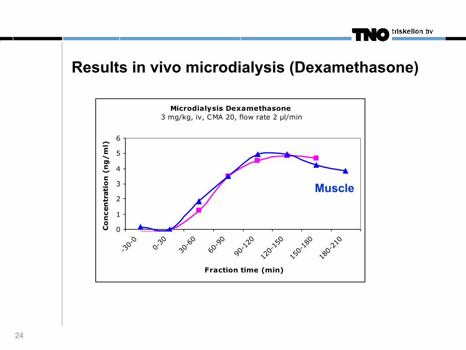
Results in vivo microdialysis (Dexamethasone)
24
Microdialysis Dexamethasone 3 mg/kg, iv, CMA 20, flow rate 2 µl/min
0
1
2
3
4
5
6
-30-
00-
3030
-60
60-9
0
90-1
20
120-
150
150-
180
180-
210
Fraction time (min)
Co
nce
ntr
atio
n (
ng
/m
l)
Muscle

FDA and microdialysis
25
! “While the FDA does not require microdialysis studies at this point in time, the agency is receptive to microdialysis data as part of an overall preclinical and clinical pharmacology package” ! “Microdialysis may contribute to the FDA Critical Path Initiative to facilitate innovation in drug development”.
From: Handbook of Microdialysis; Method, Applications and Clinical Aspects, Westerink and Cremers (2007) and AAPS Journal 2007; 9 (1) Article 6.

Conclusions
26
! Microdialysis has evolved into a mature technique. ! One of the few techniques for studying tissue concentrations. ! Semi-quantitative information. ! Potential to further evolve:
! Current generation UPLC-MS ! Metabolite identification ! Metabolomics platforms (endogenous compounds)
! Valuable tool in drug research and development.

Acknowledgements
27
Astrid Capello Florence Salmon Nicole Cnubben Ria Brust-van Schaik


