Microencapsulation of Bifidobacterium animalis subsp. lactis INL1 using wheyproteins and dextrans conjugates as wall materials
P.A. Loyeaua,∗, M.J. Spottia, N.L. Vanden Braberb, Y.E. Rossib, M.A. Montenegrob, G. Vinderolac,C.R. Carraraa
a Área de Estudios Fisicoquímicos de Alimentos, Instituto de Tecnología de Alimentos (ITA), Facultad de Ingeniería Química, Universidad Nacional Del Litoral, Santa Fe,Argentinab Centro de Investigaciones y Transferencia de Villa María (CIT), Universidad Nacional de Villa María - CONICET, Córdoba, Argentinac Instituto de Lactología Industrial (INLAIN), Universidad Nacional Del Litoral - CONICET, Santa Fe, Argentina
The incorporation of probiotic bacteria to food products is restricted by their instability, so microencapsulationcould provide them better protection during storage and gastrointestinal digestion. In this study Bifidobacteriumanimalis subsp. lactis INL1 was microencapsulated by spray drying using whey proteins isolate (WPI) and dex-trans (DX of 6, 70 and 450 kDa) conjugates obtained by Maillard reaction as wall materials. The stability duringstorage time and temperature, the viability after simulated gastrointestinal digestion and the antioxidant ca-pacity of the microcapsules were assayed. The cell viability was negatively affected by the gastrointestinaldigestion and also over the storage time (12 months). Conjugate with DX 6 kDa was the most stable system at25 °C and showed improved antioxidant capacity whit ABTS%+ technique; meanwhile with %OH technique nodifferences were found among the samples. Free cells also showed antioxidant activity in all their fractionsanalyzed using the same techniques.
1. Introduction
In the last years, worldwide food trends aimed at the consumptionof food that in addition provides benefits beyond nutrition. In this sensenew foods called “functional foods” have emerged, which enhancehealth conditions, wellness or reduce the risk of certain diseases (Kaur& Pal Singh, 2017). Functional foods not only can incorporate micro-nutrients, vitamins, essential oils and antioxidants but also, live beneficbacteria. Probiotics are “live microorganisms that, when administeredin adequate amounts, confer a health benefit on the host” (FAO/WHO,2002).
The incorporation of probiotic bacteria to food products is condi-tioned by bacterial instability and possible unwanted impacts in orga-noleptic properties of foods. Consequently, the preservation of viablecell and their incorporation in foods products in the required amounts(106–108 CFU/g of product) is challenging (Champagne, Paul Ross,Saarela, Flemming Hansen, & Charalampopoulos, 2011). The protectionof food-sensitive strains of probiotic bacteria may be achieved by mi-croencapsulation, employing spray drying and encapsulating wall ma-terials, such as: maltodextrin (Slavutsky, Chávez, Favaro-Trindade, &Bertuzzi, 2016), milk (Páez et al., 2013) or whey proteins (Doherty
et al., 2011).Whey proteins are widely used in food industry for different pur-
poses (Beaulieu, Savoie, Paquin, & Subirade, 2002; Wang, Xiong,Rentfrow, & Newman, 2013). These proteins have demonstrated tohave good functional properties. These properties can be improved bydifferent approaches, being Maillard reaction one of them. Maillardreaction, also known as non-enzymatic browning reaction or glicosila-tion reaction consist on the reaction between not protonated aminogroup of a protein and a carbonil group of a reducing sugar. Maillardreaction products (MRP) may have better functional properties than thestarting compounds, for example, better emulsifying properties (Zhu,Damodaran, & Lucey, 2011), antioxidant capacities (Chawla, Chander,& Sharma, 2009) as well as improved nutritional quality and solubility(Wang & Ismail, 2012). In this way, MPR are considered as wall ma-terials with potential to encapsulate not only probiotic bacteria, butalso other compounds of interest in food (Choi, Ryu, Kwak, & Ko, 2010;Qu & Zhong, 2017).
The aim of this work was to microencapsulate the breast-milk de-rived probiotic strain Bifidobacterium animalis subsp. lactis INL1 usingwhey protein and dextrans conjugates, obtained by Maillard reaction,through spray drying and to evaluate the stability and antioxidant
https://doi.org/10.1016/j.foodhyd.2018.06.051Received 1 March 2018; Accepted 29 June 2018
Whey protein isolate (WPI) (BIPRO™) was kindly provided byDavisco Foods International Inc. (Minnesota, USA). The compositionwas 0.4% (w/w) fat, 2.0% (w/w) ashes, 4.8% (w/w) moisture and lessthan 0.5% (w/w) lactose. According to Kjeldhal method, the proteincontent was 97.9% (w/w) (dry basis). Dextrans (DX) of 6, 70 and450 kDa molecular weight were obtained from Sigma-Aldrich(Germany).
The strain used in this work was Bifidobacterium animalis subsp.lactis INL1, which was isolated from human breast milk (Zacarías,Binetti, Laco, Reinheimer, & Vinderola, 2011) and showed probioticpotential in animal studies (Burns et al., 2017; Zacarías, Reinheimer,Forzani, Grangrette, & Vinderola, 2014).
2.2. Production and characterization of WPI/DX conjugated systems
The conjugates were synthesized by mixing WPI and DX of differentmolecular weight (6, 70 and 450 kDa), in a WPI:DX weight ratio of1:0.6. All the solutions were dehydrated in a laboratory-scale spraydryer ADL311S (Yamato, Japan) at a constant inlet air temperature(Tin) of 130 °C and outlet air temperature (Tout) of 71 ± 1 °C. Theobtained powders were incubated for 5 days at 60 °C, and 65% relativehumidity to develop the Maillard reaction (Spotti et al., 2013). Foursystems were obtained: WPI control (WPI native incubated without DX)called WPIc, and WPI/DX conjugates of DX 6, 70 and 450 kDa called: C-WPI/DX 6, C-WPI/DX 70 and C-WPI/DX 450, respectively.
2.2.1. UV–visible absorption spectroscopyEarly and intermediate compounds of Maillard reaction (called
Schiff bases and Amadori products) can be determined at 284 nm(Chawla et al., 2009) and at 304 nm (Amadori products) (Wang &Ismail, 2012).
Late Maillard reaction products (called melanoidins, which producebrowning effect) (Chawla et al., 2009; Lertittikul, Benjakul, & Tanaka,2007) were determined at 420 nm (Jimenez-Castaño, Villamiel, &Lopez-Fandiño, 2007; Miralles, Martinez-Rodriguez, Santiago, van deLagemaat, & Heras, 2007).
All the samples were dissolved at protein concentration of 0.5% (w/w) and the absorbance measurements were carried out in a Lambda-20spectrophotometer (Perkin-Elmer, USA). For this analysis a WPI native(WPIn) was also used as a reference.
2.2.2. Free amino groups detection using O-phthaldialdehyde (OPA)technique
To detect free amino groups of WPI (not conjugate to poly-saccharides) the O-phthaldialdehyde test (OPA, Sigma-Aldrich,Germany) was used according to Sun et al. (2011) with some mod-ifications. Samples were prepared at protein concentration of 4% (w/w). To 200 μl of each sample, 100 μl β-mercaptoethanol (Sigma-Al-drich, Germany), 100 μl of 20% (w/v) SDS and 800 μl of 0.1M sodiumtetraborate buffer solution were added. Samples were heated in a waterbath for 5min at 90 °C. OPA reagent was prepared as follow: 200mgSDS and 7.62 g disodium tetraborate decahydrated were dissolved in150mL of demineralized water. Then, 160mg of OPA were dissolved in4mL of ethanol and this solution was mixed to 400 μl of β-mercap-toethanol. Finally, the solution prepared before was taken to 200mLwith demineralized water. The reaction between 2mL of OPA and 50 μlof sample solutions was carried out for 7min. Absorbance at 340 nmwas measured in a spectrophotometer 7305 (Jenway, U.K.). To calcu-late the decrease in free amino groups (FAG) of conjugate samples, WPInative (WPIn) was used as a reference (100%) using equation (1):
⎜ ⎟= ⎛⎝
⎞⎠
FAG AA
x(%) 100S
WPIn (1)
Where AS is the total absorbance of the sample and AWPIn the absor-bance of WPI native.
2.3. Production of B. animalis subsp. lactis INL1 microcapsules
B. animalis subsp. lactis INL1 was encapsulated using WPIc or theWPI/DX conjugates as wall material. Fresh cultures of the probioticbacteria were harvested by centrifugation (5836×g, 10min, 8 °C) atthe end of the exponential growth phase in MRS-C broth (MRS brothwith cysteine; Biokar Diagnostic, France). The pellet was washed twicewith phosphate-buffered saline (PBS) solution pH 7.4 and resuspendedin different solutions: 10% (w/v) of WPI/DX conjugates and WPIc. Cellsuspensions were dehydrated in a laboratory scale spray dryer ADL311S(Yamato, Japan). Drying conditions were: feeding rate: 270mL/h, Tin
170 °C, Tout 81 ± 1 °C and air flux: 0.1MPa. The microcapsules (WPIcor WPI/DX conjugates with probiotics) were called: M-WPIc, M-WPI/DX 6, M-WPI/DX 70 and M-WPI/DX 450.
2.3.1. Characterization of B. animalis subsp. lactis INL1 microcapsulesMoisture content of the microcapsules was determined by gravi-
metry at 105 °C until constant weight. Water activity (aw) was measuredat 25 °C using awmeter Aqualab Systems (USA). These determinationswere carried out in triplicates.
The morphology of encapsulated cells was analyzed by scanningelectron microscopy (SEM) with Phenom World ProX microscope.
2.3.2. Survival to spray dryingCell viability was determined before and after spray drying.
Microcapsules obtained were reconstituted at 10% (w/v) in 0.1% (w/v)peptone water. Serial dilutions were plated on MRS-C agar and plateswere incubated for 72 h at 37 °C in anaerobiosis (AnaeroPack-Anaero,Japan). Counts were expressed in Log CFU/mL units.
2.3.3. Survival to simulated gastrointestinal digestionThis assay was carried out according to Saito et al. (2014). 1 g of
encapsulated was mixed with 20mL of a simulated saliva-gastric solu-tion (NaCl (16.2 g/L), CaCl2 (0.22 g/L), KCl (2.2 g/L), NaHCO3 (1.2 g/L)). Bovine pepsin (Merck, Darmstadt, Germany) was added to a 0.3%(w/v) final concentration. A cell count was performed (t0) and pH wasquickly lowered down to 2.5, with 0.5 N HCl. Samples were brought to37 °C in a water bath for 90min. Cell counts were performed as de-scribed before every 30min (Steps named GD t30; GD t60 and GD t90).After 90min of simulated saliva-gastric digestion, the samples werecentrifuged (6200×g, 5min, 8 °C) (Thermo Scientific, Sorvall LegendMicro 21R, Massachusetts, USA) and the pellets were washed twicewith PBS (pH 7.4), and it was resuspended to the original volume in 1%(w/v) bovine bile (Sigma-Aldrich, Germany) at pH 8.0. The cell sus-pensions were incubated in a water bath for 10min at 37 °C (step called“Duodenal shock of bile”, DSB). A sample was collected for cell viabilityassessment and another sample was centrifuged (6200×g, 5min,8 °C), the pellet was washed twice with PBS (pH 7.4) and resuspendedto the original volume in a solution containing 0.3% (w/v) bovine bile(Sigma-Aldrich, Germany) and 0.1% (w/v) pancreatin (Sigma-Aldrich,Germany) at pH 8.0. Cell counts were performed after an incubation of90min (Step named: tend). The test was performed in duplicate and cellviability was expressed in Log CFU/g.
2.3.4. Survival of B. animalis subsp. lactis INL1 during storageMicrocapsules were fractionated, vacuum-sealed, and stored pro-
tected from the light at 4 °C and 25 °C. Cell counts were performedevery 2 months for one year. Cell counts were performed on MRS-Cagar plate (72 h, 37 °C, anaerobic incubation) and expressed as LogCFU/g units.
P.A. Loyeau et al. Food Hydrocolloids 85 (2018) 129–135
130
2.3.5. Antioxidant capacityThe antioxidant activity was evaluated through the study of free
radicals scavenging activity of free cells, conjugates (wall materials)and microcapsules. Scavenging activity was evaluated against 2,2′-azinobis (3-ethylbenzothiazoline-6-sulfonic acid) diammonium salt ra-dical cation (ABTS%+) and hydroxyl radical (%+OH) techniques.
The radical scavenging percentage RS (%) for ABTS%+ and %+OHwas calculated with equation (2):
= −RS A A x(%) ( ) 100x0 (2)
Where A0 is the absorbance of the control and AX is the absorbance inpresence of conjugates, microcapsules and free cells samples.
The EC50 value, defined as the concentration of the sample (mgprot./mL) leading to 50% reduction of free initial radical concentrationwas obtained from the linear regression of plots (slope) of RS (%) vs.protein concentration.
2.3.5.1. Antioxidant capacity of free cells. The antioxidant capacity ofthe strain was assayed on different fractions: culture supernatant, intactcell and intracellular cell-free extracts. The preparation process wasconducted according to the method of Shen, Shang, and Li (2011). Inthe preparation process, total cell numbers were adjusted to about1× 109 CFU/mL. Through centrifugation at 1084×g for 10min,culture supernatant was separated from intact cells. After beingwashed three times, intact cells were obtained and resuspended indeionized and sterile water. For the preparation of intracellular cell-freeextracts, 500 μl of buffer SET (25mM EDTA; 20mM TRIS; 75mM NaCl;pH 7.0) were added. Then, three cycles of: heating (boiling bath, 5min)– freezing (−4 °C, 5min) were applied. After that, 5 μl of lysozymesolution (10mg/mL) were added and the cell suspension was incubatedfor 1 h at 37 °C. The sample was brought to its initial volume with steriledistilled water, and after homogenizing, it was centrifuged for 10min at3145×g to obtain two intracellular cell-free extracts: lysatesupernatant and lysate pellet. Each fraction (culture supernatant,intact cell, lysate supernatant and lysate pellet) was evaluated at1× 109 CFU (1mL) to determine the ABTS%+ and %+OH radicalsscavenging activity, according to the methodology described in the nextsections. Results were express as Radical scavenging percentage (RS %).
2.3.5.2. Assay of ABTS cation radical scavenging activity. Scavengingcapacity against ABTSc+ radical was determined by decolorization testas described by Sarkis, Michel, Tessaro, and Ferreira Marczak (2014)with some modifications. ABTS%+ radical was produced by mixing anABTS solution (7mM) with potassium persulfate (2.45 mM), andkeeping the mixture in the dark at room temperature for 12 h.Afterwards, the ABTS%+ solution was diluted in water untilabsorbance at 734 nm reached 0.70 ± 0.02 units. Aliquots from100 μl to 600 μL of conjugates or microcapsules, and the volume offresh culture equivalent to 1× 10⁹ CFU (1mL) were mixed with 1mL
of the ABTS%+ solution, and absorbance was determined after 6min.Results were express as EC50 value.
2.3.5.3. Assay of hydroxyl radical scavenging activity. Hydroxyl radical(%OH) was generated via Fenton reaction at pH 7.4. In presence of 2-Deoxy-D-ribose, the %OH reacts producing malonaldehyde (MDA)among other products, which forms a pinkish adduct in presence ofthiobarbituric acid (TBA) allowing its quantification by UV–Visspectroscopy. The %OH scavenging effect of encapsulates and freecells, were determined as previously described by Boiero et al.(2014). The reaction was performed in 50mM phosphate buffer (pH7.4) containing 10mM 2-deoxy-D-ribose, 100 mMH2O2, 1mM FeCl3,5 mM EDTA, in presence and absence of the samples. The reactionstarted with the addition of ascorbic acid in a final concentration of5mM. The reaction mixture was incubated for 1 h at 37 °C in a waterbath; then, 1% (w/v) TBA and 5.6% (w/v) cold trichloroacetic acidwere added and heated up to boiling temperature (95–100 °C) for20min to cause the colored adduct formation, which was measured at532 nm. Results were express as EC50 value.
2.4. Statistical analysis
Experiments were replicated at least twice in independent assays.Data was analyzed using one-way ANOVA (p˂0.05) with StatGraphicsCenturion XV software (StatGraphics.net, Madrid, Spain). LeastSignificant Differences Test (LSD) (α=0.05) was carried out.
3. Results and discussion
3.1. Production and characterization of WPI/DX conjugated systems
3.1.1. UV–visible absorption spectroscopyEarly and intermediate products of Maillard reaction absorb at 284
and 304 nm, and later products at 420 nm (Chawla et al., 2009;Jimenez-Castaño et al., 2007; Miralles et al., 2007; Wang & Ismail,2012).
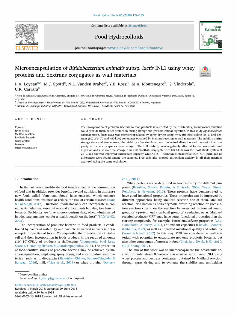
In Fig. 1 A the absorbance at 284 nm, which corresponds to Schiff'sreagent formation and in Fig. 1 B the absorbance at 304 nm, whichcorresponds to Amadori products, can be observed. In both graphs, theabsorbance of the C-WPI/DX 6 system was higher than for the otherssystems, indicating that DX of 6 kDa is much more reactive than highermolecular weight DX.
In Fig. 1 C the absorbance at 420 nm of the systems under study ispresented. This measurement is indicative of browning in the advancedstages of Maillard reaction (Lertittikul et al., 2007; Sun, Hayakawa, &Izumori, 2004). The C-WPI/DX 6 was the most reactive, meanwhile C-WPI/DX 70 and C-WPI/DX 450 exhibited similar values of absorbance,and WPIn and WPIc were the less reactive systems. Jimenez-Castañoet al. (2007), who studied β-lactoglobulin, α-lactalbumin and bovine
Fig. 1. UV–visible absorbance at 284 (A), 304 (B) and 420 nm (C) of WPIn (WPI native, ), WPIc (WPI Control, ) and conjugated systems: C-WPI/DX 6 ( ), C-WPI/DX 70 ( ) and C-WPI/DX 450 kDa ( ). The values plotted correspond of three independent experiments with its standard deviation. The different smallletters indicate significant differences according to the least significant differences test (LSD) (p < 0.05).
P.A. Loyeau et al. Food Hydrocolloids 85 (2018) 129–135
serum albumin (BSA) conjugated to DX of 10 and 20 kDa, found thesame tendency, those DX of smaller molecular weight exhibited greaterabsorbance at 420 nm.
In the three figures WPIc presented slightly higher absorbance thatWPIn, probably due to the residual lactose that can react after in-cubation. However, these systems did not present differences whenANOVA was performed.
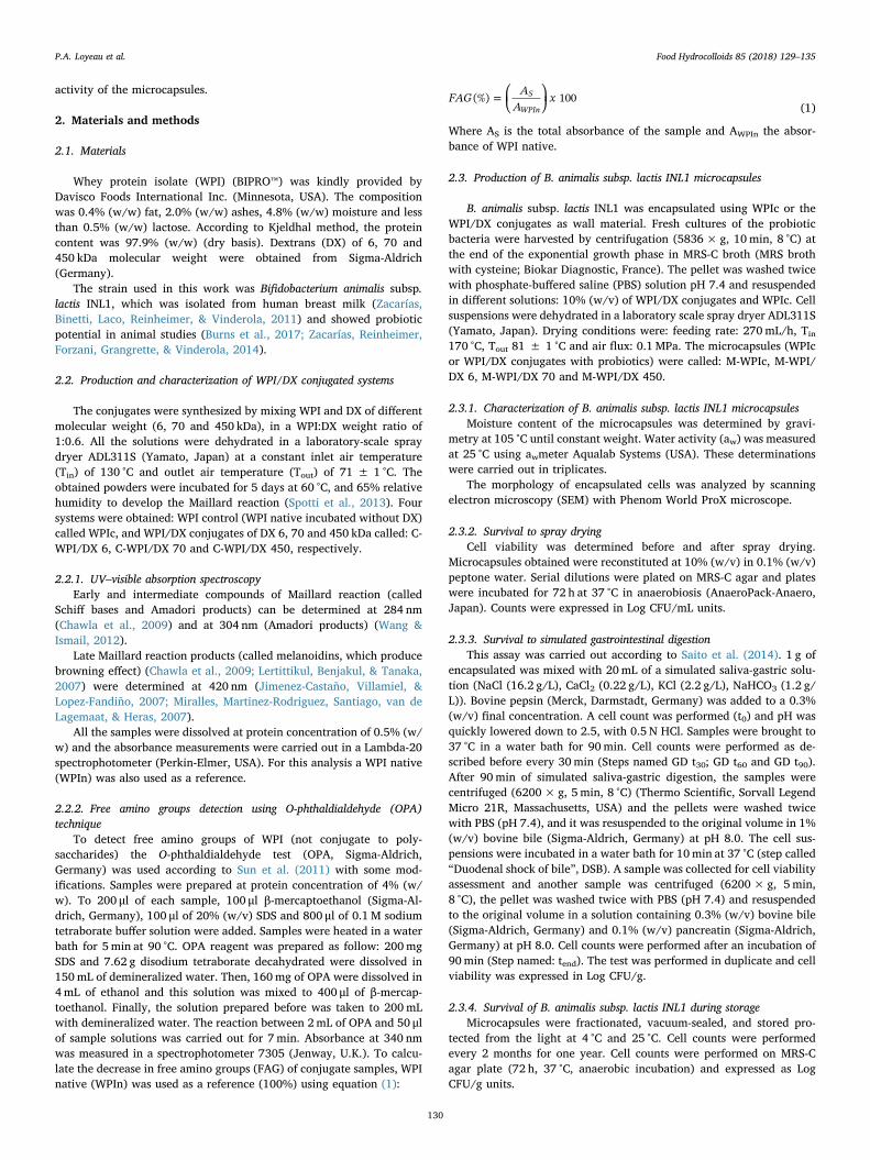
3.1.2. Determination of free amino groupsThe quantification of free amino groups in conjugate samples
(Fig. 2) could provide an estimation of the extent of the Maillard re-action. All WPI/DX conjugates systems presented a decrease in freeamino group content with decreasing DX molecular weight, when re-garding with WPIn: 90%, 87% and 84% for C-WPI/DX 450, 70 and6 kDa, respectively. A small decrease in free amino groups was observedfor WPIc which could be due to the incubation promotes the chemicalreaction between the minimum content of lactose present in the WPIand its proteins during incubation.
As can be observe in Fig. 2, molecular weight of DX significantlyaffected the percentage of free amino groups, since the smallest DX(6 kDa) is the most reactive one. This result could be explained in termsof the minor steric hindrance and a greater reactivity that smallermolecules could present. Instead, the bulky structures of 70 and450 kDa DX limit the access at the reactive sites of amino groups. Si-milarly, Wooster and Augustin (2007) determined the free aminogroups of WPI/DX conjugates incubated at 5 days and obtained values
of 95, 93 and 87% for conjugates produced with DX of 42.4, 29.4 and5.9 kDa respectively.
The results of this essay can be correlated with the results obtainedin the previous section (3.1.1). C-WPI/DX 6 presented the lowest con-tent in free amino groups and it was the system with the highest ab-sorbances at 284, 304 and 420 nm, indicating that DX 6 kDa was themost reactive DX. Therefore, the lower the molecular weight of DX, thegreater extent of Maillard reaction.
3.2. Characterization of B. animalis subsp. lactis INL1 microcapsules
3.2.1. Water activity (aw), moisture content (%H) and SEMThe water activity (aw) values of spray-dried microcapsules varied
between 0.30 and 0.41, and moisture content (%H) from 7.97 to10.93%. No tendency in the water activity or moisture content with DXmolecular weight was observed in these samples.
Passot, Cenard, Douania, Tréléa, and Fonseca (2012), showed thataw values close to 0.21 is required to maintain the normal acidificationactivity of lactic acid bacteria. According to Gardiner et al. (2000), thespray dried powders should have moisture content lower than 4% inorder to be stable. Therefore, in line with the bibliography cited, theobtained values of aw and %H of WPI/DX microcapsules were higherthan those recommended.
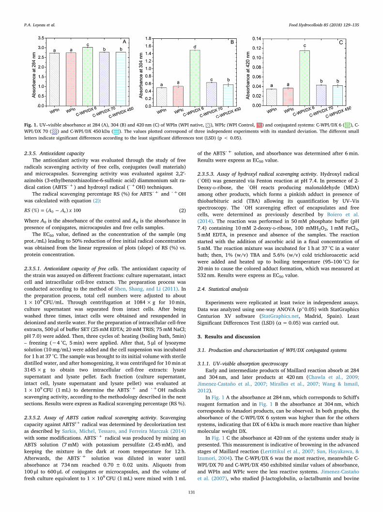
The SEM microphotographs of microcapsules are shown in Fig. 3.No differences were found in shape or size for the different systemsunder study. The morphology of the microcapsules was spherical,without cracks or pores, but many of them exhibited typical concavitiesas a consequence of the spray drying process (Saéns, Tapia, Chávez, &Robert, 2009). It was observed that the size of the capsules was around10 μm, similar to that reported by Slavutsky et al. (2016), who workedwith probiotic encapsulation using low methoxil pectin, maltodextrinand milk power, under comparable operative conditions. Páez et al.(2013), who characterized the functionality of three lactobacilli strainsafter spray drying in skim milk, also found similar shapes on the en-capsulated systems.
3.2.2. Survival of B. animalis subsp. lactis INL1 to spray dryingThe cell counts were carried out before and after the spray drying
because the heat exposure might affect the probiotic survival. The cellcounts of all the microcapsules prior to spray drying were close to9 ± 0.5 Log CFU/g. The differences between this value and the cellcounts after spray drying were: 0.73, 0.50, 0.20 and 0.01 Log orders forM-WPIc, M-WPI/DX 6, M-WPI/DX 70 and M-WPI/DX 450, respectively.M-WPIc, which was the only system that showed a significant difference(p < 0.05) between the counts before and after spray drying, was theless protecting system. This results suggests that conjugates maintainedthe cell viability against of spray drying, probably because they havehigher molecular weight. Ramakrishnan, Adzahan, Yusof, andMuhammad (2018) found that higher molecular weight of wall mate-rials resulted in a higher viscosity, which can advance the formation of
Fig. 2. Quantification of free amino groups respect to WPI native (*consider at100%; represented by ) of WPIc ( ) and conjugated systems: C-WPI/DX 6( ), C-WPI/DX 70 ( ) and C-WPI/DX 450 kDa ( ). Results are means of twoindependent experiments with its standard deviation. The different small lettersindicate significant differences according to the least significant differences test(LSD) (p < 0.05).
Fig. 3. SEM photomicrographs of spray dried microcapsules (magnification of 5600×), corresponding to the systems: M-WPI/DX 6 (A), M-WPI/DX 70 (B) and M-WPI/DX 450 (C).
P.A. Loyeau et al. Food Hydrocolloids 85 (2018) 129–135
132
a semipermeable surface during drying and protect the bioactivecompounds.
The use of whey proteins or conjugates as encapsulating agents forproduction of microcapsules can result in different physical properties,depending on the structure and the characteristics of each agent(Fritzen-Freire et al., 2012).
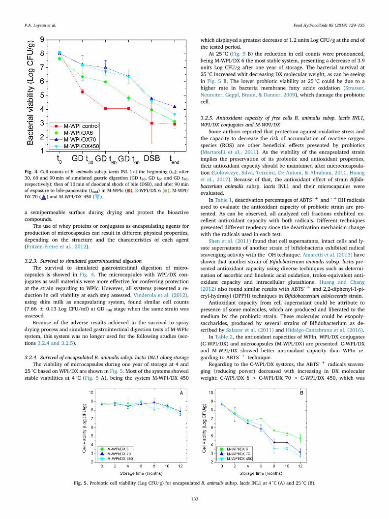
3.2.3. Survival to simulated gastrointestinal digestionThe survival to simulated gastrointestinal digestion of micro-
capsules is showed in Fig. 4. The microcapsules with WPI/DX con-jugates as wall materials were more effective for conferring protectionat the strain regarding to WPIc. However, all systems presented a re-duction in cell viability at each step assessed. Vinderola et al. (2012),using skim milk as encapsulating system, found similar cell counts(7.66 ± 0.13 Log CFU/ml) at GD t90 stage when the same strain wasassessed.
Because of the adverse results achieved in the survival to spraydrying process and simulated gastrointestinal digestion tests of M-WPIcsystem, this system was no longer used for the following studies (sec-tions 3.2.4 and 3.2.5).
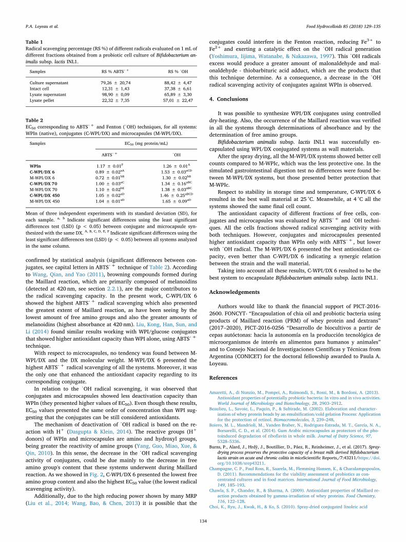
3.2.4. Survival of encapsulated B. animalis subsp. lactis INL1 along storageThe viability of microcapsules during one year of storage at 4 and
25 °C based on WPI/DX are shown in Fig. 5. Most of the systems showedstable viabilities at 4 °C (Fig. 5 A), being the system M-WPI/DX 450
which displayed a greatest decrease of 1.2 units Log CFU/g at the end ofthe tested period.
At 25 °C (Fig. 5 B) the reduction in cell counts were pronounced,being M-WPI/DX 6 the most stable system, presenting a decrease of 3.9units Log CFU/g after one year of storage. The bacterial survival at25 °C increased whit decreasing DX molecular weight, as can be seeingin Fig. 5 B. The lower probiotic viability at 25 °C could be due to ahigher rate in bacteria membrane fatty acids oxidation (Strasser,Neureiter, Geppl, Braun, & Danner, 2009), which damage the probioticcell.
3.2.5. Antioxidant capacity of free cells B. animalis subsp. lactis INL1,WPI/DX conjugates and M-WPI/DX
Some authors reported that protection against oxidative stress andthe capacity to decrease the risk of accumulation of reactive oxygenspecies (ROS) are other beneficial effects presented by probiotics(Martarelli et al., 2011). As the viability of the encapsulated strainimplies the preservation of its probiotic and antioxidant properties,their antioxidant capacity should be maintained after microencapsula-tion (Golowczyc, Silva, Teixeira, De Antoni, & Abraham, 2011; Huanget al., 2017). Because of that, the antioxidant effect of strain Bifido-bacterium animalis subsp. lactis INL1 and their microcapsules wereevaluated.
In Table 1, deactivation percentages of ABTS%+ and %+OH radicalsused to evaluate the antioxidant capacity of probiotic strain are pre-sented. As can be observed, all analyzed cell fractions exhibited ex-cellent antioxidant capacity with both radicals. Different techniquespresented different tendency since the deactivation mechanism changewith the radicals used in each test.
Shen et al. (2011) found that cell supernatants, intact cells and ly-sate supernatants of another strain of bifidobacteria exhibited radicalscavenging activity with the %OH technique. Amaretti et al. (2013) haveshown that another strain of Bifidobacterium animalis subsp. lactis pre-sented antioxidant capacity using diverse techniques such as determi-nation of ascorbic and linolenic acid oxidation, trolox-equivalent anti-oxidant capacity and intracellular glutathione. Huang and Chang(2012) also found similar results with ABTS%+ and 2,2-diphenyl-1-pi-cryl-hydrazyl (DPPH) techniques in Bifidobacterium adolescentis strain.
Antioxidant capacity from cell supernatant could be attribute topresence of some molecules, which are produced and liberated to themedium by the probiotic strain. These molecules could be exopoly-saccharides, produced by several strains of Bifidobacterium as de-scribed by Salazar et al. (2011) and Hidalgo-Cantabrana et al. (2016).
In Table 2, the antioxidant capacities of WPIn, WPI/DX conjugates(C-WPI/DX) and microcapsules (M-WPI/DX) are presented. C-WPI/DXand M-WPI/DX showed better antioxidant capacity than WPIn re-garding to ABTS%+ technique.
Regarding to the C-WPI/DX systems, the ABTS%+ radicals scaven-ging (reducing power) decreased with increasing in DX molecularweight: C-WPI/DX 6 > C-WPI/DX 70 > C-WPI/DX 450, which was
Fig. 4. Cell counts of B. animalis subsp. lactis INL 1 at the beginning (t0); after30, 60 and 90min of simulated gastric digestion (GD t30, GD t60 and GD t90,respectively); then of 10min of duodenal shock of bile (DSB), and after 90minof exposure to bile-pancreatin (tend) in M-WPIc ( ), E-WPI/DX 6 ( ), M-WPI/DX 70 ( ) and M-WPI/DX 450 ( ).
Fig. 5. Probiotic cell viability (Log CFU/g) for encapsulated B. animalis subsp. lactis INL1 at 4 °C (A) and 25 °C (B).
P.A. Loyeau et al. Food Hydrocolloids 85 (2018) 129–135
133
confirmed by statistical analysis (significant differences between con-jugates, see capital letters in ABTS%+ technique of Table 2). Accordingto Wang, Qian, and Yao (2011), browning compounds formed duringthe Maillard reaction, which are primarily composed of melanoidins(detected at 420 nm, see section 2.2.1), are the major contributors tothe radical scavenging capacity. In the present work, C-WPI/DX 6showed the highest ABTS%+ radical scavenging which also presentedthe greatest extent of Maillard reaction, as have been seeing by thelowest amount of free amino groups and also the greater amounts ofmelanoidins (highest absorbance at 420 nm). Liu, Kong, Han, Sun, andLi (2014) found similar results working with WPI/glucose conjugatesthat showed higher antioxidant capacity than WPI alone, using ABTS%+
technique.With respect to microcapsules, no tendency was found between M-
WPI/DX and the DX molecular weight. M-WPI/DX 6 presented thehighest ABTS%+ radical scavenging of all the systems. Moreover, it wasthe only one that enhanced the antioxidant capacity regarding to itscorresponding conjugate.
In relation to the %OH radical scavenging, it was observed thatconjugates and microcapsules showed less deactivation capacity thanWPIn (they presented higher values of EC50). Even though these results,EC50 values presented the same order of concentration than WPI sug-gesting that the conjugates can be still considered antioxidants.
The mechanism of deactivation of %OH radical is based on the re-action with H+ (Dasgupta & Klein, 2014). The reactive groups (H+
donors) of WPIn and microcapsules are amino and hydroxyl groups,being greater the reactivity of amine groups (Yang, Guo, Miao, Xue, &Qin, 2010). In this sense, the decrease in the %OH radical scavengingactivity of conjugates, could be due mainly to the decrease in freeamino group's content that these systems underwent during Maillardreaction. As we showed in Fig. 2, C-WPI/DX 6 presented the lowest freeamino group content and also the highest EC50 value (the lowest radicalscavenging activity).
Additionally, due to the high reducing power shown by many MRP(Liu et al., 2014; Wang, Bao, & Chen, 2013) it is possible that the
conjugates could interfere in the Fenton reaction, reducing Fe3+ toFe2+ and exerting a catalytic effect on the %OH radical generation(Yoshimura, Iijima, Watanabe, & Nakazawa, 1997). This %OH radicalsexcess would produce a greater amount of malonaldehyde and mal-onaldehyde - thiobarbituric acid adduct, which are the products thatthis technique determine. As a consequence, a decrease in the %OHradical scavenging activity of conjugates against WPIn is observed.
4. Conclusions
It was possible to synthesize WPI/DX conjugates using controlleddry-heating. Also, the occurrence of the Maillard reaction was verifiedin all the systems through determinations of absorbance and by thedetermination of free amino groups.
Bifidobacterium animalis subsp. lactis INL1 was successfully en-capsulated using WPI/DX conjugated systems as wall materials.
After the spray drying, all the M-WPI/DX systems showed better cellcounts compared to M-WPIc, which was the less protective one. In thesimulated gastrointestinal digestion test no differences were found be-tween M-WPI/DX systems, but those presented better protection thatM-WPIc.
Respect to stability in storage time and temperature, C-WPI/DX 6resulted in the best wall material at 25 °C. Meanwhile, at 4 °C all thesystems showed the same final cell count.
The antioxidant capacity of different fractions of free cells, con-jugates and microcapsules was evaluated by ABTS%+ and %OH techni-ques. All the cells fractions showed radical scavenging activity withboth techniques. However, conjugates and microcapsules presentedhigher antioxidant capacity than WPIn only with ABTS%+, but lowerwith %OH radical. The M-WPI/DX 6 presented the best antioxidant ca-pacity, even better than C-WPI/DX 6 indicating a synergic relationbetween the strain and the wall material.
Taking into account all these results, C-WPI/DX 6 resulted to be thebest system to encapsulate Bifidobacterium animalis subsp. lactis INL1.
Acknowledgements
Authors would like to thank the financial support of PICT-2016-2600. FONCYT- “Encapsulation of chia oil and probiotic bacteria usingproducts of Maillard reaction (PRM) of whey protein and dextrans”(2017–2020), PICT-2016-0256 “Desarrollo de biocultivos a partir decepas autóctonas: hacia la autonomía en la producción tecnológica demicroorganismos de interés en alimentos para humanos y animales”and to Consejo Nacional de Investigaciones Científicas y Técnicas fromArgentina (CONICET) for the doctoral fellowship awarded to Paula A.Loyeau.
References
Amaretti, A., di Nunzio, M., Pompei, A., Raimondi, S., Rossi, M., & Bordoni, A. (2013).Antioxidant properties of potentially probiotic bacteria: In vitro and in vivo activities.World Journal of Microbiology and Biotechnology, 28, 2903–2912.
Beaulieu, L., Savoie, L., Paquin, P., & Subirade, M. (2002). Elaboration and character-ization of whey protein beads by an emulsification/cold gelation Process: Applicationfor the protection of retinol. Biomacromolecules, 3, 239–248.
Boiero, M. L., Mandrioli, M., Vanden Braber, N., Rodriguez-Estrada, M. T., García, N. A.,Borsarelli, C. D., et al. (2014). Gum Arabic microcapsules as protectors of the pho-toinduced degradation of riboflavin in whole milk. Journal of Dairy Science, 97,5328–5336.
Burns, P., Alard, J., Hrdỳ, J., Boutillier, D., Páez, R., Reinheimer, J., et al. (2017). Spray-drying process preserves the protective capacity of a breast milk derived Bifidobacteriumlactis strain on acute and chronic colitis in miceScientific Reports,/7:43211/https://doi.org/10.1038/srep43211.
Champagne, C. P., Paul Ross, R., Saarela, M., Flemming Hansen, K., & Charalampopoulos,D. (2011). Recommendations for the viability assessment of probiotics as con-centrated cultures and in food matrices. International Journal of Food Microbiology,149, 185–193.
Chawla, S. P., Chander, R., & Sharma, A. (2009). Antioxidant properties of Maillard re-action products obtained by gamma-irradiation of whey proteins. Food Chemistry,116, 122–128.
Choi, K., Ryu, J., Kwak, H., & Ko, S. (2010). Spray-dried conjugated linoleic acid
Table 1Radical scavenging percentage (RS %) of different radicals evaluated on 1mL ofdifferent fractions obtained from a probiotic cell culture of Bifidobacterium an-imalis subsp. lactis INL1.
Table 2EC50 corresponding to ABTS%+ and Fenton (%OH) techniques, for all systems:WPIn (native), conjugates (C-WPI/DX) and microcapsules (M-WPI/DX).
Samples EC50 (mg protein/mL)
ABTS%+ %OH
WPIn 1.17 ± 0.01F 1.26 ± 0.01A
C-WPI/DX 6 0.89 ± 0.02aA 1.53 ± 0.03aCD
M-WPI/DX 6 0.72 ± 0.01bB 1.30 ± 0.02bB
C-WPI/DX 70 1.00 ± 0.03aC 1.34 ± 0.16aBC
M-WPI/DX 70 1.10 ± 0.02bE 1.38 ± 0.03aBC
C-WPI/DX 450 1.05 ± 0.02aD 1.46 ± 0.25aBCD
M-WPI/DX 450 1.04 ± 0.01aD 1.65 ± 0.09aD
Mean of three independent experiments with its standard deviation (SD), foreach sample. a, b Indicate significant differences using the least significantdifferences test (LSD) (p < 0.05) between conjugate and microcapsule syn-thesized with the same DX. A, B, C, D, E, F Indicate significant differences using theleast significant differences test (LSD) (p < 0.05) between all systems analyzedin the same column.
P.A. Loyeau et al. Food Hydrocolloids 85 (2018) 129–135
encapsulated with maillard reaction products of whey proteins and maltodextrin.Food Science Biotechnology, 19(4), 957–965.
Dasgupta, A., & Klein, K. (2014). Methods for measuring oxidative stress in the labora-tory. In A. Dasgupta, & K. Klein (Eds.). Antioxidants in food, vitamins and supplements.Prevention and treatment of disease (pp. 19–40). United States of America: Elsevier.
Doherty, S. B., Gee, V. L., Ross, R. P., Stanton, C., Fitzgerald, G. F., & Brodkorb, A. (2011).Development and characterisation of whey protein micro-beads as potential matricesfor probiotic protection. Food Hydrocolloids, 25, 1604–1617.
FAO/WHO (2002). Report of a joint FAO/WHO expert consultation on evaluation of healthand nutritional properties of probiotics in food including powder milk with live lactic acidbacteriaLondon, Ontario, Canada: World Health Organization and Food andAgriculture Organization of the United Nations.
Fritzen-Freire, C. B., Prudêncio, E. S., Amboni, R. D. M. C., Pinto, S. S., Negrão-Murakami,A. N., & Murakami, F. S. (2012). Microencapsulation of bifidobacteria by spraydrying in the presence of prebiotics. Food Research International, 45, 306–312.
Gardiner, G. E., O'Sullivan, E., Kelly, J., Auty, M. A. E., Fitzgerald, G. F., Collins, J. K.,et al. (2000). Comparative survival rates of human-derived probiotic lactobacillusparacasei and L. salivarius strains during heat treatment and spray drying. Appliedand Environmental Microbiology, 66(6), 2605–2612.
Golowczyc, M. A., Silva, J., Teixeira, P., De Antoni, G. L., & Abraham, A. G. (2011).Cellular injuries of spray-dried Lactobacillus spp. isolated from kefir and their impacton probiotic properties. International Journal of Food Microbiology, 144(3), 556–560.
Hidalgo-Cantabrana, C., Algieri, F., Rodriguez-Nogales, A., Vezza, T., Martínez-Camblor,P., Margolles, A., et al. (2016). Effect of a ropy exopolysaccharide-producingBifidobacterium animalis subsp. lactis strain orally administered on dss-induced colitismice model. Frontiers in Microbiology, 7 Art. 868.
Huang, H. C., & Chang, T. M. (2012). Antioxidative properties and inhibitory effect ofBifidobacterium adolescentis on melanogenesis. World Journal of Microbiology andBiotechnology, 28, 2903–2912.
Huang, S., Vignolles, M.-L., Dong Chen, X., Le Loir, Y., Jan, G., Schuck, P., et al. (2017).Spray drying of probiotics and other food-grade bacteria: A review. Trends in FoodScience & Technology, 63, 1–17.
Jimenez-Castaño, L., Villamiel, M., & Lopez-Fandiño, R. (2007). Glicosilation of indivualwhey proteins by maillard reaction using dextran of different molecular mass. FoodHydrocolloids, 21, 433–443.
Kaur, N., & Pal Singh, D. (2017). Deciphering the consumer behaviour facets of functionalfoods: A literature review. Appetite, 112, 167–187.
Lertittikul, W., Benjakul, S., & Tanaka, M. (2007). Characteristics and antioxidative ac-tivity of maillard reaction products from a porcine plasma protein–glucose modelsystem as influenced by pH. Food Chemistry, 100, 669–677.
Liu, Q., Kong, B., Han, J., Sun, C., & Li, P. (2014). Structure and antioxidant activity ofwhey protein isolate conjugated with glucose via the Maillard reaction under dry-heating conditions. Food Structure, 1, 145–154.
Martarelli, D., Verdenelli, M. C., Scuri, S., Cocchioni, M., Silvi, S., Cecchini, C., et al.(2011). Effect of a probiotic intake on oxidant and antioxidant parameters in plasmaof athletes during intense exercise training. Current Microbiology, 62(6), 1689–1696.
Miralles, B., Martinez-Rodriguez, A., Santiago, A., van de Lagemaat, J., & Heras, A.(2007). The ocurrence of a maillard-type protein-polysaccharide reaction between β-lactoglobulin and chitosan. Food Chemistry, 100, 1071–1075.
Páez, R., Lavari, L., Audero, G., Cuatrin, A., Zaritzky, N., Reinheimer, J., et al. (2013).Study of the effects of spray-drying on the functionality of probiotic lactobacilli.International Journal of Dairy Technology, 66(2), 155–161.
Passot, S., Cenard, S., Douania, I., Tréléa, I. C., & Fonseca, F. (2012). Critical water ac-tivity and amorphous state for optimal preservation of lyophilised lactic acid bac-teria. Food Chemistry, 132, 1699–1705.
Qu, B., & Zhong, Q. (2017). Casein-maltodextrin conjugate as an emulsifier for fabricationof structured calcium carbonate particles as dispersible fat globule mimetics. FoodHydrocolloids, 66, 61–70.
Ramakrishnan, Y., Adzahan, N. M., Yusof, Y. A., & Muhammad, K. (2018). Effect of wallmaterials on the spray drying efficiency, powder properties and stability of bioactivecompounds in tamarillo juice microencapsulation. Powder Technology, 328, 406–414.
Saénz, C., Tapia, S., Chávez, J., & Robert, P. (2009). Microencapsulation by spray dryingof bioactive compounds from cactus pear (Opuntiaficus-indica). Food Chemistry, 114,616–622.
Saito, V. S. T., dos Santos, T. F., Vinderola, C. G., Romano, C., Nicoli, J. R., Araújo, L. S.,et al. (2014). Viability and resistance of lactobacilli isolated from cocoa fermentationto simulated gastrointestinal digestive steps in soy yogurt. Journal of Food Science,79(2), M208–M213.
Salazar, N., Binetti, A., Gueimonde, M., Alonso, A., Garrido, P., González del Rey, C., et al.(2011). Safety and intestinal microbiota modulation by the exopolysaccharide-pro-ducing strains Bifidobacterium animalis IPLA R1 and Bifidobacterium longum IPLAE44 orally administered to Wistar rats. International Journal of Food Microbiology, 144,342–351.
Sarkis, J. R., Michel, I., Tessaro, I. C., & Ferreira Marczak, L. D. (2014). Optimization ofphenolics extraction from sesame seed cake. Separation and Purification Technology,122, 506–514.
Shen, Q., Shang, N., & Li, P. (2011). In vitro and in vivo antioxidant activity ofBifidobacterium animalis 01 isolated from centenarians. Current Microbiology, 62,1097–1103.
Slavutsky, A., Chávez, M., Favaro-Trindade, C., & Bertuzzi, M. (2016). Encapsulation ofLactobacillus acidophilus in a pilot-plant spray dryer. Effect of process parameters oncell viability. Journal of Food Process Engineering, 40(2), e12394.
Spotti, J., Perduca, M., Piagentini, A., Santiago, L. G., Rubiolo, A., & Carrara, C. (2013).Gel mechanical properties of milk whey protein-dextran conjugates obtained bymaillard reaction. Food Hydrocolloids, 32, 204–210.
Strasser, S., Neureiter, M., Geppl, M., Braun, R., & Danner, H. (2009). Influence of lyo-philization, fluidized bed drying, addition of protectants, and storage on the viabilityof lactic acid bacteria. The society for applied microbiology. Journal of AppliedMicrobiology, 107, 167–177.
Sun, Y., Hayakawa, S., & Izumori, K. (2004). Modification of ovalbumin with a rare keto-hexose through the maillard reaction, effect on protein structure and gel properties.Journal of Agricultural and Food Chemistry, 52, 1293–1299.
Sun, W., Yu, S., Yang, X., Wang, J., Zhang, J., Zhang, Y., et al. (2011). Study on therheological properties of heat-induced whey protein isolate-dextran conjugate gel.Food Research International, 44, 3259–3263.
Vinderola, G., Zacarías, M. F., Bockelmann, W., Neve, H., Reinheimer, J., & Heller, K. J.(2012). Preservation of functionality of Bifidobacterium animalis subsp. lactis INL1after incorporation of freeze-dried cells into different food matrices. FoodMicrobiology, 30, 274–280.
Wang, W. Q., Bao, Y. H., & Chen, Y. (2013). Characteristics and antioxidant activity ofwater-soluble Maillard reaction products from interactions in a whey protein isolateand sugars system. Food Chemistry, 139, 355–361.
Wang, Q., & Ismail, B. (2012). Effect of Maillard-induced glycosylation on the nutritionalquality, solubility, thermal stability and molecular configuration of whey protein.International Dairy Journal, 25, 112–122.
Wang, H. Y., Qian, H., & Yao, W. R. (2011). Melanoidins produced by the Maillard re-action: Structure and biological activity. Food Chemistry, 128, 573–584.
Wang, Y., Xiong, Y. L., Rentfrow, G. K., & Newman, M. C. (2013). Oxidation promotescross-linking but impairs film-forming properties of whey proteins. Journal of FoodEngineering, 115, 11–19.
Wooster, T. J., & Augustin, M. A. (2007). Rheology of whey protein–dextran conjugatefilms at the air/water interface. Food Hydrocolloids, 21, 1072–1080.
Yang, S., Guo, Z., Miao, F., Xue, Q., & Qin, S. (2010). The hydroxyl radical scavengingactivity of chitosan, hyaluronan, starch and their O-carboxymethylated derivatives.Carbohydrate Polymers, 82, 1043–1045.
Yoshimura, Y., Iijima, T., Watanabe, T., & Nakazawa, H. (1997). Antioxidative effect ofmaillard reaction products using Glucose-Glycine model system. Journal ofAgricultural and Food Chemistry, 45, 4106–4109.
Zacarías, M. F., Binetti, A., Laco, M., Reinheimer, J., & Vinderola, G. (2011). Preliminarytechnological and potential probiotic characterization of bifidobacteria isolated frombreast milk for use in dairy products. International Dairy Journal, 21, 548–555.
Zacarías, M. F., Reinheimer, J., Forzani, L., Grangrette, C., & Vinderola, G. (2014).Mortality and translocation assay to study the protective capacity of Bifidobacteriumlactis INL1 against Salmonella Typhimurium infection on mice. Beneficial Microbes,5(4), 427–436.
Zhu, D., Damodaran, S., & Lucey, J. A. (2011). Physicochemical and emulsifying prop-erties of whey protein isolate (WPI)-Dextran conjugates produced in aqueous solu-tion. Journal of Agricultural and Food Chemistry, 58, 2988–2994.
P.A. Loyeau et al. Food Hydrocolloids 85 (2018) 129–135