CHAPTER Microfabrication for Novel Products in Drug Delivery: An Example 8 CHAPTER CONTENTS 8.1 Microneedle Research at University of Twente and its Spin-Off .................... 237 8.1.1 Desk Research: Microneedle Arrays, Microfabrication and Transdermal Delivery of Insulin ........................................ 241 8.1.2 Is There a Need for Microneedles? .................................... 241 8.1.3 Microneedles by Microfabrication Technologies ....................... 244 Research Status ........................................................ 244 Fabrication and Design Concepts ...................................... 245 Materials ............................................................... 247 Ceramic Nanoporous Microneedles .................................... 248 Summarizing the Technology Requirements from a Commercialization Point of View ....................................... 250 8.1.4 Are Microneedles Ready for Insulin Delivery? ......................... 253 8.1.5 Design Aspects for Microneedle Insulin Delivery ...................... 258 Fabrication Attributes and Their Clinical Relevance ................... 259 Clinical Perspective .................................................... 263 8.2 MNA-4-Insulin: A Brief Evaluation .................................................... 265 8.3 Conclusions ............................................................................ 266 References ................................................................................... 267 In Chapter 1 the importance of technological innovation in the market place was discussed, and the previous chapter gave a successful example of tech- nology transfer from university to spin-off. This chapter is dedicated to academic-driven product innovation. A new technology starts with results from various different scientific disciplines, together with an analysis of the existing state of the art in industry. One may suggest that there is already enough knowledge (business intelligence) in industry, and universities (oper- ating on public money) are not the place that competitive products should Microfabrication for Industrial Applications. DOI: 10.1016/B978-0-8155-1582-1.00008-3 c 2011 Elsevier Inc. All rights reserved. 235
In Chapter 1 the importance of technological innovation in the market placewas discussed, and the previous chapter gave a successful example of tech-nology transfer from university to spin-off. This chapter is dedicated toacademic-driven product innovation. A new technology starts with resultsfrom various different scientific disciplines, together with an analysis of theexisting state of the art in industry. One may suggest that there is alreadyenough knowledge (business intelligence) in industry, and universities (oper-ating on public money) are not the place that competitive products should
be developed; they should stick to the provision of stimulating educationalenvironments. Universities do, however, have a clear responsibility to taketheir place within the innovation chain. But what is that place exactly? Is itpossible to provide the best academic training for new engineers without engi-neering sciences leading to products in the (competitive) industrial marketplace? I would immediately say that it must be both. With respect to mod-ern learning, we believe that experience with real-world problems is almostas important to a student’s education as instruction in the basics of his/herdiscipline.
Universities, therefore, have to create a motivating environment, in whichthe students actually get in touch with real-world problems, at least to acertain level. Normally, as an individual student and as a member of soci-ety, we easily recognize this need for gaining knowledge within universities.So, what is in this for industry? Since industry is simply the profit-makingpart of society, universities should bring their results to the attention of theindustry, and similarly industry should bring their pre-competitive researchquestions (encountered in the real world) to the attention of academic scien-tists. Today, both actors operate together in a variety of extensive networks,in part stimulated by public money. Within these networks, an individual’seffort can become linked to others and impact is created.
Well, can one expect that every individual’s research effort makes a dif-ference? This is unrealistic, but the sum of all the incremental results clearlymakes a difference to society. Here, I would like to focus on a specificexample, in which the results gained from researching a new technology arepre-competitive and open access, but the utilization thereof may well not be.Public resources must be used to benefit society as a whole, without discrim-ination and without private economical benefit to the individuals within theresearch teams. However, patents can still be obtained and the technologiesdescribed therein can be validated on industrial terms.
Technology transfer occupies the interface between knowledge creationand product development. Part of this process is the selection of which pro-ducts to develop. Ideas which present immediate answers to specific problemsin a market place have the highest chance of being developed; however, it isnot always obvious which ideas will have value after a product developmentpath which takes five, ten or more years towards completion prior to emergingon the market. For technology to emerge into the market place, visions areimportant.
8.1 Microneedle Research at University of Twente and its Spin-Off 237
This chapter focuses on the development of a new technology for drugdelivery through the skin. In it, we will discuss state-of-the-art microneedlearrays, and a specific microfabricated device which can transport a drug orvaccine through the skin by means of nanoscale pores.
Before building a future scenario for the use of this type of device, I haveto first thank the co-workers at University of Twente’s MESA+ Institutefor Nanotechnology, specifically Dr. S.N. Bystrova, M. Domanski and Dr.A.J.A. Winnubst, for their technological and scientific inputs in the develop-ment of a novel nanoporous microneedle array technology. The support ofProf. Dr. J.A. Bouwstra of the Leiden/Amsterdam Center for Drug Research(LACDR), Leiden University, is also acknowledged. She helped to define thereal-world problem (enhancement of transdermal drug delivery) to us as engi-neering scientists and supported the valorization project through her expertisein the field of the characterization of skin-transport processes, and kindly sug-gested that fluorescent ovalbumin can be used as a model compound for theevaluation of the nanoporous microneedle array technology developed at Uni-versity of Twente’s MESA+ Institute for Nanotechnology spin-off MyLifeTechnologies. Further, I would like to acknowledge the financial support ofthe original research project, as well as the valorization project related toMyLife Technologies, from the Dutch Science and Technology FoundationSTW, Applied Science Division of NWO, and the technology program ofthe Ministry of Economic Affairs, The Netherlands. Finally, I would like tomention that dedicating time and expert knowledge to the compilation of thischapter would not have been possible without the funding of my personalVENI grant, also by STW.
8.1 MICRONEEDLE RESEARCH AT UNIVERSITY OF TWENTEAND ITS SPIN-OFF
Chapter 7 discussed the LICETAS project. In this section, we will discussnovel products based on microneedle array technology (MNA) from the pointof the commercial exploitation of high-tech research results by academically-driven product development. This route involves obtaining a patent andconsists of two main elements: (1) desk research and (2) validating researchfor technology–product combinations. An effective academic-driven productdevelopment and innovation path always requires the collection of insight
into the current needs of society. However, in science-driven technologicaldevelopment this path may take quite a long time in order to fully comprehendall its complexities. From idea, to proposal writing, to receiving the grant andhiring personnel takes approximately 1.5 years. Conducting the research andinterpreting the results before being able to actually attract industrial attentionmay take another 2–3 years.
I have focused on the practical application of a specific microfabrica-tion technology for microneedle arrays since 2006. This technology wasdeveloped by publically funded research at the MESA+ institute between2003 to 2007 in collaboration with Leiden University. We investigated theenhancement of transdermal drug delivery processes within a pre-competitiveresearch setting. In 2006, the project team at MESA+, headed by Prof. Dr. Ir.van den Berg (BIOS, The Lab-on-a-Chip group), made a patent application.To enhance the chance of utilizing the collected know-how, and to offer inter-ested industrial partners (of which some were already part of the user com-mittee consulting on the original research project) the opportunity to evaluatethis technology further, the creation of a spin-off activity was initiated, andgranted with a first phase valorization grant by STW of 25,000.00 Euros.
This money was required to transfer the know-how gained from theresearch results in the project into the first non-tangible asset of the spin-off: a patent application. Similarly to the LICETAS project, the first step inthe project was necessary to secure a second stage pre-seed technology fund,which would then allow the technology to be developed to its full poten-tial, thus reducing the economical risk involved in pre-clinical evaluationof the MNA technology. Based on the know-how built in the valorizationteam and the prospective patent application different business cases could bedeveloped. This second, STW funded, valorization project (conducted from2008 to 2010 within MESA+ at the research group Mesoscale Chemical Sys-tems, headed by Prof. Dr. Gardeniers) increased the chances of a start-upwithin the highly competitive, but highly conservative medical field. This200,000.00 Euro project allowed the team to validate the technology to iden-tify a specific technology–product match, to create a first business niche forthe novel patented nanoporous microneedle array product. We did not justlook at the drug delivery market. To increase the opportunity for initiatinga successful start-up, it is important to identify the unique selling points ofthe technology at hand. The publicly supported valorization project allowedus to bring the potential of the original research results to the attention ofdifferent parties with specific application needs. In November 2010, indeed,
8.1 Microneedle Research at University of Twente and its Spin-Off 239
The University of Twente’s MESA+ Institute for Nanotechnology spin-offMyLife Technologies received an initial, first phase (50,000.00 Euros), life-science, pre-seed grant to evaluate the use of nanoporous MNA technologyfor making an ultra-minimally invasive, easy-to-use vaccination patch.
Through this process of pre-seeding, the organizational actors in the net-work could evaluate the business potential of the MNA technology, and supportthe efforts of the valorization team by the suitable instruments of technol-ogy transfer to the market. These efforts reduce the financial risks for anentrepreneur, allowing the public sector to get involved in the process of patentapplication roll-out to the national phase. At present, the MyLife Technologiesteam is negotiating the full terms and conditions of commercial exploitationof the patent with the university board and its funding agent. Bringing thesenegotiations to a successful close will enable the birth of a new company.
In the LICETAS case a company (Medimate BV) was started quickly afterfinalizing the original research project and know-how was transferred to thisstart-up directly. Why was this not possible in the MNA case?
In brief, the idea for LICETAS started from a project proposal inspiredby the needs of a practicing psychiatrist, and funding was received in 2001.The PhD project was entirely dedicated to one specific appliation: quantita-tive lithium measurement from a droplet of finger stick blood. In 2005, a fullproof-of-principle was presented to the user committee of the STW project,and at the same time it happened that a young entrepreneur was looking for abusiness case to start his own company. With the initial support of the originalresearch team, and members of the user committee, it was decided to initiallymake the entrepreneur a part of the research team, through (1) an extensionof 25,000.00 Euros of the original project and (2) the subsequent preparationof an STW valorization phase-one grant. Similarly to MyLife Technologies,it was because of this initial seed money that Medimate BV was able to takeoff. The entrepreneur was not part of the academic community; he had toprovide a different instrument to become one of the actors in the network, bystarting his privately held company (S. Staal BV) directly in the very first val-orization phase. Eventually, after settling the knowledge transfer conditionswith the university board and its funding agent STW, S. Staal BV and theother involved parties became owner of Medimate BV to continue the val-orization path. Since this spin-off was created by the original research fromSTW funds, the University of Twente was also eligible to apply for fundingin the second STW valorization round with its spin-off Medimate BV as theproject conducting valorization team.
It is probably the case that a BV could not be started straight away in theMyLife Technologies case because different actors, at a different time, in adifferent location and in different circumstances of the actors’ career pathswere involved. Also, when the LICETAS research project had been finalized,no patent had been applied for, and the risk of shelving the project results wasextremely high. Capillary electrophoresis on chip was not novel by itself, anddetecting lithium by this technique in body fluids was not novel either, as ithad been previously performed in serum. At the time, the accurate detectionof lithium in a whole blood sample by capillary electrophoresis on chip mighthave had the potential to be patentable, but when the decision was made totransfer the know-how to the entrepreneur he already had a company in place,which increased the chances for utilization of the research results gained inthe research project.
This scenario was not an option for MyLife Technologies, which bringsme to another question: What can I do as an academic actor to manage therisk of shelving valuable research results versus the financially high risksinvolved in high-tech innovation? How can I conduct my academic researchso as to generate output that is of direct value to a privately-held business thatinteracts with the academic network?
A comprehensive answer is probably not possible within the context ofthis book, hence I will restrict myself to the context of microfabrication forindustrial applications. One future application of MNA technology may bethe creation of a method for treating diabetes.
Today, insulin is given to a diabetes sufferer to control their glucose level.It is estimated that there will be 230 million diabetes cases worldwide inapproximately 20 years from now, and today’s methods are insufficient forproviding a solution to this problem, so novel technologies will be required.
Many different researchers world-wide have been involved in the develop-ment of microneedle array technology for the ultra-minimally invasive deliv-ery of insulin via the skin. Due to my involvement in publicly funded researchon microneedle array technology for drug delivery and the patenting of a tech-nology based on the research result we gained from the academic project, Iwas able to put my entrepreneurial head on and think towards a unique sellingpoint for this technology in the identified application of insulin delivery. Is ita new business case? A significant amount of desk research was needed toanswer this question. In the following sections I will describe its results andevaluate in depth a possible match for microneedle array technology withinsulin delivery.
8.1 Microneedle Research at University of Twente and its Spin-Off 241
8.1.1 Desk Research: Microneedle Arrays, Microfabrication andTransdermal Delivery of Insulin
Microneedles are a next-generation drug delivery device, that targets the skin.As yet, few original research papers consider this type of insulin delivery, soa review of microneedle array microfabrication technology and its possibleapplications in transdermal delivery of insulin is presented. A small sub-setof publications has been identified that demonstrate the proof-of-principle forinsulin delivery by microneedles. The results of these papers are compared.However, the specific criteria that a microneedle device must fulfil to be usedin this way are unclear, and a variety of hypotheses can be derived from thisdesk research.
A variety of microneedle designs (tip shapes, length, diameter, materials,etc.) in three distinct design categories have been produced. These are: sili-con micromachined, replicated and fine-mechanically manufactured arrays.Despite the fact that microneedles can be used to target the skin as a routefor delivery in a nearly painless manner, none of the designs show an appro-priate level of technological readiness for clinical applications. Many designshave been presented in the scientific literature, but so far, clinical evalua-tion is limited. To bring any of these minimally invasive devices to market,one has to develop a suitable model which allows clinical researchers tobenchmark the emerging microneedle techniques with respect to the conven-tional hypodermic needle injection for insulin. The case study discussed herewill highlight the essential research and development stages concerned withmicrofabrication of microneedle arrays.
8.1.2 Is There a Need for Microneedles?An arsenal of specific microfabrication techniques has been developed forminiaturization technologies, of which photolithography has been the mostsuccessful. Inspired by the success of the microelectronics industry, a wealthof devices, not necessarily electronic, has been developed where small size isa prerequisite. Some contain both electronic and non-electronic components.These devices are called microsystems or micro(opto)electromechanical sys-tems (MEMS, MOEMS). Since the 1980s, these architectures also have beenexplored for the manipulation of minute amounts of fluids, as has been dis-cussed in preceding chapters. This activity has presented us with an entirelynew field of research named microfluidics [1], which has made its mark in thelife sciences by the introduction of so called Laboratory-on-a-Chip (LoaC)
devices. These devices now present us with a multitude of applications, ofwhich some are concerned directly with clinical applications [2]. One type ofdevice is the microneedle array, either developed as a stand-alone device orintegrated with sensors and actuators [3]. They may offer advantages overhypodermic needles or chemical penetration enhancers. For certain appli-cations, whereby the target is actually in the skin, the direct insertion ofactive compounds into the viable epidermis will also increase efficacy ofthe therapy [4–8]. This is especially beneficial in the cases where the deliv-ery of macromolecular compounds is desired, e.g., DNA vaccine [9]. Theviable epidermis hosts antigen-presenting cells and these can be addressedby microneedle array therapy directly (see work by Birchall’s group, CardiffUniversity, UK) [10–13]. Bouwstra’s group at the Leiden/Amsterdam Centerfor Drug Research (LACDR), Leiden University, NL, presented two otherexamples in this area describing “Immune modulation by adjuvants com-bined with diphtheria toxoid administered topically in BALB/c mice aftermicroneedle array pre-treatment” and “Microneedle arrays for the transcu-taneous immunization of diphtheria and influenza” by Ding et al. [14, 15].In some studies, microneedle arrays are used for skin pretreatment [16–19].Other, more sophisticated miniaturized devices for drug delivery alsoexist [20–22].
Reviews of microneedle technology published to date [23–30] show theyare virtually pain free compared to the hypodermic needle. Micromachinedneedles are intended to be limited in their penetration depth and should bepainless in use because of their very sharp tips and defined length. Of course,passive patches and topical creams also avoid pain, however, they often workonly for very potent, small molecule drugs, or in specific formulations withchemical penetration enhancers. Microneedle array devices, by contrast, cre-ate a multitude of highly-defined pathways through the stratum corneum,which allow the passage of molecules without the side effects of chemicalenhancers.
Can this be applied to insulin delivery? When it comes to the micro-manufacture of microneedles two distinct concepts exist: (1) the in-planeand (2) the out-of-plane approach, as depicted in Figure 8.1. During in-planemanufacture, the microneedle tips lie within the plane of the material. Inthe out-of-plane approach the microneedle tips evolve perpendicularly tothe plane of the material’s working surface, which offers two importantadvantages: (1) a huge number of parallel injection sites, and (2) seamlessintegration of micro/nanofunctionality from the needles into the back plate.The schematic top and the cross-sectional views shown in Figure 8.1 present
an overview of the key parameters which define needle geometry. Whilein-plane processing can deliver very long microneedles (Lshaft), the out-of-plane process is generally preferred because of its shallow, self-limitinginsertion depth. However, the preferred technique depends on the individualapplication.
Combinations of these two approaches can be applied, for exampleMartanto et al. [33]. Microneedle fabrication uses the MEMS technologies,as outlined in Chapters 2 and 3, with the sequence of processing steps basedon whether the needles are to be in-plane or out-of-plane, and can differ quitesignificantly per type of microneedle array (MNA).
The development of a clinically superior MNA delivery technology hasbeen hampered by a limited understanding of the underlying transport mech-anisms and the pharmacodynamics of the different tissue layers. The deliveryof insulin, in particular, has been paid less attention than the more obvi-ous applications of targeted delivery for vaccines and immuno-enhancingagents (adjuvants). Because diabetes is a severe disease with a rapidly grow-ing patient population, it is important to investigate whether MNA devicespossess distinctive benefits for its treatment.
In summary, this desk research provides a model case for entrepreneurialscience and is organized in three sections. First a general literature screenof recent fabrication technology for microneedles is performed, including adiscussion of the choice of materials and the technology requirements forcommercialization. In the second section, a literature review of micronee-dle insulin delivery is presented. Thirdly, a sub-set of eight publicationsis derived, that describe successful experimental studies reaching proof-of-principle for insulin delivery by microneedles. In the latter section, specificattributes of these microneedle designs are discussed in more detail, whichcan eventually help to decipher the required criteria for the application ofMNA to insulin delivery. Finally, the conclusion reflects on the state-of-the-art microneedle arrays for insulin delivery in clinical applications.
8.1.3 Microneedles by Microfabrication TechnologiesResearch StatusPioneering research in the field of high-precision out-of-plane micronee-dle array fabrication and their medical applications were first studied byresearch groups at the Berkeley Sensor and Actuator Center, Universityof California (Berkeley, CA, USA), the Royal Institute of Technology(Stockholm, Sweden), MESA+ Institute for Nanotechnology, University ofTwente (Enschede, The Netherlands) and the Georgia Institute of Technol-ogy (Atlanta, GA, USA) [3, 34–41]. A large number of publications haveappeared over the last 15 years concerning the fabrication and testing ofmicroneedles in a diversity of applications, including brain–computer inter-faces, microneedle probing and stimulating for various brain regions, scalpelectroencephalography, drug delivery, diagnostics and applications wherespiked microstructures are used as nozzles and pipettes.
MNAs containing microneedles of diverse shape, length, diameter anddensity have been demonstrated to be capable of bypassing the skin barrier.
8.1 Microneedle Research at University of Twente and its Spin-Off 245
Both in-plane and out-of-plane fabrication are well suited to the creationof microneedle arrays, but each requires a different approach if it is to beintegrated into a skin patch. Only the out-of-plane method will directly pro-duce a two-dimensional array seamlessly connected to a back plate, whichis preferable for integration into higher functionality [3]. MNAs are alsofine-mechanically manufactured by assembly from conventionally producedhypodermic needles, and successfully demonstrated in certain types of trans-dermal delivery studies [42]. Nonetheless, lithographic exposure techniquesutilizing microsystems technology (MEMS), (i.e., reproducing a structuralelement multiple times from a mask or mold), are now established in drugdelivery research.
Lithographic techniques result in arrays with a tip radius of <100 nm,microneedle outer diameters below 300µm, very well-defined length in therange of a few to hundred micrometers, and varying needle densities. Batchprocessing allows some cost reduction. Hollow MNAs require a flow-throughdelivery system and have been tested by gluing the back plate of the MNAto a syringe. These devices are not yet applicable to self-administered trans-dermal insulin delivery, but they demonstrate a clear step forward. Figure 8.2summarizes complementary examples of out-of-plane MNAs, and demon-strates early prototypes of microneedles coupled to a syringe for intradermaldrug delivery [33, 43–46]. Recent publications have discussed microfabri-cated microneedle technology, and evaluated their performance against otherinsulin delivery techniques [24, 47–49].
Fabrication and Design ConceptsSome processes form the MNA device directly, while others first produce amaster. Silicon micromachining and stereomicrolithography are two exam-ples of direct techniques forming three-dimensional work pieces [3, 37, 51].SU-8 photolithography on pre-patterned silicon substrates combining lithog-raphy with micromolding, titanium micromachining, and the use of two pho-ton induced polymerization of organic–inorganic hybrid biomaterials [52–54]have also been attempted experimentally.
Some remarkably simple methods have been recorded for micromoldingmicroneedles. For example, Lee et al. introduced drawing lithography forthe three-dimensional fabrication of an ultrahigh-aspect-ratio microneedle,and Zhu et al. recently demonstrated a fabrication process that uses chemi-cally etched silica needles as a template for a replication process [55, 56].Unfortunately, Zhu et al. [56] omit a detailed description of the fabrication
of the silica template. Although their paper does not describe the fabricationmethod of the template the authors clearly identify micromolding as a low-costmanufacturing scheme, which is suitable for further drug delivery research.
Many different processes have been tried to make extremely sharpmicroneedles. Unfortunately, there is no consensus on how one should define
8.1 Microneedle Research at University of Twente and its Spin-Off 247
sharpness in this context. Many different research groups have elaborated onvarious fabrication processes to generate particular needle geometries, withthe intention of reducing the tip radius, which will obviously lower the inser-tion energy. The density, the total number of needles within a defined arrayarea, and the mechanism of insertion (manual or impact) are likely to be themajor factors influencing insertion performance. Knife-like features, such asthat introduced by Gardeniers et al. [38], should be assessed against otherdesigns with respect to pain sensation, skin reaction, intrinsic skin varia-tions from volunteer to volunteer, place of incision and relevant parametersof wound healing in relation to the size and geometry of the incision. Devel-opment of a benchmark for these parameters is not trivial because the skinis an anisotropic biomaterial, which changes its mechanical characteristicsunder stress. Moreover it is unclear how the tip radius of a microneedle (i.e.,sharpness) can be translated into a meaningful parameter for such an incisionexperiment.
Some studies have explored needle radius together with other geometricaland application parameters through mechanical modeling [57, 58]. However,the ease of insertion of the microneedles or the successful introduction ofdrug does not just depend on the individual needle tip, or the configuration ofthe array, but also on the apparatus that is used to administer the MNA deviceto the skin and the drug formulation [59]. Furthermore, a trade-off betweensharpness and microneedle stability must be sought for a clinical application.The insertion of microneedle arrays in human subjects was reported in a studyby Gill and Prausnitz [60].
MaterialsMicroneedles are made either directly from active agents embedded in a dis-solving material, or the microneedle platform acts as a carrier, which releasesactive compounds from a coating (solid microneedles), through a fluid con-duit from a reservoir (hollow microneedles) or from the material’s intrinsicpores (porous microneedles). The range of materials used in research orcommercial activities includes glass or silica, glass-like plastics and ceramiccomposites, silicon, silicon nitride, stainless steel, nickel-plated needle arraysand titanium, amongst various types of polymers, carbohydrates and micro-nanoparticle hybrid materials. Each of these materials may require their ownfabrication techniques, for example, stainless steel or titanium microneedlearrays are cut by a laser, which limits their physical dimensions to the reso-lution of the laser cutter. This limited resolution will also affect the radius ofthe needle tips, and the overall geometrical dimensions of the needles.
Silicon, which forms a native oxide layer, is much used in microsys-tems technology and has been accepted for use in transdermal pre-clinicalresearch, although no commercial application yet exists. Ji et al. have donesome preliminary work in the area of porous microneedles. They used selec-tive electrochemical etching, with a photoresist as a protective mask to formthe porous silicon tip, thus forming one of the first silicon out-of-planemicroneedle arrays that contain tips of two types of materials with very dis-tinct properties [61]. Other publications also discuss the range of materialsused in out-of-plane micro- and nanomanufacture of MNAs. For example,Park et al. investigated a polymer double-layer molding process, as shownin Figure 8.2f, which was generated from a mold structure. The mold wasfabricated by means of a combination of SU-8 photolithography, reactive ionetching and polydimethylsiloxane soft lithography [45, 50]. They producedtapered tips, using poly(lactic-co-glycolic acid) as a material for the formationof dissolving microneedles and encapsulated calcein in the tips. The particlestructure remains intact inside the molded microneedle.
Similarly, Moon et al. fabricated a mold by soft-lithography, from a mas-ter made by inclined deep x-ray exposure, then the soft-mold is used for thereplication of MNAs into biodegradable poly-L-lactide acid [62]. Carbohy-drates have also been used for molding MNAs, and Donnelly et al. discussedthe processing difficulties and instability of the material in MNA produc-tion [63]. Kolli and Banga, and Li et al. have found maltose to be more easilyadapted to molding microneedles [64, 65]. Both Kolli and Li are membersof Banga’s group affiliated with the Mercer University (Atlanta, GA, USA),which holds an impressive record on microneedle array technology.
Ceramic Nanoporous MicroneedlesWithin microneedle research, some groups are attempting to make eversharper and smaller point-like microfeatures, while others are looking furtheron in the development chain, towards low-cost production and applications.One particularly exciting concept is the possibility of making arrays ofmicroneedles in one platform, so combining a multitude of microneedles inparallel without the need for additional assembly steps. Such arrays could beused to make hundreds of miniscule incisions into the skin in one step, with-out causing pain. These incisions could then be used to transport moleculesacross the skin barrier. Figure 8.3 shows a ceramic-based nanoporous MNAplatform technology developed at University of Twente’s MESA+ Insti-tute for Nanotechnology (patent pending WO2009/113856). Here, the MNAis replicated from a soft-mold that is generated by polydimethylsiloxane
8.1 Microneedle Research at University of Twente and its Spin-Off 249
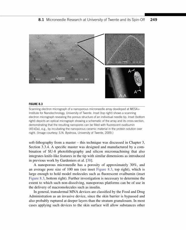
FIGURE 8.3
Scanning electron micrograph of a nanoporous microneedle array developed at MESA+Institute for Nanotechnology, University of Twente. Inset (top right) shows a scanningelectron micrograph revealing the porous structure of an individual needle tip. Inset (bottomright) depicts an optical micrograph showing a schematic of the array and its cross-section,demonstrating that the resulting nanopores can be filled with fluorescent ovalbumin(45 kDa), e.g., by incubating the nanoporous ceramic material in the protein solution overnight. (Image courtesy: S.N. Bystrova, University of Twente, 2009.)
soft-lithography from a master – this technique was discussed in Chapter 3,Section 3.3.4. A specific master was designed and manufactured by a com-bination of SU-8 photolithography and silicon micromachining that alsointegrates knife-like features in the tip with similar dimensions as introducedin previous work by Gardeniers et al. [38].
A nanoporous microneedle has a porosity of approximately 30%, andan average pore size of 100 nm (see inset Figure 8.3, top right), which islarge enough to hold model molecules such as fluorescent ovalbumin (insetFigure 8.3, bottom right). Further investigation is necessary to determine theextent to which such non-dissolving, nanoporous platforms can be of use inthe delivery of macromolecules such as insulin.
In general, transdermal MNA devices are classified by the Food and DrugAdministration as an invasive device, since the skin barrier is bypassed andalso probably ruptured at deeper layers than the stratum granulosum. In mostcases applying such devices to the skin surface will allow substances other
than the drug to enter the body. Any skin rupture event will interfere withthe body’s defence mechanism, generating additional adverse reactions inthe skin, or even unfavorable systemic effects through infection or poison-ing, hence this classification is necessary. All components of the microneedleadministration device that come into direct contact with tissue, or risk contactduring operation of the device must, therefore, obey stringent rules to gainmedical approval.
At this stage of development, the exact material properties of a MNA arenot fully understood, and have to be assessed as they stand and also when theyare packaged within a drug-applying device. Despite their general potentialbeing recognized in the drug delivery community the benefits of a specificmaterial for insulin delivery still have to be addressed for all the differentnovel platform technologies described above.
Figure 8.4 gives an overview of the nanoneeedle fabrication process devel-oped at University of Twente’s MESA+ Institute for Nanotechnology. Thisprocess starts with a hard mold, manufactured by photolithography and sili-con micromachining, to combine a pre-patterned silicon substrate and athick-film photoresist (SU-8) into one hard-mold structure for the fabricationof a soft mold for subsequent multiple production of ceramic green bodies.The individual MNAs are cut from the green body and sintered, forming thesemi-finished product ready for integration into a delivery patch.
Figure 8.5 depicts the integration of the ceramic MNA into a patch-typesystem. With a resulting materials porosity of approximatly 30%, a singlemicroneedle within the array has a liquid loading capacity in the order of1–2 nl, and the backing plate can contain up to 5µl. These minute amountsmay suffice for vaccination purposes. Currently, the application of Univer-sity of Twente’s MNA technology is being investigated in collaboration withProf. Dr. Scheper’s and Dr. de Gruijl’s groups at the VUmc Free UniversityMedical Center, Amsterdam, The Netherlands, for the purpose of vaccinedelivery. This type of nanoporous MNA is probably not appropriate forinsulin delivery, but one needs further research to be conclusive.
Summarizing the Technology Requirements from a CommercializationPoint of ViewMicrofabricated devices compete with the delivery of insulin by standard,ultra-fine needle systems, either integrated in a patient-friendly, pen-likedelivery system, or as a classical hypodermic needle mounted on a syringe.This type of needle costs about 0.05–0.20 US Dollars per disposable needle.
8.1 Microneedle Research at University of Twente and its Spin-Off 251
a
a
c
e
b
c
d
PDMS
PDMS
1st soft mold
Ceramic filler
Ceramic green body
Hard mold:SU-8 on silicon
2nd soft mold
FIGURE 8.4
Process sequence of micromolding for ceramic microneedle arrays including the fabricationof a soft mold for production. Patent pending. (Explanation of a–e can be found elsewhere,[patent pending WO2009/113856])
MNA replication technology can probably compete with such a low pricingregime, but the pharmacodynamics of insulin uptake from the epidermal layerneeds much more research, and is not yet clinically identified as a beneficialroute for insulin delivery.
8.1 Microneedle Research at University of Twente and its Spin-Off 253
8.1.4 Are Microneedles Ready for Insulin Delivery?In the last two decades a number of papers discussing the fabrication ofmicroneedle arrays, either starting from the in-plane or the out-of-planemanufacturing concept, have been published. An evaluation of their appli-cation to insulin delivery is described in this section. One of the earliestpapers addressing insulin delivery by means of MNA is the publication“Solid and hollow microneedles for transdermal protein delivery” by McAl-lister et al. [40]. Solid silicon microneedles were inserted across the stratumcorneum and painlessly disrupted the skin’s barrier function, as demonstratedby an increase in permeability to calcein by four orders of magnitude. Addi-tionally, an in vitro experiment was carried out that delivered insulin acrossthe human epidermis at approximately four orders of magnitude larger levelsthan passive permeation.
These experiments utilized solid microneedles, and were carried out asa microneedle pretreatment intended to generate aqueous pathways into theepidermis. The publication also addressed fabrication concepts for hollowsilicon and metal (nickel) microneedles, producing out-of-plane micronee-dles that permit fluids (water) to flow through their bores for the first time.Although this work was highly experimental, it provided a clear milestonein the field of microneedle array fabrication technology. The two pioneer-ing researchers in this field were A. Pisano and M. Prausnitz. Althoughthis review focuses on out-of-plane microneedles, Pisano and collaboratorshave also published an in-plane polysilicon micromolding process that wasdeveloped to fabricate hollow tubes by a combination of various micro-machining techniques (see Figure 8.1(A) in Section 8.1.2). The researchersclaimed that these types of devices could be useful for microfluidic applica-tions, specifically including the continuous delivery of insulin to a diabeticpatient [31]. In 2000, Zahn et al. (Pisano’s group) also considered the useof their microneedles for sample collection in biological analysis [31]. Theirfindings particularly mention microneedle strength and achievable bendingmoments, and optimization thereof. They further studied the flow capacity oftheir microneedles given a pressure at the inlet up to 138 kPa. Despite theirapplication claim, no results on insulin delivery were described at this stageof their research. In the same year an overview of microneedle technologywas published by the Prausnitz’s group which extended the list of possiblemicroneedle applications to gene and macromolecular drug delivery, and thatmay include insulin [67].
Trebotich et al. then presented a thorough study of the geometries and flowcharacteristics of microneedles [68, 69]. Although the analysis of in-planedesign differs from the required analysis for out-of-plane designs, it is worthmentioning that these mechanical parameter studies should be translated toexperiments evaluating skin insertion performance. Zahn et al. have alsopublished “Components of an integrated microfluidic devices for continuousglucose monitoring with responsive insulin delivery” in Diabetes Technol-ogy and Therapeutics [70], in which they suspect that their microneedle drugdelivery technology could be applied to the intradermal injection of insulin.They hypothesized that intradermally injected insulin is quickly absorbed bythe capillary bed into the bloodstream – an important postulation that stillrequires further clarification. They also postulate that the microfluidic admin-istration of insulin could be performed by an integrated active device andmay therefore allow complex drug delivery profiles compared to the classicalbolus injection of insulin.
The methods under investigation to pass the skin’s barrier have beenreviewed, as have fabrication methods and transport studies of microfabri-cated needles [41, 71]. Amongst other large molecular weight compoundsand nanoparticles, this study included the first proof-of-concept that showedthe flow of microliter quantities of insulin into skin utilizing an individualhollow glass microneedle in vivo, and reducing the blood glucose level indiabetic rats.
The fabrication review was a follow-up of the preliminary work carriedout on hollow microneedles by McAllister et al. in 1999 [40]. Based on siliconmicromachining, a novel out-of-plane microneedle fabrication technologywas demonstrated by Gardeniers et al. in 2002 [38]. In their follow-up pub-lication in 2003, they demonstrated in vivo insulin delivery into rats for thefirst time utilizing these hollow silicon microneedles coupled to a syringe [3].Prausnitz and his various co-workers have also continued their work oninsulin delivery at different levels of integration. For example, in 2004 Mar-tanto et al. described laser-cut, stainless steel microneedles for transdermaldelivery of insulin in vivo by a solid microneedle pre-treatment [33]. Praus-nitz then showed that microneedles can dramatically increase transdermaldelivery rates, especially for macromolecules [24]. These findings confirmedthe earlier postulation of Zahn et al., although the mechanism of insulinuptake still remains unclear [70]. Together with an overview of proven trans-port studies, including the example of insulin facilitated by microneedlepre-treatment, Praunsnitz’ findings incorporate the concern of skin insertion
8.1 Microneedle Research at University of Twente and its Spin-Off 255
force (i.e., margin of safety). Prausnitz also extended his view on advanceddelivery mechanisms that could be achieved by combining microneedleswith iontophoresis. To the best of my knowledge, this concept was firstdemonstrated for insulin delivery by Chen et al. in 2009 [17].
Inspired by the side-open microneedle design introduced earlier by Grissand Stemme [37], Zhang and Jullien described “Microneedle arrays for drugdelivery and fluid extraction” including a side-open microneedle out-of-planedesign with very sharp points, optimized through the use of a bi-mask tech-nique [72]. They claimed that their needles are useful for insulin deliveryinto the human body, amongst other applications. Unfortunately, they didnot include any results of a study on humans. In 2009, they used the samemicroneedle array design to demonstrate ink delivery into chicken skin [73].Similarly to Gardeniers et al., Teo et al. coupled their microneedle arrays to asyringe [3, 44] and performed experiments with straight-walled microneedlearrays made from silicon. These results are of a controversial nature, sincethey used blunt microneedles despite all other systems using pointed ones.They demonstrated an increase in transport rates through dermatomed skin,while their findings in vivo restrict the potential benefits of this MNA design.Teo et al. state the following in their conclusion:
“In vitro tests using isolated skin demonstrated that the application of themicroneedles resulted in a 10–20 fold increase in transdermal transport dependingon the diameter of the microneedles.”
Furthermore, Teo et al. concluded:
“In vivo test in diabetic animals, however, demonstrated that arrays of thesemicroneedles were not effective in the delivery of a systemic drug, such as insulin.”
However, in the same year an additional study by Davis et al. (Prausnitz’sgroup) presented further work on insulin delivery enabled by the microneedlearray technology introduced by McAllister et al. [74, 41]. These contradictoryfindings may be explained solely by the difference in the geometry of theneedles, their array density and the experimental differences between the set-ups of the in vivo trials.
In 2006, other work on MNA technology for transdermal insulin deliverywas published. For example, Ma et al. introduced “A PZT insulin pump inte-grated with a silicon microneedle array for transdermal drug delivery” [75],but this research does not demonstrate any insulin delivery either in vitro or
in vivo. Lv et al. published “Modeling of transdermal drug delivery with amicroneedle array”, in which their theoretical studies were directed at insulindelivery [58]. Computer modeling of the fluid injection performance from aMNA is an important design aspect in the development of such applications.For example, transport into the skin depends on factors such as injectionvelocity and blood circulation. These initial modeling efforts are extremelyvaluable in optimizing this class of devices for clinical practice, and theymust be continued as an integral part of product development.
Roxhed et al. have presented a higher level integration concept for drugdelivery by introducing “Compact, seamless integration of active dosing andactuation with microneedles for transdermal drug delivery” [76], giving theexample of insulin, which has been eventually proven in a publication byNordquist et al. entitled “Novel microneedle patches for active insulin deliv-ery are efficient in maintaining glycaemic control: an initial comparison withsubcutaneous administration” [32]. This paper provides the first proof-of-principle of insulin delivery by an integrated microsystem that uses a hollowsilicon microneedle array in the form of a patch.
At the same time, Ito et al. introduced their microneedle concept for thepercutaneous absorption of insulin, in the European Journal of Pharma-ceutical Sciences [77]. In this study, a total of five microneedles deliveredinsulin into the skin. The microneedles were fabricated forming a thread withpolypropylene tips. This technique is not comparable to the high-precisionmicrofabricated array described previously, but their work eventually ledto the development of micromolded, self-dissolving microneedle arrays forinsulin delivery, introduced in 2009 [78].
Just one year before, Roxhed et al. presented controlled administration ofinsulin in their publication on “Painless drug delivery through microneedle-based transdermal patches featuring active infusion” in rats [79], whichcomplemented their earlier studies on “Membrane-sealed hollow micronee-dles and related administration schemes for transdermal drug delivery” [43],which covers a specific design element of a dissolving or bursting mem-brane at the tip, gaining a higher integration level and thus potentially morecontrol over the various parameters involved in the delivery mechanism.A relatively new review by Prausnitz and Langer published in 2008 twoadditional research papers, and discuss how insulin delivery depends onMNA geometry. They were followed in 2009 by “Optimizing microneedlearrays for transdermal drug delivery: extension to non-square distribution ofmicroneedles” [57, 80, 81].
8.1 Microneedle Research at University of Twente and its Spin-Off 257
These three papers are entirely dedicated to modeling and their findingsmust be evaluated further in clinical translational experiments. Finally, arelatively recent study by Gupta et al. shows data that compare human invivo studies by means of microneedles with bolus injections [49]. Althoughthis very recent study by the Praunsnitz’s group uses a single pulled glassneedle for the insulin delivery experiments, as opposed to their earlier out-of-plane hollow microneedle arrays, it is still important in evaluating the keyparameters of microneedle insulin delivery. In summary, only a few signifi-cant out-of-plane microneedle technologies have been presented throughoutthe last decade. Figure 8.6 depicts the three design categories. Category (I)illustrates direct silicon micromachining from top to bottom: blunt needles by
(I) (II) (III)
FIGURE 8.6
Design categories: (I) direct, (II) replication, (III) fine-mechanical approach. The drawingsreflect on the alternative designs. The generated shapes and dimensions are strongly linkedto a selected fabrication process. All design varieties have proven to rupture the skin. Everyspecific shape, i.e., fabrication process, has dimensional restrictions.
Teo et al., sophisticated knife-like needles by Gardeniers et al., very pointyneedles with side open release channels introduced by Griss et al., optimizedby Roxhed et al., and finally utilized patches in insulin delivery experimentsby Nordquist et al. [44, 3, 37, 76, 32].
Category (II) introduces replication processes containing two extremeexamples of this category: a dissolving polymer microneedle introduced byIto et al. [78] and the metal microneedles described by Davis et al. [74]. Cat-egory (III) illustrates one of the examples of a fine-mechanical approach.This needle is manufactured by laser cutting and bending of a metal sheet.This type of microneedle has been used as a pre-treatment device, by meansof which insulin delivery in a diabetic rat model has been confirmed [33].All these designs are inherently different, however, they all give a proof-of-principle for microneedle insulin delivery. These design differences can onlybe further evaluated within an application-oriented design process.
As an example given on insulin, innovation in medical interventions isfocused on the development of more effective therapies, having less sideeffects and lower costs. Injection by a needle-and-syringe system for theadministration of drugs or vaccines is an example of such a medical interven-tion. Although this method is well established and very effective in medicalpractice (inserting a molecular substance in to the body that normally wouldnot pass the skin passively) it does have drawbacks. The most obvious is thata needle generally causes pain, when it contacts the sensitive nerves inside ofthe skin sending signals of pain to our brains. Therefore, a variety of othermechanisms of transdermal delivery and micromachined microneedle sys-tems have been invented, including microneedle patches as described abovefor the delivery of insulin. Figure 8.7 gives another example of such a trans-dermal application, whereas a commerically available microneedle systemacts as a skin portal, which is developed by Zosano Pharma Inc. (previouslyALZA Corporation).
8.1.5 Design Aspects for Microneedle Insulin DeliveryAt present, different types of insulin formulations are available with differ-ent pharmacological specifications. They are needed because patients requireboth a basal level of insulin and rapid-acting insulin to deal with food intakeand exercise. Clearly, to be useful to patients, microneedle delivery must beable to supply both long- and short-acting insulin supply. Eventually, clinicalscenarios could include the use of different microneedle configurations andthe use of different insulin formulations.
However, as yet, only eight publications were found that confirm the proof-of-principle of using MNAs in insulin delivery in the diabetic rat model,while one additional publication [33] discusses microneedle insulin deliv-ery in human subjects of Diabetes Type 1 using a single glass microneedle[3, 17, 32, 43, 44, 74, 78, 79].
Fabrication Attributes and Their Clinical RelevanceShaping of the MNAs directly, by silicon micromachining techniques(category I, Figure 8.6), is considered to be an expensive technical solutiondue to the relatively high cost of high-precision silicon wafers as a base mate-rial. These techniques allow a high level of integration, and are advantageouswhen a certain amount of electronic integration is needed for the device.These processes will also be capable of producing very sharp tips, with radiusless than 100 nm. This reduces the insertion force needed for such devices,thus enabling ease of use for manual administration. A first comprehensivetrial, benchmarking important delivery parameters in the diabetic rat model,was carried out by Nordquist et al. [32]. Figure 8.8 shows two of their find-ings: (1) the decreasing plasma glucose level, showing the effect of the routeof administration by microneedles (Figure 8.8a), and (2) the effect of the rateof intradermal administration on blood glucose (Figure 8.8b).
The second category (II), Figure 8.6, covers replication processes froma master. In general, these are cheap production schemes at a relatively lowlevel of integration. Polymer micromolding processes incorporating insulininto a matrix material were introduced by Ito et al. This group used a rela-tively expensive, lithographically-defined master to yield very well-definedfeatures in a biodegradable matrix [78]. As an alternative, batch-replicationby electrodeposition from a relatively cheap sacrificial master, made by lasermachining is less accurate but generates a very specific design with a throughhole [74]. These two process types may each have benefits and opportuni-ties for cost reduction. From the affordability point of view, high-volumereplication from a sustainable master structure is the most feasible route.
Clinically, however, these two replication concepts are very different. Thedissolving polymer microneedle can only allow a fixed amount of insulin
8.1 Microneedle Research at University of Twente and its Spin-Off 261
release per time interval. On the other hand, the hollow metal microneedlescan be coupled to an insulin reservoir [74, 78], which can in turn be linked toa closed-loop feedback sensor, which triggers release of insulin on demand.This is similar to continuously controlled release from an implanted or percu-taneous drug delivery pump, and thus should bring high patient compliance.Such concepts anticipate high efficacy and reduced side effects during ther-apy. Other hollow micromachined microneedles, e.g., as given in category(I), also offer this kind of advantage.
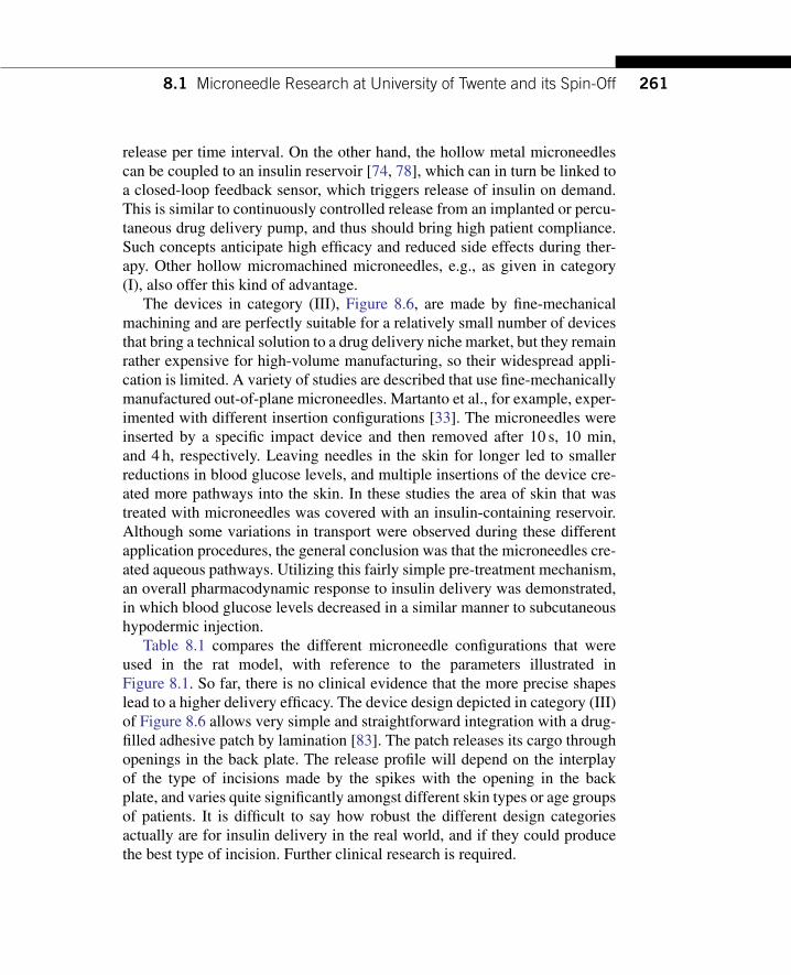
The devices in category (III), Figure 8.6, are made by fine-mechanicalmachining and are perfectly suitable for a relatively small number of devicesthat bring a technical solution to a drug delivery niche market, but they remainrather expensive for high-volume manufacturing, so their widespread appli-cation is limited. A variety of studies are described that use fine-mechanicallymanufactured out-of-plane microneedles. Martanto et al., for example, exper-imented with different insertion configurations [33]. The microneedles wereinserted by a specific impact device and then removed after 10 s, 10 min,and 4 h, respectively. Leaving needles in the skin for longer led to smallerreductions in blood glucose levels, and multiple insertions of the device cre-ated more pathways into the skin. In these studies the area of skin that wastreated with microneedles was covered with an insulin-containing reservoir.Although some variations in transport were observed during these differentapplication procedures, the general conclusion was that the microneedles cre-ated aqueous pathways. Utilizing this fairly simple pre-treatment mechanism,an overall pharmacodynamic response to insulin delivery was demonstrated,in which blood glucose levels decreased in a similar manner to subcutaneoushypodermic injection.
Table 8.1 compares the different microneedle configurations that wereused in the rat model, with reference to the parameters illustrated inFigure 8.1. So far, there is no clinical evidence that the more precise shapeslead to a higher delivery efficacy. The device design depicted in category (III)of Figure 8.6 allows very simple and straightforward integration with a drug-filled adhesive patch by lamination [83]. The patch releases its cargo throughopenings in the back plate. The release profile will depend on the interplayof the type of incisions made by the spikes with the opening in the backplate, and varies quite significantly amongst different skin types or age groupsof patients. It is difficult to say how robust the different design categoriesactually are for insulin delivery in the real world, and if they could producethe best type of incision. Further clinical research is required.
LUTTGE
—12-ch08-235-272-9780815515821
—2011/8/24
—18:05
—Page
262—
#28
Table 8.1 Microneedle Arrays for Insulin Delivery in the Diabetic Rat Model
Material, Dneedle Width Thickness Lshaft Needles/Ref. Rneedle (nm) (µm) (µm) (µm) (µm) array Animal Procedure Type of insulin Response
8.1 Microneedle Research at University of Twente and its Spin-Off 263
The various tips depicted in Figure 8.6 all present design varieties that cre-ate a “mechanical cut” and may be defined by their main function: “cutting”.However, their actual function is to release a controlled dose of insulin, andclinicians need to know how the exact size, type and geometric shape of themicroconduits into the skin influence its delivery mechanism. When studyingthe pharmacodynamics of insulin release, one should not forget that the skinis an immunosensitive organ. When incisions are made in it, allowing for-eign bodies to enter, an immune response may be triggered, which may alterthe efficacy of insulin uptake, and also provoke unfavorable side-effects. Thismay also be an explanation for the smaller reduction in blood glucose levelsobserved by Martanto et al. when microneedles were inserted for a longertime or more than once [33]. Quality control of sterility, biocompatibility andefficacy of drug delivery are key for the final product.
Quantification of insertion and delivery processes, as well as the visu-alization of the incisions made by the individual types of microneedles incombination with the use of specific applicator devices, are therefore veryimportant factors in these devices. A comparison on different types of inser-tion procedures and microneedle designs in transdermal drug delivery hasbeen made by the Bouwstra group, but as yet not with respect to its effecton controlled insulin delivery [84]. The cost of statistically relevant clinicalstudies are high, and require a sustained supply of the devices which needapproval by an ethical-medical committee. All the design varieties intro-duced and discussed above show a certain level of technological readinessthat supports this request.
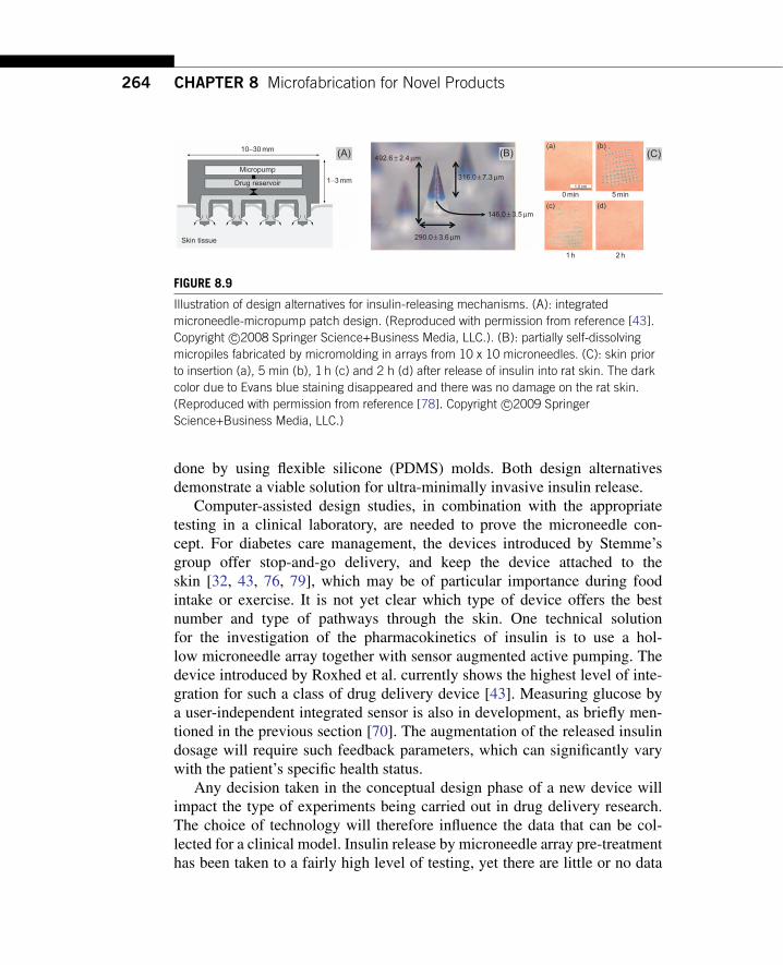
Clinical PerspectiveFrom a clinical point of view, the microneedle array devices summarizedschematically in Figure 8.6 are all of preliminary but potential interest,since the only studies that have used them have been small-scale animaltrials (n< 10). Figure 8.9 shows two extreme design alternatives that haveachieved a certain full proof-of-principle for insulin delivery, using out-of-plane microneedles as depicted in Figure 8.1(B) and (D). Figure 8.9(A)illustrates one such device integrated with a special actuator for the activerelease of insulin that is stored in a reservoir in liquid form. Figure 8.9(B)shows a simpler device, which releases insulin by a phase change, dissolv-ing the polymeric material into the skin without any further active devicecomponents [78]. These self-dissolving microneedles encapsulate the insulinin a chondroitin sulfate matrix during micromolding of arrays, which is
done by using flexible silicone (PDMS) molds. Both design alternativesdemonstrate a viable solution for ultra-minimally invasive insulin release.
Computer-assisted design studies, in combination with the appropriatetesting in a clinical laboratory, are needed to prove the microneedle con-cept. For diabetes care management, the devices introduced by Stemme’sgroup offer stop-and-go delivery, and keep the device attached to theskin [32, 43, 76, 79], which may be of particular importance during foodintake or exercise. It is not yet clear which type of device offers the bestnumber and type of pathways through the skin. One technical solutionfor the investigation of the pharmacokinetics of insulin is to use a hol-low microneedle array together with sensor augmented active pumping. Thedevice introduced by Roxhed et al. currently shows the highest level of inte-gration for such a class of drug delivery device [43]. Measuring glucose bya user-independent integrated sensor is also in development, as briefly men-tioned in the previous section [70]. The augmentation of the released insulindosage will require such feedback parameters, which can significantly varywith the patient’s specific health status.
Any decision taken in the conceptual design phase of a new device willimpact the type of experiments being carried out in drug delivery research.The choice of technology will therefore influence the data that can be col-lected for a clinical model. Insulin release by microneedle array pre-treatmenthas been taken to a fairly high level of testing, yet there are little or no data
available that compare this novel transdermal mechanism of insulin releasewith the gold standard (bolus injection) within a clinically relevant number ofhuman diabetic subjects. Although the working principle of insulin micronee-dle delivery has been confirmed by Gupta et al., utilizing a single, pulled glassneedle at various insertion depths [49], there is still insufficient evidence toshow that a delivery system equipped with an out-of-plane microneedle arraycan deliver insulin in a clinical application.
Delivery technology research can be carried out for the creation of a multi-tude of parallel injection sites with the preliminary devices presented here, butsome level of concern remains regarding the degree of control that is exertedover the device insertion mechanisms. For example, applying high pressuresduring insertion will affect the skin properties, and thus the results of thetransport studies. A delivery platform that includes sensor-augmented releaseis clearly desired, but at this stage of development many kinds of out-of-planeMNA devices can be already translated from the pure engineering sciences toresearch in drug delivery technology. Patient trials, especially those related tocomfort, user-friendliness and pain sensation during the use of such devices,have by now demonstrated an appropriate level of technology readiness forclinical translational research. The initial studies by Gupta et al. [49], pho-tographed the insertion sites to qualitatively evaluate the difference betweeninjection depths. Much more indepth studies of the efficacy of MNA injec-tions will be needed to prove their clinical effectiveness. For micromachinedmicroneedles, new benchmark methods must be adopted to evaluate the clin-ical relevance of the different designs and materials. Both of the two conceptspresented in Figure 8.9 provide a starting point for a further analysis of thespecific design criteria for the application of MNAs in insulin delivery.
From the viewpoints of microengineering, drug delivery technology andclinical translation, the current microdevices show clearly that microsystemsengineering presents certain benefits compared to those designs that use sim-ple fine mechanical solutions or even mechanical assembly. A formal designapproach can offer an exquisite tool for finding appropriate technologicalsolutions for the development of transdermal insulin delivery, based on theaforementioned out-of-plane microneedle designs [85].
8.2 MNA-4-INSULIN: A BRIEF EVALUATIONIn addition to developing MNA fabrication technology, it is also importantto create a clinical focus for the use of microneedles in insulin delivery. This
review gives an overview of the different types of microneedle technologies,which already refer to the proof-of-principle of microneedle insulin delivery.With respect to drug delivery research, devices from the three well-knowncategories of technology (silicon micromachining, replication and precisionengineering) show enhanced transdermal delivery rates for insulin. Specificefficacy/bioavailability data has not yet been gathered, however. The devicesshow few side effects (including safety), and little discomfort during applica-tion, but other clear advantages over the hypodermic needle/syringe conceptcannot yet be confirmed, and there is a lack of data on pharmacodynamicsfor systemic human insulin uptake. Nevertheless, there is no doubt that pas-sive microneedle integrated skin patches could be an affordable solution forthe painless intradermal delivery of insulin. In conclusion, a sufficiently hightechnology readiness of a variety of MNAs has been demonstrated. How-ever, none of the systems discussed in the literature are ready for the market,mainly due to the lack of clinically confirmed reference data. Verification ofthe device quality and pharmacokinetics must be benchmarked first.
8.3 CONCLUSIONSIs it too early or is it already too late to start a profitable business based on MNAtechnology? Based on this discussion of the application needs for insulin, itappears that there is not yet any significant business based on using micronee-dle arrays. Does this mean we just sit and wait for someone to take an initiative,and find a solution for the year 2030? Obviously, this is a rhetorical ques-tion. At the current stage of development it is not possible to judge whichtype of MNA will best deliver insulin via the skin. There are, of course, alsoother means of delivery, and other treatments for diabetes, but the deliveryof insulin by transdermal means should be studied seriously. A great deal ofclinical and business study is needed before further clinical decisions can betaken. Further, it is important to realize that high-tech business developmentbenefits from the collaboration between different research disciplines at thecross-over point: the application. For drug delivery applications it is importantthat researchers from the pharmaceutical, drug delivery and the engineeringsciences meet as early as possible to define a common aim to work on.
Having conducted this desk research, I must ask myself what researchquestions remain for a microfabrication expert in this field, and yes, I canthink of many. Obviously, it is important to ask this question from the
viewpoint of each discipline taking part in the work, and then combine theminto one joint-call proposal for a focused technology development in this spe-cific sector of the life sciences to provide a solution for the 2030 problem.This should be anticipated by the researchers in this field together with appro-priate industry sectors as early as possible. Only this approach will make surethat the required results from the different disciplines can come together at therequired pace of development. A world-wide science and technology fundingstructure has been created that provides research subsidies for public–privatepartnership.
Lessons learned from this case study should inspire students in the engi-neering sciences to start talking to their colleagues in the pharmaceuticaland biomedical sciences and get those partnerships running. Of course, thisdiscussion may also inspire managers of key technologies to adopt micro-fabrication principles in totally different areas to those introduced here. Thischapter may serve as a starting point for a new business case. An ultraearly technology adapter, similar to Unversity of Twente’s spin-off MyLifeTechnologies, may allow many more real-world applications of micro- andnanofabrication technology.
References[1] J. Hogan, Lab on a chip. A little goes a long way, Nature 442 (2006) 351–352.[2] E.X. Vrouwe, R. Luttge, I. Vermes, A. Van Den Berg, Microchip capillary electrophoresis
for point-of-care analysis of lithium, Clin. Chem. 53 (1) (2007) 117–123.[3] H.J.G.E. Gardeniers, R. Luttge, E.J.W. Berenschot, M.J. de Boer, S.Y. Yeshurun,
M. Hefetz, et al., Silicon micromachined hollow microneedles for transdermal liquidtransport, IEEE J. Microelectromech. Syst. 12 (6) (2003) 855–862.
[4] P.C. Mills, S.E. Cross, Transdermal drug delivery: Basic principles for the veterinarian,Vet. J. 172 (2) (2006) 218–233.
[5] S. Godefroy, M. Peyre, N. Garcia, S. Muller, D. Sesardic, C.D. Partidos, Effect of skinbarrier disruption on immune responses to topically applied cross-reacting material,crm197, of diphtheria toxin, Infect. Immun. 73 (8) (2005) 4803–4809.
[6] C.D. Partidos, S. Muller, Decision-making at the surface of the intact or barrier disruptedskin: Potential applications for vaccination or therapy, Cell. Mol. Life Sci. 62 (13) (2005)1418–1424.
[7] D.T. O’Hagan, R. Rappuoli, Novel approaches to vaccine delivery, Pharm. Res. 21 (9)(2004) 1519–1530.
[8] B.W Barry, Novel mechanisms and devices to enable successful transdermal drugdelivery, Eur. J. Pharm. Sci. 14 (2) (2001) 101–114.
[9] M. Kendall, Engineering of needle-free physical methods to target epidermal cells forDNA vaccination, Vaccine 24 (21) (2006) 4651–4656.
[10] F. Chabri, K. Bouris, T. Jones, D. Barrow, A. Hann, C. Allender, et al., Microfabri-cated silicon microneedles for nonviral cutaneous gene delivery, Br. J. Dermatol. 150(5) (2004) 869–877.
[11] J. Birchall, S. Coulman, M. Pearton, C. Allender, K. Brain, A. Anstey, et al., CutaneousDNA delivery and gene expression in ex vivo human skin explants via wet-etchmicrofabricated microneedles, J. Drug Targeting 13 (7) (2005) 415–421.
[12] S. Coulman, C. Allender, J. Birchall, Microneedles and other physical methods for over-coming the stratum corneum barrier for cutaneous gene therapy, Crit. Rev. Ther. DrugCarr. Syst. 23 (3) (2006) 205–258.
[13] M. Pearton, C. Allender, K. Brain, A. Anstey, C. Gateley, N. Wilke, et al., Gene deliveryto the epidermal cells of human skin explants using microfabricated microneedles andhydrogel formulations, Pharm. Res. 25 (2) (2008) 407–416.
[14] Z. Ding, E. Van Riet, S. Romeijn, G.F.A. Kersten, W. Jiskoot, J.A. Bouwstra, Immunemodulation by adjuvants combined with diphtheria toxoid administered topically inBALB/c mice after microneedle array pretreatment, Pharm. Res. 26 (7) (2009) 1635–1643.
[15] Z. Ding, F.J. Verbaan, M. Bivas-Benita, L. Bungener, A. Huckriede, D.J. van denBerg, et al., Microneedle arrays for the transcutaneous immunization of diphtheria andinfluenza in BALB/c mice, J. Controlled Release 136 (1) (2009) 71–78.
[16] S.A. Coulman, A. Anstey, C. Gateley, A. Morrissey, P. McLoughlin, C. Allender, et al.,Microneedle mediated delivery of nanoparticles into human skin, Int. J. Pharm. 366 (1–2)(2009) 190–200.
[17] H. Chen, H. Zhu, J. Zheng, D. Mou, J. Wan, J. Zhang, et al., Iontophoresis-driven pen-etration of nanovesicles through microneedle-induced skin microchannels for enhancingtransdermal delivery of insulin, J. Controlled Release 139 (1) (2009) 63–72.
[18] F.J. Verbaan, S.M. Bal, D.J. van den Berg, J.A. Dijksman, M. van Hecke, H. Verpoorten,et al., Improved piercing of microneedle arrays in dermatomed human skin by an impactinsertion method. J. Controlled Release 128 (1) (2008) 80–88.
[19] S.A. Coulman, D. Barrow, A. Anstey, C. Gateley, A. Morrissey, N. Wilke, et al.,Minimally invasive cutaneous delivery of macromolecules and plasmid DNA viamicroneedles. Curr. Drug Deliv. 3 (1) (2006) 66–75.
[20] D.A. La Van, D.M. Lynn, R. Langer, Moving smaller in drug discovery and delivery, Nat.Rev. Drug Discovery 1 (1) (2002) 77–84.
[21] A.C.R. Grayson, R.S. Shawgo, Y. Li, M.J. Cima, Electronic MEMS for triggereddelivery, Adv. Drug Delivery Rev. 56 (2) (2004) 173–184.
[22] S.Z. Razzacki, P.K. Thwar, M. Yang, V.M. Ugaz, M.A. Burns, Integrated microsystemsfor controlled drug delivery, Adv. Drug Delivery Rev. 56 (2) (2004) 185–198.
[23] C.D. Partidos, Delivering vaccines into the skin without needles and syringes, ExpertRev. Vaccines 2 (6) (2003) 753–761.
[24] M.R. Prausnitz, Microneedles for transdermal drug delivery, Adv. Drug Delivery Rev. 56(5) (2004) 581–587.
[25] Y.B. Schuetz, A. Naik, R.H. Guy, Y.N. Kalia, Emerging strategies for the transder-mal delivery of peptide and protein drugs, Expert Opin. Drug Deliv. 2 (3) (2005)533–548.
[26] S. Sharma, A.J. Nijdam, P.M. Sinha, R.J. Walczak, X. Liu, M.M.-C. Cheng, M. Ferrari,Controlled-release microchips, Expert Opin. Drug Deliv. 3 (3) (2006) 379–394.
[27] R.K. Sivamani, D. Liepmann, H.I. Maibach, Microneedles and transdermal applications,Expert Opin. Drug Deliv. 4 (1) (2007) 19–25.
[28] J. Vandervoort, A. Ludwig, Microneedles for transdermal drug delivery: A minireview,Front. Biosci. 13 (5) (2008) 1711–1715.
[29] A. Arora, M.R. Prausnitz, S. Mitragotri, Micro-scale devices for transdermal drugdelivery, Int. J. Pharmaceutics 364 (2) (2008) 227–236.
[30] H. Kalluri, A.K. Banga, Microneedles and transdermal drug delivery, J. Drug Deliv. Sci.Technol. 19 (5) (2009) 303–310.
[32] L. Nordquist, N. Roxhed, P. Griss, G. Stemme, Novel microneedle patches for activeinsulin delivery are efficient in maintaining glycaemic control: An initial comparisonwith subcutaneous administration, Pharm. Res. 24 (7) (2007) 1381–1388.
[33] W. Martanto, S.P. Davis, N.R. Holiday, J. Wang, H.S. Gill, M.R. Prausnitz, Transdermaldelivery of insulin using microneedles in vivo, Pharm. Res. 21 (6) (2004) 947–952.
[34] B. Stoeber, D. Liepmann, Arrays of hollow out-of-plane microneedles for drug delivery,J. Microelectromech. Syst. 14 (3) (2005) 472–479.
[35] P. Griss, H.K. Tolvanen-Laakso, P. Merilinen, G. Stemme, Characterization of micro-machined spiked biopotential electrodes, IEEE Trans. Biomed. Eng. 49 (6) (2002)597–604.
[36] P. Griss, G. Stemme, Novel, side opened out-of-plane microneedles for microfluidictransdermal interfacing, in: Proceedings of the IEEE Micro Electro Mechanical Systems(MEMS) 2002, pp. 467–470.
[37] P. Griss, G. Stemme, Side-opened out-of-plane microneedles for microfluidic transdermalliquid transfer, J. Microelectromech. Syst. 12 (3) (2003) 296–301.
[38] J.G.E. Gardeniers, J.W. Berenschot, M.J. De Boer, Y. Yeshurun, M. Hefetz,R. Van’t Oever, et al., Silicon micromachined hollow microneedles for transdermal liquidtransfer, in: Proceedings of the IEEE Micro Electro Mechanical Systems (MEMS), 2002,pp. 141–144.
[39] S. Henry, D.V. McAllister, M.G. Allen, M.R. Prausnitz, Microfabricated micronee-dles: A novel approach to transdermal drug delivery, J. Pharm. Sci. 87 (8) (1998)922–925.
[40] D.V. McAllister, S. Kaushik, P.N. Patel, J.L. Mayberry, M.G. Allen, M.R. Prausnitz, Solidand hollow microneedles for transdermal protein delivery, Proc. Controlled Release Soc.(26) (1999) 192–193.
[41] D.V. McAllister, P.M. Wang, S.P. Davis, J.-H. Park, P.J. Canatella, M.G. Allen, et al.,Microfabricated needles for transdermal delivery of macromolecules and nanoparticles:
Fabrication methods and transport studies, Proc. Natl. Acad. Sci. USA 100 (Suppl. 2)(2003) 13755–13760.
[42] F.J. Verbaan, S.M. Bal, D.J. van den Berg, W.H.H. Groenink, H. Verpoorten, R. Lttge,et al., Assembled microneedle arrays enhance the transport of compounds varying overa large range of molecular weight across human dermatomed skin. J. Controlled Release117 (2) (2007) 238–245.
[43] N. Roxhed, P. Griss, G. Stemme, Membrane-sealed hollow microneedles and relatedadministration schemes for transdermal drug delivery, Biomed. Microdevices 10 (2)(2008) 271–279.
[44] M.A. Ling Teo, C. Shearwood, K.C. Ng, J. Lu, S. Moochhala, In vitro and invivo characterization of MEMS microneedles, Biomed. Microdevices 7 (1) (2005)47–52.
[45] J.H. Park, M.G. Allen, M.R. Prausnitz, Biodegradable polymer microneedles: fabrica-tion, mechanics and transdermal drug delivery, J. Controlled Release 104 (1) (2005)51–66.
[46] M.I. Haq, E. Smith, D.N. John, M. Kalavala, C. Edwards, A. Anstey, et al., Clinicaladministration of microneedles: Skin puncture, pain and sensation, Biomed. Microdevices11 (1) (2009) 35–47.
[47] A.L. Teo, C. Shearwood, K.C. Ng, J. Lu, S. Moochhala, Transdermal microneedles fordrug delivery applications, Mater. Sci. Eng. B132 (1–2) (2006) 151–154.
[48] G. Aggarwal, A. Garg, S. Dhawan, Transdermal drug delivery: Evolving technologiesand expanding opportunities, Indian J. Pharm. Educ. Res. 43 (3) (2009) 251–259.
[49] J. Gupta, E.I. Felner, M.R. Prausnitz, Minimally invasive insulin delivery in subjectswith type 1 diabetes using hollow microneedles, Diabetes Technol. Ther. 11 (6) (2009)329–337.
[50] J.-H. Park, M.G. Allen, M.R. Prausnitz, Polymer microneedles for controlled-release drugdelivery, Pharm. Res. 23 (5) (2006) 1008–1019.
[51] T. Matsuda, M. Mizutani, Liquid acrylate-endcapped biodegradable poly(-caprolactone-co-trimethylene carbonate). ii. Computer-aided stereolithographic microarchitecturalsurface photoconstructs, J. Biomed. Mater. Res. 62 (3) (2002) 395–403.
[52] R. Luttge, E.J.W. Berenschot, M.J. de Boer, D.M. Altpeter, E.X. Vrouwe, A. van denBerg, et al., Integrated lithographic molding for microneedle-based devices, J. Micro-electromech. Syst. 16 (4) (2007) 872–884.
[53] G. Zhao, W. Li, G. Tang, J. Chen, Fabrication of bulk titanium out-of-plane micronee-dles, in: 4th IEEE International Conference on Nano/Micro Engineered and MolecularSystems, NEMS 2009, 2009, pp. 428–431.
[54] A. Doraiswamy, C. Jin, R.J. Narayan, P. Mageswaran, P. Mente, R. Modi, et al., Two pho-ton induced polymerization of organic-inorganic hybrid biomaterials for microstructuredmedical devices, Acta Biomater. 2 (3) (2006) 267–275.
[55] K. Lee, H.C. Lee, D.-S. Lee, H. Jung, Drawing lithography: Three-dimensional fabrica-tion of an ultrahigh-aspect-ratio microneedle, Adv. Mater. 22 (4) (2010) 483–486.
[56] Q. Zhu, V.G. Zarnitsyn, L. Ye, Z. Wen, Y. Gao, L. Pan, et al., Immunization by vaccine-coated microneedle arrays protects against lethal influenza virus challenge, Proc. Natl.Acad. Sci. USA 106 (19) (2009) 7968–7973.
[57] B. Al-Qallaf, D.B. Das, Optimization of square microneedle arrays for increasing drugpermeability in skin, Chem. Eng. Sci. 63 (9) (2008) 2523–2535.
[58] Y.-G. Lv, J. Liu, Y.-H. Gao, B. Xu, Modeling of transdermal drug delivery with amicroneedle array, J. Micromech. Microeng. 16 (11) (2006) 2492–2501.
[59] A.K. Banga, Microporation applications for enhancing drug delivery, Expert Opin. DrugDeliv. 6 (4) (2009) 343–354.
[60] H.S. Gill, M.R. Prausnitz, Pocketed microneedles for drug delivery to the skin, J. Phys.Chem. Solids 69 (5–6) (2008) 1537–1541.
[61] J. Ji, F.E.H. Tay, J. Miao, Microfabricated hollow microneedle array using ICP etcher,J. Phys. Conf. Ser. 34 (1) (2006) 1132–1136.
[62] S.J. Moon, C.Y. Jin, S.S. Lee, A novel method of microneedle array fabrication usinginclined deep x-ray exposure, J. Phys. Conf. Ser. 34 (1) (2006) 180–186.
[63] R.F. Donnelly, D.I.J. Morrow, P.A. McCarron, A. David Woolfson, A. Morrissey,P. Juzenas, et al., Microneedle arrays permit enhanced intradermal delivery of apreformed photosensitizer, Photochem. Photobiol. 85 (1) (2009) 195–204.
[64] C.S. Kolli, A.K. Banga, Characterization of solid maltose microneedles and their use fortransdermal delivery, Pharm. Res. 25 (1) (2008) 104–113.
[65] G. Li, A. Badkar, S. Nema, C.S. Kolli, A.K. Banga, In vitro transdermal delivery of thera-peutic antibodies using maltose microneedles. Int. J. Pharm. 368 (1–2) (2009) 109–115.
[68] D. Trebotich, J.D. Zahn, D. Liepmann, Complex fluid dynamics in bioMEMS devices:Modeling of microfabricated microneedles, in: 2002 International Conference on Com-putational Nanoscience and Nanotechnology - ICCN 2002, 2002, pp. 20–23.
[69] D. Trebotich, J.D. Zahn, B. Prabhakarpandian, D. Liepmann, Modeling of microfabri-cated microneedles for minimally invasive drug delivery, sampling and analysis, Biomed.Microdevices 5 (3) (2003) 245–251.
[70] J.D. Zahn, Y.-C. Hsieh, M. Yang, Components of an integrated microfluidic device forcontinuous glucose monitoring with responsive insulin delivery, Diabetes Technol. Ther.7 (2005) 536–545.
[71] M.R. Prausnitz, Overcoming skin’s barrier: The search for effective and user-friendlydrug delivery, Diabetes Technol. Ther. 3 (2) (2001) 233–236.
[72] P. Zhang, G.A. Jullien, Microneedle array for drug delivery and fluid extraction, in:Proceedings, (2005) International Conference on MEMS, NANO and Smart Systems,ICMENS Digital Object Identifier: 10.1109/ICMENS.2005.71.
[73] R. Zhang, P. Zhang, C. Dalton, G.A. Jullien, Modeling of drug delivery into tissues witha microneedle array using mixture theory, Biomech. Model. Mechanobiol. (2009) 1–10.
[74] S.P. Davis, W. Martanto, M.G. Allen, M.R. Prausnitz, Hollow metal microneedles forinsulin delivery to diabetic rats, IEEE Trans. Biomed. Eng. 52 (5) (2005) 909–915.
[75] B. Ma, S. Liu, Z. Gan, G. Liu, X. Cai, H. Zhang, et al., A PZT insulin pump integratedwith a silicon microneedle array for transdermal drug delivery, Microfluid. Nanofluid. 2(5) (2006) 417–423.
[76] N. Roxhed, B. Samel, L. Nordquist, P. Griss, G. Stemme, Compact, seamless integra-tion of active dosing and actuation with microneedles for transdermal drug delivery, in:Proceedings of the IEEE International Conference on Micro Electro Mechanical Systems(MEMS), vol. 2006, 2006, pp. 414–417.
[77] Y. Ito, E. Hagiwara, A. Saeki, N. Sugioka, K. Takada, Feasibility of microneedles forpercutaneous absorption of insulin, Eur. J. Pharm. Sci. 29 (2006) 82–88.
[78] Y. Ito, T. Yamazaki, N. Sugioka, K. Takada, Self-dissolving micropile array tips forpercutaneous administration of insulin, J. Mater. Sci. Mater. Med. 21 (2) (2009) 835–841.
[79] N. Roxhed, B. Samel, L. Nordquist, P. Griss, G. Stemme, Painless drug delivery throughmicroneedle-based transdermal patches featuring active infusion, IEEE Trans. Biomed.Eng. 55 (3) (2008) 1063–1071.
[80] A. Davidson, B. Al-Qallaf, D.B. Das, Transdermal drug delivery by coated microneedles:Geometry effects on effective skin thickness and drug permeability, Chem. Eng. Res. Des.86 (11) (2008) 1196–1206.
[81] B. Al-Qallaf, D.B. Das, Optimizing microneedle arrays for transdermal drug delivery:Extension to non-square distribution of microneedles, J. Drug Targeting 17 (2) (2009)108–122.
[82] M. Cormier, B. Johnson, M. Ameri, K. Nyam, L. Libiran, D.D. Zhang, P. Daddona,Transdermal delivery of desmopressin using a coated microneedle array patch system,J. Controlled Release 97 (3) (2004) 503–511.
[83] D.P. Wermeling, S.L. Banks, D.A. Hudson, H.S. Gill, J. Gupta, M.R. Prausnitz, et al.,Microneedles permit transdermal delivery of a skin-impermeant medication to humans,Proc. Natl. Acad. Sci. USA 105 (6) (2008) 2058–2063.
[84] S.M. Bal, J. Caussin, S. Pavel, J.A. Bouwstra, In vivo assessment of safety of microneedlearrays in human skin, Eur. J. Pharm. Sci. 35 (3) (2008) 193–202.