Microparticles from Patients with Metabolic Syndrome Induce Vascular Hypo-Reactivity via Fas/Fas-Ligand Pathway in Mice Abdelali Agouni 1,2¤ , Pierre-Henri Ducluzeau 1,3 , Tarek Benameur 1 , Se ´ bastien Faure 1 , Martina Sladkova 1,4 , Lucie Duluc 1 , Georges Leftheriotis 5 , Olga Pechanova 5 , Mirela Delibegovic 2 , Maria Carmen Martinez 1 , Ramaroson Andriantsitohaina 1 * 1 INSERM, U694, Angers, France; Universite ´ d’Angers, Angers, France, 2 Institute of Biological and Environmental Sciences, University of Aberdeen, Aberdeen, Scotland, United Kingdom, 3 De ´partement d’Endocrinologie et Diabe ´tologie, CHU d’Angers, Angers, France, 4 Institute of Normal and Pathological Physiology, Slovak Academy of Sciences, Bratislava, Slovak Republic, 5 INSERM, U771, Angers, France Abstract Microparticles are membrane vesicles with pro-inflammatory properties. Circulating levels of microparticles have previously been found to be elevated in patients with metabolic syndrome (MetS). The present study aimed to evaluate the effects of in vivo treatment with microparticles, from patients with MetS and from healthy subjects (HS), on ex vivo vascular function in mice. Microparticles isolated from MetS patients or HS, or a vehicle were intravenously injected into mice, following which vascular reactivity in response to vasoconstrictor agonists was assessed by myography with respect to cyclo-oxygenase pathway, oxidative and nitrosative stress. Injection of microparticles from MetS patients into mice induced vascular hypo- reactivity in response to serotonin. Hypo-reactivity was associated with up-regulation of inducible NO-synthase and increased production of NO, and was reversed by the NO-synthase inhibitor (N G -nitro-L-arginine). The selective COX-2 inhibitor (NS398) reduced the contractile effect of serotonin in aortas from mice treated with vehicle or HS microparticles; however, this was not observed within mice treated with MetS microparticles, probably due to the ability of MetS microparticles to enhance prostacyclin. MetS microparticle-mediated vascular dysfunction was associated with increased reactive oxygen species (ROS) and enhanced expression of the NADPH oxidase subunits. Neutralization of the pro- inflammatory pathway Fas/FasL completely prevented vascular hypo-reactivity and the ability of MetS microparticles to enhance both inducible NO-synthase and monocyte chemoattractant protein-1 (MCP-1). Our data provide evidence that microparticles from MetS patients induce ex vivo vascular dysfunction by increasing both ROS and NO release and by altering cyclo-oxygenase metabolites and MCP-1 through the Fas/FasL pathway. Citation: Agouni A, Ducluzeau P-H, Benameur T, Faure S, Sladkova M, et al. (2011) Microparticles from Patients with Metabolic Syndrome Induce Vascular Hypo- Reactivity via Fas/Fas-Ligand Pathway in Mice. PLoS ONE 6(11): e27809. doi:10.1371/journal.pone.0027809 Editor: Pieter H. Reitsma, Leiden University Medical Center, The Netherlands Received July 6, 2011; Accepted October 25, 2011; Published November 15, 2011 Copyright: ß 2011 Agouni et al. This is an open-access article distributed under the terms of the Creative Commons Attribution License, which permits unrestricted use, distribution, and reproduction in any medium, provided the original author and source are credited. Funding: This work was supported in part by grants from Fonds Europe ´en pour le De ´veloppement Re ´gional (R.A. nu 8891), Fondation pour la Recherche Me ´ dicale (R.A. nu INE20050303433 and MCM nu INE20060306500), CNRS, INSERM and Universite ´ d’Angers. A.A. and T.B. are recipients of doctoral fellowships from the French Education Ministry (MENRT). R.A. is supported by a ‘‘Contrat d’Interface’’ INSERM. The funders had no role in study design, data collection and analysis, decision to publish, or preparation of the manuscript. Competing Interests: The authors have declared that no competing interests exist. * E-mail: [email protected]¤ Current address: Faculty of Health and Medical Sciences, University of Surrey, Guildford, England, United Kingdom Introduction Metabolic syndrome (MetS) is associated with glucose intoler- ance, obesity, ageing, elevated blood pressure and dyslipidaemia, all of which are risk factors associated with cardiovascular morbidity and mortality [1,2]. The prevalence of MetS is increasing and continues to provide challenges for medical research beyond its clinical and public health importance. The pathophysiology of MetS seems to be largely attributable to insulin resistance with the implication of excessive flux of fatty acids [1,2], but also to a pro-inflammatory state resulting from the production of cytokines from adipocytes and macrophages [1,3,4,5]. Thus, increased inflammatory factors and reactive oxygen species (ROS) are associated with detrimental cardiovas- cular alterations linked to MetS. Inflammation is orchestrated by the interactions between inflammatory cells (such as leukocytes) and vascular cells (endothelial and smooth muscle cells) which under activation or apoptosis (for example) lead to the release of circulating microparticles (MPs) [6,7]. MPs are membrane vesicles with pro-coagulant and pro- inflammatory properties [6,7]. The mechanism of MP formation is complex and has yet to be clearly elucidated, due to cell type and stimuli specificity. However, evidence suggest that following cell activation or apoptosis, MP formation occurs due to the sustained elevation in cytosolic calcium concentration in addition to the consequent activation of calpain and protein kinases and the inhibition of phosphatases. In addition to MP formation, these changes result in cytoskeletal reorganization and membrane blebbing [6,8,9,10]. The mechanism of MPs clearance from the circulation is currently unknown, but due to their small size, MPs are believed to be more readily diffusible than cells, and are able to escape phagocytosis [11]. PLoS ONE | www.plosone.org 1 November 2011 | Volume 6 | Issue 11 | e27809
Transcript
Microparticles from Patients with Metabolic SyndromeInduce Vascular Hypo-Reactivity via Fas/Fas-LigandPathway in MiceAbdelali Agouni1,2¤, Pierre-Henri Ducluzeau1,3, Tarek Benameur1, Sebastien Faure1, Martina
Sladkova1,4, Lucie Duluc1, Georges Leftheriotis5, Olga Pechanova5, Mirela Delibegovic2, Maria Carmen
Martinez1, Ramaroson Andriantsitohaina1*
1 INSERM, U694, Angers, France; Universite d’Angers, Angers, France, 2 Institute of Biological and Environmental Sciences, University of Aberdeen, Aberdeen, Scotland,
United Kingdom, 3 Departement d’Endocrinologie et Diabetologie, CHU d’Angers, Angers, France, 4 Institute of Normal and Pathological Physiology, Slovak Academy of
Sciences, Bratislava, Slovak Republic, 5 INSERM, U771, Angers, France
Abstract
Microparticles are membrane vesicles with pro-inflammatory properties. Circulating levels of microparticles have previouslybeen found to be elevated in patients with metabolic syndrome (MetS). The present study aimed to evaluate the effects ofin vivo treatment with microparticles, from patients with MetS and from healthy subjects (HS), on ex vivo vascular function inmice. Microparticles isolated from MetS patients or HS, or a vehicle were intravenously injected into mice, following whichvascular reactivity in response to vasoconstrictor agonists was assessed by myography with respect to cyclo-oxygenasepathway, oxidative and nitrosative stress. Injection of microparticles from MetS patients into mice induced vascular hypo-reactivity in response to serotonin. Hypo-reactivity was associated with up-regulation of inducible NO-synthase andincreased production of NO, and was reversed by the NO-synthase inhibitor (NG-nitro-L-arginine). The selective COX-2inhibitor (NS398) reduced the contractile effect of serotonin in aortas from mice treated with vehicle or HS microparticles;however, this was not observed within mice treated with MetS microparticles, probably due to the ability of MetSmicroparticles to enhance prostacyclin. MetS microparticle-mediated vascular dysfunction was associated with increasedreactive oxygen species (ROS) and enhanced expression of the NADPH oxidase subunits. Neutralization of the pro-inflammatory pathway Fas/FasL completely prevented vascular hypo-reactivity and the ability of MetS microparticles toenhance both inducible NO-synthase and monocyte chemoattractant protein-1 (MCP-1). Our data provide evidence thatmicroparticles from MetS patients induce ex vivo vascular dysfunction by increasing both ROS and NO release and byaltering cyclo-oxygenase metabolites and MCP-1 through the Fas/FasL pathway.
Citation: Agouni A, Ducluzeau P-H, Benameur T, Faure S, Sladkova M, et al. (2011) Microparticles from Patients with Metabolic Syndrome Induce Vascular Hypo-Reactivity via Fas/Fas-Ligand Pathway in Mice. PLoS ONE 6(11): e27809. doi:10.1371/journal.pone.0027809
Editor: Pieter H. Reitsma, Leiden University Medical Center, The Netherlands
Received July 6, 2011; Accepted October 25, 2011; Published November 15, 2011
Copyright: � 2011 Agouni et al. This is an open-access article distributed under the terms of the Creative Commons Attribution License, which permitsunrestricted use, distribution, and reproduction in any medium, provided the original author and source are credited.
Funding: This work was supported in part by grants from Fonds Europeen pour le Developpement Regional (R.A. nu 8891), Fondation pour la RechercheMedicale (R.A. nu INE20050303433 and MCM nu INE20060306500), CNRS, INSERM and Universite d’Angers. A.A. and T.B. are recipients of doctoral fellowships fromthe French Education Ministry (MENRT). R.A. is supported by a ‘‘Contrat d’Interface’’ INSERM. The funders had no role in study design, data collection and analysis,decision to publish, or preparation of the manuscript.
Competing Interests: The authors have declared that no competing interests exist.
by waist circumference), enhanced triglyceridemia, and increased
blood pressure. HbA1c values were not higher than 7.5% both in
MetS patients and HS. Insulin levels were significantly higher in
MetS patients, indicating insulin resistance. In addition, MetS
patients exhibited lower levels of adiponectin compared to control
subjects, although leptin levels were similar in both groups,
supporting insulin resistance within the MetS patients [31]. Over
half of the MetS patients exhibited 4 out of 5 MetS criteria
(Table 1).
MP-Induced Hypo-Reactivity in Metabolic Syndrome
PLoS ONE | www.plosone.org 3 November 2011 | Volume 6 | Issue 11 | e27809
Consistent with our previous findings [12], we found that total
circulating levels of MPs and platelet-, endothelial- and red cell-
derived MPs were significantly enhanced when compared with HS
MPs (Table S1).
MetS MPs induce ex vivo vascular hypo-reactivity inmouse aorta
5-HT produced a concentration-dependent increase in tension
in aortic rings from all groups of mice; however, vascular reactivity
to the agonist was markedly decreased in mice treated with MetS
MPs compared to those treated with either vehicle or HS MPs
(Fig. 1A). Interestingly, the aortas from MetS MP-treated mice,
compared to those from vehicle- or HS MP-treated animals, also
exhibited an impaired contractile response to the concomitant
application of KCl (80 mmoll21) and a single concentration of
another vaso-constrictor agonist, thromboxane A2 agonist U46619
(100 nmoll21) (Fig. 1B). These data suggest that the MetS MP-
induced vascular hypo-reactivity is agonist-independent.
Involvement of NO in MetS MP-induced vascular hypo-reactivity
To investigate the role of NO, the effect of the NO-synthase
inhibitor, L-NA, was studied in response to 5-HT treatment.
Interestingly, we found that inhibition of the NO pathway
completely prevented the vascular hypo-reactivity induced by
MetS MPs (Fig. 2A), suggesting that NO may be involved in the
mechanism of this vascular hypo-reactivity. Direct in situ
measurements of NO production were performed by EPR
spectroscopy using Fe(DETC)2 as a spin trap. Aortas from vehicle,
HS MP- and MetS MP-treated mice, exhibited an EPR feature of
signals derived from NO-Fe(DETC)2. The NO-Fe(DETC)2 EPR
signal was greater in aortas from MetS MPs-treated mice
compared to vehicle- and HS MP-treated mice (Fig. 2B).
Moreover, MetS MPs markedly increased iNOS expression in
mouse aorta compared to vehicle or HS MPs (Fig. 2C) indicating
elevated enhanced NO production.
MetS MPs increase ROS production and increase NADPHoxidase expression
Aorta sections from mice treated with MetS MPs displayed an
increase in vascular wall (endothelium and in the media layer)
ROS production compared to vessels from vehicle or HS MP-
treated mice, measured through EtBr fluorescence (Fig. 3A).
We evaluated expression of membrane (gp91phox) and cytosolic
(p47phox and p67phox) subunits of NADPH oxidase, a major source
Table 1. Baseline characteristics of subjects.
Control subjects MetS patients
Number 17 21
Mean age (years) 5462 5662
Sex ratio (male/female) 12/5 17/4
BMI (kg/m2) 2861.3 3461.1c
Waist circumference (cm) 9463.2 11362.3c
Ratio Waist/Hips 0.9560.02 1.0160.01a
Systolic blood pressure (mm Hg) 12162 13563c
Diastolic blood pressure (mm Hg) 7462 7962a
Glycemia (mmol/l) 5.3360.11 6.5560.22c
Insulinemia (pmol/l) 61.669.1 163.7617.8c
HbA1c (%) 5.660.06 6.1760.24a
Total cholesterol (mmol/l) 660.34 5.360.25
HDL cholesterol (mmol/l) 1.9260.16 1.6360.13
LDL cholesterol (mmol/l) 3.260.26 2.6460.21
Triglycerides (mmol/l) 1.360.11 2.4260.33c
Leptin (mg/l) 15.364.8 15.263.11
Adiponectin (mg/l) 8.160.8 5.360.34b
Number of MetS components (%)
0 36 -
1 41 -
2 23 -
3 - 38
4 - 52
5 - 10
Treatments (%)
Oral antidiabetic 0 41
Insulin 0 2
Antihypertensive 1 70
Statins 2 2
Baseline characteristics of MetS patients (n = 21) compared to control subjects(n = 17). Subjects were fasted before blood collection. All values are expressedin International System (SI) units.aP,0.05;bP,0.01;cP,0.001.doi:10.1371/journal.pone.0027809.t001
Figure 1. MetS MPs induce vascular hypo-reactivity in mouse aortas. Concentration-response curves to 5-HT (A) and contractile response tothe concomitant application of KCl and a single concentration of U46619 (B) in aortic rings from mice treated with either vehicle (CTL, n = 7), healthysubject MPs (HS MPs, n = 8) or MetS patient MPs (MetS MPs, n = 8). ***P,0.001 MetS MPs vs. CTL; ## P,0.001 MetS MPs vs. HS MPs.doi:10.1371/journal.pone.0027809.g001
MP-Induced Hypo-Reactivity in Metabolic Syndrome
PLoS ONE | www.plosone.org 4 November 2011 | Volume 6 | Issue 11 | e27809
of cellular superoxide anion (O22). Interestingly, MetS MPs
markedly enhanced the expression of gp91phox, as well as p47phox
without affecting the level of p67phox in mouse aorta (Fig. 3B),
accounting probably for an increase in NADPH oxidase activity,
while vehicle or HS MPs produced no effect. To evaluate the
capacity of the vessels to reduce O22 in the presence of MPs, we
examined expression of different isoforms of SOD. MPs from both
HS and MetS patients did not significantly affect Mn-SOD, Cu/
Zn-SOD or Ec-SOD expression compared to the vehicle control
(Fig. 3C).
Involvement of COX metabolites in MetS MP-inducedvascular hypo-reactivity
To investigate the role of COX metabolites in 5-HT-induced
vaso-reactivity, the effects of both a non-selective inhibitor of
COX (indomethacin) and a selective inhibitor of COX-2 (NS398),
were examined.
In the presence of indomethacin, contractile response to 5-HT
was reduced in aortas from all groups of mice (Fig. 4A–4C). Thus,
vascular hypo-reactivity to 5-HT was still present in aortas from
mice treated with MetS MPs compared to HS MPs or vehicle.
These results highlight that vasoconstrictor metabolite(s) sensitive
to indomethacin participate in 5-HT-induced contraction
(Fig. 4D). When COX-2 was specifically inhibited using NS398,
the response to 5-HT was impaired in vessels from vehicle and HS
MP injected mice (Fig. 4E–4F), suggesting the contribution of
COX-2-derived vasoconstrictor metabolites. By contrast, inhibi-
tion of COX-2 did not modify the response induced by MetS MPs
(Fig. 4G). These results suggest that MetS MPs treatment leads to
the release of both vasodilator and vasoconstrictor metabolites and
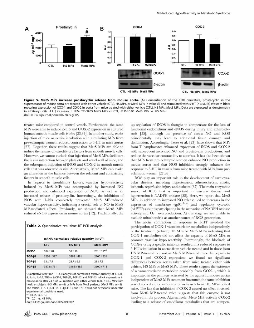
the former blunted the effect of the latter. Interestingly, MetS MPs
enhanced the production of prostacyclin, a vasorelaxant metab-
olite, independently of modification in either COX-1 or COX-2
expressions (Fig. 5B).
MetS MPs increase MCP-1 expression in mice aortasIn order to evaluate the effect(s) of MPs on inflammatory
processes, we analyzed the mRNA expression of pro-inflammatory
cytokines. As shown in Table 2, MetS MPs increased the
expression of MCP-1, a potent agonist of monocytes, memory T
cells, and basophils. However, MPs either from MetS patients or
HS had no effect on mRNA expression of TGF-b1, TGF-b2 and
TGF-b3 (Table 2). The mRNA expression of the other cytokines
(IL-6, IL-8, IL-1a, IL-1b, IL-10 and TNF-a) was not detectable
under the experimental conditions used. MCP-1 mRNA increase
was reinforced by our observation that aortas from mice treated
with MetS MPs exhibited enhanced MCP-1 protein expression
compared to those from mice treated either with vehicle or HS
MPs (Fig. 6D).
Involvement of Fas/FasL signaling pathway in thevascular MetS MP-induced hypo-reactivity
Interestingly, we found that MPs from MetS patients expressed
FasL (Fig. 6A). When we neutralized FasL harbored by MetS
MPs, using specific antibodies, prior to their injection into mice,
we completely prevented the hypo-reactivity previously observed
in mouse aorta (Fig. 6B). This indicates that Fas/FasL pathway is
involved in MP-induced vascular dysfunction. Interestingly,
neutralizing FasL also prevented the MetS MP-induced increase
of iNOS (Fig. 6C) and MCP-1 (Fig. 6D).
Figure 2. Involvement of NO in MetS MP-induced vascularhypo-reactivity. (A) Concentration-response curves to 5-HT of aorticrings from mice treated with either vehicle (CTL, n = 7), HS MPs (n = 8),or MetS MPs (n = 8) in the presence of NO-synthase inhibitor (L-NA,100 mmol/l). (B) Quantification of the amplitude of NO-Fe(DETC)2 signalsin aorta from mice treated with either vehicle, HS MPs or MetS MPs.Values are expressed as units/mg weight of dried (dW) aorta (n = 7). (C)Western blot revealing expression of inducible NO-synthase (iNOS) inaorta from mice treated with either vehicle, HS MPs or MetS MPs. Dataare expressed as denstometry in arbitrary units (A.U.) as mean 6 SEM.* P,0.05 vs. CTL; # P,0.05 MetS MPs vs. HS MPs; ## P,0.01 MetS MPsvs. HSMPs.doi:10.1371/journal.pone.0027809.g002
MP-Induced Hypo-Reactivity in Metabolic Syndrome
PLoS ONE | www.plosone.org 5 November 2011 | Volume 6 | Issue 11 | e27809
MP-Induced Hypo-Reactivity in Metabolic Syndrome
PLoS ONE | www.plosone.org 6 November 2011 | Volume 6 | Issue 11 | e27809
Discussion
In the present study, we demonstrated that i.v. injection of MetS
MPs promotes vascular hypo-reactivity in mice. This may be an
effect of the release of vasodilatory products from different cellular
origins (for example endothelial cells, smooth muscle cells, or
fibroblasts). Interestingly, we found that the observed vascular
hypo-reactivity was associated with overproduction of vasodilator
mediators, such as NO and prostacyclin, as well as increased ROS
in the aorta. These effects resulted in an up-regulation of iNOS
without changes in COX-1 and COX-2 expression, and enhanced
expression of the NADPH-oxidase subunits, gp91phox and p47phox.
Of particular interest is the finding that Fas/FasL pathway is
involved in MetS MP-induced vascular hypo-reactivity. MetS MPs
also markedly increased MCP-1 mRNA and protein levels,
without changing the mRNA levels of other pro-inflammatory
cytokines in the aorta. These data provide valuable information in
our understanding of some of the paracrine roles that MPs play as
vectors of trans-cellular messengers in promoting vascular
dysfunction during MetS.
MPs contribute, at least in part, to the alterations of vascular
function in many cardiovascular diseases (for review [7,9,32]). In
our previous work [12], we showed that MetS patients had
increased circulating levels of MPs compared to HS. In particular,
we observed an increase in endothelial-, erythrocyte-, and platelet-
derived MPs in addition to pro-coagulant (annexin V+) MPs in
MetS patients. We also reported previously an association between
increased circulating MPs and their properties in inducing
endothelial dysfunction both in vitro and in vivo. We showed that
the effect of MetS MPs on endothelial cells is driven by non
platelet-derived MPs [12]. In the present study, total circulating
MPs from HS or MetS patients were injected into mice, in order to
mimic the in vivo situation where all subpopulations of MPs co-
exist. Interestingly, we provide further evidence that MetS MPs
are also able to promote vascular hypo-reactivity in response to
vaso-constrictor agonists in mice aortas. These findings are in line
with our previous studies where MPs generated in vitro from
apoptotic T lymphocytes or MPs obtained either from diabetic
patients [23] or pre-eclamptic women [27,33] induced vascular
hypo-reactivity in mice aortas. In these previous studies, vascular
hypo-reactivity was observed even in vessels without functional
endothelium. In addition, lymphocyte-derived MPs injected in vivo
were able to interact directly with smooth muscle as evidenced by
an increased CD4 labelling in the media layer of aortas from MP-
Figure 3. MetS MPs increase ROS production and enhance NADPH oxidase expression. (A) Detection of ROS production in the vascularwall. Vessel sections of aorta from mice treated with either vehicle, HS MPs or MetS MPs were incubated 30 min with DHE and visualized by confocalmicroscopy. Results are representative pictures of three independent experiments. Bargraphs show the quantification of ROS. Typical examples ofROS staining in each group are shown. (B and C) Western blots revealing expression of NADPH oxidase subunits (gp91phox, p47phox and p67phox) (B)and expression of SOD isoforms (Mn-SOD, Cu/Zn-SOD and Ec-SOD) (C) in aorta from mice treated with either vehicle, HS MPs or MetS MPs. Data areexpressed as denstometry in arbitrary units (A.U.) as mean 6 SEM. * P,0.05 vs. CTL; ** P,0.01 vs. CTL # P,0.05 vs. HS MPs; ## P,0.01 vs. HSMPs.doi:10.1371/journal.pone.0027809.g003
Figure 4. Involvement of COX metabolites in MetS MP-induced vascular hypo-reactivity. (A–D) Concentration-response curves to 5-HT ofaortic rings from mice treated with either vehicle (CTL, n = 7), HS MPs (n = 8) or MetS MPs (n = 8) in the absence or presence of COX inhibitor(indomethacin, 10 mmol/l). (E–H) Concentration-response curves to 5-HT of aortic rings from mice treated with either vehicle (CTL, n = 7), HS MPs(n = 8) or MetS MP (n = 8) in the absence or presence of COX-2 inhibitor (NS398, 1 mmol/l). ***P,0.001 vs. absence of the inhibitor; ## P,0.01 MetSMPs vs. CTL.doi:10.1371/journal.pone.0027809.g004
MP-Induced Hypo-Reactivity in Metabolic Syndrome
PLoS ONE | www.plosone.org 7 November 2011 | Volume 6 | Issue 11 | e27809
treated mice compared to control vessels. Furthermore, the same
MPs were able to induce iNOS and COX-2 expression in cultured
human smooth muscle cells in vitro [23,34]. In another study, in vivo
injection of mice or ex vivo incubation with circulating MPs from
pre-eclamptic women reduced contraction to 5-HT in mice aortas
[27]. Together, these results suggest that MetS MPs are able to
induce the release of vasodilatory factors from smooth muscle cells.
However, we cannot exclude that injection of MetS MPs facilitates
the in vivo interaction between platelets and vessel wall of mice, and
the subsequent induction of iNOS and COX-2 in smooth muscle
cells that was observed ex vivo. Alternatively, MetS MPs can evoke
an alteration in the balance between the relaxant and constricting
factors in smooth muscle cells.
In regards to vasodilatory factors, vascular hypo-reactivity
induced by MetS MPs was accompanied by increased NO
production and enhanced expression of iNOS, as well as an
increased release of prostacyclin. Interestingly, the inhibition of
NOS with L-NA completely prevented MetS MP-induced
vascular hypo-reactivity, indicating a crucial role of NO in MetS
MP-mediated effects. Previously, we showed that MetS MPs
reduced eNOS expression in mouse aortas [12]. Traditionally, the
up-regulation of iNOS is thought to compensate for the loss of
functional endothelium and eNOS during injury and atheroscle-
rosis [35], although the presence of excess NO and ROS
coincidentally may lead to additional tissue damage and
dysfunction. Accordingly, Tesse et al. [23] have shown that MPs
from T lymphocytes enhanced expression of iNOS and COX-2
with subsequent increased NO and prostacyclin productions, and
reduce the vascular contractility to agonists. It has also been shown
that MPs from pre-eclamptic women enhance NO production in
mouse aortas and that NOS inhibition strongly enhances the
response to 5-HT in vessels from mice treated with MPs from pre-
eclamptic women [27,36].
ROS play an important role in the development of cardiovas-
cular diseases, including hypertension, atherosclerosis, stroke,
ischemia-reperfusion injury and diabetes [37]. The main enzymatic
source of ROS that is important in vascular disease and
hypertension is NADPH oxidase [38]. Here, we report that MetS
MPs, in addition to increased NO release, led to increases in the
expression of membrane (gp91phox) and regulatory cytosolic
(p47phox) subunits participating in the activation of NADPH oxidase
activity and O22 overproduction. At this stage we are unable to
exclude mitochondria as another source of ROS generation.
The aortic contraction in response to 5-HT involved the
participation of COX-1 vasoconstrictor metabolites independently
of the treatment (vehicle, HS MPs or MetS MPs) indicating that
COX-1 metabolites did not affect the capacity of MetS MPs to
promote vascular hypo-reactivity. Interestingly, the blockade of
COX-2 using a specific inhibitor resulted in a reduced response to
5-HT stimulation in aortas from vehicle-treated and in those from
HS MP-treated but not in MetS MP-treated mice. In regards to
COX-1 and COX-2 expression, we found no significant
differences between aortas taken from mice treated either with
vehicle, HS MPs or MetS MPs. These results suggest the existence
of a vasoconstrictor metabolite probably from COX-1, which is
implicated in the pathway activated by the agonist in mouse aortas
independent of MetS MPs treatment inasmuch the same inhibition
was observed either in control or in vessels from HS MPs-treated
mice. The fact that inhibition of COX-2 caused no effect in vessels
from MetS MP-treated mice suggests that this enzyme is not
involved in the process. Alternatively, MetS MPs activate COX-2
leading to a release of vasodilator metabolites that are compen-
Figure 5. MetS MPs increase prostacyclin release from mouse aorta. (A) Concentration of the COX derivative, prostacyclin in thesupernatants of mouse aorta pre-treated with either vehicle (CTL), HS MPs, or MetS MPs (n values?) and stimulated with 5-HT (n = 5). (B) Western blotsrevealing expression of COX-1 and COX-2 in aorta from mice treated with either vehicle (CTL), HS MPs, MetS MPs. Data are expressed as denstometryin arbitrary units (A.U.) as mean 6 SEM. *P,0.05 MetS MPs vs. CTL; # P,0.05 MetS MPs vs. HS MPs.doi:10.1371/journal.pone.0027809.g005
Table 2. Quantitative real time RT-PCR analysis.
mRNA normalized relative quantity (6103)
CTL HS MPs MetS MPs
MCP-1 104628 51616 201617a,b
TGF-b1 32266377 33826481 29606331
TGF-b2 3367.5 28.766.6 2867.3
TGF-b3 38736731 31686485 36006713
Quantitative real time RT-PCR analysis of normalized relative quantity of IL-6, IL-8, IL-1a, IL-1b, TNF-a, MCP-1, TGF-b1, TGF-b2 and TGF-b3 mRNA expressions inmouse aorta after 24 h of i.v. injection with either vehicle (CTL, n = 4), MPs fromhealthy subjects (HS MPs, n = 4) or MPs from MetS patients (MetS MPs, n = 4).The mRNA: IL-6, IL-8, IL-1a, IL-1b, IL-10 and TNF-a was not detectable under theexperimental conditions used.aP,0.05 vs. CTL;bP,0.01 vs. HS MPs.doi:10.1371/journal.pone.0027809.t002
MP-Induced Hypo-Reactivity in Metabolic Syndrome
PLoS ONE | www.plosone.org 8 November 2011 | Volume 6 | Issue 11 | e27809
sated by a release of vasoconstrictor metabolites. The latter
hypothesis is supported by the fact that MetS MPs, but not HS
MPs, were able to induce an increase in the release of prostacyclin.
It is possible that under the conditions studied, prostacyclin may
attenuate the vascular damage induced by NO overproduction,
and may participate to the adaptive response of vascular cells to
MetS MPs.
Evidences from clinical and experimental studies support the
hypothesis that inflammation plays an important role in a wide
range of cardiovascular diseases and have focused attention on the
signals that initiate cellular infiltration of vascular tissues [31,39].
In the current study, we report that MetS MPs increased vascular
expression of MCP-1, which could potentially lead to increased
recruitment of leukocytes under conditions associated with
vascular inflammation. Previously, we reported that MetS MPs
did not affect in vitro expression of MCP-1 mRNA in cultured
endothelial cells [12]. A possible reason for this discrepancy may
be due to additional interactions between MetS MPs and
circulating cells following injection into mice.
FasL is a 40-kDa cytotoxic type II trans-membrane protein
belonging to the tumour necrosis factor1 family. Unlike Fas, which
is constitutively expressed by various cell types, FasL is expressed
primarily by cells of the immune system such as activated T cells
[18,40]. Fas and FasL play a crucial role in the induction of
apoptosis in various cell types [18,40]. By deleting auto-reactive
lymphocytes, Fas/FasL ensure the development of normal T and
B cell repertoires [18,40], preventing autoimmune disorders. In
vascular smooth muscle cells, it has been shown that the activation
of the Fas/FasL pathway results in the increased expression of a
specific program of inflammatory genes [21,22]. Functionally,
FasL has been shown to be pro-inflammatory, and implicated in
pathophysiological processes of various cardiovascular diseases,
such as coronary heart disease, arteriosclerosis, and ischemia-
reperfusion injury [19]. In human atherosclerosis, FasL is
expressed together with markers of apoptosis in inflammatory
regions of plaques [41]. FasL-mediated smooth muscle cell
apoptosis within the vulnerable plaque may lead to plaque
instability and rupture, events well known to cause myocardial
infarction and stroke. Given the role of the pro-inflammatory
pathway Fas/FasL in the expression of inflammatory genes in
smooth muscle cells [21,22], we hypothesized that the vascular
effects of MetS MPs may be mediated by the interaction of FasL,
harbored by MetS MPs, and Fas receptor expressed by smooth
muscle cells from the vascular wall [41]. In the present study, we
find that MetS MPs express FasL and that its neutralization, using
a specific antibody, restores the reactivity in vessels from MetS
MP-injected mice towards reactivity of aortas either from vehicle-
or HS MP-treated mice. Interestingly, neutralization of FasL also
normalizes the expression of iNOS and MCP-1 in aortas from
MetS MP-treated mice indicating the importance of inflammation
and NO in MetS MPs effects. In line with our findings, it has been
reported that proapoptotic stimuli, including FasL, or over-
Figure 6. Involvement of Fas/FasL signaling pathway in the MetS MP-induced hypo-reactivity in mouse aorta. (A) Western Blotshowing FasL expression in MPs obtained from two different patients (P1 and P2). (B) Concentration-response curves to 5-HT of aortic rings from micetreated with vehicle (CTL, n = 7), MetS MPs (n = 8), or MetS MPs pre-incubated with FasL antibody (n = 6). (C–D) Western Blots showing iNOS (C) andMCP-1 (D) expressions in aorta from mice treated with either vehicle, HS MPs MetS MPs, or MetS MPs pre-incubated with FasL antibody. Data areexpressed as denstometry in arbitrary units (A.U.) as mean 6 SEM. ***P,0.001 vs. CTL; # P,0.05 vs.HS MPs; P,0.001 vs.HS MPs; 1 P,0.05 vs. MetSMPs + FasL antibody; 11 P,0.01 vs. MetS MPs + FasL antibody.doi:10.1371/journal.pone.0027809.g006
MP-Induced Hypo-Reactivity in Metabolic Syndrome
PLoS ONE | www.plosone.org 9 November 2011 | Volume 6 | Issue 11 | e27809
expression of Fas-associated death domain protein causes local
accumulation of macrophages and triggers transcriptional upre-
gulation of MCP-1 in vitro and in vivo [21] through a mechanism
involving calpains and caspase 8 [22]. Furthermore, Fas/FasL
interaction is capable to induce NFkB signalling [40] and may
therefore induce iNOS expression [42]. The fact that we found
that neutralizing FasL carried by MPs prevented vascular
dysfunction supports of the hypothesis of an interaction between
FasL from MetS MPs with Fas from the vessel wall of the treated/
injected mouse. In support of this, we showed previously that MPs
injected in vivo are able to interact directly with smooth muscle by
increasing CD4 labelling in the media layer of aortas from MP-
treated mice compared to vessels from control mice [23].
Interestingly, in the current study we noticed that MetS MPs did
not affect the sensitivity to vasoconstrictors as shown by the
absence of differences in the pD2. Nevertheless, MetS MPs act via
other pathways, including but not limited to Fas/FasL, to increase
vascular inflammation and enhance release of vasodilator agents
(NO, prostacyclins) which participate in decreasing the Emax
(hypo-reactivity).
In conclusion, we provide evidence that MetS MPs induce ex
vivo vascular hypo-reactivity to vasoconstrictor agents in aorta by
increasing both oxidative and nitrosative stresses and by increasing
the release of COX-2-derived prostacyclin. In addition, we show
that MetS MPs increase the expression of MCP-1 mRNA in the
vessel wall. The critical role of MPs as a vector of biological
messages leading to vascular dysfunction in MetS involving Fas/
FasL pathway is also underlined.
These effects of MetS MPs, in addition to their capacity to
reduce endothelial vasodilatation, strengthen the notion that MPs
cannot only be considered as surrogate markers of endothelial
dysfunction or injury, but also as effectors able to amplify pre-
existing vascular dysfunction, including vascular hypo-reactivity
and inflammation. Thus, MetS MPs may interfere with mecha-
nisms leading to atherosclerotic plaque development and vascular
thrombosis during the evolution of MetS.
Supporting Information
Table S1 Circulating microparticle levels in patientswith metabolic syndrome compared to healthy subjects.Total MP levels and different populations: platelet- (CD41+),
endothelial- (CD146+), erythrocyte-(CD235+) derived and proco-
agulant (annexin V+) microparticles.
(DOC)
Acknowledgments
Authors thank Dr. Melissa Page for her critical reading the manuscript.
Author Contributions
Conceived and designed the experiments: RA AA. Performed the
experiments: AA P-HD TB SF MS LD GL MCM. Analyzed the data:
AA RA MCM. Contributed reagents/materials/analysis tools: OP MD.
Wrote the paper: AA RA MCM.
References
1. Chew GT, Gan SK, Watts GF (2006) Revisiting the metabolic syndrome.Med J Aust 185: 445–449.
2. Sesti G (2006) Pathophysiology of insulin resistance. Best Pract Res ClinEndocrinol Metab 20: 665–679.
3. Fulop T, Tessier D, Carpentier A (2006) The metabolic syndrome. Pathol Biol
(Paris) 54: 375–386.
4. Magliano DJ, Shaw JE, Zimmet PZ (2006) How to best define the metabolic
intercellular vectors of biological messages. Mol Interv 11: 88–94.
11. Freyssinet JM (2003) Cellular microparticles: what are they bad or good for?J Thromb Haemost 1: 1655–1662.
12. Agouni A, Lagrue-Lak-Hal AH, Ducluzeau PH, Mostefai HA, Draunet-Busson C, et al. (2008) Endothelial dysfunction caused by circulating
microparticles from patients with metabolic syndrome. Am J Pathol 173:1210–1219.
13. Mostefai HA, Meziani F, Mastronardi ML, Agouni A, Heymes C, et al. (2008)Circulating microparticles from patients with septic shock exert protective role in
vascular function. Am J Respir Crit Care Med 178: 1148–1155.
(2004) Elevated plasma endothelial microparticles: preeclampsia versus gesta-tional hypertension. Am J Obstet Gynecol 191: 1418–1424.
15. Priou P, Gagnadoux F, Tesse A, Mastronardi ML, Agouni A, et al. Endothelialdysfunction and circulating microparticles from patients with obstructive sleep