MicroRNAs in oral fluids (saliva and gingivalcrevicular fluid) as biomarkers inorthodontics: systematic review andintegrated bioinformatic analysisPriyanka Kapoor1,2* , Aman Chowdhry1,3 , Dinesh Kumar Bagga4, Deepak Bhargava5 and S. Aishwarya6
Abstract
Background: MicroRNAs (miRNAs) are non-coding short, single-stranded RNA molecules that may serve asbiomarkers for various inflammatory and molecular mechanisms underlying bone and tissue remodelingconsequent to orthodontic force application.
Methods: A thorough literature search in major databases was conducted in March 2021 to generate evidence formiRNAs in orthodontics, with prior PROSPERO registration. The initial search revealed 920 articles, subjected to strictselection criteria according to PRISMA, and resulted in final inclusion of four studies. Quality assessment byQUADAS-2 classified three studies as unclear risk-of-bias while the applicability was high. Further, bioinformaticanalysis was performed to identify the target genes from the miRNA database (miRDB) and TargetScan databasesand their protein-protein interaction pathways with the STRING analysis.
Results: Multiple miRNAs in gingival crevicular fluid (GCF) of orthodontic patients were seen, including miRNA-21,27(a/b), 29(a/b/c), 34,146(a/b), 101, and 214 along with matrix metalloproteinases (MMPs)-1, 2, 3, 8, 9, 14 in onestudy. A statistically significant increase in expression of miRNA-29a/b/c,101, 21 from pre-treatment (before initiationof retraction) was seen to reach a peak at 4–6 weeks (wk) of retraction. On the contrary, miRNA-34a showeddownregulation from the 1 day to 4 wk of retraction and also, negatively correlated with MMPs-2,9,14 levels at thesame observation times. The distance of canine movement showed mild correlation with miRNA-27a/b, 214 at 2 wkof retraction. Bioinformatics revealed 1213 mutual target genes which were analyzed for inter-relational pathwaysusing Cytoscape plugin, MCODE. Further, 894 prominent protein interactions were identified from the STRINGdatabase and SMAD4, IGF1, ADAMTS6, COL4A1, COL1A1, COL3A1, FGFR1, COL19A1, FBN1, COL5A1, MGAT4A, LTBP1,MSR1, COL11A1, and COL5A3 were recognized as the hub genes. Their interactions were able to isolate multiplemiRNAs: hsa-miR-34a-5p, hsa-miR-29b-2-5p, hsa-miR-29b-3p, hsa-miR-34a-3p, hsa-miR-27a-5p, hsa-miR-29a-5p, hsa-miR-29b-1-5p, hsa-miR-29c-3p, hsa-miR-214-5p, hsa-miR-27a-3p, hsa-miR-29a-3p, hsamiR-146-5p, which were foundpromising as biomarkers for tooth movement.
* Correspondence: [email protected] of Dental Sciences, Sharda University, Greater Noida, UP, India2Department of Orthodontics, Faculty of Dentistry, Jamia Millia Islamia, NewDelhi 110025, IndiaFull list of author information is available at the end of the article
Kapoor et al. Progress in Orthodontics (2021) 22:31 https://doi.org/10.1186/s40510-021-00377-1
Conclusions: Our results support using miRNAs as biomarkers in varied orthodontic study designs and for inter-relationships with pathological settings like periodontal disease, pre-malignancies, or conditions like obesity ormetabolic irregularities, etc. The identified target genes and their protein interaction pathways can be used topropose precision therapies, focusing on ideal tooth movement with minimal iatrogenic side-effects.
BackgroundMicroRNAs (miRNAs) are non-coding short regulatoryRNAs, usually known as RNA-interfering systems or gene si-lencing entities, that have a significant role in various patho-logical and physiological activities in the body. They work byregulating gene expression by imperfect complementarybinding of target messenger (mRNA) strands and hence,their derangement can easily serve as biomarkers for differ-ent disease processes or altered growth and their prognosis.The three different kinds of miRNAs, pri-miRNAs (largeRNA precursors in the nucleus), pre-miRNAs (stem-loopstructures transported into the cytoplasm), or mature-miRNAs and their mechanism of action has been depicteddiagrammatically in Fig. 1 [1, 2]. miRNAs can singularlyaffect multiple strands of mRNAs, alter the gene expressionprofile of target genes and their pathways, hence can influ-ence multiple cell-regulating functions by altering the pro-tein–protein interactions, ultimately affecting causation ofdisease and its severity [3, 4].
MiRNAs in dentistryLiterature evidence suggests an association of miRNAs, spe-cifically miR-34a with the development of the tooth, remod-eling of bone, as well as differentiation of dental stem cells[5–7]. Besides, various miRNAs are associated with oral can-cers as well as with a higher risk of conversion of oral prema-lignant lesions to malignancy. Change in expression ofseveral miRNAs has been documented with related risk fac-tors for oral cancer, e.g., increased miR-31 and miR-138 ex-pression and decreased miR-10b, miR-92a, miR-200b, miR-372, miR-375, miR-378a, and miR-145 expression withsmokeless tobacco use. Further, research is being conductedto explore deregulated miRNAs as biomarkers for clinic-pathological indicators, precision treatment, and generationof expression profiles for oral cancer [8].
miRNAs in craniofacial aberrationsmiRNAs expression and function have also been associ-ated with the development of the face, specifically
Fig. 1 A simplified diagram explaining the pathway of microRNA (miRNA) dependent gene expression. Shows processing of three different typesof miRNA, pri-miRNA in nucleus, pre-miRNA migration to cytoplasm, processing of mature mi-RNA in cytoplasm. Inputs from [1, 2]
Kapoor et al. Progress in Orthodontics (2021) 22:31 Page 2 of 15
secondary palate growth [9, 10]. A recent systematic re-view by Mendes et al. 2020 has reported upregulation of57 and downregulation of 43 out of 100 mi-RNAs in dif-ferent categories of cleft lip and palate (CLP) and add-itionally, differential expression of 9 mi-RNAs in cleftpalate (CP) patients [11]. A single study on salivary miR-NAs in CLP patients by Grassia et al. (2017) has revealedmiR-324-3, p miR-141, and miR223 as probable bio-markers of CLP malformation [3].
miRNAs in orthodonticsAttempts have been made to study miRNAs, both inmice and humans, upon application of orthodonticforces as mechanical stimuli are known to initiate a cas-cade of cellular and biochemical processes after the re-lease of various pro-inflammatory markers whichmediate the remodeling of bone and periodontium.These mediators are known to play an active role in os-teoclastogenesis [e.g., interleukin (IL-1β), tumor necrosisfactor (TNF)-α, receptor activator of nuclear kappa lig-and (RANKL), etc.] as well as osteoblastogenesis (e.g.,osteoprotegrin (OPG), IL-4, IL-10, etc.] [12, 13]. Add-itionally, the expression of different miRNAs at multipleobservation times in orthodontic tooth movement(OTM) are correlated with osteoclastic/osteoblastic dif-ferentiation associated with the release of multiple bio-markers in the paracrine environment [7, 14, 15].
miRNA detectionSecretory miRNA can be detected in body fluids like thewhole serum, urine, and cerebrospinal fluid as well as inoral fluids like saliva and gingival crevicular fluid (GCF).Their presence is usually detected in exosomes, whichare micro-vesicles, very small (30–100 nm) in size, andcarry various nucleic acids and proteins [16]. It is welldocumented by utilizing quantitative polymerase chainreaction (qPCR) that miRNA in exosomes in saliva/serum,when compared to exosome-depleted serum/saliva andwhole serum, are much more in number and can be de-tected even in very small quantities [14]. Besides, in ortho-dontic treatment, oral biofluids including GCF and salivamay be the most convenient and patient-compliant sourceof identifying these miRNA biomarkers.
Bioinformatics in orthodonticsBioinformatics involves biological abstracting in the con-text of macromolecules and application of principles of“informatics” (using computational domains: appliedmaths, statistics, and computer science) to organize theevidence gathered from these molecules and compre-hend the derivations on a larger scale for application inmultiple fields of biology [17]. The advent of bioinfor-matics has shown tremendous potential to bridge thegap between basic research and medical or dental
applied sciences. Clinical orthodontics entails a complexphenomenon of hard tissue and soft tissue remodelinginvolving complex interaction of a multitude of cellsconsequent to force application, which is difficult tosummarize and interpret. Janjic et al. (2018) have uti-lized bioinformatics to investigate stress adaptation ofcells upon application of different types of in vitro mech-anical loading models in a systematic review and usedthe STRING analysis to outline the most significantprotein-protein interaction (PPI) networks and signalingpathways [18]. Another in vitro study used bioinformaticanalysis to screen the candidate genes and pathways inresponse to mechanical force on osteocytes, to derive thathypoxia might be the key factor of differential response[19]. A recent in vivo study in mice by Klein Y et al.(2020) identified differential gene expression (DEG) uponcoil spring activation at different time points and pre-sented a heat map of “top 50” genes showing significantchanges, along with two distinct DEG clusters: one show-ing initial downregulation in 3 days (d), followed by upreg-ulation (representing formative events of cellularproliferation, migration, etc.) until 14 d while the otherpeaked initially, only to decrease in 14 d (representing in-flammatory degradative, innate/adaptive immune re-sponses, etc.) [4]. Continuing research in this domain hasbeen contemplated to merge genetics, biology, and ortho-dontic therapeutics to formulate the basis for precision or-thodontics and a personalized treatment approach [20].
Gap in literatureAlthough various biomarkers have been identified in oralfluids at the biochemical level, the evidence is still sparserelated to post-transcriptional regulation of the associ-ated changes by the miRNAs. Hence, a critical evalu-ation of existing literature is required to identify themost potent miRNA biomarkers in OTM, their time-dependent alteration, and their effect on the gene ex-pression profile and protein-protein interactions (PPI),to understand the biology of hard and soft tissue alter-ations and remodeling.Additionally, target genes of the identified functional
miRNAs were corroborated, and a regulatory co-expression network of the target genes and miRNAs wasconstructed to analyze the underlying protein interac-tions. This will provide a basis for understanding specificbiological collaborations in tooth movement and mayfurther the scope of precision therapies and research op-portunities in this domain. Further, the protein interac-tions causing iatrogenic side-effects like apical rootresorption or caries-risk can also be identified.
Aim and objectivesThe current study was conducted with the followingobjectives:
Kapoor et al. Progress in Orthodontics (2021) 22:31 Page 3 of 15
1. To study expression of miRNAs in oral fluids (GCFand saliva) of patients undergoing orthodontictreatment.
2. To identify the target genes of the miRNAs isolatedin orthodontic patients by using miRNA database(miRDB) and TargetScan databases.
Fig. 2 Details of the preferred reporting items of systematic reviews and meta-analysis (PRISMA)
Kapoor et al. Progress in Orthodontics (2021) 22:31 Page 4 of 15
3. To analyze protein-protein interactions and identifyHUB genes using Cytoscape plugin, MolecularComplex Detection (MCODE).
Material and methodsA critical review of the literature was performed to explorethe association of miRNA in oral fluids orthodontics, afterregistration of protocol in PROSPERO (CRD42021244200).A thorough literature search was conducted in major data-bases—PubMed (P), Web of Science (WOS), J-Gate, Direc-tory of Open access journals (DOAJ), Scopus (S), Embase,open access thesis and dissertations (OATD), hand search(HS), and reference tracking in March 2021. There was nolimitation of date or language in search. Both MeSH and freetext terms were used for the search: “microRNA,” “miRNA,”and “Orthodontics.”The following search strategy was used:PubMed/ web of Science/Scopus: (microRNAs) OR
(miRNA) AND (Orthodontic*)Embase: (“microrna”/exp OR microrna) AND
(“mirna”/exp OR mirna) AND (“orthodontics”/exp ORorthodontics)DOAJ/ OATD/J-Gate: (microRNAs) OR (miRNA)
AND (Orthodontics)The initial search was applied by two researchers inde-
pendently (PK and AC). It revealed 920 articles includ-ing 215 in (P), 364 in (S), 261 in Embase, 18 in WOS, 55in J-Gate, 3 in DOAJ, 3 in OATD, and 1 in (HS) [detailsof the preferred reporting items of systematic reviewsand meta-analysis (PRISMA) given in Fig. 2]. After du-plicates removal, strict inclusion and exclusion criteria(Table 1) were applied which yielded six articles [4 in(P), 1 in OATD, 1 in (HS)]. Further exclusion of 2 arti-cles was done due to studies on animal experiments .This led to shortlisting of 4 studies [2 in (P), 1 in (HS), 1in OATD] which were subjected to subsequent qualityassessment. The data extraction of the final studies wasperformed by two investigators (PK, AC) independentlybased on participant characteristics, study design,miRNA studied, oral fluid for isolation, and outcomes(Table 2). Any discordance in any of the data was dis-cussed with a third researcher (DKB) and resolution wasdone after reaching a consensus.
Quality assessmentThe QUADAS (Quality Assessment of Diagnostic Ac-curacy Studies)-2 tool was used to evaluate risk of bias(ROB) for each study [23]. It was performed
independently by two researchers and in discrepancy,consensus by referring to a third evaluator (an additionalfile shows this in more detail [see Additional file 1]).Three out of four studies had an unclear ROB while ap-plicability was high.
Target gene predictionFurther, the target genes for the identified functionalmiRNAs were analyzed by TargetScan which predictsthe targets by computing the 8mer, 7mer, and 6merbinding sites of miRNAs [24], as well as MicroRNA Tar-get Prediction Database (miRDB) which identifies thetargets from the high throughput sequencing experi-ments and machine learning methods [25]. To enhancethe credibility and reliability of target prediction, mutualgenes were selected from both miRDB and TargetScandatabases.
Gene annotation and pathway analysisAnnotation of the target genes offers significant meaningto the genes. Inter-relational gene annotation was per-formed with the ClueGO application (version 2.5.7) [26]installed in the Cytoscape software (version 3.5.10). Ben-jamini Hochberg statistical validation with a kappa scoreof 3, p value < 0.05 annotated the genes for their bio-logical process, molecular function, cellular components,Kyoto Encyclopedia of Genes and Genomes (KEGG)pathways, and immune reactions [27].
Protein interaction network and hub genes predictionProtein-protein interaction (PPI) and co-expression net-works for the encoding of mutual target genes were eval-uated using the STRING database [28]. The potentialinteractions were filtered out with the MCODE applica-tion installed in the Cytoscape software. Interacting pro-teins and the co-expressing proteins were clusteredbased on the haircut option with the networks scoringdegree cutoff of 2, K-score of 2, and node score cutoff of0.2. The cluster with the highest cluster score was se-lected as the hub genes [27].
ResultsThe results have been presented in the followingsequence:
I. Dynamics of miRNAs in oral fluidsII. Target gene identification and annotation
Table 1 Inclusion and Exclusion criteria for studies
Inclusion Exclusion
All original studies on humans including clinical trials, prospective orretrospective cohort studies, studies mentioning both miRNA and toothmovement
In-vitro studies, animal studies, studies on miRNAs but not in orthodontics,studies in orthodontics but not in miRNAs, miRNAs not measured in oralfluids, case reports, and reviews/opinions/guidelines
Kapoor et al. Progress in Orthodontics (2021) 22:31 Page 5 of 15
Table
2Presen
tatio
nof
eviden
cerelatedto
microRN
Asin
orthod
onticsbasedon
participantsandstud
ycharacteristics,GCFhand
ling,
andou
tcom
es
S No.
Autho
rsAge/sexof
stud
yparticipan
ts
Oral
fluid
miRNA
stud
ied
Observation
times
Stud
ych
aracteristics
GCF
Collection
Han
dlin
gAna
lysis
Results
Con
clusion
1[21]
15(10–17
y)patientsfor
testsample,
4M(26–28
y)forstd.
sample
GCF
miRNA-
29(a/b/
c)
5tim
es,T0-priorto
retr.,T1-1
hrpo
stretr.,T2-1
dpo
stretr.,T3-7
dpo
stretr.,T4-6
wk
postretr.
CaninerRetr.force
of250g
Perio
pape
r,Mesialsite
ofcanine
1min.
placem
ent,
repe
at4tim
es
Quan.&qu
al.o
fRN
A;Bioanalyzer,
spectrop
hotometer,
secretorymiRNA
presen
ce:m
iScript
PCRarray,miRNA-
29presen
ce:RT-
real-im
ePC
R
Stat.sign.increase
inlevels
ofmiRNA-29family
from
T0-T4,-spe
cificall
yin
miRNA-29b
:fro
mT0-T1,T0-T3,no
stat.
diffb/w
levelsof
miRNA
29-a,b,c
Presen
ceof
miRNA-29
inGCF(m
ostly
inexo-
some)
inOTM
,rolein
regu
latio
nof
osteo-
clasts&EC
Mmolecule
expression
2[7]
20(12–18
y)GCF
miR-34a
and
MMPs-1,
2,3,8,
9,14
6tim
es,T0-dayof
forceapplication,
T1-1
hrpo
stretr.,
T2-1
dpo
stretr.,
T3-1
wkpo
stretr.,
T4-4
wkpo
stretr.,
T5-12wkpo
stretr.
Canineretr.as
explaine
din
[48],oral
hygien
eprotocol
of0.12%
CLX
glucon
ate
rinse
for1wk,rand
omcanine
asinde
xtooth,
contra.canineas
control,canine
retr.
Perio
pape
r,tension&
compr.
sites
1min.
placem
ent
RT-PCR
T2–T4:mi-34a
decrease
&MMPs
increase
compared
toT0,T1,T5:b
aseline
levels,
-vecorrel.ofm
iR-34a
with
MMP-2,9,14,nodiff.b/w
tension&compr.sites,no
variatio
nin
controlteeth
miRNA-34a
rolein
OTM
,reg
ulates
osteog
enicfunctio
n,Wnt/b-caten
inpathway
3[2]
11(3M,8F,
10-18y),eth-
nicdistr.:9
Hispanics,1
AfricanAmeri-
can,
1Caucasian.
GCF
miRNA
29 (a,b,c),
21,101
6tim
es,T0-be
fore
bond
ing,
T1-0
dof
canine
retr.(be
fore
retr.),T2-1
hrpo
stretr.,T3-1
dpo
stretr.,T4-7
dpo
stretr.,T5-5wkpo
stretr.
Canineretr.250
g,by
E-chainactivn
Perio
pape
r,MBDBsites
1–2mm
insertion,4
times
collection(60
seceach)
TotalR
NA:
spectrop
hotometer,
specificmiRNA:
qRT-PC
R,RN
Aassay,
real-tim
ePC
Rsystem
miRNA29a:increase
b/w
T1–T2,fallin
T3,p
eakat
T5(stat.sign
.)miRNA29b;
stat.
sign
increase
b/w
T1–T2,
T1–T4,T1–T5,miRNA29c
stat.sign.increase
b/w
T1–
T5,m
iRNA101:stat.sign.
increase
b/w
T1–T4,T1–T5,
miRNA21:stat.sign
.increase
b/w
T1-T5,no
stat.
diff.b/w
levelsof
29a,b,c
miRNAs-29a/b/c,
miRNA-21rolein
osteoclast&
osteo-
blastregu
latio
n,miRNA101:against
fibroge
nesis
4[22]
11 (4M,7F,14.75
yav.age
))
GCF
miRNAs
27(a/b),
146(a/
b),214
5tim
es,T0-priorto
bond
ing,
T1-0
d(beforecoilsprin
gactivn.),T2-2
wk
postretr.,T3-5
wk
postretr.,T4-7
wk
postretr.
Canineretr.w
ith150cNclosed
coil
sprin
ganchored
tosectionalL-lo
op&T
ADs
Perio
pape
r5pe
riopape
rstrip
sat
each
timepo
int(60
seceach),1st
forvolumetric
&rest4for
miRNA
measuremen
ts
Real-tim
eqR
T-PC
R,spectrop
hotometer
Caninem/m
entdistance
b/w
T1-T2show
edstat.sign.
+ve
correl.w
ithprofile
ex-
press.of
miRNA-27a
atT2,
miRNA27bat
T2,m
iRNA
214at
T2,
b/w
T1-T3with
profile
ex-
press.of
miRNA27bat
T3,
nochange
inmiRNA146a/
b,mild
-vecorrel.b
/wGCF
volume&miRNA-27a
change
atT4
Roleof
miRNA27a/b,
146a/b,214
inOTM
,of
which
expression
profile
ofmiRN27a/b,
214show
edcorrel
with
canine
m/m
ent,
rolein
catbolic&
anabolicpathways
+ve
positiv
e,−ve
nega
tive,
activnactiv
ation,
b/wbe
tween,
CLXChlorhe
xidine
,com
pr.com
pression
,correl.correlation,
dda
y(s),D
Bdistob
uccal,diff.d
ifferen
ce,d
istr.d
istribution,
E-chainelastic
chain,
ECM
extracellular
matrix
,hrho
ur(s),GCF
ging
ival
crevicular
fluid,m
ax.m
axim
um,M
Bmesiobu
ccal,m
in.(s)minutes,m
iR/m
iRNAmicroRN
A(m
atureform
),m/m
entmov
emen
t,MMPmatrix
metalloproteina
ses,mon
mon
ths,NiTin
ickel
titan
ium,O
PGosteop
rotegrin,O
TMorthod
ontic
toothmov
emen
t,PC
Rpo
lymerasechainreactio
n,pd
lperiodo
ntal
ligam
ent,Qua
l.qu
ality
,Qua
n.qu
antity,RA
NKL
receptor
activ
ator
ofn-Ka
ppalig
and,
retr.retraction,
sec.
second
(s),sign
.significan
t,stat.statistically,std.stand
ard,
TADtempo
rary
anchorag
ede
vices(TADs),w
kweek(s),W
Twild
type
,yyear(s)
Kapoor et al. Progress in Orthodontics (2021) 22:31 Page 6 of 15
III. Regulatory co-expression network and hub geneprediction
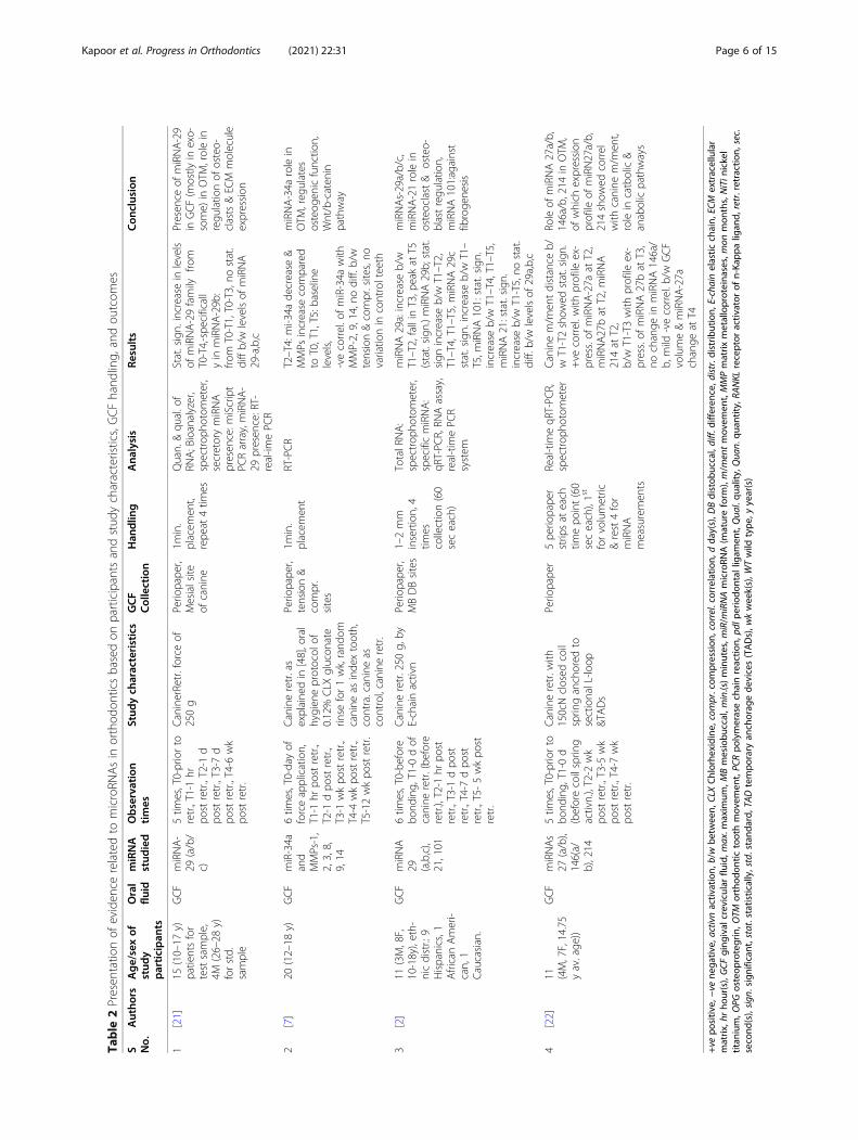
Dynamics of miRNAs in oral fluidsThe results of the studies are tabulated and presented inTable 2, divided into the following categories:
Participant characteristicsThe sample size in the included studies was 11 in twostudies [2, 22], n = 15 and n = 20 in one study each.Studies also specify dropouts, as well as the exclusion ofparticipants from the study due to gingival conditions[220] [2, 22]. Male to female ratio has not been men-tioned in two studies [7, 21]. Ethnic distribution of par-ticipants was mentioned in one study, although thesample size was small [2].
Study characteristicsAll four studies were longitudinal where sample collec-tion was done at multiple observation points [2, 7, 21,22]. The baseline levels of all miRNAs served as internalcontrols. One study also mentioned contralateral canineas control [7]. All four studies have evaluated a protocolof canine retraction with the extraction of first premo-lars. Observation times ranged from five times [21, 22]to six times in two studies [2, 7] over 5–6 weeks (wk)after initiation of retraction. Observation times: beforeinitiation of retraction, immediately after retraction start,1 day (d), 7 days were considered in all the studies. Allstudies analyzed miRNAs in gingival crevicular fluid(GCF) and none of the studies analyzed saliva.
Collection of GCFGCF collection was done with a periopaper in all fourstudies [2, 7, 21, 22], with the depth of insertion of papermentioned in one study [2]. The time of placement of 1min (min.) was specified in all four studies but repeatedplacement of up to 4 times as specified in one study [2].Site-specific collection of GCF was done in three out offour studies with the mesial site of canine in one study[21] and both the mesial and distal sites mentioned intwo studies [2, 7] of which one study analyzed the differ-ence in miRNA level between tension and compressionsites [2].
miRNA studiedDifferent miRNAs have been evaluated in all the studies:miRNA-29 in two studies [214] [2, 21], miR-34a alongwith inflammatory mediators-MMPs [7], miRNA-21 [2],101 [2], 27 [22], 146 [22], and 214 [22] in one study each.
Upregulation or downregulation of miRNAsExpression profile of miRNA varied in GCF at differentobservation times. There was a statistically significant
increase in levels of miRNA-29 family from pre-retrac-tion levels to 5–6 weeks post-retraction, with no differ-ence in levels of miRNA 29a/b/c [214] [2, 21]. Specificcomponents of miRNA-29 family showed differential ex-pression with the application of retraction forces:miRNA-29a increases from baseline to 1 h (h) post-re-traction followed by a decrease at 1day and further peakat 5 weeks [2]. miRNA-29b showed a statisticallysignificant increase from pre-retraction to 1 h and 7 days[214] [2, 21]. miRNA-29c showed a significant increasefrom 1 h post- retraction to peak at 5 weeks [2]. Besides,miRNA-34a showed a significant decrease from 1 day to4 weeks post retraction compared to baseline and 1 hpost-retraction while a negative correlation was seen inmatrix metalloproteinases (MMPs)-2,9,14 with an in-crease in levels in the same time intervals, both on ten-sion and compression sites [7]. miRNA-101 showed astatistically significant increase from 1 h post-retractionto 7 days and 5 weeks while miRNA-21 showed a signifi-cant increase at 5-weeks time interval [2]. Family ofmiRNA-27, 214, 146 showed a variation related to thedistance of canine movement during retraction: levels ofmiRNA-27a, miRNA-27b, and miRNA-214 showed apositive correlation with the canine distance in these2 weeks. miRNA-27b levels also showed a positivecorrelation of canine distance at 5 weeks of retractionbut no significant correlation was observed inmiRNA-146(a/b) [22].
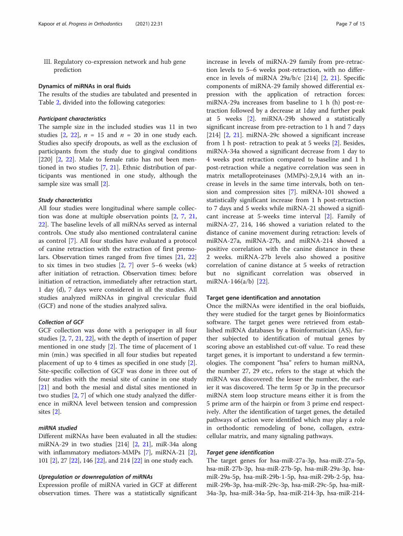
Target gene identification and annotationOnce the miRNAs were identified in the oral biofluids,they were studied for the target genes by Bioinformaticssoftware. The target genes were retrieved from estab-lished miRNA databases by a Bioinformatician (AS), fur-ther subjected to identification of mutual genes byscoring above an established cut-off value. To read thesetarget genes, it is important to understand a few termin-ologies. The component “hsa” refers to human miRNA,the number 27, 29 etc., refers to the stage at which themiRNA was discovered: the lesser the number, the earl-ier it was discovered. The term 5p or 3p in the precursormiRNA stem loop structure means either it is from the5 prime arm of the hairpin or from 3 prime end respect-ively. After the identification of target genes, the detailedpathways of action were identified which may play a rolein orthodontic remodeling of bone, collagen, extra-cellular matrix, and many signaling pathways.
Kapoor et al. Progress in Orthodontics (2021) 22:31 Page 7 of 15
5p, hsa-miR-146a-3p, and hsa-miR-146a-5p as identifiedfrom the review were predicted by both TargetScan andmiRDB databases. The genes were retrieved frommiRDB with the score > 90 and from TargetScan rankedby cumulative weighted context++ score >− 80. Therewere 1213 mutual genes from both the databases thatwere analyzed further (Fig. 3).
Target gene annotationInter-relational gene annotation resulted in detailingand pathways of the mutual target genes. MCODE, a
Cytoscape plugin, found out the most significant clus-ters of protein interactions (highly interconnected re-gions) with the highest score of 68.37, with 40 geneontology (GO)-enriched terms with 100% confidence.Figure 4 represents the complex of collagen trimers,extracellular matrix (ECM) structural constituent con-ferring tensile strength, collagen degradation, collagenbiosynthesis, ECM degradation were significant GOterms annotated for the cluster. The target genes sup-pressed by the miRNAs were also responsible in insu-lin growth factor (IGF-1) binding, transforming
Fig. 3 Results of target gene identification from miRDB and TargetScan databases
Kapoor et al. Progress in Orthodontics (2021) 22:31 Page 8 of 15
growth factor (TGF)-β signaling pathways, scavengingclass A receptors, and many more.
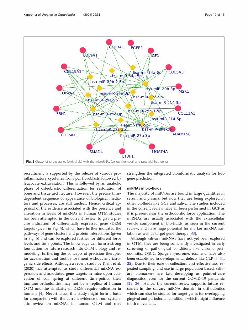
Regulatory co-expression network and Hub genepredictionProtein-protein interactions (PPI) encoded by the signifi-cant targets identified from the STRING databaseshowed 894 prominent interactions. These interactionswere used to identify the Hub genes, which are de-scribed as genes with elevated association in candidatemodules, which in this case are the protein -protein in-teractions. The score depicting the high connectivitygives an indication of the significance of HUB genes. Inthe current review, the hub genes as identified fromMCODE resulted in a prominent cluster with the high-est score of 8.98 with 15 nodes (hub genes) and 34 de-grees (interactions). The hub genes were identified asSMAD4, IGF1, ADAMTS6, COL4A1, COL1A1,COL3A1, FGFR1, COL19A1, FBN1, COL5A1,MGAT4A, LTBP1, MSR1, COL11A1, and COL5A3. Theinteractions of hub genes along with the promising miR-NAs are shown in Fig. 4. The promising miRNAs werehsa-miR-34a-5p, hsa-miR-29b-2-5p, hsa-miR-29b-3p,hsa-miR-34a-3p, hsa-miR-27a-5p, hsa-miR-29a-5p, hsa-miR-29b-1-5p, hsa-miR-29c-3p, hsa-miR-214-5p, hsa-miR-27a-3p, hsa-miR-29a-3p, and hsamiR-146-5p (Fig.
5). These hub genes can further be studied for their indi-vidualized effects on orthodontic treatment.
DiscussionmiRNAs as biomarkersMicroRNAs (miRNAs) are small (22 nucleotides lengthapproximately) single-stranded, noncoding RNAs thatregulate distinct biological processes by post-translational regression or destruction of mRNA targetgenes. They have been explored previously as bio-markers in multiple pathologic conditions like neo-plasms, malignancies, transplant rejection, infection,cardiac injury, etc. [29]. Specifically, in dentistry, theirrole has been established as prognostic and diagnosticmarkers in OSCC (oral squamous cell carcinoma) [30],premalignant conditions, e.g., dysplasia in leukoplakiaand conversion to malignancy [31], periodontal diseaseand homeostasis [29, 32], and also in craniofacial malfor-mations including CLP [3]. Nevertheless, the role ofmiRNAs in OTM and remodeling has not been exploredsufficiently to date [29].
Scope for miRNA markers in OTMThe orthodontic force application is known to initiatean aseptic inflammatory cascade mediated by underlyingcellular and molecular pathways. At cellular levels, theinitial catabolic phase of destruction with osteoclast
Fig. 4 Inter-relational pathway analysis and GO enrichment of the target gene clusters. The pathways are shown in arrow and triangle representationand the GO in ellipses (biological process), hexagon (molecular function), and parallelogram (cellular components)
Kapoor et al. Progress in Orthodontics (2021) 22:31 Page 9 of 15
recruitment is supported by the release of various pro-inflammatory cytokines from pdl fibroblasts followed byleucocyte extravasation. This is followed by an anabolicphase of osteoblastic differentiation for restoration ofbone and tissue architecture. However, the precise time-dependent sequence of appearance of biological media-tors and processes, are still unclear. Hence, critical ap-praisal of the evidence associated with the presence andalteration in levels of miRNAs in human OTM studieshas been attempted in the current review, to give a pre-cise indication of differentially expressed gene (DEG)targets (given in Fig. 4), which have further indicated thepathways of gene clusters and protein interactions (givenin Fig. 5) and can be explored further for different forcelevels and time points. The knowledge can form a strongfoundation for future research into OTM biology and re-modeling, furthering the concepts of precision therapiesfor acceleration and tooth movement without any iatro-genic side effects. Although a recent study by Klein et al.(2020) has attempted to study differential miRNA ex-pression and associated gene targets in mice upon acti-vation of coil spring at different time-points, theirimmuno-orthodontics may not be a replica of humanOTM and the similarity of DEGs require validation inhumans [4]. Nevertheless, this study might form a basisfor comparison with the current evidence of our system-atic review on miRNAs in human OTM and may
strengthen the integrated bioinformatic analysis for hubgene prediction.
miRNAs in bio-fluidsThe majority of miRNAs are found in large quantities inserum and plasma, but now they are being explored inother biofluids like GCF and saliva. The studies includedin the current review have all been performed in GCF asit is present near the orthodontic force application. ThemiRNAs are usually associated with the extracellularvesicle component in bio-fluids, as seen in the currentreview, and have huge potential for marker miRNA iso-lation as well as target gene therapy [33].Although salivary miRNAs have not yet been explored
in OTM, they are being sufficiently investigated in earlyscreening of pathological conditions like chronic peri-odontitis, OSCC, Sjorgen syndrome, etc., and have alsobeen established in developmental defects like CLP [3, 34,35]. Due to their ease of collection, cost-effectiveness, re-peated sampling, and use in large population based, saliv-ary biomarkers are fast developing as point-of-carediagnostics, even for the current COVID-19 pandemic[29, 36]. Hence, the current review supports future re-search in the salivary miRNA domain in orthodonticswhich can also be studied for target genes for overlappinggingival and periodontal conditions which might influencetooth movement.
Fig. 5 Cluster of target genes (pink circle) with the microRNAs (yellow rhombus) and potential hub genes
Kapoor et al. Progress in Orthodontics (2021) 22:31 Page 10 of 15
Epigenetic regulation of miRNA in OTM and target geneidentificationThe present review identified multiple families of mi-RNAs in humans during orthodontic canine retraction,including miRNAs-21, 27, 29, 34,146, 214, 101. Each ofthem is associated with varied molecular mechanismsplaying a role in the remodeling of bone and surround-ing structures in tooth movement.Of these, miRNA-21, which is one of the earliest discov-
ered miRNAs, show a steady rise in miRNA-21 from 1st–5th week of application of 250 g of retraction force in astudy by Lazari P. 2016, included in our review [2]. Therole of miRNA-21 has been studied in tooth movement inmice which indicates that the post-transcription control ofprotein mediated by miRNA-21 leads to inhibition of tar-get gene PDCD4, which in turn regulates C-Fos, furtherpotentiating osteoclastogenesis [37]. Thus, the phase ofthe tooth movement which supports rise in miRNA-21leads to heightened resorptive activity. This finding is sup-ported by Zhang et al. (2020) in a study on 36 maleSprague-Dawley rats which evaluated miR-21and its targetgenes in periodontally accelerated osteogenic orthodontics(PAOO) with an orthodontic loading of 25 g by a tensionspring between central incisors and maxillary first molar.Results showed a rise in miRNA-21 levels in combinedPAOO and tooth movement compared to the latter alone,after 7 days of surgery [37]. Further, this study also re-ported higher miRNA-21 associated with down streamingof target mRNA and PDCD4 protein expression leadingto increased expression of RANKL and C-Fos proteins.Similar target genes modulating clastogenesis like RANKLhave been identified in TargetScan and miRDB databasesin the current review and also in in-vivo gene expressionin mice in a study by Klein et al. 2020 [4]. Another studyby Cheng et al. (2016) used 30 g force to evaluate miRNA-21 expression in 3-month-old WT (wild type) and miR-21−/− mice and showed that OTM distance in miR-21−/−
mice was 30% that of WT mice and also, a significantlyhigher miR-21 was observed at 7 days of OTM com-pared with baseline levels, thus supporting role ofmiRNA-21 in tooth movement, as seen in the currentreview [38]. Additionally, Wu et al. (2020) also showedretarded tooth movement in miR-21−/− mice due to de-creased osteoclast numbers and suppressed RANKLpathway, thus supporting the target gene for resorptionidentified in the bioinformatic analysis in the currentreview [15]. The role of miRNA-21 in osteoclastogene-sis has also been established by in-vitro studies, one byKagiya and Nakamura (2013) on cells treated withTNF-α/RANKL, which showed increased expression ofmiRNA-21 and in-turn differentiation of osteoclastswhile another study by Sugatani et al. (2011) showedthat miRNA-21 silencing downregulated osteoclasto-genesis [39, 40].
Another family of miRNA studied in the present re-view was miRNA-29 and its role in orthodontic tooth re-traction evaluated in two studies. While one study in thecurrent review showed a statistically significant peakin the levels of miRNA-29a/b/c at 5 weeks of retractioncompared to baseline (before retraction) [2], anotherstudy showed an immediate increase in secretorymiRNA-29(a/b/c), followed by leveling at 1 h and a grad-ual increase till 6 weeks post retraction [2, 14]. This ex-pression profile of miRNA-29 has been previouslyassociated with osteoclast differentiation, TRAP+ cellformation, RANKL production, multinucleated osteo-clast formation, and extracellular matrix trabecular syn-thesis [39, 41, 42]. One previous study by Franceschettiet al. (2013) demonstrated the target genes influenced bymiRNA-29 to better comprehend their role in osteoclastregulation. They identified negative regulation of Srgap2(SLIT-ROBO Rho GTPase-activating protein 2) andCdc42 (cell division control protein 42) in the cytoskel-etal organization as well as targets like Gpr85 (Gprotein-coupled receptor 85), Nfia (nuclear factor I/A),Cd93 of macrophage lineage, and Calcr (calcitonin re-ceptor) for osteoclast survival [41] Another aspect ofperiodontal remodeling in OTM, which is extracellularmatrix (ECM) gene regulation, was also studied by Chenet al. (2015) by subjecting periodontal ligament (pdl)cells to cyclic stretch and compression forces and studyinteractions of miR-29b mimic/inhibitor and collagenregulator genes (COL1A1, COL3A1, COL5A1) genes,thus giving evidence of its role in ECM regulation [43].The same target genes have been identified in the over-lap of TargetScan and miRDB databases in the currentreview and the pathway of their action has beenhighlighted in Fig. 4. Contrastingly, miR-29 is alsoknown to be involved in the anabolic process leading todifferentiation of osteoblasts and downregulation ofRANKL by the Wnt pathway [44, 45]. A study by Liet al. (2009) provides evidence of miRNA-29b reaching apeak at 28 d during the deposition period and causingECM accumulation and targeting inhibitors of osteo-blastogenesis [45] Similar results were seen by Kapinaset al. (2009) who additionally provided the gene targetsof miRNA-29b by downregulating inhibitors of osteo-blastogenesis as well as showed increased miRNA-29a/cduring osteoblastic differentiation [44]. This may wellexplain the late peak in miRNA-29 at 5–6 wk of retrac-tion in the current review, which corresponds to thedepository phase in the studies included in our review.Another marker miRNA, miRNA-101, showed a statis-
tically significant increase at 1 wk and 5 wk of retractionfrom the pre-treatment levels in a study by Lazari et al.(2016) in the current review [2]. This increase in expres-sion is supported by a previous study by Li et al. (2012)which identified the proteins modulated by miRNA-101-
Kapoor et al. Progress in Orthodontics (2021) 22:31 Page 11 of 15
periodontal ligament-associated protein-1 (PLAP-1) aswell as transforming growth factor (TGF-β), which areboth responsible for anabolic processes, thus explainingthe late increase in levels of miRNA-101 [46]. TGF-βsignaling has also been indicated in the gene targetsidentified in the bioinformatic analysis of the current re-view, highlighted in Fig. 3. Additionally, the bone appos-ition process has also been linked to the presence ofmiR-34a, which is known for osteogenesis and angiogen-esis [47].The current review also shows miR-34a downregula-
tion in orthodontic canine retraction from 1 d to 1month (mon) post-retraction, both on tension and com-pression sites and in a negative correlation of MMPs-2,9,14 expression at the same observation times [7],whereby MMPs are known for matrix degradation [13].MiRNA-27 family, studied in the current review, is
known to suppress the pro-inflammatory response con-sequent to the force application. MiRNA-27(a/b) sup-ports an increase in differentiation of osteoblasts,enabling the Wnt pathway, hence the anabolic processbut the change is not statistically significant, probablydue to sampling limitations [22]. Distance moved by thecanine in 2 weeks and 5 weeks has shown a mild positivecorrelation with miRNA-27(a/b) which may correspondto the peak in osteoid mineralization and differentiationof osteoblasts [48].An action similar to miRNA-27 is performed by
miRNA-146(a/b) which is elevated in acute inflamma-tion of OTM but when inflammation subsides, onlymiRNA-146a was shown to return to baseline in thecurrent review [22], as has been witnessed in previousstudies [49]. The role of miR-146 in the inflammatorycascade is potentiated by suppressing the pro-inflammatory mitogen-activated protein kinase (MAPK)and NF-κB pathway, regressing the early growth re-sponse and endothelial activation, thus serving as nega-tive feedback mechanisms for the inflammation [49].Besides, another unpublished thesis by Christyne
Chmil (May 2020) in support of the current review, alsoshowed an evidence of significant change in miRNA-155expression in GCF in 2 weeks of retraction by micro-implant supported retraction using closed coil spring (pvalue = 0.006). Other miRNAs, miRNA-21, and miRNA-29b showed mild correlations at 2 weeks, thus strength-ening the role of miRNAs in tooth movement [50].Hence, the current review provides insight into the ex-
citing domain of epigenetic regulation of microRNA in-fluencing the molecular mechanisms underlying boneand tissue remodeling associated with tooth movement.
Clinical significance of the studyThe current study supports the expression of miRNApresent in exosomes existing in GCF of teeth undergoing
tooth movement. While change in levels of various me-diators like cytokines, enzymes, etc. have been studiedearlier in association with varying orthodontic forcelevels and time intervals, their role is limited to indica-tion of underlying remodeling process consequent tochange in mediator expression. On the other hand,miRNA studied in the current review respond to mech-anical stimuli and modulate osteoblastogenesis, osteo-clastogenesis, and extra-cellular matrix regulation post-transcriptionally at a genetic level. Further, this studyhas been able to identify the various target genes, e.g.,PDCD4 associated with miRNA-21, which can furtherindicate the pathway of C-Fos regulation directly in-volved in resorption. Similarly, various other miRNAsstudied in tooth movement have been analyzed for iden-tification of mutual target genes for a cumulative effectand further gene ontology performed for HUB geneidentification, e.g., COL1A1, COL3A1, COL5A1 etc.which can serve as promising biomarkers for resorptionor apposition. It helps in the study of protein-proteininteractions which can interpret complex inter-relationships of various processes associated with toothmovement including periodontal inflammation, root re-sorption or accompanying systemic conditions. This canformulate the basis of target gene therapies for precisionor personalized treatment or individualize treatment byupregulation or downregulation of specific genes tobring faster tooth movement with minimal side-effects.
Limitations of the current studyAlthough the role of microRNAs is being slowly estab-lished in tooth movement, the literature evidence is stillscanty and has an unclear risk of bias. The sample size issmall with a disproportionate male-female ratio, whichmay introduce bias in the results of the study. Therandomization of subjects or teeth was not performed inthe majority of studies; hence the quality of includedstudies was compromised. The GCF collection protocolwas not standardized in all studies as well as oral hy-giene regimen and maintenance are not specified. Thereis no study conducted in saliva in tooth movement,hence only one oral fluid has been studied. The bioinfor-matics component may not be all-inclusive, as no studyto date has been performed in humans to outline all dif-ferentially expressed genes in OTM, showing time-dependent variation.
Conclusions
1. Various microRNAs have been studied in humanGCF which may serve as biomarkers for toothmovement, primarily orthodontic canine retraction,including miRNA-21, 27(a/b), 29(a/b/c), 34,146(a/b), 101, and 214. But the literature is scanty with an
Kapoor et al. Progress in Orthodontics (2021) 22:31 Page 12 of 15
unclear risk of bias and hence, there is an urgent re-quirement of outlining a full complement of DEGsto identify the most promising miRNAs as bio-markers for OTM.
2. A statistically significant increase in expression ofmiRNA-29a/b/c, miRNA-21, miRNA-101 from pre-treatment (prior to initiation of retraction) levels topeak at 4–6 weeks of retraction was seen, but the out-comes are depicted in studies with an unclear ROB.On the other hand, a single study with a low ROBreported miRNA-34a downregulation in negativecorrelation with MMPs-2,9,14 levels, thus supportingits role in anabolic osteogenesis and apposition.
3. The concentration of miRNAs was found to behigher in the exosome-associated than depletedcomponent of GCF, hence secretory miRNAs caneasily be detected in various bio-fluids in exosomesand can further be explored for marker miRNAsand target gene therapy.
4. Bioinformatic analysis revealed 1213 mutual targetgenes and SMAD4, IGF1, ADAMTS6, COL4A1,COL1A1, COL3A1, FGFR1, COL19A1, FBN1,COL5A1, MGAT4A, LTBP1, MSR1, COL11A1, andCOL5A3 as the hub genes. These can be furtheranalyzed for integrated pathways with otherassociated periodontal and systemic conditions,which might influence tooth movement.
5. Multiple promising miRNA biomarkers wereidentified: hsa-miR-34a-5p, hsa-miR-29b-2-5p, hsa-miR-29b-3p, hsa-miR-34a-3p, hsa-miR-27a-5p, hsa-miR-29a-5p, hsa-miR-29b-1-5p, hsa-miR-29c-3p,hsa-miR-214-5p, hsa-miR-27a-3p, hsa-miR-29a-3p,hsamiR-146-5p. These can be used in future ortho-dontic models of tooth movement using differentforce levels and observation intervals.
Future directions
1. Differentially expressed gene expression in OTM:Full complement of differentially expressedgenes (DEGs) in human tooth movement usingnovel gene testing technologies of RNA sequencingfor accuracy in smaller samples. Expression shouldbe monitored in serum, and additionally in oral bio-fluids (GCF and saliva) due to ease in sampling andrepeated evaluation.
2. Robust orthodontic study designs: Differences insex, ethnicity, age (adults and juveniles) need to beaddressed to comprehend differences in the boneand tissue remodeling associated with OTM. Largersample preferred to minimize bias. Differentmechanics: orthodontic/orthopedic, functional oraccelerated tooth movement require isolation ofmiRNA markers.
3. Basic research in mechanisms controlling miRNAactivity: A detailed analysis for overlapping proteininteractions to identify links between various localand systemic conditions including periodontaldiseases, neoplasms, etc.
4. Optimal use of miRNA datasets: For computationaldiagnostic and prediction models and bioinformaticanalysis in various orthodontic situations andproposing marker miRNA biosensors for the same.
5. Precision and personalized treatments: Usinginformation of target genes and their pathways bylocal delivery of miRNA-associated exosomes. Thismay be beneficial to accelerate treatment time, re-duce side effects of root resorption, caries risk, gin-gival inflammatory conditions, etc.
6. Propose integrated basic research infrastructure inorthodontic specialty: Requirement of trainedmanpower, adequate bioinformatics training, as wellas infrastructure resources for basic researchassociated to orthodontic departments.
AbbreviationsALP: Alkaline phosphatase; AST: Aspartate aminotransferase; Calcr: Calcitoninreceptor; Cdc42: Cell division control protein 42; CLP: Cleft lip and palate;DEG: Differential gene expression; ELISA: Enzyme-linked immunoassay;GCF: Gingival crevicular fluid; Gpr85: G protein-coupled receptor 85;IL: Interleukin; MAPK: Mitogen-activated protein kinase; miRDB: miRNAdatabase; miRNA: MicroRNA; MMPs: Matrix metalloproteinases; Nfia: Nuclearfactor I/A; OPG: Osteoprotegrin; OSCC: Oral squamous cell carcinoma;OTM: Orthodontic tooth movement; PCR: Polymerase chain reaction;pg: Picogram; PPI: Protein- protein interaction; PRISMA: Preferred reportingitems of systematic reviews and meta-analysis; PLAP-1: Periodontal ligament-associated protein-1; QUADAS: Quality assessment tool for diagnosticaccuracy studies; RANKL: Receptor activator of nuclear kappa B ligand;Srgap2: SLIT-ROBO Rho GTPase-activating protein 2; TGF-β: Transforminggrowth factor -β; TNF- α: Tumor necrosis factor- α; TRAP: Tartrate-resistantacid phosphatase
Supplementary InformationsThe online version contains supplementary material available at https://doi.org/10.1186/s40510-021-00377-1.
Additional file 1.
AcknowledgementsNot applicable.
Availability of supporting data and materialData set necessary to interpret, replicate, and build upon findings reportedin the article are provided. Further raw data if required (raw data is notpublic) can be obtained from the corresponding author on reasonablerequest.
Authors’ contributionsPK has made substantial contributions to the conception and design of thework, analysis and interpretation of data, drafted the work or substantivelyrevised it, and approved the submitted version. AC has made substantialcontributions to the analysis and interpretation of data, drafted the work orsubstantively revised it, and approved the submitted version. DKB has madesubstantial contributions to the analysis and interpretation of data, revision,and approved the submitted version. DB has made substantial contributionsto the analysis and interpretation of data, revision, and approved thesubmitted version. All authors read and approved the final manuscript.
Kapoor et al. Progress in Orthodontics (2021) 22:31 Page 13 of 15
Ethics approval and consent to participateNot applicable.
Consent for publicationNot applicable.
Competing interestsThe authors declare that they have no competing interests.
Author details1School of Dental Sciences, Sharda University, Greater Noida, UP, India.2Department of Orthodontics, Faculty of Dentistry, Jamia Millia Islamia, NewDelhi 110025, India. 3Department of Oral Pathology & Microbiology, Facultyof Dentistry, Jamia Millia Islamia, New Delhi 110025, India. 4Department ofOrthodontics & Dentofacial Orthopaedics, School of Dental Sciences, ShardaUniversity, Greater Noida, UP, India. 5Department of Oral Pathology &Microbiology, School of Dental Sciences, Sharda University, Greater Noida,UP, India. 6Department of Bioinformatics, Stella Maris College (Autonomous),Chennai, India.
Received: 11 May 2021 Accepted: 18 July 2021
References1. Escors D, Kochan G, Stephenson H, Breckpot K. Cell and tissue gene
targeting with Lentiviral vectors. In: Lentiviral vectors and gene therapy[Internet]. 2012. Available from: 10.1007/978-3-0348-0402-8_3
2. Lazari P. Secretory micro-RNA 29 in gingival crevicular fluid during canineretraction [Internet] [thesis]. University of Illinois at Chicago; 2016 [cited2021 Mar 12]. Available from: /articles/thesis/Secretory_Micro-RNA_29_in_Gingival_Crevicular_Fluid_During_Canine_Retraction/10812575/1
3. Grassia V, Lombardi A, Kawasaki H, Ferri C, Perillo L, Mosca L, et al. SalivarymicroRNAs as new molecular markers in cleft lip and palate: a new frontierin molecular medicine. Oncotarget. 2018 Apr 10;9(27):18929–38. https://doi.org/10.18632/oncotarget.24838.
4. Klein Y, Fleissig O, Polak D, Barenholz Y, Mandelboim O, Chaushu S.Immunorthodontics: in vivo gene expression of orthodontic toothmovement. Sci Rep. 2020 May 18;10(1):8172. https://doi.org/10.1038/s41598-020-65089-8.
5. Sun F, Wan M, Xu X, Gao B, Zhou Y, Sun J, et al. Crosstalk between miR-34aand notch signaling promotes differentiation in Apical Papilla stem cells(SCAPs). J Dent Res. 2014 Jun;93(6):589–95. https://doi.org/10.1177/0022034514531146.
6. Grilli A, Sciandra M, Terracciano M, Picci P, Scotlandi K. Integratedapproaches to miRNAs target definition: time-series analysis in anosteosarcoma differentiative model. BMC Med Genet. 2015 Jun 30;8(1):34.https://doi.org/10.1186/s12920-015-0106-0.
7. Zhang B, Yang L, Zheng W, Lin T. MicroRNA-34 expression in gingivalcrevicular fluid correlated with orthodontic tooth movement. Angle Orthod.2020 Mar 12;90(5):702–6. https://doi.org/10.2319/090219-574.1.
8. Yete S, Saranath D. MicroRNAs in oral cancer: biomarkers with clinicalpotential. Oral Oncol. 2020 Nov 1;110:105002. https://doi.org/10.1016/j.oraloncology.2020.105002.
9. Pan Y, Li D, Lou S, Zhang C, Du Y, Jiang H, et al. A functional polymorphismin the pre-miR-146a gene is associated with the risk of nonsyndromicorofacial cleft. Hum Mutat. 2018 May;39(5):742–50. https://doi.org/10.1002/humu.23415.
10. Schoen C, Aschrafi A, Thonissen M, Poelmans G, Von den Hoff JW, CarelsCEL. MicroRNAs in Palatogenesis and Cleft Palate. Front Physiol [Internet].2017 [cited 2021 Mar 17];8. Available from: https://www.frontiersin.org/articles/10.3389/fphys.2017.00165/full
11. Mendes SMDA, Espinosa DDSG, Moreira PEDO, Marques D, Fagundes NCF,Ribeiro-Dos-Santos Â. miRNAs as biomarkers of orofacial clefts: a systematicreview. J Oral Pathol. 2020 Mar;49(3):201–9. https://doi.org/10.1111/jop.12950.
12. Kapoor P, Kharbanda OP, Monga N, Miglani R, Kapila S. Effect of orthodonticforces on cytokine and receptor levels in gingival crevicular fluid: asystematic review. Prog Orthod. 2014 Dec 9;15(1):65. https://doi.org/10.1186/s40510-014-0065-6.
13. Kapoor P, Monga N, Kharbanda OP, Kapila S, Miglani R, Moganty R. Effect oforthodontic forces on levels of enzymes in gingival crevicular fluid (GCF): asystematic review. Dent Press J Orthod. 2019 May 20;24(2):40.e1–40.e22.
14. Atsawasuwan P, Lazari P, Chen Y, Zhou X, Viana G, Evans CA. SecretorymicroRNA-29 expression in gingival crevicular fluid during orthodontictooth movement. PLoS One. 2018;13(3):e0194238. https://doi.org/10.1371/journal.pone.0194238.
15. Wu L, Su Y, Lin F, Zhu S, Wang J, Hou Y, et al. MicroRNA-21 promotesorthodontic tooth movement by modulating the RANKL/OPG balance in Tcells. Oral Dis. 2020;26(2):370–80. https://doi.org/10.1111/odi.13239.
16. Gallo A, Tandon M, Alevizos I, Illei GG. The majority of microRNAs detectablein serum and saliva is concentrated in exosomes. PLoS One. 2012;7(3):e30679. https://doi.org/10.1371/journal.pone.0030679.
17. Luscombe NM, Greenbaum D, Gerstein M. What is bioinformatics? Aproposed definition and overview of the field. Methods Inf Med. 2001;40(4):346–58. https://doi.org/10.1055/s-0038-1634431.
19. Wang Z, Ishihara Y, Ishikawa T, Hoshijima M, Odagaki N, Ei Hsu Hlaing E,et al. Screening of key candidate genes and pathways for osteocytesinvolved in the differential response to different types of mechanicalstimulation using a bioinformatics analysis. J Bone Miner Metab. 2019 Jul;37(4):614–26. https://doi.org/10.1007/s00774-018-0963-7.
20. Iwasaki LR, Covell DA, Frazier-Bowers SA, Huja SS, Kapila S, Nickel JC. Prefaceto COAST 2018 Innovators’ Workshop: bridging the biology and technologygap in orthodontics and craniofacial care. Orthod Craniofacial Res. 2019;22(S1):5–7. https://doi.org/10.1111/ocr.12303.
21. Atsawasuwan P, Lazari P, Chen Y, Zhou X, Viana G, Evans CA. SecretorymicroRNA-29 expression in gingival crevicular fluid during orthodontictooth movement. PloS One. 2018;13(3):e0194238.
22. Seagraves AL. Circulatory microRNA-27, -146, and -214 in gingival crevicularfluid during orthodontic tooth movement [Internet] [thesis]. University ofIllinois at Chicago; 2020 [cited 2020 Dec 24]. Available from: /articles/thesis/Circulatory_MicroRNA-27_-146_and_-214_in_Gingival_Crevicular_Fluid_During_Orthodontic_Tooth_Movement/13475250/1
23. Whiting PF, Rutjes AWS, Westwood ME, Mallett S, Deeks JJ, Reitsma JB, et al.QUADAS-2: a revised tool for the quality assessment of diagnostic accuracystudies. Ann Intern Med. 2011;155(8):529–36. https://doi.org/10.7326/0003-4819-155-8-201110180-00009.
24. Chmil, Christyne (2020): miRNA-21, -29 Family and -155 Expression ingingival crevicular fluid during orthodontic tooth movement. University ofIllinois at Chicago. Thesis. https://doi.org/10.25417/uic.13476501.v1
25. Agarwal V, Bell GW, Nam J-W, Bartel DP. Predicting effective microRNAtarget sites in mammalian mRNAs. eLife. 2015;12:4.
26. miRDB: an online database for prediction of functional microRNA targets[Internet]. [cited 2021 May 2]. Available from: https://www.ncbi.nlm.nih.gov/pmc/articles/PMC6943051/
27. Bindea G, Mlecnik B, Hackl H, Charoentong P, Tosolini M, Kirilovsky A, et al.ClueGO: a Cytoscape plug-in to decipher functionally grouped geneontology and pathway annotation networks. Bioinformatics. 2009 Apr 15;25(8):1091–3. https://doi.org/10.1093/bioinformatics/btp101.
28. Aishwarya S, Gunasekaran K, Margret AA. Computational gene expressionprofiling in the exploration of biomarkers, non-coding functional RNAs anddrug perturbagens for COVID-19. J Biomol Struct Dyn. :1–16.
29. Szklarczyk D, Morris JH, Cook H, Kuhn M, Wyder S, Simonovic M, et al. TheSTRING database in 2017: quality-controlled protein–protein associationnetworks, made broadly accessible. Nucleic Acids Res. 2017;45(Databaseissue):D362–8. https://doi.org/10.1093/nar/gkw937.
30. Kim S-H, Lee S-Y, Lee Y-M, Lee Y-K. MicroRNAs as biomarkers for dental diseases.Singap Dent J. 2015 Dec 1;36:18–22. https://doi.org/10.1016/j.sdj.2015.09.001.
31. Ghosh RD, Pattatheyil A, Roychoudhury S. Functional landscape ofdysregulated microRNAs in oral squamous cell carcinoma: clinicalimplications. Front Oncol [Internet]. 2020 [cited 2021 May 3];10. Availablefrom: https://www.frontiersin.org/articles/10.3389/fonc.2020.00619/full
Kapoor et al. Progress in Orthodontics (2021) 22:31 Page 14 of 15
32. Brito JAR, Gomes CC, Guimarães ALS, Campos K, Gomez RS. Relationshipbetween microRNA expression levels and histopathological features ofdysplasia in oral leukoplakia. J Oral Pathol Med Off Publ Int Assoc OralPathol Am Acad Oral Pathol. 2014 Mar;43(3):211–6.
33. Luan X, Zhou X, Trombetta-eSilva J, Francis M, Gaharwar AK, AtsawasuwanP, et al. MicroRNAs and periodontal homeostasis. J Dent Res. 2017 May;96(5):491–500. https://doi.org/10.1177/0022034516685711.
34. Holliday LS, Truzman E, Zuo J, Han G, Torres-Medina R, Rody WJ.Extracellular vesicle identification in tooth movement models. OrthodCraniofacial Res. 2019;22(S1):101–6. https://doi.org/10.1111/ocr.12287.
35. Fujimori K, Yoneda T, Tomofuji T, Ekuni D, Azuma T, Maruyama T, et al.Detection of salivary miRNAs reflecting chronic periodontitis: a pilot study.Mol Basel Switz. 2019 Mar 15;24(6).
36. Park NJ, Zhou H, Elashoff D, Henson BS, Kastratovic DA, Abemayor E, et al.Salivary microRNA: discovery, characterization, and clinical utility for oralcancer detection. Clin Cancer Res. 2009 Sep 1;15(17):5473–7. https://doi.org/10.1158/1078-0432.CCR-09-0736.
37. Zhang Y, Tian Y, Yang X, Zhao Z, Feng C, Zhang Y. MicroRNA-21 serves animportant role during PAOO-facilitated orthodontic tooth movement. MolMed Rep. 2020;22(1):474–82. https://doi.org/10.3892/mmr.2020.11107.
38. Chen N, Sui BD, Hu CH, Cao J, Zheng CX, Hou R, Yang ZK, Zhao P, Chen Q,Yang QJ, Jin Y, Jin F. microRNA-21 Contributes to Orthodontic ToothMovement. J Dent Res. 2016;95(12):1425–33. https://doi.org/10.1177/0022034516657043.
39. Kagiya T, Nakamura S. Expression profiling of microRNAs in RAW264.7 cellstreated with a combination of tumor necrosis factor alpha and RANKLduring osteoclast differentiation. J Periodontal Res. 2013;48(3):373–85.https://doi.org/10.1111/jre.12017.
40. Sugatani T, Vacher J, Hruska KA. A microRNA expression signature ofosteoclastogenesis. Blood. 2011 Mar 31;117(13):3648–57. https://doi.org/10.1182/blood-2010-10-311415.
41. Franceschetti T, Kessler CB, Lee S-K, Delany AM. miR-29 Promotes murineosteoclastogenesis by regulating osteoclast commitment and migration. JBiol Chem. 2013 Nov 15;288(46):33347–60. https://doi.org/10.1074/jbc.M113.484568.
42. Villarreal G, Oh D-J, Kang MH, Rhee DJ. Coordinated regulation ofextracellular matrix synthesis by the microRNA-29 family in the trabecularmeshwork. Invest Ophthalmol Vis Sci. 2011;52(6):3391–7. https://doi.org/10.1167/iovs.10-6165.
43. Chen Y, Mohammed A, Oubaidin M, Evans CA, Zhou X, Luan X, et al. Cyclicstretch and compression forces alter microRNA-29 expression of humanperiodontal ligament cells. Gene. 2015 Jul 15;566(1):13–7. https://doi.org/10.1016/j.gene.2015.03.055.
44. Kapinas K, Kessler CB, Delany AM. miR-29 suppression of osteonectin inosteoblasts: regulation during differentiation and by canonical Wnt signaling. JCell Biochem. 2009 Sep 1;108(1):216–24. https://doi.org/10.1002/jcb.22243.
45. Li Z, Hassan MQ, Jafferji M, Aqeilan RI, Garzon R, Croce CM, et al. Correction:biological functions of miR-29b contribute to positive regulation ofosteoblast differentiation. J Biol Chem. 2019 Jun 21;294(25):10018. https://doi.org/10.1074/jbc.AAC119.009552.
46. Li C, Li C, Yue J, Huang X, Chen M, Gao J, et al. miR-21 and miR-101regulate PLAP-1 expression in periodontal ligament cells. Mol Med Rep.2012 May;5(5):1340–6. https://doi.org/10.3892/mmr.2012.797.
47. Zha X, Sun B, Zhang R, Li C, Yan Z, Chen J. Regulatory effect of microRNA-34a on osteogenesis and angiogenesis in glucocorticoid-inducedosteonecrosis of the femoral head. J Orthop Res Off Publ Orthop Res Soc.2018;36(1):417–24.
48. Holland R, Bain C, Utreja A. Osteoblast differentiation during orthodontictooth movement. Orthod Craniofacial Res. 2019;22(3):177–82. https://doi.org/10.1111/ocr.12308.
49. Cheng HS, Sivachandran N, Lau A, Boudreau E, Zhao JL, Baltimore D, et al.MicroRNA-146 represses endothelial activation by inhibiting pro-inflammatory pathways. EMBO Mol Med. 2013 Jul;5(7):1017–34. https://doi.org/10.1002/emmm.201202318.
50. Chmil, Christyne. miRNA-21, -29 Family and -155 Expression in gingivalcrevicular fluid during orthodontic tooth movement. University of Illinois atChicago. Thesis. 2020. https://doi.org/10.25417/uic.13476501.v1.
Publisher’s NoteSpringer Nature remains neutral with regard to jurisdictional claims inpublished maps and institutional affiliations.
Kapoor et al. Progress in Orthodontics (2021) 22:31 Page 15 of 15