Microscopical study of the digestive tract of Blue and Yellow macaws
M. N. Rodrigues1, J. A. P. Abreu1, C. Tivane1, P. G. Wagner2, D. B. Campos3, R. R. Guerra3, R. E. G. Rici 1 and M. A. Miglino1 1Surgery Department , Faculty of Veterinary Medicine and Animal Science (FMVZ-USP), São Paulo University (USP),
Cidade Universitária, Av. Prof. Dr. Orlando Marques de Paiva 87, São Paulo SP 05508270, Brazil. *Corresponding author: [email protected]
2IBAMA/PB – National Forest of the restinga of Cabedelo, BR 230, estrada de Cabedelo, Cabedelo PB 58310-000 Brazil. 3Center of Agricultural Sciences, Federal University of Paraiba, S/N, Cidade Universitária II, Areia PB, 58397-000 Brazil
Studies on the gastrointestinal tract of macaws are scarce. This study aims to address the lack of meaningful anatomical information on the digestive tract of Blue and Yellow macaw’s (Ara ararauna) by presenting light microscopic features of the digestive tract. The light microscopy (LM) study was supplemented by a detailed scanning electron microscopy (SEM) of sampled surface features. Three adult Blue and Yellow macaws of either sex were used in this investigation. Samples of the tongue, oesophagus, crop, proventriculus, ventriculus and intestines were collected and processed routinely for LM and SEM (tongue and ventriculus). The most remarkable feature in the tongue was the presence of Herbst corpuscles located in the core of loose connective tissue or dense connective tissue of the lamina propria. In the oesophagus mucous glands were confined to the caudal part of the crop and the muscular layer was composed by a single layer of fibers. The crop was structuraly similar to the oesophagus. In the proventriculus and ventriculus the muscular layer exhibit circular orientation; this layer was notably well developed in the ventriculus. In the intestines the villi decreased in height and goblet cells increased in number caudally. SEM of the tongue revealed that the dorsal surface of the apex and body " creases" and wrinkles, being the duct openings of the underlying lingual glands were the most obvious feature and were seen to contain mucus and desquamated epithelial cells. Caudally directed conical papillae were present in the root which" sat" in a slightly wrinkled surface. The ventriculus cuticle presented overlapped layers of flat plates with irregular edges. The present study in addition to confirm the basic structure of the digestive tract of birds in general, also provides not previously reported data of the digestive tract of macaws that can be useful in the study of nutrition and health of this bird.
Keywords: morphology, Ara ararauna, digestive system, microscopy, macaws
1. Introduction
Brazil is the country with the major number of Psittacidae birds in the world. The Parakeets, parrots and macaws are part of this family, the latter being the most representative of the group and extremely important specie of Brazilian wildlife and also in other countries of the world [1]. The blue and yellow macaws (Ara ararauna) can reach up to 80 cm long, exhibiting blue color in the top feathers, yellow color in the bottom feathers and lines of black feathers in the neck and face. Its thick and sensitive tongue is used as a tactile organ [2]. In recent studies on macaws, aspects such as activity, animal husbandry, feeding habits and habitat have been discussed. However, morphological data regarding the digestive system of this bird still scarce. The Brazilian Institute of Environment and Renewable Natural Resources (IBAMA) regulates trade in wildlife animals coming from breeding, fact that increases the use of these animals as pets [3]. It is believed that studies on the digestive tract of blue and yellow macaw could provide valuable information that can be used by other professional working in the management, and nutrition of this specie. In this view, the present study will describe in detail microscopic features of the gastrointestinal tract of the Araa rarauna, in order to supply the lack of data regarding the morphology of this species and thereby assist in the maintenance of this important bird in captivity and / or free life.
2. Material and method
Three adult blue and yellow macaws of either sex were used in this experiment. The birds were preserved in 10% buffered formalin in the Anatomy lab of the Faculty of Veterinary Medicine of Sao Paulo University and were donated by CETAS-PB (02019.00129/2009-12 IBAMA REFERENCE NUMBER). The animals were ventrally incised from the oesophagus to the cloaca in order to expose the gastrointestinal tract. For light microscopy, were collected sections of approximately 0.5 cm from the tongue, oesophagus, crop, proventriculus, ventriculus and intestines. The samples were kept in fixative 10% formaldehyde solution and dehydrated in a series of ethanols in increasing concentrations (70-100%) and diaphanized in xylene followed by inclusion in histological paraffin similar - Ervplast. Sections of 5μmwere obtained on a LEICA 2165 microtome (Faculty of Veterinary Medicine) and stained in Hematoxylin-Eosin. The gathering of material, histological sections as well as the preparation and staining of the slides were performed based on the methodology previously described [4].
Current Microscopy Contributions to Advances in Science and Technology (A. Méndez-Vilas, Ed.)
The photos were taken with Zeiss Axio Cam camera coupled to a microscope (Faculty of Veterinary Medicine of São Paulo University). For scanning electron microscopy samples from the tongue and ventriculus, were collected post-fixed in 1% tetroxide osmium solution and 0.1% PBS for 2 hours. Subsequently the samples were washed for 45 minutes with 0.1% PBS and 20 minutes with distilled water. The final material was dehydrated in a series of increasing concentrations of alcohol 50%, 70%, 90% and 100% for 30 minutes in each. Then the specimens were fitted on aluminum metal stubs, suitable for scanning electron microscopy, using carbon glue. In the sequence were subjected to metallic sputtering with gold in the sputtering device EMITECHK550 (Faculty of Veterinary Medicine of São Paulo University) and analyzed and photomicrographed by the scanning gelecron microscope LEO 435VP (Faculty of Veterinary Medicine of São Paulo University).
3. Results
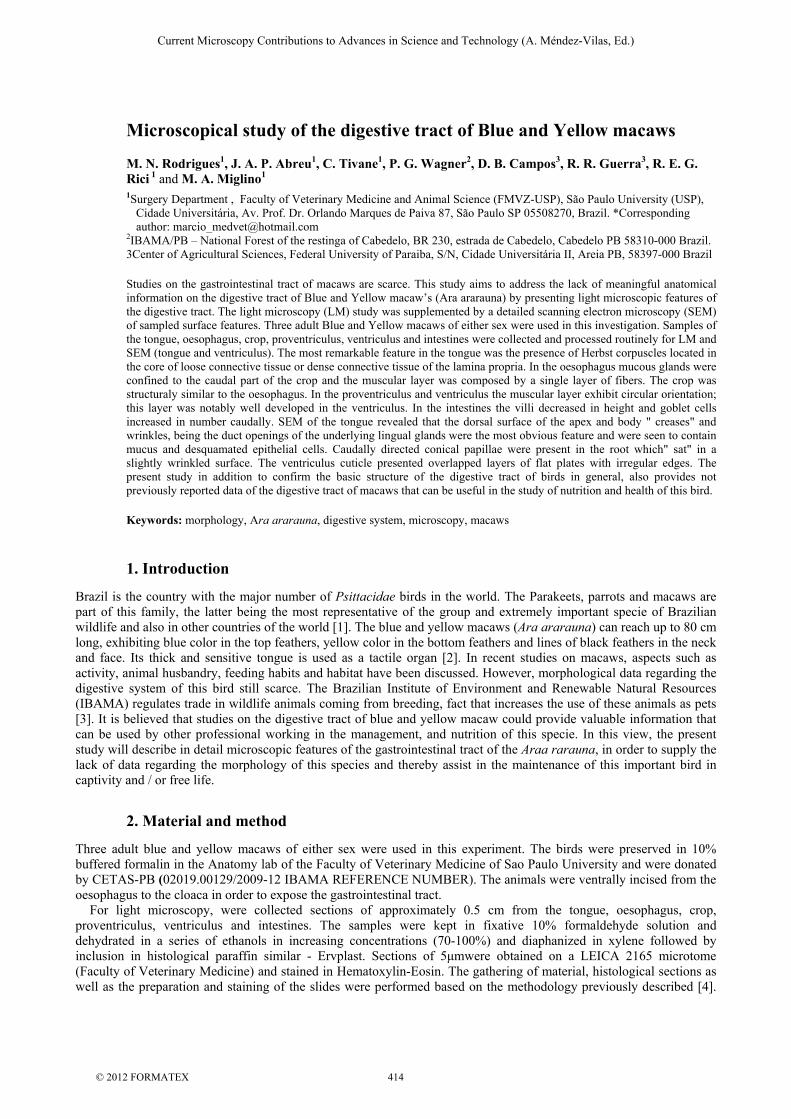
Light Microscopy confirmed that the tongue of the macaw was a caudal projection of the ventral floor of the oral cavity. Its mucosa was lined by a lamina propria and its epithelium was stratified squamous keratinized. The keratinization was greater at the apex of the tongue. The dermis consisted of connective tissue present in the mucosal lamina propria that merged with the submucosal layer and the epimysium of the adjacent lingual muscles. No taste buds were seen in the macaw tongue. Papillae of connective tissue penetrated the lingual epithelium (Figure 1A). These papillae were deeper where the epithelium was thicker and lower and bulbous where the epithelium was thin. Were observed in the lamina propria of irregular dense connective tissue, lymphoid tissue associated with Herbst corpuscles. These corpuscles were similar to Pacinian corpuscles of mammals and were ovaland/or circular structures, with a connective tissue capsule laminated interiorly and in the center of the corpuscle sensory nervous ending (Figures1A and1B). Such structures were found mainly at the apex but were also present in the body and lingual root exhibiting different sizes. The submucosa of loose connective tissue was rich in mucus-secreting glands but also cartilage, muscles, infiltrated lymphoid tissue and a thick layer of adipose tissue (figure 1B). Single branched tubular mucous glands were found in major number than salivary glands. The glands were covered by a connective tissue capsule with septa separating the glandular tubules (Figures 1B and 1C). Hyaline cartilage (Figure1D) of the entoglossal bone (Figure1E) supported the tongue of the macaw. The axis of this organ had a considerable amount of striated skeletal muscle. No lingual papillae was observed. At the root of the tongue, encapsulated aggregates of lymphoid tissue were observed probably representing lingual tonsils (Figure1F). In scanning electron microscopy view, the stratified squamous keratinized epitheliumof the dorsal surface of the apex of the tongue presented folds and ridges, openings of the mucous glands and some dead cells desquamation. Dense connective tissue formed the lamina propria (Figure 1G). On the dorsal surface of the body of the tongue were also evident folds and openings of mucous glands, some of which releasing mucus (Figure 1H). In the root of the tongue conic papillae directed caudally were viewed in a slightly wrinkled surface (Figure 1I).
Fig. 1. Photomicrography of a histological section of the tongue. In A Lingual apex. Herbst corpuscles (arrows). Connective tissue papillae (asterisks). (1) Lymphoid tissue. In B Lingual body. (1) Stratified squamous keratinized epithelium. (2) Adipose tissue. (3) Lymphoid tissue. (4) Mucous gland. Connective tissue papillae (asterisks). Herbst corpuscle (arrow). In C Lingual root. Unit of a simple branched tubular mucous gland (1) which consists of several tubular or single glands (2) bounded by a connective tissue capsule(3). In D Lingual apex. (1) Adipose tissue. (2) Hyaline cartilage. In E Lingual Body. Hyaline cartilage (1). Bone (2). In F Lingual root. (1) circular aggregates of lymphoid tissue (lingual tonsils) and striated skeletal muscle(2). At Scanning electron photomicrograph of the tongue. In G Cross-section of lingual apex showing the division between the dense connective tissue of the lamina propria (1) and the stratified squamous keratinized epithelium (2) represented by the red arrow. In H Dorsal surface of the body of the tongue, with folds (1) and ridges (2), opening of the mucous glands (yellow arrows) some of which were freeing mucus
Current Microscopy Contributions to Advances in Science and Technology (A. Méndez-Vilas, Ed.)
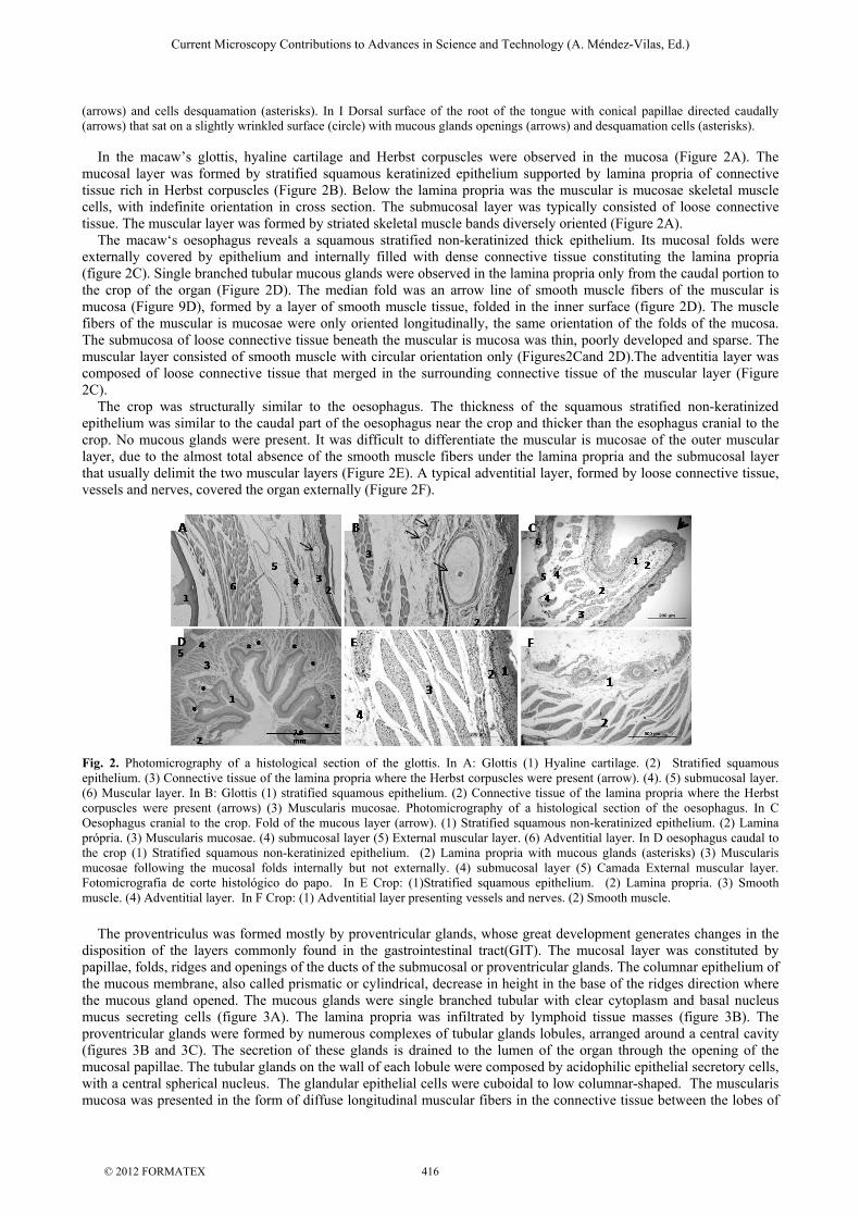
(arrows) and cells desquamation (asterisks). In I Dorsal surface of the root of the tongue with conical papillae directed caudally (arrows) that sat on a slightly wrinkled surface (circle) with mucous glands openings (arrows) and desquamation cells (asterisks). In the macaw’s glottis, hyaline cartilage and Herbst corpuscles were observed in the mucosa (Figure 2A). The mucosal layer was formed by stratified squamous keratinized epithelium supported by lamina propria of connective tissue rich in Herbst corpuscles (Figure 2B). Below the lamina propria was the muscular is mucosae skeletal muscle cells, with indefinite orientation in cross section. The submucosal layer was typically consisted of loose connective tissue. The muscular layer was formed by striated skeletal muscle bands diversely oriented (Figure 2A). The macaw‘s oesophagus reveals a squamous stratified non-keratinized thick epithelium. Its mucosal folds were externally covered by epithelium and internally filled with dense connective tissue constituting the lamina propria (figure 2C). Single branched tubular mucous glands were observed in the lamina propria only from the caudal portion to the crop of the organ (Figure 2D). The median fold was an arrow line of smooth muscle fibers of the muscular is mucosa (Figure 9D), formed by a layer of smooth muscle tissue, folded in the inner surface (figure 2D). The muscle fibers of the muscular is mucosae were only oriented longitudinally, the same orientation of the folds of the mucosa. The submucosa of loose connective tissue beneath the muscular is mucosa was thin, poorly developed and sparse. The muscular layer consisted of smooth muscle with circular orientation only (Figures2Cand 2D).The adventitia layer was composed of loose connective tissue that merged in the surrounding connective tissue of the muscular layer (Figure 2C). The crop was structurally similar to the oesophagus. The thickness of the squamous stratified non-keratinized epithelium was similar to the caudal part of the oesophagus near the crop and thicker than the esophagus cranial to the crop. No mucous glands were present. It was difficult to differentiate the muscular is mucosae of the outer muscular layer, due to the almost total absence of the smooth muscle fibers under the lamina propria and the submucosal layer that usually delimit the two muscular layers (Figure 2E). A typical adventitial layer, formed by loose connective tissue, vessels and nerves, covered the organ externally (Figure 2F).
Fig. 2. Photomicrography of a histological section of the glottis. In A: Glottis (1) Hyaline cartilage. (2) Stratified squamous epithelium. (3) Connective tissue of the lamina propria where the Herbst corpuscles were present (arrow). (4). (5) submucosal layer. (6) Muscular layer. In B: Glottis (1) stratified squamous epithelium. (2) Connective tissue of the lamina propria where the Herbst corpuscles were present (arrows) (3) Muscularis mucosae. Photomicrography of a histological section of the oesophagus. In C Oesophagus cranial to the crop. Fold of the mucous layer (arrow). (1) Stratified squamous non-keratinized epithelium. (2) Lamina própria. (3) Muscularis mucosae. (4) submucosal layer (5) External muscular layer. (6) Adventitial layer. In D oesophagus caudal to the crop (1) Stratified squamous non-keratinized epithelium. (2) Lamina propria with mucous glands (asterisks) (3) Muscularis mucosae following the mucosal folds internally but not externally. (4) submucosal layer (5) Camada External muscular layer. Fotomicrografia de corte histológico do papo. In E Crop: (1)Stratified squamous epithelium. (2) Lamina propria. (3) Smooth muscle. (4) Adventitial layer. In F Crop: (1) Adventitial layer presenting vessels and nerves. (2) Smooth muscle.
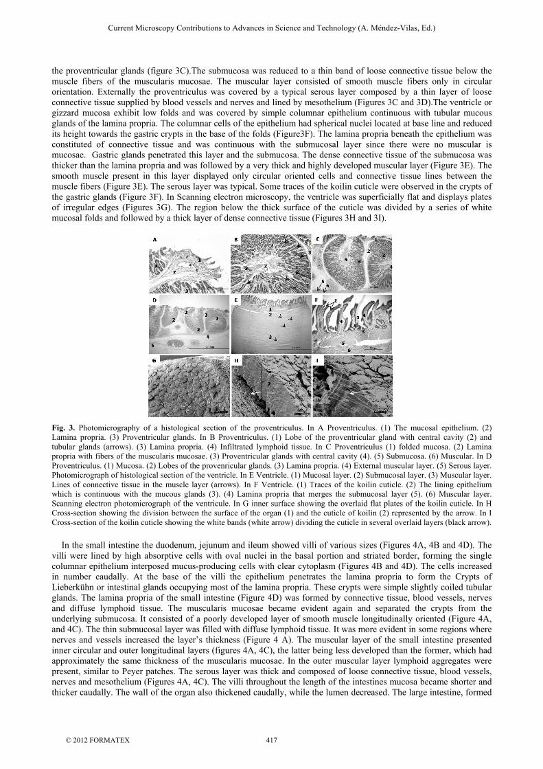
The proventriculus was formed mostly by proventricular glands, whose great development generates changes in the disposition of the layers commonly found in the gastrointestinal tract(GIT). The mucosal layer was constituted by papillae, folds, ridges and openings of the ducts of the submucosal or proventricular glands. The columnar epithelium of the mucous membrane, also called prismatic or cylindrical, decrease in height in the base of the ridges direction where the mucous gland opened. The mucous glands were single branched tubular with clear cytoplasm and basal nucleus mucus secreting cells (figure 3A). The lamina propria was infiltrated by lymphoid tissue masses (figure 3B). The proventricular glands were formed by numerous complexes of tubular glands lobules, arranged around a central cavity (figures 3B and 3C). The secretion of these glands is drained to the lumen of the organ through the opening of the mucosal papillae. The tubular glands on the wall of each lobule were composed by acidophilic epithelial secretory cells, with a central spherical nucleus. The glandular epithelial cells were cuboidal to low columnar-shaped. The muscularis mucosa was presented in the form of diffuse longitudinal muscular fibers in the connective tissue between the lobes of
Current Microscopy Contributions to Advances in Science and Technology (A. Méndez-Vilas, Ed.)
the proventricular glands (figure 3C).The submucosa was reduced to a thin band of loose connective tissue below the muscle fibers of the muscularis mucosae. The muscular layer consisted of smooth muscle fibers only in circular orientation. Externally the proventriculus was covered by a typical serous layer composed by a thin layer of loose connective tissue supplied by blood vessels and nerves and lined by mesothelium (Figures 3C and 3D).The ventricle or gizzard mucosa exhibit low folds and was covered by simple columnar epithelium continuous with tubular mucous glands of the lamina propria. The columnar cells of the epithelium had spherical nuclei located at base line and reduced its height towards the gastric crypts in the base of the folds (Figure3F). The lamina propria beneath the epithelium was constituted of connective tissue and was continuous with the submucosal layer since there were no muscular is mucosae. Gastric glands penetrated this layer and the submucosa. The dense connective tissue of the submucosa was thicker than the lamina propria and was followed by a very thick and highly developed muscular layer (Figure 3E). The smooth muscle present in this layer displayed only circular oriented cells and connective tissue lines between the muscle fibers (Figure 3E). The serous layer was typical. Some traces of the koilin cuticle were observed in the crypts of the gastric glands (Figure 3F). In Scanning electron microscopy, the ventricle was superficially flat and displays plates of irregular edges (Figures 3G). The region below the thick surface of the cuticle was divided by a series of white mucosal folds and followed by a thick layer of dense connective tissue (Figures 3H and 3I).
Fig. 3. Photomicrography of a histological section of the proventriculus. In A Proventriculus. (1) The mucosal epithelium. (2) Lamina propria. (3) Proventricular glands. In B Proventriculus. (1) Lobe of the proventricular gland with central cavity (2) and tubular glands (arrows). (3) Lamina propria. (4) Infiltrated lymphoid tissue. In C Proventriculus (1) folded mucosa. (2) Lamina propria with fibers of the muscularis mucosae. (3) Proventricular glands with central cavity (4). (5) Submucosa. (6) Muscular. In D Proventriculus. (1) Mucosa. (2) Lobes of the provenricular glands. (3) Lamina propria. (4) External muscular layer. (5) Serous layer. Photomicrograph of histological section of the ventricle. In E Ventricle. (1) Mucosal layer. (2) Submucosal layer. (3) Muscular layer. Lines of connective tissue in the muscle layer (arrows). In F Ventricle. (1) Traces of the koilin cuticle. (2) The lining epithelium which is continuous with the mucous glands (3). (4) Lamina propria that merges the submucosal layer (5). (6) Muscular layer. Scanning electron photomicrograph of the ventricule. In G inner surface showing the overlaid flat plates of the koilin cuticle. In H Cross-section showing the division between the surface of the organ (1) and the cuticle of koilin (2) represented by the arrow. In I Cross-section of the koilin cuticle showing the white bands (white arrow) dividing the cuticle in several overlaid layers (black arrow).
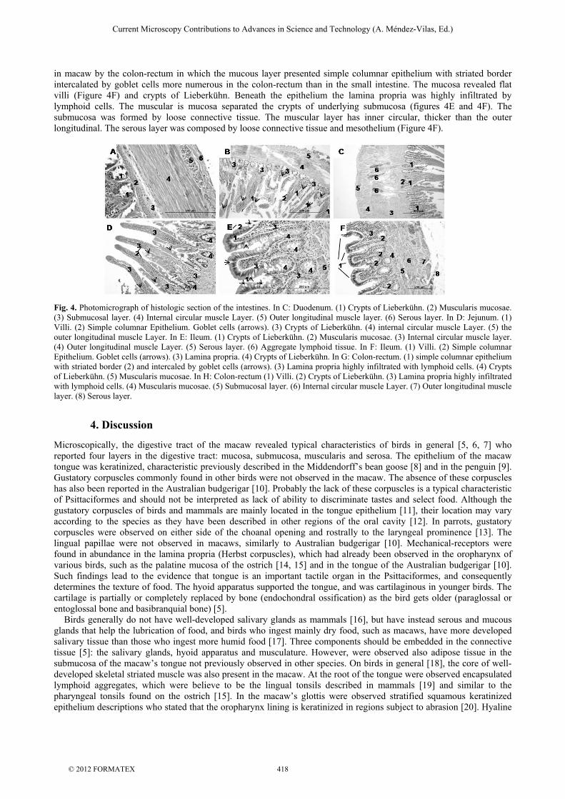
In the small intestine the duodenum, jejunum and ileum showed villi of various sizes (Figures 4A, 4B and 4D). The villi were lined by high absorptive cells with oval nuclei in the basal portion and striated border, forming the single columnar epithelium interposed mucus-producing cells with clear cytoplasm (Figures 4B and 4D). The cells increased in number caudally. At the base of the villi the epithelium penetrates the lamina propria to form the Crypts of Lieberkühn or intestinal glands occupying most of the lamina propria. These crypts were simple slightly coiled tubular glands. The lamina propria of the small intestine (Figure 4D) was formed by connective tissue, blood vessels, nerves and diffuse lymphoid tissue. The muscularis mucosae became evident again and separated the crypts from the underlying submucosa. It consisted of a poorly developed layer of smooth muscle longitudinally oriented (Figure 4A, and 4C). The thin submucosal layer was filled with diffuse lymphoid tissue. It was more evident in some regions where nerves and vessels increased the layer’s thickness (Figure 4 A). The muscular layer of the small intestine presented inner circular and outer longitudinal layers (figures 4A, 4C), the latter being less developed than the former, which had approximately the same thickness of the muscularis mucosae. In the outer muscular layer lymphoid aggregates were present, similar to Peyer patches. The serous layer was thick and composed of loose connective tissue, blood vessels, nerves and mesothelium (Figures 4A, 4C). The villi throughout the length of the intestines mucosa became shorter and thicker caudally. The wall of the organ also thickened caudally, while the lumen decreased. The large intestine, formed
Current Microscopy Contributions to Advances in Science and Technology (A. Méndez-Vilas, Ed.)
in macaw by the colon-rectum in which the mucous layer presented simple columnar epithelium with striated border intercalated by goblet cells more numerous in the colon-rectum than in the small intestine. The mucosa revealed flat villi (Figure 4F) and crypts of Lieberkühn. Beneath the epithelium the lamina propria was highly infiltrated by lymphoid cells. The muscular is mucosa separated the crypts of underlying submucosa (figures 4E and 4F). The submucosa was formed by loose connective tissue. The muscular layer has inner circular, thicker than the outer longitudinal. The serous layer was composed by loose connective tissue and mesothelium (Figure 4F).
Fig. 4. Photomicrograph of histologic section of the intestines. In C: Duodenum. (1) Crypts of Lieberkühn. (2) Muscularis mucosae. (3) Submucosal layer. (4) Internal circular muscle Layer. (5) Outer longitudinal muscle layer. (6) Serous layer. In D: Jejunum. (1) Villi. (2) Simple columnar Epithelium. Goblet cells (arrows). (3) Crypts of Lieberkühn. (4) internal circular muscle Layer. (5) the outer longitudinal muscle Layer. In E: Ileum. (1) Crypts of Lieberkühn. (2) Muscularis mucosae. (3) Internal circular muscle layer. (4) Outer longitudinal muscle Layer. (5) Serous layer. (6) Aggregate lymphoid tissue. In F: Ileum. (1) Villi. (2) Simple columnar Epithelium. Goblet cells (arrows). (3) Lamina propria. (4) Crypts of Lieberkühn. In G: Colon-rectum. (1) simple columnar epithelium with striated border (2) and intercaled by goblet cells (arrows). (3) Lamina propria highly infiltrated with lymphoid cells. (4) Crypts of Lieberkühn. (5) Muscularis mucosae. In H: Colon-rectum (1) Villi. (2) Crypts of Lieberkühn. (3) Lamina propria highly infiltrated with lymphoid cells. (4) Muscularis mucosae. (5) Submucosal layer. (6) Internal circular muscle Layer. (7) Outer longitudinal muscle layer. (8) Serous layer.
4. Discussion
Microscopically, the digestive tract of the macaw revealed typical characteristics of birds in general [5, 6, 7] who reported four layers in the digestive tract: mucosa, submucosa, muscularis and serosa. The epithelium of the macaw tongue was keratinized, characteristic previously described in the Middendorff’s bean goose [8] and in the penguin [9]. Gustatory corpuscles commonly found in other birds were not observed in the macaw. The absence of these corpuscles has also been reported in the Australian budgerigar [10]. Probably the lack of these corpuscles is a typical characteristic of Psittaciformes and should not be interpreted as lack of ability to discriminate tastes and select food. Although the gustatory corpuscles of birds and mammals are mainly located in the tongue epithelium [11], their location may vary according to the species as they have been described in other regions of the oral cavity [12]. In parrots, gustatory corpuscles were observed on either side of the choanal opening and rostrally to the laryngeal prominence [13]. The lingual papillae were not observed in macaws, similarly to Australian budgerigar [10]. Mechanical-receptors were found in abundance in the lamina propria (Herbst corpuscles), which had already been observed in the oropharynx of various birds, such as the palatine mucosa of the ostrich [14, 15] and in the tongue of the Australian budgerigar [10]. Such findings lead to the evidence that tongue is an important tactile organ in the Psittaciformes, and consequently determines the texture of food. The hyoid apparatus supported the tongue, and was cartilaginous in younger birds. The cartilage is partially or completely replaced by bone (endochondral ossification) as the bird gets older (paraglossal or entoglossal bone and basibranquial bone) [5]. Birds generally do not have well-developed salivary glands as mammals [16], but have instead serous and mucous glands that help the lubrication of food, and birds who ingest mainly dry food, such as macaws, have more developed salivary tissue than those who ingest more humid food [17]. Three components should be embedded in the connective tissue [5]: the salivary glands, hyoid apparatus and musculature. However, were observed also adipose tissue in the submucosa of the macaw’s tongue not previously observed in other species. On birds in general [18], the core of well-developed skeletal striated muscle was also present in the macaw. At the root of the tongue were observed encapsulated lymphoid aggregates, which were believe to be the lingual tonsils described in mammals [19] and similar to the pharyngeal tonsils found on the ostrich [15]. In the macaw’s glottis were observed stratified squamous keratinized epithelium descriptions who stated that the oropharynx lining is keratinized in regions subject to abrasion [20]. Hyaline
Current Microscopy Contributions to Advances in Science and Technology (A. Méndez-Vilas, Ed.)
cartilage and Herbst corpuscles were clearly visible in the glottis. These corpuscles were restricted to keratinized regions in the oral cavity, such as the glottis. The oesophagus showed typical tunica mucosa [21, 5, 7]. The epithelium in the macaw differs from the findings of [22, 23, 6] who described the epithelium as keratinized. In the esophagus lamina propria of macaw's few mucous glands was present only in the portion of the oesophagus caudal to the crop. The glands were similar to the mucous glands observed in the tongue contrarily to [5, 6, 7, 22] reports on birds. The longitudinal folds were more evident in the caudal portion different to ostriches [15]. The submucosal layer of a thin layer of connective tissue [22]. The muscular layer in macaws revealed a band of smooth muscle circularly oriented similarly to ostriches [15]. Different arrangement was reported in other birds where the muscular layer is divided in inner circular and thin longitudinal outer layer [5, 6, 21]; three subdivisions in the muscular layer of rheas were described [24]: outer longitudinal, circular intermediary and inner longitudinal according with the founded in macaw`s. In the macaw´s crop the lamina propria was less developed than in the oesophagus with few mucous glands in accordance with descriptions in the chicken [7]. The absence of mucous glands, except in regions near the oesophagus represents only a significant histological difference between the two structures as observed [5]. In the macaw the limit between muscularis mucosae and muscular layer was not clear, due to the almost absence of muscle fibres between these two layers. The proventricle of macaws was structurally similar to other birds as described by several authors [5, 6, 18], rich in proventricular glands. Other layers were present, but modified or reduced due to the pressure exerted by the large well-developed submucosal glands. The mucosa of the epithelium was columnar, as described in the hen [25, 5] in the rock dove [26] in the yellow quail [27] and in the house sparrow [28].The connective tissue lamina propria with lymphoid vessels infiltration [25, 5]. Diffuse muscle fibers of the muscularis mucosae were found near the lamina propria, but the longitudinal external stratum of the layer was not evident, contrasting the observations who reported an extremely thin external muscular layer in the proventriculus of the budgerigar and parrot [29]. The submucosal layer was very thin as described in birds [25, 5, 22] beneath the isolated muscle fibers of the muscularis mucosae as reported in chickens [30] and in the burrowing owl [31]. The glandular epithelial cells varied from cuboidal to low columnar with serrated appearance similarly to findings in birds [6, 18]. The difference observed microscopically in the macaw’s proventriculus in comparison with other birds was the presence of a single layer of smooth muscle fibers in the muscular layer, contrarily to the three layers identified in chickens [30], in the red-gartered coot [32] and in birds [6]. The mucous membrane was observed with two layers [25]: outer longitudinal and inner circular. Supporting the evidences [5, 29, 27, 31] the circular layer in the macaws was thicker than the longitudinal. The structural layers of the ventriculus also showed typical characteristics of birds [5, 7, 18]. The lamina propria has loose connective tissue than the submucosa, which consists of dense connective tissue as observed in the macaw [5]. Some authors have verified the presence of a more developed muscularis mucosae in birds [31], with longitudinal muscle fibers in burrowing owl (carnivorous bird), or less developed muscularis mucosae in the ring-necked parakeet(fruit bird) [33]. Although have described three layers in the tunica muscular of the red-gartered coot [32], in the love bird [34] and observed two layers in the ring-necked parakeet [33], in the burrowing owl [31] and in birds [22], were identified only one layer in the macaw. In the histological sections examined in our study were observe only a thick layer of circular muscle fibers. “The luminal surface should be coated with a keratin-like secretion product (koilin cuticle), produced by the mucous glands of the lamina propria” [5, 22, 6, 7, 18]. However, this cuticle has not been observed and may have been destroyed in the preparation of our histological sections, since the setting of the keratin-like substance in the epithelium is only moderately firm. The small intestine was not divided in different histological regions, and was structurally similar to mammals [6, 21, 18], in the chicken [5, 7] and in the Australian budgerigar descriptions [10]. Studies with the different segments of small intestine of ostriches [35], describing them as covered by villi, lined by a simple cylindrical epithelium with microvilli located on the edges of the apical cells. Although the muscularis mucosae of the duodenum and jejunum has two layers [36] it have three layers in the ileum and the muscular layer is thin, with circular and longitudinal layer of different sizes. Except for the muscularis mucosae of the ileum which differs from the macaw by presenting two layers, the description of the lining of the small intestine [36, 35]. The villi increased the absorptive surface of organ and the intestinal glands or crypts of Lieberkühn opened between the villi. These lands release mucus enzymes and hormones secreting cells, and, progenitor cells which repair and restore the intestinal epithelium [7]. Lymphoid aggregates, similar to Peyer patches were present in the submucosa and in the external longitudinal layer contrarily to [6] who described them as only present in the caudal portion of digestive tract. Structurally the large intestine is similar to small intestine, the most significant difference being the shortening and thickening of the mucosal villi [7]. Accordingly the mucosa of the macaw’s large intestine possessed villi similar to those described in birds [6, 7]. This feature differs from observations in the Australian budgerigar [10], who also refer to the absence of external longitudinal muscular layer and villi, and folds in the epithelial surface. Were convinced that our findings and the presence of layers depending on the degree of the intestine distension [5]. When it is fully distended the villi become flattened. Additionally, the number of goblet cells increase in the large intestine as described in birds [6]. The scanning electron microscopy findings confirmed the observations of the light microscopy. The keratinized dermal papillae observed macroscopically in the root of the tongue were also observed in scanning electron microscopy.
Current Microscopy Contributions to Advances in Science and Technology (A. Méndez-Vilas, Ed.)
Additionally, the presence of glands described in LM was confirmed in the SEM; the openings of the glands in the dorsal surface of the tongue, some releasing mucus. Desquamation In the surface of the apex was greater than in the body and the root of the tongue thus confirming the histology evidences where the apex was more keratinized than other portions of the organ. The dorsal surface of the apex of the tongue was extremely folded similarly to the region of the tongue’s body in the Middendorff’s bean goose [8]. The macroscopic observation showed that the dorsal surface of the root of the tongue in the macaw was smooth like the body of the goose’s tongue [8], the little tern’s tongue in chicken [8]. This characteristic seems to be a natural adjustment to facilitate swallowing, since many authors relate the form and structure of the tongue to the feeding habits of the species [22, 8]. However at the root of the macaw’s tongue were also observed caudally oriented papillae previously reported in earlier studies. Giant conical papillae, called '' lingual spikes '' [37] have been observed in various birds, such as the duck by [37], the owl [38], the goose [8] and the white-tailed eagle [39]. However these structures were more numerous and arranged in the form of a row between the body and the lingual root, whereas in the macaw they are located in the lingual root in two rows formed a "V" with the apex facing rostrally. Another bird in which the disposal of conical papillae differs from that found in many avian species is the penguin, where they are numerous covering the entire dorsal surface of the tongue to the laryngeal prominence [9]. The function of the papillae is not fully clear [8], but it is believed that they are useful to help the transfer of food towards the oesophagus preventing regurgitation [39]. Even though SEM of the ventral surface of the macaw’s tongue was not carried out were convinced (from the LM ) that we would find high levels of keratinization since macroscopically we the area was anatomically described as lingual nail or “cuticula cornea lingualis” due to its appearance similar to nail [22]. This lingual nail was described in the white-tailed eagle [39]. Similarly to ostrich [15] descriptions in the ostrich, the dorsal surface of the tongue of the macaw also presented duct openings of the subjacent lingual glands. Scanning microscopy of the macaw’s ventricle demonstrated the presence of a hard and thick internal gastric cuticle observed macroscopically and few folds of mucosa of dense connective tissue band. The surface of the koilin cuticle was thick and composed by plates that probably deposited one over the other, koilin clusters observed between the folds of the mucosa probably produced by "glands" in chickens [40], who states that birds whose diet consists of hard material have a thick and abrasive inner membrane of koilin. The inner surface of the cuticle in the macaw, despite being abrasive, differs from that described in chickens [40]. This author describes the cuticle as being narrow clusters of koilin tips in the form of vertical rodlets formed by the tubular glands; among these rodlets there’s a horizontal rough matrix rounded by less rigid material formed by layers of superficial epithelial cells that die and desquamate alternate by less harsh koilin layers. However the layers of the macaw’s koilin cuticle only display koilin plates and desquamated dead epithelial cells. Even though a brief note of the appearance of the mucosal surface of the pigeon’s ventriculus has been published [41], no further information was found in the literature about the characteristics of the surface of the koilin cuticule, in other species, besides the description in the chicken [5, 40]. As for scanning electron microscopy, few remarkable particularities described in our study include the presence of openings representing the mucous glands duct in all lingual portions, the presence of conical papillae on the tongue’s root and the reduction of folds and ridges from the apex to the root presented on the dorsal surface of the tongue. Because of the almost total absence of morphological information about the ventricle of Psittaciformes and other birds in general, this study used only hens as comparison standard. Structurally it was noted that the harsh and greenish koilin present in cuticle of the macaw’s ventricle, with rodlets on the surface differs from the harsh and yellowish cuticle on the chicken, with plates of irregular edges on its surface. The structural peculiarities found in the microscopy may be attributed to different dietary habits of the macaw.
References
[1] TUBELIS, D.P..Feeding ecology of Ara Araruna(Aves, Psitttacidae)at firebreaks in western cerrado, Brazil.Revista Biotemas, 22, junho de 2009,pp105-115.
[2] COLOBINI, F. Maravilhas do Brasil (Wonders of Brazil) Aves (Birds). Escrituras. São Paulo, 2006, p. 106. [3] VALLE, S. F., et al. Parâmetros de bioquímica sérica de machos, fêmeas e filhotes de arara Canindé (Ara ararauna) saudáveis
mantidas em cativeiro comercial. Ciência Rural, v.38, n.3, p. 711-716, mai-jun, 2008. [4] TOLOSA, E. M. C.; RODRIGUES, C. J.; BEHEMER, O. A.; FREITAS NETO, A. G. Manual de técnicas normal e patológica. 2.
ed. São Paulo: Manole, 2003. [5] HODGES, R. D. The histology of the fowl. London: Academic Press, 1974. pp 35-88. [6] BANKS, W. J. Histologia veterinária aplicada. 2. ed. São Paulo: Manole, 1991. 629pp. [7] BACHA, W. J.; BACHA, L. M. 2003. Atlas colorido de histologiaveterinária. 2ª ed., Roca, São Paulo, Brasil, 457pp. [8] IWASAKI, S. et al. Ultrastructural study of the keratinization of the dorsal epithelium of the tongue of Middendorff’s bean goose,
Anserfabalismiddendorffi.Anatomical Records,v. 247, p.149-163, 1997. [9] KOBAYASHI, K. et al. Fine structure of the tongue and lingual papillae of the penguin.Archives of Histology and Cytology
,v.61, p.37-46, 1998. [10] MATSUMOTO, F.S. Topografia e morfologia das vísceras do perquito-australiano (Melopsittacusundulatus, SHAW 1805).
Ciência Animal Brasileira. v. 10, n. 4. p.1263-1270, 2009. [11] KENT GC Comparative Anatomy of the Vertebrates. Saint Louis: Mosby Co, 1978.
Current Microscopy Contributions to Advances in Science and Technology (A. Méndez-Vilas, Ed.)
[12] GANCHROW, J. R., D. GANCHROW, M. OPPENHEIMER. Chorda tympani innervationof anterior mandibular taste buds in the chicken (Gallus gallusdomesticus). Anat. Rec. pp. 2163,434, 1986.
[13] COLES, B. H.: Essentials of Avian Medicine and Surgery. 3 ed. UK:Blackwell Publishing, 2007. [14] GUIMARÃES, J. P. et al. Mecanoreceptores da mucosa palatina de avestruz (Struthiocamelus): estudo ao microscópio de
luz. Pesq. Vet. Bras. v.27, n 12, p.491-494, Dez 2007. [15] TIVANE, C.A morphological study of the oropharynx and oesophagus of the ostrich (Struthiocamelus).2008.M.Sc. thesis,
University of Pretoria, South Africa. [16] IWASAKI, S. Evolution of the structure and function of the vertebrate tongue.Journal of Anatomy. v. 201, pp. 1-13, 2002. [17] OROSZ, S. Anatomy of the digestive system. In: Altman R.B.et al. Avian medicine andsurgery. Philadelphia: WB
SaundersCompany, 1997. p. 412–5. [18] DELLMANN, H. D. and EURELL, J. Textbook of Veterinary Histology. 6th Ed. UK:Blackwell Publishing, 2006. [19] COSTA, M.M.B. Anatomia funcional da faringe. In: Andy Petroiani. Anatomia Cirúrgica. Guanabara Koogan. Rio de Janeiro,
1999.pp. 206-216. [20] OLSEN, G.H. Oral biology and beak disorders ofbirds. In: CROSSLEY, D.A. Oral biology, dental and beak disorders. The
Veterinary Clinics of North America: exotic animal practice. W.B. Saunders Company, Philadelphia, 2003, v.6, n.3, pp.505-522.
[21] AUGHEY, E. and FRYE, F. L. Comparative Veterinary Histology with Clinical Correlates. Iowa: Iowa State University Press, 2001.
[22] McLELLAND, J. Digestive system. In: KING, A. S., J.McLELLAND. Form and Function in Birds. London: Academic Press, 1979. p. 69-181.
[23] FOWLER, M.E. Comparative clinical anatomy of ratites.Journal of Zoo and Wildlife Medicine. v. 22, pp. 204-227, 1991. [24] BARTHELS, P. BeitragzurHistologie des Ösophagus der Vögel.Zeitschriftfür wissenschafttlicheZoologie. v.59, pp. 655-689,
1895. [25] CALHOUN, M. L. Microscopic anatomy of the digestive system of the chicken. Ames, Iowa State College Press, 1954.108 pp. [26] LIMA, M. A. I. & SASSO, W. S. Histochemical detection of glycoproteins in the gastric epithelia of Columba livia.Acta
Histochem. v.76.pp.145-50, 1985. [27] FIERI, W. J. Aspectos anatômicos e histológicos do tubo digestivo da codorna Nothuramaculosa maculosa, (TEMMINCK,
1815). 1984. Tesedoutor. Univ. Mackenzie, São Paulo. 109 pp [28] KLEM JR., PARKER, M. A.; SPRAGUE, W. L.; TARUFI, S. A.; VELTRI, C. J. & WALKER, M. J. Gross morphology and
general histology of the alimentary tract of the american robin (Turdusmigratorius). Proc. Pa. Acad. Sci. v.58 pp. 151-8, 1984.
[29] BEE DE SPERONI, N. & CHIKILIAN, M. Estudiomorfohistologico e histoquimico comparado de la primeira porciondeltracto digestivo de Zenaidaauriculatachrysauchenia y Myiopsittamonachamonacha (aves Columbidae y psittacidae). Hist. Nat. Corrientes. v.3 pp. 21-32, 1983.
[30] BRADLEY, O. C. & GRAHAME, T. The structure of the fowl. 3. ed. Philadelphia, J.B. Lippincot, 1951. pp 29-48. [31] ROCHA, S.O. Aspectos morfológicos (histológicos) do tubo digestivo da coruja-buraqueira Speotytocunicularia, (Molina,
1782). São Paulo, 1991. 73pp. [Dissertação (mestr.) - Escola Paulista de Medicina]. [32] ESPINOLA, L.V. & GALLIUSSI, E.A. Estudio anátomo-histológico del tracto digestivo de Fulica armillata (VELELLOT,
1817) Aves (Gruiformes, Rallidae) Iheringia Sér. Zool.v.70, pp. 93-108, 1990. [33] JAIN, D. K. Histomorphology and proteolytic activity in the gastric apparatus of frugivorous,carnivorous and omnivorous
species of birds. Acta Biol. Acad. Sci. hung. v.27, pp.135-45, 1976. [34] IMAIZUMI, M. & HAMA, K.An electron microscopic study on the interstitial cells of the gizzard in the love-bird
(Urolonchadomestica), Z. Zellforch.v. 97, pp. 351-7, 1969. [35] ILLANES, J.et al.;. Descripción histológica de los diferentes segmentos del aparato digestivo de avestruz (Struthiocamelus var.
domesticus).InternationalJournalofMorphology.. v. 24, pp. 205-214, 2006. [36] BEZUIDENHOUT, A. J.; VAN ASWEGWN, G.A light microscopic and immunocytochemical study of the gastrointestinal
tract of the ostrich (Struthiocamelus L.).Onderstepoort Journal of Veterinarian Research.v 57:, pp. 37-48, 1990. [37] KOOLOOS, J.G.M. A conveyer belt model for pecking in the mallard (Anasplatyrhynchos).Neth. J. Zool. v. 36, pp. 47–87,
1986. [38] EMURA, S & CHEN, H. Scanning electron microscopic study of the tongue in the owl (Strixuralensis). AnatHistolEmbryol.
v. 37, pp. 475-478. 2008. [39] JACKOWIAK, H., AND S. GODYNICKI: Light and scanning electron microscopic study of the tongue in the white tailedeagle
(Haliaeetusalbicilla, Accipitridae, Aves). Ann. Anat. v. 187, pp. 197–205 , 2005. [40] AKESTER, A. R. Structure of the glandular layer and koilin membrane in the gizzard of the adult domestic fowl (Gallus
gallusdomesticus).Journal of Anatomy. v. 147, pp.1 - 25, 1986. [41] DESMETH, M. Scanning electron microscopy of the proventriculus and gizzard of the pigeon.Zeitschrift far mikroskopisch-
anatomischeForschung . v.9, pp. 180-182, 1982.
Current Microscopy Contributions to Advances in Science and Technology (A. Méndez-Vilas, Ed.)


![The extinct macaws of the West Indies, with special ... · macaws were ‘as numerous in all these islands [West Indies] as sparrows or other small birds are with us’ [Europeans],](https://static.documents.pub/doc/80x56/5f2a10a375fc6f0ea638bcdd/the-extinct-macaws-of-the-west-indies-with-special-macaws-were-aas-numerous.jpg)


