Microstructure and strain relaxation of orthorhombic TmMnO 3 epitaxial thin films Y. Yu a , X. Zhang a,n , J.J. Yang b , J.W. Wang b , Y.G. Zhao b a Beijing National Center for Electron Microscopy, Laboratory of Advanced Materials, Department of Materials Science and Engineering, Tsinghua University, Beijing 100084, China b Department of Physics and the Key Laboratory of Atomic and Nanosciences, Ministry of Education, Tsinghua University, Beijing 100084, China article info Article history: Received 5 October 2011 Received in revised form 7 November 2011 Accepted 14 November 2011 Communicated by A. Ohtomo Available online 19 November 2011 Keywords: A1. Crystal structure A1. Transmission electron microscopy B1. Manganites B1. Perovskites B2. Ferroelectric materials B2. Magnetic materials abstract Orthorhombic TmMnO 3 (o-TMO) thin films have been epitaxially stabilized on (110) SrTiO 3 substrates by pulsed laser deposition (PLD) technique. The microstructure and strain relaxation mechanism of o-TMO thin films are analyzed using transmission electron microscopy. It is shown that major defects in the films are misfit dislocations with Burgers vectors of type a p /010S and a p /110S, whereas a p /110S dislocations tend to dissociate into partial dislocations with Burgers vectors of type 1/2a p /110S. Strain in o-TMO films is relaxed by misfit dislocations as well as surface fluctuations, which is different from most of the previous studies of the perovskite thin films. & 2011 Elsevier B.V. All rights reserved. 1. Introduction Nowadays, multiferroic materials have garnered tremendous research interests due to their unique physical properties and the potential for future applications in multifunctional devices [1]. Orthorhombic (o-) rare-earth manganites RMnO 3 (R belonging to lanthanum series) represent an important class of ‘‘improper ferro- electrics’’ [2], where the ferroelectricity is not only coexisting with, but also intrinsically correlating with the magnetic order. A rich variety of subtle interplay among spin, charge, orbital, and lattice degrees of freedom are responsible for the distinctive features in this system. As the orthorhombic phase of RMnO 3 with R¼ Y, Ho-Lu is not a thermodynamically stable phase at the ambient conditions, obtain of o-RMnO 3 (R¼ Y, Ho-Lu) is therefore a prerequisite for the follow-up studies. Epitaxial stabilization of this orthorhombic phase in thin films were achieved by metal–organic chemical vapor deposition (MOCVD) [3] and pulsed laser deposition (PLD) [4–6]. Meanwhile, recent studies have reported the strain-induced effects on ferroelec- tricity [7], magnetic ordering [8], magnetocapacitance [9], and mag- netoelectric coupling [10] in o-RMnO 3 thin films. Large electric polarization and magnetoelectric coupling have just been found in o-TmMnO 3 (o-TMO) thin films [6]. In order to have a better under- standing of the physical properties of these materials and the mechanisms involved, it is of vital importance to analyze the microstructure of o-RMnO 3 (R¼ Y, Ho-Lu) thin films. However, an intensive microstructural study including defect structure and strain relaxation of o-RMnO 3 epitaxial thin films is still lacking. In this paper, we report on the characterization of microstructure and strain relaxation of o-TMO epitaxial thin films by transmission electron microscopy (TEM). 2. Experiment o-TMO thin films were grown on (110)-oriented SrTiO 3 (STO) single-crystal substrates using PLD with a 248 nm KrF excimer laser. The films were all deposited under a 10 Pa atmosphere of flowing oxygen at 940 1C with a single-phased hexagonal TMO target, which was prepared by solid state sintering with stoichio- metric high-purity Tm 2 O 3 and MnO 2 powders as the starting materials. TEM cross-sectional specimens were then prepared by cutting, gluing, mechanical polishing, and dimpling, followed by argon ion milling in a stage cooled with liquid nitrogen. TEM studies were carried out on a JEOL JEM-2011 and a JEOL JEM- 2010F field-emission gun transmission electron microscope (FEG- TEM) operated at 200 kV. 3. Results and discussion Fig. 1(a) shows the low-magnification cross-sectional image of o-TMO/STO, where o-TMO is about 20 nm in thickness with rough Contents lists available at SciVerse ScienceDirect journal homepage: www.elsevier.com/locate/jcrysgro Journal of Crystal Growth 0022-0248/$ - see front matter & 2011 Elsevier B.V. All rights reserved. doi:10.1016/j.jcrysgro.2011.11.031 n Corresponding author. Tel.: þ86 10 62773999; fax: þ86 10 62771160. E-mail address: [email protected] (X. Zhang). Journal of Crystal Growth 338 (2012) 280–282
Transcript
Journal of Crystal Growth 338 (2012) 280–282
Contents lists available at SciVerse ScienceDirect
Microstructure and strain relaxation of orthorhombic TmMnO3 epitaxialthin films
Y. Yu a, X. Zhang a,n, J.J. Yang b, J.W. Wang b, Y.G. Zhao b
a Beijing National Center for Electron Microscopy, Laboratory of Advanced Materials, Department of Materials Science and Engineering, Tsinghua University, Beijing 100084, Chinab Department of Physics and the Key Laboratory of Atomic and Nanosciences, Ministry of Education, Tsinghua University, Beijing 100084, China
a r t i c l e i n f o
Article history:
Received 5 October 2011
Received in revised form
7 November 2011
Accepted 14 November 2011
Communicated by A. Ohtomo/110S dislocations tend to dissociate into partial dislocations with Burgers vectors of type 1/2ap
Orthorhombic TmMnO3 (o-TMO) thin films have been epitaxially stabilized on (110) SrTiO3 substrates
by pulsed laser deposition (PLD) technique. The microstructure and strain relaxation mechanism of
o-TMO thin films are analyzed using transmission electron microscopy. It is shown that major defects
in the films are misfit dislocations with Burgers vectors of type ap/010S and ap/110S, whereas ap
/110S. Strain in o-TMO films is relaxed by misfit dislocations as well as surface fluctuations, which is
different from most of the previous studies of the perovskite thin films.
& 2011 Elsevier B.V. All rights reserved.
1. Introduction
Nowadays, multiferroic materials have garnered tremendousresearch interests due to their unique physical properties and thepotential for future applications in multifunctional devices [1].Orthorhombic (o-) rare-earth manganites RMnO3 (R belonging tolanthanum series) represent an important class of ‘‘improper ferro-electrics’’ [2], where the ferroelectricity is not only coexisting with,but also intrinsically correlating with the magnetic order. A richvariety of subtle interplay among spin, charge, orbital, and latticedegrees of freedom are responsible for the distinctive features in thissystem. As the orthorhombic phase of RMnO3 with R¼Y, Ho-Lu is nota thermodynamically stable phase at the ambient conditions, obtainof o-RMnO3 (R¼Y, Ho-Lu) is therefore a prerequisite for the follow-upstudies. Epitaxial stabilization of this orthorhombic phase in thinfilms were achieved by metal–organic chemical vapor deposition(MOCVD) [3] and pulsed laser deposition (PLD) [4–6]. Meanwhile,recent studies have reported the strain-induced effects on ferroelec-tricity [7], magnetic ordering [8], magnetocapacitance [9], and mag-netoelectric coupling [10] in o-RMnO3 thin films. Large electricpolarization and magnetoelectric coupling have just been found ino-TmMnO3 (o-TMO) thin films [6]. In order to have a better under-standing of the physical properties of these materials and themechanisms involved, it is of vital importance to analyze the
ll rights reserved.
x: þ86 10 62771160.
ng).
microstructure of o-RMnO3 (R¼Y, Ho-Lu) thin films. However, anintensive microstructural study including defect structure and strainrelaxation of o-RMnO3 epitaxial thin films is still lacking. In thispaper, we report on the characterization of microstructure and strainrelaxation of o-TMO epitaxial thin films by transmission electronmicroscopy (TEM).
2. Experiment
o-TMO thin films were grown on (110)-oriented SrTiO3 (STO)single-crystal substrates using PLD with a 248 nm KrF excimerlaser. The films were all deposited under a 10 Pa atmosphere offlowing oxygen at 940 1C with a single-phased hexagonal TMOtarget, which was prepared by solid state sintering with stoichio-metric high-purity Tm2O3 and MnO2 powders as the startingmaterials. TEM cross-sectional specimens were then prepared bycutting, gluing, mechanical polishing, and dimpling, followed byargon ion milling in a stage cooled with liquid nitrogen. TEMstudies were carried out on a JEOL JEM-2011 and a JEOL JEM-2010F field-emission gun transmission electron microscope (FEG-TEM) operated at 200 kV.
3. Results and discussion
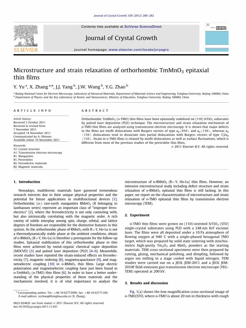
Fig. 1(a) shows the low-magnification cross-sectional image ofo-TMO/STO, where o-TMO is about 20 nm in thickness with rough
Fig. 1. (a) Low-magnification cross-sectional image of o-TMO/STO. (b) Typical [001] SAED pattern of o-TMO/STO. The subscript f and s in the indices denote diffraction
spots coming from the film and the substrate, respectively. Splitting of the spots due to lattice mismatch becomes obvious in high-order reflections, such as (600)f and
(330)s. (c) HRTEM image of the interface.
Y. Yu et al. / Journal of Crystal Growth 338 (2012) 280–282 281
surface. The substrate STO has a cubic perovskite structure (spacegroup Pm-3m) with lattice constant of 3.905 A. The space groupof bulk o-TMO is determined to be Pbnm with lattice constantsao-TMO¼ap
ffiffiffi
2p¼5.228 A, bo-TMO¼ap
ffiffiffi
2p¼5.809 A, and co-TMO¼
2ap¼7.318 A [11], where ap denotes the pseudocubic perovskiteunit-cell lattice parameter. A selected area electron diffraction(SAED) pattern, obtained from the interface area including boththe TMO thin film and the STO substrate, is shown in Fig. 1(b).This result reveals that the a-axis oriented o-TMO epitaxial thinfilm was successfully grown on the STO substrate without any secondphase. The epitaxial relationship is TMO(100)[010]99STO(110)[110],showing the o-TMO thin film suffers a tensile strain of 6.3% in [001]direction and a compressive strain of 5.2% in [110] direction. High-resolution TEM (HRTEM) image of the interface is shown in Fig. 1(c),which further confirms the ‘‘cube-on-cube’’ epitaxy of o-TMO on STOand the existence of a sharp and clean interface.
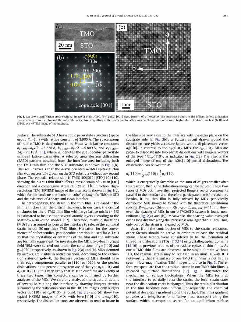
In heteroepitaxy, the strain in the thin film is released if thefilm is thicker than the critical thickness. In our case, the criticalthickness for the o-TMO thin film with large lattice misfit (�5%)is estimated to be less than several atomic layers according to theMatthews–Blakeslee model [12]. Therefore, misfit dislocations(MDs) are assumed to form at the interface to release the epitaxialstrain in our 20 nm-thick TMO films. Hereafter, for the conve-nience of defect studies, pseudocubic notation is used for o-TMOso that the crystalline orientations of the film and the substrateare formally equivalent. To investigate the MDs, two-beam brightfield TEM were carried out under the conditions of g¼[110] andg¼[020], respectively, as shown in Fig. 2(a) and (b). MDs, denotedby arrows, are visible in both situations. According to the extinc-tion criterion g�b¼0, the Burgers vectors of MDs should havetheir edge components parallel to [110] or [020]. As the perfectdislocations in the perovskite system are of the type ap/110S andap/010S [13], it is very likely that MDs in our films are exactly ofthese two types. This conjecture can be confirmed by furtheranalyses of the MDs. We carefully analyzed the structural detailsof several MDs along the interface by drawing Burgers circuitssurrounding the dislocation cores in the HRTEM images, only Burgersvector ap/110S or ap/010S is found. Fig. 2(c) and (d) show twotypical HRTEM images of MDs with b¼ap[110] and b¼ap[010],respectively. The dislocation cores are observed to tend to locate in
the film side very close to the interface with the extra plane on thesubstrate side. In Fig. 2(d), a Burgers circuit drawn around thedislocation core yields a closure failure with a displacement vectorap[010]. In contrast to the ap/010S MDs, the ap/110S MDs areprone to dissociate into two partial dislocations with Burgers vectorsof the type 1/2ap/110S, as indicated in Fig. 2(c). The inset is theenlarged image of one of the 1/2ap[110] partial dislocations. Thedissociation can be written as
ap½110� ¼1
2ap½110�þ
1
2ap½110�,
which is energetically favorable as the sum of b2 gets smaller afterthis reaction, that is, the dislocation energy can be reduced. These twotypes of MDs both have their projected Burgers vector componentsparallel to the interface and, therefore, participate in misfit relaxation.Besides, if the thin film is fully relaxed by MDs, periodicallydistributed MDs should be formed with the theoretical equilibriumspacing D¼bo-TMO�2dSTO/110S/(bo-TMO�2dSTO/110S)¼11 nm. How-ever, the spacing of MDs in our o-TMO/STO system is found non-uniform (Fig. 2(a) and (b)). Meanwhile, the spacing value averagedover a long distance along the interface is also lager than 11 nm. Thus,only part of the strain is released by MDs.
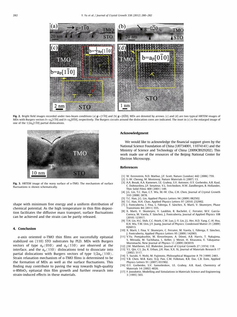
Apart from the contribution of MDs to the strain relaxation,other factors should be active in order to release the residualstrain. These factors were considered to be the formation ofthreading dislocations (TDs) [13,14] or crystallographic domains[15,16] in previous studies of perovskite epitaxial thin films. Asour o-TMO thin films are observed to be single domain withoutTDs, the residual strain may be released in an unusual way. It isnoteworthy that the surface of our TMO thin films is not flat, asseen in low-magnification TEM images and also in Fig. 3. There-fore, we speculate that the residual strain in our TMO thin films isreleased by surface fluctuations [17]. Fig. 3 illustrates themechanism of surface fluctuations. When the MDs form atthe interface to partially relax the strain, the local strain statenear the dislocation cores is changed. Thus the strain distributionin the film becomes non-uniform. Consequently, the chemicalpotential develops a gradient along the surface. Then this gradientprovides a driving force for diffusive mass transport along thesurface, which attempts to search for an equilibrium surface
Fig. 2. Bright field images recorded under two-beam conditions (a) g¼[110] and (b) g¼[020]. MDs are denoted by arrows. (c) and (d) are two typical HRTEM images of
MDs with Burgers vectors b¼ap[110] and b¼ap[010], respectively. The Burgers circuits around the dislocation cores are indicated. The inset in (c) is the enlarged image of
one of the 1/2ap[110] partial dislocations.
Fig. 3. HRTEM image of the wavy surface of o-TMO. The mechanism of surface
fluctuations is shown schematically.
Y. Yu et al. / Journal of Crystal Growth 338 (2012) 280–282282
shape with minimum free energy and a uniform distribution ofchemical potential. As the high temperature in thin film deposi-tion facilitates the diffusive mass transport, surface fluctuationscan be achieved and the strain can be partly released.
4. Conclusion
a-axis oriented o-TMO thin films are successfully epitaxialstabilized on (110) STO substrates by PLD. MDs with Burgersvectors of type ap/010S and ap/110S are observed at theinterface, and the ap/110S dislocations tend to dissociate intopartial dislocations with Burgers vectors of type 1/2ap/110S.Strain relaxation mechanism of o-TMO films is determined to bethe formation of MDs as well as the surface fluctuations. Thisfinding may contribute to paving the way towards high-qualityo-RMnO3 epitaxial thin film growth and further research intostrain-induced effects in these materials.
Acknowledgment
We would like to acknowledge the financial support given by theNational Science Foundation of China (U0734001, 11074141) and theMinistry of Science and Technology of China (2009CB929202). Thiswork made use of the resources of the Beijing National Center forElectron Microscopy.
[10] X. Marti, I. Fina, V. Skumryev, C. Ferrater, M. Varela, L. Fabrega, F. Sanchez,J. Fontcuberta, Applied Physics Letters 95 (2009) 142903.
[11] V.Yu. Pomjakushin, M. Kenzelmann, A. Donni, A.B. Harris, T. Nakajima,S. Mitsuda, M. Tachibana, L. Keller, J. Mesot, H. Kitazawa, E. Takayama-Muromachi, New Journal of Physics 11 (2009) 043019.
[12] J.W. Matthews, A.E. Blakeslee, Journal of Crystal Growth 27 (1974) 118.[13] Y.L. Qin, C.L. Jia, K. Urban, J.H. Hao, X.X. Xi, Journal of Materials Research 17
(2002) 3117.[14] T. Suzuki, Y. Nishi, M. Fujimoto, Philosophical Magazine A 79 (1999) 2461.[15] Y.B. Chen, M.B. Katz, X.Q. Pan, C.M. Folkman, R.R. Das, C.B. Eom, Applied
Physics Letters 91 (2007) 031902.[16] O.Y. Gorbenko, S.V. Samoilenkov, I.E. Graboy, A.R. Kaul, Chemistry of
Materials 14 (2002) 4026.[17] F. Jonsdottir, Modelling and Simulations in Materials Science and Engineering