Leishmania (Viannia) panamensis expresses a nuclease with molecular andbiochemical features similar to the Endonuclease G of higher eukaryotes
MIGUEL A. TORO-LONDOÑO, MSC1, EDWIN BAIRON PATIÑO, PHD2,SARA MARÍA ROBLEDO, PHD3, ANTONIO JIMÉNEZ-RUIZ, PHD4, JUAN FERNANDO ALZATE, PHD5
SUMMARY
Objective: To characterize the molecular and biochemical features of the Endonuclease G of Leishmania (Viannia)panamensis.
Methods: The gene of the putative L. (V.) panamensis Endonuclease G was amplified, cloned, and sequenced. Therecombinant protein was produced in a heterologous expression system and biochemical assays were run to determine its ion,temperature, and pH preferences.
Results: The L. (V.) panamensis rENDOG has biochemical features similar to those found in other trypanosomatids andhigher eukaryotes. In addition, phylogenetic analysis revealed a possible evolutionary relationship with metazoan ENDOG.
Conclusions: L. (V.) panamensis has a gene that codifies an ENDOG homologous to those of higher organisms. Thisenzyme can be produced in Escherichia coli and is able to degrade covalently closed circular double-stranded DNA. It hasa magnesium preference, can be inhibited by potassium, and is able to function within a wide temperature and pH range.
Leishmania (Viannia) panamensis expresa una nucleasa molecular y bioquímicamente similar a la Endonucleasa Gde eucariotas superiores
RESUMEN
Objetivo: Caracterizar molecular y bioquímicamente la Endonucleasa G (EndoG) de Leishmania (Viannia) panamensis.Métodos: El gen de la putativa Endonucleasa G de L. (V.) panamensis fue amplificado, clonado y secuenciado. La proteína
recombinante se produjo en un sistema de expresión heterólogo y la proteína activa se sometió a pruebas bioquímicas paradeterminar la preferencia de iones, temperatura y pH.
Resultados: La rEndoG de L. (V.) panamensis muestra características bioquímicas similares a aquellas descritas en otrostrypanosomatidos y en eucariotas superiores. Además, los análisis filogenéticos muestran una posible relación evolutiva conla Endonucleasa G de metazoos.
Conclusiones: Leishmania (V.) panamensis posee un gen que codifica para una endonucleasa homóloga a la EndoG deotros organismos superiores, que se puede producir de forma recombinante en Escherichia coli y que es capaz de degradarADN circular cerrado de doble cadena. Tiene una preferencia por los iones magnesio y manganeso para usarlos como cofactory es inhibida por el potasio. Además, funciona en un amplio rango de pH y temperatura.
1. Researcher, Program for the Study and Control of Tropical Diseases (PECET), Medical School, Universidad deAntioquia, Medellín, Colombia. e-mail: [email protected]
2. Professor, Institute of Chemistry, Universidad de Antioquia, Medellín, Colombia. e-mail: [email protected]. Professor, Program for the Study and Control of Tropical Diseases (PECET), Medical School, Universidad de Antioquia,
Medellín, Colombia. e-mail: [email protected]. Professor, Biochemistry and Molecular Biology Departament, Universidad de Alcalá, Madrid, Spain.
e-mail: [email protected]. Professor, Parasitology Group, Microbiology and Parasitology Department, Medical School, Universidad de Antioquia,
Medellín, Colombia. e-mail: [email protected] for publication March 11, 2010 Accepted for publication June 30, 2010
Leishmaniasis is a zoonosis caused by protozoanparasites of the Leishmania genus and transmitted byphlebotomine sand flies (Diptera: Psychodidae). Thisdisease occurs in all continents except Antarctica and itis estimated that approximately 350 million peopleworldwide are at risk of infection with approximately2.3 million new cases each year1. Leishmaniasis presentsa wide clinical spectrum ranging from cutaneous formsthat resolve spontaneously to visceral infections thatmay be fatal. The different clinical manifestations ofthe disease depend on the immune status of the host andthe species of parasite involved. In Colombia, the mostcommon clinical form is cutaneous leishmaniasis (CL)with 10,248 cases in 2008; Leishmania (Viannia)panamensis is the species most commonly implicated2.
Leishmania has a digenic life cycle, characterizedby the presence of flagellate promastigotes in the insectvector and intracellular amastigotes within the phago-somes of host mammal macrophages. During theinfection process in mammals, Leishmania parasitesenter and remain within the host cell, evading theinflammatory response before causing the lesion3. Ithas been suggested that one of mechanisms by whichthe parasite evades the immune response is byphosphatidylserine (PS) expression in the cell mem-branes, both of promastigotes in the inoculum4 andamastigotes that simulate apoptosis5. After beingrecognized by phagocytic cells, this PS inducesproduction of the anti-inflammatory cytokine TGB-βand causes negative regulation of the pro-inflammatorycytokine TNF-α6,7; thus, reducing inflammation andallowing the parasite to evade the immune system.Despite the importance of processes similar to apoptosisin these parasites, the mechanisms and moleculesinvolved have been poorly studied. Recently, thepresence of a mitochondrial nuclease, which migratesto the nucleus in response to apoptotic stimuli8,9 hasbeen identified, both in species of Trypanosoma (T.brucei) and Leishmania, i.e., L. (Leishmania) donovaniand L. (L.) infantum. This protein is thought to behomologous to the Endonuclease G (EndoG) of higher
eukaryotes, responsible for DNA degradation in caspase-independent processes of programmed cell death10,11.
EndoG has been characterized in different organisms,both multi- and uni-cellular, and it is known forparticipating in apoptotic processes. EndoG also playsa role in the repair and replication of mitochondrialDNA12. This enzyme is a non-specific DNA/RNAnuclease and belongs to a family of nucleases deno-minated ββα-me finger, a name based on the dispositionof the catalytic site, which comprises two beta sheetsand an alpha helix13. This site is also characterized bythe moiety DRGH, histidine (H) being responsible forthe activation of the water molecule needed to carry outhydrolysis of the phosphodiester bond9,14. It should benoted that in trypanosomatids this characteristic moietyis replaced by SRGH8,9. The change from aspartic acidto serine does not affect enzyme activity because thissite is not critical for catalytic activity14. Furthermore,its activity is known to be principally dependent onMg+2, Mn+2, or Co+2, which distinguishes it fromDNAses I and II12.
In this study we demonstrate for the first time that L.(V.) panamensis also possesses a gene that codes forthis enzyme, which upon being expressed in a recom-binant manner presents endonuclease activity underconditions known to be present during the apoptoticprocess. Additionally, a phylogenetic relationshipbetween this protein in trypanosomatids and itspresumed homologue in higher eukaryotes is establishedfor the first time.
MATERIALS AND METHODS
Parasite culture and isolation of nucleic acids.Promastigotes of the L. (V.) panamensis strain MHOM/CO/87/UA140 were cultured in modified biphasic Novy-MacNeal-Nicolle (NNN) medium supplemented withpenicillin (2000 U/ml). The cultures were initiated withan inoculum of 106 parasites/ml and the parasitesharvested in the logarithmic or stationary phase afterincubation at 26°C for 5 days. DNA was purified fromthese promastigotes using standard protocols withphenol/chloroform.
Amplification and cloning of the putative endog ofL. (V.) panamensis. The primers 5EGbrcx (sense) 5’GGGGATCCGCCCAGGCCTCCACGCTCA 3’ and3EGbrcx (anti-sense) 5’ GGCTCGAGCTGCCGGTA
156
Colombia Médica Vol. 42 Nº 2, 2011 (Abril-Junio)
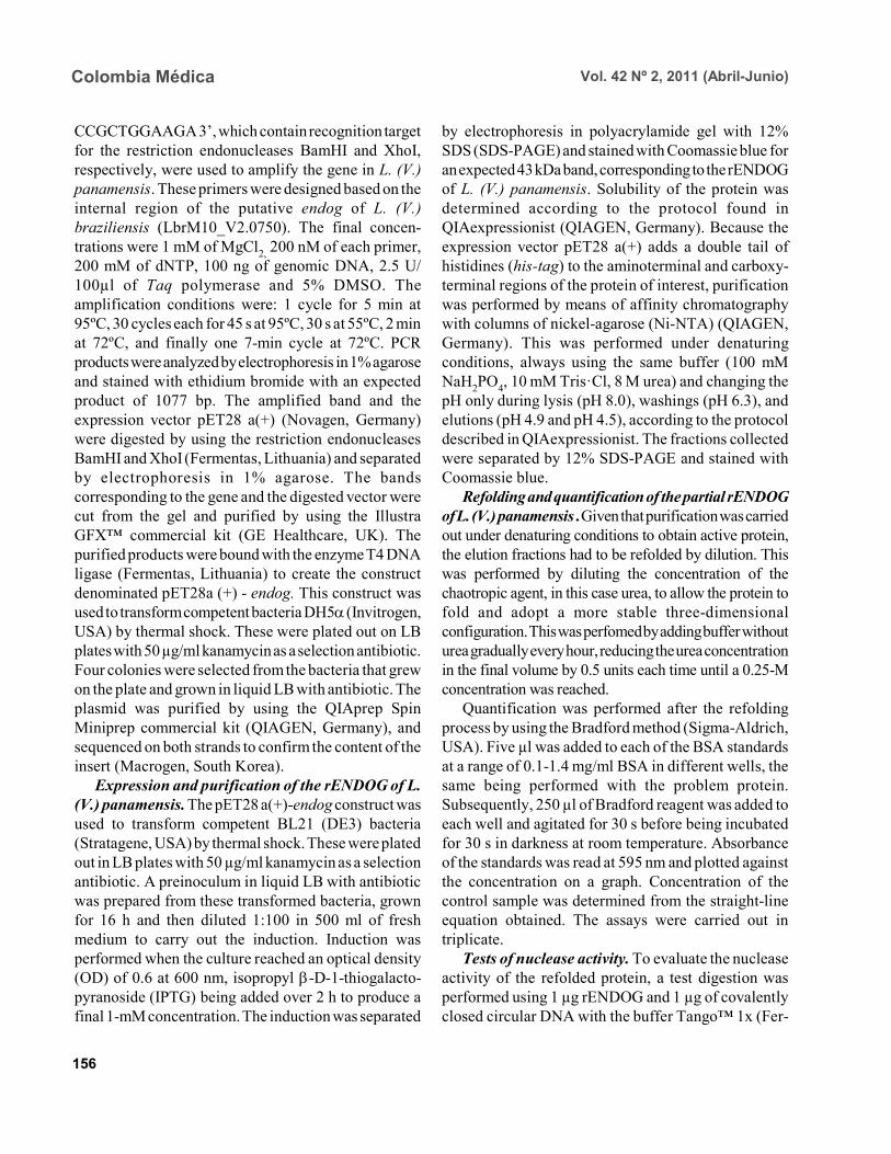
CCGCTGGAAGA 3’, which contain recognition targetfor the restriction endonucleases BamHI and XhoI,respectively, were used to amplify the gene in L. (V.)panamensis. These primers were designed based on theinternal region of the putative endog of L. (V.)braziliensis (LbrM10_V2.0750). The final concen-trations were 1 mM of MgCl2, 200 nM of each primer,200 mM of dNTP, 100 ng of genomic DNA, 2.5 U/100µl of Taq polymerase and 5% DMSO. Theamplification conditions were: 1 cycle for 5 min at95ºC, 30 cycles each for 45 s at 95ºC, 30 s at 55ºC, 2 minat 72ºC, and finally one 7-min cycle at 72ºC. PCRproducts were analyzed by electrophoresis in 1% agaroseand stained with ethidium bromide with an expectedproduct of 1077 bp. The amplified band and theexpression vector pET28 a(+) (Novagen, Germany)were digested by using the restriction endonucleasesBamHI and XhoI (Fermentas, Lithuania) and separatedby electrophoresis in 1% agarose. The bandscorresponding to the gene and the digested vector werecut from the gel and purified by using the IllustraGFX™ commercial kit (GE Healthcare, UK). Thepurified products were bound with the enzyme T4 DNAligase (Fermentas, Lithuania) to create the constructdenominated pET28a (+) - endog. This construct wasused to transform competent bacteria DH5α (Invitrogen,USA) by thermal shock. These were plated out on LBplates with 50 µg/ml kanamycin as a selection antibiotic.Four colonies were selected from the bacteria that grewon the plate and grown in liquid LB with antibiotic. Theplasmid was purified by using the QIAprep SpinMiniprep commercial kit (QIAGEN, Germany), andsequenced on both strands to confirm the content of theinsert (Macrogen, South Korea).
Expression and purification of the rENDOG of L.(V.) panamensis. The pET28 a(+)-endog construct wasused to transform competent BL21 (DE3) bacteria(Stratagene, USA) by thermal shock. These were platedout in LB plates with 50 µg/ml kanamycin as a selectionantibiotic. A preinoculum in liquid LB with antibioticwas prepared from these transformed bacteria, grownfor 16 h and then diluted 1:100 in 500 ml of freshmedium to carry out the induction. Induction wasperformed when the culture reached an optical density(OD) of 0.6 at 600 nm, isopropyl β-D-1-thiogalacto-pyranoside (IPTG) being added over 2 h to produce afinal 1-mM concentration. The induction was separated
by electrophoresis in polyacrylamide gel with 12%SDS (SDS-PAGE) and stained with Coomassie blue foran expected 43 kDa band, corresponding to the rENDOGof L. (V.) panamensis. Solubility of the protein wasdetermined according to the protocol found inQIAexpressionist (QIAGEN, Germany). Because theexpression vector pET28 a(+) adds a double tail ofhistidines (his-tag) to the aminoterminal and carboxy-terminal regions of the protein of interest, purificationwas performed by means of affinity chromatographywith columns of nickel-agarose (Ni-NTA) (QIAGEN,Germany). This was performed under denaturingconditions, always using the same buffer (100 mMNaH2PO4, 10 mM Tris·Cl, 8 M urea) and changing thepH only during lysis (pH 8.0), washings (pH 6.3), andelutions (pH 4.9 and pH 4.5), according to the protocoldescribed in QIAexpressionist. The fractions collectedwere separated by 12% SDS-PAGE and stained withCoomassie blue.
Refolding and quantification of the partial rENDOGof L. (V.) panamensis . Given that purification was carriedout under denaturing conditions to obtain active protein,the elution fractions had to be refolded by dilution. Thiswas performed by diluting the concentration of thechaotropic agent, in this case urea, to allow the protein tofold and adopt a more stable three-dimensionalconfiguration. This was perfomed by adding buffer withouturea gradually every hour, reducing the urea concentrationin the final volume by 0.5 units each time until a 0.25-Mconcentration was reached.
Quantification was performed after the refoldingprocess by using the Bradford method (Sigma-Aldrich,USA). Five µl was added to each of the BSA standardsat a range of 0.1-1.4 mg/ml BSA in different wells, thesame being performed with the problem protein.Subsequently, 250 µl of Bradford reagent was added toeach well and agitated for 30 s before being incubatedfor 30 s in darkness at room temperature. Absorbanceof the standards was read at 595 nm and plotted againstthe concentration on a graph. Concentration of thecontrol sample was determined from the straight-lineequation obtained. The assays were carried out intriplicate.
Tests of nuclease activity. To evaluate the nucleaseactivity of the refolded protein, a test digestion wasperformed using 1 µg rENDOG and 1 µg of covalentlyclosed circular DNA with the buffer Tango™ 1x (Fer-
Effects of pH and temperature on enzymaticactivity. The effects on enzymatic activity of pH over arange of 3-12 and temperature at 20-84°C weredetermined under the conditions mentioned previouslyand the obtained optimal concentration of magnesium.
Bioinformatical analysis. Bioinformatic analysessuch as the search for homologous sequences to constructthe phylogenetic tree were performed by using theExPASy database (http://ca.expasy.org/). The searchfor the signal peptide was performed on the amino acidsequence of L. (V) braziliensis using the SP-NNalgorithm in the TriTrypDB database (http://tritrypdb.org/tritrypdb/). This same database was used to deter-mine the position of the gene on the genomes of thedifferent species of trypanosomatids (synteny).
Functional domains were sought in the proteinexpressed by using the conserved domains database(CCD) tool from the NCBI (http://www.ncbi.nlm.nih.gov/Structure/cdd/cdd.shtml). Extremity sequencesbelonging to L. (V.) braziliensis were used to determinethe relevance of the amino and carboxyterminal endsabsent in the rENDOG of L. (V.) panamensis. Secondarystructures (alpha helix and beta sheets) in thesesequences that could give indications of their importancewere sought by using the APSSP2 algorithm (http://www.imtech.res.in/raghava/apssp2/).
Phylogenetic relationship. To make a phylogeneticreconstruction of the Endo G of L. (V.) panamensis, 26sequences of proteins homologous to human Endo Gwere identified from a wide variety of organisms,including bacteria, fungi, amphibians, birds, andmammals. The sequences collected were aligned usingthe Clustal algorithm and the BLOSUM matrix. To
determine the evolutionary relationship of the 26 taxaanalyzed, the alignment data were loaded onto theMEGA 4 program and a distance matrix obtained thatshowed the estimated divergence between the sequences.These results were obtained from pairwise analysisbetween the 26 alignments using Poisson’s correctionmethod. Based on the sequence alignment data obtained,a tree was constructed by the Neighbour-Joining (NJ)method to determine evolutionary relationships amongthe 26 organisms of the gene studied. The bootstrapused was 1000 and those branches reproduced in lessthan 50% of the replicates were collapsed.
RESULTS
Amplification and cloning of the putative endog ofL. (V.) panamensis. A 1077 bp internal region of theputative endog gene of L. (V.) panamensis was amplifiedby PCR. This amplification was performed after adding5% DMSO to the PCR mix, given that without thisadditive a non-specific band of approximately 1500bp(Figure 1A) was amplified. This region was cloned intothe plasmid pET 28 (+), creating the construct pET28a(+) – endog (Figure 1B). The construct insert wassequenced for both strands and aligned against theputative endog gene of L. (V.) braziliensis using theClustal W algorithm, yielding a 98% identity as a result.This way, it was confirmed that the amplified andcloned fragment was the internal region of the putativeendog gene of L. (V.) panamensis. The sequence obtainedcan be found by using GeneBank access numberGQ119624.1.
Expression and purification of the rENDOG of L.(V.) panamensis. Based on its nucleotide sequence, theprotein deduced was of 401 amino acids with a molecularmass of 43.3 kDa. After induction with 1 mM IPTG for2 h at 37°C, bacteria BL21 (DE3) transformed withpET28a (+)-endog expresses a protein of approximately45 kDa, corresponding to the expected size of therENDOG, was observed (Figure 2A). Solubility studiesof the recombinant protein revealed that this wasexpressed in an insoluble form (data not shown), hencepurification had to take place under denaturingconditions (Figure 2B). After purifying 0.96 g of abacterial precipitate, 65 mg of total soluble protein wasobtained and then, 10.5 mg of purified recombinantprotein was obtained from the elutions. Since the
158
Colombia Médica Vol. 42 Nº 2, 2011 (Abril-Junio)
Figure 2. Induction and purification of rENDOG of L. (V.) panamensis. A. Induction. M. Molecular weightmarker Pre. Pre-induction BL21 (DE3), Post. Post-induction BL21 (DE3) 2 h with 1 mM IPTG. B.Purification. M. Molecular weight marker Lys. Lysate, FT. Flow-through, W. Washings (pH 6.5), E. Elutionfractions. (pH 5.9 and 4.5). Gels represent similar patterns obtained from three independent experiments.
6.5 5.9 4.5
116.0-66.2-
45.0-
35.0-
25.0-
18.4-
14.0-
Figure 1. Amplification and cloning of the putative endog of L. (V.) panamensis. A. Amplification. M.Molecular weight marker. Amplification of a fragment of the expected size, only achieved when 5% DMSOwas used in the reaction. C(-). Negative control. This gel represents similar patterns of 3 independentexperiments. B. Construct pET28a (+) – endog. Orientation of the cloned fragment, as well as the positionof both restriction sites used and sequences that code for the histidine tails can be observed.
159
Colombia Médica Vol. 42 Nº 2, 2011 (Abril-Junio)
Bradford methodology is incompatible with ureaconcentrations above 3 M, these fractions werequantified by spectrophotometry at 280 nm.
Refolding and quantification of the partialrENDOG of L. (V.) panamensis . After refolding, afinal 30-ml volume was obtained with a proteinconcentration of 100 ng/ml. Quantification wasperformed via the Bradford methodology. Given thatduring the refolding process by dialysis, as well asdilution of the chaotropic agent the protein was alsodiluted, precipitation with trichloroacetic acid (TCA)was carried out before quantification. When sampleswere treated with 400 mM dithiothreitol (DTT) thebands of highest molecular weight observed in purifiedfractions of rENDOG disappeared (Figure 3A).
3B). Enzymatic activity was shown by the appearanceof a DNA smear, a clear sign of degradation of thismacromolecule.
Optimal concentration of MgCl2 and inhibitoryconcentration of KCl. After testing the preliminaryactivity, a nuclease activity assay was performed withMgCl2. This revealed that the optimal concentration ofMg2+ for 1µg of rENDOG is 15 mM, no further increaseof nuclease activity being seen above this concentration(Figure 4A). Furthermore, it was determined that theinhibitory concentration of K1+ is 140 mM. A gradualreduction in nuclease activity was observed as potassiumconcentation increased, total inhibition was observedat 230 mM (Figure 4B).
Effect of different divalent cations on nuclease activity.Because the enzyme uses divalent cations present inthe cellular environment as cofactors, the activity wasevaluated by using divalent cations other than magnesiumsuch as: barium, manganese, and calcium. Despite beinga divalent cation, barium does not function as a cofactor(Figure 5A). Manganese does behave as a cofactor, albeit
1.5-
1-
0.7-
0.5-
0.2-
Figure 3. Refolding and Nuclease Activity Assay. A. Refolding. M. Molecular weight marker, FP: Refoldingfraction precipitated with TCA, 2. FNP: Fraction not precipitated. B. Activity assay. M. Molecular weightmarker, C(-). Negative control. Two reactions were performed, one observed at room temperature (RT) andthe other at 37°C. These were performed in the absence of rENDOG (0 µg) and using 1 µg rENDOG. Gelsrepresent similar patterns in three independent experiments.
160
Colombia Médica Vol. 42 Nº 2, 2011 (Abril-Junio)
Figure 5. Evaluation of different cations as cofactors. Concentrations of 0, 1, 5, 10, and 20 mM of bariumchloride (BaCl2), calcium chloride (CaCl2) and manganese chloride (MnCl2) evaluated. M. Molecular weightmarker. C(-). Negative control. Gels represent similar patterns obtained from three independent experiments.
Figure 4. Optimal con-centration of MgCl2 andInhibitory Concentra-tion of KCl. A. OptimalMgCl2 concentration. M.Molecular weight mar-ker. Increasing concen-trations of magnesiumchloride (MgCl2) from0-350 mM are shown.C(-). Negative control.B. Inhibitory concen-tration of KCl. M. Mole-cular weight marker.Rising concentrations(90-250 mM) of pota-ssium chloride (KCl)using a fixed concen-tration of 15 mM ofMgCl2 are shown. Gelsrepresent similar pa-tterns obtained forthree independent ex-periments.
less effectively than magnesium (Figure 5C). For calcium,concentrations of 1-5 mM presented endonuclease activity(Figure 5B); although, much less than that observed withmagnesium and manganese, indicating that it is a lessefficient cofactor.
Effects of pH and temperature on enzymaticactivity. It was found that the enzyme maintained its
nuclease activity over a wide pH range of 3-12 (Figure6A). It also worked over a wide range of temperatures,remaining active from 20-50°C (Figure 6B). In similarexperiments performed in our laboratory (results notshown), we demonstrated that the enzyme was able tomaintain its activity for up to 2 h at 84°C, inactivationoccurring after exposure to 100°C for more than 5 min.
161
Colombia Médica Vol. 42 Nº 2, 2011 (Abril-Junio)
Figure 6. Effects of temperature and pH on rENDOG activity. Effect of pH. M. Molecular weight marker.rENDOG activity at different pH values (3-12). C(-). Control without rENDOG. B. Effect of temperature M.Molecular weight marker. Activity of rENDO G at different temperatures (20-50°C). C(+). Control at 37ºC,C(-). Control without rENDOG. Gels represent similar patterns obtained for three independent experiments.
Bioinformatic analysis. Bioinformatic analysisrevealed that EndoG possesses a signal peptide ofapproximately 32 amino acids on the aminoterminalend. It was also determined that the gene coding forEndoG is located on chromosome 10 in the threeLeishmania species for which the genome is completelyknown i.e., L. (L.) infantum, L. (L.) major, and L. (V.)braziliensis. It was also determined that this is a highlysyntenic single-copy gene in these species. The enzymewas found to possess three known domains, onedenominated NUC referring to a super family ofnucleases, one binding to magnesium, and a thirdbinding to the substrate (DNA). In the absent amino-terminal region, it was determined that the missingamino acids did not form any secondary structure thatcould indicate a biological function. The algorithmpredicted the formation of an alpha helix on thecarboxyterminal region with high probability (>0.8),
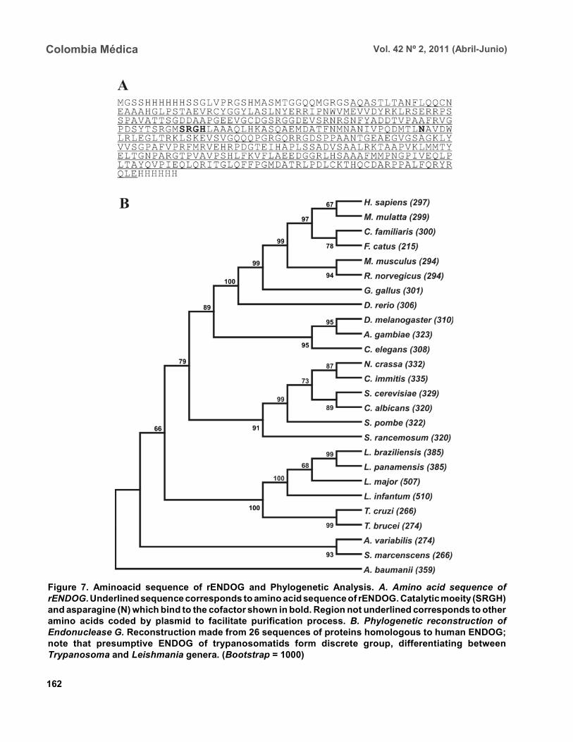
although no known functional domain was identified.Phylogenetic relationship. Sequences were aligned
based on the amino acid sequence obtained for the EndoG of L. (V.) panamensis (Figure 7A), as well as on theother 26 protein sequences presumably homologous tothis. A phylogenetic tree was constructed from thisalignment by the Neighbour Joining (NJ) method inwhich the phylogenetic relationship that exists betweenthe protein of L. (V.) panamensis and the other knownhomologues is shown (Figure 7B).
DISCUSSION
Although various studies have been carried out onTrypanosoma and Leishmania to show the existence ofENDOG4,8,9, all Leishmania studies were performed onspecies of the Leishmania subgenus and their relevanceto members of Viannia species is unknown. It has been
162
Colombia Médica Vol. 42 Nº 2, 2011 (Abril-Junio)
Figure 7. Aminoacid sequence of rENDOG and Phylogenetic Analysis. A. Amino acid sequence ofrENDOG. Underlined sequence corresponds to amino acid sequence of rENDOG. Catalytic moeity (SRGH)and asparagine (N) which bind to the cofactor shown in bold. Region not underlined corresponds to otheramino acids coded by plasmid to facilitate purification process. B. Phylogenetic reconstruction ofEndonuclease G. Reconstruction made from 26 sequences of proteins homologous to human ENDOG;note that presumptive ENDOG of trypanosomatids form discrete group, differentiating betweenTrypanosoma and Leishmania genera. (Bootstrap = 1000)
163
Colombia Médica Vol. 42 Nº 2, 2011 (Abril-Junio)
established that certain genes that exist in one subgenusare lacking in the other and vice-versa15; hence, it isimportant to carry out independent studies on Vianniaspecies. Certain differences were clear from the start ofthe study given that it was not possible to amplify theentire gene in L. (V.) panamensis using oligonucleotidesdesigned from sequences of the endog of L. (L.) major,L. (L.) infantum, and L. (V.) braziliensis (results notshown). Thus, we may conclude that the 5’ and 3’terminal sequences of the coding region (CDS) ofendog differ from those of the reference speciesmentioned and for this reason did not adequatelyhybridize the oligonucleotides in amplification PCR ofthe entire gene. For this reason, new oligonucleotideswere designed based on the internal region of the geneof L. (V.) braziliensis, allowing amplification of a bandof approximately 1500 bp, which however did notconcur with the expected size of the internal 1077-bpregion we attempted to amplify. By using 5% DMSO asan additive, it was posible to amplify and clone a 1077-bp fragment of the internal coding region of the Endo Ggene of L. (V.) panamensis. It should be noted thatalthough it is an internal region of the gene, it containsthe triplet codons of the amino acids included in theputative catalytic site of the enzyme. This fragmentcorresponds to 71% of the expected size of the gene,based on the 1515-bp sequence reported for this gene inL. (V.) braziliensis (LbrM10_V2.0750). In lacking the5’ end of the coding region, we know that 86 aminoacids are missing from the aminoterminal end of theenzyme of which the first 40 correspond to the signalpeptide that naturally directs this enzyme to themitochondria of the parasite9. Our in-silico analysisrevealed the presence of a signal peptide of 32 aminoacids on the amino terminal. According to the predictionsperformed, the other missing amino acids do not formany secondary structure and, thus, cannot be performingany function. Likewise, by missing part of the 3’ region,the enzyme lacks 58 amino acids in the carboxy-termi-nal region, which according to the predictions has avery high probability of forming a pair of alpha helix.This indicates that the region may code for somebiological function; nevertheless, more studies arerendered. Despite these deletions in the amino andcarboxyterminal ends, the rENDOG of L. (V.)panamensis is an active endonuclease able to degradecovalently closed circular DNA, demonstrating that
these regions are not critical to the functioning of thisenzyme. This is to be expected given that in Leishmaniathis enzyme is 53 kDa, much larger than the 32 kDafound in other eukaryotic organisms such as humansand fungi, as well as the 37 kDa of the nuclease ofSerratia marcescens, which is also thought to be relatedto the ENDOG of higher organisms10,12.
The high expression levels obtained for the rENDOGof L. (V.) panamensis in E. coli facilitated the formationof inclusion bodies16, requiring a denaturation processof solubilization of the protein for purification. Thehigh amount of protein produced during expression issurprising, given that in some species such as L. (L.)major, the toxicity of the enzyme precludes it frombeing produced in recombinant manner in E. coli8.Although L. (L.) infantum ENDOG has been producedin E. coli, its levels of expression are not as high9. It,therefore, remains to be determined whether the highlevels seen in L. (V.) panamensis are due to the lowertoxicity of recombinant proteins from New Worldspecies per se, or if the absence of the signal peptideand/or the carboxy-terminal region reduces their toxicity,allowing better expression to occur.
On carrying out rENDOG purification, highmolecular weight proteins were found as contaminantsin the elution fractions. However, these disappearedwhen the quantity of DDT (dithiothreitol) in the bufferof the protein sample was duplicated to 400 mM,indicating that they were actually multimeric aggregatesof the recombinant protein bound to each other bydisulphate bridges. This enzyme is known to require adivalent cation as a cofactor, thought to be involved instabilization of the transition state during nucleophilicattack and in coordination of the water molecule thatprotonates the salient group during hydrolysis14. Amongthe ions known to function as cofactors in orthologousenzymes are cobalt (Co2+), manganese (Mn2+),magnesium (Mg 2+), nickel (Ni2+), calcium (Ca2+), andzinc (Zn2+)12. Even though the enzyme is able to functionwith several of the ions mentioned, it is widely acceptedthat the most important is Mg2+. It is known that theENDOG of C. elegans, which has been implicated inapoptosis, is dependent only on magnesium11. Giventhe importance of Mg2+ as the principal cofactor ofENDOG, a test was run to determine the optimalconcentration of this cation. It was thus determined thatthe optimal concentration for 1 µg of recombinant
164
Colombia Médica Vol. 42 Nº 2, 2011 (Abril-Junio)
enzyme is 15 mM of MgCl2, without increase inenzymatic activity above this concentration. However,a concentration of 50 mM of MgCl2 appeared to inhibitactivity. Rather than being due to excess cofactor, thisinhibitory effect could be explained by the effect ofmagnesium on DNA because the magnesium can bindto the major and minor grooves of the DNA molecule,altering the DNA structure17. It is noteworthy that theintracellular concentration of magnesium dissolved inthe cytosol lies between 0.5 and 1 mM18, concentrationat which rENDOG is active. Different mono- anddivalent cations, such as barium (Ba2+), manganese(Mn2+), calcium (Ca2+), and potassium (K1+) wereevaluated in addition to magnesium. As expected, bothmanganese and calcium function as cofactors, clearsigns of degradation were visible after 2 h of digestion.Although greater signs of degradation were observedwith magnesium and manganese than with calcium, theimportance of the latter in the activation of nucleaseactivity during apoptosis has been demonstrated inmammals19,20. However, no degradation whatsoeverwas evident for barium, perhaps due to its greater sizethan other divalent cations. This would cause a stericimpediment, which would not permit the enzyme to usethe cation as a cofactor. After determining the optimalmagnesium concentration for the enzyme, an inhibitionassay was performed by using increasing concentrationsof potassium. It was determined that concentrationsabove 140 mM of KCl inhibited enzymatic activity,with complete inhibition at 230 mM. These resultsconcur with the intracellular potassium concentrationof 140 mM both in mammalian cells and Leishmaniapromastigotes. The potassium concentration, never-theless, falls to 30 mM in mammalian cells and 65 mMin promastigotes during apoptosis21-23. It is clear thatunder normal intracellular conditions, the enzyme isslighly inhibited due to the intracellular concentrationsof potassium. Because Leishmania is a parasite with aheteroxenous life cycle, exposed to different tempe-ratures both in the insect vector and in the mammalianhost, we decided to evaluate the temperature range atwhich ENDOG mantains its activity and determinedthat this enzyme is active above 20°C, presenting peakactivity at 40-46°C. Enzymatic activity began to beaffected above 48°C, with residual nuclease activityobserved up to 84°C, which means that this enzyme ishighly thermostable. No major changes were observed
in its degradation capacity when activity was evaluatedat pH levels of 3-12. The changes observed in electro-phoretic migration can be attributed to the highconcentrations of NaOH used to reach the most alkalinepH values (unpublished laboratory observations). It isnot surprising that the enzyme is tolerant to a wide pHrange because it is known that the pH tends to acidifyduring apoptotic processes.
The homologous relationships among the endo-nucleases G of distinct organisms have been establishedmainly based on similarity of enzyme functions than onphylogenetic analysis. For this reason, 26 proteinsequences presumably homologous to human ENDOGwere collected and a phylogenetic tree was constructedto determine the evolutionary relationships among theseproteins. ENDOG belongs to the super family of non-specific nucleases denominated ββα-Me-finger,although the catalytic moiety DRGH present in mostmembers of this family14 is modified in trypanosomatidssince a serine residue occupies the position of theaspartic acid (SRGH). Although it has been suggestedthat this aspartic acid acts on conformation of thecatalytic site, mutagenesis site assays suggest that thechange from aspartic acid to alanine produces a 54%reduction in bovine ENDOG activity. The function ofaspartic acid in trypanosomatids appears to be performedwithout any problem by serine. The other residuescritical to function, including the asparagine responsiblefor binding to the cofactor24, are present in the rENDOGof L. (V.) panamensis. Even with this change in thesequence, it aligned adequately with the other 25sequences analyzed and did not require mayor editing.Based on this alignment, a distance matrix wasconstructed from this tree by the Neighbour-Joiningmethod. Proteins belonging to trypanosomatids aregrouped together in this tree, as are those from bacteria,fungi, insects, and mammals. From these results, it canbe inferred that the putative ENDOG of these parasitesis a homologue of the one found in humans and otherhigher eukaryotes. Thus, from the point of view ofpossible structure and function9 this similarity, togetherwith the hypothesis of a possible common origin of thisgene shown by the construction of this tree, strengthensthe hypothesis for homology among these enzymes.
In summary, we may conclude that L. (V.) panamensispossesses a gene that codes for an endonucleasehomologous to the ENDOG of higher organisms, which
165
Colombia Médica Vol. 42 Nº 2, 2011 (Abril-Junio)
can be produced in recombinant form in E. coli and isable to degrade covalently closed circular double-stranded DNA. It has a preference for magnesium andmanganese ions, using them as cofactors, and is inhibitedby potassium. It also functions over a wide range of pHand temperature. The similarity in the biochemicalcharacteristics and relationship of this enzyme withthose found in metazoans, suggest that it could beinvolved in the process of programmed cell death thathas been described for these organisms.
Conflict of interest. None of the authors has conflictsof interest related to this study.
ACKNOWLEDGEMENTS
This project received joint funding from the researchsupport foundation at Banco de la República de Colom-bia (Project N° 2204), Universidad de Antioquia andCOLCIENCIAS (Project N° 1101-00018-9999 and1102-343-19319) and Centro para el Desarrollo deProductos (CIDEPRO).
REFERENCES
1. Desjeux P. Leishmaniasis: current situation and newperspectives. Comp Immunol Microbiol Infect Dis. 2004; 27:305-18.
2. Vélez ID, Gilchrist K, Arbeláez MP, Rojas CA, Puerta JA,Antunes CM, et al. Failure of a killed Leishmania amazonensisvaccine against American cutaneous leishmaniasis in Colom-bia. Trans R Soc Trop Med Hyg. 2005. 99: 593-8.
3. Belkaid Y, Méndez S, Lira R, Kadambi N, MilonG, Sacks D.A natural model of Leishmania major infection reveals aprolonged «silent» phase of parasite amplification in the skinbefore the onset of lesion formation and immunity. JImmunol. 2000; 165: 969-77.
4. Zandbergen V, Bollinger GA, Wenzel A, Kamhawi S, Voll R,Klinger M, et al. Leishmania disease development depends onthe presence of apoptotic promastigotes in the virulentinoculum. Proc Natl Acad Sci USA. 2006; 103: 13837-42.
5. de Freitas JM, Moreira ME, Bonomo A, Bozza PT, AmaranteG, Pirmez C, et al. Apoptotic mimicry by an obligateintracellular parasite downregulates macrophage microbicidalactivity. Curr Biol. 2001; 11: 1870-3.
6. Voll RE, Herrmann M, Roth EA, Stach C, Kalden JR,Girkontaite I. Immunosuppressive effects of apoptotic cells.Nature. 1997; 390: 350-1.
7. Fadok VA, Bratton DL, Konowal A, Freed PW, Westcott JY,Henson PM, Macrophages that have ingested apoptotic cellsin vitro inhibit proinflammatory cytokine production throughautocrine/paracrine mechanisms involving TGF-beta, PGE2,and PAF. J Clin Invest. 1998; 101: 890-8.
8. Gannavaram S, Vedvyas C, Debrabant A, Conservation of the
pro-apoptotic nuclease activity of endonuclease G in unicellulartrypanosomatid parasites. J Cell Sci. 2008; 121: 99-109.
9. Rico E, Alzate JF, Arias AA, Moreno D, Clos J, Gago F, et al.Leishmania infantum expresses a mitochondrial nucleasehomologous to EndoG that migrates to the nucleus in responseto an apoptotic stimulus. Mol Biochem Parasitol. 2009; 163:28-38.
10. Li LY, Luo X, Wang X. Endonuclease G is an apoptoticDNase when released from mitochondria. Nature. 2001; 412:95-9.
11. Parrish J, Li L, Klotz K, Ledwich D, Wang X, Xue D,Mitochondrial endonuclease G is important for apoptosis in C.elegans. Nature. 2001; 412: 90-4.
12. Low RL, Mitochondrial Endonuclease G function in apoptosisand mtDNA metabolism: a historical perspective. Mito-chondrion. 2003; 2: 225-36.
13. Kuhlmann UC, Moore GR, James R, Kleanthous C, HemmingsAM. Structural parsimony in endonuclease active sites: shouldthe number of homing endonuclease families be redefined?FEBS Lett. 1999; 463: 1-2.
14. Schafer P, Scholz SR, Gimadutdinow O, Cymerman IA,Bujnicki JM, Ruiz A, et al. Structural and functionalcharacterization of mitochondrial EndoG, a sugar non-specificnuclease which plays an important role during apoptosis. JMol Biol. 2004; 338: 217-28.
15. Peacock CS, Seeger K, Harris D, Murphy L, Ruiz JC, QuailMA, et al. Comparative genomic analysis of three Leishmaniaspecies that cause diverse human disease. Nat Genet. 2007;39: 839-47.
16. Marston FA. The purification of eukaryotic polypeptidessynthesized in Escherichia coli. Biochem J. 1986; 240: 1-12.
17. Jerkovic B, Bolton PH. Magnesium increases the curvature ofduplex DNA that contains dA tracts. Biochemistry. 2001; 40:9406-11.
23. Sen N, Das BB, Ganguly A, Mukherjee T, Bandyopadhyay S,Majumder HK. Camptothecin-induced imbalance inintracellular cation homeostasis regulates programmed celldeath in unicellular hemoflagellate Leishmania donovani. JBiol Chem. 2004; 279: 52366-75.
24. Friedhoff P, Kolmes B, Gimadutdinow O, Wende W, KrauseKL, Pingoud A. Analysis of the mechanism of the Serratianuclease using site-directed mutagenesis. Nucleic Acids Res.1996; 24: 2632-9.