Page 1
RESEARCH PAPER
Miniaturization of immunoassay by using a novel module-levelimmunosensor with polyaniline-modified nanoprobesthat incorporate impedance sensing and paper-based sampling
Cheng-Hsin Chuang • Yuan-Chu Yu •
Da-Huei Lee • Ting-Feng Wu • Cheng-Ho Chen •
Shih-Min Chen • Hsun-Pei Wu • Yao-Wei Huang
Received: 25 September 2013 / Accepted: 13 February 2014
� Springer-Verlag Berlin Heidelberg 2014
Abstract This work presents a module-level impedance
measurement system integrated with a disposable immu-
nosensor for the immunoassay of bladder cancer cell lysate
(T24) to a specific antibody (galectin-1). The immuno-
sensor consisted of a flexible printed circuit patterned with
an interdigital microelectrode array which immobilized
polyaniline-modified nanoprobes on an electrode surface
by dielectrophoresis. A quantitative sampling of cell lysate
without a pump was made by using paper as the cell lysate
carrier and sweeping a moistened paper over the sensing
area of interdigital microelectrode array for sampling. In
this study, the impedance measurement results of the
module-level system were compared with those measured
by the precision LCR meter, in which the error is \2 %.
Additionally, the normalized impedance variation in im-
munosensing linearly increased with the cell lysate con-
centration. With a sensitivity based on a normalized
impedance variation of 124.4 % per mg/ml, the immuno-
sensor can rapidly detect the lowest concentration of cell
lysate for 0.0626 mg/ml in 10 min. Therefore, this work
has demonstrated the accuracy of the module-level
immunosensor as well as the reliability of impedance-
based sensing for bladder cancer cell lysate. The proposed
disposable sensor and portable impedance system module
are highly promising for use in point-of-care diagnostics.
Keywords Dielectrophoresis � Polyaniline �Immunosensor � Impedance sensing
1 Introduction
Bladder cancer ranks as the sixth most common cancer in
industrialized countries. According to the Urology Care
Foundation, in 2013, 72,570 Americans were diagnosed
with bladder cancer and 15,210 died of the disease. Recent
decades have witnessed a gradual increase in the incidence
of bladder cancer. Although up to 75–80 % of new cases
are diagnosed as a non-invasive (pathological stage Ta),
stroma invasive (T1) or carcinoma in situ (Tis) disease, the
remaining 20–25 % of tumors are presented as muscle
invasive or more advanced disease (T2–4) with a poor
prognosis. Moreover, although approximately 20 % of Ta
and T1 tumors are cured, after an initial removal, 60–70 %
of those tumors recur at least once in 5 years while
10–20 % progress to muscle invasive cancer (Jemal et al.
2010). During the diagnosis of bladder cancer, the grade
must be identified when deciding upon the cancer treat-
ment. Although cystoscopy is the most effective means of
examining the grade of bladder cancer from a biopsy of the
bladder lining, patients may need an anesthesia during this
procedure. Therefore, despite the need to routinely
administer cystoscopy in order to detect the recurrence of
bladder cancer in patients, the subsequent clinical burden
C.-H. Chuang (&) � Y.-C. Yu � S.-M. Chen � H.-P. Wu �Y.-W. Huang
Department of Mechanical Engineering, Southern Taiwan
University of Science and Technology, Tainan 71005, Taiwan
e-mail: [email protected]
D.-H. Lee
Department of Electrical Engineering, Southern Taiwan
University of Science and Technology, Tainan 71005, Taiwan
T.-F. Wu
Department of Biotechnology, Southern Taiwan University of
Science and Technology, Tainan 71005, Taiwan
C.-H. Chen
Department of Chemical and Materials Engineering, Southern
Taiwan University of Science and Technology, Tainan 71005,
Taiwan
123
Microfluid Nanofluid
DOI 10.1007/s10404-014-1364-4
Page 2
on patients is of priority concern. Besides cystoscopy, a
few urinary markers have been developed to reduce the
frequency of cystoscopy (Cheung et al. 2013). However,
their sensitivity and selectivity remain unsatisfactory,
necessitating the development of a highly accurate, non-
invasive and in vitro method for point-of-care diagnostics
of bladder cancer to help postoperative patients.
Using a few biomarkers for bladder cancer, recent works
have verified the relevance of their expression levels to the
prognosis of recurrence, such as annexin 1 (Li et al. 2010),
lactate dehydrogenase B (LDH-B) (Liao et al. 2011),
galectin-1 (Memon et al. 2005). Based on the expression
level, doctors can decide whether cystoscopy is necessary
as a routine check for postoperative patients. However, the
protein expression is usually determined by immunohisto-
chemistry (IHC), Western blotting or enzyme-linked
immunosorbent assay (ELISA). Despite the widespread use
of these analytical methods to detect specific proteins in the
given sample of tissue homogenate or extract, their pro-
cedures require both complex sample preparation and
instrumentation. Alternatively, microfluidic devices have
received considerable attention for use in immunoassay,
owing to their desirable features such as a low quantity of
the required sample, rapid response, low fabrication cost
and addressable sensing area for array manner. The most
widely used detection method is fluorescence, followed by
electrochemistry. Theoretically, the immunosensor sur-
faces are immobilized with antibodies; the specific proteins
thus bind with antibodies based on antibody–antigen
interaction. Once the proteins are labeled with fluorescent
dye, the fluorescence response can accurately reflect the
expression level (Song et al. 2011; Seo et al. 2011).
However, for label-free detection, the immunoreaction can
be measured by cyclic voltammetry (CV) and electro-
chemical impedance spectroscopy (EIS) (Samanman et al.
2012; Moreira et al. 2013; Lin et al. 2013). Despite mini-
aturization of the immunosensor, the measurement instru-
ments (e.g., fluorescent microscopy, electrochemical
analyzer and impedance analyzer) are still bulky and
expensive for point-of-care purpose. This work develops a
novel module system of impedance measurement to
achieve a portable size and reliable signal processing for
immunoassay. An attempt is also made to immobilize
antibodies on the immunosensor by using dielectrophoresis
(DEP) in order to condense the nanoprobes on the surface
of interdigital microelectrodes array (IDMA). The nanop-
robes are coated with conductive polymer (polyaniline) and
conjugated with specific antibodies, galectine-1, for
immunoassay of bladder cancer cell lysate (T24).
Polyaniline (PANI) is an important member of intrin-
sically conducting polymer. Chemical oxidization and
electrochemical synthesis are two major routes for syn-
thesizing PANI (Pouget et al. 1992). Among the
advantages of PANI over other conducting polymers
include ease of synthesis, low cost, high environmental
stability, a unique doping/dedoping mechanism, and
physical properties that can be controlled by both oxida-
tion and protonation states. Correspondingly, related
works have demonstrated the feasibility of using NPs
coated with PANI in immunoassay (Yuk et al. 2009; Gu
et al. 2009). The synthesis of PANI typically requires a
strong protonic acid as the dopant to yield a moderately
high conductivity of up to 100 s/cm. However, residual
strong protonic acid in PANI can decrease the pH value
and degrade protein during immunoassay. Hence, in this
work, a polyaniline called PANDB is synthesized using a
weak protonic acid, dodecyl benzene sulfonic acid
(DBSA) as the dopant (Chuang et al. 2013). As a bulky
molecule containing a hydrophilic head (sulfonic acid) and
a long hydrophobic chain (–C12H25), DBSA functions both
as a surfactant and dopant. The sulfonic acid group
interacts strongly with the polyaniline backbone. The
strong interaction is sufficient to prevent the DBSA mol-
ecule washed out from the polyaniline backbone by the
aqueous solution. Therefore, PANDB is a promising
electrical material for biomedical applications, owing to
its thermal and chemical stability in the doped form. As is
well known, the conductive polymer coated on the nano-
particles yields a porous microstructure as nanoprobes
immobilized on the electrode surface and reduces the base
line of initial impedance of IDMA. Therefore, as is
expected, the modification scheme increases the sensitivity
of immunosensor. Additionally, an attempt is made to
achieve an inexpensive and disposable immunosensor
without a pumping device by designing an open sensing
area in order to avoid the complex fabrication processes of
a microchannel and flow chamber. Although micropipette
is the most common sampling procedure in the laboratory,
it is infeasible for patient use at home. Therefore, based on
a relatively easy approach in which paper is used as the
sample carrier, this work presents a novel immunosensor
based on DEP trapping of nanoprobes. Moreover, accu-
racy of the proposed module-level impedance measure-
ment system is verified using the precision LCR meter.
Furthermore, the immunoassay sample is a clinical cell
lysate instead of commercial biomarker kits. Thus, the
feasibility of using point-of-care diagnostics to treat
bladder cancer is demonstrated as well.
2 Theory of dielectrophoresis
Dielectrophoresis was first described by Pohl (1951, 1978).
A dielectric object with a permittivity different from the
surrounding medium situated in a non-uniform electric
field experiences a net force given as follows:
Microfluid Nanofluid
123
Page 3
FDEP ¼ 2peR3pRe½KðxÞ�rE2
rms ð1Þ
where em is the electrical permittivity of the surrounding
medium; Rp is the radius of the particle; rE2rms
��
�� ¼
ffiffiffiffiffiffiffiffiffiffiffiffiffiffiffiffiffiffiffiffiffiffiffiffiffiffiffiffiffiffiffiffiffiffiffiffiffiffiffi
rE2x þrE2
y þrE2z
q
is the gradient of the square of
applied electric field magnitude; and K(x) is the frequency-
dependent Claussius–Mosotti (CM) factor for a dielectric
uniform sphere. For instance, for a bead, it is expressed as
follows:
KðxÞ ¼e�p � e�me�p þ 2e�m
ð2Þ
where e* is the complex permittivity of the medium (m) or
particle (p) and is defined (by OR as follows:)
e� ¼ e� jrx
ð3Þ
where e is the permittivity of the medium or particle; r is
the conductivity of the medium or particle; x is the
angular frequency; and j is H(-1). Therefore, the CM
factor can be viewed as the ratio of electrical conduc-
tivities between the particle and the medium at a low
frequency; this factor can also be regarded as the ratio of
permittivities between the particle and the medium at a
high frequency. The sign of the CM factor signifies
whether it is a positive DEP (p-DEP) or negative DEP
(n-DEP). When the real part of the CM factor is a positive
value, Re e�p � e�m
.
e�p þ 2e�m
h i
[ 0; particles suspended in
the medium are moved toward the region possessing a
high-intensity electric field by a p-DEP force. Conversely,
when Re e�p � e�m
.
e�p þ 2e�m
h i
\0, the DEP force moves
the particles toward the region possessing a low-intensity
electric field, which is the so-called n-DEP. Furthermore,
when Re e�p � e�m
.
e�p þ 2e�m
h i
¼ 0, the DEP force is equal
to zero, implying that the suspended particles are unaf-
fected by the DEP force; corresponding frequency of the
AC signal is called the cross-over frequency. Thus, in a
non-uniform electric field, the direction of particle
movement depends on the CM factor, and the magnitude
of the DEP force is determined by the imposed electric
field at the particle position, as well as the particle size.
Owing to that the DEP force decreases with particle size,
other factors should be considered when the particle size
shrinks to a nanoscale size such as Brownian motion and
Stokes force. Chuang and Huang (2012, 2013) numeri-
cally and experimentally demonstrated the feasibility of
manipulating nanoparticles. In this work, the positive
DEP force is applied to immobilized biomodified nano-
particles onto the electrode surface.
3 Materials
3.1 Synthesis of PANDB/Al2O3 NPs
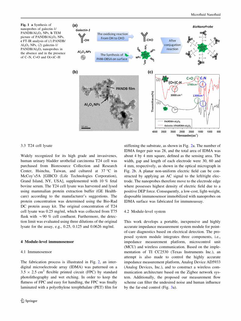
PANDB surrounding the Al2O3 NPs was synthesized, owing
to not only the ability of the amino groups (–NH2) of PANDB
to bind to the aldehyde groups (–CHO) of antibodies
(Fig. 1a), but also the ability of the conductivity of PANDB to
reduce the initial impedance of IDMA after PANDB-modi-
fied Al2O3 NPs immobilized on an electrode. The synthesis of
PANDB/Al2O3 NPs is described as follows. In this work,
commercially available aluminum oxide nanoparticles (Al2O3
NPs) from Evonik Degussa Taiwan Ltd. (AEROXIDE� Alu
C, Evonik Degussa), were used, in which their average size
ranges from approximately 20 to 30 nm. PANDB–Al2O3 NPs
were first synthesized using 3 g of aniline monomer, 8.4115 g
of DBSA, 7.35 g of ammonium persulfate (APS) and 2 g of
Al2O3 NPs. Al2O3 NPs were then added to the aniline solution
for 10 min and ultrasonically agitated, followed by the
introduction of the DBSA solution and subsequent stirring for
10 min. Finally, the APS solution was added and the system
was allowed to react for 2 h. PANDB was polymerized by a
standard oxidative polymerization of the aniline monomer,
with APS used as the oxidizing agent. Following polymeri-
zation for 2 h, darkish green PANDB/Al2O3 NPs suspension
was filtered and washed with acetone and reverse deionized
(DI) water until the filtrate became as colorless as the RO
water. Finally, 20 ml of DI water was mixed with powders of
PANDB/Al2O3 to yield an NP suspension. According to
Fig. 1b, average size of the PANDB-coated Al2O3 NPs ran-
ges from 120 to 200 nm, indicating that PANDB encloses and
links Al2O3 NPs into an irregular shape. This morphology can
cause the stacking of porous microstructures on the electrode
surface by DEP force, as well as reduce the impedance due to
the interconnection of conductive PANDB. Hence, the
modification scheme benefits impedance sensing approaches.
3.2 The preparation of the nanoprobes
Antibodies (galectin-1) were conjugated with PANDB/
Al2O3 NPs oxidizing 2 lg of the antibody against galectin-
1 protein for 5 min at room temperature with 1 mM
sodium metaperiodate with the pH value adjusted to 5.5
using 0.1 M sodium acetate. During the oxidizing reaction,
the hydroxyl groups (–OH) of carbohydrate moieties of
antibody were oxidized to aldehyde groups (–CHO), which
subsequently reacted with the amino groups exposed on the
surface of PANDB/Al2O3 NPs. The oxidized antibody was
then mixed with PANDB/Al2O3 NPs with rotation at room
temperature for 30 min. Following conjugation reaction,
antibody-modified PANDB–Al2O3 NPs were collected by
centrifugation at 18,0009g for 5 min; the nanoprobes are
referred to as galectin-1/PANDB/Al2O3 NP.
Microfluid Nanofluid
123
Page 4
3.3 T24 cell lysate
Widely recognized for its high grade and invasivenes,
human urinary bladder urothelial carcinoma T24 cell was
purchased from Bioresource Collection and Research
Center, Hsinchu, Taiwan, and cultured at 37 �C in
McCoy’s5A [GIBCO (Life Technologies Corporation),
Grand Island, NY, USA], supplemented with 10 % fetal
bovine serum. The T24 cell lysate was harvested and lysed
using mammalian protein extraction buffer (GE Health-
care) according to the manufacturer’s suggestions. The
protein concentration was determined using the Bio-Rad
DC protein assay kit. The original concentration of T24
cell lysate was 0.25 mg/ml, which was collected from T75
flask with *90 % cell confluent. Furthermore, the detec-
tion limit was evaluated using three dilutions of the original
lysate for the assay, e.g., 0.25, 0.125 and 0.0626 mg/ml.
4 Module-level immunosensor
4.1 Immunosensor
The fabrication process is illustrated in Fig. 2, an inter-
digital microelectrode array (IDMA) was patterned on a
3.5 9 2.5 cm2 flexible printed circuit (FPC) by standard
photolithography and wet etching. In order to keep the
flatness of FPC and easy for handling, the FPC was finally
laminated with a polyethylene terephthalate (PET) film for
stiffening the substrate, as shown in Fig. 2a. The number of
IDMA finger pair was 28, and the total area of IDMA was
about 4 by 4 mm square, defined as the sensing area. The
width, gap and length of each electrode were 30, 60 and
4 mm, respectively, as shown in the optical micrograph in
Fig. 2b. A planar non-uniform electric field can be con-
structed by applying an AC signal to the left/right elec-
trode. The nanoprobes therefore move to the electrode edge
where possesses highest density of electric field due to a
positive DEP force. Consequently, a low-cost, light-weight,
disposable immunosensor immobilized with nanoprobes on
IDMA surface was fabricated for immunoassay.
4.2 Module-level system
This work develops a portable, inexpensive and highly
accurate impedance measurement system module for point-
of-care diagnostics based on electrical detection. The pro-
posed system module integrates three components, i.e.,
impedance measurement platform, microcontrol unit
(MCU) and wireless communication. Based on the imple-
mentation of TI CC2530 (Texas Instruments Inc.), an
attempt is also made to control the highly accurate
impedance measurement platform, Analog Device AD5933
(Analog Devices, Inc.), and to construct a wireless com-
munication architecture based on the Zigbee network sys-
tem. Additionally, the proposed our measurement flow
scheme can filter the undesired noise and human influence
by the far-end control (Fig. 3a).
Fig. 1 a Synthesis of
nanoprobes of galectin-1/
PANDB/Al2O3 NPs, b TEM
picture of PANDB/Al2O3 NPs,
c FT-IR analysis of (1) PANDB/
Al2O3 NPs, (2) galectin-1/
PANDB/Al2O3 nanoprobes in
the absence and in the presence
of C–N, C=O and O(=)C–H
Microfluid Nanofluid
123
Page 5
AD5933, which contains a direct digital synthesis (DDS)
module and a digital Fourier transfer (DFT) module, pro-
vides a highly accurate impedance measurement perfor-
mance. An attempt is also made to provide high control
flexibility of AD5933 using the improved 8-bit MCU in
CC2530 to achieve measurement control on single frequency
and sweep frequency modes through an I2C interface. First,
AD5933 initiates the stimulus signal by the internal 27-bit
DDS module. The impendence response signal is then
sampled by an internal 12-bit analog-to-digital converter
(ADC) module. Additionally, the embedded 1024-point FFT
module calculates all of the impedance results with respect to
each frequency pin. Furthermore, to communicate the final
results through a wireless system, CC2530, which contains
Fig. 2 a Fabrication of
immunosensor with interdigital
microelectrode array (IDMA);
b schematic picture of non-
uniform electrode field of
IDMA and the finished
disposable immunosensor
Fig. 3 a Experimental setup of
immunosensing based on
impedance system module and
paper sampling; b photograph
of module-level impedance
system
Microfluid Nanofluid
123
Page 6
8-bit MCU and the Zigbee physical layer, is responsible for
the control of impedance measurements (AD5933) and far-
end communication with personal computer (PC). In the
proposed Zigbee network topology, the impedance is mea-
sured by the terminal of an end device, which controls the
AD5933 through I2C interface. As a serial communication
protocol, the I2C standard contains two bidirectional open
drain wires, i.e., SDA and SCL. This standard is a master and
slave-based architecture. Based on the I2C standard, the
master (i.e., end device of CC2530) sends commands to I2C
bus including the start frequency command, incremental
frequency command, cycle numbers command and sweep
frequency number command. The device (AD5933) may
then issue the impedance measurement tasks. Finally, the
frequency responses of impedance results may be sent to an
end device in the form of real and imaginary data parts. When
the terminal of coordinator obtains the impedance results
from the terminal of end device through Zigbee wireless
communication, the coordinator forwards the impedance
results to a PC by the RS-232 interface. Using the LabVIEW-
based platform, the proposed measurement system achieves
the far-end control and computation system in the PC, which
can complete the final frequency response. Importantly, the
large amount of final data can be stored on a far-end server
for further accounting analysis. Therefore, the proposed
measurement system can communicate over an extremely
long distance by CC2530, possibly around 1 K meter, thus
resolving the signal decade problem on a wire-based com-
munication system. Furthermore, the highly accurate results
achieved by AD5933 can be stored in the far-end server for
advance analysis. Above features facilitate point-of-care
diagnosis for elderly individuals, whom require frequent
monitoring and telehealthcare at home.
5 Experimental method
Figure 4 illustrates the five steps of the experimental
procedure. First, 10 ll of nanoprobes suspension with a
measured conductivity of about 500 ls/cm (SC-170,
Suntex Instruments Co., Ltd.) was dripped on the sensing
area of an immunosensor by using a micropipette. The
AC signals were then applied to IDMA for trapping
nanoprobes on IDMA surface by a positive DEP force;
the AC signals were 10 Vpp at 50 kHz through a function
generator (AFG3022, Tektronix). Owing to that the DEP
trapping was performed in the atmospheric environment
at room temperature, AC signals were applied to IDMA
for only 10 min before the nanoprobes suspension dried
out. Following DEP trapping, the immunosensor was
washed by DI water and dried by a N2 gun in the second
step. Notably, although the DEP force was not applied
during the washing procedure, the immobilized
nanoprobes were not removed in the experimental
observation. This formation can be attributed to the layer-
like microstructure of PANDB nanoparticles and surface
charge of proteins. Therefore, based on van der Waals’
force and electrostatic force, physisorption between the
immobilized nanoprobes and electrode surface was
improved. Meanwhile, the initial impedance at 50 kHz of
immunosensor was measured by the proposed impedance
system module and denoted as Zprobe. To obtain a stable
value of Zprobe, the above procedures were iterated for six
times to investigate the variation of Zprobe that corre-
sponded to DEP trapping within 60 min. In the third step
of the experimental procedure, a chartula (i.e., a folded
paper for containing medicinal powder) with a size of
7 mm 9 25 mm was immersed into the T24 cell lysate
for 2 s, and then, the moistened paper was swept across
the sensing area. Chartula rather than conventional copy
paper was chosen owing to its smoother texture of char-
tula and greater density. Therefore, the absorbed protein
remains on the chartula surface rather than in the texture.
Additionally, the quantity of T24 cell lysate adsorbed on
the paper was measured about 0.01 g. In the fourth step
of the experimental procedure, T24 cell lysate which
contained various proteins was incubated for 10 min of
immunoreaction. However, only the galectin-1 protein in
T24 cell lysate conjugated with nanoprobes, owing to the
specific binding between the antibody and antigen. In the
fifth step of the experimental procedure, an attempt was
made to eliminate the non-binding proteins on the
immunosensor by washing the sensor surface with DI
water and then drying it by a N2 gun. The impedance of
IDMA at 50 kHz after immunoassay was then measured
by the proposed impedance system module and denoted
as ZT24. The variation of impedance due to specific
binding of galectin-1 protein can be attributed to
DZ = ZT24 - Zprobe. Moreover, an attempt was made to
eliminate the difference of initial impedance (Zprobe)
between different immunosensors by normalizing DZ with
Zprobe, i.e., DZ/Zprobe, which is denoted as a normalized
variation of impedance. In our experiments, three T24 cell
lysate concentrations were evaluated with respect to the
sensitivity of an immunosensor based on DZ/Zprobe.
6 Results and discussion
6.1 Characterization of nanoprobes
This work also demonstrated the feasibility of imple-
menting the final step of nanoprobes preparation, i.e.,
binding antibodies on PANDB/Al2O3 nanoparticles, by
comparing two FT-IR spectra of PANDB/Al2O3 nano-
particles before and after binding with antibodies in
Microfluid Nanofluid
123
Page 7
Fig. 1c. According to this figure, three regions in the
spectra changed after binding with antibodies, implying
that these functional groups explicitly stretched or
changed after the modification. As a result, two regions
appearing around 1,780 and 3,000 cm-1 are, respec-
tively, assigned to C=O and O(=)C–H; another peak
appearing at 1,300 cm-1 reveals the presence of C–N
bond, especially for the NH2 group. The functional
group C=O may originate from the carboxylic acid
(–COOH) groups of galectin-1. Therefore, we can infer
that the free amino groups on PANDB surface (NH2)
bond with the carbohydrate groups (O(=)C–H) of oxi-
dized antibodies, as shown in Fig. 1a. Consequently, the
PANDB nanoprobes were successfully synthesized for
protein immobilization in the immunosensing system.
Additionally, the antigen binding region (Fab region, the
Y-shape arms) is only 10 % of the antibody structure,
explaining why most of the carbohydrate group bridges
the amino groups; the Fab region can be exposed as the
receptor of antigen as well. This modification scheme
thus can assure the receptor of antibody exposed on the
NP surface for immunosensing.
6.2 Characterization of impedance system module
Accuracy of the proposed impedance system module
was verified using a precision LCR meter (WK 6420,
Wayne Kerr Electronics). The impedance of an immu-
nosensor with IDMA before coating was measured by
the proposed impedance system module and precision
LCR meter at 50 kHz for 10 times, respectively. The
average results of impedance were 577 and 588 kXfrom impedance system module and precision LCR
meter, respectively. Notably, the error is only 1.89 % if
the value from the LCR meter is used as the reference,
allowing us to achieve a highly accurate and reliable
impedance measurement based on the proposed imped-
ance system module.
6.3 Time effects of DEP trapping and immunosensing
Using the DEP force, this work also attempted to immo-
bilize nanoprobes onto the IDMA surface in order to
achieve a lower baseline of Zprobe after DEP trapping, in
which the impedance change was measured for 60 min
with 6 iterations of 10 min DEP trapping. According to the
blue curve in Fig. 5, the impedance decreased slightly
within 20 min, yet it descended a large range when DEP
trapping was performed for 30 min. The impedance then
remained stable in a low level until 60 min. Therefore, the
PANDB-coated nanoprobes effectively reduce the initial
impedance of IDMA after immobilization on the electrode
surface by DEP trapping. Thus, 30-min DEP trapping is
Fig. 4 Experimental procedure
for DEP trapping of nanoprobes
and immunosensing by
impendence sensing
Fig. 5 Impedance variation during trapping nanoprobes and immu-
nosensing for 60 min, blue line represents the decrease of impedance
when nanoprobes are immobilized on the electrode surface by DEP
force, where the red line represents the increase in impedance when
proteins binding with nanoprobes (color figure online)
Microfluid Nanofluid
123
Page 8
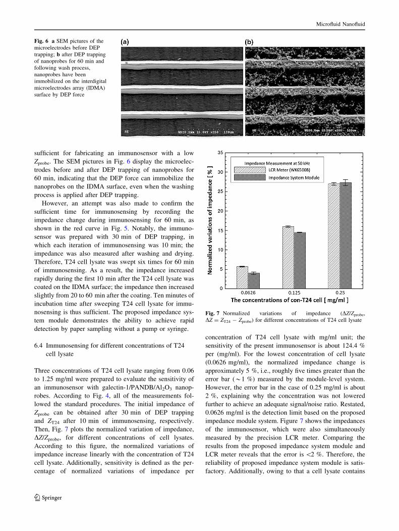
sufficient for fabricating an immunosensor with a low
Zprobe. The SEM pictures in Fig. 6 display the microelec-
trodes before and after DEP trapping of nanoprobes for
60 min, indicating that the DEP force can immobilize the
nanoprobes on the IDMA surface, even when the washing
process is applied after DEP trapping.
However, an attempt was also made to confirm the
sufficient time for immunosensing by recording the
impedance change during immunosensing for 60 min, as
shown in the red curve in Fig. 5. Notably, the immuno-
sensor was prepared with 30 min of DEP trapping, in
which each iteration of immunosensing was 10 min; the
impedance was also measured after washing and drying.
Therefore, T24 cell lysate was swept six times for 60 min
of immunosensing. As a result, the impedance increased
rapidly during the first 10 min after the T24 cell lysate was
coated on the IDMA surface; the impedance then increased
slightly from 20 to 60 min after the coating. Ten minutes of
incubation time after sweeping T24 cell lysate for immu-
nosensing is thus sufficient. The proposed impedance sys-
tem module demonstrates the ability to achieve rapid
detection by paper sampling without a pump or syringe.
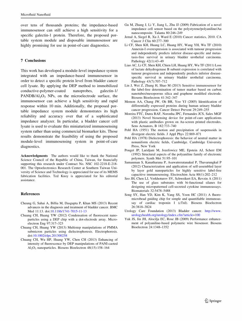
6.4 Immunosensing for different concentrations of T24
cell lysate
Three concentrations of T24 cell lysate ranging from 0.06
to 1.25 mg/ml were prepared to evaluate the sensitivity of
an immunosensor with galectin-1/PANDB/Al2O3 nanop-
robes. According to Fig. 4, all of the measurements fol-
lowed the standard procedures. The initial impedance of
Zprobe can be obtained after 30 min of DEP trapping
and ZT24 after 10 min of immunosensing, respectively.
Then, Fig. 7 plots the normalized variation of impedance,
DZ/Zprobe, for different concentrations of cell lysates.
According to this figure, the normalized variations of
impedance increase linearly with the concentration of T24
cell lysate. Additionally, sensitivity is defined as the per-
centage of normalized variations of impedance per
concentration of T24 cell lysate with mg/ml unit; the
sensitivity of the present immunosensor is about 124.4 %
per (mg/ml). For the lowest concentration of cell lysate
(0.0626 mg/ml), the normalized impedance change is
approximately 5 %, i.e., roughly five times greater than the
error bar (*1 %) measured by the module-level system.
However, the error bar in the case of 0.25 mg/ml is about
2 %, explaining why the concentration was not lowered
further to achieve an adequate signal/noise ratio. Restated,
0.0626 mg/ml is the detection limit based on the proposed
impedance module system. Figure 7 shows the impedances
of the immunosensor, which were also simultaneously
measured by the precision LCR meter. Comparing the
results from the proposed impedance system module and
LCR meter reveals that the error is \2 %. Therefore, the
reliability of proposed impedance system module is satis-
factory. Additionally, owing to that a cell lysate contains
Fig. 6 a SEM pictures of the
microelectrodes before DEP
trapping; b after DEP trapping
of nanoprobes for 60 min and
following wash process,
nanoprobes have been
immobilized on the interdigital
microelectrodes array (IDMA)
surface by DEP force
Fig. 7 Normalized variations of impedance (DZ/Zprobe,
DZ = ZT24 - Zprobe) for different concentrations of T24 cell lysate
Microfluid Nanofluid
123
Page 9
over tens of thousands proteins; the impedance-based
immunosensor can still achieve a high sensitivity for a
specific galectin-1 protein. Therefore, the proposed por-
table system module and disposable immunosensor are
highly promising for use in point-of-care diagnostics.
7 Conclusions
This work has developed a module-level impedance system
integrated with an impedance-based immunosensor in
order to detect a specific protein level from bladder cancer
cell lysate. By applying the DEP method to immobilized
conductive-polymer-coated nanoprobes, galectin-1/
PANDB/Al2O3 NPs, on the microelectrode surface, the
immunosensor can achieve a high sensitivity and rapid
response within 10 min. Additionally, the proposed por-
table impedance system module demonstrates its high
reliability and accuracy over that of a sophisticated
impedance analyzer. In particular, a bladder cancer cell
lysate is used to evaluate this miniaturized immunosensing
system rather than using commercial biomarker kits. Those
results demonstrate the feasibility of using the proposed
module-level immunosensing system in point-of-care
diagnostics.
Acknowledgments The authors would like to thank the National
Science Council of the Republic of China, Taiwan, for financially
supporting this research under Contract No. NSC 102-2218-E-218-
001. The Optoelectronics Research Center at Southern Taiwan Uni-
versity of Science and Technology is appreciated for use of its MEMS
fabrication facilities. Ted Knoy is appreciated for his editorial
assistance.
References
Cheung G, Sahai A, Billia M, Dasgupta P, Khan MS (2013) Recent
advances in the diagnosis and treatment of bladder cancer. BMC
Med 11:13. doi:10.1186/1741-7015-11-13
Chuang CH, Huang YW (2012) Condensation of fluorescent nano-
particles using a DEP chip with a dot-electrode array. Micro-
electron Eng 97:317–323
Chuang CH, Huang YW (2013) Multistep manipulations of PMMA
submicron particles using dielectrophoresis. Electrophoresis.
doi:10.1002/elps.201300258
Chuang CH, Wu HP, Huang YW, Chen CH (2013) Enhancing of
intensity of fluorescence by DEP manipulations of PANI-caoted
Al2O3 nanoparticles. Biosens Bioelectron 48(15):158–164
Gu M, Zhang J, Li Y, Jiang L, Zhu JJ (2009) Fabrication of a novel
impedance cell sensor based on the polystyrene/polyaniline/Au
nanocomposite. Talanta 80:246–249
Jemal A, Siegel R, Xu J, Ward E (2010) Cancer statistics, 2010. CA
Cancer J Clin 60:277–300
Li CF, Shen KH, Huang LC, Huang HY, Wang YH, Wu TF (2010)
Annexin-I overexpression is associated with tumour progression
and independently predicts inferior disease-specific and metas-
tasis-free survival in urinary bladder urothelial carcinoma.
Pathology 42(1):43–49
Liao AC, Li CF, Shen KH, Chien LH, Huang HY, Wu TF (2011) Loss
of lactate dehydrogenase B subunit expression is correlated with
tumour progression and independently predicts inferior disease-
specific survival in urinary bladder urothelial carcinoma.
Pathology 43(7):707–712
Lin J, Wei Z, Zhang H, Shao M (2013) Sensitive immunosensor for
the label-free determination of tumor marker based on carbon
nanotubes/mesoporous silica and graphene modified electrode.
Biosens Bioelectron 41:342–347
Memon AA, Chang JW, Oh BR, Yoo YJ (2005) Identification of
differentially expressed proteins during human urinary bladder
cancer progression. Cancer Detect Prevent 29:249–255
Moreira FTC, Dutra RAF, Noronha JPC, Fernandes JCS, Sales MGF
(2013) Novel biosensing device for point-of-care applications
with plastic antibodies grown on Au-screen printed electrodes.
Sens Actuators, B 182:733–740
Pohl HA (1951) The motion and precipitation of suspensoids in
divergent electric fields. J Appl Phys 22:869–871
Pohl HA (1978) Dielectrophoresis: the behavior of neutral matter in
nonuniform electric fields, Cambridge. Cambridge University
Press, New York
Pouget JP, Laridjani M, Jozefowicz ME, Epstein AJ, Scherr EM
(1992) Structural aspects of the polyaniline family of electronic
polymers. Synth Met 51:95–101
Samanman S, Kanatharana P, Asawatreratanakul P, Thavarungkul P
(2012) Characterization and application of self-assembled layer
by layer gold nanoparticles for highly sensitive label-free
capacitive immunosensing. Electrochim Acta 80(1):202–212
Seo JH, Chen LJ, Verkhoturov SV, Schweikert EA, Revzin A (2011)
The use of glass substrates with bi-functional silanes for
designing micropatterned cell-secreted cytokine immunoassays.
Biomaterials 32:5478–5488
Song SY, Han YD, Kim K, Yang SS, Yoon HC (2011) A fluoro-
microbead guiding chip for simple and quantifiable immunoas-
say of cardiac troponin I (cTnI). Biosens Bioelectron
26:3818–3824
Urology Care Foundation (2013) Bladder cancer. http://www.
urologyhealth.org/urology/index.cfm?article=100
Yuk JS, Jin JH, Alocilja EC, Rose JB (2009) Performance enhance-
ment of polyaniline-based polymeric wire biosensor. Biosens
Bioelectron 24:1348–1352
Microfluid Nanofluid
123