miR-216b suppresses tumor growth and invasion by targeting KRAS in nasopharyngeal carcinoma Min Deng 1,2 , Hailin Tang 1,2 , Yanhong Zhou 1 , Ming Zhou 1 , Wei Xiong 1 , Ying Zheng 1 , Qiurong Ye 1 , Xi Zeng 1,2 , Qianjin Liao 1,2 , Xiaofang Guo 1 , Xiaoling Li 1 , Jian Ma 1, * and Guiyuan Li 1, * 1 Cancer Research Institute, Central South University, 110 Xiang-Ya Road, Changsha 410078, Hunan, People’s Republic of China 2 Cancer Research Institute, University of South China, 28 Changsheng West Road, Hengyang 421001, Hunan, People’s Republic of China * Authors for correspondence ([email protected]; [email protected]) Accepted 6 May 2011 Journal of Cell Science 124, 2997–3005 ß 2011. Published by The Company of Biologists Ltd doi: 10.1242/jcs.085050 Summary MicroRNAs (miRNAs) are small noncoding RNAs that are involved in various diseases, including cancer. In the present study, we found that miR-216b was downregulated in nasopharyngeal carcinoma (NPC) cell lines and specimens. Decreased expression of miR- 216b was directly related to advanced clinical stage and lymph node metastasis. miR-216b levels correlated inversely with levels of KRAS protein during nasopharyngeal tumorigenesis. Furthermore, we demonstrated that miR-216b can bind to the 39 untranslated region (UTR) of KRAS and inhibit expression of the KRAS protein. Both in vitro and in vivo assays revealed that miR-216b attenuated NPC cell proliferation, invasion and tumor growth in nude mice. miR-216b exerts its tumor suppressor function through inhibition of the KRAS-related AKT and ERK pathways. Our findings provide, for the first time, significant clues regarding the role of miR-216b as a tumor suppressor by targeting KRAS in NPC. Key words: microRNA-216b, KRAS, Nasopharyngeal carcinoma Introduction Nasopharyngeal carcinoma (NPC) is a malignancy arising from the nasopharyngeal epithelium (NET) with an incidence in southern China of 20–30 cases per 100,000, a frequency 25-fold higher than that of other countries (Yu and Yuan, 2002). Many factors, including tumor suppressors and oncogenes, are involved in nasopharyngeal tumorigenesis (Deng et al., 1998; Lo et al., 1996; Qian et al., 2002; Sarac et al., 2001; Sheu et al., 1995; Xiang and Zhang, 2005; Xiong et al., 2004). Recently, the classical family of protein-encoding genes recognized as tumor suppressors and oncogenes has been expanded to include a type of non-protein-coding RNA molecule known as microRNA (miRNA) (Calin and Croce, 2006; Esquela-Kerscher and Slack, 2006). miRNAs are 19–24 nucleotides in length, and they regulate gene expression by imperfect base-pairing with complementary sequences located mainly, but not exclusively, in the 39 untranslated regions (UTRs) of target mRNAs. Hence, miRNAs represent one of the major regulatory families of genes in eukaryotic cells, and they work by inducing translational repression and transcript degradation (Doench and Sharp, 2004; German et al., 2008; Pillai et al., 2007). miRNAs regulate genes involved in functions ranging from development, differentiation and proliferation to stress processes (German et al., 2008). Rapidly emerging evidence strongly suggests crucial roles of miRNAs in tumorigenesis (Slack and Weidhaas, 2008; Vasilatou et al., 2010). For example, miR-182 is upregulated in melanoma and promotes metastasis by repressing FOXO3 and microphthalmia-associated transcription factor (MITF) (Segura et al., 2009). The let-7 family of miRNAs has been shown to regulate KRAS (Johnson et al., 2005), and let-7 levels have been correlated with prognosis in lung cancer (Takamizawa et al., 2004). miR-96 expression is decreased in pancreatic cancer, and it functions as a tumor suppressor by targeting KRAS (Yu et al., 2010). Recently, Sengupta et al. reported that miR-216 was significantly downregulated (an 85% reduction) in NPC relative to NET when examined by a miRNA microarray analysis (Sengupta et al., 2008). Two members of the miR-216 family, miR-216a and miR-216b, are located at chromosome 2p16.1, a region frequently deleted in NPC (Shao et al., 2001). These findings suggest that deregulation of miR-216 is associated with nasopharyngeal carcinogenesis. However, to our knowledge, the role of miR-216 in tumorigenesis remains undefined. Here, we determined that two members of miR-216 family (miR-216a and especially mir-216b) are downregulated in NPC, and we found an inverse correlation between the levels of miR- 216b and KRAS protein. miR-216b directly targets the 39-UTR of the KRAS transcript and elicits a specific and robust knockdown of KRAS protein; it also inhibits NPC cell proliferation, invasion and tumor growth in nude mice. miR- 216b mediates its tumor suppressor function, at least in part, by suppressing pathways downstream of KRAS, such as the PI3K- AKT and MEK-ERK pathways. Our findings provide, for the first time, significant clues regarding the role of miR-216b as a tumor suppressor in NPC. Results miR-216b is downregulated in NPC cell lines and clinical specimens Recently, Sengupta et al. identified eight miRNAs showing robust differential expression between 31 NPC and ten normal healthy NET samples by a miRNA microarray analysis. Among Research Article 2997 Journal of Cell Science
Transcript
miR-216b suppresses tumor growth and invasion bytargeting KRAS in nasopharyngeal carcinoma
Min Deng1,2, Hailin Tang1,2, Yanhong Zhou1, Ming Zhou1, Wei Xiong1, Ying Zheng1, Qiurong Ye1, Xi Zeng1,2,Qianjin Liao1,2, Xiaofang Guo1, Xiaoling Li1, Jian Ma1,* and Guiyuan Li1,*1Cancer Research Institute, Central South University, 110 Xiang-Ya Road, Changsha 410078, Hunan, People’s Republic of China2Cancer Research Institute, University of South China, 28 Changsheng West Road, Hengyang 421001, Hunan, People’s Republic of China*Authors for correspondence ([email protected]; [email protected])
Accepted 6 May 2011Journal of Cell Science 124, 2997–3005� 2011. Published by The Company of Biologists Ltddoi: 10.1242/jcs.085050
SummaryMicroRNAs (miRNAs) are small noncoding RNAs that are involved in various diseases, including cancer. In the present study, wefound that miR-216b was downregulated in nasopharyngeal carcinoma (NPC) cell lines and specimens. Decreased expression of miR-216b was directly related to advanced clinical stage and lymph node metastasis. miR-216b levels correlated inversely with levels of
KRAS protein during nasopharyngeal tumorigenesis. Furthermore, we demonstrated that miR-216b can bind to the 39 untranslatedregion (UTR) of KRAS and inhibit expression of the KRAS protein. Both in vitro and in vivo assays revealed that miR-216b attenuatedNPC cell proliferation, invasion and tumor growth in nude mice. miR-216b exerts its tumor suppressor function through inhibition of theKRAS-related AKT and ERK pathways. Our findings provide, for the first time, significant clues regarding the role of miR-216b as a
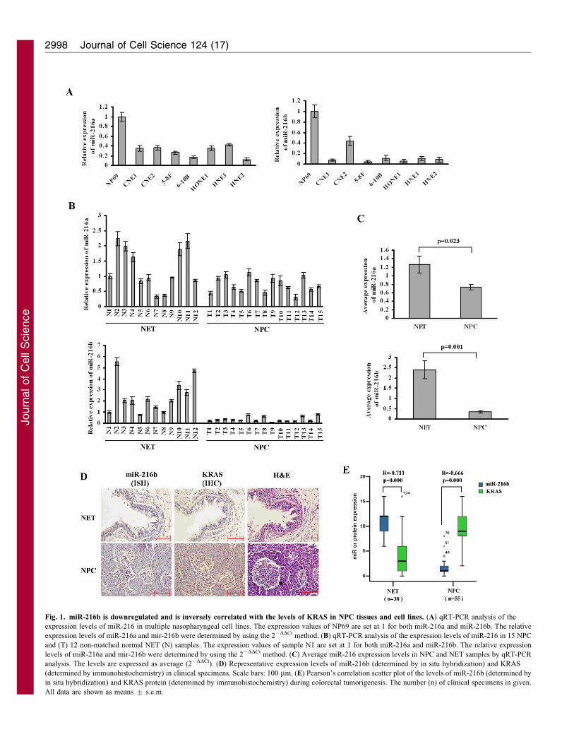
Fig. 1. miR-216b is downregulated and is inversely correlated with the levels of KRAS in NPC tissues and cell lines. (A) qRT-PCR analysis of the
expression levels of miR-216 in multiple nasopharyngeal cell lines. The expression values of NP69 are set at 1 for both miR-216a and miR-216b. The relative
expression levels of miR-216a and mir-216b were determined by using the 22DDCt method. (B) qRT-PCR analysis of the expression levels of miR-216 in 15 NPC
and (T) 12 non-matched normal NET (N) samples. The expression values of sample N1 are set at 1 for both miR-216a and miR-216b. The relative expression
levels of miR-216a and mir-216b were determined by using the 22DDCt method. (C) Average miR-216 expression levels in NPC and NET samples by qRT-PCR
analysis. The levels are expressed as average (22DDCt). (D) Representative expression levels of miR-216b (determined by in situ hybridization) and KRAS
(determined by immunohistochemistry) in clinical specimens. Scale bars: 100 mm. (E) Pearson’s correlation scatter plot of the levels of miR-216b (determined by
in situ hybridization) and KRAS protein (determined by immunohistochemistry) during colorectal tumorigenesis. The number (n) of clinical specimens in given.
All data are shown as means ¡ s.e.m.
Journal of Cell Science 124 (17)2998
Journ
alof
Cell
Scie
nce
those miRNAs, the level of miR-216 expression in cancer cells
was reduced by 85% compared with the level of expression innormal epithelial cells. Here, we used quantitative real-time PCR(qRT-PCR) to measure mature miR-216a and -216b expression
levels in eight NET cell lines, 15 NPC and 12 NET tissuesamples. These two members of the miR-216 family showedreduced expression levels in all NPC cell lines with respect tobenign immortalized NP69 NET cells (Fig. 1A). Similarly, both
of them showed lower expression in NPC tumors relative to NETtissues (Fig. 1B). The overall average expression levels of miR-216a and -216b were lower in NPCs than in normal tissues (NET)
(Fig. 1C). In particular, the difference in expression (betweennormal and tumor tissues) of miR-216b was several times higherthan that of miR216a (miR-216a, fold difference51.7, P50.023;
miR-216b, fold difference56.7, P50.001, Fig. 1C). For thisreason, we focused on miR-216b in this study.
To determine how the downregulation of miR-216b mightcontribute to NPC phenotypes or clinical pathological features,
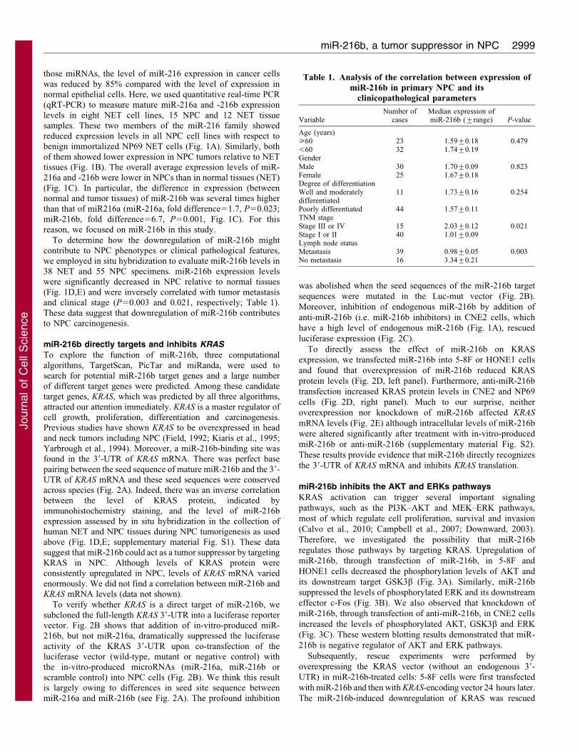
we employed in situ hybridization to evaluate miR-216b levels in38 NET and 55 NPC specimens. miR-216b expression levelswere significantly decreased in NPC relative to normal tissues
(Fig. 1D,E) and were inversely correlated with tumor metastasisand clinical stage (P50.003 and 0.021, respectively; Table 1).These data suggest that downregulation of miR-216b contributes
to NPC carcinogenesis.
miR-216b directly targets and inhibits KRAS
To explore the function of miR-216b, three computational
algorithms, TargetScan, PicTar and miRanda, were used tosearch for potential miR-216b target genes and a large numberof different target genes were predicted. Among these candidatetarget genes, KRAS, which was predicted by all three algorithms,
attracted our attention immediately. KRAS is a master regulator ofcell growth, proliferation, differentiation and carcinogenesis.Previous studies have shown KRAS to be overexpressed in head
and neck tumors including NPC (Field, 1992; Kiaris et al., 1995;Yarbrough et al., 1994). Moreover, a miR-216b-binding site wasfound in the 39-UTR of KRAS mRNA. There was perfect base
pairing between the seed sequence of mature miR-216b and the 39-UTR of KRAS mRNA and these seed sequences were conservedacross species (Fig. 2A). Indeed, there was an inverse correlationbetween the level of KRAS protein, indicated by
immunohistochemistry staining, and the level of miR-216bexpression assessed by in situ hybridization in the collection ofhuman NET and NPC tissues during NPC tumorigenesis as used
above (Fig. 1D,E; supplementary material Fig. S1). These datasuggest that miR-216b could act as a tumor suppressor by targetingKRAS in NPC. Although levels of KRAS protein were
consistently upregulated in NPC, levels of KRAS mRNA variedenormously. We did not find a correlation between miR-216b andKRAS mRNA levels (data not shown).
To verify whether KRAS is a direct target of miR-216b, we
subcloned the full-length KRAS 39-UTR into a luciferase reportervector. Fig. 2B shows that addition of in-vitro-produced miR-216b, but not miR-216a, dramatically suppressed the luciferase
activity of the KRAS 39-UTR upon co-transfection of theluciferase vector (wild-type, mutant or negative control) withthe in-vitro-produced microRNAs (miR-216a, miR-216b or
scramble control) into NPC cells (Fig. 2B). We think this resultis largely owing to differences in seed site sequence betweenmiR-216a and miR-216b (see Fig. 2A). The profound inhibition
was abolished when the seed sequences of the miR-216b target
sequences were mutated in the Luc-mut vector (Fig. 2B).
Moreover, inhibition of endogenous miR-216b by addition of
anti-miR-216b (i.e. miR-216b inhibitors) in CNE2 cells, which
have a high level of endogenous miR-216b (Fig. 1A), rescued
luciferase expression (Fig. 2C).
To directly assess the effect of miR-216b on KRAS
expression, we transfected miR-216b into 5-8F or HONE1 cells
and found that overexpression of miR-216b reduced KRAS
protein levels (Fig. 2D, left panel). Furthermore, anti-miR-216b
transfection increased KRAS protein levels in CNE2 and NP69
cells (Fig. 2D, right panel). Much to our surprise, neither
overexpression nor knockdown of miR-216b affected KRAS
mRNA levels (Fig. 2E) although intracellular levels of miR-216b
were altered significantly after treatment with in-vitro-produced
miR-216b or anti-miR-216b (supplementary material Fig. S2).
These results provide evidence that miR-216b directly recognizes
the 39-UTR of KRAS mRNA and inhibits KRAS translation.
miR-216b inhibits the AKT and ERKs pathways
KRAS activation can trigger several important signaling
pathways, such as the PI3K–AKT and MEK–ERK pathways,
most of which regulate cell proliferation, survival and invasion
(Calvo et al., 2010; Campbell et al., 2007; Downward, 2003).
Therefore, we investigated the possibility that miR-216b
regulates those pathways by targeting KRAS. Upregulation of
miR-216b, through transfection of miR-216b, in 5-8F and
HONE1 cells decreased the phosphorylation levels of AKT and
its downstream target GSK3b (Fig. 3A). Similarly, miR-216b
suppressed the levels of phosphorylated ERK and its downstream
effector c-Fos (Fig. 3B). We also observed that knockdown of
miR-216b, through transfection of anti-miR-216b, in CNE2 cells
increased the levels of phosphorylated AKT, GSK3b and ERK
(Fig. 3C). These western blotting results demonstrated that miR-
216b is negative regulator of AKT and ERK pathways.
Subsequently, rescue experiments were performed by
overexpressing the KRAS vector (without an endogenous 39-
UTR) in miR-216b-treated cells: 5-8F cells were first transfected
with miR-216b and then with KRAS-encoding vector 24 hours later.
The miR-216b-induced downregulation of KRAS was rescued
Table 1. Analysis of the correlation between expression of
Age (years)>60 23 1.59¡0.18 0.479,60 32 1.74¡0.19GenderMale 30 1.70¡0.09 0.823Female 25 1.67¡0.18Degree of differentiationWell and moderatelydifferentiated
11 1.73¡0.16 0.254
Poorly differentiated 44 1.57¡0.11TNM stageStage III or IV 15 2.03¡0.12 0.021Stage I or II 40 1.01¡0.09Lymph node statusMetastasis 39 0.98¡0.05 0.003No metastasis 16 3.34¡0.21
miR-216b, a tumor suppressor in NPC 2999
Journ
alof
Cell
Scie
nce
upon the introduction of KRAS (Fig. 3D), and the phosphorylation
levels of AKT and ERK were altered in a similar manner. The
downregulation of phosphorylated AKT and phosphorylated ERK
by miR-216b could be rescued by re-expression of KRAS
(Fig. 3D). These observations suggest that miR-216b inhibits the
AKT and ERK pathways by targeting KRAS.
miR-216b suppresses NPC cell proliferation and invasion
by targeting KRAS
In light of the above findings, we decided to explore the
biological significance of miR-216b in NPC tumorigenesis. We
found that overexpression of miR-216b in CNE2 and 5-8F cells
markedly attenuated cell proliferation and that re-expression of
KRAS (without an endogenous 39-UTR) rescued this inhibition,
suggesting that miR-216b targets KRAS specifically (Fig. 4A).
Overexpression of miR-216b inhibited the in vitro invasive
potentials of 5-8F and HONE1 cells, whereas re-expression of
KRAS rescued this inhibition (Fig. 4C). In contrast with in-vitro-
produced miR-216, anti-miR-216b induced cell growth and
invasive phenotypes in CNE2 cells (Fig. 4B,D).
Next, we tested whether miR-216b could play a role in
tumorigenesis by using nude mice xenograft models. Nude mice
Fig. 2. KRAS is a direct target of miR-216b. (A) Sequence alignment of microRNAs of the miR-216 family and the KRAS 39-UTR. There are differences in seed
site sequence between miR-216a and miR-216b. The KRAS 39-UTR contains one predicted miR-216b-binding site. The seed regions of miR-216 and the
seed-recognizing sites in the KRAS 39-UTR are indicated in red, and all nucleotides in seed-recognizing sites are completely conserved in several species. Hsa,
Fea, Felis catus. (B) Luciferase assay on 5-8F cells (left-hand side) and HONE1 cells (right-hand side), which were co-transfected with miR-216 and a luciferase
reporter containing the full length of KRAS 39-UTR (Luc-wt) or a mutant (Luc-mut) in which the first five nucleotides of the miR-216b-binding site were mutated.
An empty luciferase reporter construct was used as a negative control (Luc-ctrl). Luciferase activities were measured 48 hours post transfection. miR-216b
markedly suppressed luciferase activity in Luc-wt reporter constructs. The data are means ¡ s.e.m. for separate transfections (n54). (C) Luciferase assay on CNE2
cells co-transfected with a luciferase reporter (Luc-wt) and either anti-miR-216a or anti-miR-216b. Inhibition of endogenous miR-216b rescued the luciferase
expression. The data are means ¡ s.e.m. for separate transfections (n54). (D) miR-216b or anti-miR-216b transfection affects KRAS protein levels. 5-8F and
HONE1 cells were transfected with miR-216b or scramble control, and CNE2 and NP69 cells were transfected with anti-miR-216b or anti-scramble. (E) KRAS
mRNA levels were analyzed upon miR-216b or anti-miR-216b transfection by qRT–PCR. All data are shown as means ¡ s.e.m. **P,0.01.
Journal of Cell Science 124 (17)3000
Journ
alof
Cell
Scie
nce
transplanted with 5-8F cells transfected either with scramble or
miR-216b developed solid tumors in 25 days. However, tumor
growth (including tumor volume and weight) was significantly
reduced when the miR-216b was stably expressed in 5-8F cells
(P,0.05, Fig. 4E, left-hand panels). CNE2 cells infected with the
miR-216b-knockdown lentivirus or the control lentivirus were
also transplanted into nude mice. After 25 days, tumor growth
(including tumor volume and weight) was significantly increased
in the miR-216b-knockdown CNE2 cells (P,0.05, Fig. 4E,
right-hand panels). In addition, we confirmed that changes in
miR-216b expression altered KRAS protein expression levels in
lysates from mouse tumors (Fig. 4F; supplementary material Fig.
S3).
Our findings demonstrate that miR-216b has properties
consistent with tumor suppressor function. The ability to
modulate KRAS protein levels might explain, at least in part,
why miR-216b can inhibit cell proliferation and invasion in NPC.
DiscussionAlthough the first microRNAs were discovered in C. elegans
several years ago [lin-4 in 1993 (Lee et al., 1993) and let-7 in
2000 (Reinhart et al., 2000)], it is only more recently that the
study of this class of small regulatory RNAs in humans has
become widespread (Ruvkun et al., 2004). Until now, only a
handful of studies have identified specific miRNAs involved in
human tumorigenesis and metastasis. Therefore, we believe more
effort should be made, not only towards the identification of
relevant miRNAs but also into the specific mechanisms by which
they accomplish their specific functions, particularly with regard
to the oncogenesis of different types of tumors.
A recent report has shed light on which miRNAs are involved
in NPC tumorigenesis. Sengupta et al. reported that miR-216
expression levels were significantly downregulated (an 85%
reduction) in NPC relative to NET, although little is known about
the function and mechanism of miR-216 in tumorigenesis
(Sengupta et al., 2008). Here, we used qRT-PCR and in situ
hybridization to assay expression of mir-216 family of miRNAs
in NPC cell lines and clinical specimens and found that these
miRNAs were indeed downregulated in NPC tumor tissues.
Importantly, the decrease in levels of miR-216b was several
times greater than that of the levels of miR-216a in NPC samples
as opposed to in normal tissues (Fig. 1C), and the levels of miR-
216b were found to be inversely correlated to tumor metastasis
and clinical stage (Table 1). In the same samples, the copy
numbers of miR-216 and miR-216b showed a positive
correlation, even although they were not exactly equal,
implying that their mechanisms of regulation are somehow
different (data not shown).
We next explored the possible targets of miR-216b in NPC
cells through different computational algorithms. Silicon analysis
revealed KRAS as a candidate target of miR-216b, which
attracted our attention immediately. Like other members of the
RAS family, KRAS is a GTPase and an early player in many
signal transduction pathways. Active GTP-bound KRAS
associates with a wide variety of effectors, including Raf,
PI3K, Ral-GDS, Rho GTPases and other molecules, to transmit
downstream signals controlling distinct cellular events, including
cell proliferation, survival, differentiation and invasion
(Campbell et al., 2007; Downward, 2003). Active point
mutations in the KRAS gene have been found at high rates in a
wide range of human tumors (Almoguera et al., 1988; Anderson
et al., 1992; Burmer and Loeb, 1989). For example, ,90% of
pancreatic cancers contain a KRAS point mutation, as do ,50%
of colon cancers and thyroid cancers (Bos, 1989). Interestingly,
KRAS gene mutations are rare in breast cancer (Slamon et al.,
1984), but its upregulation has been found in 60% of the tumors
analyzed (Thor et al., 1986). The same situation has been
observed in head and neck tumors, including NPC. Hence, KRAS
mutations do not appear to play a major role in these types of
tumors, but several studies have revealed that KRAS
Fig. 3. miR-216b inhibits AKT and ERK pathways by targeting KRAS.
(A,B) miR216b overexpression reduced the activity of AKT (A) and ERK
pathways (B) in 5-8F and HONE1 cells. (C) Knockdown of miR-216b by anti-
miR-216b increased the activity of AKT and ERK signaling in CNE2 cells.
(D) miR-216b (or scramble control) transfection followed by KRAS (or mock
vector) transfection 24 hours later in 5-8F cells affects AKT and ERK
signaling. AKT pathway activity was measured by examining expression of
phosphorylated AKT (pAKT) and phosphorylated GSK3b (pGSK3b), whereas
EKR pathway activity was measured by examining expression of
phosphorylated ERK (pERK) and c-Fos.
miR-216b, a tumor suppressor in NPC 3001
Journ
alof
Cell
Scie
nce
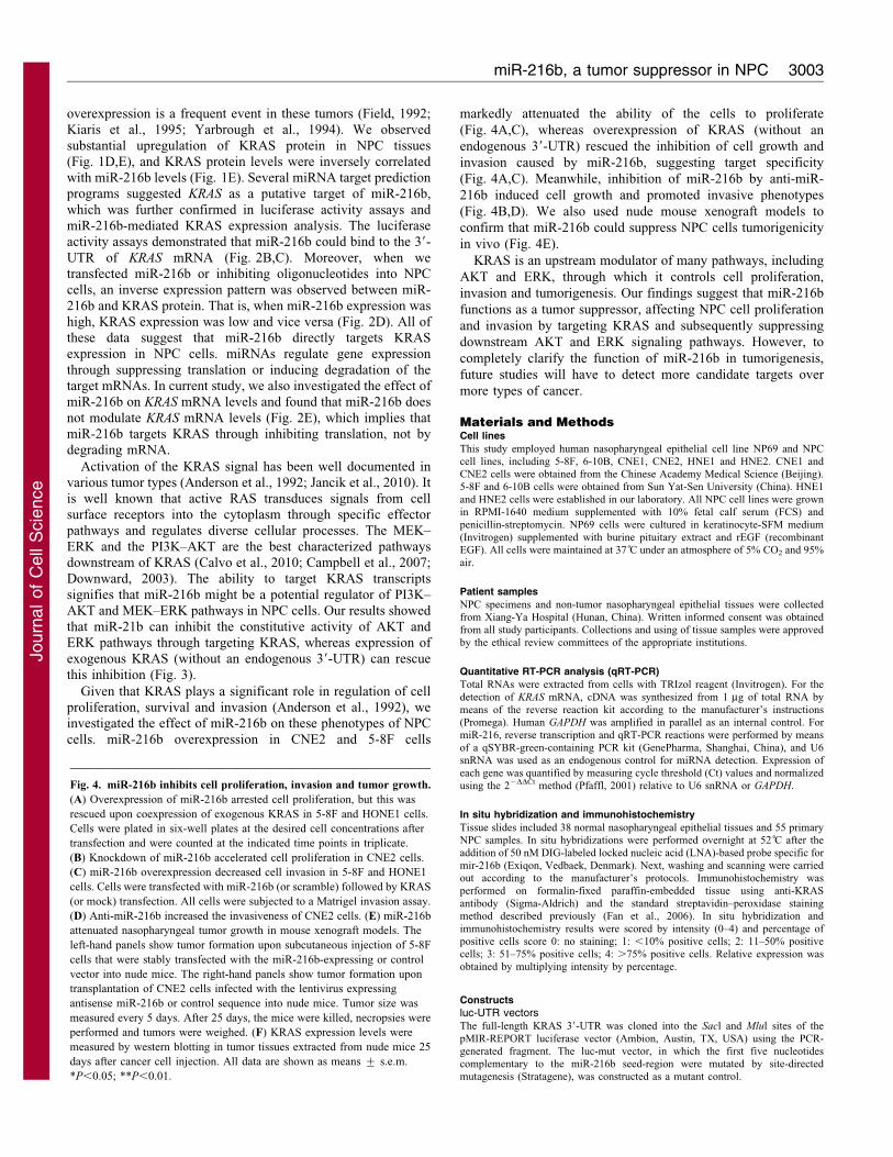
Fig. 4. See next page for legend.
Journal of Cell Science 124 (17)3002
Journ
alof
Cell
Scie
nce
overexpression is a frequent event in these tumors (Field, 1992;Kiaris et al., 1995; Yarbrough et al., 1994). We observed
substantial upregulation of KRAS protein in NPC tissues(Fig. 1D,E), and KRAS protein levels were inversely correlatedwith miR-216b levels (Fig. 1E). Several miRNA target predictionprograms suggested KRAS as a putative target of miR-216b,
which was further confirmed in luciferase activity assays andmiR-216b-mediated KRAS expression analysis. The luciferaseactivity assays demonstrated that miR-216b could bind to the 39-
UTR of KRAS mRNA (Fig. 2B,C). Moreover, when wetransfected miR-216b or inhibiting oligonucleotides into NPCcells, an inverse expression pattern was observed between miR-
216b and KRAS protein. That is, when miR-216b expression washigh, KRAS expression was low and vice versa (Fig. 2D). All ofthese data suggest that miR-216b directly targets KRASexpression in NPC cells. miRNAs regulate gene expression
through suppressing translation or inducing degradation of thetarget mRNAs. In current study, we also investigated the effect ofmiR-216b on KRAS mRNA levels and found that miR-216b does
not modulate KRAS mRNA levels (Fig. 2E), which implies thatmiR-216b targets KRAS through inhibiting translation, not bydegrading mRNA.
Activation of the KRAS signal has been well documented invarious tumor types (Anderson et al., 1992; Jancik et al., 2010). Itis well known that active RAS transduces signals from cellsurface receptors into the cytoplasm through specific effector
pathways and regulates diverse cellular processes. The MEK–ERK and the PI3K–AKT are the best characterized pathwaysdownstream of KRAS (Calvo et al., 2010; Campbell et al., 2007;
Downward, 2003). The ability to target KRAS transcriptssignifies that miR-216b might be a potential regulator of PI3K–AKT and MEK–ERK pathways in NPC cells. Our results showed
that miR-21b can inhibit the constitutive activity of AKT andERK pathways through targeting KRAS, whereas expression ofexogenous KRAS (without an endogenous 39-UTR) can rescue
this inhibition (Fig. 3).
Given that KRAS plays a significant role in regulation of cellproliferation, survival and invasion (Anderson et al., 1992), weinvestigated the effect of miR-216b on these phenotypes of NPC
cells. miR-216b overexpression in CNE2 and 5-8F cells
markedly attenuated the ability of the cells to proliferate
(Fig. 4A,C), whereas overexpression of KRAS (without an
endogenous 39-UTR) rescued the inhibition of cell growth and
invasion caused by miR-216b, suggesting target specificity
(Fig. 4A,C). Meanwhile, inhibition of miR-216b by anti-miR-
216b induced cell growth and promoted invasive phenotypes
(Fig. 4B,D). We also used nude mouse xenograft models to
confirm that miR-216b could suppress NPC cells tumorigenicity
in vivo (Fig. 4E).
KRAS is an upstream modulator of many pathways, including
AKT and ERK, through which it controls cell proliferation,
invasion and tumorigenesis. Our findings suggest that miR-216b
functions as a tumor suppressor, affecting NPC cell proliferation
and invasion by targeting KRAS and subsequently suppressing
downstream AKT and ERK signaling pathways. However, to
completely clarify the function of miR-216b in tumorigenesis,
future studies will have to detect more candidate targets over
more types of cancer.
Materials and MethodsCell lines
This study employed human nasopharyngeal epithelial cell line NP69 and NPC
cell lines, including 5-8F, 6-10B, CNE1, CNE2, HNE1 and HNE2. CNE1 andCNE2 cells were obtained from the Chinese Academy Medical Science (Beijing).
5-8F and 6-10B cells were obtained from Sun Yat-Sen University (China). HNE1
and HNE2 cells were established in our laboratory. All NPC cell lines were grownin RPMI-1640 medium supplemented with 10% fetal calf serum (FCS) and
penicillin-streptomycin. NP69 cells were cultured in keratinocyte-SFM medium
(Invitrogen) supplemented with burine pituitary extract and rEGF (recombinantEGF). All cells were maintained at 37 C̊ under an atmosphere of 5% CO2 and 95%
air.
Patient samples
NPC specimens and non-tumor nasopharyngeal epithelial tissues were collected
from Xiang-Ya Hospital (Hunan, China). Written informed consent was obtainedfrom all study participants. Collections and using of tissue samples were approved
by the ethical review committees of the appropriate institutions.
Quantitative RT-PCR analysis (qRT-PCR)
Total RNAs were extracted from cells with TRIzol reagent (Invitrogen). For the
detection of KRAS mRNA, cDNA was synthesized from 1 mg of total RNA by
means of the reverse reaction kit according to the manufacturer’s instructions(Promega). Human GAPDH was amplified in parallel as an internal control. For
miR-216, reverse transcription and qRT-PCR reactions were performed by means
of a qSYBR-green-containing PCR kit (GenePharma, Shanghai, China), and U6snRNA was used as an endogenous control for miRNA detection. Expression of
each gene was quantified by measuring cycle threshold (Ct) values and normalized
using the 22DDCt method (Pfaffl, 2001) relative to U6 snRNA or GAPDH.
In situ hybridization and immunohistochemistry
Tissue slides included 38 normal nasopharyngeal epithelial tissues and 55 primaryNPC samples. In situ hybridizations were performed overnight at 52 C̊ after the
addition of 50 nM DIG-labeled locked nucleic acid (LNA)-based probe specific for
mir-216b (Exiqon, Vedbaek, Denmark). Next, washing and scanning were carriedout according to the manufacturer’s protocols. Immunohistochemistry was
performed on formalin-fixed paraffin-embedded tissue using anti-KRAS
antibody (Sigma-Aldrich) and the standard streptavidin–peroxidase stainingmethod described previously (Fan et al., 2006). In situ hybridization and
immunohistochemistry results were scored by intensity (0–4) and percentage of
luc-UTR vectorsThe full-length KRAS 39-UTR was cloned into the Sacl and Mlul sites of the
pMIR-REPORT luciferase vector (Ambion, Austin, TX, USA) using the PCR-
generated fragment. The luc-mut vector, in which the first five nucleotidescomplementary to the miR-216b seed-region were mutated by site-directed
mutagenesis (Stratagene), was constructed as a mutant control.
Fig. 4. miR-216b inhibits cell proliferation, invasion and tumor growth.
(A) Overexpression of miR-216b arrested cell proliferation, but this was
rescued upon coexpression of exogenous KRAS in 5-8F and HONE1 cells.
Cells were plated in six-well plates at the desired cell concentrations after
transfection and were counted at the indicated time points in triplicate.
(B) Knockdown of miR-216b accelerated cell proliferation in CNE2 cells.
(C) miR-216b overexpression decreased cell invasion in 5-8F and HONE1
cells. Cells were transfected with miR-216b (or scramble) followed by KRAS
(or mock) transfection. All cells were subjected to a Matrigel invasion assay.
(D) Anti-miR-216b increased the invasiveness of CNE2 cells. (E) miR-216b
attenuated nasopharyngeal tumor growth in mouse xenograft models. The
left-hand panels show tumor formation upon subcutaneous injection of 5-8F
cells that were stably transfected with the miR-216b-expressing or control
vector into nude mice. The right-hand panels show tumor formation upon
transplantation of CNE2 cells infected with the lentivirus expressing
antisense miR-216b or control sequence into nude mice. Tumor size was
measured every 5 days. After 25 days, the mice were killed, necropsies were
performed and tumors were weighed. (F) KRAS expression levels were
measured by western blotting in tumor tissues extracted from nude mice 25
days after cancer cell injection. All data are shown as means ¡ s.e.m.
*P,0.05; **P,0.01.
miR-216b, a tumor suppressor in NPC 3003
Journ
alof
Cell
Scie
nce
Constructs for miR-216 overexpression and knockdownSynthesized RNA duplexes of scramble miRNA, miR-216a and miR-216b, andtheir inhibitors anti-scramble and anti-miR-216b were obtained from Ambion. Toconstruct a vector expressing miR-216b, the precursor sequence of miR-216b(MI0005569) was synthesized, annealed and then inserted into the BamHI–HindIIIfragment of the pGCsi/U6 vector (GeneChem, Shanghai, China). A constructincluding the nonspecific miRNA was used as a negative control. miR-216b-knockdown lentiviruses were purchased from SunBio (Shanghai, China).
KRAS-expressing vectorFull-length KRAS cDNA entirely lacking the 39-UTR was purchased fromGeneCopeia (Rockville, MD, USA) and subcloned into the eukaryotic expressionvector pcDNA3.1(+) (Invitrogen). The empty pcDNA3.1(+) vector was used as anegative control.
Cell transfection and infection
5-8F cells were transfected with the miR-216b-expressing vector or the control vectorexpressing a scrambled miRNA, using Lipofectamine 2000 (Invitrogen). All cellswere selected with 400 mg/l G418 to generate two stable monoclonal cell lines (astable cell line expressing miR-216b, 5-8F-miR-216b, and a control stable cell line, 5-8F-miR-scramble). In-vitro-produced miR-216a and miR-216b and the negativescramble control were transfected into 5-8F and HONE1 cells. The inhibitors anti-miR-216b and anti-scramble were also transfected into CNE2 and NP69 cells.
For miRNA and pcDNA3.1KRAS combination experiments, 5-8F and HONE1cells were transfected with pcDNA3.1-KRAS and empty vector 24 hours aftertransfection of the in-vitro-produced miR-216b. To establish stable miR-216b-knockdown cell lines, CNE2 cells were transduced with the miR-216b knockdownlentivirus or the control lentivirus and selected with 5 mg/l puromycin.
Luciferase assay
Luc-wt, Luc-mut and Luc-ctrl were co-transfected with in-vitro-produced miR-216a or miR-216b into 5-8F and HONE1 cells. In addition, Luc-wt was co-transfected with miR-216b inhibitor into CNE2 cells. The pMIR-REPORT b-galactosidase control vector was transfected as a control. Luciferase activity wasmeasured in cell lysates 48 hours after transfection using the Dual-Lightluminescent reporter gene assay kit (Applied Biosystems). Results werenormalized against b-galactosidase activity.
Western blotting
Western blotting was carried out as described previously (Zhou et al., 2008). Anti-KRAS antibody was obtained from Sigma-Aldrich. Antibodies against ERK,phosphorylated ERK, AKT, phosphorylated AKT, GSK3b, phosphorylatedGSK3b and c-Fos antibodies were obtained from Cell Signaling Technology.Anti-GAPDH antibody was from Santa Cruz Biotechnology.
Cell proliferation assay
After transfection, cells were plated into six-well plates at the desired cellconcentrations. Cell counts were estimated by trypsinizing the cells andperforming analysis in triplicate with a Coulter counter (Beckman Coulter,Fullerton, CA) at the indicated time points.
Cell invasion assay
At 48 hours after transfection, cells were seeded onto the basement membranematrix (EC matrix, Chemicon, Temecula, CA) present in the insert of a 24-wellculture plate. Fetal bovine serum was added to the lower chamber as achemoattractant. After a further 48 hours, the non-invading cells and EC matrixwere gently removed with a cotton swab. Invasive cells located on the lower sideof the chamber were stained with Crystal Violet, air-dried and photographed.
In vivo tumorigenesis
Five-week-old male nude athymic BALB/c nu/nu mice were used for examiningtumorigenicity. To evaluate the role of miR-216b in tumor formation, 5-8F cellsstably overexpressing miR-216 or scramble control were propagated andinoculated subcutaneously into the dorsal flanks of nude mice (26106 cells in0.2 ml volume). CNE2 cells infected with the miR-216b-knockdown lentivirus orthe control lentivirus were also transplanted into nude mice (1.56106 cells in0.2 ml volume). Tumor size was measured every 5 days. After 25 days, the micewere killed, necropsies were performed and the tumors were weighed. Tumorvolumes were determined according to the following formula: A6B2/2, where Ais the largest diameter and B is the diameter perpendicular to A. The experimentswere performed using five or six mice per group, and all animal procedures wereperformed in accordance with institutional guidelines.
Statistical analysis
Student’s unpaired t-tests were used to evaluate statistical significance.Spearman’s correlation tests were used to evaluate the pair-wise expression
correlation between miR-216b and KRAS. Data are expressed as means ¡ s.e.m.P,0.05 was considered statistically significant.
This work was supported by grants from the China National KeyScientific Technology Programs (2006CB910502), 111 Project (111-2-12), National 863 High Technology Program (2007AA02Z170),National Nature Scientific Foundation of China (30871282), andHunan Province Nature Scientific Foundation (08JJ3051). Theauthors declare they have no conflict of interest.
ReferencesAlmoguera, C., Shibata, D., Forrester, K., Martin, J., Arnheim, N. and Perucho, M.
(1988). Most human carcinomas of the exocrine pancreas contain mutant c-K-rasgenes. Cell 53, 549-554.
Anderson, M. W., Reynolds, S. H., You, M. and Maronpot, R. M. (1992). Role ofproto-oncogene activation in carcinogenesis. Environ. Health Perspect. 98, 13-24.
Bos, J. L. (1989). ras oncogenes in human cancer: a review. Cancer Res. 49, 4682-4689.
Burmer, G. C. and Loeb, L. A. (1989). Mutations in the KRAS2 oncogene duringprogressive stages of human colon carcinoma. Proc. Natl. Acad. Sci. USA 86, 2403-2407.
Calin, G. A. and Croce, C. M. (2006). MicroRNA signatures in human cancers. Nat.
Rev. Cancer 6, 857-866.
Calvo, F., Agudo-Ibanez, L. and Crespo, P. (2010).The Ras-ERK pathway:understanding site-specific signaling provides hope of new anti-tumor therapies.BioEssays 32, 412-421.
Campbell, P. M., Groehler, A. L., Lee, K. M., Ouellette, M. M., Khazak, V. and Der,
C. J. (2007). K-Ras promotes growth transformation and invasion of immortalizedhuman pancreatic cells by Raf and phosphatidylinositol 3-kinase signaling. Cancer
Res. 67, 2098-2106.
Deng, L., Jing, N., Tan, G., Zhou, M., Zhan, F., Xie, Y., Cao, L. and Li, G. (1998). Acommon region of allelic loss on chromosome region 3p25.3-26.3 in nasopharyngealcarcinoma. Genes Chromosomes Cancer 23, 21-25.
Doench, J. G. and Sharp, P. A. (2004). Specificity of microRNA target selection intranslational repression. Genes Dev. 18, 504-511.
Downward, J. (2003). Targeting RAS signalling pathways in cancer therapy. Nat. Rev.
Cancer 3, 11-22.
Esquela-Kerscher, A. and Slack, F. J. (2006). Oncomirs-microRNAs with a role incancer. Nat. Rev. Cancer 6, 259-269.
Fan, S. Q., Ma, J., Zhou, J., Xiong, W., Xiao, B. Y., Zhang, W. L., Tan, C., Li, X. L.,
Shen, S. R., Zhou, M. et al. (2006). Differential expression of Epstein-Barr virus-encoded RNA and several tumor-related genes in various types of nasopharyngealepithelial lesions and nasopharyngeal carcinoma using tissue microarray analysis.Hum. Pathol. 37, 593-605.
Field, J. K. (1992). Oncogenes and tumour-suppressor genes in squamous cellcarcinoma of the head and neck. Eur. J. Cancer B Oral Oncol. 28B, 67-76.
German, M. A., Pillay, M., Jeong, D. H., Hetawal, A., Luo, S., Janardhanan, P.,
Kannan, V., Rymarquis, L. A., Nobuta, K., German, R. et al. (2008). Globalidentification of microRNA-target RNA pairs by parallel analysis of RNA ends. Nat.
Biotechnol. 26, 941-946.
Jancik, S., Drabek, J., Radzioch, D. and Hajduch, M. (2010). Clinical relevance ofKRAS in human cancers. J. Biomed. Biotechnol. 2010, 150960.
Johnson, S. M., Grosshans, H., Shingara, J., Byrom, M., Jarvis, R., Cheng, A.,
Labourier, E., Reinert, K. L., Brown, D. and Slack, F. J. (2005). RAS is regulatedby the let-7 microRNA family. Cell 120, 635-647.
Kiaris, H., Spandidos, D. A., Jones, A. S., Vaughan, E. D. and Field, J. K. (1995).Mutations, expression and genomic instability of the H-ras proto-oncogene insquamous cell carcinomas of the head and neck. Br. J. Cancer 72, 123-128.
Lee, R. C., Feinbaum, R. L. and Ambros, V. (1993). The C. elegans heterochronicgene lin-4 encodes small RNAs with antisense complementarity to lin-14. Cell 75,843-854.
Lo, K. W., Cheung, S. T., Leung, S. F., van Hasselt, A., Tsang, Y. S., Mak, K. F.,Chung, Y. F., Woo, J. K., Lee, J. C. and Huang, D. P. (1996). Hypermethylation ofthe p16 gene in nasopharyngeal carcinoma. Cancer Res. 56, 2721-2725.
Pfaffl, M. W. (2001). A new mathematical model for relative quantification in real-timeRT-PCR. Nucleic Acids Res. 29, e45.
Pillai, R. S., Bhattacharyya, S. N. and Filipowicz, W. (2007). Repression of proteinsynthesis by miRNAs: how many mechanisms? Trends Cell Biol. 17, 118-126.
Qian, C. N., Guo, X., Cao, B., Kort, E. J., Lee, C. C., Chen, J., Wang, L. M., Mai,
W. Y., Min, H. Q., Hong, M. H. et al. (2002). Met protein expression level correlateswith survival in patients with late-stage nasopharyngeal carcinoma. Cancer Res. 62,589-596.
Reinhart, B. J., Slack, F. J., Basson, M., Pasquinelli, A. E., Bettinger, J. C., Rougvie,
A. E., Horvitz, H. R. and Ruvkun, G. (2000). The 21-nucleotide let-7 RNAregulates developmental timing in Caenorhabditis elegans. Nature 403, 901-906.
Ruvkun, G., Wightman, B. and Ha, I. (2004). The 20 years it took to recognize theimportance of tiny RNAs. Cell 116, S93-S96, 2 p.p. following S96.
Sarac, S., Akyol, M. U., Kanbur, B., Poyraz, A., Akyol, G., Yilmaz, T. and Sungur,A. (2001). Bcl-2 and LMP1 expression in nasopharyngeal carcinomas. Am. J.
Otolaryngol. 22, 377-382.Segura, M. F., Hanniford, D., Menendez, S., Reavie, L., Zou, X., Alvarez-Diaz, S.,
Zakrzewski, J., Blochin, E., Rose, A., Bogunovic, D. et al. (2009). Aberrant miR-182 expression promotes melanoma metastasis by repressing FOXO3 andmicrophthalmia-associated transcription factor. Proc. Natl. Acad. Sci. USA 106,1814-1819.
Sengupta, S., den Boon, J. A., Chen, I. H., Newton, M. A., Stanhope, S. A., Cheng,
Y. J., Chen, C. J., Hildesheim, A., Sugden, B. and Ahlquist, P. (2008). MicroRNA29c is down-regulated in nasopharyngeal carcinomas, up-regulating mRNAsencoding extracellular matrix proteins. Proc. Natl. Acad. Sci. USA 105, 5874-5878.
Shao, J. Y., Huang, X. M., Yu, X. J., Huang, L. X., Wu, Q. L., Xia, J. C., Wang,H. Y., Feng, Q. S., Ren, Z. F., Ernberg, I. et al. (2001). Loss of heterozygosity andits correlation with clinical outcome and Epstein-Barr virus infection in nasophar-yngeal carcinoma. Anticancer Res. 21, 3021-3029.
Sheu, L. F., Chen, A., Tseng, H. H., Leu, F. J., Lin, J. K., Ho, K. C. and Meng, C. L.
(1995). Assessment of p53 expression in nasopharyngeal carcinoma. Hum. Pathol. 26,380-386.
Slack, F. J. and Weidhaas, J. B. (2008). MicroRNA in cancer prognosis. N. Engl. J.
Med. 359, 2720-2722.Slamon, D. J., deKernion, J. B., Verma, I. M. and Cline, M. J. (1984). Expression of
cellular oncogenes in human malignancies. Science 224, 256-262.Takamizawa, J., Konishi, H., Yanagisawa, K., Tomida, S., Osada, H., Endoh, H.,
Harano, T., Yatabe, Y., Nagino, M., Nimura, Y. et al. (2004). Reduced expressionof the let-7 microRNAs in human lung cancers in association with shortenedpostoperative survival. Cancer Res. 64, 3753-3756.
Thor, A., Ohuchi, N., Hand, P. H., Callahan, R., Weeks, M. O., Theillet, C.,
Lidereau, R., Escot, C., Page, D. L., Vilasi, V. et al. (1986). ras gene alterations and
enhanced levels of ras p21 expression in a spectrum of benign and malignant human
mammary tissues. Lab. Invest. 55, 603-615.
Vasilatou, D., Papageorgiou, S., Pappa, V., Papageorgiou, E. and Dervenoulas, J.
(2010). The role of microRNAs in normal and malignant hematopoiesis. Eur. J.
Haematol. 84, 1-16.
Xiang, Y. N. and Zhang, W. Y. (2005). The clinical significance of p16 protein non-
expression and p16 gene inactivation by deletions and hypermethylation in
nasopharyngeal carcinoma. Zhonghua Bing Li Xue Za Zhi 34, 358-361.
Xiong, W., Zeng, Z. Y., Xia, J. H., Xia, K., Shen, S. R., Li, X. L., Hu, D. X., Tan, C.,
Xiang, J. J., Zhou, J. et al. (2004). A susceptibility locus at chromosome 3p21 linked
to familial nasopharyngeal carcinoma. Cancer Res. 64, 1972-1974.
Yarbrough, W. G., Shores, C., Witsell, D. L., Weissler, M. C., Fidler, M. E. and
Gilmer, T. M. (1994). ras mutations and expression in head and neck squamous cell
carcinomas. Laryngoscope 104, 1337-1347.
Yu, M. C. and Yuan, J. M. (2002). Epidemiology of nasopharyngeal carcinoma. Semin.
Cancer Biol. 12, 421-429.
Yu, S., Lu, Z., Liu, C., Meng, Y., Ma, Y., Zhao, W., Liu, J., Yu, J. and Chen, J.
(2010). miRNA-96 suppresses KRAS and functions as a tumor suppressor gene in
pancreatic cancer. Cancer Res. 70, 6015-6025.
Zhou, Y., Zeng, Z., Zhang, W., Xiong, W., Wu, M., Tan, Y., Yi, W., Xiao, L., Li, X.,
Huang, C. et al. (2008). Lactotransferrin: a candidate tumor suppressor-deficient
expression in human nasopharyngeal carcinoma and inhibition of NPC cell
proliferation by modulating the mitogen-activated protein kinase pathway. Int. J.