SPECIAL CONTRIBUTION MIRD Pamphlet No. 22 (Abridged): Radiobiology and Dosimetry of a-Particle Emitters for Targeted Radionuclide Therapy* George Sgouros 1 , John C. Roeske 2 , Michael R. McDevitt 3 , Stig Palm 4 , Barry J. Allen 5 , Darrell R. Fisher 6 , A. Bertrand Brill 7 , Hong Song 1 , Roger W. Howell 8 , and Gamal Akabani 9 In collaboration with the SNM MIRD Committee: Wesley E. Bolch, A. Bertrand Brill, Darrell R. Fisher, Roger W. Howell, Ruby F. Meredith, George Sgouros (Chair), Barry W. Wessels, and Pat B. Zanzonico 1 Department of Radiology and Radiological Science, Johns Hopkins University, Baltimore, Maryland; 2 Department of Radiation Oncology, Loyola University Medical Center, Maywood, Illinois; 3 Departments of Medicine and Radiology, Memorial Sloan- Kettering Cancer Center, New York, New York; 4 Dosimetry and Medical Radiation Physics Section, International Atomic Energy Agency, Vienna, Austria; 5 Centre for Experimental Radiation Oncology, St. George Cancer Centre, Kogarah, Australia; 6 Radioisotopes Program, Pacific Northwest National Laboratory, Richland, Washington; 7 Department of Radiology, Vanderbilt University, Nashville, Tennessee; 8 Division of Radiation Research, Department of Radiology, New Jersey Medical School Cancer Center, University of Medicine and Dentistry of New Jersey, Newark, New Jersey; and 9 Department of Nuclear Engineering, Texas A&M University, College Station, Texas The potential of a-particle emitters to treat cancer has been rec- ognized since the early 1900s. Advances in the targeted delivery of radionuclides and radionuclide conjugation chemistry, and the increased availability of a-emitters appropriate for clinical use, have recently led to patient trials of radiopharmaceuticals la- beled with a-particle emitters. Although a-emitters have been studied for many decades, their current use in humans for tar- geted therapy is an important milestone. The objective of this work is to review those aspects of the field that are pertinent to targeted a-particle emitter therapy and to provide guidance and recommendations for human a-particle emitter dosimetry. Key Words: a-particle emitters; human a-particle emitter dosim- etry; targeted a-particle emitter therapy J Nucl Med 2010; 51:311–328 DOI: 10.2967/jnumed.108.058651 Several reviews have been published on the topic of a-particle–emitting radionuclides, which have been the subject of considerable investigation as cancer therapeutics (1–8). In the context of targeted therapy, a-particle emitters have the advantages of high potency and specificity. These advantages arise from the densely ionizing track and short path length of the emitted positively charged helium nucleus in tissue. The practical implication of these features, as well as the distinction between a-particles and the more widely used b-particle emitters for targeted radionuclide therapy, is that it is possible to sterilize individual tumor cells solely from self-irradiation with a-particle emitters. This is gener- ally not possible with b-particle emitters given achievable antibody specific activity, tumor-cell antigen expression levels, and the need to avoid prohibitive toxicity (5). These attributes combine to provide the fundamental strength and rationale for using a-particle–emitting radionuclides for cancer therapy. Current approaches to cancer treatment are largely ineffective once the tumor has metastasized and tumor cells are disseminated throughout the body. There is also increasing evidence that not all tumor cells are relevant targets for effective tumor eradication and that sterilization of a putative subpopulation of a small number of tumor stem cells may be critical to treatment efficacy (9). The eradication of such disseminated tumor cells, or of a subpopulation of tumor stem cells, requires a systemic targeted therapy that is minimally susceptible to chemo- or radioresistance, is potent enough to sterilize individual tumor cells and microscopic tumor cell clusters (even at a low dose-rate and low oxygen tension), and exhibits an acceptable toxicity profile (10). a-Particle–emitting radionuclides address this critical need. To accomplish these goals, a reliable, cost-effective source of a-particle emitters is needed for research and development and for routine use in clinical practice. Improved chemical labeling and stability will be needed to achieve the desired biodistribution and associated dose distribution necessary for successful therapy with acceptable acute and long-term toxicities. These limitations have slowed the development and clinical use of a-emitter targeted therapy relative to the use of b- and Auger-electron–emitting radionuclides. Received Sep. 29, 2008; revision accepted Jun. 29, 2009. For correspondence or reprints contact: George Sgouros, Department of Radiology and Radiological Science, CRB II 4M61/1550 Orleans St., School of Medicine, Johns Hopkins University, Baltimore, MD 21231. E-mail: [email protected]*Unabridged version of this document is available at: http://interactive. snm.org/index.cfm?PageID51110&RPID52199&FileID5144234. COPYRIGHT ª 2010 by the Society of Nuclear Medicine, Inc. a-P ARTICLE EMITTER DOSIMETRY • Sgouros et al. 311
Transcript
S P E C I A L C O N T R I B U T I O N
MIRD Pamphlet No. 22 (Abridged):Radiobiology and Dosimetry of a-ParticleEmitters for Targeted Radionuclide Therapy*
George Sgouros1, John C. Roeske2, Michael R. McDevitt3, Stig Palm4, Barry J. Allen5, Darrell R. Fisher6,A. Bertrand Brill7, Hong Song1, Roger W. Howell8, and Gamal Akabani9
In collaboration with the SNM MIRD Committee: Wesley E. Bolch, A. Bertrand Brill, Darrell R. Fisher,Roger W. Howell, Ruby F. Meredith, George Sgouros (Chair), Barry W. Wessels, and Pat B. Zanzonico
1Department of Radiology and Radiological Science, Johns Hopkins University, Baltimore, Maryland; 2Department of RadiationOncology, Loyola University Medical Center, Maywood, Illinois; 3Departments of Medicine and Radiology, Memorial Sloan-Kettering Cancer Center, New York, New York; 4Dosimetry and Medical Radiation Physics Section, International Atomic EnergyAgency, Vienna, Austria; 5Centre for Experimental Radiation Oncology, St. George Cancer Centre, Kogarah, Australia;6Radioisotopes Program, Pacific Northwest National Laboratory, Richland, Washington; 7Department of Radiology, VanderbiltUniversity, Nashville, Tennessee; 8Division of Radiation Research, Department of Radiology, New Jersey Medical School CancerCenter, University of Medicine and Dentistry of New Jersey, Newark, New Jersey; and 9Department of Nuclear Engineering, TexasA&M University, College Station, Texas
The potential of a-particle emitters to treat cancer has been rec-ognized since the early 1900s. Advances in the targeted deliveryof radionuclides and radionuclide conjugation chemistry, and theincreased availability of a-emitters appropriate for clinical use,have recently led to patient trials of radiopharmaceuticals la-beled with a-particle emitters. Although a-emitters have beenstudied for many decades, their current use in humans for tar-geted therapy is an important milestone. The objective of thiswork is to review those aspects of the field that are pertinent totargeted a-particle emitter therapy and to provide guidanceand recommendations for human a-particle emitter dosimetry.
J Nucl Med 2010; 51:311–328DOI: 10.2967/jnumed.108.058651
Several reviews have been published on the topic ofa-particle–emitting radionuclides, which have been thesubject of considerable investigation as cancer therapeutics(1–8). In the context of targeted therapy, a-particle emittershave the advantages of high potency and specificity. Theseadvantages arise from the densely ionizing track and shortpath length of the emitted positively charged helium nucleusin tissue. The practical implication of these features, as wellas the distinction between a-particles and the more widely
used b-particle emitters for targeted radionuclide therapy, isthat it is possible to sterilize individual tumor cells solelyfrom self-irradiation with a-particle emitters. This is gener-ally not possible with b-particle emitters given achievableantibody specific activity, tumor-cell antigen expressionlevels, and the need to avoid prohibitive toxicity (5). Theseattributes combine to provide the fundamental strength andrationale for using a-particle–emitting radionuclides forcancer therapy. Current approaches to cancer treatment arelargely ineffective once the tumor has metastasized and tumorcells are disseminated throughout the body. There is alsoincreasing evidence that not all tumor cells are relevant targetsfor effective tumor eradication and that sterilization ofa putative subpopulation of a small number of tumor stemcells may be critical to treatment efficacy (9). The eradicationof such disseminated tumor cells, or of a subpopulation oftumor stem cells, requires a systemic targeted therapy that isminimally susceptible to chemo- or radioresistance, is potentenough to sterilize individual tumor cells and microscopictumor cell clusters (even at a low dose-rate and low oxygentension), and exhibits an acceptable toxicity profile (10).a-Particle–emitting radionuclides address this critical need.To accomplish these goals, a reliable, cost-effective source ofa-particle emitters is needed for research and developmentand for routine use in clinical practice. Improved chemicallabeling and stability will be needed to achieve the desiredbiodistribution and associated dose distribution necessary forsuccessful therapy with acceptable acute and long-termtoxicities. These limitations have slowed the developmentand clinical use of a-emitter targeted therapy relative to theuse of b- and Auger-electron–emitting radionuclides.
Received Sep. 29, 2008; revision accepted Jun. 29, 2009.For correspondence or reprints contact: George Sgouros, Department
of Radiology and Radiological Science, CRB II 4M61/1550 Orleans St.,School of Medicine, Johns Hopkins University, Baltimore, MD 21231.
E-mail: [email protected]*Unabridged version of this document is available at: http://interactive.
snm.org/index.cfm?PageID51110&RPID52199&FileID5144234.COPYRIGHT ª 2010 by the Society of Nuclear Medicine, Inc.
a-PARTICLE EMITTER DOSIMETRY • Sgouros et al. 311
The first clinical trial of an a-particle emitter in radio-labeled antibody therapy used 213Bi conjugated to theantileukemia antibody HuM195 and was reported in 1997(11,12), 4 years after 213Bi was first suggested for therapeuticuse (13). This trial was followed by a human trial of theantitenascin antibody 81C6 labeled with the a-emitter 211Atin patients with recurrent malignant glioma (14). In additionto these 2 antibody-based trials, a clinical trial of unconju-gated 223Ra against skeletal metastases in patients withbreast and prostate cancer was recently completed (15).More recently, a patient trial of 211At targeting ovariancarcinoma has been initiated (16). Future trials of a-emittersare anticipated using antibodies labeled with 211At or 213Biand directed against tumor neovasculature (17–19). Aconjugation methodology for 225Ac was recently described
(20), and a phase I trial of this radionuclide with theantileukemia antibody HuM195 in leukemia patients hasrecently been initiated (21). Table 1 summarizes clinicaltrials involving a-particle–emitting radiopharmaceuticals.
This report focuses on a-emitter dosimetry as it relates tohuman use in targeted therapy. A review of a-particleradiobiologic studies is provided with a focus on the radio-biology of a-emitters that are relevant to targeted therapy inhumans. Closely related to the radiobiology of a-emitters isthe concept of relative biological effectiveness (RBE), whichis also reviewed. The dosimetry of a-emitters has beenaddressed in a large number of publications. The criteria formicrodosimetry, the different approaches for performing suchcalculations, and selected studies in which such calculationshave been performed are briefly described.
TABLE 1. Summary of Recently Reported Clinical Trials Using a-Particle Emitters
Radionuclide
Delivery
vehicle
Type of
cancer Comments Reference211At Antitenascin
IgG
Glioblastoma
multiforme
Ongoing phase I trial using surgical
cavity injection of labeled antitenascinIgG; median survival of 60 wk; 2 patients
with recurrent glioblastoma multiforme
survived nearly 3 y
14
MX35 F(ab9)2 Ovarian carcinoma Ongoing phase I trial using MX35 F(ab9)2;bone marrow and peritoneal absorbed
doses of 0.08 and 8 mGy/MBq, respectively
16
213Bi Anti-CD33 IgG Myelogenous
leukemia (acuteor chronic)
Phase I completed with no toxicity, substantial
reduction in circulating and bone marrow blasts;phase I/II in cytoreduced patients, 4 of 23
patients at very high risk showed lasting
complete response (up to 12 mo)
11,21
Antineurokinin
receptor peptide
Glioblastoma Two patients treated with 213Bi; 1 with
oligodendroglioma treated by distillation in
resection cavity alive more than 67 mo
148
Anti-CD20 IgG(rituximab)
Relapsed orrefractory
non-Hodgkin
lymphoma
Phase I study with 9 patients treated to date 149
9.2.27 IgG Melanoma Sixteen patients; intralesional administration ledto massive tumor cell kill and resolution of
lesions; significant decline in serum marker
melanoma-inhibitory-activity protein at 2 wkafter treatment
150
223Ra RaCl2 Skeletal breast
and prostate
cancermetastases
Phase IA dose-escalation studies completed
involving single-dose infusion of 46–250 kBq/kg
in 25 patients with no dose-limiting hematologictoxicity; phase IB study in 6 patients to
evaluate repeated injections (2 or 5 fractions)
totaling up to 250 kBq/kg; phase II randomized
trial in 33 patients with metastatic breast orprostate cancer treated with external beam
plus saline or 4 times 50 kBq/kg dose of 223Ra
at 4-wk intervals; shows significant (266%)
decrease in bone alkaline phosphatase comparedwith placebo and 15 of 31 patients with
prostate-specific antigen decrease . 50% from
baseline vs. 5 of 28 in control group
151,152
225Ac Anti-CD33 IgG Acute
myelogenous
leukemia
Phase I trial, ongoing, at first dose-level of
18.5 kBq/kg (0.5 mCi/kg); 1 of 2 patients had
elimination of peripheral blasts and reduction
in marrow blasts
21
312 THE JOURNAL OF NUCLEAR MEDICINE • Vol. 51 • No. 2 • February 2010
Therapeutic nuclear medicine is already a highly multi-disciplinary field. Therapy with a-particle emitters is easilyone of the more multidisciplinary endeavors within thisenterprise. This review is intended to provide the necessarybackground including the physics and dosimetry perspec-tive to aid in the design, conduct, and analysis of clinicaltrials using a-emitting radiotherapeutics.
a-PARTICLE RADIOBIOLOGY
Interest in a-particle–emitting radionuclides for cancertherapy is driven by the physical and radiobiologic prop-erties of a-particles as compared with those of photons andelectrons (Fig. 1). The energy deposited along the path ofan a-particle per unit path length is shown in Figure 2. Asshown in the figure, the energy deposition along the path, or
linear energy transfer (LET), of an a-particle can be 2 or 3orders of magnitude greater than the LET of b-particlesemitted by radionuclides such as 131I and 90Y.
One of the first studies demonstrating the biologic effectsof heavy charged particles was by Raymond Zirkle in 1932(22). He examined the effect of polonium a-particles oncell division in fern spores and showed a much greaterbiologic effect when the spore nucleus was placed in theBragg peak of the a-particle track, compared with theplateau region of the track (23). Much of the subsequentradiobiology of a-particles was established in a series ofseminal studies performed by Barendsen et al. in the 1960s(24–32). These studies first demonstrated the now familiarand accepted features of a-particle irradiation. A sub-sequent series of studies on the mutation and inactivation
FIGURE 1. Illustration of difference inionization density between low- andhigh-LET tracks. (Reprinted with per-mission of (153).)
FIGURE 2. LET vs. distance traveledin tissue for a-particles with 2 differentinitial kinetic energies. a-Particles emit-ted with lower initial energy are closer totheir Bragg peak and, therefore, startout with higher LET. LET of electronswith initial energy of 100–500 keV is alsoshown at bottom of plot for comparison.(Plot generated using data from (108).)
a-PARTICLE EMITTER DOSIMETRY • Sgouros et al. 313
of 3 different mammalian cell types exposed to helium,boron, or nitrogen ions spanning LET values in the range of20–470 keV�mm21 was key in evaluating the variousbiophysical models that had been posited to explain low-versus high-LET effects (33–36). The work was alsoinstrumental in providing both the experimental resultsand the biophysical analysis to help understand the RBE-versus-LET relationship established by Barendsen. Thebiophysical analysis in the last paper of the series (33)provided compelling theoretic support for the concept of 2types of radiation-induced cellular inactivation. The firsttype is that due to the accumulation of multiple events thatcan be repaired at low doses (i.e., sublethal damage) butthat saturate the cellular repair mechanisms at higher doses.This type of inactivation yields the characteristic linear-quadratic dose–response curve for low-LET radiation,corresponding to a small number, approximately 3–9 (i.e.,;100–300 eV) ionizations in a distance of about 3 nmassociated with a low probability of producing lethallesions. The second type of inactivation arises from a singlelethal event for high-LET radiation. In this case, a largernumber of ionizations, more than 10, over the 3-nmdistance depositing more than 300 eV produces lethallesions with a high probability. It is important, however,to remember that these studies were performed usingexternal beams of a-particles in which the incident a-par-ticles were generally orthogonal to an a-permeable surfaceon which the cells were cultured as a monolayer ofadherent cells.
As initially demonstrated experimentally by Fisheret al. (37), and then theoretically by Humm et al. (38),and most recently by Kvinnsland et al. (39), the spatialdistribution of a-particle emitters has an important impacton the absorbed dose distribution and, correspondingly, onthe slope of the cell-survival curve. Neti and Howellrecently provided experimental evidence of a lognormalcellular uptake of 210Po citrate among a cell populationuniformly exposed to the radiochemical and showed thatthis distribution can substantially alter the cell survivalcurve (40). Although many of the results obtained fromthe external-beam studies (summarized in Table 2) aregenerally applicable regardless of the a-particle distribu-tion, specific parameters such as the average number ofa-particle traversals to induce a lethal event or the D0
value (i.e., the absorbed dose required to reduce cellsurvival to 0.37) are highly sensitive to experimentalfactors such as the geometry of the cells, the thicknessor diameter of the cell nucleus, the distribution of DNAwithin the nucleus (i.e., the phase of the cell cycle), andthe number and spatial distribution of the a-particlesources relative to the target nuclei.
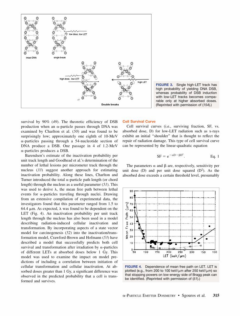
The distinction between DNA double-strand breaks(DSBs) caused by a single high-LET track versus DNAdamage caused by multiple low-LET tracks is illustrated inFigure 3. This basic observation underpins almost all theradiobiology of a-particles.
Traversals Required for Cell Kill
The average number of a-particle nuclear traversalsrequired to kill a cell, as measured by loss of thesubsequent ability to form a colony, ranges from as lowas 1 (41) to as high as 20 (42). If bystander effects areincluded, the lower end of the range would include 0.The variability in this value when bystander effects arenot considered arises because of the high sensitivity ofthis determination to the geometry of the cell andthe nucleus during irradiation and also the LET of theincident a-particles and the LET distribution within thenucleus.
Quoting from a publication of Raju et al. (43), ‘‘Thenotion that a cell will be inactivated by the passage ofa single a particle through a cell nucleus prevailed untilLloyd and her associates (42) demonstrated that 10 to 205.6 MeV a particles were required to induce one lethallesion in flattened C3H 10T1/2 cells. Studies by Bird,et al. (44) showed that approximately four 3He ions wererequired to pass through the cell nucleus to induce onelethal lesion in V79 cells at the G1/S-phase border, cellsin late S phase required five to eight 3He ions. Todd, et al.(45) investigated the effect of 3.5 MeV a particles onsynchronized T-1 cells, and observed that approximatelyone a particle out of four to five traversing a cell nucleusis effective in inducing one lethal lesion. Roberts andGoodhead (46) estimated that one out of six 3.2 MeVa-particle traversals through a C3H 10T1/2 cell nucleus islethal. Barendsen (47) concluded that the probability ofinactivation per unit track length of high-LET a particlesis approximately 0.08 mm21 for both T-1 and C3H 10T1/2 cells consistent with the results of Roberts and Good-head for C3H10T1/2 cells (46).’’ In a study comparinghigh-LET effects of Auger versus a-particle emitters,Howell et al. found that about 9 decays of 210Po wererequired to reduce cell survival to 37% (D0) when it wasdistributed between the cytoplasm and nucleus of V79cells; the energy deposited in the cell nucleus correspondsto about 2 complete (maximum chord length) traversals ofthe cell nucleus (48). In a murine lymphoma cell line,approximately 25 cell-bound a-particle–emitting 212Bimmunoconjugates were required to reduce clonogenic
TABLE 2. a-Particle Beam Findings That Are AlsoApplicable to Internally Administered a-Particle Emitters
No. Finding
1 RBE . 1 for cell sterilization, chromosomal damage/
cancer induction relative to low-LET radiation2 Reduced susceptibility to modulation by
radiosensitizers and radioprotectors
3 Reduced capacity to repair sublethal damage
4 Higher induction of DNA DSBs at low totalabsorbed doses
5 Monoexponential surviving fraction curve after
uniform irradiation (absence of a shoulder)
314 THE JOURNAL OF NUCLEAR MEDICINE • Vol. 51 • No. 2 • February 2010
survival by 90% (49). The theoretic efficiency of DSBproduction when an a-particle passes through DNA wasexamined by Charlton et al. (50) and was found to besurprisingly low; approximately one eighth of 10-MeVa-particles passing through a 54-nucleotide section ofDNA produce a DSB. One passage in 4 of 1.2-MeVa-particles produces a DSB.
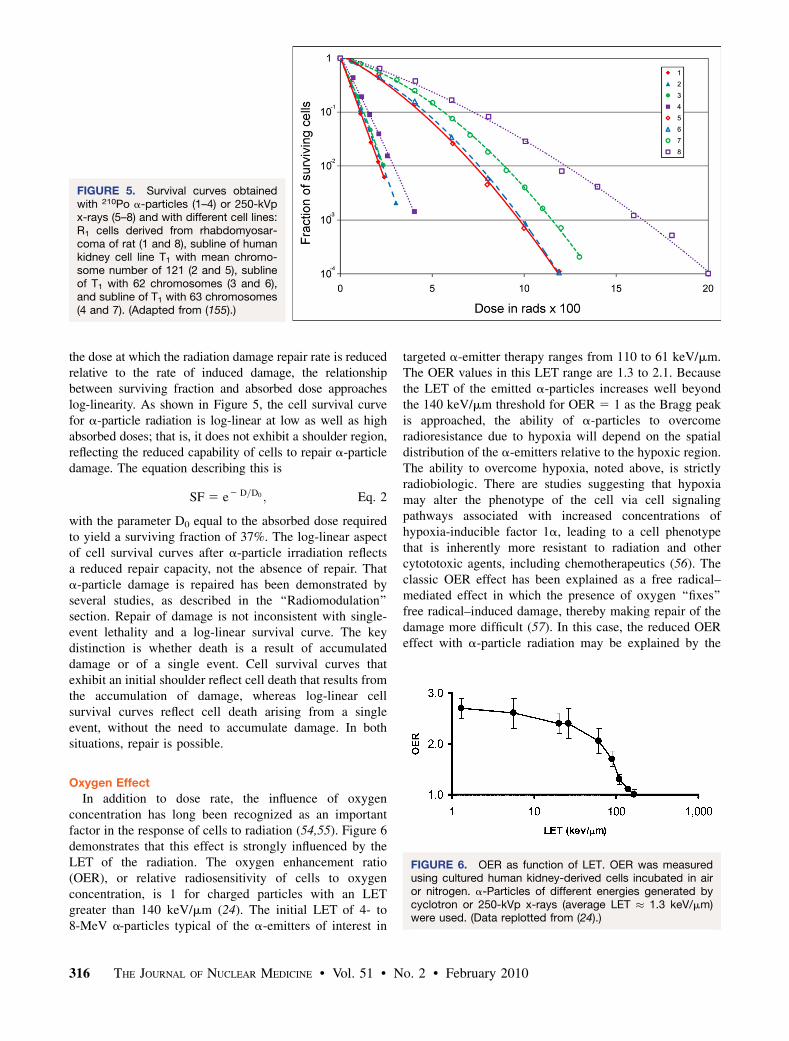
Barendsen’s estimate of the inactivation probability perunit track length and Goodhead et al.’s determination of thenumber of lethal lesions per micrometer track through thenucleus (33) suggest another approach for estimatinginactivation probability. Along these lines, Charlton andTurner introduced the total a-particle path length (or chordlength) through the nucleus as a useful parameter (51). Thiswas used to derive l, the mean free path between lethalevents for a-particles traveling through nuclei. Drawingfrom an extensive compilation of experimental data, theinvestigators found that this parameter ranged from 1.5 to64.4 mm. As expected, l was found to be dependent on theLET (Fig. 4). An inactivation probability per unit tracklength through the nucleus has also been used in a modeldescribing radiation-induced cellular inactivation andtransformation. By incorporating aspects of a state vectormodel for carcinogenesis (52) into the inactivation/trans-formation model, Crawford-Brown and Hofmann (53) havedescribed a model that successfully predicts both cellsurvival and transformation after irradiation by a-particlesof different LETs at absorbed doses below 1 Gy. Thismodel was used to examine the impact on model pre-dictions of including a correlation between initiation ofcellular transformation and cellular inactivation. At ab-sorbed doses greater than 1 Gy, a significant difference wasobserved in the predicted probability that a cell is trans-formed and survives.
Cell Survival Curve
Cell survival curves (i.e., surviving fraction, SF, vs.absorbed dose, D) for low-LET radiation such as x-raysexhibit an initial ‘‘shoulder’’ that is thought to reflect therepair of radiation damage. This type of cell survival curvecan be represented by the linear-quadratic equation
SF 5 e2aD2bD2
: Eq. 1
The parameters a and b are, respectively, sensitivity perunit dose (D) and per unit dose squared (D2). As theabsorbed dose exceeds a certain threshold level, presumably
FIGURE 3. Single high-LET track hashigh probability of yielding DNA DSB,whereas probability of DSB inductionwith low-LET tracks becomes compa-rable only at higher absorbed doses.(Reprinted with permission of (154).)
FIGURE 4. Dependence of mean free path on LET. LET isplotted (e.g., from 200 to 100 keV/mm after 250 keV/mm) sothat stopping powers on low-energy side of Bragg peak canbe identified. (Reprinted with permission of (51).)
a-PARTICLE EMITTER DOSIMETRY • Sgouros et al. 315
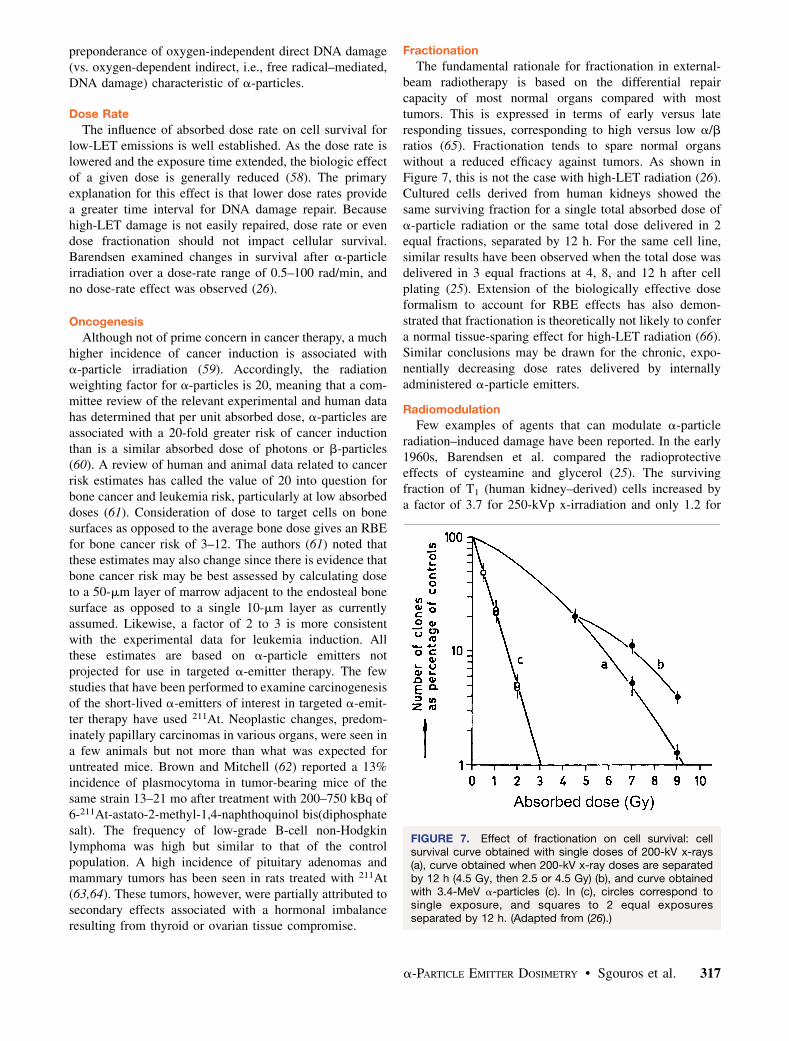
the dose at which the radiation damage repair rate is reducedrelative to the rate of induced damage, the relationshipbetween surviving fraction and absorbed dose approacheslog-linearity. As shown in Figure 5, the cell survival curvefor a-particle radiation is log-linear at low as well as highabsorbed doses; that is, it does not exhibit a shoulder region,reflecting the reduced capability of cells to repair a-particledamage. The equation describing this is
SF 5 e2 D=D0 ; Eq. 2
with the parameter D0 equal to the absorbed dose requiredto yield a surviving fraction of 37%. The log-linear aspectof cell survival curves after a-particle irradiation reflectsa reduced repair capacity, not the absence of repair. Thata-particle damage is repaired has been demonstrated byseveral studies, as described in the ‘‘Radiomodulation’’section. Repair of damage is not inconsistent with single-event lethality and a log-linear survival curve. The keydistinction is whether death is a result of accumulateddamage or of a single event. Cell survival curves thatexhibit an initial shoulder reflect cell death that results fromthe accumulation of damage, whereas log-linear cellsurvival curves reflect cell death arising from a singleevent, without the need to accumulate damage. In bothsituations, repair is possible.
Oxygen Effect
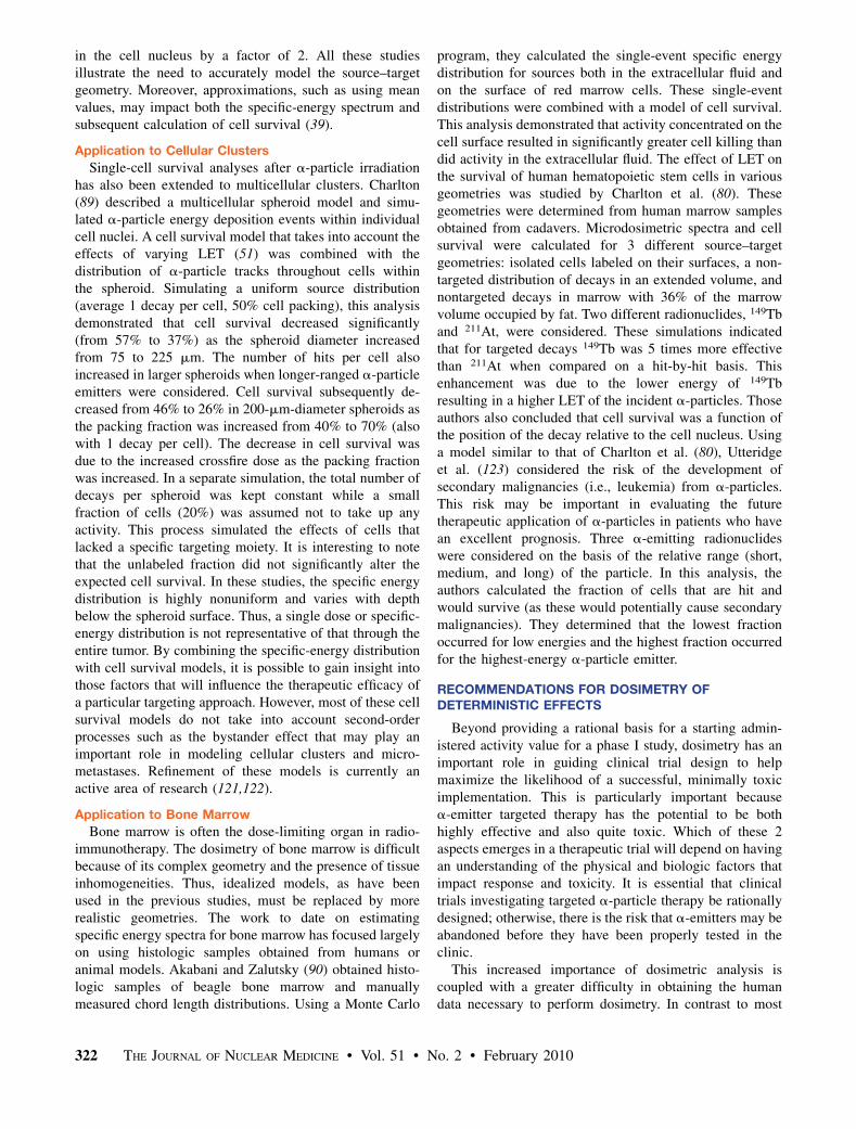
In addition to dose rate, the influence of oxygenconcentration has long been recognized as an importantfactor in the response of cells to radiation (54,55). Figure 6demonstrates that this effect is strongly influenced by theLET of the radiation. The oxygen enhancement ratio(OER), or relative radiosensitivity of cells to oxygenconcentration, is 1 for charged particles with an LETgreater than 140 keV/mm (24). The initial LET of 4- to8-MeV a-particles typical of the a-emitters of interest in
targeted a-emitter therapy ranges from 110 to 61 keV/mm.The OER values in this LET range are 1.3 to 2.1. Becausethe LET of the emitted a-particles increases well beyondthe 140 keV/mm threshold for OER 5 1 as the Bragg peakis approached, the ability of a-particles to overcomeradioresistance due to hypoxia will depend on the spatialdistribution of the a-emitters relative to the hypoxic region.The ability to overcome hypoxia, noted above, is strictlyradiobiologic. There are studies suggesting that hypoxiamay alter the phenotype of the cell via cell signalingpathways associated with increased concentrations ofhypoxia-inducible factor 1a, leading to a cell phenotypethat is inherently more resistant to radiation and othercytototoxic agents, including chemotherapeutics (56). Theclassic OER effect has been explained as a free radical–mediated effect in which the presence of oxygen ‘‘fixes’’free radical–induced damage, thereby making repair of thedamage more difficult (57). In this case, the reduced OEReffect with a-particle radiation may be explained by the
FIGURE 5. Survival curves obtainedwith 210Po a-particles (1–4) or 250-kVpx-rays (5–8) and with different cell lines:R1 cells derived from rhabdomyosar-coma of rat (1 and 8), subline of humankidney cell line T1 with mean chromo-some number of 121 (2 and 5), sublineof T1 with 62 chromosomes (3 and 6),and subline of T1 with 63 chromosomes(4 and 7). (Adapted from (155).)
FIGURE 6. OER as function of LET. OER was measuredusing cultured human kidney-derived cells incubated in airor nitrogen. a-Particles of different energies generated bycyclotron or 250-kVp x-rays (average LET � 1.3 keV/mm)were used. (Data replotted from (24).)
316 THE JOURNAL OF NUCLEAR MEDICINE • Vol. 51 • No. 2 • February 2010
preponderance of oxygen-independent direct DNA damage(vs. oxygen-dependent indirect, i.e., free radical–mediated,DNA damage) characteristic of a-particles.
Dose Rate
The influence of absorbed dose rate on cell survival forlow-LET emissions is well established. As the dose rate islowered and the exposure time extended, the biologic effectof a given dose is generally reduced (58). The primaryexplanation for this effect is that lower dose rates providea greater time interval for DNA damage repair. Becausehigh-LET damage is not easily repaired, dose rate or evendose fractionation should not impact cellular survival.Barendsen examined changes in survival after a-particleirradiation over a dose-rate range of 0.5–100 rad/min, andno dose-rate effect was observed (26).
Oncogenesis
Although not of prime concern in cancer therapy, a muchhigher incidence of cancer induction is associated witha-particle irradiation (59). Accordingly, the radiationweighting factor for a-particles is 20, meaning that a com-mittee review of the relevant experimental and human datahas determined that per unit absorbed dose, a-particles areassociated with a 20-fold greater risk of cancer inductionthan is a similar absorbed dose of photons or b-particles(60). A review of human and animal data related to cancerrisk estimates has called the value of 20 into question forbone cancer and leukemia risk, particularly at low absorbeddoses (61). Consideration of dose to target cells on bonesurfaces as opposed to the average bone dose gives an RBEfor bone cancer risk of 3–12. The authors (61) noted thatthese estimates may also change since there is evidence thatbone cancer risk may be best assessed by calculating doseto a 50-mm layer of marrow adjacent to the endosteal bonesurface as opposed to a single 10-mm layer as currentlyassumed. Likewise, a factor of 2 to 3 is more consistentwith the experimental data for leukemia induction. Allthese estimates are based on a-particle emitters notprojected for use in targeted a-emitter therapy. The fewstudies that have been performed to examine carcinogenesisof the short-lived a-emitters of interest in targeted a-emit-ter therapy have used 211At. Neoplastic changes, predom-inately papillary carcinomas in various organs, were seen ina few animals but not more than what was expected foruntreated mice. Brown and Mitchell (62) reported a 13%incidence of plasmocytoma in tumor-bearing mice of thesame strain 13–21 mo after treatment with 200–750 kBq of6-211At-astato-2-methyl-1,4-naphthoquinol bis(diphosphatesalt). The frequency of low-grade B-cell non-Hodgkinlymphoma was high but similar to that of the controlpopulation. A high incidence of pituitary adenomas andmammary tumors has been seen in rats treated with 211At(63,64). These tumors, however, were partially attributed tosecondary effects associated with a hormonal imbalanceresulting from thyroid or ovarian tissue compromise.
Fractionation
The fundamental rationale for fractionation in external-beam radiotherapy is based on the differential repaircapacity of most normal organs compared with mosttumors. This is expressed in terms of early versus lateresponding tissues, corresponding to high versus low a/bratios (65). Fractionation tends to spare normal organswithout a reduced efficacy against tumors. As shown inFigure 7, this is not the case with high-LET radiation (26).Cultured cells derived from human kidneys showed thesame surviving fraction for a single total absorbed dose ofa-particle radiation or the same total dose delivered in 2equal fractions, separated by 12 h. For the same cell line,similar results have been observed when the total dose wasdelivered in 3 equal fractions at 4, 8, and 12 h after cellplating (25). Extension of the biologically effective doseformalism to account for RBE effects has also demon-strated that fractionation is theoretically not likely to confera normal tissue-sparing effect for high-LET radiation (66).Similar conclusions may be drawn for the chronic, expo-nentially decreasing dose rates delivered by internallyadministered a-particle emitters.
Radiomodulation
Few examples of agents that can modulate a-particleradiation–induced damage have been reported. In the early1960s, Barendsen et al. compared the radioprotectiveeffects of cysteamine and glycerol (25). The survivingfraction of T1 (human kidney–derived) cells increased bya factor of 3.7 for 250-kVp x-irradiation and only 1.2 for
FIGURE 7. Effect of fractionation on cell survival: cellsurvival curve obtained with single doses of 200-kV x-rays(a), curve obtained when 200-kV x-ray doses are separatedby 12 h (4.5 Gy, then 2.5 or 4.5 Gy) (b), and curve obtainedwith 3.4-MeV a-particles (c). In (c), circles correspond tosingle exposure, and squares to 2 equal exposuresseparated by 12 h. (Adapted from (26).)
a-PARTICLE EMITTER DOSIMETRY • Sgouros et al. 317
210Po a-particle radiation. Similar results were observedwith glycerol; cell survival was increased by 2.0 and 1.2 for250-kVp x-rays and 210Po a-particles, respectively. Qual-itatively consistent but quantitatively different results havebeen obtained with the radiosensitizer Wortmannin. Thisirreversible and potent inhibitor of DNA-dependent proteinkinase is involved in the nonhomologous end-joining DNArepair pathway invoked in the repair of DNA DSBs (67).In V79 Chinese hamster cells, Wortmannin led to a 3- to4-fold increase in genotoxic damage, as measured by theinduction of micronuclei. High-LET irradiation, as deliv-ered by a boron neutron-capture reaction, leading to therelease of a-particles with an average energy of 2.3 MeV,yielded an increase in micronucleus induction of approx-imately 2-fold. This finding suggests that the more complexdouble-strand damage induced by high-LET radiation isa substrate of the nonhomologous end-joining pathway(68,69). In vivo studies in mouse testes have shown thatsoybean oil, S-(2-aminoethyl)isothiuronium bromide hy-drobromide, and cysteamine afford some protection againstthe cytotoxic effects of 5.3-MeV a-particles emitted by210Po (70–73). When spermatogonial cell survival was usedas the biologic endpoint, dose modification factors of 2.2,2.4, and 2.6, respectively, were obtained. No modificationof the spermatogonial response to a-particles was observedwhen dimethyl sulfoxide or vitamin C was used (74,75).
That DNA damage and its repair are at the core ofa-emitter radiobiologic effects is supported by many yearsof experimental and theoretic work. It is important,however, to keep in mind that all the foundation workregarding the radiobiology of a-emitters was performedwell before modern molecular biology came into existence.In light of the remarkable and far-reaching gains in ourunderstanding of the molecular mechanisms involved incancer genesis, the cellular response to radiation, and DNAsingle and DSB repair, a reexamination of a-particleradiobiology using modern tools is warranted.
RBE
The biologic effect of ionizing radiation is influenced bythe absorbed dose, the dose rate, and the quality ofradiation. Radiation quality is characterized by the spatialdistribution of the energy imparted and by the density ofionizations per unit path length, referred to as the LET orstopping power of a charged particle (22,60). Depending onthe effect considered, greater ionization density alonga track will increase the probability of inducing a biologiceffect. Compared with electrons and b-particles, a-particlesexhibit a high density of ionization events along their track(76). Electrons and b-particles that are emitted by radionu-clides generally range in energy from several megaelectronvolts to as low as several kiloelectron volts, with corre-sponding LET values ranging from about 0.1 to 1 keV/mm(b-particles actually are characterized by a spectrum ofenergies; the bottom end of the spectrum is zero). Theexception to these is Auger electrons, which have energies
as low as several electron volts and corresponding LETvalues as high as 25 keV/mm. a-Particles emitted byradionuclides range in energy from 2 to 10 MeV, withinitial LET values ranging from 60 to 110 keV/mm. A giventissue-absorbed dose resulting from a-particles, therefore,is likely to yield considerably greater biologic effects(again depending on the effect being considered) than thesame absorbed dose delivered by typical electrons orb-particles. To account for differences in energy depositionpattern exhibited by different quality radiations, the conceptof RBE has been established. An authoritative review ofthis concept, its derivation, and appropriate application hasbeen published by the International Commission on Radio-logical Protection (ICRP) (60,77), and the reader is en-couraged to consult this source for additional information.In radiobiology, RBE equals the ratio of absorbed doses of2 types of radiation that produce the same specifiedbiologic effect.
RBE Defined
RBE is calculated as the absorbed dose of a referenceradiation (e.g., x-rays, g-rays, b-particles), Dr(x), requiredto produce a biologic effect, x, divided by the absorbeddose of the test radiation, Dt(x), required to produce thesame biologic effect:
RBEðxÞ 5DrðxÞDtðxÞ
: Eq. 3
RBE is thus an experimentally determined value definedfor a particular biologic effect and therefore for a particularbiologic system.
The experimentally determined value can be influencedby the variability of the biologic system across differentlaboratories. This issue has been examined for studies invitro (78). The methodology used for calculating theabsorbed dose of the 2 radiation types will also impactthe result. Ideally, this should not be the case. Themethodology used should provide the true absorbed dosevalue or specific energy distribution (‘‘Case for Micro-dosimetry’’ section) to the relevant biologic target for boththe test and the reference radiations. In practice, however,this is a challenge even for studies in vitro (79). In thesetting of human a-particle emitter dosimetry, consistencyand reproducibility will be as important as accuracy. Thisissue is discussed in greater detail in ‘‘Recommendationsfor Dosimetry of Deterministic Effects’’ section.
The fact that the RBE is related to the pattern of ionizingenergy deposition along a particle track leads to a thirdfactor that will impact the results. The RBE for a particularradiation type will also depend on the initial emissionenergy of the particle (i.e., how close the particle is to theend of its track [the Bragg peak]). This factor has beenexamined by Charlton et al. (80) and Howell et al. (81). Inthe studies by Howell et al., a uniform distribution ofdecays was assumed to calculate the D0 for 7 a-emitting
318 THE JOURNAL OF NUCLEAR MEDICINE • Vol. 51 • No. 2 • February 2010
isotopes covering a wide range of initial energies. Using theD0 obtained for x-rays for the cell line used in the a-emittercalculations, a linear relationship between RBE and initiala-particle energy was obtained over an initial a-energyranging from 5 to 8.5 MeV. The straight line was given byRBE 5 2.9–0.167Ei, where Ei is the initial a-particleenergy in megaelectron volts. This is an approximatescaling of the equation derived from in vivo experimentaldata by Howell et al. (81). In addition to effects related tothe Bragg peak, nonuniform biodistribution of the a-emit-ters also leads to microdosimetric effects that impact RBEand the slope of the cell-survival curve (37–39).
If the reference radiation yields a dose–response re-lationship that is not log-linear for the biologic systemexamined, the RBE value will depend on the specificbiologic quantitative endpoint selected (e.g., D50, D37 (5D0), D10, etc., which determines whether the comparisonfalls in the shoulder or in the log-linear region of a dose–response or survival curve). The type of biologic endpoint(e.g., survival, mutation) and the dose rates of the test andreference radiations will also influence the RBE value.Strictly speaking, the test radiation should be delivered ina manner identical to that of the reference radiation (e.g.,chronic or acute). However, acute externally administeredx- and g-rays are often used as the reference radiation whenRBE values are determined for internally administeredradionuclides. Given the often-sizeable difference in bi-ologic responses to acute-versus-chronic low-LET radia-tion, the dose rate at which the reference radiation isdelivered can impact the resulting RBE (48). The dose-ratepattern delivered by radiopharmaceuticals is generally wellrepresented by multicomponent exponential functions.Howell et al. have delivered such patterns with externalbeams of 137Cs g-rays (82). This approach was used tostudy the bone marrow response to exponentially decreas-ing dose rates of 137Cs g-rays (83). The response ofgranulocyte–macrophage colony-forming cells in the mar-row to decreasing dose rates with half-times ranging from62 h to N (i.e., constant dose rate) were studied andcompared with the response to acute exposures. Meanlethal doses for chronic irradiation were up to 40% higherthan those for acute exposures. Thus, care must be takenwhen comparing RBE values based on different referenceradiations.
Based on a review of experimental literature, an RBEvalue of between 3 and 5 was recommended for cell killingby a panel convened by the Department of Energy in 1996(84). Because human studies using a-particle emitters haveyet to be analyzed for deterministic effects, an RBE of 5was recommended for projecting the possible deterministicbiologic effects associated with an estimated a-particleabsorbed dose.
RBE, Q, and wR
The discussion thus far has focused on RBE. RBE isoccasionally confused with quality factors, Q, and radiation
weighting factors, wR. This confusion reflects the historicalevolution of RBE which was originally defined as relativebiological efficiency and intended to apply to both radio-biology (deterministic effects) and protection (stochasticeffects). As currently recommended by the ICRP, however,RBE is not to be used directly in radiation protection butonly as a starting quantity to derive the radiation weightingfactor wR, which replaced the quality factor Q in the mostrecent ICRP recommendations (85,86). The RBE valuesused to arrive at wR relate to stochastic endpoints such ascancer induction, rather than deterministic endpoints suchas normal-tissue toxicity and tumor cell sterilization incancer therapy patients. The ICRP radiation weightingfactor for a-particles is 20. This value, intended only forstochastic effects caused by a-particle irradiation, is basedon animal experiments and from analysis of historicala-emitter exposures. In contrast to RBE values, weightingfactors are not directly measured values but rather areconsensus recommendations of the ICRP (60).
The radiation weighting factor wR is a unitless factor thatconverts average absorbed dose (in units of grays) toequivalent dose in an organ or tissue. The SI unit forequivalent dose is referred to by the special name sievert.The sievert is not a unit in the conventional sense but isintended to indicate that the absorbed dose value has beenadjusted to reflect a biologic risk that is associated withstochastic effects. Although the sievert is often used in thecontext of deterministic effects, this use is not strictlycorrect because the ICRP has stipulated that the sievertshould be used only to designate the risk of incurringstochastic biologic effects such as cancer. The ICRP hasreported on RBE for deterministic effects (RBEM), but nospecial name has been chosen by the ICRP for the productof absorbed dose and a factor such as RBE that specificallyreflects similar scaling for a deterministic effect (77).
a-PARTICLE DOSIMETRY
Radiation dosimetry offers a means for standardizing andcomparing the efficacy of different radiation-based treat-ments. It provides a logical basis for understanding theeffects that various radiation qualities have on biologicmatter. For a-particle emitters, accurate dosimetry calcu-lations require knowledge of the activity distribution asa function of time at the cellular and subcellular levels (87).Furthermore, an accurate representation of the geometry atthis level is also required. For in vitro experiments (i.e., cellsurvival studies), the activity distribution is straightforward,consisting of uptake on the surface or within the cell, alongwith a known fraction in the surrounding solution. In theseexperiments, the cell and nucleus can be approximated asconcentric spheres, the dimensions of which can easily bemeasured. However, for clinical applications, these ideali-zations give way to complex activity and tissue geometries.In these cases, modeling the 3-dimensional geometry ofa spheroid (88,89) or using microscopic data from tissuebiopsy samples (90) can provide information on the target
a-PARTICLE EMITTER DOSIMETRY • Sgouros et al. 319
geometry. Determining the activity distribution, however,remains difficult. Autoradiography (91) may provide a snap-shot of the activity distribution at a single instance intime. However, the determination of the activity as a func-tion of time may require mathematic modeling (92–94) ofthe carrier molecules as they diffuse through tissue andbind to markers on cell surfaces. Ideally, such modelingshould be validated using animal model measurements invivo.
Case for Microdosimetry
There are 2 methods for calculating the energy depositedby individual a-particles. One method uses the MIRDformalism to calculate the average dose to the target (cellnucleus) for a variety of source compartments (cell surface,cytoplasm, and nucleus). Extensive tables have been pro-duced for various combinations of a-particle–emittingradionuclides and cellular geometries (95,96). The basisfor using mean absorbed dose is related to the biologicproperties of low-LET radiations such that a large number,often several thousands, of statistically independent radia-tion deposition events in a single cell nucleus is required toinduce a demonstrable biologic effect. In such a case, thestatistical variation of the energy imparted to different cellnuclei is minimal. In contrast, for high-LET irradiation,such as a-particles, the effect of even a single event in thecell nucleus is so great that the mean absorbed dose can bea misleading index of biologic effect. This is due to severalreasons. Foremost is that the number of a-particles thattraverse a cell nucleus is often few, and therefore stochasticvariations become important. In addition, the path of thea-particle through the cell nucleus is also critical. Ana-particle that crosses directly through a cell nucleus willdeposit a large amount of energy, whereas one that merelygrazes the surface will deposit little or no energy. Thus,a second method for a-particle dosimetry—microdosime-try—takes into account the stochastic nature of energydeposited in small targets. The fundamental quantities inclassic microdosimetry are specific energy (energy per unitmass) and lineal energy (energy per unit path lengththrough the target) (97). Microdosimetry was originallyproposed by Rossi (98) to explain the stochastic nature ofenergy deposited in matter by external ionizing radiation. Ithas subsequently been adapted to the case of internallydeposited a-particle emitters (99–101).
Microdosimetric Techniques
Microdosimetric spectra may be calculated using eitheranalytic or Monte Carlo methods (102). Analytic methodsuse convolutions (via Fourier transforms) of the single-event spectrum to calculate multievent distributions (98).The single-event spectrum represents the pattern of specificenergy depositions for exactly 1 a-particle hit. Kellererdeveloped a method to efficiently determine the multiple-event spectrum through the use of Fourier transforms (103).Although analytic codes are computationally efficient, theyare often limited to simple source–target geometries be-
cause the single-event spectrum must be known for eachsource–target configuration. Monte Carlo codes offergreater flexibility than analytic methods and can simulatea wide variety of geometries and source configurations.Idealizations are often made to simplify the coding andreduce calculation time. In nearly all Monte Carlo codes,a-particles are assumed to travel in straight lines. Thisapproximation is valid for a-particles having energies lessthan 10 MeV (97). In addition, the range of d-rays(energetic electrons originating from the a-particle trackthat cause secondary ionizations in the vicinity of the track)and the width of the a-particle track (;100 nm) are oftenignored because the targets that are studied (i.e., cellnucleus) are much larger than these dimensions (104).The rate of a-particle energy loss is characterized by thestopping power. These data for a variety of media can beobtained from the literature (105–108). Inherent in thestopping-power formulation is the continuous slowing-down approximation. As the name implies, this approxi-mation assumes that a-particles lose energy continuouslyas they traverse matter. Thus, the calculated specific energyimparted depends on the choice of stopping powers used.
Criterion for Adopting Microdosimetry
The rationale for microdosimetry was outlined byKellerer and Chmelevsky (109). They suggested that thestochastic variations of energy deposited within the targetmust be considered when the relative deviation of the localdose exceeds 20%. For example, a small cell nucleus witha diameter of 5 mm irradiated by a-particles would requirean average dose of at least 100 Gy for the relativedeviations to be less than the 20% threshold. Thus, the ne-cessity for microdosimetric methods will depend on thesource distribution, the target size and shape, and the ex-pected mean dose. For small average doses (such asthose expected by nontargeted tissues) microdosimetrymay be important in characterizing the pattern of energydeposition and in understanding how this pattern relates toclinical outcomes. However, in tumor, where the mean dosemay be large, a microdosimetric treatment may not benecessary.
Microdosimetry Implementation Techniques
Although microdosimetry has increased our understand-ing of stochastic patterns of energy deposition by a-parti-cles in both simple and complex geometries and has madeit possible to explain in vitro observations, application toclinical practice has been limited because time-dependentactivity distributions at the subcellular level are complexand not well characterized in vivo. Roeske and Stinchcomb(110) described a technique for determining dosimetricparameters that are important in a-particle dosimetry.These parameters consist of the average dose, SD ofspecific energy, and the fraction of cells receiving zerohits. The individual values are determined using tables ofthe ‘‘S’’ value (111), and the first and second moments ofthe single-event spectra. The average dose is determined by
320 THE JOURNAL OF NUCLEAR MEDICINE • Vol. 51 • No. 2 • February 2010
multiplying the S value by the cumulated activity within thesource compartment. Dividing the average dose by the firstmoment of the single-event spectrum yields the averagenumber of hits. Subsequently, the fraction of cells receivingzero hits (or any number of hits) can be determined usingthe average number of hits and the Poisson distribution.The SD is the product of the average number of hits and thesecond moment of the single-event spectrum. Individualmoments may be determined using either analytic methodsor Monte Carlo calculations. Stinchcomb and Roeske (112)have produced tables of the S value and the individualmoments for several geometries and source configurationsappropriate for a-particle therapy. These tables were alsoused in the analysis of cell survival after a-particleirradiation (112).
Applications of Microdosimetry
Early applications of microdosimetry were performed toassess the probability of cancer induction after exposure toa-emitters. These exposures were generally not intended fortherapeutic purposes, and carcinogenesis was of concern. Inone such application, the specific energy distributions forplutonium oxide in dog lung were calculated. The calcula-tions accounted for the size distribution of the inhaledaerosol and the associated deposition probabilities in thelung for various particle sizes. The distribution of targetsites; the probability of an a-particle intersecting a targetsite; and the range, energy loss, straggling characteristics,and d-ray production of a-particle tracks were also consid-ered. The analysis provided an improved understanding ofthe relationship between dose, as described by microdosi-metric specific energy spectra, and response, as measuredby the incidence of lung tumors in beagle dogs (113).
In radioimmunotherapy, microdosimetry has been usedin several a-particle applications. These applications can bebroadly characterized as theoretic studies of simple cellulargeometries, experimental analysis of cell survival aftera-particle irradiation, and the microdosimetry of realisticgeometries such as multicellular spheroids and bone mar-row. The work in each of these categories will be discussedseparately.
Roesch (99) described an approach for calculatingmicrodosimetric spectra. Fisher et al. (37) subsequentlyapplied this approach to several geometries that havetherapeutic application, including sources distributed onand within individual cells, sources distributed withinspheric clusters of cells, and sources located in cylinders(i.e., blood vessels) that deposited energy within sphericcell nuclei a short distance away. These calculationsshowed the number of a-particle emissions originatingfrom cell surfaces that would be needed to inactivatecancer cells with high efficiency. The basic geometries thatdescribed the spatial distribution of a-emitters relative tothe spatial distribution of target spheres have served as thebasis of those used in several theoretic studies. In one suchstudy, Humm (114) used a Monte Carlo method with
a model of cell survival to estimate the surviving fractionof cells located outside a capillary and cells located withina tumor with uniformly distributed 211At. Although themean dose was similar for these 2 types of geometries,there was a significant variation in the expected cellsurvival due to the differences in the specific energyspectra. In particular, the fraction of cells receiving noa-particle hits increased with distance from the capillary(due to the short range of the a-particles). The survivingfraction versus mean specific energy was biexponential.That is, for low doses, the slope of this curve was similar tothat of a uniformly irradiated tumor. However, with in-creasing doses, the curve was less steep and asymptoticallyapproached a value corresponding to the fraction of nonhitcells. Building on the previous analysis, Humm and Chin(38) analyzed how specific energy spectra are affected bycell nucleus size, binding fraction, cell volume fraction, andnonuniform binding. Their results indicated that nonuni-form distributions of a-particle emitters can result inexpected survival curves that deviate significantly fromthe classic monoexponential curves produced by a uniform,external source of a-particles. In these studies, although theinherent cell sensitivity (zo) was held constant, the slope ofthe cell survival curve as a function of absorbed dose to themedium was highly dependent on the source configuration.Furthermore, simulations in which cells were more uni-formly irradiated resulted in steeper cell survival curvesthan those in which the distribution of a-emitters washighly heterogeneous. The effects of cell size and shape onexpected cell survival were further studied by Stinchcomband Roeske (115). In their analysis, the cell and nucleuswere assigned various shapes ranging from spheres toellipsoids where the ratio of the major-to-minor axis wasvaried from 1 to 5 while the volume of the nucleus was heldconstant. Separately, the dimensions of the nucleus werevaried while the shape was held constant. Calculations ofspecific energy spectra and resulting cell survival demon-strated that the expected surviving fraction was not a strongfunction of the target shape, provided the volume was fixed.However, significant variations in cell survival were ob-served as the volume of the nucleus was varied. Morerecently, Aubineau-Laniece et al. developed a Monte Carlocode to simulate cylindric geometries as a model forbronchial airway bifurcations (116). In a series of reportson a-particles from radon progeny, Fakir et al. (117–119)demonstrated that for uniform surface emissions, there weresignificant variations in cellular energy deposition. Largervariations in the hit frequencies and energy depositedwere observed when a nonuniform distribution of activitywas also considered. Palm et al. (120) examined themicrodosimetric effects of daughter products from 211At.Separate simulations were performed assuming the daugh-ter products decayed at the site of 211At emission or thatthey diffused away from the site. Based on an analysis ofexperimental data, the 210Po daughter product seemed todiffuse from the decay site, decreasing the energy deposited
a-PARTICLE EMITTER DOSIMETRY • Sgouros et al. 321
in the cell nucleus by a factor of 2. All these studiesillustrate the need to accurately model the source–targetgeometry. Moreover, approximations, such as using meanvalues, may impact both the specific-energy spectrum andsubsequent calculation of cell survival (39).
Application to Cellular Clusters
Single-cell survival analyses after a-particle irradiationhas also been extended to multicellular clusters. Charlton(89) described a multicellular spheroid model and simu-lated a-particle energy deposition events within individualcell nuclei. A cell survival model that takes into account theeffects of varying LET (51) was combined with thedistribution of a-particle tracks throughout cells withinthe spheroid. Simulating a uniform source distribution(average 1 decay per cell, 50% cell packing), this analysisdemonstrated that cell survival decreased significantly(from 57% to 37%) as the spheroid diameter increasedfrom 75 to 225 mm. The number of hits per cell alsoincreased in larger spheroids when longer-ranged a-particleemitters were considered. Cell survival subsequently de-creased from 46% to 26% in 200-mm-diameter spheroids asthe packing fraction was increased from 40% to 70% (alsowith 1 decay per cell). The decrease in cell survival wasdue to the increased crossfire dose as the packing fractionwas increased. In a separate simulation, the total number ofdecays per spheroid was kept constant while a smallfraction of cells (20%) was assumed not to take up anyactivity. This process simulated the effects of cells thatlacked a specific targeting moiety. It is interesting to notethat the unlabeled fraction did not significantly alter theexpected cell survival. In these studies, the specific energydistribution is highly nonuniform and varies with depthbelow the spheroid surface. Thus, a single dose or specific-energy distribution is not representative of that through theentire tumor. By combining the specific-energy distributionwith cell survival models, it is possible to gain insight intothose factors that will influence the therapeutic efficacy ofa particular targeting approach. However, most of these cellsurvival models do not take into account second-orderprocesses such as the bystander effect that may play animportant role in modeling cellular clusters and micro-metastases. Refinement of these models is currently anactive area of research (121,122).
Application to Bone Marrow
Bone marrow is often the dose-limiting organ in radio-immunotherapy. The dosimetry of bone marrow is difficultbecause of its complex geometry and the presence of tissueinhomogeneities. Thus, idealized models, as have beenused in the previous studies, must be replaced by morerealistic geometries. The work to date on estimatingspecific energy spectra for bone marrow has focused largelyon using histologic samples obtained from humans oranimal models. Akabani and Zalutsky (90) obtained histo-logic samples of beagle bone marrow and manuallymeasured chord length distributions. Using a Monte Carlo
program, they calculated the single-event specific energydistribution for sources both in the extracellular fluid andon the surface of red marrow cells. These single-eventdistributions were combined with a model of cell survival.This analysis demonstrated that activity concentrated on thecell surface resulted in significantly greater cell killing thandid activity in the extracellular fluid. The effect of LET onthe survival of human hematopoietic stem cells in variousgeometries was studied by Charlton et al. (80). Thesegeometries were determined from human marrow samplesobtained from cadavers. Microdosimetric spectra and cellsurvival were calculated for 3 different source–targetgeometries: isolated cells labeled on their surfaces, a non-targeted distribution of decays in an extended volume, andnontargeted decays in marrow with 36% of the marrowvolume occupied by fat. Two different radionuclides, 149Tband 211At, were considered. These simulations indicatedthat for targeted decays 149Tb was 5 times more effectivethan 211At when compared on a hit-by-hit basis. Thisenhancement was due to the lower energy of 149Tbresulting in a higher LET of the incident a-particles. Thoseauthors also concluded that cell survival was a function ofthe position of the decay relative to the cell nucleus. Usinga model similar to that of Charlton et al. (80), Utteridgeet al. (123) considered the risk of the development ofsecondary malignancies (i.e., leukemia) from a-particles.This risk may be important in evaluating the futuretherapeutic application of a-particles in patients who havean excellent prognosis. Three a-emitting radionuclideswere considered on the basis of the relative range (short,medium, and long) of the particle. In this analysis, theauthors calculated the fraction of cells that are hit andwould survive (as these would potentially cause secondarymalignancies). They determined that the lowest fractionoccurred for low energies and the highest fraction occurredfor the highest-energy a-particle emitter.
RECOMMENDATIONS FOR DOSIMETRY OFDETERMINISTIC EFFECTS
Beyond providing a rational basis for a starting admin-istered activity value for a phase I study, dosimetry has animportant role in guiding clinical trial design to helpmaximize the likelihood of a successful, minimally toxicimplementation. This is particularly important becausea-emitter targeted therapy has the potential to be bothhighly effective and also quite toxic. Which of these 2aspects emerges in a therapeutic trial will depend on havingan understanding of the physical and biologic factors thatimpact response and toxicity. It is essential that clinicaltrials investigating targeted a-particle therapy be rationallydesigned; otherwise, there is the risk that a-emitters may beabandoned before they have been properly tested in theclinic.
This increased importance of dosimetric analysis iscoupled with a greater difficulty in obtaining the humandata necessary to perform dosimetry. In contrast to most
322 THE JOURNAL OF NUCLEAR MEDICINE • Vol. 51 • No. 2 • February 2010
targeted therapy trials to date, collection of biodistributiondata for dosimetry from pretreatment imaging studies willnot be possible for most a-particle–emitting radionuclideswith therapeutic potential. This places a greater emphasison preclinical studies and extrapolation of results obtainedfrom such studies to the human. Several of the a-emittingradiotherapeutics decay to a-emitting daughters whosedistribution may not be that of the carrier. Aside fromunderstanding the biodistribution and dosimetry of thea-emitter–labeled carrier, therefore, the biodistributionand dosimetry of the daughter must also be considered(124–131).
In this section, the focus of the discussion and therecommendations that are made are specific to determinis-tic effects.
Recommendations
After stability and radiochemical purity of the radio-pharmaceutical have been established and an appropriatetarget identified, the following progression of studies isproposed. Elements of these recommendations have alsobeen described elsewhere (132–134).
1. Determine cellular targeting kinetics and properties.A. Determine number of sites per cell and fraction of cells
expressing target.B. Determine distribution of binding sites per cell among the
targeted cells.C. Determine binding and dissociation constants for cell
targeting (e.g., antibody affinity).D. Determine internalization rate and fraction internalized.E. Determine fate of internalized radionuclide.F. Determine median lethal dose in targeted versus nontargeted
cells.G. Determine cell-level dosimetry for targeted and nontargeted
cells.
2. Perform animal (xenograft or transgenic) model studies.A. Evaluate maximum tolerated administered activity.B. Identify likely dose-limiting organs.C. Collect macroscopic (whole-organ) pharmacokinetics.D. Collect microscopic (e.g., by autoradiography or optical
imaging) biodistribution in dose-limiting organs.E. Evaluate stability of the radiopharmaceutical in vivo.F. Evaluate efficacy at maximum tolerated administered
activity.G. Perform cell- and organ-level dosimetry for the animal
model.
3. Extrapolate data obtained in steps 1 and 2 to the humanto arrive at initial activity for a phase I study.
A. Develop and fit a pharmacokinetic model to data obtained insteps 1 and 2.
B. Replace model parameter values with estimated humanvalues; simulate biodistribution in humans.
C. Use model-derived biodistribution to estimate absorbed doseto dose-limiting organs identified in step 2B.
4. Assess radiopharmaceutical distribution during thephase I study.
A. Image (if possible).B. Collect and count blood samples.C. Collect, count, and autoradiograph biopsy samples (if
practical).
If there are concerns (not addressed by animal studies)about possible renal, urinary bladder wall, or gastrointes-tinal toxicity related to the localization of activity inluminal contents versus the organ wall:
D. Collect and count urine samples.E. Collect and count fecal samples.
Steps 1–3 are general guidelines. The primary objectiveis to collect adequate preclinical data so as to have anunderstanding of the a-emitters’ likely biodistribution andkinetics in humans. This objective is particularly importantbecause pretherapy patient imaging will not be possible.It is essential that this approach not be seen as mandatoryfor moving a-emitter–labeled radiopharmaceuticals to theclinic; in particular, step 3 may be replaced by a projectedconservative (worst-case) scenario analysis or by a directtranslation of small-animal pharmacokinetics to the hu-man using standard methods to adjust for differences inbody size and organ mass (135). The autoradiographyproposed in steps 2D and 4C will clearly be subject to thepractical constraint of a-emitter half-life. For short-liveda-emitters, microscopic imaging of fluorescently taggedagents may be a viable alternative to autoradiography inanimal models.
Conventional Versus Cell-Level Dosimetry. In mostcases, a microdosimetric analysis will not be necessaryfor targeted therapy applications because the activity leveladministered and mean absorbed doses to targeted cells arelarger than in the cases described here and the resultingstochastic deviation is expected to be substantially less than20%. In such cases, standard dosimetry methods may beapplied (111,136). The standard approach to dosimetrycalculations has been described by the MIRD Committee(111). In this formalism, the absorbed dose to a targetvolume from a source region is given as the total number ofdisintegrations in the source region multiplied by a factor(the S value) that provides the absorbed dose to a targetvolume per disintegration in the source region. The sum ofthese products across all source regions gives the totalabsorbed dose to the target. MIRD cellular S values havebeen published for cell level dosimetry calculations forsituations in which the number of disintegrations indifferent cellular compartments can be measured or mod-eled (95). With these S values, the absorbed dose to thenucleus may be calculated from a-particle emissionsuniformly distributed on the cell surface, in the cytoplasm,or in the nucleus.
Conventional Dosimetry for Organs and Tumors. Esti-mation of the average absorbed dose to a particular normalorgan or tumor volume is based on the assumption that theradioactivity is uniformly distributed in the organ and that
a-PARTICLE EMITTER DOSIMETRY • Sgouros et al. 323
the energy deposited by the emitted a-particles is alsodistributed uniformly within the organ. With some excep-tions (137–141), the cross-organ dose from a-particle andelectron emissions can be assumed negligible for humanorgan and tumor dosimetry. Care is required in applying Svalues for a-emitters because a-emitters may have multipledecay pathways and multiple radioactive daughters thatshould be considered. For example, S values for 213Bi willnot include the emissions from the 213Po daughter, whichhas a 4-ms half-life and contributes 98% of the a-particlesemitted by 213Bi decay (the remaining 2% come fromdecay of 213Bi itself). This consideration and also theimportance of separately accounting for absorbed dosedue to electron and photon emissions from that due toa-particles requires that the dosimetry calculations bebased on absorbed fraction calculations rather than on Svalues. The methodology is described by the followingequations (presented using the recently published updatedMIRD schema) (142):
DaðrT ; TDÞ 5 A~ðrS; TDÞ �+i
Dai fðrT )rS; Ea
i Þ
MðrTÞ; Eq. 4
DeðrT ; TDÞ 5 A~ðrS; TDÞ �+i
Dei fðrT )rS; Ee
i Þ
MðrTÞ; Eq. 5
DphðrT ; TDÞ 5
+rS
A~ðrS; TDÞ �+i
Dphi fðrT )rS; Eph
i Þ� �
MðrTÞ;
Eq. 6
DRBEðrT ;TDÞ 5 RBEa � DaðrT ; TDÞ1RBEe
� DeðrT ;TDÞ1RBEph � DphðrT ;TDÞ; Eq. 7
where Dx rT ; TDð Þ is absorbed dose to the target region, rT,from emission type x, over the dose integration period, TD;DRBE rT ; TDð Þ is RBE-weighted dose to the target region, rT;rT and rS are the target and source region (or tissue),respectively; A~ rT ; TDð Þ is time-integrated activity or totalnumber of nuclear transitions in the target region, rT; M(rT)is the mass of the target region; Dx
i is mean energy emittedper nuclear transition for the ith emission of particle type x(alpha, electron, or photon); f rT )rS; EX
i
� �is the fraction
of energy emitted per nuclear transition in the sourceregion, rS, that is absorbed in the target region, rT, by theith emission of particle type x that is emitted with initialenergy E; and RBEa, RBEe, and RBEph are RBEs fora-particles (a), electrons (e), and photons (ph), respectively(RBEe 5 RBEph 5 1).
The total number of nuclear transitions in a particulartissue or region is typically obtained by longitudinalimaging, or counting tissue samples for radioactivity.Values for the Di’s are obtained from decay-schemetabulations published for each radionuclide (143). Theabsorbed fraction for each decay type, f, must be calcu-lated from tabulations of absorbed fractions for the partic-ular tissue geometry. In almost all cases, non–cell-leveldose calculations, the absorbed fractions for a-particles,
FIGURE 8. Decay scheme for 213Bi.
TABLE 3. Electron Emissions Considered in Absorbed Dose Calculations
Mean energy and range values are listed for b-emissions. Dominant contributors to electron absorbed dose are shown in bold.
324 THE JOURNAL OF NUCLEAR MEDICINE • Vol. 51 • No. 2 • February 2010
can be assumed equal to 1; the absorbed fractions forelectrons are likewise usually assumed equal to 1. Adescription of the methods used to calculate these valuesis beyond the scope of this review but are provided in thereferences (141,144,145), one of which (141), in particular,describes absorbed fractions that are tabulated by a-particleenergy for bone marrow trabeculae. For a-emitters thatdecay via a branched decay scheme, as in 213Bi, forexample (Fig. 8), it is important to account for the relativeyield of each branch in determining the total energy emittedby each type of emission (i.e., the Di’s). In the case of 213Bi,Tables 3 and 4 summarize the electron and a-particleemissions. The tables illustrate how to tally the totalelectron and a-particle energy. As shown, 2.2% of 213Bidecays results in 209Tl with the emission of an a-particle;the initial energy of the emitted a-particle is either 5.5 or5.8 MeV, with the probability of each given by the yieldsshown in Table 2. In the remaining 97.8% of decays, 213Bidecays to 213Po with the emission of a b-particle. 213Poitself decays rapidly via the emission of an 8.4-MeVa-particle to 209Pb, which in turn decays to 209Bi with theemission of a 198-keV b-particle. The exercise illustratesthat a careful accounting of emissions is required in tallyingthe energy emitted per disintegration of the administereda-emitter, even when the decay scheme is relativelysimple as for 213Bi. Although outside the scope of thisreview, the photon S values (Table 5) can be calculated onthe basis of tabulations of photon absorbed fractions todifferent source–target organ combinations and photonenergies (146).
Units
The issue of identifying the most appropriate dosimetryquantities and units is particularly important for a-emittersbecause, as noted earlier, there can be confusion regardingthe calculation of dosimetry quantities that relate tostochastic versus deterministic effects. It is incorrect toassign the unit sievert to the quantity defined by Equation 7.The sievert is not a unit in the conventional sense but,rather, is intended to indicate that the absorbed dose valuehas been scaled to reflect a biologic risk that is associatedwith stochastic effects. Although the product of determin-istic RBEs and absorbed dose in grays has been referred toas a sievert, this is not strictly correct because sievert shouldbe used only to designate the risk of incurring stochastic
biologic effects such as cancer. No special named unit hasbeen widely adopted to reflect a dose value that has beenmultiplied by an RBE and that specifically reflects themagnitude of deterministic effects. The MIRD Committeehas proposed that the barendsen (Bd) be defined as thespecial named unit for the product of deterministic RBE andabsorbed dose and has published a commentary to this effect(147). To avoid confusion during the transition period, theMIRD Committee recommends that the 3 absorbed dosevalues, for a-, electron, and photon emissions, be providedseparately and reported in the absorbed dose unit, gray. Thisremoves any ambiguity as to interpretation of reportedabsorbed doses for a-emitter therapy applications.
Daughters
The example provided above is for an a-emitter witha relatively simple decay scheme. Each disintegration of theparent 213Bi leads to a single a-particle emission; there areno long-lived a-emitting daughters. This is not the case forthe longer-lived a-emitters 223Ra, 225Ac, and 227Th, whichdecay via a-emitting daughters. Because emission of ana-particle by the parent atom leads to a 50- to 100-nmrecoil of the resulting daughter, daughter atoms may notremain conjugated to the molecular carrier. In the mostcomplex scenario, the biologic distribution of the daughterwill depend on the site of parent decay (124). In practice,the biologic distribution of long-lived daughters tends to bedominated by the chemical fate of the daughter atom. Forexample, 213Bi, the longest-lived daughter of 225Ac, con-centrates in the kidneys. Likewise, 223Ra, the daughter of227Th, localizes to bone. Dosimetry calculations for suchradionuclides must, therefore, account for the biodistribu-tion of both the parent and all daughters.
TABLE 4. a-Particle Emissions Considered in Absorbed Dose Calculations
15. Nilsson S, Larsen RH, Fossa SD, et al. First clinical experience with alpha-
emitting radium-223 in the treatment of skeletal metastases. Clin Cancer Res.
2005;11:4451–4459.
16. Hultborn R, Andersson H, Back T, et al. Pharmacokinetics and dosimetry of211At-MX35 F(AB9)(2) in therapy of ovarian cancer: preliminary results from
an ongoing phase I study [abstract]. Cancer Biother Radiopharm. 2006;21:395.