CASE REPORT Mitochondrial DNA depletion syndrome presenting with ataxia and external ophthalmoplegia: Case report Laila Selim a, * , Dina Mehaney b , Fayza Hassan b , Randa Sabry b , Reham Zeyada b , Sawsan Hassan c , Iman Gamal Eldin d , Enrico Bertini e a Pediatrics Neurology Department, Faculty of Medicine, Cairo University, Egypt b Clinical and Chemical Pathology Department, Faculty of Medicine, Cairo University, Egypt c Genetics Department, Cairo University Children’s Hospital, Egypt d Metabolic Unit, Cairo University Children’s Hospital, Egypt e Unit of Neuromuscular and Neurodegenerative Disorders, Bambino Gesu ` Hospital, Rome, Italy Received 17 April 2012; accepted 30 May 2012 Available online 24 July 2012 KEYWORDS Mitochondrial; Nuclear; Sequencing; Depletion; Mutations Abstract The mitochondrial DNA depletion syndromes are autosomal recessive disorders character- ized by decreased mitochondrial DNA copy number in affected tissues. Mutations in 2 genes involved in deoxyribonucleotide metabolism, the deoxyguanosine kinase gene and the thymidine kinase 2 gene, had been related to this syndrome. This study aims to describe the clinical, histochemical, biochemical and molecular diagnosis of one Egyptian pediatric patient with the myopathic form of mitochondrial depletion syndrome. The patient presented to Cairo University Pediatric Hospital with the clinical sus- picion of mitochondrial encephalomyopathy. Histochemical and biochemical studies of the respiratory chain complexes were performed on the muscle biopsy specimen from the patient. Molecular diagnosis was done by quantitative radioactive Southern blot and sequencing analysis of the whole coding regions of the TK2 gene. Histochemical staining revealed cytochrome oxidase negative fibers and increased staining for succinate dehydrogenase. The activity of complex I was not detected and complex IV activ- ity was about 46% of age matched controls. Southern blot analysis showed reduction of the mitochon- drial/nuclear DNA ratio, the degree of depletion was around 30% of aged-matched controls. Sequencing analysis of the TK2 gene revealed no sequence variation. Targeted molecular diagnosis based on the biochemical analysis of the respiratory chain enzymes makes the molecular evaluation of mitochondrial disorders much easier. Involvement of other nuclear genes rather than TK2 gene in the pathogenesis of the myopathic form of mitochondrial depletion syndrome should be considered. Ó 2012 Ain Shams University. Production and hosting by Elsevier B.V. All rights reserved. * Corresponding author. E-mail address: [email protected](L. Selim). Peer review under responsibility of Ain Shams University. Production and hosting by Elsevier The Egyptian Journal of Medical Human Genetics (2012) 13, 351–357 Ain Shams University The Egyptian Journal of Medical Human Genetics www.ejmhg.eg.net www.sciencedirect.com 1110-8630 Ó 2012 Ain Shams University. Production and hosting by Elsevier B.V. All rights reserved. http://dx.doi.org/10.1016/j.ejmhg.2012.05.003
Transcript
The Egyptian Journal of Medical Human Genetics (2012) 13, 351–357
Ain Shams University
The Egyptian Journal of Medical Human Genetics
www.ejmhg.eg.netwww.sciencedirect.com
CASE REPORT
Mitochondrial DNA depletion syndrome presenting
with ataxia and external ophthalmoplegia: Case report
Laila Selim a,*, Dina Mehaney b, Fayza Hassan b, Randa Sabry b,
Reham Zeyada b, Sawsan Hassan c, Iman Gamal Eldin d, Enrico Bertini e
a Pediatrics Neurology Department, Faculty of Medicine, Cairo University, Egyptb Clinical and Chemical Pathology Department, Faculty of Medicine, Cairo University, Egyptc Genetics Department, Cairo University Children’s Hospital, Egyptd Metabolic Unit, Cairo University Children’s Hospital, Egypte Unit of Neuromuscular and Neurodegenerative Disorders, Bambino Gesu Hospital, Rome, Italy
Received 17 April 2012; accepted 30 May 2012Available online 24 July 2012
*
E-
Pe
11
ht
KEYWORDS
Mitochondrial;
Nuclear;
Sequencing;
Depletion;
Mutations
Corresponding author.
mail address: LSelim83@gm
er review under responsibilit
Production an
10-8630 � 2012 Ain Shams
tp://dx.doi.org/10.1016/j.ejmh
ail.com (
y of Ain
d hostin
Universit
g.2012.0
Abstract The mitochondrial DNA depletion syndromes are autosomal recessive disorders character-
ized by decreased mitochondrial DNA copy number in affected tissues. Mutations in 2 genes involved
in deoxyribonucleotide metabolism, the deoxyguanosine kinase gene and the thymidine kinase 2 gene,
had been related to this syndrome. This study aims to describe the clinical, histochemical, biochemical
and molecular diagnosis of one Egyptian pediatric patient with the myopathic form of mitochondrial
depletion syndrome. The patient presented to Cairo University Pediatric Hospital with the clinical sus-
picion of mitochondrial encephalomyopathy. Histochemical and biochemical studies of the respiratory
chain complexes were performed on the muscle biopsy specimen from the patient. Molecular diagnosis
was done by quantitative radioactive Southern blot and sequencing analysis of thewhole coding regions
of the TK2 gene. Histochemical staining revealed cytochrome oxidase negative fibers and increased
staining for succinate dehydrogenase. The activity of complex I was not detected and complex IV activ-
ity was about 46% of age matched controls. Southern blot analysis showed reduction of the mitochon-
drial/nuclear DNA ratio, the degree of depletion was around 30% of aged-matched controls.
Sequencing analysis of the TK2 gene revealed no sequence variation. Targeted molecular diagnosis
based on the biochemical analysis of the respiratory chain enzymes makes the molecular evaluation
of mitochondrial disorders much easier. Involvement of other nuclear genes rather than TK2 gene in
the pathogenesis of the myopathic form of mitochondrial depletion syndrome should be considered.� 2012 Ain Shams University. Production and hosting by Elsevier B.V. All rights reserved.
L. Selim).
Shams University.
g by Elsevier
y. Production and hosting by Elsevier B.V. All rights reserved.
Mitochondria are keys to many cellular processes. One of themost important mechanisms is oxidative phosphorylation
(OXPHOS) resulting in the production of cellular energy inthe form of ATP. The OXPHOS system consists of five com-plexes (I–V) and two mobile electron carriers (coenzyme Q
and cytochrome c) embedded in the inner mitochondrial mem-brane [1].
The mitochondrial genome encodes 13 essential polypep-tides of the OXPHOS system and the necessary RNAs machin-
ery. The remaining structural proteins and those involved inimport, assembly and mitochondrial DNA (mtDNA) replica-tion are encoded by the nuclear DNA and are targeted to
the mitochondria [2].Disorders of mitochondrial origin are a heterogeneous
group of diseases commonly manifesting in tissues with a
high-energy demand, for example, muscle and nerve, hencethe name ‘‘mitochondrial encephalomyopathies’’ [3].
Mutations in respiratory chain protein subunits encoded by
either mitochondrial (mt) DNA or nuclear (n) DNA areresponsible for such diseases [4]. Over 100 point mutationsof mtDNA that are known to cause mitochondrial dysfunctionhad been identified [5].
Recent developments in the molecular diagnostics allowedfor the exploration of many of pathogenic mutations, thus pro-viding more clues about the molecular basis of these disorders.
The aim of the present study is to describe the clinical, histo-chemical, biochemical and molecular diagnosis of one Egyptianpediatric patient with the myopathic form of mitochondrial
DNA depletion syndrome.
2. Case report
A 2.5 years old girl, the 3rd child of unrelated parents, born bya caesarean section at term after normal pregnancy. She had
Figure 1 MRI brain of the proband showing abnormal T2 and F
normal developmental history. By the age of 1.5 years, mothernoticed unsteadiness of gait and repeated falling followed at2 years by loosing the ability of independent walking, with
the appearance of head nodding, nystagmus and squint fol-lowed by seizures. Her clinical examination revealed ataxiaand generalized trunkal and limb hypotonia, bilateral ptosis,
external ophthalmoplegia and fine nystagmoid movements.Her laboratory investigations showed persistent lactic acide-mia, 2.8 mmol/L (normal, up to 2.2 mmol/L). Liver and kid-
ney functions, creatine kinase (CK) and plasma ammoniawere normal. Brain magnetic resonance imaging (MRI)showed abnormal lesions in the basal ganglia (appearing hyp-odense in T1 weighted images (WIs) and hyperintense on T2
WIs, cerebellar demylination and abnormal signal intensityin the brain stem. This picture was suggestive of SURF1 genemutation Fig. 1. Echocardiographic (ECHO) and chest radio-
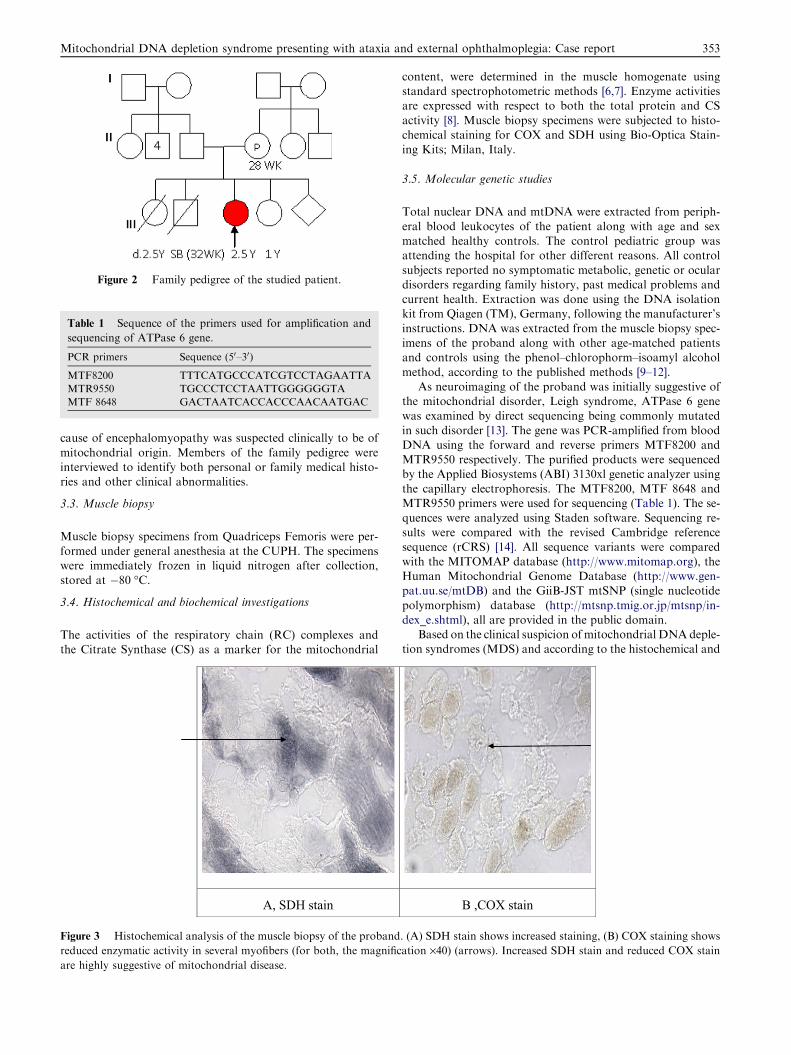
graphic findings were normal. An electroencephalogram(EEG) showed epileptiform activity. The parents have onehealthy daughter (1 year old). Their first child (III-1) had pre-
sented in a similar manner at 8 months of age and died by theage of 2.5 years, the cause of death was uncertain. They had aboy (III-2) who died by 32 weeks of gestation (S.B.) Fig. 2.
3. Methods
3.1. Ethical issue
Verbal consent was obtained from the parents for all the pro-
cedures performed. Parental written informed consent was ob-tained for the undergoing muscle biopsy.
3.2. Family history
The patient who is a regular visitor of the neurometabolicclinic of Cairo University Pediatric Hospital (CUPH) pre-sented clinically with encephalomyopathy. The underlying
LAIR hyperintense signals are noted at: dentate nuclei (1), peri-
(arrows).
Table 1 Sequence of the primers used for amplification and
sequencing of ATPase 6 gene.
PCR primers Sequence (50–30)
MTF8200 TTTCATGCCCATCGTCCTAGAATTA
MTR9550 TGCCCTCCTAATTGGGGGGTA
MTF 8648 GACTAATCACCACCCAACAATGAC
Figure 2 Family pedigree of the studied patient.
Mitochondrial DNA depletion syndrome presenting with ataxia and external ophthalmoplegia: Case report 353
cause of encephalomyopathy was suspected clinically to be of
mitochondrial origin. Members of the family pedigree wereinterviewed to identify both personal or family medical histo-ries and other clinical abnormalities.
3.3. Muscle biopsy
Muscle biopsy specimens from Quadriceps Femoris were per-
formed under general anesthesia at the CUPH. The specimenswere immediately frozen in liquid nitrogen after collection,stored at �80 �C.
3.4. Histochemical and biochemical investigations
The activities of the respiratory chain (RC) complexes andthe Citrate Synthase (CS) as a marker for the mitochondrial
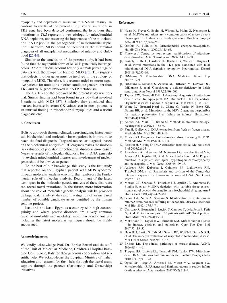
Figure 3 Histochemical analysis of the muscle biopsy of the proband
reduced enzymatic activity in several myofibers (for both, the magnific
are highly suggestive of mitochondrial disease.
content, were determined in the muscle homogenate usingstandard spectrophotometric methods [6,7]. Enzyme activitiesare expressed with respect to both the total protein and CS
activity [8]. Muscle biopsy specimens were subjected to histo-chemical staining for COX and SDH using Bio-Optica Stain-ing Kits; Milan, Italy.
3.5. Molecular genetic studies
Total nuclear DNA and mtDNA were extracted from periph-
eral blood leukocytes of the patient along with age and sexmatched healthy controls. The control pediatric group wasattending the hospital for other different reasons. All control
subjects reported no symptomatic metabolic, genetic or oculardisorders regarding family history, past medical problems andcurrent health. Extraction was done using the DNA isolationkit from Qiagen (TM), Germany, following the manufacturer’s
instructions. DNA was extracted from the muscle biopsy spec-imens of the proband along with other age-matched patientsand controls using the phenol–chlorophorm–isoamyl alcohol
method, according to the published methods [9–12].As neuroimaging of the proband was initially suggestive of
the mitochondrial disorder, Leigh syndrome, ATPase 6 gene
was examined by direct sequencing being commonly mutatedin such disorder [13]. The gene was PCR-amplified from bloodDNA using the forward and reverse primers MTF8200 andMTR9550 respectively. The purified products were sequenced
by the Applied Biosystems (ABI) 3130xl genetic analyzer usingthe capillary electrophoresis. The MTF8200, MTF 8648 andMTR9550 primers were used for sequencing (Table 1). The se-
quences were analyzed using Staden software. Sequencing re-sults were compared with the revised Cambridge referencesequence (rCRS) [14]. All sequence variants were compared
with the MITOMAP database (http://www.mitomap.org), theHuman Mitochondrial Genome Database (http://www.gen-pat.uu.se/mtDB) and the GiiB-JST mtSNP (single nucleotide
polymorphism) database (http://mtsnp.tmig.or.jp/mtsnp/in-dex_e.shtml), all are provided in the public domain.
Based on the clinical suspicion ofmitochondrial DNAdeple-tion syndromes (MDS) and according to the histochemical and
biochemical analyses results, P32-radioactive Southern blot andquantitation of the mt/n DNA ratio were conducted as previ-ously described [15,16]. For confirmation of themyopathic form
of MDS, TK2 gene was sequenced using the 3130xl ABI auto-mated sequencer [17].
4. Results
Results of histochemical staining of the muscle biopsy speci-men were compatible with mitochondrial myopathy. There
was increased staining for SDH Fig. 3A and COX negative fi-bers Fig. 3B. The SDH stain evaluates complex II of the RC(totally encoded by the nuclear DNA) while [18], the COX
stain evaluates complex IV [19] encoded for by both n/mtDNA. The presence of increased SDH activity and decreasedCOX activity is highly suggestive of mtDNA involvement
[20,21].Biochemical analysis of the muscle biopsy specimen was
suggestive of mtDNA involvement. Marked reduction of theactivities of the RC complexes had been detected. Complex I
Table 3 Nucleotide changes found in ATPase 6 genes.
Nucleotide
locus
Nucleotide
position (a)
Sequence
change
Amino acid
change (b)
Previously
reported references
ATPase 6 8292 g-a Non-coding [22]
ATPase 6 8527 a-g M-V [23]
ATPase 6 8723 g-a R-Q [24]
(a) Changes at the following nucleotides positions are frequently
occurring variants in the general population. (b) The first letter is
the amino acid residue predicted by the revised Cambridge
sequence. The last letter is the predicted amino acid in the protein of
the patient. Amino acid changes are expressed as one letter code:
http://molbio.info.nih.gov/gcode.html.
Table 4 Quantitation of mt/n DNA ratio of the proband and
other myopathic patients (2–6) along with control cases using
phosphorus imager. The results are expressed as the copy
number of mitochondrial DNA per haploid nuclear gene. The
control value of the muscle mtDNA copy number was obtained
through the pooling of muscle specimens of 9 age-matched
controls (1–9). The degree of depletion in the proband of the
present study was around 50% of age-matched controls.
Figure 4 Autoradiograph of Southern blot of total DNA
extracted from muscle of the proband. The total (nuclear and
mitochondrial) DNA was hybridized simultaneously with two 32P-
labeled probes––human mtDNA and a cloned fragment of the
human 18S ribosomal RNA gene (nuclear DNA). Arrows.
Mitochondrial DNA depletion syndrome presenting with ataxia and external ophthalmoplegia: Case report 355
activity was not detected. Complex IV activity was about 46%
of age-matched healthy controls. The activities were also foundto be reduced when referred to CS. Activity of SDH was nor-mal. Results of biochemical analysis are shown in Table 2.
Sequencing analysis of the ATPase 6 gene revealed 3 recog-nized benign polymorphisms [22–24] Table 3. Southern blotanalysis showed reduction of the mt/n DNA ratio of the stud-ied case and confirmed mitochondrial DNA depletion, the de-
gree of depletion was around 50% of age-matched controlsTable 4, Fig. 4. Sequencing analysis of the TK2 gene revealedno sequence variations.
5. Discussion
Mitochondrial DNA depletion syndromes (MDS) are autoso-
mal recessive disorders characterized by a reduction in mito-chondrial DNA copy number in the clinically affected tissues[4,15].
A balanced supply of deoxyribonucleoside triphosphates(DNTPs) is required for DNA polymerase c to faithfully rep-licate the mitochondrial genome, the mitochondrial deoxynu-
cleotide triphosphates (dNTPs) pool is maintained boththrough the import of cytosolic dNTPs and by salvagingdeoxyribonucleosides within the organelle itself [4,25].
A number of nuclear-encoded enzymes are involved in these
processes, including thymidine kinase 1 (TK1) within the cyto-sol, thymidine kinase 2 (TK2) and deoxyguanosine kinase(dGK) within mitochondria. The last two are key enzymes of
the mitochondrial salvage pathway [4,26].In the past few years, there had been a substantial increase
in the understanding of the molecular basis of mtDNA deple-
tion syndromes [8]. The TK2 gene is located on chromosome16 and encodes a 234-amino acid polypeptide, which is synthe-sized in the cytoplasm, then imported into the mitochondrial
matrix [20]. It catalyzes the transfer of a phosphate group fromadenosine triphosphate to thymidine or deoxycytidine [28,29],whereas dGK efficiently phosphorylates deoxyguanosine anddeoxyadenosine to the corresponding deoxynucleotide mono-
phosphates [30]. The combined action of the 2 enzymes allowsthe synthesis of all 4 dNTPs needed for mtDNA replication[30,31].
Mutations in the TK2 gene in patients with myopathicMDS [28] and mutations in the dGK gene in patients withhepatocerebral MDS [32,33] were identified confirming the
concept that imbalances in mitochondrial dNTP pool canaffect mtDNA integrity. This imbalance leads to inefficientreplication and, therefore, depletion of mtDNA [18,34]. More
recently, mutations in RRM2B, encoding the small subunit ofp53-inducible ribonucleotide reductase, have also been identi-fied in patients with myopathic mtDNA depletion disorder
[35].The MDS differs from other mitochondrial disorders be-
cause it is a quantitative defect rather than a qualitative defect.The low level of mtDNA in some tissues causes insufficient
synthesis of respiratory chain components [15]. Depletion ofmtDNA can occur as a secondary phenomenon, for exampleas a result of antiretroviral nucleoside analogue therapy [36].
In MDS, however, mtDNA depletion is considered a primaryprocess, and the degree of depletion is generally proportionalto the severity of the phenotype [27].
Patients with the myopathic form of MDS usually presentat or soon after birth with progressive weakness, hypotonia,hyporeflexia, and die of respiratory failure before 1 year of
age (congenital form) or before 10 years of age (juvenile form)[27].
Since the first description of TK2 mutations leading to themyopathic form of mtDNA depletion syndrome in 2001,
approximately 20 different pathogenic mutations have nowbeen reported, distributed throughout the TK2 coding se-quence [37].
The proband of the present study was clinically suspectedto have mitochondrial encephalomyopathy. Based on MRIfindings, the initial diagnosis was suggestive of Leigh syndrome
(LS) with SURF1 gene mutation. Bilateral putaminal T2 pro-longation has long been considered a consistent feature of LSon MRI studies [38]. LS with COX deficiency caused by
SURF1 gene mutations represents a relatively homogeneousclinical entity. Similar to the proband of this study, affectedpatients present at around 1 year of age with progressiveencephalopathy, generalized hypotonia, trunk ataxia, oculo-
motor abnormalities and central respiratory problems[39,40]. However, the biochemical evidence of deficiency ofcomplex I and complex IV in such case was against the sugges-
tion of SURF1 gene mutation which is exclusively associatedwith isolated complex IV deficiency.
As high index of suspicion of MDS is warranted in any
child with proximal weakness and hypotonia of unclear etiol-ogy, the diagnosis needed to be confirmed by Southern blotanalysis for mtDNA quantitation and subsequent TK2 muta-tion screening [37].
Based on the results of histochemical, biochemical andmolecular analyses, the proband had fulfilled the diagnosticcriteria for the myopathic form of MDS [41,42]. These criteria
include clear evidence of mtDNA depletion by Southern blot,histochemical evidence of COX deficiency, plus at least two ofthe following features: ragged-red fibers (RRF), lactic acidosis,
multiple defects of respiratory chain enzymes [25]. Based onthese findings, it was mandatory to screen this patient formutations in TK2 gene, in order to better define the frequency
of mutations in this gene.Similar studies had been conducted by Tiranti et al., Johans-
son et al., Mandel et al. and Finsterer [4,25,37,43]. Similar tothe probands of the present study, those patients developed
356 L. Selim et al.
myopathy and depletion of muscular mtDNA in infancy. Incontrast to results of the present study, several mutations inTK2 gene had been detected confirming the hypothesis that
mutations in TK2 represent a new etiology for mitochondrialDNA depletion, underscoring the importance of the mitochon-drial dNTP pool in the pathogenesis of mitochondrial deple-
tion. Therefore, MDS should be included in the differentialdiagnosis of all unexplained myopathies of infancy and child-hood [27,44].
Similar to the conclusion of the present study, it had beenfound that the myopathic form of MDS is genetically heteroge-neous. TK2 mutations account for only a small percentage ofpatients with the myopathic form of MDS [25]. This suggests
that defects in other genes must be involved in the etiology ofmyopathic MDS. Therefore, it is recommended to screen nega-tive patients for mutations in other candidate genes rather than
TK2 and dGK genes involved in dNTP metabolism.The CK level of the proband of the present study was nor-
mal. Similar finding had been found in a study that involved
4 patients with MDS [37]. Similarly, they concluded thatmarked increase in serum CK values seen in most patients isan unusual finding in mitochondrial myopathies and a useful
diagnostic clue.
6. Conclusion
Holistic approach through clinical, neuroimaging, histochemi-cal, biochemical and molecular investigations is important toreach the final diagnosis. Targeted molecular diagnosis basedon the biochemical analysis of RC enzymes makes the molecu-
lar evaluation of pediatric mitochondrial disorders more easier.Negative results of molecular diagnosis for mtDNA genes donot exclude mitochondrial diseases and involvement of nuclear
genes should be always suspected.To the best of our knowledge, this study is the first study
that reported on the Egyptian patient with MDS syndrome
through molecular analysis which further reinforces the funda-mental role of molecular analysis. Recruitment of the latesttechniques in the molecular genetic analysis of these disorders
can reveal novel mutations. In the future, more informationabout the role of molecular genetic analysis will be providedby large scale family studies and investigation of an increasednumber of possible candidate genes identified by the human
genome project.Last and not least, Egypt as a country with high consan-
guinity and where genetic disorders are a very common
cause of morbidity and mortality, molecular genetic analysisincluding the latest molecular techniques should be highlyencouraged.
Acknowledgements
We kindly acknowledge Prof. Dr. Enrico Bertini and the staffof the Unit of Molecular Medicine, Children’s Hospital Bam-bino Gesu, Rome, Italy for their generous cooperation and sci-
entific help. We acknowledge the Egyptian Ministry of highereducation and research for their help through the travel grantsupport through the parown (Partnership and Ownership)