19
MLAB 1415: Hematology MLAB 1415: Hematology Keri Brophy-Martinez Keri Brophy-Martinez Chapter 8: Anemia Part Four
| Date post: | 02-Jan-2016 |
| Category: |
Documents |
| Upload: | sylvester-potts |
| View: | 39 times |
| Download: | 0 times |
MLAB 1415: HematologyMLAB 1415: HematologyKeri Brophy-MartinezKeri Brophy-Martinez
Chapter 8: AnemiaPart Four

Variations in RBC Variations in RBC DistributionDistribution
2
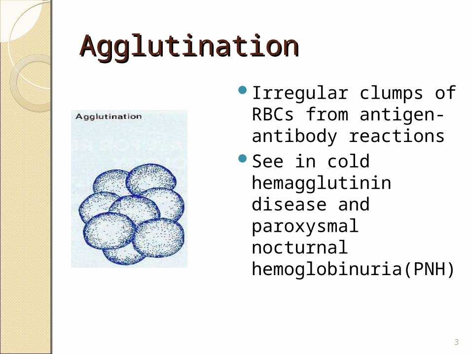
AgglutinationAgglutinationIrregular clumps of
RBCs from antigen-antibody reactions
See in cold hemagglutinin disease and paroxysmal nocturnal hemoglobinuria(PNH)
3
AgglutinationAgglutination
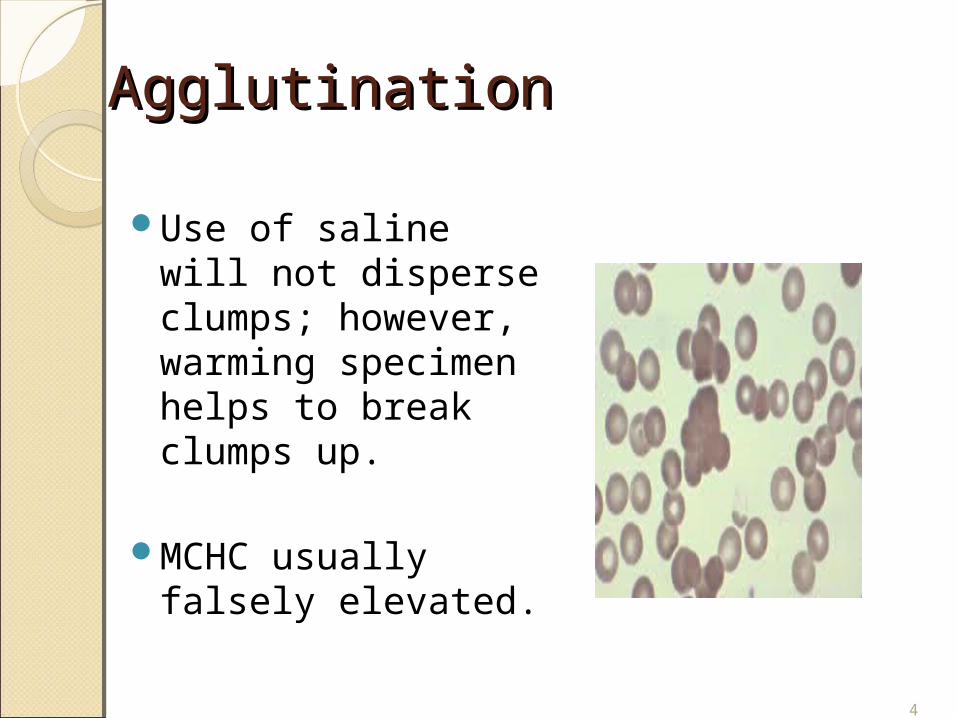
Use of saline will not disperse clumps; however, warming specimen helps to break clumps up.
MCHC usually falsely elevated.
4
RouleauxRouleaux
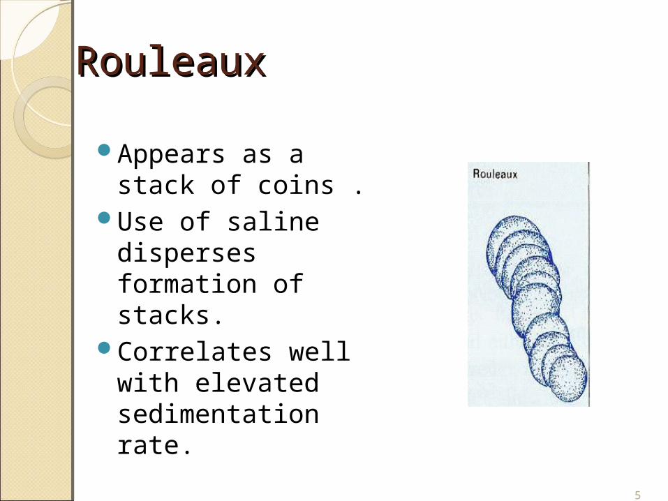
Appears as a stack of coins .
Use of saline disperses formation of stacks.
Correlates well with elevated sedimentation rate.
5
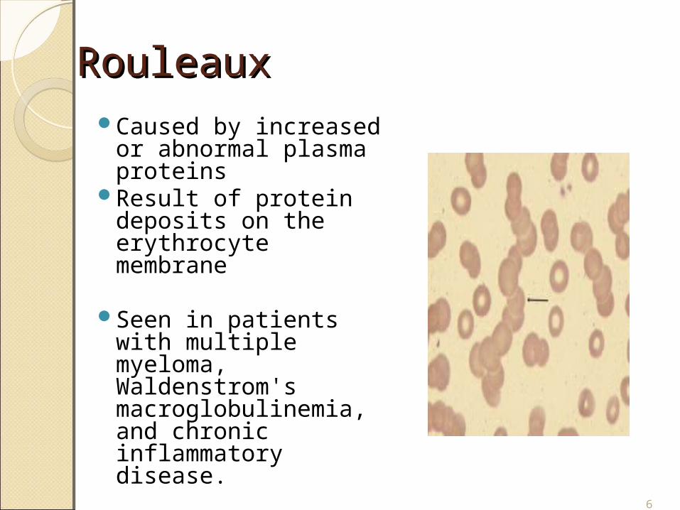
RouleauxRouleauxCaused by increased
or abnormal plasma proteins
Result of protein deposits on the erythrocyte membrane
Seen in patients with multiple myeloma, Waldenstrom's macroglobulinemia, and chronic inflammatory disease.
6

RBC InclusionsRBC Inclusions
7
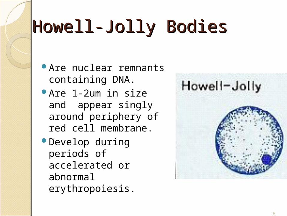
Howell-Jolly BodiesHowell-Jolly Bodies
Are nuclear remnants containing DNA.
Are 1-2um in size and appear singly around periphery of red cell membrane.
Develop during periods of accelerated or abnormal erythropoiesis.
8

Howell-Jolly BodiesHowell-Jolly Bodies
Spleen usually removes them; however, during times of erythroid stress, spleen cannot keep up with formation of inclusions.
Seen following splenectomy, in thalassemia, hemolytic anemias, and in megaloblastic anemias.
9
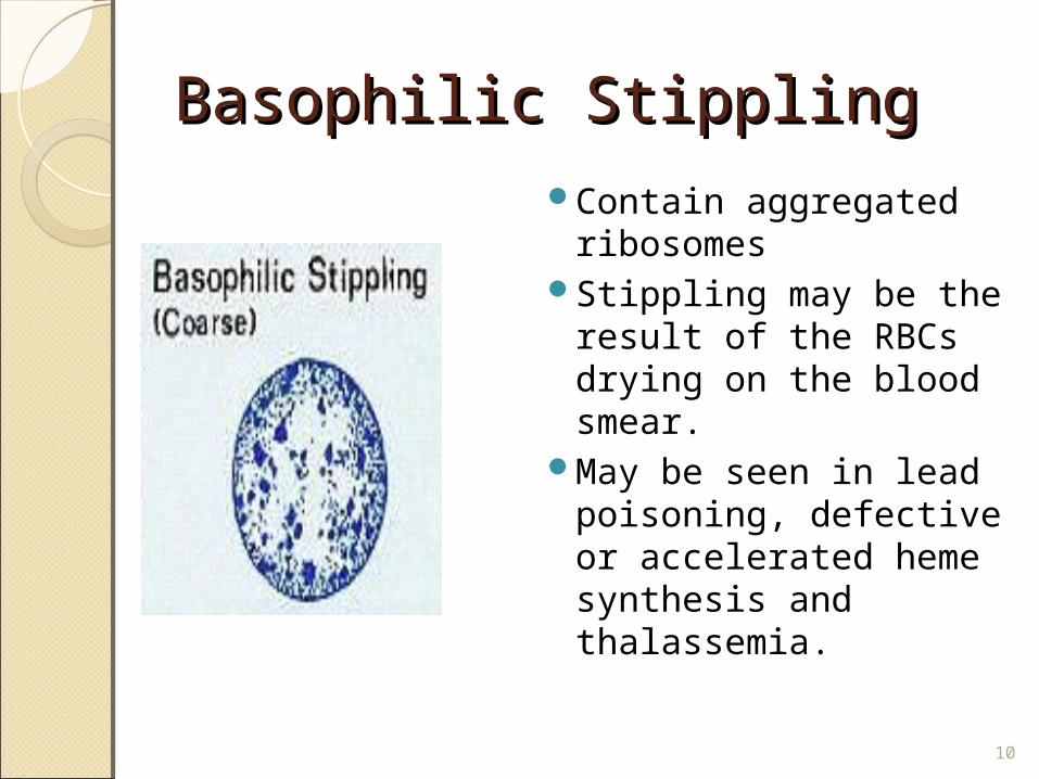
Basophilic StipplingBasophilic StipplingContain aggregated
ribosomes Stippling may be the
result of the RBCs drying on the blood smear.
May be seen in lead poisoning, defective or accelerated heme synthesis and thalassemia.
10
Basophilic StipplingBasophilic Stippling
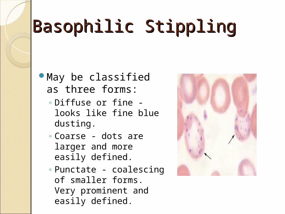
May be classified as three forms: ◦ Diffuse or fine - looks
like fine blue dusting. ◦ Coarse - dots are
larger and more easily defined.
◦ Punctate - coalescing of smaller forms. Very prominent and easily defined.
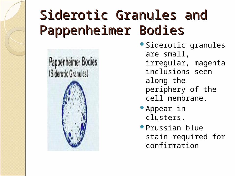
Siderotic Granules and Siderotic Granules and Pappenheimer BodiesPappenheimer Bodies
Siderotic granules are small, irregular, magenta inclusions seen along the periphery of the cell membrane.
Appear in clusters. Prussian blue stain
required for confirmation
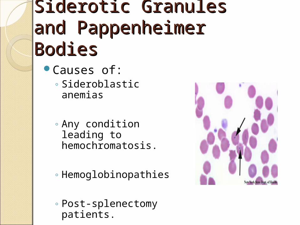
Siderotic Granules and Siderotic Granules and Pappenheimer BodiesPappenheimer Bodies
Causes of:◦ Sideroblastic anemias
◦ Any condition leading to hemochromatosis.
◦ Hemoglobinopathies
◦ Post-splenectomy patients.
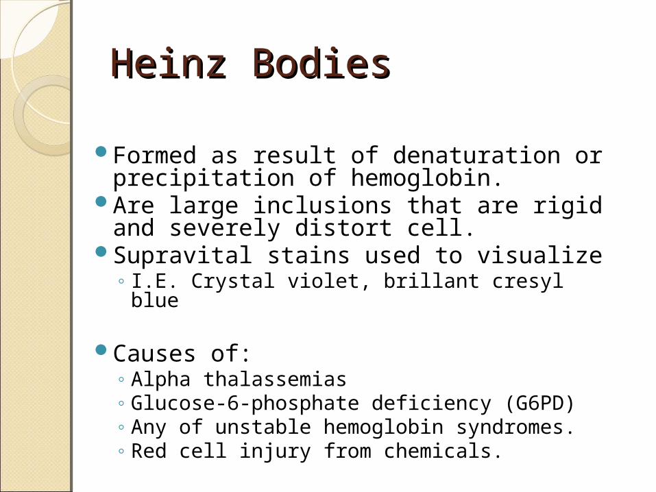
Heinz BodiesHeinz Bodies
Formed as result of denaturation or precipitation of hemoglobin.
Are large inclusions that are rigid and severely distort cell.
Supravital stains used to visualize◦ I.E. Crystal violet, brillant cresyl blue
Causes of:
◦ Alpha thalassemias◦ Glucose-6-phosphate deficiency (G6PD)◦ Any of unstable hemoglobin syndromes.◦ Red cell injury from chemicals.
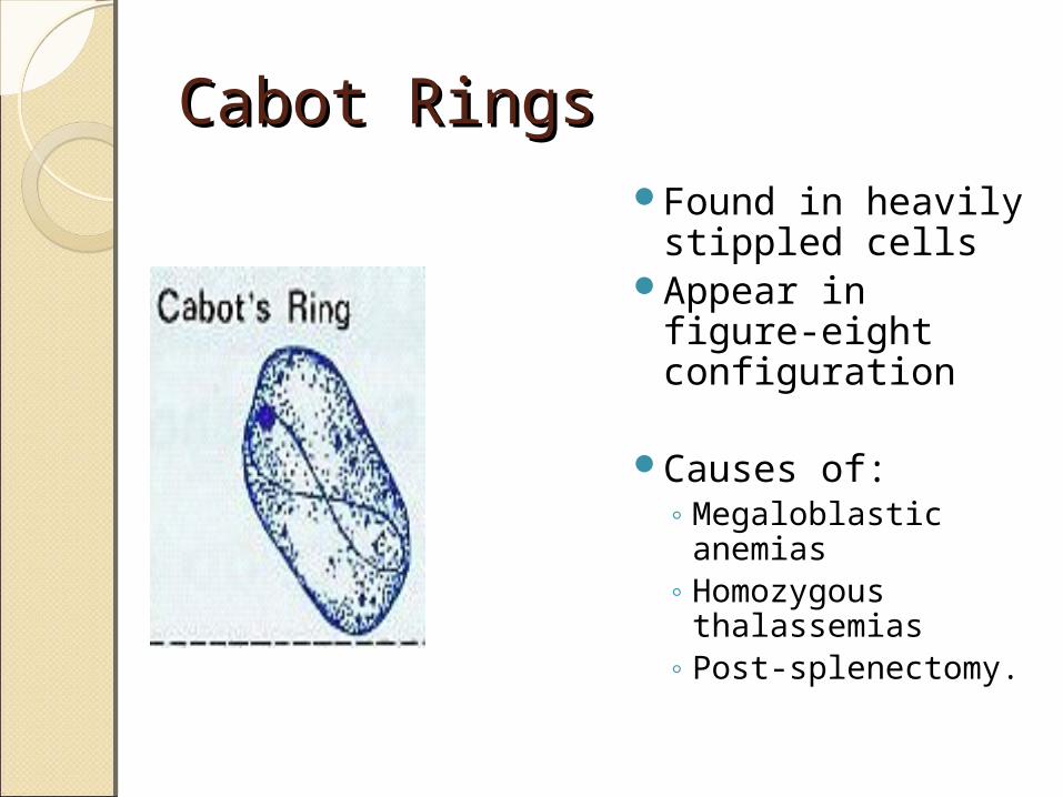
Cabot RingsCabot RingsFound in heavily
stippled cells Appear in figure-
eight configuration
Causes of:
◦ Megaloblastic anemias
◦ Homozygous thalassemias
◦ Post-splenectomy.
16
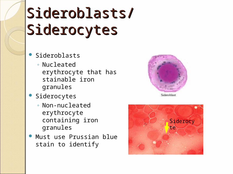
Sideroblasts/SiderocytesSideroblasts/Siderocytes
Sideroblasts◦ Nucleated erythrocyte
that has stainable iron granules
Siderocytes◦ Non-nucleated
erythrocyte containing iron granules
Must use Prussian blue stain to identify
Siderocyte
ReferencesReferences• Harmening, D. M. (2009). Clinical
Hematology and Fundamentals of Hemostasis. Philadelphia: F.A Davis.
• McKenzie, S. B., & Williams, J. L. (2010). Clinical Laboratory Hematology . Upper Saddle River: Pearson Education, Inc.
• http://www.ezhemeonc.com/index.php/hematological-disorders/
• http://www.wiwe.net/irene/lab/chemheme/heme/microscope/stomatocyte.htm
ReferencesReferences• http://home.ccr.cancer.gov/oncology/
oncogenomics/WEBHemOncFiles/Review%20of%20Terms.html
• http://tiny.cc/85k0b• http://image.bloodline.net/stories/
storyReader$1214http://www.comlexflashcards.com/wp-
content/uploads/2010/06/image970.png


